Article

Genetic Landscape of Non-Remitting Neutropenia in Children and Chronic Idiopathic Neutropenia in Adults

1
Laboratory of Genetics and Genomics of Rare Diseases, IRCCS Istituto Giannina Gaslini, 16147 Genova, Italy
2
Hemopoiesis Research Laboratory, School of Medicine, University of Crete, 71003 Heraklion, Greece
3
Clinical Bioinformatics, IRCCS Istituto Giannina Gaslini, 16147 Genova, Italy
4
Haematology Unit, IRCCS Istituto Giannina Gaslini, 16147 Genova, Italy
5
Genomics Facility, Italian Institute of Technology (IIT), 16163 Genova, Italy
6
Department of Hematology, University Hospital of Heraklion, 71110 Crete, Greece
*
Author to whom correspondence should be addressed.
Int. J. Mol. Sci. 2025, 26(14), 6929; https://doi.org/10.3390/ijms26146929
Submission received: 11 June 2025 / Revised: 11 July 2025 / Accepted: 13 July 2025 / Published: 18 July 2025
(This article belongs to the Special Issue New Insights into Immune Dysregulation Disorders)

Abstract

Non-remitting neutropenia in children and chronic idiopathic neutropenia (CIN) in adults have been described previously as peculiar subgroups of neutropenic patients carrying similar clinical and immunological features. The present collection comprising 25 subjects (16 adults and 9 children) mostly affected with mild (84%) and moderate (16%) neutropenia aimed to identify the underlying (possibly common) genetic background. The phenotype of these patients resemble the one described previously: no severe infections, presence of rheumathological signs, leukopenia in almost all patients and lymphocytopenia in one-third of the cohort. The pediatric patients did not share common genes with the adults, based on the results of the multisample test, while some singular variants in neutropenia potentially associated with immune dysregulation likely consistent with the phenotype were found. SPINK5, RELA and CARD11 were retrieved and seem to be consistent with the clinical picture characterized by neutropenia associated to immune dysregulation. The enrichment and burden tests performed in comparison with a control group underline that the products of expression by the variants involved belong to the autoimmunity and immune regulation pathways (i.e., SPINK5, PTPN22 and PSMB9). Even with the limitation of this study’s low number of patients, these results may suggest that non-remitting neutropenia and CIN in adults deserve deep genetic study and enlarged consideration in comparison with classical neutropenia.

1. Introduction

Children affected with non-remitting neutropenia, defined as late onset (LO) or long lasting (LL) neutropenia either with or without antibodies against neutrophils, have been classified within the provisional category of “likely Acquired Neutropenia”, according to the International Neutropenia Guidelines [1]. This group of patients shows a peculiar immunological pattern characterized by a reduction in absolute lymphocyte count and an immunophenotypic T- and B-cell profile similar to known autoimmune disorders, i.e., increased HLADR+ and CD3+ TCRγδ cells, reduced T-regulatory cells, increased CD27IgD (double negative) naïve B-cells and a tendency toward reduced B-memory cells. In a minority of patients, pathogenic and likely pathogenic variants related to primary immuno-deregulatory disorders have been found by next generation sequencing (NGS) [2].
Adult patients with persistent and unexplained neutropenia who do not fulfil the diagnostic criteria of any underlying disease are characterized as chronic idiopathic neutropenia (CIN). A significant proportion of CIN patients also display an immune dysregulation pattern similar to LO/LL neutropenic children [3,4,5,6]. Specifically, CIN patients frequently display immunoglobulin disturbances, lymphopenia, activated T-lymphocytes with increased expression of HLA-DR and reduced memory B-cells [3,4,5,6]. The shared immunoregulatory disturbances between LO/LL neutropenia and CIN prompted us to investigate for a common genetic background between the two entities, testing the hypothesis that they may both belong to the same disease spectrum. We have, therefore, analyzed, by whole exome sequencing (WES), samples from pediatric LO/LL neutropenia and adult CIN patients, and the results are presented herein. To the best of our knowledge, no similar data are currently available in the literature.

2. Results

2.1. Immunological and Clinical Characteristics

The most important immunological characteristics are presented in Table 1. Leukopenia, defined as white blood cell counts below 4.0 × 10⁹/L in adults [7] and according to a different threshold for age in children, adolescents and young adults [8], was present in almost all patients (22/25; i.e., 88%), while lymphocytopenia, defined as counts below 1.5 × 10⁹ in adults [9] and below the lower limit according to age [8] in the younger group, affected 7/25 (i.e., 28%) patients. Based on the age-related reference values, none of the patients had decreased main class immunoglobulin levels. However, in accordance with the previously reported data, 5/25 (i.e., 20%) showed IgM above the threshold, and 3/25 (i.e., 12%), 7/25 (i.e., 28%) and 5/25 (i.e., 20%) patients displayed low IgG1, IgG3 and IgG4 subclass levels [10]. The most relevant reduction in lymphocyte subsets was related to natural killer cells (8/25, 32%), followed by depletion of CD19 in 3/25 subjects (12%). All patients had hemoglobin and platelet values within the normal range. As for the clinical/laboratory characteristics (Table 1), arthralgia without objective signs of arthritis was present in 7/25 (i.e., 28%) patients, fatigue was shown in 4/25 (i.e., 16%), mostly pediatric cases, and aphthae was referred by 4/25 (i.e., 16%) subjects. Two subjects had psoriasis, and one showed HLA-B27+ positivity with no symptoms/signs of ankylosis spondylitis. Anti-nuclear antibodies (ANA) were positive in 5/25 (i.e., 20%), antibodies against thyroid were present in 2/25 (i.e., 8%), and antibodies against neutrophils were present in 9/25 (i.e., 36%).

2.2. Genes Potentially Associated with Neutropenia

The results obtained through the multisample genetic analysis, the singleton analysis and the in silico panel of 538 genes associated with IEI (inborn error of immunity) (Figure 1) are summarized in Table 2 (for details, see Supplementary Tables S1–S4). Only genes known to be associated with the phenotype or potentially related to immunological disorders are reported.
Table 2 shows all genes shared by at least two patients within the entire cohort or present only in the pediatric or in the adult cohort, with variants characterized by a CADD (combined annotation dependent depletion) score greater than or equal to 15 and present in heterozygosity in a maximum of 10 controls from the Gaslini dataset. The genes carrying variants present only in a single subject, according to more stringent criteria such as a CADD score greater than or equal to 20, and variants not present in any healthy control in the Gaslini dataset are also shown. Lastly, the genes specifically associated with IEI but not emerged from the previous analyses because they (i) were not shared among patients, (ii) had a CADD score lower than 20 and (iii) were present in heterozygosity in not more than 10 controls from the Gaslini dataset are also presented. The Franklin database (https://franklin.genoox.com/clinical-db/home, accessed on 29 January 2025) was used to verify the prediction of each variant according to the ACMG criteria [11].
Overall, variants of uncertain significance (VUS), likely pathogenic (LP) variants and pathogenic (P) variants are reported. The Franklin database allows for the classification of VUS as either “hot” or “cold”, depending on whether the parameters used by the system lean towards a likely benign or LP variant, respectively. Hot VUS, represented in red according to this system, are “relevant”, having some probable roles in pathogenicity, while the cold VUS may not have any impact.
Hot VUS/LP/P variants that exhibit zygosity consistent with the gene’s inheritance pattern are highlighted in red. Furthermore, variants requiring special attention are marked in orange; these include (i) those affecting cancer predisposing genes, (ii) variants in genes involved in DNA repair or telomere maintenance, (iii) heterozygous variants in autosomal recessive (AR) inherited genes and (iv) variants in genes not yet associated with disease but potentially related to immune dysregulation. Finally, the cold VUS are indicated in grey.
In the following paragraphs, only the P/LP/hot VUS variants consistent with zygosity emerged through (i) the multisample test as for variants shared by children and adults, (ii) singleton analysis as for individual variants and (iii) genes belonging to an IEI panel by using a “more sensible” filter are described. The mechanisms of the disorders caused by the single variant and the consistency with the clinical features are extensively discussed.

2.2.1. Multisample Analysis

A pathogenic homozygous variant in SPINK5 (c.1302+4A>T) was found in Patient 10 (Pt#10), while Pt#4 is a heterozygous carrier of the SPINK5 (p.R217*) variant (Table 2 and Supplementary Table S1). SPINK5 is a serine protease inhibitor important for the anti-inflammatory and/or antimicrobial protection of mucous epithelia. It contributes to the integrity and protective barrier function of the skin by regulating the activity of defense-activating and desquamation-involved proteases. Interestingly, Pt#10 is affected by psoriasis (Table 1) [12].
Heterozygous BRCA1 variants were seen in three patients, but one variant has a pathogenic prediction (LP) and was found in Pt#3 (p.D1692G) (Table 2 and Supplementary Table S1). A BRCA1 mutation in a homozygous state is causative of Fanconi anemia (MIM #617883); otherwise, cancer predisposition is related to a single mutated allele. Breast cancer type 1 susceptibility protein plays a central role in DNA repair by facilitating cellular responses to DNA damage. Pt#10 has a family history of a very aggressive and precocious form of cancer. He inherited the mutation from his father who has been affected by prostatic carcinoma very early in life [13].
Two MSTIR variants, c.1880+2T>G and p.V323Wfs*15, classified as two hot VUS, were detected in Pt#21 and Pt#1, respectively (Table 2 and Supplementary Table S1). Macrophage-stimulating protein receptor alpha chain (MSTIR) regulates many physiological processes by binding to the MST1 ligand, including cell survival, migration and differentiation. Ligand binding at the cell surface induces autophosphorylation of RON (alias for MSTIR) on its intracellular domain that provides docking sites for downstream signaling molecules. Following activation by the ligand, MSTIR interacts with the PI3-kinase subunit PIK3R1, PLCG1 or the adapter GAB1 [14].

2.2.2. Singleton Analysis

This analysis showed two hot VUS and one LP variant only in the adult cohort, affecting the GATA2, GJB4 and ITGB3 genes, with a zygosity consistent with the expected inheritance (Table 2 and Supplementary Table S2).
In detail, the GATA2 (p.H127N) hot VUS was identified in Pt#15. The patient has mild neutropenia discovered in a routine checkup and remains asymptomatic to date. She also screened as HLA-B27 positive but with the absence of any musculoskeletal symptoms. The GATA transcription factors have emerged as candidate regulators of gene expression in hematopoietic cells, usually dendritic cells, monocytes, B lymphocytes and NK lymphocytes. Interestingly, this patient had lymphopenia with particularly low NK cells [15,16].
GATA2 is involved in familial leukemia and in a complex congenital immunodeficiency that evolves over decades and leads to predisposition to infection and myeloid malignancy. The patient is not showing any signs of dysplasia or any susceptibility to infections to date.
The GJB4 hot VUS (p.I30T) was identified in Pt#20. Erythrokeratodermia variabilis et progressive (MIM #617524) is the phenotype associated with the presence of a causative variant in this gene, characterized by persistent plaque-like or generalized hyperkeratosis and transient red patches of variable size, shape and location. The patient has had mild neutropenia for the last 35 years and does not show any signs of skin abnormities or infections.
The ITGB3 (p.A754T) LP variant was detected in Pt#23. The ITGB3 gene encodes glycoprotein IIIa (GP IIIa), the beta subunit of the platelet membrane adhesive protein receptor complex GP IIb/IIIa. The mutation in this gene is responsible for a bleeding disorder, platelet-type 24 and Glanzmann thrombasthenia 2 (MIM #607759), which is a feature not consistent with neutropenia. Indeed, the patient is suffering from mild neutropenia, with no major infections and diffuse bone pain and myalgia [17,18].

2.2.3. Inborn Error of Immunity Panel Analysis

According to the 538 gene custom panel compiled from the International Union of Immunological Societies (IUIS) Expert Committee and PanelApp England (Primary immunodeficiency or monogenic inflammatory bowel disease (Version 4.0)), two hot VUS and one P variant were found (Table 2 and Supplementary Table S3): TGFBR2 (p.R224H) in Pt#2; CARD11 (p.S1026C) in Pt#25; MEFV (p.M694V) in Pt#20.
The hot VUS of TGFBR2 (p.R224H), which is basically associated with Loeys–Dietz syndrome (MIM #610168), has no clinical consistency in Pt#2. On the contrary, heterozygous VUS in the RELA gene (p.S536del), carried again by Pt#2, consistently causes familial Bechet-like autoinflammatory disease-3 (MIM #618287). Patients’ ulcerations occur specifically in the oral, gastrointestinal and vaginal mucosa rich in microbiota, suggesting they may have served as inflammatory stimuli inducing TNF release, resulting in mucosal injury and impaired epithelial recovery [19]. The symptoms of Pt#2 fit with this description.
Pt#25 carried an LP variant (p.S1026C) in CARD11. The gene encodes a protein that acts as a scaffold for nuclear factor kappa-B (NF-κB) activity in the adaptive immune response controlling peripheral B-cell differentiation and a variety of critical T-cell effector functions. CARD11 is associated with immunodeficiency (MIM #617638/615206/616452). Indeed, the patient displays reduced levels (borderline) of IgG1 and IgG3 subclasses.
MEFV is the gene of familial Mediterranean fever (FMF) (IM #134610/249100), one of the most common monogenic autoinflammatory diseases. Pt#20 carried a P variant (p.M694V) in MEFV. This gene is involved in inflammatory reactions through altered leukocyte apoptosis, secretion of interleukin-1beta (IL-1β) and activation of the NF-κB pathway, and, thereby, the degree of inflammation. The patient had no features of clinical FMF and was just suffering from mild neutropenia [20].

2.3. Gene Clustering

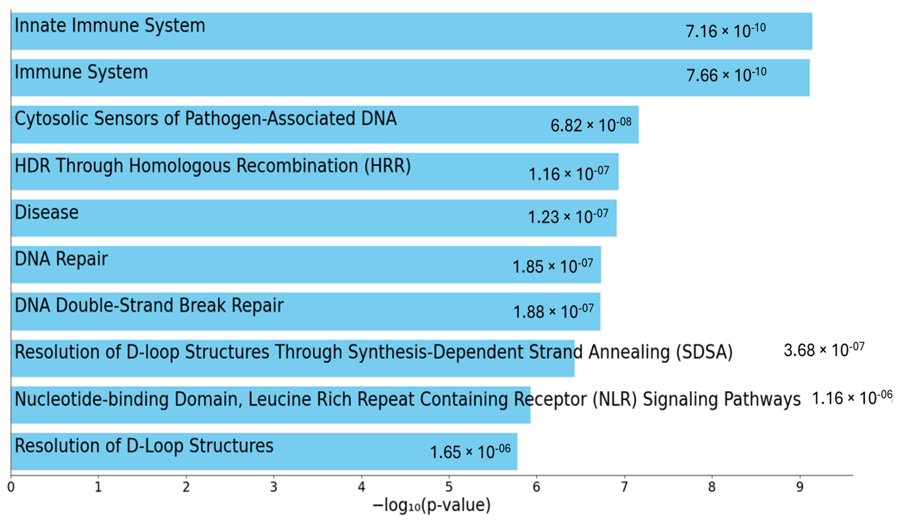
In Supplementary Table S4, all genes resulting from the previous analyses are collected, and the contribution of the variants found in each gene is highlighted with different colors according to the pathogenicity prediction described by Franklin. The genes are divided according to the functional pathway they belong to using EnrichR and Reactome Pathways 2024 (https://maayanlab.cloud/Enrichr/, accessed on 11 July 2025). The categorization of the genes revealed a diverse array of biological functions, highlighting the complexity of cellular processes that may be associated with the neutropenia phenotype. Prominent among these are pathways related to the innate immune system and adaptive immune system (response and inflammation), suggesting a potential strong link between immune dysregulation and neutropenia occurrence. Additionally, a significant portion of the genes contribute to DNA repair and genome stability. Overall, this classification underscores the diverse roles these genes may have in contributing to the neutropenia phenotype (Figure 2).

Multisample Analysis of Total Patient Group vs. Healthy Controls

The multisample dataset, including both neutropenic patients (n = 25) and healthy controls (n = 75), was used to assess the enrichment of rare, deleterious, hot VUS, LP, and P variants. Several comparisons were performed, including LL/LO patients versus healthy controls, CIN patients versus healthy controls, and total neutropenia patients (i.e., LL/LO plus CIN) versus healthy controls. In this latter test, SPINK5 almost reached the nominal p threshold (p-value: 0.06), suggesting a potential burden of pathogenic variants. Additionally, in the CIN versus healthy controls comparison, RAD50 exhibited a significant enrichment after multiple testing correction (p-value: 0.019), indicating an enrichment of suggestive variants in this gene. The same vcf file was used for testing rare variant association, focusing on rare and deleterious variants with high, moderate or modifier impact on transcripts. Using RVTEST, different statistical models were applied: (1) the CMC model (Burden test) considers variants as if they contribute equally, and in this particular model, all variants are collapsed into a single score; (2) for the SKAT analysis, the variants are analyzed individually, with weights assigned based on MAF (rarer variants have higher weights); (3) the SKAT-O approach is considered the optimal test, combining SKAT and Burden. If all variants have a similar effect (all deleterious or all protective), it behaves like Burden. If variants have mixed effects (some increase and some decreased risk), it behaves like SKAT.
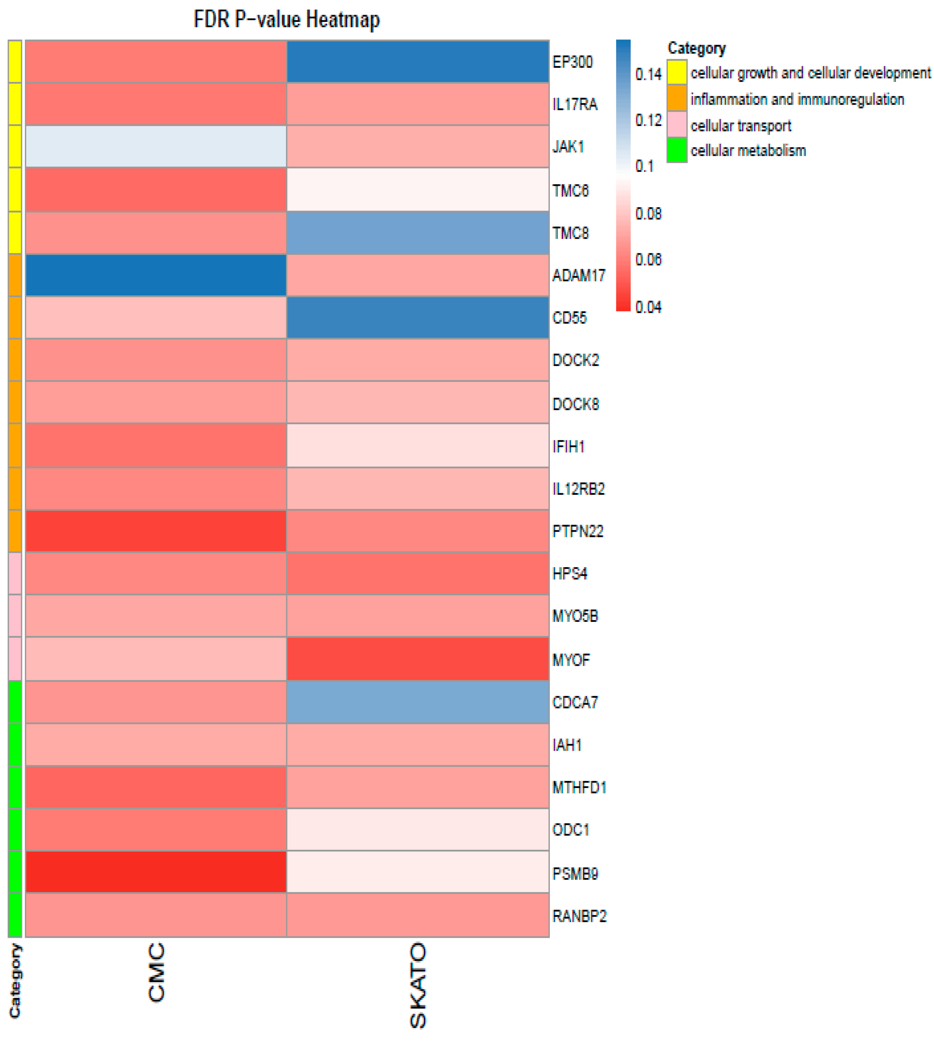
Among these analyses, PTPN22 and PSMB9 showed a significant enrichment of rare variants using the CMC model (Supplementary Table S5), even after multiple testing correction (Benjamini–Hochberg method). Additionally, MYOF reached an FDR < 0.05 in the SKATO analysis (Supplementary Table S6).
Overall, 23 genes from the 538 genes tested in the custom panel showed at least one FDR-adjusted p-value ≤ 0.08 between the two approaches. These genes were further categorized into four key biological processes: cellular growth and cellular development, inflammation and immunological regulation, cellular metabolism and cellular transport (Figure 3).
Among the significant enriched genes, PTPN22 is a lymphoid-specific intracellular phosphatase that interacts with the adaptor protein CBL and plays a role in regulating T-cell receptor signaling. The gene PSMB9 is particularly important for inflammation and immunoregulatory pathways. Notably, this latter gene, located in the MHC class II region, encodes a beta subunit of the 20S core of the immunoproteasome, whose expression is induced by interferon-gamma and is involved in antigen processing. Finally, MYOF encodes a type II membrane protein structurally related to dysferlin, involved in membrane repair and regeneration through calcium-mediated fusion events, with mutations leading to muscle weakness affecting both proximal and distal muscles.

3. Discussion

CIN in adults and LL/LO neutropenia in children and adolescents, respectively, are two categories of neutropenic patients who seem to have peculiar clinical and biochemical features when compared to other forms of neutropenia, either acquired or congenital. In these patients, previous studies identified a subgroup whose immune dysregulation background was sustained by the presence of pathogenic variants of IEI [2,3,4,5]. The hypothesis is that neutropenia may not represent “the” disease but rather an epiphenomenon of immune dysregulation.
In this perspective, the application of WES analysis intended to look for any common genetic background in CIN and in LO/LL able to justify a common “root” of these entities. The immunological and clinical characteristic of the population described in the present paper basically resembled those described previously [2,3,4,5], thus delineating again the different composition of these groups compared to the “classical neutropenia” categories; this was further confirmed by the absence of any common neutropenia variants.
The genes found with the multisample test using “less stringent” parameters than the ones applied in the IEI panel were SPINK5, BRCA1 and MSTIR1. SPINK5 is related to a skin disorder and is consistent with the clinical picture of Pt#5, who is affected with psoriasis; the possible connection with neutropenia might be explained by the expected reduced expression of LEF1, which in turn deregulates lymphocyte activity.
SPINK5 is a serine protease inhibitor that plays a pivotal role in a wide variety of immune and inflammatory processes, including T- and B-cell differentiation, activation of cytokines and complement and recruitment of inflammatory cells. It is difficult to say whether neutropenia may be the result of such mechanisms [21].
Pt#3 was found to be a carrier of BRCA1; indeed, a high incidence of early and severe cancer in this patient’s family has been documented. His father, affected with an aggressive form of prostatic cancer, did carry the same mutation. As for the MSTIR gene, only hot VUS have been found; being that this gene is involved in cellular traffic, it could be hypothesized that neutropenia could be the result of neutrophil migration, but again, this remains only a hypothesis. As for the singleton analysis, GATA2, GJB4 and ITGB3 mutations were identified. GATA2 is a transcription factor that plays an essential role in the development and proliferation of hematopoietic and endocrine cell lineages. Pathogenic variants in this gene cause autosomal-dominant GATA2 deficiency, which may present clinically as Emberger syndrome, immunodeficiency 21, susceptibility to acute myeloid leukemia or myelodysplastic syndrome, WILD syndrome (warts, immunodeficiency, lymphedema), MonoMAC or DCML (dendritic cells, monocytopenia and lymphopenia) deficiency. GATA2 germline mutations are also reported in patients with myeloid malignancy without evidence of preceding immunodeficiency or other disturbances [22].
In our patient (Pt#15), we detected the GATA2 (p.H127N) variant, a missense mutation affecting codon 127 that is predicted to be deleterious and has not been reported in GnomAD before. The patient is not showing any GATA2-related clinical signs. Regarding the hot VUS GJB4 (p.I30T) variant identified in Pt#20, an association with the neutropenia phenotype cannot be established. The same applies for the ITGB3 (p.A754T) LP variant detected in Pt#23. When the panel of IEI is studied with a high sensitivity, some interesting data are found, at least for RELA and CARD11, showing more consistent connection with the neutropenia feature.
RELA haploinsufficiency causes an autosomal-dominant, mucocutaneous disease named autoinflammatory disease, familial, Bechet-like-3. This disorder provides a counterpoint to autoinflammatory diseases characterized by increased inflammatory cytokine signaling. Defects in the genes encoding the NF-κB regulatory proteins result in NF-κB overactivation and multisystemic autoinflammation characterized by oral ulcers, arthritis, uveitis and/or vasculitis [23,24]. The history of Pt#2 began as isolated chronic mild symptomatic autoimmune neutropenia with occasional mouth ulcers. At the age of 17 years, the patient developed frank arthritis, which required anti-TNF therapy. Autoimmune cytopenia has been reported in patients carrying this variant too [24].
CARD11 is a membrane protein acting as a key signaling scaffold that controls antigen-induced lymphocyte activation during the adaptive immune response (NF-κB, JNK and mTOR pathway). Germline CARD11 mutations can result in gain/loss of function of the protein, leading to different phenotypes (SCID, BENTA disease, atopy, CVID). Several heterozygous hypomorphic/dominant negative variants of CARD11 have demonstrated a loss of protein function, exhibiting high penetrance and variable expressivity [25,26]. The largest heterozygous germline loss-of-function case series is composed of 48 patients with a broad phenotype, including atopy, viral infections of the skin and respiratory tract, hypogammaglobulinemia and autoimmunity. Nonetheless, 14% of this cohort were neutropenic [27]. In the case of our patient, neutropenia is chronic and asymptomatic, and the patient has no history of infections and other immunodeficiency-related clinical signs apart from slightly reduced IgG1 and IgG3 levels.
Pt#20 was a heterozygous carrier of the MEFV gene (p.M694V) encoding pyrin, which functions as an innate immune sensor that can trigger the formation of an inflammasome, allowing for the production of inflammatory mediators during infection [28].
The p.M694V mutation is very common in the Mediterranean population, probably due to the selective advantage based on heightening the inflammatory response to some pathogens endemic in this geographic region [29,30]. The clinical spectrum of p.M694V carriers is wide, from a silent phenotype (mainly in heterozygous individuals) to severe forms (i.e., amyloidosis); several reports outline that fever, chest pain and abdominal pain are the main presentation symptoms shown either from homozygous or heterozygous carriers. Cytopenia and, in particular, neutropenia are not described as associated factors; probably, this mutation is not consistent with the actual picture. Indeed, the patient does not demonstrate any typical clinical manifestations associated with FMF.
The lack of shared genes between the adults and the pediatric population may be due to the small size of our sample. Moreover, the direct correspondence between the LP/P variants and the role of a number of hot VUS in the clinical phenotype have to be better defined to understand the real weight in the clinical settings.
We can hypothesize that the neutropenia phenotype may result from the combined effects of multiple genetic variants rather than a single causative mutation, thus considering that these disorders are rather far from the classical monogenic disorders. This concept, known as oligogenic or polygenic inheritance, is increasingly recognized in complex diseases such as ulcerative colitis [31,32]. In this view, the pathway enrichment analysis, utilizing tools like EnrichR and Reactome Pathways 2024 and including all the genes being mutated in our cohort of patients, was performed.
This analysis highlighted the involvement of key biological functions in neutropenia. Interestingly, the enrichment of pathways related to the innate and adaptive immune systems seems to suggest that immune dysregulation plays a critical role in the development of this condition. Furthermore, the identification of genes involved in DNA repair and genome stability indicates that genomic integrity may also be compromised in neutropenia patients. This categorization of affected pathways provides valuable insights into the cellular mechanisms that may be disrupted in the disease and underlines the importance of the longitudinal follow-up.
In the search for candidate genes, we also performed comparisons between neutropenia patients and healthy controls as well as rare variant association testing. The identification of SPINK5 and RAD50 as potentially significant suggested their involvement in the LL/LO and CIN groups, respectively. Furthermore, the rare variant association testing, using models like CMC, SKAT and SKAT-O, revealed additional genes, such as PTPN22, PSMB9 and MYOF, that may contribute to the disease phenotype. These findings underscore the utility of SKAT-O and CMC in uncovering rare variant associations within biologically relevant genes, particularly in complex immune-mediated conditions. The convergence of immune regulatory pathways and membrane dynamics observed here warrants further investigation, potentially offering novel mechanistic insights.
Moreover, the fact that 23 genes showed some level of significance across these approaches underscores the complexity of the genetic landscape of neutropenia. Despite the heterogeneity of the genetic variants identified, many of the implicated genes are involved in the regulation of immune responses.
This observation supports the hypothesis that immune dysregulation may play a crucial role in the pathogenesis of neutropenia.

4. Materials and Methods

The WES analysis was carried out according to the multisample method, combining, in a single variant call format (vcf) file, the data of 9 pediatric patients collected at the Hematology Giannina Gaslini Institute (Genoa, Italy) and 16 adult patients selected at the Department of Hematology, University Hospital of Heraklion (Crete, Greece). Variants reported in GnomAD (https://gnomad.broadinstitute.org/) v3.1.2 with an allele frequency equal to or lower than 5% or never seen before and with a combined annotation dependent depletion (CADD) score v1.6 (https://cadd.gs.washington.edu/) equal to or greater than 15 were selected. An internal database of the Giannina Gaslini Institute, consisting of over 7000 WES of pediatric patients affected with various disorders and their healthy parents, was used to verify the possible presence and consequent frequency of these variants also in the reported healthy adult population. It was arbitrarily decided to still consider variants present in heterozygosity in up to 10 healthy controls. In this multisample analysis, coding region variants and splicing variants shared by at least two patients were selected, regardless of whether the variants were identical or distinct but located within the same gene. Applying these criteria, we looked at mutant genes and variants shared between both the pediatric and adult cohorts, only in the pediatric cohort and only in the adult cohort. Subsequently, the WES data from the 25 patients of this study were analyzed individually (singleton analysis) using more stringent criteria: unique variants with a CADD score equal to or greater than 20 and absent in the internal database of healthy controls.
Finally, an in silico custom panel of 538 genes associated with inborn errors of immunity (IEI) compiled from the International Union of Immunological Societies (IUIS) Expert Committee [33] and PanelApp England (Primary immunodeficiency or monogenic inflammatory bowel disease, Version 4.0) (https://panelapp.genomicsengland.co.uk, accessed on 11 July 2025) was applied to the multisample file, looking for variants present in a single patient, with CADD more than or equal to 15 and more likely associated with the phenotype.
A case-control approach was also adopted comparing the total patient group with a control group consisting of 75 healthy individuals (median age 62 years, range 52–71 years) from the cohort described above. This cohort included patients with mild COVID-19 and no need for hospitalization or advanced respiratory support. Clinical and laboratory parameters did not indicate either leukopenia or neutropenia.
Starting from this enlarged dataset, two types of analyses were conducted: (1) a Fisher exact test corrected for false discovery rate (FDR) to compare the total patient group (LL/LO plus CIN) with healthy controls, testing the presence and enrichment of rare, deleterious, hot VUS, likely pathogenic and pathogenic variants; (2) a rare variant association test by means of the RVTESTS software (version: 20181226) [34] using the silico gene panel composed of 538 associated genes cited above and considering rare variants with minor allele frequency (MAF) less than 0.01 in the study cohort; CADD ≥ 20 or without CADD; and high, moderate or modifier impact. The variants were first annotated with ANNO (github.com/zhanxw/anno, accessed on 11 July 2025) and then analyzed using different tests: Kernel method (SKAT and SKAT-O), Burden test (ExactCMC) and variable threshold method (Price model). The significance threshold was determined by applying the FDR correction.

5. Conclusions

Even with the limits of the present collection, mainly related to the low numbers of enrolled patients whose ancestry was not extensively collected, the working hypothesis of neutropenia as a result of genetically driven disimmunity takes shape.
The genetic landscape of LO/LL and CIN neutropenias is complex, involving a combination of rare and common variants in genes mainly involved in immune regulation.
The present findings suggest that these conditions are not classical monogenic disorders but rather complex diseases resulting from the interplay of multiple genetic and environmental factors.
All the premises are the basis to design future goals aimed, firstly, at increasing the sample size to find out shared variants and to analyze any possible epigenetic/epigenomic influence. Recognizing specific patterns of immune dysregulation emerged as neutropenia, interpreting complex genetic testing and choosing any possible target treatment can be challenging but might definitely change the future of the affected subjects.

Supplementary Materials

The supporting information can be downloaded at: https://www.mdpi.com/article/10.3390/ijms26146929/s1.

Author Contributions

Conceptualization, F.F., H.A.P. and C.D.; methodology, A.G., F.R., G.T., M.C.G., D.V. and I.M.; validation, A.G., I.C., F.R. and P.U.; formal analysis, A.G., I.C., F.R., G.T. and M.R.; writing—original draft preparation, A.G., F.F. and F.R.; writing—review and editing, I.C., H.A.P., G.T. and F.F.; supervision, C.D. and F.F. All authors have read and agreed to the published version of the manuscript.

Funding

This research received funding by (a) the European Hematology Association Scientific Working Group grant and (b) the Cooperation in Science and Technology (COST) Action CA18233 “The Network for Innovative Diagnosis and treatment of Chronic Neutropenias, EuNet-INNOCHRON” (https://www.eunet-innochron.eu/).

Institutional Review Board Statement

This study was conducted in accordance with the Declaration of Helsinki and approved by the Institutional Review Board of University of Crete (151/11 October 2022)” for studies involving humans. For the pediatric branch, this study has been approved by the Regional Ethical Committee (Registro CER Liguria: id: 4966) on 1 March 2021.

Informed Consent Statement

Informed consent was obtained from all subjects involved in this study.

Data Availability Statement

The original contributions presented in this study are included in the article/Supplementary Materials. Further inquiries can be directed to the corresponding author(s).

Conflicts of Interest

The authors declare no conflicts of interest.

Abbreviations

The following abbreviations are used in this manuscript:
ARAutosomal Recessive
ACMGAmerican Council of Medical Genetic
CADDCombined Annotation Dependent Depletion
CINChronic Idiopathic Neutropenia
CVIDCommon Variable ImmunoDeficiency
IEIInborn Errors of Immunity
LEF1Lymphoid Enhancer-Binding Factor 1
LLLong Lasting
LOLate Onset
MHCMajor Histocompatibility Complex
NF-κBNuclear Factor Kappa-Light-Chain-Enhancer of Activated B-Cells
NGSNext Generation Sequencing
PPathogenic
LPLikely Pathogenic
TNFTumor Necrosis Factor
WESWhole Exome Sequencing
PLCG1Phospholipase C (PLC) γ1
GAB1GRB2-Associated Binder 1
JNKc-Jun N-Terminal Kinase
mTORMammalian Target of Rapamycin
SCIDSevere Combined Immunodeficiency
VUSVariant of Unknown Significance

References

  1. Fioredda, F.; Skokowa, J.; Tamary, H.; Spanoudakis, M.; Farruggia, P.; Almeida, A.; Guardo, D.; Höglund, P.; Newburger, P.E.; Palmblad, J.; et al. The European Guidelines on Diagnosis and Management of Neutropenia in Adults and Children: A Consensus Between the European Hematology Association and the EuNet-INNOCHRON COST Action. Hemasphere 2023, 7, e872. [Google Scholar] [CrossRef] [PubMed]
  2. Fioredda, F.; Beccaria, A.; Casartelli, P.; Turrini, E.; Contratto, C.; Giarratana, M.C.; Bagnasco, F.; Saettini, F.; Pillon, M.; Marzollo, A.; et al. Late-onset and long-lasting neutropenias in the young: A new entity anticipating immune-dysregulation disorders. Am. J. Hematol. 2024, 99, 534–542. [Google Scholar] [CrossRef] [PubMed]
  3. Kyriakou, D.; Papadaki, H.A.; Sakellariou, D.; Eliopoulos, A.G.; Kapsimali, V.; Eliopoulos, G.D. Flow-cytometric analysis of peripheral blood lymphocytes in patients with chronic idiopathic neutropenia of adults. Ann. Hematol. 1997, 75, 103–110. [Google Scholar] [CrossRef] [PubMed]
  4. Papadaki, H.A.; Stamatopoulos, K.; Damianaki, A.; Gemetzi, C.; Anagnostopoulos, A.; Papadaki, T.; Eliopoulos, A.G.; Eliopoulos, G.D. Activated T-lymphocytes with myelosuppressive properties in patients with chronic idiopathic neutropenia. Br. J. Haematol. 2005, 128, 863–876. [Google Scholar] [CrossRef] [PubMed]
  5. Mavroudi, I.; Eliopoulos, A.G.; Pontikoglou, C.; Pyrovolaki, K.; Damianaki, A.; Koutala, H.; Zervou, M.I.; Ximeri, M.; Mastrodemou, S.; Kanellou, P.; et al. Immunoglobulin and B-cell disturbances in patients with chronic idiopathic neutropenia. Clin. Immunol. 2017, 183, 75–81. [Google Scholar] [CrossRef] [PubMed]
  6. Pyrovolaki, K.; Mavroudi, I.; Papadantonakis, N.; Velegraki, M.; Ximeri, M.; Kalpadakis, C.; Gvazava, G.; Klaus, M.; Eliopoulos, G.D.; Papadaki, H.A. Transforming growth factor-beta1 affects interleukin-10 production in the bone marrow of patients with chronic idiopathic neutropenia. Eur. J. Haematol. 2007, 79, 531–538. [Google Scholar] [CrossRef] [PubMed]
  7. McKenzie, S.B.; Williams, J.L. Clinical Laboratory Hematology, 3rd ed.; Pearson Clinical Laboratory Science Series; Pearson: Boston, MA, USA, 2014. [Google Scholar]
  8. van Gent, R.; van Tilburg, C.M.; Nibbelke, E.E.; Otto, S.A.; Gaiser, J.F.; JAnseen, P.L.; Sanders, A.M.; Borgans, J.A.M.; Wulffraat, N.M.; Bierings, M.B.; et al. Refined characterization and reference values of the pediatric T- and B-cell compartments. Clin. Immunol. 2009, 133, 95–107. [Google Scholar] [CrossRef] [PubMed]
  9. Bisset, L.R.; Lung, T.L.; Kaelin, M.; Ludwig, E.; Dubs, R.W. Reference values for peripheral blood lymphocyte phenotypes applicable to the healthy adult population in Switzerland. Eur. J. Haematol. 2004, 72, 203–212. [Google Scholar] [CrossRef] [PubMed]
  10. Ugazio, A.G.; Duse, M.; Notarangleo, L.D.; Plebani, A.; Porta, F. Il Bambino Immunodepresso Perche’ lo e’ e Come va Difeso, 2nd ed.; CEA: Milano, Italy, 1995. [Google Scholar]
  11. Richards, S.; Aziz, N.; Bale, S.; Bick, D.; Das, S.; Gastier-Foster, J.; Grody, W.W.; Hegde, M.; Lyon, E.; Spector, E.; et al. Standards and guidelines for the interpretation of sequence variants: A joint consensus recommendation of the American College of Medical Genetics and Genomics and the Association for Molecular Pathology. Genet. Med. 2015, 17, 405–424. [Google Scholar] [CrossRef] [PubMed]
  12. Chavanas, S.; Garner, C.; Bodemer, C.; Ali, M.; Hamel-Teillac, D.; Wilkinson, J.; Bonafe, J.-L.; Paradisi, M.; Kelsell, D.P.; Ansai, S.; et al. Localization of the Netherton syndrome gene to chromosome 5q32, by linkage analysis and homozygosity mapping. Am. J. Hum. Genet. 2000, 66, 914–921. [Google Scholar] [CrossRef]
  13. Farmer, H.; McCabe, N.; Lord, C.J.; Tutt, A.N.; Johnson, D.A.; Richardson, T.B.; Santarosa, M.; Dillon, K.J.; Hickson, I.; Knights, C.; et al. Targeting the DNA repair defect in BRCA mutant cells as a therapeutic strategy. Nature 2005, 434, 917–921. [Google Scholar] [CrossRef] [PubMed]
  14. Wang, M.-H.; Ronsin, C.; Gesnel, M.-C.; Coupey, L.; Skeel, A.; Leonard, E.J.; Breathnach, R. Identification of the RON gene product as the receptor for the human macrophage stimulating protein. Science 1994, 266, 117–119. [Google Scholar] [CrossRef] [PubMed]
  15. Tsai, F.Y.; Keller, G.; Kuo, F.C.; Weiss, M.; Chen, J.; Rosenblatt, M.; Alt, F.W.; Orkin, S.H. An early haematopoietic defect in mice lacking the transcription factor GATA-2. Nature 1994, 371, 221–226. [Google Scholar] [CrossRef] [PubMed]
  16. Hsu, A.P.; Sampaio, E.P.; Khan, J.; Calvo, K.R.; Lemieux, J.E.; Patel, S.Y.; Frucht, D.M.; Vinh, D.C.; Auth, R.D.; Freeman, A.F.; et al. Mutations in GATA2 are associated with the autosomal dominant and sporadic monocytopenia and mycobacterial infection (MonoMAC) syndrome. Blood 2011, 118, 2653–2655. [Google Scholar] [CrossRef] [PubMed]
  17. Bray, P.F.; Rosa, J.P.; Johnston, G.I.; Shiu, D.T.; Cook, R.G.; Lau, C.; Kan, Y.; McEver, R.P.; Shuman, M.A. Platelet glycoprotein IIb. Chromosomal localization and tissue expression. J. Clin. Investig. 1987, 80, 1812–1817. [Google Scholar] [CrossRef] [PubMed]
  18. Bajt, M.L.; Ginsberg, M.H.; Frelinger, A.L., 3rd; Berndt, M.C.; Loftus, J.C. A spontaneous mutation of integrin alpha IIb beta 3 (platelet glycoprotein IIb-IIIa) helps define a ligand binding site. J. Biol Chem. 1992, 267, 3789–3794. [Google Scholar] [CrossRef] [PubMed]
  19. Badran, Y.R.; Dedeoglu, F.; Leyva Castillo, J.M.; Bainter, W.; Ohsumi, T.K.; Bousvaros, A.; Goldsmith, J.D.; Geha, R.S.; Chou, J. Human RELA haploinsufficiency results in autosomal-dominant chronic mucocutaneous ulceration. J. Exp. Med. 2017, 214, 1937–1947. [Google Scholar] [CrossRef] [PubMed]
  20. Batu, E.D.; Basaran, O.; Bilginer, Y.; Ozen, S. Familial Mediterranean Fever: How to Interpret Genetic Results? How to Treat? A Quarter of a Century After the Association with the Mefv Gene. Curr. Rheumatol. Rep. 2022, 24, 206–212. [Google Scholar] [CrossRef] [PubMed]
  21. Roberts, R.M.; Mathialagan, N.; Duffy, J.Y.; Smith, G.W. Regulation and regulatory role of proteinase inhibitors. Crit. Rev. Eukaryot. Gene Expr. 1995, 4, 385–436. [Google Scholar] [CrossRef] [PubMed]
  22. Wlodarski, M.W.; Collin, M. Horwitz MS GATA2 defciency and related myeloid neoplasms. Semin. Hematol. 2017, 54, 81–86. [Google Scholar] [CrossRef] [PubMed]
  23. Lecerf, K.; Koboldt, D.C.; Kuehn, H.S.; Jayaraman, V.; Lee, K.; Mosher, T.M.; Yonkof, J.R.; Mori, M.; Hickey, S.E.; Franklin, S.; et al. Case report and review of the literature: Immune dysregulation in a large familial cohort due to a novel pathogenic RELA variant. Rheumatology 2023, 62, 347–359. [Google Scholar] [CrossRef] [PubMed]
  24. Comrie, W.A.; Faruqi, A.J.; Price, S.; Zhang, Y.; Rao, V.K.; Su, H.C.; Lenardo, M.J. RELA haploinsufficiency in CD4 lymphoproliferative disease with autoimmune cytopenias. J. Allergy Clin. Immunol. 2018, 141, 1507–1510. [Google Scholar] [CrossRef] [PubMed]
  25. Turvey, S.E.; Durandy, A.; Fischer, A.; Fung, S.Y.; Geha, R.S.; Gewies, A.; Giese, T.; Greil, J.; Keller, B.; McKinnon, M.L.; et al. The CARD11-BCL10-MALT1 (CBM) signalosome complex: Stepping into the limelight of human primary immunodeficiency. J. Allergy Clin. Immunol. 2014, 134, 276–284. [Google Scholar] [CrossRef] [PubMed]
  26. Hara, H.; Yokosuka, T.; Hirakawa, H.; Ishihara, C.; Yasukawa, S.; Yamazaki, M.; Koseki, H.; Hiroki, H.; Yoshida, H.; Saito, T. Clustering of CARMA1 through SH3-GUK domain interactions is required for its activation of NF-κB signalling. Nat. Commun. 2015, 6, 5555. [Google Scholar] [CrossRef] [PubMed]
  27. Dorjbal, B.; Stinson, J.R.; Ma, C.A.; Weinreich, M.A.; Miraghazadeh, B.; Hartberger, J.M.; Frey-Jakobs, S.; Weidinger, S.; Moebus, L.; Franke, A. AHypomorphic caspase activation and recruitment domain 11 (CARD11) mutations associated with diverse immunologic phenotypes with or without atopic disease. J. Allergy Clin. Immunol. 2019, 143, 1482–1495. [Google Scholar] [CrossRef] [PubMed]
  28. Masters, S.L.; Lagou, V.; Jeru, I.; Baker, P.J.; Van Eyck, L.; Parry, D.A.; Lawless, D.; De Nardo, D.; Grcia-Perez, J.E.; Dagley, L.F.; et al. Familial autoinflammation with neutrophilic dermatosis reveals a regulatory mechanism of pyrin activation. Sci. Transl. Med. 2016, 8, 332ra45. [Google Scholar] [CrossRef] [PubMed]
  29. Touitou, I. The spectrum of Familial Mediterranean fever (FMF) mutations. Eur. J. Hum. Genet. 2001, 9, 473–483. [Google Scholar] [CrossRef] [PubMed]
  30. Aksentijevich, I.; Centola, M.; Deng, Z.; Sood, R.; Balow, J.E., Jr.; Wood, G.; Zaks, N.; Mansfield, E.; Chen, X.; Eisenberg, S.; et al. Missense Mutations in a New Member of the RoRet Gene Family Are Likely to Cause Familial Mediterranean Fever. Cell 1997, 90, 797–807. [Google Scholar] [CrossRef] [PubMed]
  31. Lee, Y.; Kim, J. Unraveling the mystery of oligogenic inheritance under way? Mol. Cells 2023, 4, 100003. [Google Scholar] [CrossRef] [PubMed]
  32. Yan, P.; Wang, Y.; Meng, X.; Yang, H.; Liu, Z.; Qian, J.; Zhou, W.; Li, J. Whole Exome Sequencing of Ulcerative Colitis–associated Colorectal Cancer Based on Novel Somatic Mutations Identified in Chinese Patients. Inflamm. Bowel Dis. 2019, 25, 1293–1300. [Google Scholar] [CrossRef] [PubMed]
  33. Tangye, S.G.; Al-Herz, W.; Bousfiha, A.; Cunningham-Rundles, C.; Franco, J.L.; Holland, S.M.; Klein, C.; Morio, T.; Oksenhendler, E.; Picard, C.; et al. Human Inborn Errors of Immunity: 2022 Update on the Classification from the International Union of Immunological Societies Expert Committee. J. Clin. Immunol. 2022, 42, 1473–1507. [Google Scholar] [CrossRef] [PubMed]
  34. Zhan, X.; Hu, Y.; Li, B.; Abecasis, G.R.; Liu, D.J. RVTESTS: An efficient and comprehensive tool for rare variant association analysis using sequence data. Bioinformatics 2016, 32, 1423–1426. [Google Scholar] [CrossRef] [PubMed]
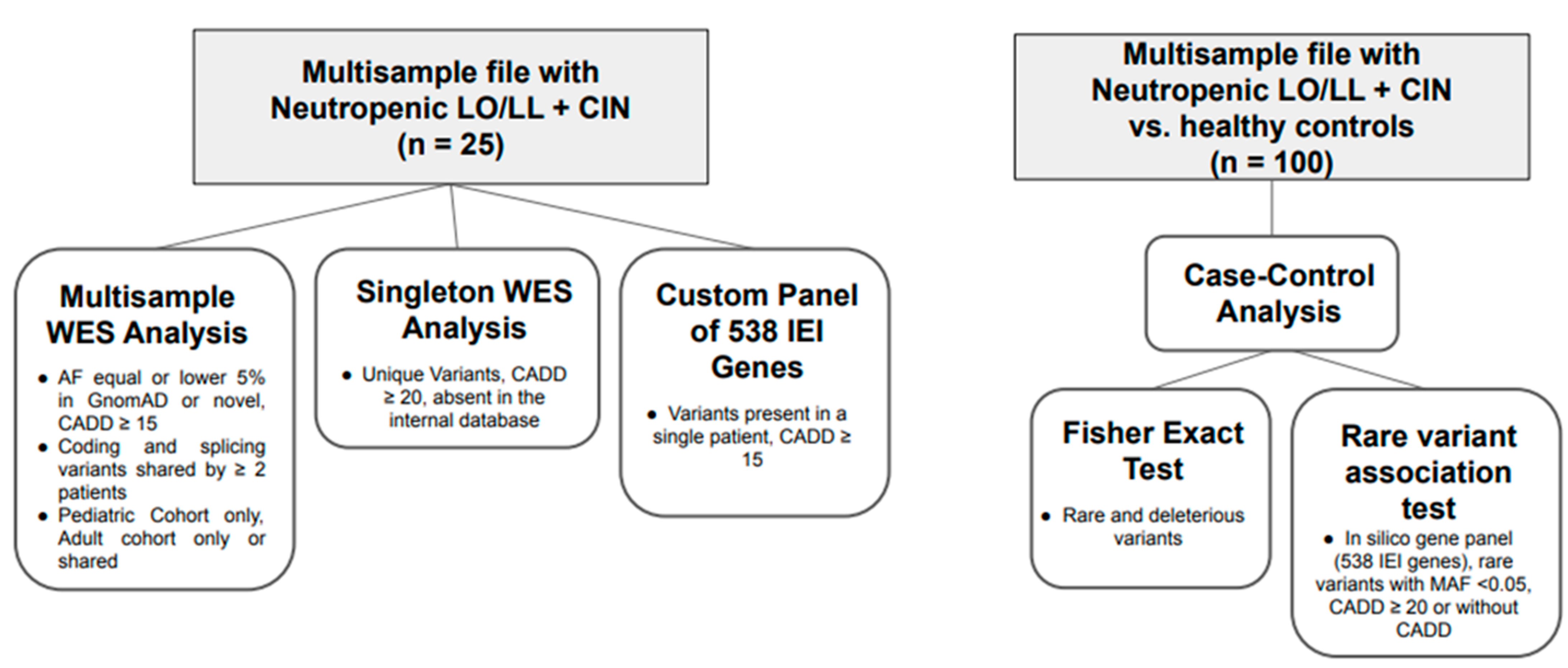
Figure 1. WES analysis pipeline. The flowchart summarizes the WES analysis pipeline used in this study. It begins with multisample variant calling from pediatric and adult cohorts, followed by filtering based on population frequency and deleteriousness (CADD score). Variants were compared across groups (shared, pediatric only, adult only), with additional singleton analysis applying stricter criteria. A custom gene panel (538 IEl-related genes) was then used to identify potentially pathogenic variants. Finally, a case-control approach was applied, incorporating statistical association tests (Fisher’s exact test, SKAT, SKAT-o, Cm C, VT) to detect gene-level enrichment of rare damaging variants in patients versus healthy controls.
Ijms 26 06929 g001
Figure 2. The top 10 enriched terms for the input gene set are displayed based on the −log10(p-value), with the actual p-value shown next to each term.
Ijms 26 06929 g002
Figure 3. Heatmap showing genes identified as slightly significant (FDR-corrected p-value between 0.08 and 0.05, light red) and significant (FDR-corrected p-value < 0.05, dark red). The columns indicate, with corresponding colors, genes identified as nominally significant or significant using the CMC and SKAT approaches, as described in the Materials and Methods Section 4. The legend categorizes these genes into broader functional groups.
Ijms 26 06929 g003
Table 1. Clinical and hemato/immunological features of the whole cohort.
PTGenderEthnicityConsanguinityDate of BirthAge at Onset (y)Age at Diagnosis of NP (y)Duration FUP (mo)Main ClinicalFamily HistoryAutoimmunityIncluding AbANWBC (×109/L)Neu (×109/L)Lymph (×109/L)Mono (×109/L)IgA (48–368 mg/dL)IgG (701–1600 mg/dL)IgM (25–170 mg/dL)IgG1 (490–1140 mg/dL)IgG2 (150–640 mg/dL)IgG3 (20–110 mg/dL)IgG4 (8–140 mg/dL)CD3%CD3/CD4%CD3/CD8%CD19%CD3-(CD16/56)+%
Pt1FC *1991162741Fatigue/Apthae/
Arthralgias
NR #ANA+
Ab AN+
2.30.51.30.3153117119375236938.49.282.750.318.411.64.0
Pt2FC *200321632Fatigue/Apthae/
Arthralgias
NR #Ab AN+1.70.50.970.1522010286754643519.35.384.957.020.15.48.3
Pt3MC *2002121372ApthaeEarly Cancer aANA+ Ab AN+3.50.42.20.322211961569493281098478.045.027.78.613.4
Pt4FC *2003111366Fatigue/ApthaeNeurodegenerative disease bANA+ Ab AN+2.70.451.60.4136143630588143737.211278.058.72.414.35.0
Pt5MC *20112464NothingNR #Ab AN+2.90.951.50.241161207671060189748077.443.425.212.16.6
Pt6FC *201500.366NothingEarly Cancer cAb AN+4.80.481.90.355490575677221362467.039.019.011.018.4
Pt7FC *2001171912NothingNR #Anti Thy Ab+2.20.321.40.458132222387331337.112.282.154.420.813.04.0
Pt8FC *2000171841NothingCancer
in the family d
Ab AN+1.90.221.10.4185130868748692521278.439.522.312.05.6
Pt9MC *2003151534FatigueNR #Anti Thy Ab+ Ab AN+1.90.351.10.3165959757501892211881.334.431.19.96.9
Pt10MC *1993252648Mild psoriasisNeutropenic siblingAb AN+2.90.81.30.6245134016678541729.219.276.548.025.314.77.8
Pt11FC *1968505060ArthralgiasNR #Neg3.11.51.10.4117123013082239352.10.766.344.019.416.616.8
Pt12FC *19643638240ArthralgiasNR #Neg3.01.21.50.314410408261539125.42.568.652.015.014.414.3
Pt13FC *19791818372ArthralgiasNeutropenic siblingNeg3.31.21.60.3253110013565159834.041.171.038.0
Pt14MC *19573232156NothingNR #Neg2.80.71.40.53077204536735513.535.674.730.941.29.614.8
Pt15FC *1963545636Ankylosis spondylitisNR #Neg2.41.11.00.322293018761034035.348.479.752.422.511.68.8
Pt16FC *19513535180ArthralgiasNR #ANA+3.31.31.50.418913303792522667.43.078.058.223.86.4
Pt17MC *19732727209NothingNR #Neg3.20.52.00.4252134064770357031.057.579.738.938.513.65.3
Pt18MC *1973263096PsoriasisNR #Neg2.71.41.00.326514206389543613.950.051.229.226.510.728.2
Pt19FC *1953606660NothingNR #Neg2.50.41.50.51201380799494238.3108.065.039.035.016.313.7
Pt20FC *19543535276NothingNR #ANA+3.81.61.70.438111204887218518.10.371.037.034.018.08.0
Pt21FC *19683232288NothingNR #Neg3.71.51.70.3155120019371726024.0107.0
Pt22FC *19582325276NothingNR #Neg4.01.51.60.514579012036337016.716.972.244.227.716.311.4
Pt23FC *1968415072ArthralgiasNR #Neg3.01.01.60.324498112163038816.394.763.835.023.012.313.8
Pt24MC *19991818120NothingNRNeg2.81.01.50.262115018372126928.759.477.057.419.68.1
Pt25FC *19664040216NothingNRNeg4.01.61.80.51048362944020119.1108.084.954.928.89.111.7
In light grey (from Pt1 to Pt9) is highlighted the pediatric population. Ethnicity: C * = Caucasian, Consanguinity: N° = Non-consanguineous, # NR = Not reported. Cancer in detail: (a) Early prostate cancer (father); (b) Neurodegenerative disease (sister); (c) Early Hodgkin disease (mother); (d) Pediatric acute lymphoblastic leukemia (first cousin). WBCs, white blood cells; ANA, anti-nuclear antibodies; Anti Thy Ab, antibodies against thyroid; Ab AN, anti-neutrophil antibodies.
Table 2. Overview of all genetic variants found with three different methods.
Pt_IDMULTISAMPLES TestSINGLETON Test Inborn Errors of Immunity (IEI) PANEL (538 Genes)
Pt1MST1RMPEG1CARD10
CASP8 LAT
Pt2DNM2/MRE11/IRF7/CD36
XRCC2/NBAS TGFBR2
SIGLEC1/MPO/ZNFX1 RELA/TRNT1
Pt3BRCA1/MPO/CASP5 IFNAR1/SLC7A7
EPX/RIF1
Pt4RANBP2/DOCK1/DOCK8/PLCH2/PIEZO1
SPINK5
Pt5EP300/VPS13B/ZNFX1 AP3D1
ATP7B/PLCL2
Pt6BCLAF1/EPX/MYO9B/PDGFRB/PIK3C2GCASP4ADAM17/IL18BP/IPO8/LRRC32
Pt7FANCM/MRE11 SLCO2A1/NLRP1
Pt8PLCG2 POLD1
ATP7B/CXCR1/CD36
Pt9POLE/DOCK8/MAP3K21/SIGLEC1/DNM2 HS3ST6
Pt10SPINK5/CXCR1/DOCK1/PLCL2 POLR3A
HYOU1/POLE/RANBP2
Pt11EP300/ORAI1PSMB11
CHD7/PIK3C3/RAD50
Pt12MYO9B/NBAS/PDGFRB/XRCC2 NLRC4
ORAI1/PIK3C3/CTC1/PEPDCBLB
Pt13DOCK1/EPX/FANCM/HYOU1 TICAM1
Pt14ADAMTS13/PRKCH/ZNFX1HAX1CTNNB1
EP300
Pt15CASP5/DOCK8/PIEZO1GATA2
PRKCH
Pt16ARHGAP21/PRKCHTET2PSMG2/SP110
Pt17BRCA1/MRE11/RIF1/ADAMTS13 RFXAP
PEPD/RAD50
Pt18ABCB6/IRF7/MST1R/PLCG2AEN/ATG4D/RSF1
ALPI/LYST/PEPD/RAD50/RASAL3
Pt19BRCA1/MAP3K21/RIF1 ARPC1B/C6
VPS13B/RASAL3/CTC1/MYOF
Pt20CASP8/DOCK8/SIGLEC1GJB4/OVCA2/ZYXMEFV/PLG
Pt21ATP7B/N4BP2L2/PEPD/RAD50 DCLRE1C
MST1R RTEL1
Pt22EP300/MSH4ACTN1/G6PC3FANCC
PLCH2/ALPITCF3/PGD
N4BP2L2STEAP3
Pt23PIK3C2G/MYOFITGB3/IGSF6/MSRA/SERPINB3EXTL3/POLA1
Pt24PDGFRB/ARHGAP21LTBNFAT5/TLN1
MSH4/LYST
Pt25BCLAF1/PIEZO1SAMD9CARD11
CHD7/PEPDCXCR4/RAD23A
In red: P/LP/hot VUS homozygous or compound heterozygosity in AR transmitted diseases or heterozygous in AD transmitted diseases. In orange: P/LP/hot VUS variants (i) in heterozygosity in AR transmitted disease or (ii) in gene of unknown inheritance or (iii) that would deserve attention due to their role in cancer predisposition, DNA repair or telomer disorders or other disorders (i.e., bleeding). Light grey: cold VUS.
Disclaimer/Publisher’s Note: The statements, opinions and data contained in all publications are solely those of the individual author(s) and contributor(s) and not of MDPI and/or the editor(s). MDPI and/or the editor(s) disclaim responsibility for any injury to people or property resulting from any ideas, methods, instructions or products referred to in the content.