Article

Design, Synthesis, and Testing of 1,2,3-Triazolo-Quinobenzothiazine Hybrids for Cytotoxic and Immunomodulatory Activity

by
Klaudia Giercuszkiewicz-Haśnik
1,2,
Magdalena Skonieczna
1,2,
Beata Morak-Młodawska
3 and
Małgorzata Jeleń
3,*
1
Department of Systems Biology and Engineering, The Silesian University of Technology, Akademicka Street 16, 44-100 Gliwice, Poland
2
Centre of Biotechnology, Silesian University of Technology, Krzywoustego Street 8, 44-100 Gliwice, Poland
3
Department of Organic Chemistry, Faculty of Pharmaceutical Sciences in Sosnowiec, Medical University of Silesia in Katowice, Jagiellońska Street 4, 41-200 Sosnowiec, Poland
*
Author to whom correspondence should be addressed.
Int. J. Mol. Sci. 2025, 26(14), 6920; https://doi.org/10.3390/ijms26146920
Submission received: 20 June 2025 / Revised: 14 July 2025 / Accepted: 17 July 2025 / Published: 18 July 2025
(This article belongs to the Special Issue Techniques and Strategies in Drug Design and Discovery, 3rd Edition)

Abstract

Phenothiazines, mainly known for their antipsychotic activity, have recently attracted attention as potential compounds with anticancer and immunomodulatory activity In this study, 20 new quinobenzothiazines (MJ1MJ20) were synthesized and their effects on normal cell lines (BEAS-2B, NHDF) and cancer cell lines (HCT116, MCF7, A549, SH-SY5Y, U2OS) were investigated. The studies included cytotoxicity assessment, analysis of the expression of genes (BCL2, AIFM2, MDM2) and pro-inflammatory cytokines (IL6, IL8) using the RT-qPCR method, and prediction of biological activity using the PASS platform. The results indicate that the compounds MJ19 and MJ20 have the greatest effect on the induction of pro-inflammatory (IL6, IL8) and antiapoptotic (BCL2, MDM2) genes, suggesting their potential use in therapies for inflammatory and autoimmune diseases. Gene expression analysis showed that compound MJ2 in BEAS-2B cells significantly induced the expression of AIFM2, a protein responsible for protecting against ferroptosis, while moderately increasing the expression of BCL2 and MDM2, suggesting a potential role for MJ2 in the modulation of protective mechanisms of healthy cells, e.g., avoiding apoptosis death. These results emphasize the potential of quinobenzothiazines as multifunctional bioactive compounds, which require further studies to determine their mechanisms of action and specificity.

1. Introduction

The phenothiazine derivatives are the oldest synthetic group of tricyclic neuroleptic drugs and have been known for over 100 years and are one of the most versatile compounds from the point of view of biological activity. In recent years, hundreds of articles have been published on the synthesis and biological activity of these compounds. This research has made it possible to create many derivatives, some of which are used in therapy. Currently, these compounds are used in psychiatry, particularly in the treatment of schizophrenia, manic states, and various psychoses [1,2,3].
Classical phenothiazines, substituted in position 10 with dialkylaminoalkyl groups and additionally in position 2 with small groups (-H, -Cl, -CF3, SCH3), exhibit significant activities such as neuroleptic, antiemetic, antihistaminic, antipuritic, analgesic, and antihelmintic. For some time there have also been reports of promising anticancer, antibacterial, antiplasmid, multidrug resistance (MDR) reversal activities and potential treatment in Alzheimer’s and Creutzfeldt-Jakob diseases of classical phenothiazines [4,5,6,7,8,9,10,11,12,13].
Modifications of phenothiazine is mostly carried out mainly by changing the substituent in the thiazine nitrogen atom, introduction of substituents into the benzene ring, oxidation of the sulfur atom of the thiazine ring to the sulfoxide or sulfone system and replacing one or two benzene rings by the heteroaromatic system (pyridine, pyridazine, pyrimidine, pyrazine, 1,2,4-triazine, quinoline, quinoxaline) or bicyclic homoaromatic system (naphthalene) [14,15,16,17,18,19].
The techniques of modifying the neuroleptic phenothiazine skeleton lead to modified phenothiazine systems that show a significant spectrum of biological activities, including: anticancer, antiplasmid, antiviral, anti-inflammatory, and antibacterial activities, reversal of multidrug resistance, potential treatment in Alzheimer’s and Creutzfeldt-Jakob diseases, butyrylcholinesterase inhibition, while they also exhibit antioxidant and antihyperlipidemic activity [1,20,21,22].
Triazoles are heterocyclic organic compounds containing five member rings with three nitrogen atoms and two carbon atoms. There were two isomeric forms of triazoles, namely 1,2,3-triazole and 1,2,4-triazole [23,24,25].
The 1,2,3-triazoles, due to their unique chemical and structural properties, have received a great deal of attention over the past several decades and have been well recognized for their wide range of pharmacological properties. The 1,2,3-triazole derivatives have been well exploited for the generation of many medicinal scaffolds that exhibit anti-HIV, anticancer, and antibacterial activities. The 1,2,3-triazole ring when coupled with another heterocyclic ring has manifested wide therapeutic potential such as: antitumor, anti-inflammatory, analgesic, anti-HIV, antifungal, antimicrobial, anticonvulsant, antioxidant, antitubercular, antiparasitic, antithrombotic, and antidepressant. 1,2,3-Triazole derivatives have also been used as enzyme inhibitors such as histone deacetylase, PDE4 (phosphodiesterase 4), alkaline phosphatase, cysteine protease and acetylcholinesterase. Several triazoles (for example, cefatrizine, tazobactam) were used in medicine as β-lactam antibiotics. Furthermore, 1,2,3-triazoles can form hydrogen bonds, which play an important role in bioavailability and solubility. The triazole ring is a biological linker and exhibits bioisosteric effects on various heteroaromatic and aromatic rings [26,27,28,29,30,31,32,33].
The structural modifications of the classical phenothiazine system are carried out by replacing the substituent at the nitrogen, among others, by incorporating the 1,2,3-triazole ring. Liu et al. [34] investigated the anticancer activity of the new phenothiazine-1,2,3-triazole hybrids. Their antiproliferative activity against three gastric cancer cell lines (MKN28, MGC-803, and MKN45) was evaluated. One of the described compounds showed high inhibitory activity against MGC-803 gastric cancer cells with an IC50 value of 1.2 μM (compound A, Scheme 1). It was found that this derivative could inhibit migration by regulating the expression level of N-cadherin, E-cadherin, vimentin and active d-MMP2. Furthermore, it could regulate the wnt/β-catenin signaling pathway in MGC-803 cells in a concentration-dependent manner by reducing the expression level of Wnt5α, β-catenin and TCF4. This hybrid was also a novel inhibitor of tubulin polymerization. In an oral study, it was demonstrated that this compound could effectively inhibit the growth of xenografted MGC-803 tumor in vivo without apparent side effects [34]. Zhang et al. [35] investigated how introducing a 1,2,3-triazole substituent into the phenothiazine system affects anticancer activity. The researchers designed a series of novel phenothiazine-1,2,3-triazole hybrids and evaluated their antiproliferative activity against three cancer cell lines: MDA-MB-231, MDA-MB-468, and MCF-7. When the 1,2,3-triazole group was removed, inhibitory activity against all cancer cell lines was decreased (IC50 > 100 μM). The most active compound showed IC50 values ranging from 0.8 μM to 1.7 μM (compound B, Scheme 1). It was found to induce apoptosis of MCF-7 cells through regulation of apoptosis-related proteins (Bcl-2, Bax, Bad, Parp, and DR5) [35]. 1,2,3-Triazole-phenothiazines have also been reported to exhibit in vitro activity against human gastric cancer (MGC-803), human esophageal cancer (EC-109), human prostate cancer (PC-3), human breast cancer (MCF-7), and hepatocellular carcinoma (HepG-2). The IC50 values for these hybrids ranged from 0.5 to 27.3 μM. The introduction of a fluorine atom into the benzene ring of the substituent was judged to lead to an increase in activity (compound C, Scheme 1) [36].
The aim of our studies was to obtain new phenothiazine hybrids by replacing one of the benzene rings with a quinoline system and introducing a 1,2,3-triazole ring-containing substituent to the thiazine nitrogen atom. Some of the previously obtained quinobenzothiazines with different substituents on the thiazine nitrogen atom showed promising immunosuppressive and antitumor activity against human cancer cell lines derived from the colon, breast, kidney, lung, ovary, prostate, central nervous system, melanoma and leukemia. Scheme 2 shows the overall structures of the quinobenzothiazines that have been synthesized and tested for various biological activities [37,38,39].
Recent studies indicate that phenothiazines may have potential anticancer effects by influencing various molecular mechanisms in cells [40,41,42]. Their actions include inhibiting cell proliferation, modulating signaling pathways such as PDK1/Akt and MAPK/ERK1/2, inducing apoptosis by inhibiting the Akt/mTOR pathway, and blocking angiogenesis by inhibiting VEGF (vascular endothelial growth factor) production and VEGF-dependent signaling. This study focused on evaluating the effect of guinobenzothiazines on cell survival, morphology, and expression of genes related to apoptosis, ferroptosis, and the inflammatory response, which may contribute to a better understanding of their potential therapeutic properties.
The selected research genes—BCL2 (B-cell CLL/lymphoma 2), MDM2 (mouse double minute 2 homolog) and AIFM2 (Apoptosis-Inducing Factor, Mitochondrion-Associated, 2)—play key roles in cell survival processes. The BCL2 gene encodes an antiapoptotic protein that protects mitochondria from damage by inhibiting the apoptosis process. BCL2 overexpression is often observed in cancer cells and is associated with resistance to cytotoxic therapy [38]. MDM2 is a negative regulator of the p53 protein, which controls the cell cycle and apoptosis in response to cellular stress [43]. AIFM2, also known as Ferroptosis Suppressor Protein 1 (FSP1), plays a key role in protecting cells from ferroptosis, a regulated form of cell death induced by oxidative stress and lipid peroxidation [44]. High expression of AIFM2 may support cell survival under environmental stress.
The study used the normal BEAS-2B cell line, representing human bronchial epithelial cells, NHDF (Normal Human Dermal Fibroblast cells) and various cancer cell lines, including HCT116 (colon cancer), A549 (lung cancer), MCF7 (breast cancer), SH-SY5Y (neuroblastoma) and U2OS (osteosarcoma). These lines were selected due to their biological diversity, which allows for the assessment of the activity of the tested compounds in different molecular and tissue contexts.
The aim of this study was to evaluate the effect of quinobenzothiazines (MJ1MJ20) on cell survival, changes in their morphology, and the expression of genes related to the mechanisms of cell survival and death. These studies are not only to identify potential mechanisms of action of quinobenzothiazines but also to assess their usefulness in the context of anticancer therapies and modulation of the inflammatory response.

2. Results and Discussion

2.1. Synthesis of 1,2,3-Triazolo-Quinobenzothiazine Hybrids

One of the modern drug design strategies is hybrid drug design (molecular hybridization), which involves the rational combination of structural fragments derived from two (or more) known drugs or ligands that exhibit biological activity against a specific molecular target. The goal of this method is to obtain a new chemical compound that combines the favorable pharmacodynamic properties of the two starting structures, and at the same time may have a better pharmacokinetic profile, reduced toxicity or the ability to overcome drug resistance [45,46,47]. The hybridization strategy has found applications in the design of enzyme inhibitors, anticancer, anti-infective or neuroprotective drugs, among others. An example is the synthesis of hybrid acetylcholinesterase and β-amyloid inhibitors for the treatment of Alzheimer’s disease, in which fragments of donepezil and other inhibitors were combined in a single molecule to achieve a multi-targeted effect [48]. Hybrid drug design is considered a promising approach in the context of polypharmacology, enabling the creation of compounds that act on several biological targets simultaneously, which is particularly important in the treatment of complex diseases such as cancer, neurodegenerative diseases or infectious diseases [49]. As we demonstrated in the introduction, both phenothiazines and 1,2,3-triazoles are valuable chemical motifs in drug design due to their broad spectrum of biological activities. Therefore, the purpose of our study was to synthesize and evaluate for immunomodulatory and cytotoxic activity against selected cancer cell lines hybrids of quinobenzothiazines with 1,2,3-triazoles. Hybridization of phenothiazines with 1,2,3-triazoles involves the incorporation of a triazole ring to a thiazine nitrogen atom. The synthesis of such hybrids often relies on the Huisgen cycloaddition reaction, known as “click chemistry,” which allows efficient and selective formation of a triazole bond between the corresponding azide and alkyne groups [50].
As we described in the introduction, the phenothiazine-triazole hybrid compounds obtained so far have shown promising biological properties. In our approach to further modify these structures, we changed the phenothiazine ring to a quinobenzothiazine ring.
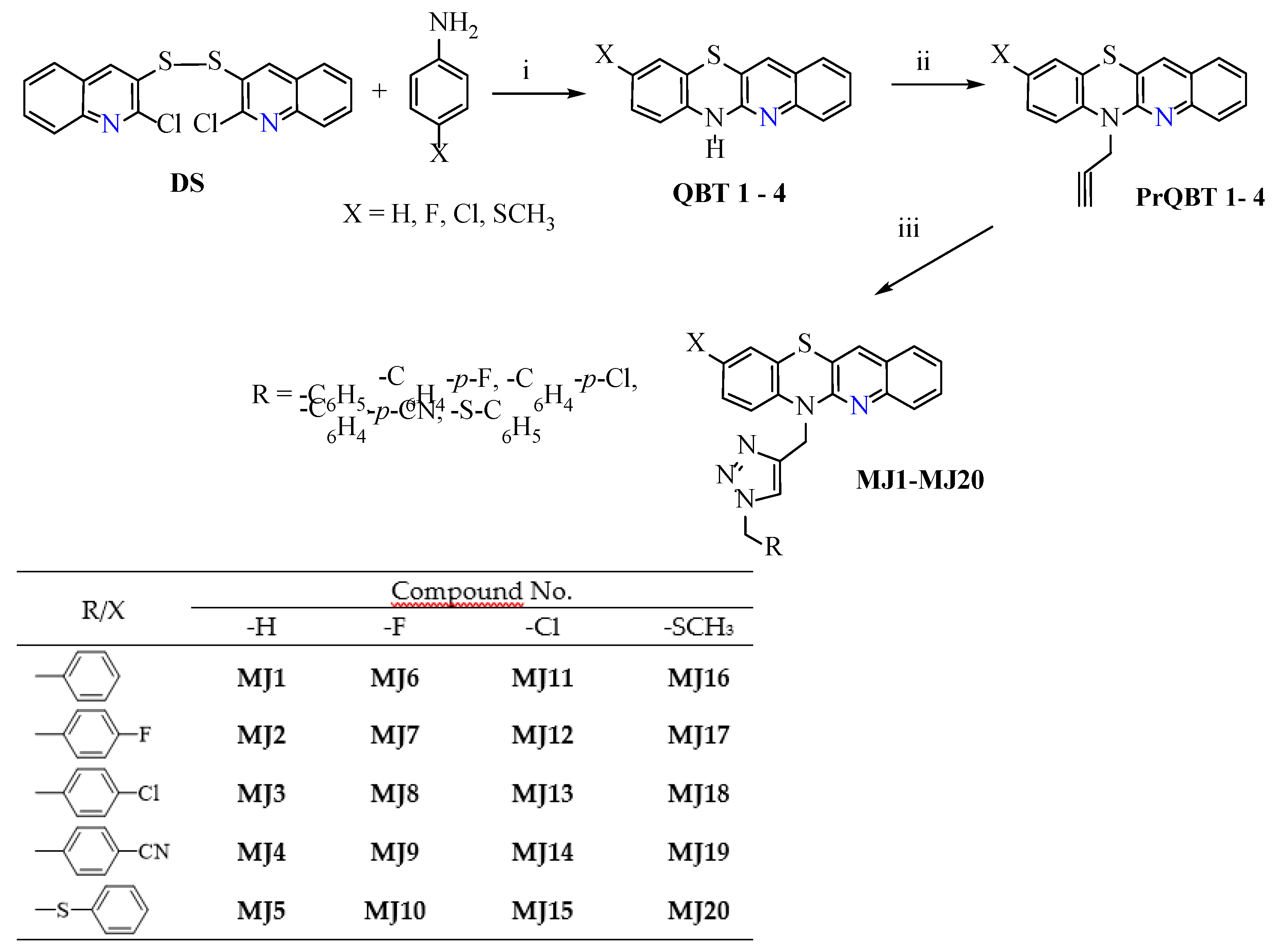
Accordingly, we began our research by developing the synthesis of structural analogs of quinobenzothiazines (quino [3,2-b]benzo [1,4]thiazine, 9-chloroquino [3,2-b]benzo [1,4]thiazine, 9-fluoroquino [3,2-b]benzo [1,4]thiazine and 9-methylthiochino [3,2-b]benzo [1,4]thiazine) containing different 1,2,3-triazolyl substituents and to investigate their anticancer activity (Scheme 3).
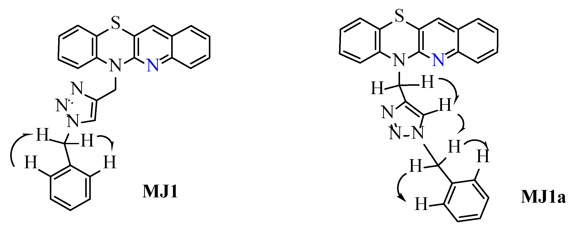
In the next step, we synthesized these 20 derivatives initially evaluated in silico for anticancer activity. Syntheses were started by obtaining appropriate quinobenzothiazine analogs—NH-quinobenzothiazines QBT 14 according to the literature procedures, which were the starting material for further steps [51]. The NH-quinobenzothiazines QBT 14 were then transformed in the next step by reaction with 3-bromo-1-propyne into 6-propynyl derivatives PrQBT 14 according to the methodology previously developed and described [52,53]. Substituted triazole derivatives of quinobenzothiazine were synthesized using copper-catalyzed azide-alkyne cycloaddition (CuAAC, with selected azides, in the presence of copper catalyst, in toluene). The identification of the product structure was based on 1H, 13C NMR spectra, 2D NMR experiments: COSY and ROESY and HR MS mass spectrometry. The crude reaction products were separated by column chromatography to obtain pure final derivatives MJ1MJ20 in good yield. As is known from previous studies, the reaction of 2-propynyl derivatives with organic azides can lead to 1,4- or 1,5- regioisomers [23,27]. To confirm this dependence, the structure of the obtained products was unequivocally confirmed using the 2D NMR ROESY experiment for quinobenzothiazine MJ1 (Scheme 4). Analyzing the results of this experiment, it can be seen that the protons of the benzyl group were correlated with the aromatic proton of the 1,2,3-triazole ring and the ortho-protons of the benzene ring. However, if the reaction resulted in the formation of the 1,5-regioisomer of the substitution quinobenzothiazine MJ1a, a correlation between the CH2 group on the nitrogen atom of the thiazine and the CH2 group on the triazole ring should be visible. No such correlations were observed in the obtained spectra of quinobenzothiazine MJ1.

2.2. Prediction of Biological Activity Using the PASS Web Platform

In the next step, compounds were tested in silico. For in silico research using the PASS platform [54] we chose 1,2,3-triazole-quinobenzothiazine hybrids containing benzyl, 4-fluorobenzyl, 4-chlorobenzyl, 4-cyanobenzyl and methylthiophenyl units.
Based on the data obtained from the predictive analysis for the tested substances MJ1MJ20 (Table 1), conclusions can be drawn about their potential biological properties. Most of the tested compounds show potential activity as anaphylatoxin receptor antagonists, while quinobenzothiazines MJ1, MJ3, MJ5, MJ6, MJ11, MJ13, and MJ15 show the highest Pa (probability of activity) values in this range. Anaphylatoxins C3a and C5a are polypeptides that are released during complement activation at the site of activation of the inflammatory reaction [55]. This causes the activation of granulocytes, macrophages, and mast cells and their chemotaxis, as well as increased vascular permeability [56]. In addition to immunomodulatory properties, anaphylatoxins regulate tissue regeneration [57]. This property suggests the possibility of modulating inflammatory responses, which may be useful in the treatment of autoimmune diseases or inflammatory conditions. Compounds such as MJ2, MJ16, MJ17, and MJ19 are highly likely to have a positive effect on the therapy of autoimmune diseases.
Most of the compounds studied (e.g., MJ1, MJ2, MJ3, MJ6, MJ11, MJ15, MJ20) have relatively high Pa values as therapeutic agents for atherosclerosis, indicating their possible influence on lipid metabolism or inflammatory processes associated with this disease [58].
Compounds such as MJ3, MJ5, MJ6, MJ11, MJ13, and MJ20 have high predictions for glycosylphosphatidylinositol-related phospholipase D inhibitor properties. This feature may be important in the regulation of signaling pathways related to cell membranes and immune responses [59]. The quinobenzothiazines MJ5, MJ6, MJ10 and MJ20 may have properties of the interleukin 2 agonist, suggesting the potential to stimulate the immune response. Such activity may be important in supportive therapy in oncology or infectious diseases [60].
Some compounds (e.g., MJ4, MJ19) can potentially exhibit neurotransmitter uptake inhibitor properties, suggesting their use in the treatment of neurological disorders such as depression or neurodegenerative diseases [61].

2.3. Microscopic Observations

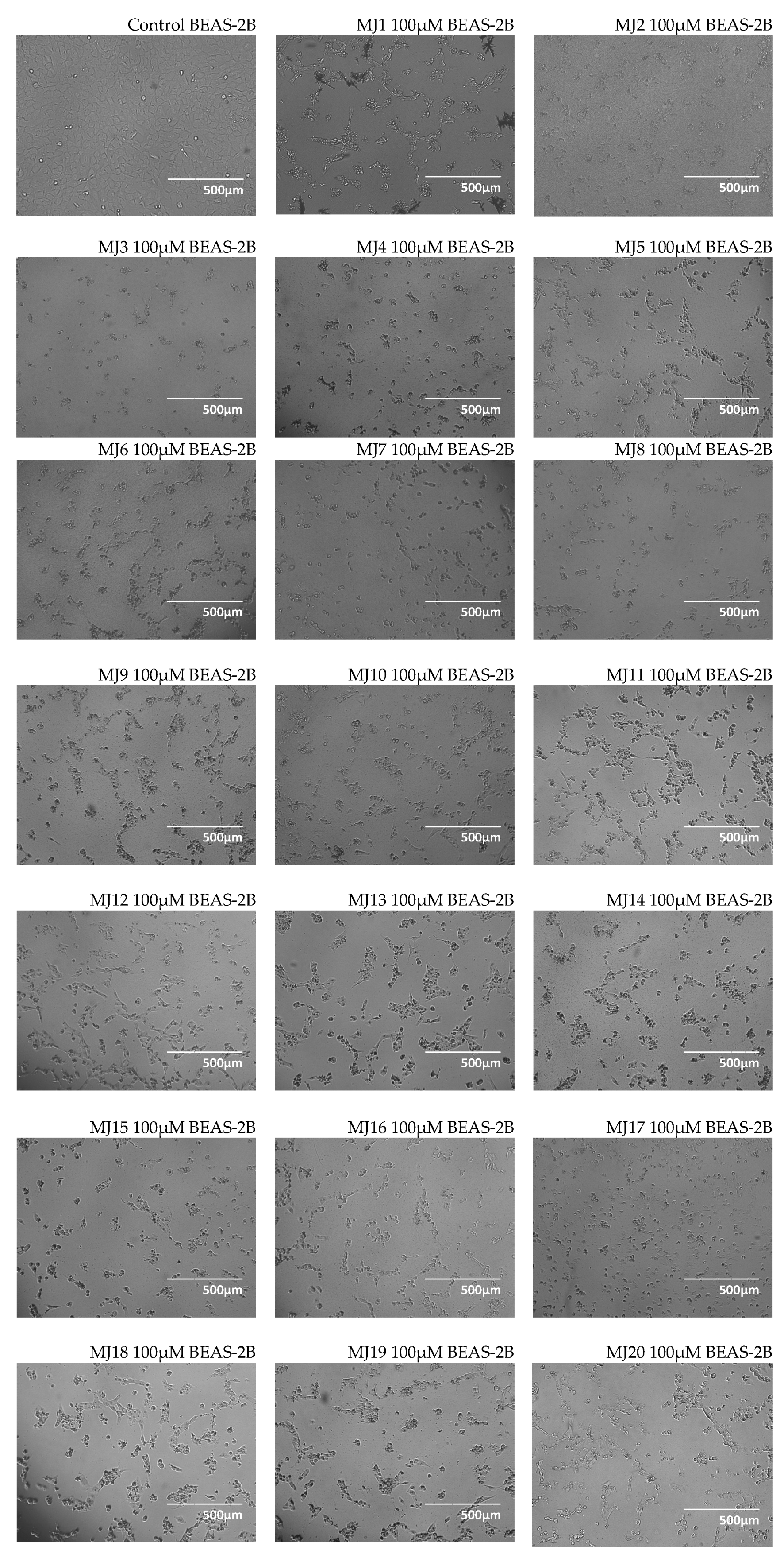
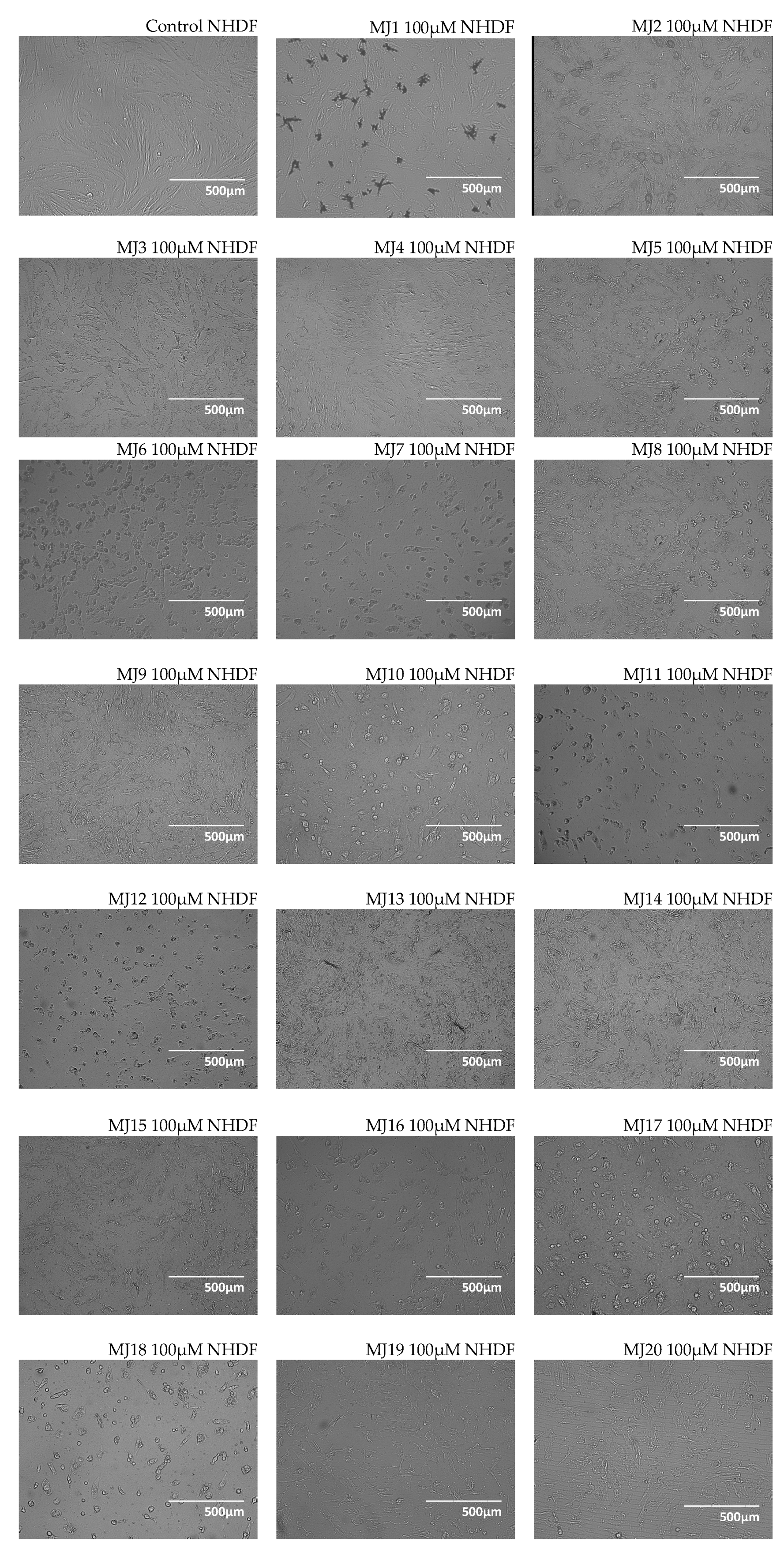
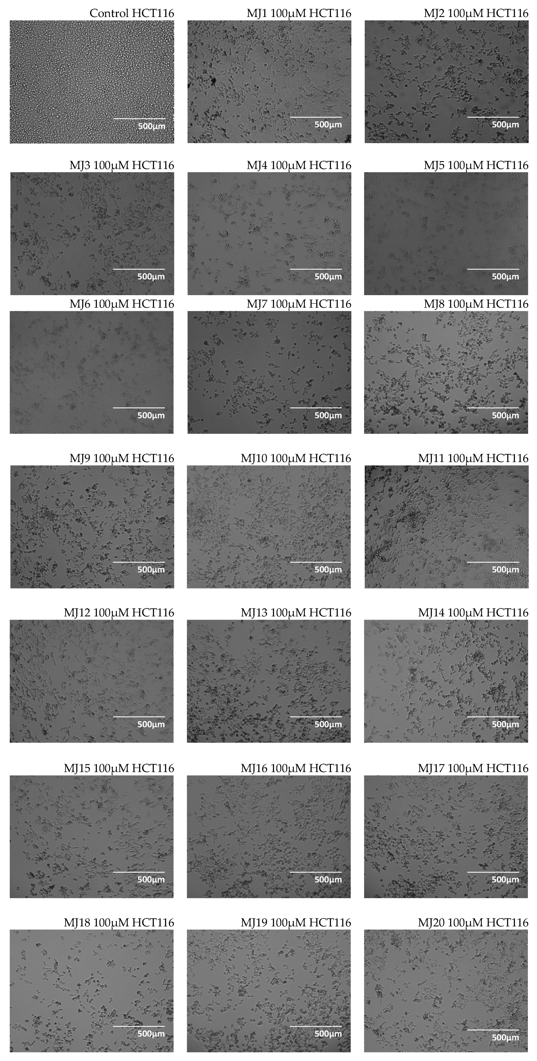
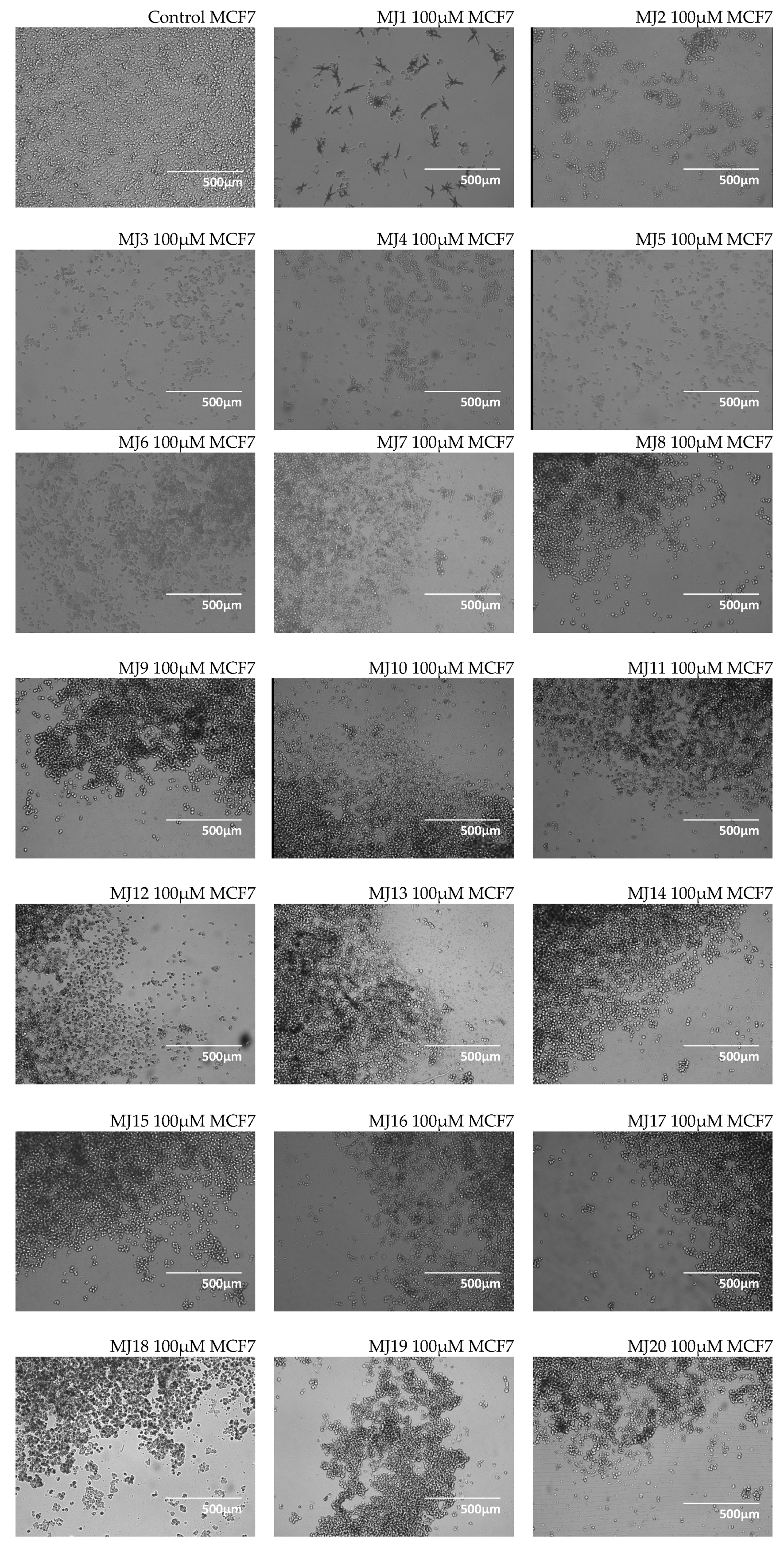
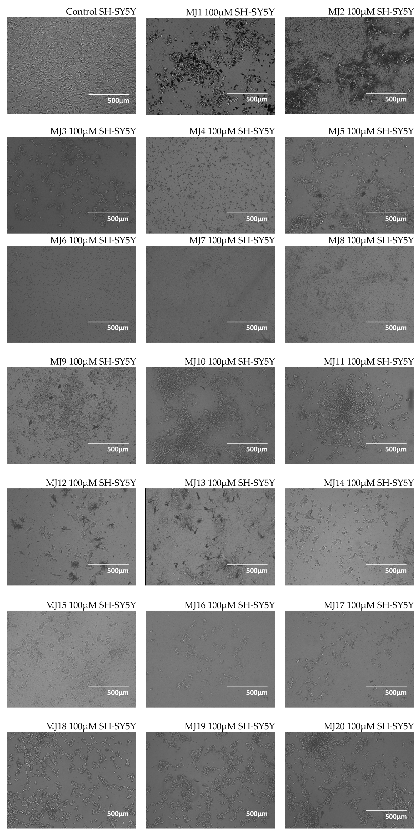
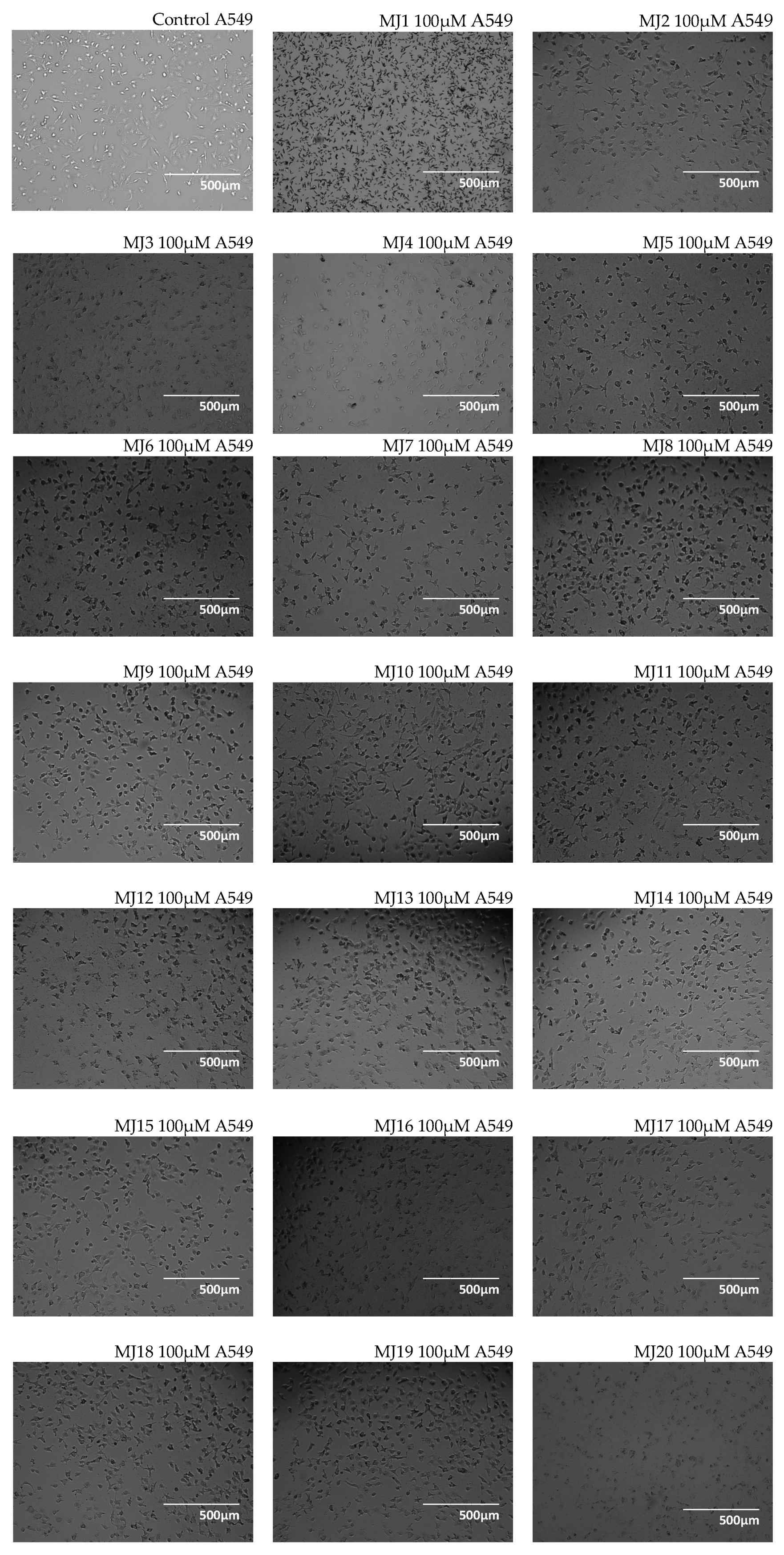
Microscopic studies were performed using BEAS-2B, NHDF, HCT116, MCF7, SH-SY5Y, A549 cell lines. Examples of BEAS-2B, NHDF, HCT116, MCF7, SH-SY5Y, and A549 cells images after 24 h incubation with MJ1MJ20 compounds at a concentration of 100 µM are shown in Figure 1, Figure 2, Figure 3, Figure 4, Figure 5 and Figure 6. Additional images showing the cell morphology of these lines in response to compounds MJ1MJ20 at concentrations of 6.25–100 µM are provided in Supplementary Materials File S2.
Microscopic observations showed that the quinobenzothiazines MJ1MJ20 affected cell morphology in a concentration-dependent manner. At lower concentrations (6.25–25 µM), the changes were minimal or invisible, and the cells maintained their typical shape and even distribution on the substrate, indicating their moderate effect under these conditions. High concentrations (50–100 µM) caused significant morphological changes, such as detachment of cells from the substrate and their adoption of a spherical shape, which may suggest the initial stages of cell death processes such as apoptosis or necrosis. Cancer lines such as HCT116, MCF7, SH-SY5Y, and A549 showed a greater susceptibility to morphological changes in response to high concentrations of quinobenzothiazines compared to normal cells such as BEAS-2B or NHDF, which may indicate differences in the mechanisms of cell death between cancer and healthy cells. Although images at 100 µM concentration are included in the article, detailed images showing cell morphology in response to lower concentrations of compounds (6.25–50 µM) are provided in the Supplementary Materials (the full range of concentrations the BEAS-2B and HCT116 lines, the 100 µM concentration the NHDF, MCF7, SH-SY5Y, and A549 lines) and provide additional information on morphological changes, allowing for a more complete interpretation of the effects of the compounds tested. These results indicate that the quinobenzothiazines MJ1MJ20 can affect cell survival and behavior in a concentration-dependent manner, making them interesting candidates for further investigation of their therapeutic potential.

2.4. Alamar Blue Test

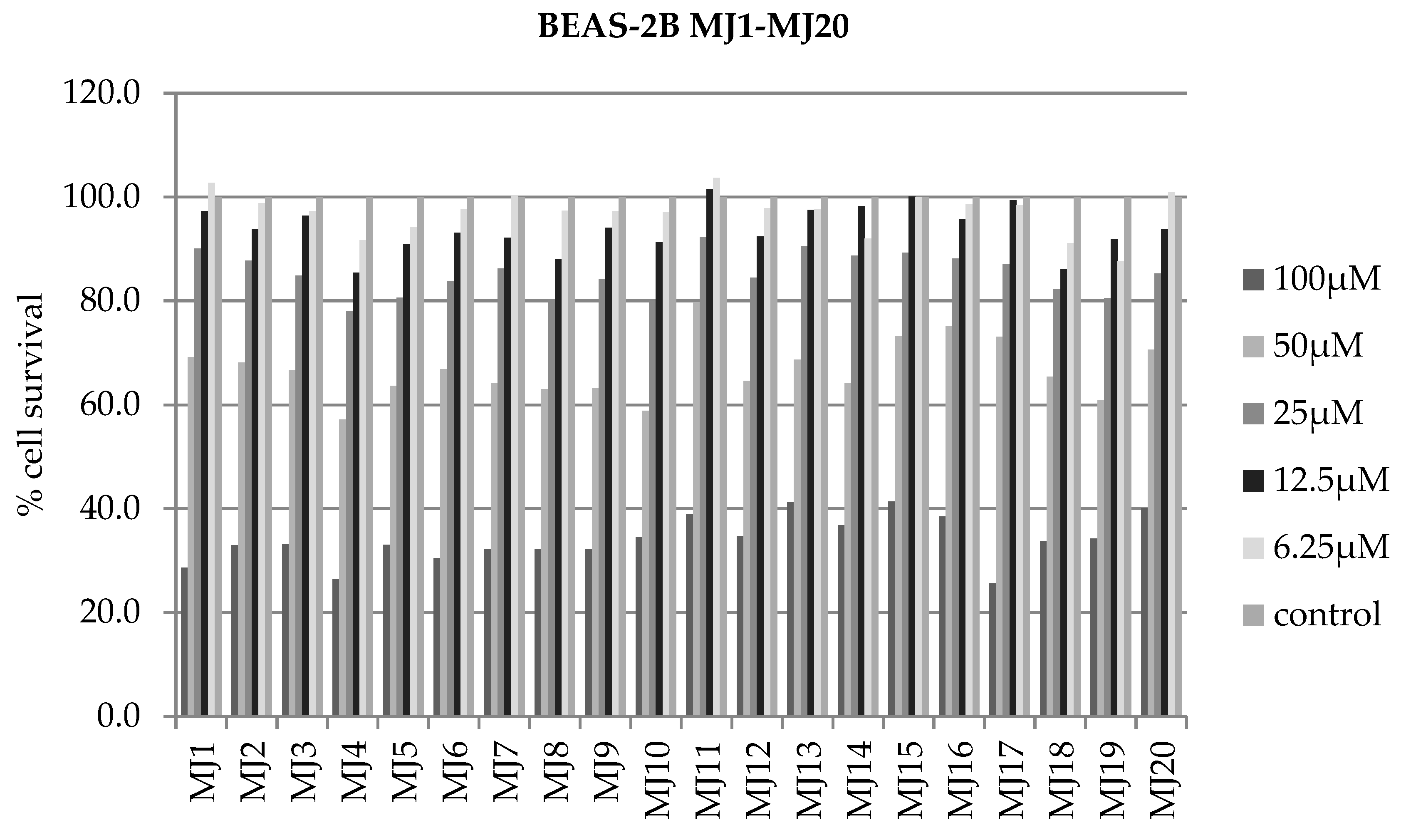
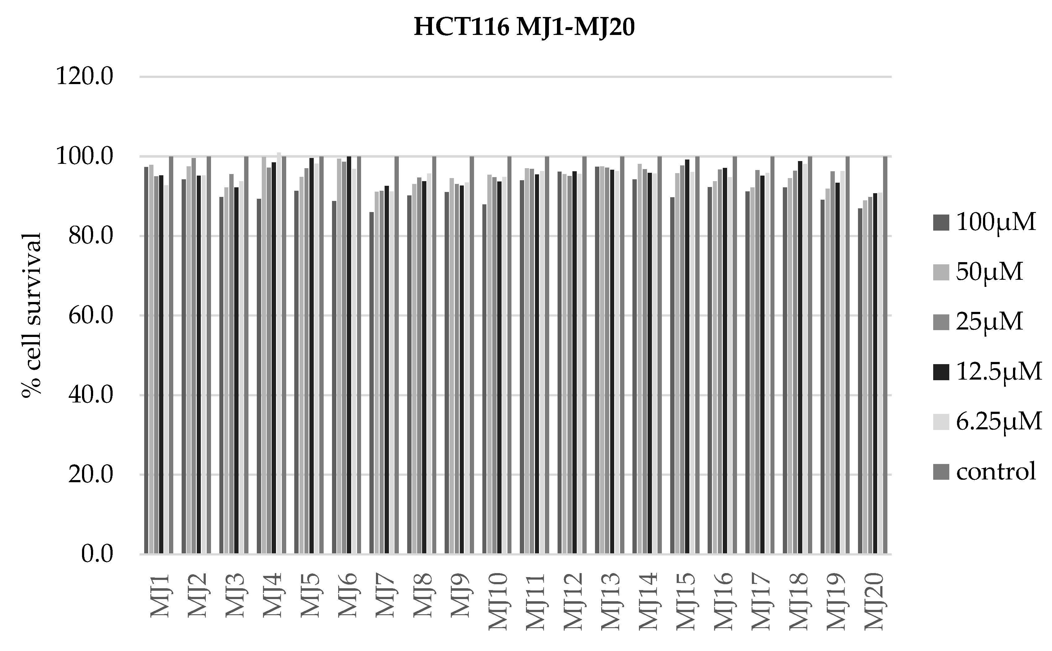
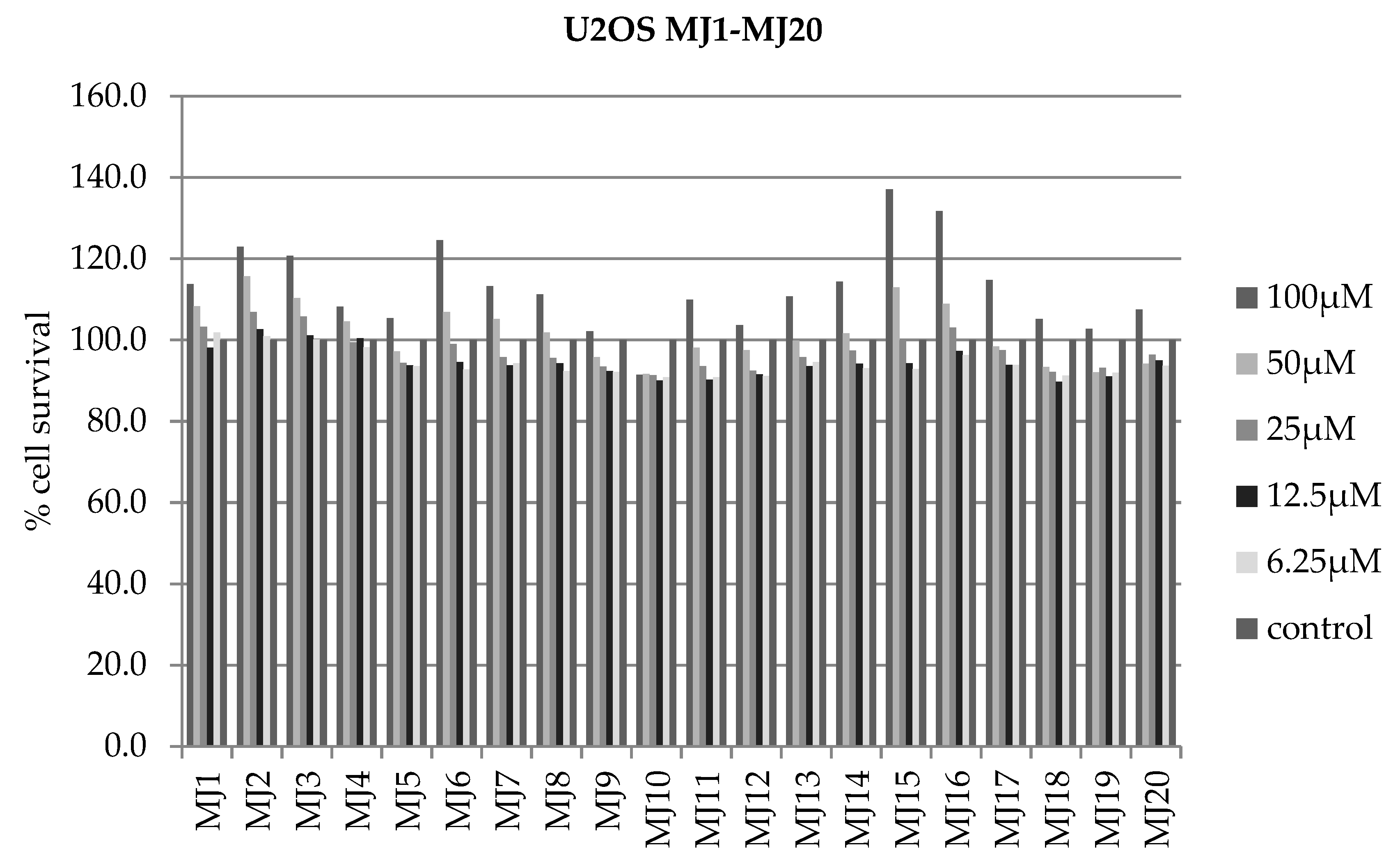
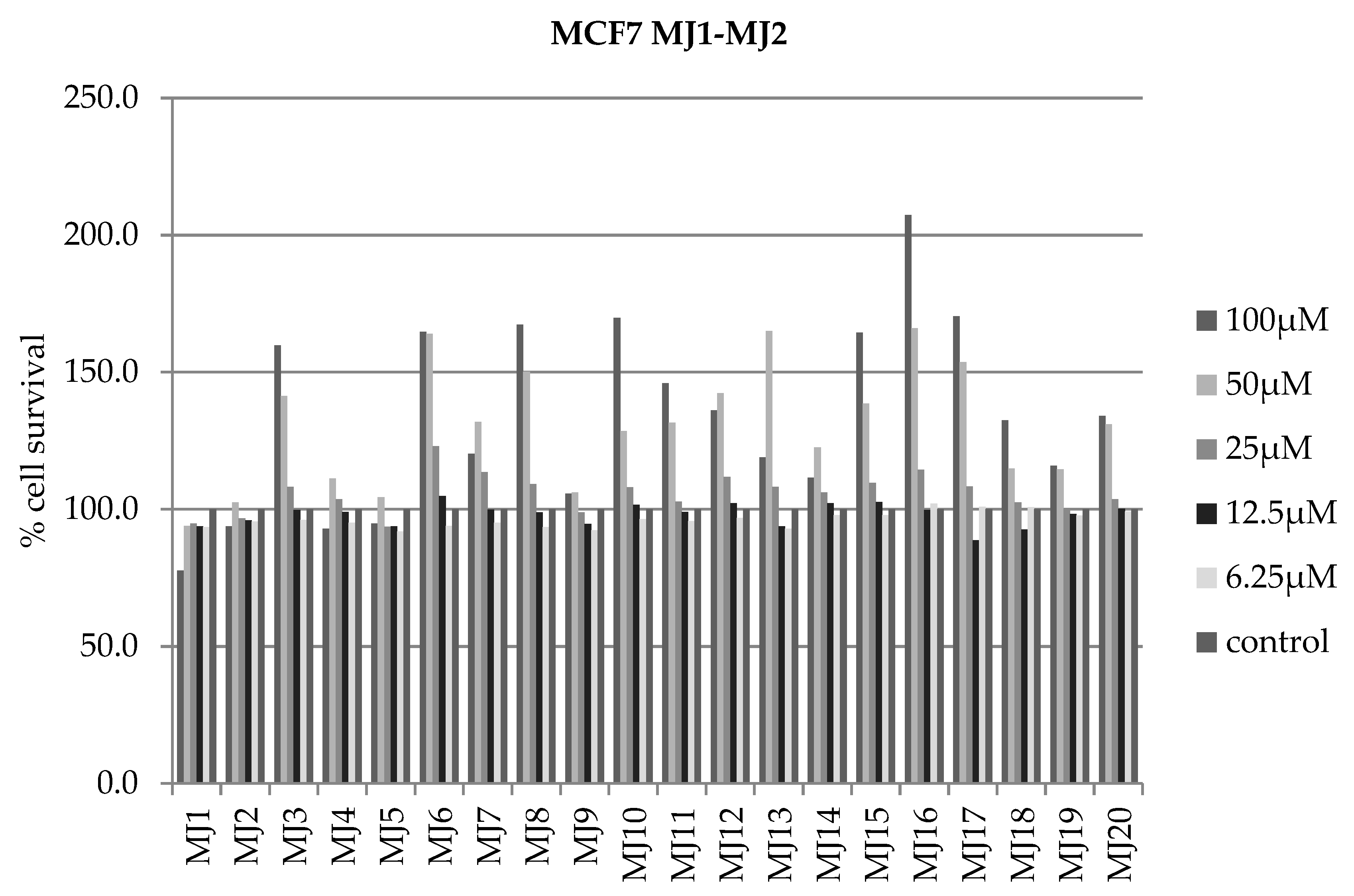
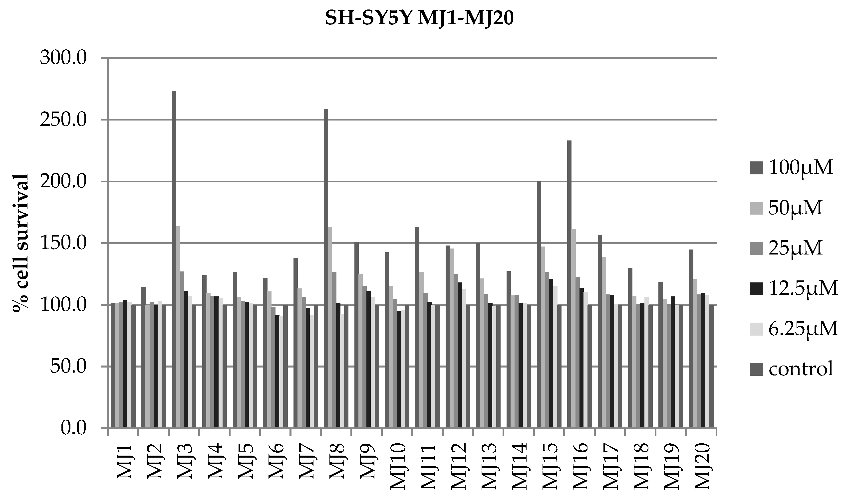
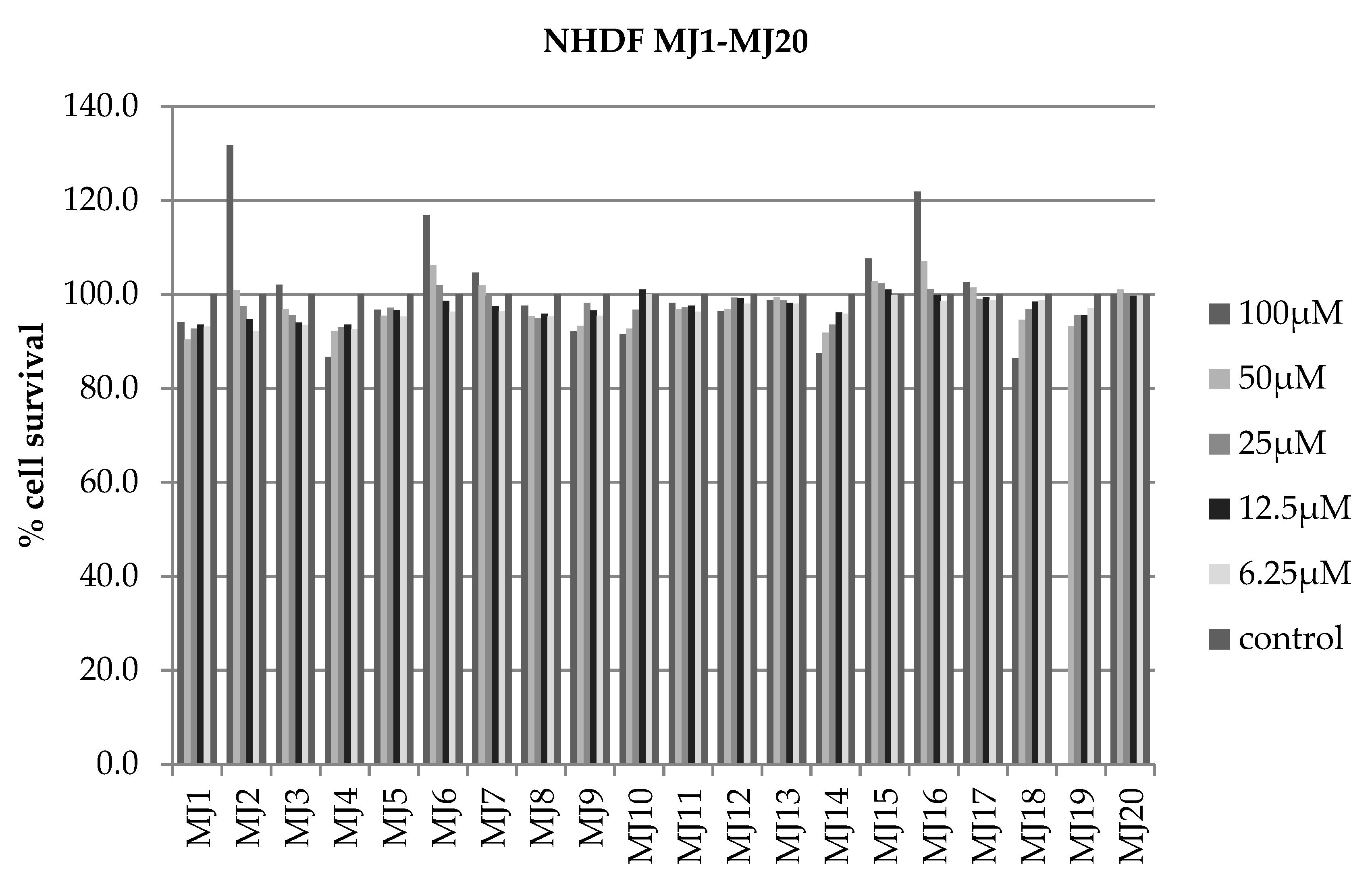
The cytotoxicity of the tested chemical compounds MJ1MJ20 was tested using the Alamar Blue test. The experiment was conducted on cell lines BEAS-2B (Figure 7), HCT116 (Figure 8), U2OS (Figure 9), MCF7 (Figure 10), SH-SY5Y (Figure 11), NHDF (Figure 12), and A549 (Figure 13). Cells were seeded in 96-well plates and incubated with the tested compounds for 24 h. The test result is the IC50 parameter, which is a measure of cytotoxic activity expressed by the concentration of the substance that causes the death of half of the analyzed cells compared to the control.
For the analyzed compounds, the IC50 parameter could only be calculated for the BEAS-2B line. For the remaining cell lines, the percentage of cell survival was so high that it prevented the calculation of IC50. The obtained results are summarized in Table 2.
The obtained results indicate that the tested compounds are not good cytostatics for the tested cell lines, as evidenced by the high survival percentages compared to the control after the use of MJ1MJ20.
The results of the Alamar Blue test showed that for the BEAS-2B line it was possible to determine the IC50 values, but they were high, indicating limited cytotoxicity of the tested quinobenzothiazines against these cells. In the case of cancer lines such as HCT116, A549, MCF7 or SH-SY5Y, the IC50 values could not be determined, which suggests their significant resistance to the action of MJ1MJ20 compounds at the applied concentrations. This phenomenon may result from the activation of cellular resistance mechanisms, such as overexpression of antiapoptotic proteins (e.g., BCL2) or effective drug efflux by transport pumps. Cancer cell lines that demonstrate this resistance may be a valuable model for studying the mechanisms of chemoresistance and assessing potential strategies to overcome it in the context of designing new anticancer therapies.
Alamar Blue test results showed that the highest concentrations of the quinobenzothiazines tested (100 µM) could lead to interference in the reading of the results. Cell viability values exceeding 100% were observed in some samples, suggesting the possibility of interference of the compound with the Alamar Blue reagent itself. This phenomenon could be caused by chemical reactions between the compounds tested and the dye or excessive metabolic activity of the cells in response to high concentrations of the compounds, which could lead to an erroneous increase in the fluorescence signal. These results indicate the need for caution in interpreting data for high concentrations of the compounds.

2.5. RT-qPCR

Real-time polymerase chain reaction was performed for the reference gene RPL41 (ribosomal protein L41) and the tested genes: BCL2, AIFM2, MDM2, IL6 and IL8. The RPL41 gene was chosen as a reference gene because it is stable and does not undergo significant changes under different experimental conditions or in different cell types. The BEAS-2B line was selected for the experiment. The expression level of selected genes was measured after 24 h of incubation with the tested substances MJ1MJ20 at a concentration of 100 μM (Table 3).

2.5.1. BEAS-2B’s BCL2 Gene Expression

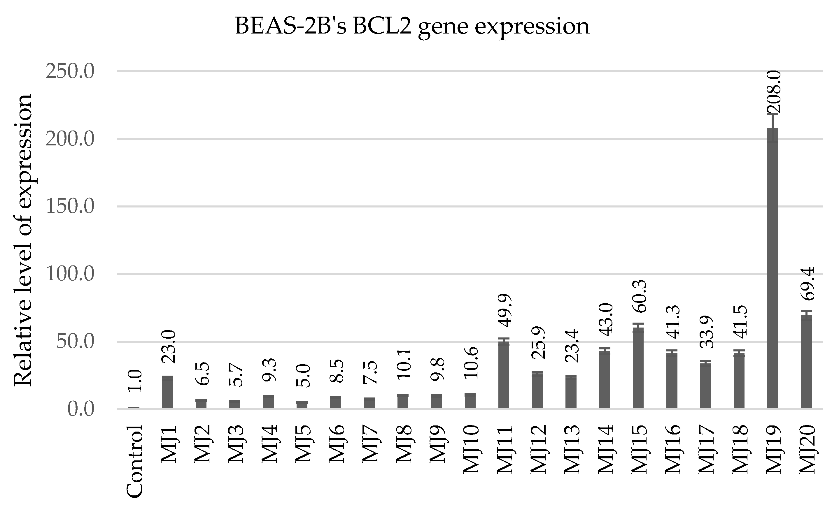
The RT-qPCR experiment was performed for the BEAS-2B cell line. In this experiment, the BCL2 gene was studied, and RPL41 was used as the reference gene (Figure 14).
BCL2 is a gene encoding an antiapoptotic protein that inhibits the process of programmed cell death. An increase in its expression was observed in response to most of the compounds tested, with the highest induction observed for MJ19 (208-fold increase) and MJ20 (69.4-fold increase). These results indicate that these quinobenzothiazines can enhance cell survival mechanisms. In the context of anticancer therapy, such activity is undesirable because it may promote the survival of cancer cells, especially under conditions of oxidative stress or exposure to cytotoxic drugs. However, in degenerative or inflammatory diseases, increased BCL2 may help protect healthy cells from damage.

2.5.2. BEAS-2B’s MDM2 Gene Expression

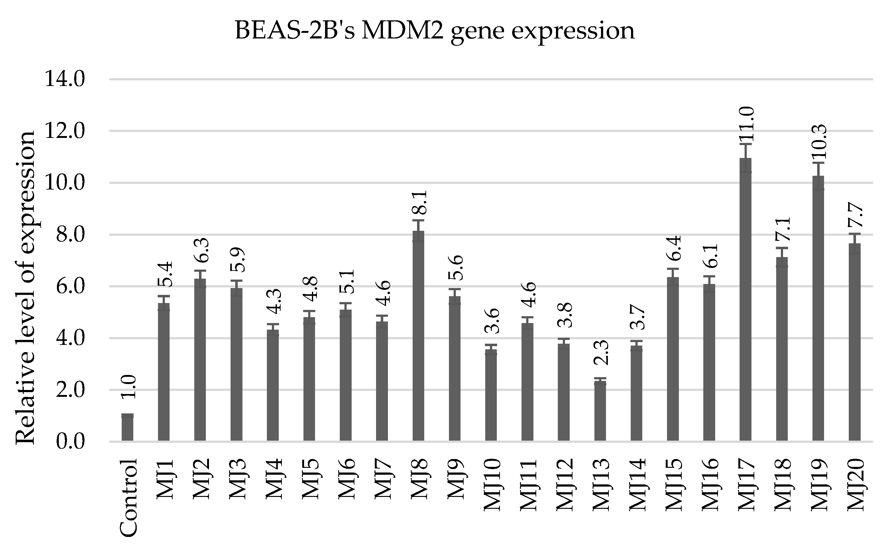
The RT-qPCR experiment was performed for the BEAS-2B cell line. In this experiment, the MDM2 gene was studied and RPL41 was used as reference gene (Figure 15).
MDM2 is a key regulator of p53, which plays an important role in DNA damage response and cell cycle regulation. The high expression of MDM2 in response to MJ17 (11.0-fold increase) and MJ19 (10.3-fold increase) suggests the ability of these compounds to modulate cell survival mechanisms. Similarly to BCL2, this may be beneficial in the context of protecting healthy cells.

2.5.3. BEAS-2B’s AIFM2 Gene Expression

The RT-qPCR experiment was performed for the BEAS-2B cell line. In this experiment, the AIFM2 gene was studied and RPL41 was used as reference gene (Figure 16).
Analysis of AIFM2 gene expression showed a significant increase in its level in BEAS-2B cells after the application of MJ2 (11.4-fold increase) and MJ19 (14.9-fold increase) compounds compared to the control, indicating the activation of protective mechanisms against ferroptosis. AIFM2 plays a key role in the regulation of ferroptosis, a form of cell death associated with lipid peroxidation and the accumulation of reactive oxygen species (ROS) in the presence of iron. Increased expression of AIFM2 in BEAS-2B cells after the application of MJ2 and MJ19 may suggest that the tested compound induces moderate oxidative stress, which stimulates adaptive mechanisms, such as the activation of antiferroptotic pathways. The high expression of AIFM2 may play a key role in the protection of BEAS-2B cells against ferroptotic death, which emphasizes the potential of MJ2 and MJ19 in the study of diseases in which the regulation of ferroptosis may be beneficial, such as neurodegenerative or inflammatory diseases.

2.5.4. BEAS-2B’s IL6 and IL8 Gene Expression

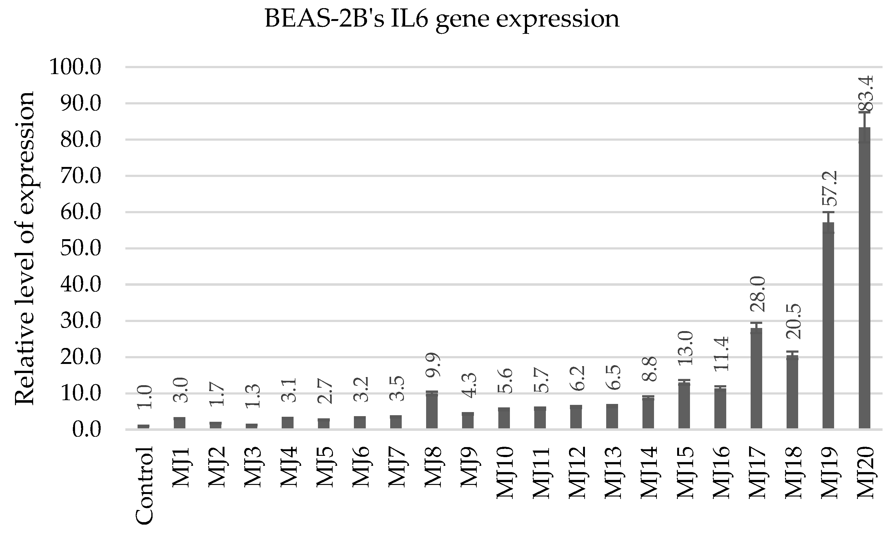
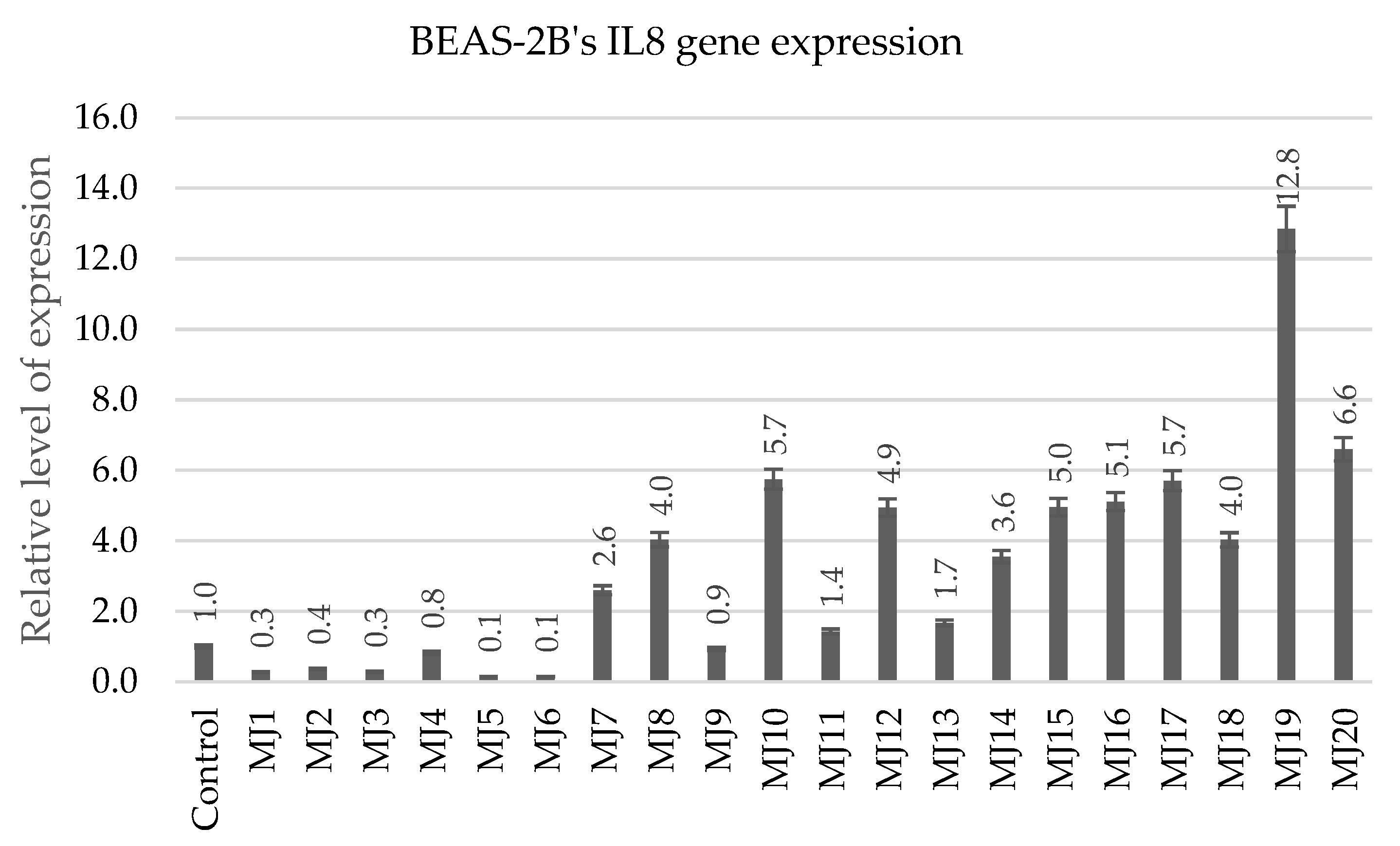
The RT-qPCR experiment was performed for the BEAS-2B cell line. In this experiment, the IL6 and IL8 genes were studied and RPL41 was used as a reference gene (Figure 17 and Figure 18).
The quinobenzothiazines MJ19 and MJ20 showed a strong ability to induce pro-inflammatory cytokines. In case of IL6, the highest expression was noted for MJ20 (83.4-fold increase), while MJ19 induced an increase of 57.2-fold. A similar trend was observed for IL8, where MJ19 induced an increase of 12.85 and MJ20 of 6.6. These interleukins play a key role in the regulation of inflammatory responses. Their induction may indicate the ability of quinobenzothiazines to modulate the immune system, making them potential candidates for adjuvant therapies in inflammatory and autoimmune diseases.
The quinobenzothiazines obtained in the present study differ in their 9-position substituents and in their 1,2,3-triazole ring substituents. MJ1MJ5 quinobenzothiazines do not have a substituent in position 9; MJ6MJ10 quinobenzothiazines are substituted with fluorine in this position; and MJ11MJ15 quinobenzothiazines are substituted with a chlorine atom. The last group, quinobenzothiazines MJ16MJ20, have a thiomethyl substituent in position 9. The results demonstrate how different substituents impact the biological activity of the compounds, particularly with respect to gene expression. MJ19 and MJ20 compounds, which contain a sulfur-containing thiomethyl substituent in position 9 of the quinobenzothiazine system and a methylthiophenyl substituent in the triazole ring (MJ20 only), exhibited enhanced activity in modulating BCL2, MDM2, and pro-inflammatory cytokine gene expression levels. This enhanced activity can be attributed to the electron-donating nature of the sulfur substituents, which potentially increases the compounds’ affinity for biological targets that regulate gene expression pathways. Additionally, these derivatives lack halogen atoms in the phenyl ring of the triazole substituent, demonstrating a negative inductive effect. The compounds also differed in the type of substituents in the triazole ring, including benzyl, 4-halobenzyl, 4-cyanobenzyl or phenylthiomethyl groups. It has been shown that compounds containing 4-halobenzyl substituents (e.g., MJ2, MJ3, MJ7, MJ8) can show moderate activity, but did not achieve as high gene induction as compounds with a thiomethyl group. Substitution of the phenyl ring with fluorine or chlorine atoms increases bond polarity and can affect the lipophilicity of molecules, which is important for cell membrane permeability. However, the presence of halogens may also have an inductive effect that decreases the activity of certain signaling pathways, as observed in the lower expression levels of IL6 and IL8 for these derivatives compared to MJ19 and MJ20.

2.5.5. HCT116′s BCL2 Gene Expression

In each of the four compound groups: MJ1MJ5, MJ6MJ10, MJ11MJ15, and MJ16MJ20, one compound was selected based on the best IC50 value and a distinctive gene expression profile (BCL2, MDM2, AIFM2, IL6, IL8). The comparison is presented in Table 3. Based on these data, four of the most promising compounds (MJ2; MJ8; MJ15; MJ19)- one from each group—were selected for further studies in the HCT116 tumor cell line.
Table 3. Summary of the most promising MJ1MJ20 compounds from each chemical group based on their biological activity profile in BEAS-2B cells (IC50 and relative level of gene expression).
MJ1–MJ5MJ6–MJ10MJ11–MJ15MJ16–MJ20
IC50MJ4
(64.526 µM)
MJ8
(71.858 µM)
MJ12
(75.636 µM)
MJ17
(72.595 µM)
BCL2MJ1
(23.0)
MJ10
(10.6)
MJ15
(60.3)
MJ19
(208.0)
MDM2MJ2
(6.3)
MJ8
(8.1)
MJ15
(6.4)
MJ17
(11.0)
AIFM2MJ2
(11.4)
MJ9
(4.5)
MJ15
(2.6)
MJ19
(14.9)
IL6MJ1
(3.0)
MJ8
(9.9)
MJ15
(13.0)
MJ20
(83.4)
IL8MJ4
(0.8)
MJ10
(5.7)
MJ15
(5.0)
MJ19
(12.8)
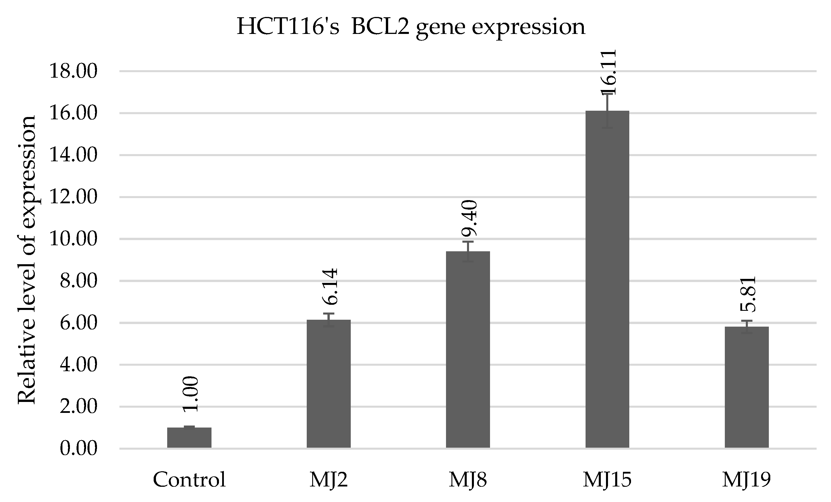
The results of the analysis of BCL2 gene expression levels in the HCT116 cancer line for compounds MJ2, MJ8, MJ15, and MJ19 indicate an increase compared to the control, suggesting that the tested compounds may also influence the regulation of mechanisms related to cell survival in cancer cells (Figure 19). Of particular note, although BCL2 expression levels in HCT116 were moderately elevated (from 5.81 to 16.11), they exhibited a clear modulatory effect, which may indicate a potential protective or adaptive effect under conditions of oxidative stress. An even more significant effect was observed in the healthy BEAS-2B lines, in which BCL2 expression was significantly higher, particularly for MJ15 (60.3) and MJ19 (208.0). This relationship may indicate a selective protective effect of these compounds on normal cells, while having a limited effect on cancer cells, but this requires further study. The observed effects support the hypothesis of a favorable action profile of the tested compounds as molecules capable of modulating survival mechanisms in a manner dependent on the cell type and its biological state.

2.5.6. HCT116′s MDM2 Gene Expression

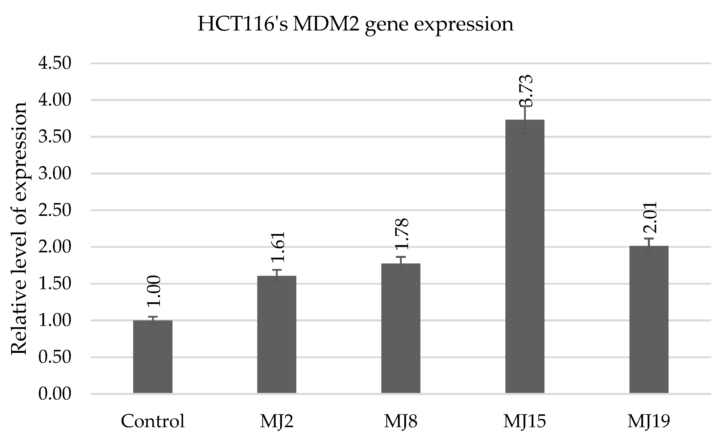
The results of MDM2 gene expression in the HCT116 cancer line after treatment with MJ2, MJ8, MJ15, and MJ19 compounds showed a small but noticeable increase compared to the control, with the highest value for MJ15 (3.73), which may suggest that some of the tested compound derivatives are capable of mildly modulating the p53-MDM2 regulatory axis in cancer cells (Figure 20). On the other hand, significantly higher levels of MDM2 expression were observed in healthy BEAS-2B cells, with values ranging from 6.3 for MJ2 to as much as 10.3 for MJ19. This significant difference may indicate a selective effect of these compounds, which manifests itself in a stronger increase in MDM2 in normal than cancer cells. Because MDM2 is a key inhibitor of p53, its increased expression in healthy cells may provide physiological protection against excessive activation of cellular stress and apoptotic pathways. Under the influence of bioactive compounds, this may be a homeostatic mechanism that promotes the survival of healthy cells. In cancer cells, however, a moderate increase in MDM2 levels may be considered therapeutically beneficial because it does not lead to a strong inhibition of p53 function, which could potentially reduce treatment efficacy. Mild induction of MDM2 may therefore play a regulatory role without inducing severe resistance to apoptosis.

2.5.7. HCT116 AIFM2

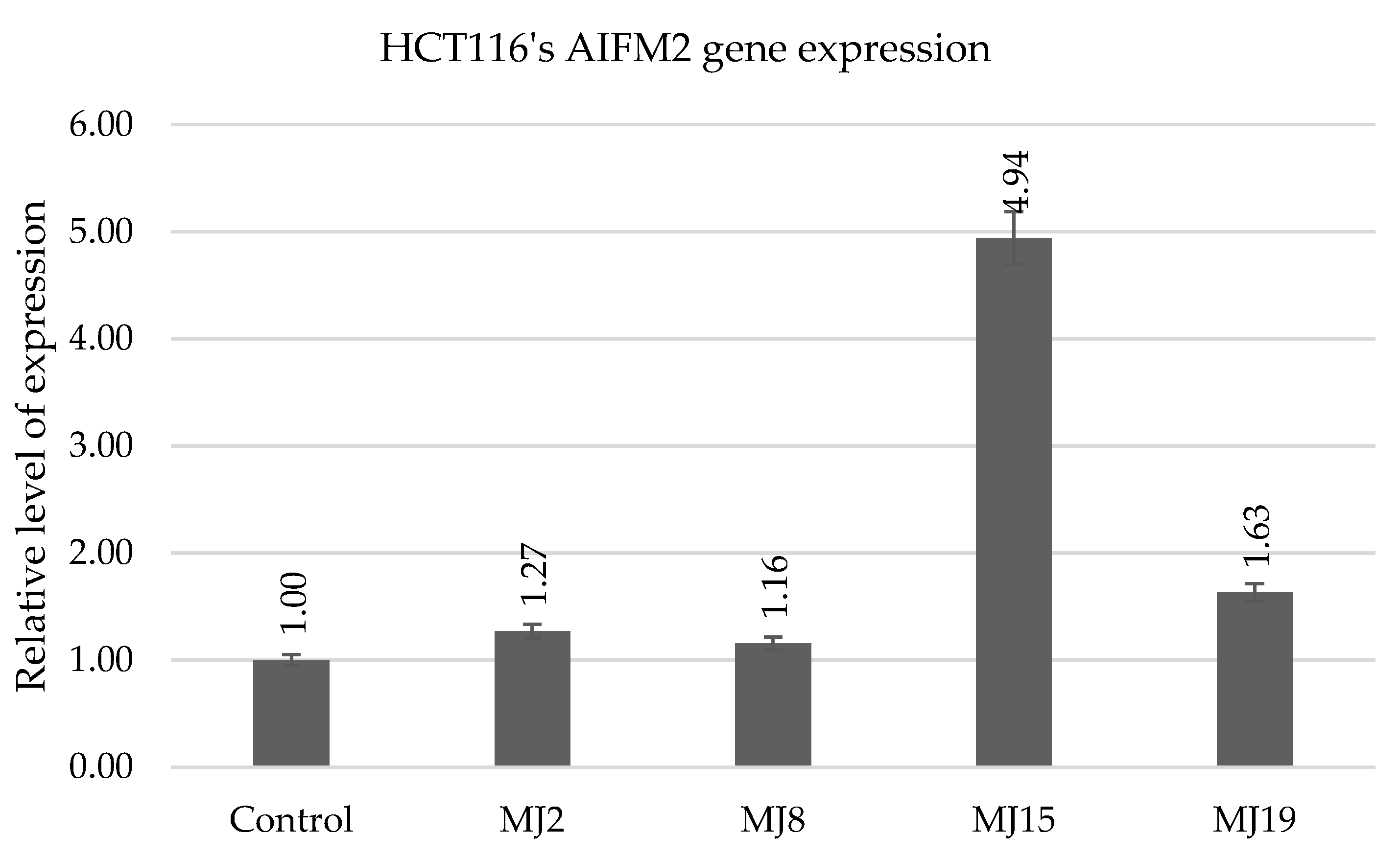
Analysis of AIFM2 gene expression levels in the HCT116 cancer line after treatment with MJ2, MJ8, MJ15, and MJ19 compounds revealed a slight increase in expression compared to the control for MJ2, MJ8, and MJ19 compounds, and a moderate increase compared to the control, particularly for MJ15, which reached a relative value of 4.94 (Figure 21). This result may indicate the ability of some of the tested compounds, especially MJ15, to activate protective mechanisms related to redox balance and prevent ferroptosis in cancer cells. However, the most significant difference was observed in AIFM2 expression in healthy BEAS-2B cells, where the values were significantly higher—up to 14.9 for MJ19 and 11.4 for MJ2. This suggests that the tested compounds can selectively and more effectively induce AIFM2 expression in normal cells than in cancer cells, which may be a significant advantage in the context of their potential use as protective agents, which requires further research. AIFM2, known as an important suppressor of ferroptosis, protects cells from oxidative stress and lipid peroxidation, so its activation may indicate the ability of phenothiazines to enhance the natural defense mechanisms of healthy cells. At the same time, moderate induction of AIFM2 in cancer cells may be beneficial because it does not block potentially effective pathways leading to cancer cell death, such as ferroptosis.

2.5.8. HCT116′s IL6 andIL8 Gene Expression

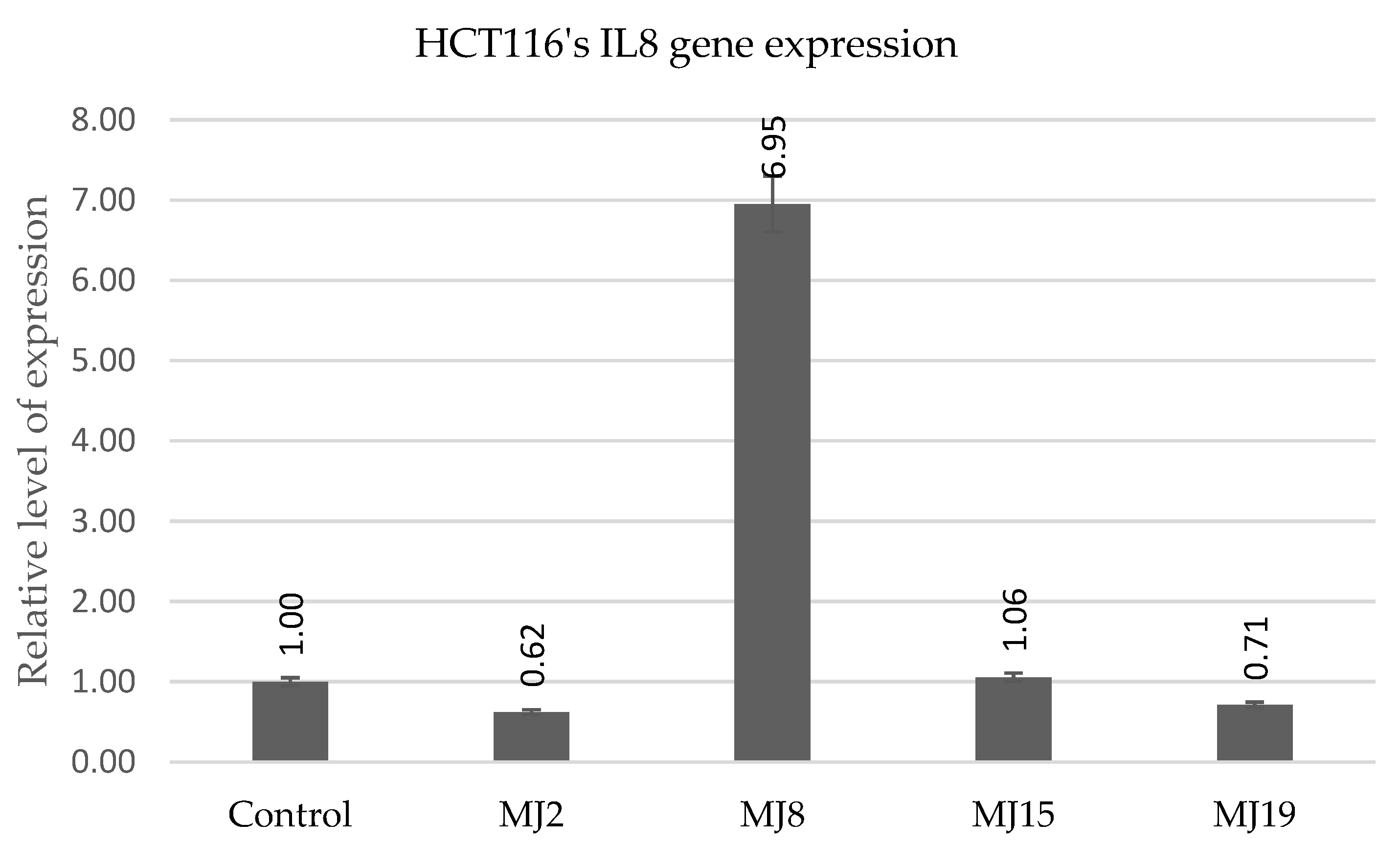
Analysis of IL6 and IL8 gene expression following the administration of MJ2, MJ8, MJ15, and MJ19 compounds demonstrates that these phenothiazines have the ability to modulate the inflammatory response in a manner that is dependent on both the compound type and the cell type (Figure 22 and Figure 23). A moderate increase in IL6 expression was observed in HCT116 cancer cells, particularly after the administration of MJ8 (6.55) and MJ15 (5.56), suggesting activation of a local inflammatory response in the tumor microenvironment. At the same time, IL6 expression was low for MJ2 and MJ19, indicating a more subtle effect of these compounds on pro-inflammatory pathways in cancer cells. In the normal BEAS-2B cell line, IL6 expression values were significantly higher, especially for MJ19, which reached a level of 57.2, suggesting its strong potential to activate the immune system in non-neoplastic cells. A similar pattern was observed for IL8, an interleukin that plays a key role in chemotaxis and angiogenesis. In the HCT116 cell line, only MJ8 significantly increased IL8 expression (6.95), while the remaining compounds induced only a slight or even decreased expression of this gene. In BEAS-2B cells, a significant increase in IL8 expression was observed after treatment with MJ15 and MJ19, with MJ19 reaching a value as high as 12.85, which may indicate its ability to strongly activate the inflammatory response in healthy cells. This feature could be exploited to enhance anti-infective defenses or to treat conditions where activation of a local inflammatory response is desirable. These results suggest the immunomodulatory potential of the tested compounds and indicate their selective, cell-type-dependent action.

2.6. DAPI Staining for Nuclear Morphology Assessment and Apoptosis Observation

During microscopic observations after the application of selected compounds MJ2, MJ8, MJ15, and MJ19 at a concentration of 100 μM and 24 h of incubation, no significant apoptotic changes were observed in the nuclei of BEAS-2B and HCT116 cells, which is consistent with the increased expression of antiapoptotic genes such as BCL2 compared to the control. Example images of cell nuclei are presented in Supplementary Materials File S2. No significant level of apoptotic bodies was detected.

3. Materials and Methods

Melting points were determined on a Boetius apparatus in open capillaries. 1H NMR spectra were recorded on a Bruker Avance spectrometer (Bruker, Billerica, MA, USA) at 600 MHz, and 13C NMR and 19F NMR spectra were recorded on the same spectrometer at 150 MHz and 564 MHz, respectively. CDCl3 was used as a solvent. Two-dimensional COSY, NOESY, HSQC, and HMBC spectra of selected compounds were recorded on a Bruker Avance spectrometer at 600 MHz, using CO-SYGPSW, NOESYGPPHSW, HSQCGPPH, and HMBCGP experiments. EI HRMS spectra were recorded on a Brucker Impact II spectrometer (Bruker, Billerica, MA, USA). The 1H NMR, 13C NMR, and HRMS spectra are provided in Supplementary Materials File S1. Thin layer chromatography was performed on alumina 60 F254 neutral (type E) (Merck 1.05581) with CH2Cl2 as an eluent. 6H-Quino [3,2-b]benzo [1,4]thiazine QBT1, 6H-9-chloroquino [3,2-b]benzo-[1,4]thiazine QBT2, 6H-9-fluoroquino [3,2-b]benzo [1,4]thiazine QBT3, and 6H-9-methyltioquino [3,2-b]benzo [1,4] thiazine QBT4 were obtained according to previously described methods [51]. These quinobenzothiazines were then converted to their 6-propynyl derivatives PrQBT1PrQBT4 according to previously described procedures [52].

3.1. In Silico Study

The biological properties of the analyzed compounds were predicted using the PASS web platform. PASS enables the prediction of the potential biological activities of chemical compounds based on their structure, assuming that biological activity results from the molecular structure of the substance. The tool provides predictions for specific biological properties expressed as the ratio of “probability of activity (Pa)” to the “probability of inactivity (Pi)”. The higher the Pa value, the higher the probability that a given compound exhibits a given activity [62].

3.2. General Procedure for the Synthesis of Compounds MJ1MJ20

To a solution of 6-propynyl quinobenzothiazine PrQBT 14 (0.5 mmol) and copper iodide (I) (0.006 mg) in dry toluene (10 mL), a corresponding organic azide (0.510 mmol) was added. The reaction mixture was heated at 70 °C and stirred for 48 h. The mixture was then cooled, and toluene was distilled off on a vacuum evaporator. The residue was dissolved in CH2Cl2 and purified by column chromatography (silica gel, CH2Cl2 as eluent) to obtain pure triazole derivatives MJ1MJ20:
[(1-Benzyl-1H-1,2,3-triazol-4-yl)-methyl]-quino[3,2-b]benzo[1,4]thiazine (MJ1):
Yield: 80% M.p.: 165–166 °C. 1H NMR (600 MHz, CDCl3) δ: 5.38 (s, 2H, CH2), 5.56 (s, 2H, N-CH2), 6.88 (t, 1H, H-9, J = 7.8 Hz), 7.00 (dd, 1H, H-7, J = 7.8 Hz, J = 1.8 Hz), 7.07–7.09 (m, 2H, 2H-Ph), 7.25–7.19 (m, 5H, 3H-Ph, H-8, H-10), 7.28 (d 1H, H-2, J = 8.2 Hz), 7.42–7.46 (m, 2H, H-1, H-3), 7.60 (s, 1H, H-12), 7.78 (m, 1H, H-4), 7.80 (s, 1H, CH). 13C NMR (150 MHz, CDCl3) δ: 43.94, 54.08, 117.33, 119.77, 120.40, 123.17, 123.86, 124.78, 125.36, 125.69, 126.63, 127.81, 128.16, 128.58, 129.01, 129.22, 130.02, 132.62, 133.01, 134.68, 140.68, 144.36, 151.49. HR MS (ESI) calcd for: C25H20N5S [M + H]+: 422.1439, found: 422.1430.
6-((1-(4-Fluorobenzyl)-1H-1,2,3-triazol-4-yl)methyl)-quino[3,2-b]benzo[1,4]thiazine (MJ2):
Yield: 82% M.p.: 145–146 °C. 1H NMR (600 MHz, CDCl3) δ: 5.44 (s, 2H, CH2), 5.53 (s, 2H, N-CH2), 6.93 (t, 1H, H-9, J = 7.2 Hz), 6.99–7.03 (m, 2H, H-Ph), 7.08 (d, 1H, H-7, J = 7.8 Hz), 7.14 (t, 1H, H-8, J = 15.6 Hz), 7.19–7.21 (m, 2H, H-Ph), 7.32 (t, 1H, H-2, J = 15 Hz), 7.38 (d, 1H, H-10, J = 7.8 Hz), 7.51 (t, 1H, H-3, J = 8.4 Hz), 7.55 (d, 1H, H-1, J = 7.8 Hz), 7.55–7.7.64 (m, 1H, H-4), 7.66 (s, 1H, H-12), 7.74 (s, 1H, CH). 13C NMR (150 MHz, CDCl3) δ: 44.10, 53.33, 115.98 (J = 22.5 Hz), 117.41, 120.01, 120.53, 122.70, 124.08, 124.78, 125.29, 125.60, 126.41, 126.70, 128.22, 129.70, 129.81 (J = 9 Hz), 130.21, 130.54 (J = 3 Hz), 133.29, 140.81, 144.23, 151.36, 162.72 (J = 246 Hz). 19F NMR (564 MHz, CDCl3) δ: -112.71. HR MS (ESI) calcd for: C25H19FN5S [M + H]+: 440.1345, found: 440.1350.
6-[(1-(4-Chlorobenzyl)-1H-1,2,3-triazol-4-yl)methyl]-quino[3,2-b]benzo[1,4]thiazine (MJ3):
Yield: 80% M.p.: 165–166 °C. 1H NMR (600 MHz, CDCl3) δ: 5.32 (s, 2H, CH2), 5.45 (s, 2H, N-CH2), 6.83 (t, 1H, H-9, J = 15.6 Hz), 6.97 (dd, 1H, H-7, J = 9 Hz, 1.5 Hz), 7.01 (d, 2H, H-Ph, J = 8.5 Hz), 7.03 (t, 1H, H-8, J = 8.8 Hz), 7.16 (d, 2H, H-Ph, J = 8.5 Hz), 7.20 (t, 1H, H-2, J = 15.9 Hz), 7.26 (d, 1H, H-10, J = 9 Hz), 7.40–7.43 (m, 2H, H-1, H-3), 7.54 (s, 1H, H-12), 7.58 (d, 1H, H-4, J = 7 Hz), 7.69 (s, 1H, CH). 13C NMR (150 MHz, CDCl3) δ: 43.35, 53.30, 116.88, 119.11, 120.04, 123.40, 124.61, 124.74, 125.91, 126.13, 126.34, 126.53, 128.03, 129.21, 129.29, 129.33, 129.62, 132.37, 133.23, 134.59, 141.04, 145.18, 151.61. HR MS (ESI) calcd for: C25H19ClN5S [M + H]+: 456.1050, found: 456.1046.
4-((4-((Quino[3,2-b]benzo[1,4]thiazin-6-yl)methyl)-1H-1,2,3-triazol-1-yl)methyl)benzonitrile (MJ4):
Yield: 80% M.p.: 165–166 °C. 1H NMR (600 MHz, CDCl3) δ: 5.56 (s, 2H, CH2), 5.71 (s, 2H, N-CH2), 7.01–7.05 (m, 2H, H-7, H-9), 7.22 (d, 2H, Ph, J = 8.3 Hz), 7.33 (d, 1H, H-2, J = 1.8 Hz), 7.41 (t, 1H, H-8, J = 7.1 Hz), 7.56–7.60 (m, 5H, H-1, H-3, H-10, 2H-Ph), 7.77 (s, 1H, H-12), 7.97–7.98 (m, 2H, H-4, H-CH). 13C NMR (150 MHz, CDCl3) δ: 46.72, 53.39, 112.66, 118.08, 119.49, 121.18, 124.71, 124.83, 126.02, 126.36, 126.84, 127.61, 128.00, 128.04, 128.15, 128.30, 132.74, 132.81, 132.83, 134.88, 137.13, 139.73, 141.19, 150.33. HR MS (ESI) calcd for: C26H19N6S [M + H]+: 447.1393, found: 447.1386.
6-((1-((Phenylthio)methyl)-1H-1,2,3-triazol-4-yl)methyl)-quino[3,2-b]benzo[1,4]thiazine (MJ5):
Yield: 80% M.p.: 113–114 °C. 1H NMR (600 MHz, CDCl3) δ: 5.44 (s, 2H, CH2), 5.46 (s, 2H, NCH2), 6.85 (t, 1H, H-9, J = 7.6 Hz), 6.96–6.99 (m, 3H, H-Ph, H-7, H-10), 7.03–7.06 (m, 2H, H-Ph), 7.08–7.09 (m, 2H, H-Ph), 7.20 (dd, 1H, H-8, J = 8.8 Hz, 0.5 Hz), 7.22 (t, 1H, H-2, J = 8 Hz), 7.43 (t, 1H, H-3, J = 7.1 Hz), 7.45 (d, 1H, H-1, J = 8 Hz), 7.58 (s, 1H, H-12), 7.61 (s, 1H, CH), 7.66 (d, 1H, H-4, J = 7.1 Hz). 13C NMR (150 MHz, CDCl3) δ: 43.36, 54.14, 116.91, 119.12, 120.03, 123.49, 124.06, 124.83, 125.91, 126.30, 126.56, 128.03, 128.76, 129.31, 129.46, 129.70, 131.40, 131.55, 132.44, 132.89, 140.88, 144.99, 151.52. HR MS (ESI) calcd for: C25H20N5S2 [M + H]+: 454.1160, found: 454.1155.
9-Fluoro-6-[(1-benzyl-1H-1,2,3-triazol-4-yl)-methyl]-quino[3,2-b]benzo[1,4]thiazine (MJ6):
Yield: 77% M.p.: 149–150 °C. 1H NMR (600 MHz, CDCl3) δ: 5.37 (s, 2H, 2CH2), 5.41 (s, 2H, CH2), 6.69–6.74 (m, 2H, H-7, H-8), 7.09–7.09 (m, 2H, 2H-Ph), 7.22–7.20 (m, 4H, H-2, 3H-Ph), 7.25–7.27 (m, 1H, H-10), 7.40–7.43 (m, 2H, H-1, H-3), 7.54 (s, 1H, H-12), 7.59 (d, 1H, H-4), 7.71 (s, 1H, CH). 13C NMR (150 MHz, CDCl3) δ: 43.47, 54.10, 113.44 (d, J = 24 Hz), 114.32 (d, J = 22.5 Hz), 117.83 (d, J = 7.5 Hz), 118.07, 121.90, 121.93 (d, J = 9 Hz), 124.71, 125.81, 126.25, 126.39, 127.89, 128.07, 128.63, 129.04, 129.75, 132.51, 134.66, 137.40 (d, J = 1.5 Hz), 144.43, 144.79, 151.56, 158.78 (d, J = 243 Hz). 19F NMR (564 MHz, CDCl3) δ: −119.53. HR MS (ESI) calcd for: C25H19FN5S [M + H]+: 440.1345, found: 440.1338.
9-Fluoro-6-((1-(4-fluorobenzyl)-1H-1,2,3-triazol-4-yl)methyl)-quino[3,2-b]benzo[1,4]thiazine (MJ7):
Yield: 78% M.p.: 146–147 °C. 1H NMR (600 MHz, CDCl3) δ: 5.45 (s, 2H, CH2), 5.54 (s, 2H, CH2), 6.73–6.78 (m, 2H, H-7, H-10), 6.89–6.92 (m, 2H, 2H-Ph), 7.08–7.11 (m, 2H, 2H-Ph), 7.23–7.27 (m, 2H, H-2, H-8), 7.43–7.46 (m, 2H, H-1, H-3), 7.59 (s, 1H, H-12), 7,67 (d, 1H, H-4), 7.76 (s, 1H, CH).13C NMR (150 MHz, CDCl3) δ: 43.73, 53.37, 113.55 (d, J = 22.5 Hz), 114.44 (d, J = 22.5 Hz), 116.04 (d, J = 21 Hz), 117.98 (d, J = 9 Hz), 118.41, 122.15 (d, J = 7.5 Hz), 124.70, 124.98, 125.70, 126.45, 129.79, 129.84, 130.00, 130.48 (d, J = 3 Hz), 132.90, 137.29 (d, J = 3 Hz), 143.77, 144.60, 151.44, 158.92 (d, J = 243 Hz), 162.77 (d, J = 246 Hz). 19F NMR (564 MHz, CDCl3) δ: -119.26, -112.68. HR MS (ESI) calcd for: C25H18F2N5S [M + H]+: 458.1251, found: 458.1254.
9-Fluoro-6-[(1-(4-chlorobenzyl)-1H-1,2,3-triazol-4-yl)methyl]-quino[3,2-b]benzo-[1,4]thiazine (MJ8):
Yield: 83% M.p.: 137–138 °C. 1H NMR (600 MHz, CDCl3) δ: 5.45 (s, 2H, CH2), 5.54 (s, 2H, CH2), 6.80–6.81 (m, 2H, H-7, H-10), 7.14–7.16 (m, 2H, 2H-Ph), 7.30–7.31 (m, 3H, H-2, 2H-Ph), 7.36–7.37 (d, 1H, H-8), 7.52–7.54 (m, 2H, H-1, H-3), 7.58 (d, 1H, H-4), 7.64 (s, 1H, H-12), 7.80 (s, 1H, CH). 13C NMR (150 MHz, CDCl3) δ: 42.98, 53.47, 113.39 (d, J = 25.5 Hz), 114.22 (d, J = 22.5 Hz), 117.43 (d, J = 9 Hz), 117.63, 121.64 (d, J = 7.5 Hz), 124.50, 125.00, 125.98, 126.39, 126.78, 129.26, 129.31, 129.49, 132.03, 133.13, 134.70, 137.53 (d, J = 1.5 Hz), 145.26, 151.65, 158.63 (d, J = 241.5 Hz). 19F NMR (564 MHz, CDCl3) δ: −119.18. HR MS (ESI) calcd for: C25H18ClFN5S [M + H]+: 474.0955, found: 474.0959.
4-((4-((9-Fluoroquino [3,2-b]benzo[1,4]thiazin-6-yl)methyl)-1H-1,2,3-triazol-1-yl)methyl)-benzonitrile (MJ9):
Yield: 78% M.p.: 179–180 °C. 1H NMR (600 MHz, CDCl3) δ: 5.46 (s, 2H, CH2), 5.55 (s, 2H, CH2), 6.76–6.80 (m, 2H, H-7, H-8), 7.16 (d, 2H, 2H-Ph, J = 8.34 Hz), 7.23–7.25 (m, 1H, H-10), 7.28 (t, 1H, H-2, J = 6.96 Hz), 7.46–7.49 (m, 4H, H-1, H-3, 2H-Ph), 7.64 (s, 1H, H-12), 7.80 (m, 1H, H-4), 7.90 (s, 1H, CH). 13C NMR (150 MHz, CDCl3) δ: 44.09, 53.33, 112.66, 112.66, 113.73 (d, J = 25.5 Hz), 114.61 (d, J = 21 Hz), 118.15 (d, J = 9 Hz), 118.91, 122.51 (d, J = 7.5 Hz), 125.14, 125.40, 125.56, 126.56, 128.26, 128.29, 128.34, 130.40, 132.91, 133.11, 133.51, 137.11, 139.81, 144.52, 151.26, 159.15 (d, J = 244.5 Hz). 19F NMR (564 MHz, CDCl3) δ: −114.49. HR MS (ESI) calcd for: C26H18FN6S [M + H]+: 465.1298, found: 465.1286.
9-Fluoro-6-((1-((phenylthio)methyl)-1H-1,2,3-triazol-4-yl)methyl)-quino[3,2-b]-benzo[1,4]thiazine (MJ10):
Yield: 75% M.p.: 121–122 °C. 1H NMR (600 MHz, CDCl3) δ: 5.34 (s, 2H, CH2), 5.43 (s, 2H, CH2), 6.69–6.72 (m, 2H, H-7, H-8), 6.96–6.98 (m, 2H, 2H-Ph), 7.02–7.03 (m, 1H, H-10), 7.08–7.10 (m, 2H, 2H-Ph), 7.17–7.19 (m, 1H, H-2), 7.20–7.23 (m, 1H, H-3), 7,41–7.45 (m, 2H, H-1, 1H-Ph), 7.53–7.54 (m, 2H, H-4, H-12), 7.57 (s, 1H, CH). 13C NMR (150 MHz, CDCl3) δ: 43.12, 54.08, 113.43 (d, J = 25.5 Hz), 114.23 (d, J = 22.5 Hz), 117.54 (d, J = 7.5 Hz), 117.74, 121.70, 121.76, 124.15, 124.66, 125.91, 126.36, 126.71, 128.74, 129.30, 129.65, 131.57, 132.28, 132.79, 137.34, 144.82, 145.07, 151.46, 158.69 (d, J = 243 Hz). 19F NMR (564 MHz, CDCl3) δ: −115.40. HR MS (ESI) calcd for: C25H19FN5S2 [M + H]+: 472.1066, found: 472.1067.
9-Chloro-6-[(1-benzyl-1H-1,2,3-triazol-4-yl)-methyl]-quino[3,2-b]benzo[1,4]thiazine (MJ11):
Yield: 78% M.p.: 127–128 °C. 1H NMR (600 MHz, CDCl3) δ: 5.38 (s, 2H, CH2), 5.52 (s, 2H, CH2), 7.97 (d, 1H, H-7, J = 2.2 Hz), 7.01 (dd, 1H, H-10, J = 11.2 Hz, 2.4 Hz), 7.08–7.10 (m, 2H, 2H-Ph), 7.21–7.27 (m, 4H, 3H-Ph, H-8), 7.26 (t, 1H, H-2, J = 7.7 Hz), 7.43–7.47(m, 2H, H-1, H-3), 7.61 (s, 1H, H-12), 7.77 (d, 1H, H-4, J = 8.2 Hz), 7.79 (m, 1H, CH). 13C NMR (150 MHz, CDCl3) δ: 43.91, 54.15, 118.19, 118.75, 122.27, 124.80, 125.30, 125.34, 125.47, 125.67, 126.12, 126.45, 127.86, 127.90, 127.95, 128.66, 129.05, 130.26, 133.35, 134.57, 139.56, 144.01, 151.15. HR MS (ESI) calcd for: C25H19ClN5S [M + H]+: 456.1050, found: 456.1053.
9-Chloro-6-((1-(4-fluorobenzyl)-1H-1,2,3-triazol-4-yl)methyl)-quino[3,2-b]benzo-[1,4]thiazine (MJ12):
Yield: 79% M.p.: 158–159 °C. 1H NMR (600 MHz, CDCl3) δ: 5.36 (s, 2H, CH2), 5.57 (s, 2H, CH2), 6.88–6.91 (m, 2H, 2H-Ph), 6.99 (d, 1H, H-7, J = 2.3 Hz), 7.09 (dd, 1H, H-10, J = 11 Hz, 2.3 Hz), 7.08–7.10 (m, 2H, 2H-Ph), 7.21–7.23 (m, 2H, H-2, H-8), 7.28 (t, 1H, H-1, J = 9.5 Hz), 7.46–7.49 (m, 3H, H-3, 2H-Ph,), 7.65 (s, 1H, H-12), 7.83 (d, 1H, H-4, J = 2.4 Hz), 7.85 (m, 1H, CH). 13C NMR (150 MHz, CDCl3) δ: 44.16, 53.40, 116.05 (d, J = 21.8 Hz), 118.33, 119.08, 122.46, 124.81, 124.95, 125.55, 126.21, 126.51, 128.00, 129.40, 129.79 (d, J = 9 Hz), 129.92, 130.43 (J = 3 Hz), 130.52, 133.74, 139.47, 142.50, 143.80, 151.02, 162.77 (J = 246.0 Hz). 19F NMR (564 MHz, CDCl3) δ: −112.66. HR MS (ESI) calcd for: C25H18ClFN5S [M + H]+: 474.0955, found: 474.0965.
9-Chloro-6-[(1-(4-chlorobenzyl)-1H-1,2,3-triazol-4-yl)methyl]-quino[3,2-b]benzo-[1,4]thiazine (MJ13):
Yield: 79% M.p.: 173–174 °C. 1H NMR (600 MHz, CDCl3) δ: 5.35 (s, 2H, CH2), 5.47 (s, 2H, CH2), 7.06 (d, 1H, H-7, J = 2.4 Hz), 7.01 (dd, 1H, H-10, J = 11.2 Hz, 2.4 Hz,), 7.03–7.05 (m, 2H, 2H-Ph), 7.17–7.20 (m, 2H, 2H-Ph), 7.22 (d, 1H, H-8, J = 11 Hz), 7.26 (t, 1H, H-1), 7.45–7.47 (m, 3H, h-3, 2H-Ph), 7.60 (s, 1H, H-12), 7.67 (d, 1H, H-4, J = 8.2 Hz), 7.76 (s, 1H, CH). 13C NMR (150 MHz, CDCl3) δ: 43.81, 53.38, 118.07, 118.69, 122.22, 124.79, 125.27, 125.32, 125.44, 125.68, 126.12, 126.47, 127.46, 127.90, 129.06, 130.27, 133.09, 133.28, 134.70, 139.60, 143.21, 144.27, 151.10. HR MS (ESI) calcd for: C25H18Cl2N5S [M + H]+: 490.0660, found: 490.0665.
4-((4-((9-Chloroquino[3,2-b]benzo[1,4]thiazin-6-yl)methyl)-1H-1,2,3-triazol-1-yl)methyl)-benzonitrile (MJ14):
Yield: 65% M.p.: 192–193 °C. 1H NMR (600 MHz, CDCl3) δ: 5.46 (s, 2H, CH2), 5.67 (s, 1H, CH2), 7.03 (d, 1H, H-7, J = 2.3 Hz), 7.08 (dd, 1H, H-10, J = 11.2 Hz, 2.4 Hz), 7.08–7.14 (m, 2H, 2H-Ph), 7.20 (d, 1H, H-8, J = 8.2 Hz), 7.34 (t, 1H, H-2, J = 8 Hz), 7.46–7.48 (m, 2H, 2H-Ph), 7.49–7.52 (m, 2H, H-1, H-3), 7.70 (s, 1H, H-12), 8.00 (d, 1H, H-4, J = 8.4 Hz), 8.02 (s, 1H, CH). 13C NMR (150 MHz, CDCl3) δ: 44.57, 53.36, 112.69, 118.11, 118.56, 119.64, 122.80, 124.20, 125.30, 125.39, 126.00, 126.39, 126.64, 128.16, 128.19, 129.94, 130.94, 132.79, 132.83, 134.41, 139.29, 139.76, 143.71, 150.86. HR MS (ESI) calcd for: C26H18ClN6S [M + H]+: 481.1002, found: 481.0997.
9-Chloro-6-((1-((phenylthio)methyl)-1H-1,2,3-triazol-4-yl)methyl)-quino[3,2-b]benzo[1,4]thiazine (MJ15):
Yield: 81% M.p.: 142–143 °C. 1H NMR (600 MHz, CDCl3) δ: 5.45 (s, 4H, 2CH2), 6.97 (m, 4H, H-7, H-8, 2H-Ph), 7.05–7.08 (m, 1H, H-2), 7.10–7.11 (m, 2H, 2H-Ph), 7.16 (d, 1H, H-10, J = 9.2 Hz), 7.27 (t, 1H, H-3, J = 8 Hz), 7.45–7.49 (m, 2H, H-1, 1H-Ph), 7.62 (s, 1H, H-12), 7.65 (s, 1H, CH), 7.72 (d, 1H, H-4, J = 8.1 Hz). 13C NMR (150 MHz, CDCl3) δ: 43.51, 54.10, 117.90, 118.34, 122.03, 124.13, 125.18, 125.80, 126.08, 126.40, 127.81, 128.75, 128.83, 129.33, 129.39, 129.44, 130.10, 131.51, 132.72, 133.03, 139.51, 144.41, 151.12. HR MS (ESI) calcd for: C25H19ClN5S2 [M + H]+: 488.0770, found: 488.0754.
9-Methylthio-6-[(1-benzyl-1H-1,2,3-triazol-4-yl)-methyl]-quino[3,2-b]benzo[1,4]thiazine (MJ16):
Yield: 68% M.p.: 169–170 °C. 1H NMR (600 MHz, CDCl3) δ: 2.45 (s, 3H, CH3), 5.48 (s, 2H, CH2), 5.62 (s, 2H, CH2), 7.00 (d, 1H, H-7, J = 1.6 Hz), 7.07 (dd, 1H, H-10, J = 10.7 Hz, 2.2 Hz), 7.17–7.19, (m, 2H, 2H-Ph), 7.28–7.35 (m, 5H, 3H-Ph, H-2, H-8), 7.52–7.55 (m, 2H, H-1, H-12), 7,70 (s, 1H, H-12), 7.87–7.89 (m, 2H, H-4, CH). 13C NMR (150 MHz, CDCl3) δ: 16.68, 43.98, 54.12, 117.65, 119.23, 121.28, 121.52, 125.10, 125.18, 125.40, 125.60, 126.38, 127.00, 127.84, 127.98, 128.61, 128.70, 129.03, 129.07, 130.11, 133.09, 134.64, 138.47, 151.24. HR MS (ESI) calcd for: C26H22N5S2 [M + H]+: 468.1317, found: 468.1312.
9-Methylthio-6-((1-(4-fluorobenzyl)-1H-1,2,3-triazol-4-yl)methyl)-quino[3,2-b]-benzo[1,4]thiazine (MJ17):
Yield: 82% M.p.: 154–155 °C. 1H NMR (600 MHz, CDCl3) δ: 2.44 (s, 3H, CH3), 5.44 (s, 2H, CH2), 5.51 (s, 2H, CH2), 6.99–7.02 (m, 4H, H-7, H-10, 2H-Ph), 7.18–7.21 (m, 2H, 2H-Ph), 7.30–7.33 (m, 2H, H-2, H-8), 7.51–7.55 (m, 2H, H-1, H-3), 7.64–7.65 (m, 2H, H-4, H-12), 7.75 (s, 1H, CH). 13C NMR (150 MHz, CDCl3) δ: 16.75, 43.37, 53.35, 116.03 (d, J = 21 Hz), 117.34, 118.79, 121.02, 123.51, 124.56, 124.86, 125.22, 125.77, 125.92, 126.37, 127.03, 129.79 (d, J = 9 Hz), 130.51 (d, J = 3 Hz), 132.59, 133.10, 138.67, 143.80, 144.83, 151.33, 162.76 (d, J = 247.5 Hz). 19F NMR (564 MHz, CDCl3) δ: −112.77. HR MS (ESI) calcd for: C26H21FN5S2 [M + H]+: 486.1222, found: 486.1225.
9-Methylthio-6-[(1-(4-chlorobenzyl)-1H-1,2,3-triazol-4-yl)methyl]-quino[3,2-b]-benzo[1,4]thiazine (MJ18):
Yield: 80% M.p.: 168–169 °C. 1H NMR (600 MHz, CDCl3) δ: 2.39 (s, 3H, CH3), 5.38 (s, 2H, CH2), 5.43 (s, 2H, CH2), 6.93 (d, 1H, H-7, J = 2.2 Hz), 6.98 (dd, 1H, H-10, J = 15.7 Hz, 2.2 Hz), 7.09 (d, 2H, 2H-Ph, J = 8.4 Hz), 7.23 (d, 2H, 2H-Ph, J = 8.4 Hz), 7.25 (t, 1H, H-2), 7.30 (d, 1H, H-8, J = 8.4 Hz), 7.46–7.50 (m, 2H, H-1, H-3), 7.54 (s, 1H, H-12), 7.57 (d, 1H, H-4, J = 8.3 Hz), 7.71 (s, 1H, CH). 13C NMR (150 MHz, CDCl3) δ: 16.78, 42.79, 53.25, 116.70, 118.14, 120.71, 124.51, 125.18, 125.94, 126.35, 126.67, 126.91, 128.26, 129.19, 129.26, 129.42, 131.93, 132.45, 133.24, 134.55, 138.83, 145.00, 145.34, 151.38. HR MS (ESI) calcd for: C26H21ClN5S2 [M + H]+: 502.0926, found: 502.0926.
4-((4-((9-Methylthioquino[3,2-b]benzo[1,4]thiazin-6-yl)methyl)-1H-1,2,3-triazol-1-yl)methyl)benzonitrile (MJ19):
Yield: 74% M.p.: 161–162 °C. 1H NMR (600 MHz, CDCl3) δ: 2.34 (s, 3H, CH3), 5.54 (s, 2H, CH2), 5.66 (s, 2H, CH2), 6.89 (m, 1H, H-7), 6.93 (d, 1H, H-8), 7.16 (d, 2H, 2H-Ph, J = 8.1 Hz), 7.19–7.23 (m, 3H, H-2, H-10), 7.41–7.45 (m, 2H, H-1, H-3), 7.50 (d, 2H, 2H-Ph, J = 7.5 Hz), 7.54–7.55 (m, 2H, H-4, H-12), 7.66 (s, 1H, CH). 13C NMR (150 MHz, CDCl3) δ: 16.79. 42.87, 53.30, 112.60, 116.92, 118.18, 120.78, 120.81, 124.61, 124.67, 125.24, 125.98, 126.40, 126.63, 126.98, 128.23, 129.46, 129.50, 132.02, 132.09, 132.80, 138.79, 139.88, 145.73, 151.43. HR MS (ESI) calcd for: C27H21N6S2 [M + H]+: 493.1296, found: 493.1298.
9-Methylthio-6-((1-((phenylthio)methyl)-1H-1,2,3-triazol-4-yl)methyl)-quino[3,2-b]benzo[1,4]thiazine (MJ20):
Yield: 54% M.p.: 173–174 °C. 1H NMR (600 MHz, CDCl3) δ: 2.35 (s, 3H, CH3), 5.36 (s, 2H, CH2), 5.44 (s, 2H, CH2), 6.91 (d, 1H, H-7, J = 2.2 Hz), 6.93 (dd, 1H, H-10, J = 10.6 Hz, 2.0 Hz), 6.96–6.99 (m, 2H, 1H-Ph, H-2), 7.04 (t, 1H, H-2, J = 7.3 Hz), 7.10 (d, 2H, 2H-Ph, J = 7.2 Hz), 7.16 (d, 1H, H-8, J = 17.6Hz), 7.23 (t, 1H, H-3, J = 7.9 Hz), 7.43 (t, 1H, 1-Ph, J = 7.0 Hz), 7.45 (d, 1H, H-1, J = Hz), 7.50–7.51 (m, 2H, H-4, H-12), 7.57 (s, 1H, CH). 13C NMR (150 MHz, CDCl3) δ: 16.86, 42.83, 54.13, 116.98, 118.23, 120.68, 123.99, 124.58, 125.32, 125.98, 126.28, 126.81, 127.08, 128.77, 129.46, 131.54, 132.05, 132.17, 132.50, 132.91, 138.81, 144.88, 145.23, 151.36. HR MS (ESI) calcd for: C26H22N5S3 [M + H]+: 500.1073, found: 500.1040.

3.3. Microscopic Observations

The BEAS-2B, NHDF, HCT116, MCF7, SH-SY5Y, A549 cell lines studied were cultured in DMEM medium supplemented with 10% fetal bovine serum (FBS) and 1% penicillin/streptomycin solution. Cells were seeded at 1 × 10⁴ per well in 96-well plates and incubated for 24 h under standard culture conditions: temperature 37 °C, 5% CO2, 95% humidity. After 24 h, compounds from the MJ1MJ20 group were added to individual wells at different concentrations (6.25 µM, 12.5 µM, 25 µM, 50 µM, 100 µM). The control consisted of cells grown only in medium without the addition of chemical compounds. After 24 h of incubation, cells were observed under a microscope (JuLI™ FL Live fluorescence microscope from NanoEntek, Martinsried, Germany), documenting morphological changes. Observations included visual assessments of confluence, cell shape, arrangement, and interaction with the substrate. The Supplementary Materials include a comprehensive collection of microscopic images showing the morphology of the tested cell lines (BEAS-2B, NHDF, HCT116, MCF7, SH-SY5Y, A549) after 24 h of incubation with compounds MJ1MJ20. These images illustrate the effects of the compounds on cell shape, density, and adhesion. The images are provided as a separate PDF file for further reference. For apoptosis observations, the samples of BEAS-2B and HCT116 cells were additionally fixed with 4% paraformaldehyde (PFA) after 24 h of incubation with, or without selected compounds (Section 2.6) and DAPI stained for apoptotic bodies visualization (Fluoromount-G™ medium with DAPI dye; Thermo Fisher Scientific, Waltham, MA, USA). The samples were visualized using fluorescent microscopy AXIO IMAGER M1 by ZEISS, DAPI channel and 20x magnification. The percent of counted cells with apoptotic nucleus was defined in comparison to all visualized events.

3.4. Alamar Blue Test

In cytotoxicity studies, the Alamar Blue test was used, which is based on the ability of living cells to reduce the non-fluorescent dye resazurin to fluorescent resorufin, which reflects the metabolic activity of cells. BEAS-2B, NHDF, HCT116, MCF7, A549, SH-SY5Y and U2OS cell lines were cultured under standard conditions, in DMEM medium supplemented with 10% FBS and 1% penicillin/streptomycin, at 37 °C, 5% CO2 and 95% humidity. Cells were seeded in 96-well plates at 1 × 10⁴ cells per well and incubated for 24 h. Then, the tested compounds (MJ1MJ20) were added at different concentrations (from 6.25 µM to 100 µM). The control contained cells untreated with the tested compounds. After 24 h of incubation, 10 µL of Alamar Blue solution was added to each well and incubated for another 2–4 h under the same conditions. The signal was measured spectrophotometrically at wavelengths of 570 nm (for resorufin) and 600 nm (for resazurin) or optionally fluorimetrically, using an excitation maximum of 570 nm and an emission maximum of 585 nm. The results were presented as percentage of cell survival relative to the control, calculating the ratio of absorbance or fluorescence of the sample to the control value. The Alamar Blue assay, due to its sensitivity and non-toxicity, allows for an accurate assessment of metabolic activity and cell survival in response to the tested quinobenzothiazines.

3.5. RT-qPCR

Gene expression studies used quantitative RT-qPCR to assess the effect of the tested quinobenzothiazines (MJ1MJ20) on the expression of apoptosis-related genes (BCL2, AIFM2, MDM2) and pro-inflammatory cytokines (IL6, IL8) (Table 4).
BEAS-2B and HCT116 cells were seeded in 6-well plates at 2 × 10⁵ cells per well and cultured in RPMI-1640 (BEAS-2B) or DMEM (HCT116) medium supplemented with 10% FBS and 1% penicillin/streptomycin at 37 °C, 5% CO2 and 95% humidity. After 24 h of incubation, the tested compounds were added at a concentration of 100 µM. After 24 h, the medium was removed from the cells, washed with PBS solution, 0.5 mL of trypsin was added per well and after 5 min it was neutralized with medium. The obtained cell suspension was centrifuged for 3 min at 2000 rpm, the supernatant was removed, and the obtained pellet was suspended in 400 µL of phenozol. Samples were frozen at −20 °C and stored until further experiment and RNA isolation.
Total RNA was isolated from cells using the A&A Biotechnology Total RNA Mini kit according to the manufacturer’s protocol. RNA quality and quantity were assessed using a NanoDrop 2000 spectrophotometer at 260/280 nm. Reverse transcription was performed using the protocol (EURx NG dART RT Kit) to obtain cDNA. After incubation, samples were placed in a freezer (−20 °C) and stored until further analysis.
The experiment was performed according to the protocol (Brilliant III Ultra Fast SYBR® Green QPCR Master Mix, Quick Reference Guide for the Bio-Rad CFX96 Real-Time PCR Detection System). RT-qPCR reaction was performed using specific primers for target genes (BCL2, AIFM2, MDM2, IL6, IL8). Gene expression was normalized to the expression of the reference gene RPL41. Reaction profiles and results were performed using Bio-Rad CFX Maestro 1.1 software from Bio-Rad. Analysis and calculations of the results obtained during RT-qPCR were performed using Microsoft Excel.
The results were analyzed using the R = 2−ΔΔCt method, which allows for determining the relative level of gene expression (R) compared to the control [63]. Each experiment was performed in three technical and biological replicates. RT-qPCR allowed for precise assessment of changes in the expression of genes related to the processes of apoptosis and inflammatory response under the influence of quinobenzothiazines, which allowed for identifying their potential mechanisms of action at the molecular level.

3.6. DAPI Staining for Nuclear Morphology Assessment

BEAS-2B and HCT116 cells were seeded into culture plates at 5 × 10³ cells per well using 0.5 mL of RPMI-1640 medium (for BEAS-2B) or DMEM (for HCT116). The plates were incubated for 24 h at 37 °C in an atmosphere containing 5% CO2. After incubation, the medium was removed and compounds MJ2, MJ8, MJ15, and MJ19 were added in the appropriate medium at a concentration of 100 μM. The plates were incubated for 24 h at 37 °C in an atmosphere containing 5% CO2. After incubation, the medium was removed, and 500 µL of 4% paraformaldehyde (PFA) was added to each well to fix the cells. Incubation continued for 10 min at room temperature. After removal of the PFA, the wells were rinsed with 500 µL of PBS (5 min of incubation) and then with distilled water. The cells were allowed to dry at room temperature. After drying, the cells were stained with DAPI. Observations were made using a fluorescence microscope AXIO IMAGER M1 by ZEISS at 20x magnification.

4. Summary

Studies conducted on cell lines using quinobenzothiazines (MJ1MJ20) provided interesting results that shed light on their potential therapeutic applications in various fields of medicine. The results of analyses conducted using the PASS server indicated a wide potential of the compounds studied in the modulation of biological processes, including regulation of anaphylotoxin receptors and inflammatory mechanisms. Especially compounds MJ19 and MJ20, which showed high Pa values for activities related to inflammatory processes and modulation of the immune system, confirmed their potential also in laboratory studies. This indicates a possible direction of research, combining the ability of quinobenzothiazines to modulate the cellular microenvironment with their potential use in the therapy of inflammatory and autoimmune diseases.
Microscopic observations showed that the effect of quinobenzothiazines on cell morphology depends on their concentration. At low concentrations (6.25–25 µM), cells retained their typical morphology, and changes were minimal. High concentrations (50–100 µM) induced significant changes, such as cell detachment from the substrate and their adoption of a spherical shape, which can indicate the initial stages of cell death, such as apoptosis or necrosis. Cancer cell lines showed greater susceptibility to morphological changes in response to quinobenzothiazines compared to physiological NHDF cells, which may suggest their selectivity in action on different cell types.
The results of the Alamar Blue test indicated limited cytotoxicity of the tested compounds against the tested cell lines. For BEAS-2B cells, the IC50 values were determined, which were high, suggesting the safety of these compounds against healthy cells. On the other hand, for cancer lines (HCT116, A549, SH-SY5Y, MCF7, U2OS), the IC50 values were unmeasurable, which may indicate mechanisms of resistance to cytotoxicity of these compounds. The activation of gene expression, such as BCL2, and genes related to inflammation processes, such as IL6 and IL8, in healthy cells suggests that these compounds can induce cellular resistance. Moreover, increased AIFM2 expression indicates the potential effect of quinobenzothiazines on alternative cell death pathways, such as ferroptosis, which opens new directions for research on structural modifications of these compounds. These results emphasize the potential of quinobenzothiazines as research tools in the analysis of the mechanisms of chemoresistance of cancer cells.
Gene expression analysis performed on the BEAS-2B line showed a significant increase in the expression of genes related to cell survival and resistance to cell death. Increased expression of BCL2, MDM2, and AIFM2 indicates the activation of protective mechanisms against apoptosis and ferroptosis, which may support cell survival under stress conditions. Induction of pro-inflammatory genes, such as IL6 and IL8, especially in response to MJ19 and MJ20, suggests that the compounds studied may modulate inflammatory pathways and affect the cellular microenvironment. These properties may be beneficial in the context of immunotherapy, where controlled activation of the immune system is desirable. At the same time, increased expression of IL6 and IL8 may in some cases support processes associated with tumor progression, such as angiogenesis or cell migration.
The compound MJ19 was found to significantly increase the expression of the BCL2 and AIFM2 genes, as well as the pro-inflammatory cytokines IL6 and IL8, in the BEAS-2B cell line. This indicates its potential anti-apoptotic and protective effects on cells, as well as its ability to modulate the inflammatory response. The high IC50 value for the BEAS-2B cell line suggests that MJ19 is safe for healthy cells and does not directly cause cell death, which could make it a candidate for supportive therapy or for use in conditions requiring cell protection and control of inflammatory processes.
Moreover, compound MJ2 significantly increased the expression of the AIFM2 gene in BEAS-2B cells, indicating a potential role in activating protective mechanisms against ferroptosis. This suggests that MJ2 may contribute to the modulation of cell survival pathways, which could be beneficial in the context of inflammatory or degenerative diseases.
Based on the conducted analyses, it can be concluded that the tested compound derivatives demonstrate the ability to selectively modulate the expression of genes involved in apoptosis, oxidative stress, and the inflammatory response in a cell-type-dependent manner. Compounds MJ2, MJ8, MJ15, and MJ19 had a stronger effect on the expression of the BCL2, MDM2, AIFM2, IL6, and IL8 genes in the healthy BEAS-2B cell line than in the cancerous HCT116 cell line, which may indicate their potential protective effect on normal cells with limited effect on cancer cells. Of particular importance is the ability of MJ19 to strongly induce pro-inflammatory cytokines and antiapoptotic genes, which may make this compound an interesting candidate for further research into adjunctive therapies.

5. Conclusions

In summary, the tested quinobenzothiazines exhibit complex biological activity, including the ability to modulate cell survival pathways and immune response, making them promising candidates for further studies. The results suggest their potential in supportive therapy and as research tools in the analysis of mechanisms of chemoresistance and ferroptosis. The MJ19 compound stands out in particular, as it induced high expression of pro-inflammatory genes and protective mechanisms against cell death, which may indicate its potential use in the therapy of inflammatory, autoimmune diseases and in the protection of healthy cells from environmental stress. Quinobenzothiazines are a valuable starting point for further studies on their therapeutic application, both in oncology and in diseases of inflammatory or degenerative origin.

Supplementary Materials

The following supporting information can be downloaded at https://www.mdpi.com/article/10.3390/ijms26146920/s1, File S1: 1H NMR and 13C NMR spectra and HR MS of compounds MJ1MJ20; File S2: images of BEAS-2B, NHDF, HCT116, MCF7, SH-SY5Y, and A549 cells after 24 h incubation with MJ1MJ20 compounds at concentrations of 6.25–100 µM, images of BEAS-2B and HCT116 cell line nuclei after 24 h of incubation with MJ2, MJ8, MJ15, MJ19 compounds at concentration of 100 µM after DAPI staining.

Author Contributions

Conceptualization, M.J., M.S. and K.G.-H.; methodology, M.S., K.G.-H. and M.J.; software, K.G.-H.; validation, K.G.-H. and B.M.-M.; formal analysis, M.S., K.G.-H. and M.J.; investigation, K.G.-H. (biological part) and M.J. (chemical part); resources, K.G.-H.; data curation, K.G.-H. and B.M.-M.; writing—original draft preparation, K.G.-H., M.J. and M.S.; writing—review and editing, K.G.-H., M.J. and M.S.; visualization, K.G.-H. and B.M.-M.; supervision, M.J. and M.S.; project administration, M.J. and M.S.; funding acquisition, M.S., K.G.-H. and M.J. All authors have read and agreed to the published version of the manuscript.

Funding

This work was supported by Silesian University of Technology, grant No. 02/040/BK25/1070 (K.G.-H. and M.S.), and by the Medical University of Silesia in Katowice, Poland, grant number BNW-1-048/5/F (M.J.).

Institutional Review Board Statement

Not applicable.

Informed Consent Statement

Not applicable.

Data Availability Statement

Data are contained within the article and Supplementary Materials.

Acknowledgments

We would like to thank Małgorzata Adamiec-Organiściok for support at the RT-qPCR experimental work and data analysis.

Conflicts of Interest

The authors declare no conflicts of interest.

Abbreviations

The following abbreviations are used in this manuscript:
AIFM2Apoptosis-Inducing Factor Mitochondria-Associated 2
A549Human lung adenocarcinoma cell line
BEAS-2BHuman bronchial epithelial cell line
BCL2B-cell CLL/lymphoma 2
COSYCOrrelation SpectroscopY
CuAACCopper(I)-catalyzed Azide-Alkyne Cycloaddition
DMEMDulbecco’s Modified Eagle Medium
FBSFetal Bovine Serum
HCT116Human colon cancer cell line
IL6Interleukin 6
IL8Interleukin 8
MCF7Human breast cancer cell line
MDM2Mouse Double Minute 2 homolog
NHDFNormal Human Dermal Fibroblasts
NOESYNuclear Overhauser effect Spectroscopy
PASSPrediction of Activity Spectra for Substances
PDE3Phosphodiesterase 3
RPL41Ribosomal Protein L41
RT-qPCRquantitative Real-Time Polymerase Chain Reaction
SH-SY5YHuman neuroblastoma cell line
U2OSHuman osteosarcoma cell line

References

  1. Posso, M.C.; Domingues, F.C.; Ferreira, S.; Silvestre, S. Development of Phenothiazine Hybrids with Potential Medicinal Interest: A Review. Molecules 2022, 27, 276. [Google Scholar] [CrossRef] [PubMed]
  2. Ohlow, M.J.; Moosmann, B. Phenothiazine: The Seven Lives of Pharmacology’s First Lead Structure. Drug Discov. Today 2011, 16, 119–131. [Google Scholar] [CrossRef] [PubMed]
  3. Mitchell, S. Phenothiazine: The Parent Molecule. Curr. Drug Targets 2006, 7, 1181–1189. [Google Scholar] [CrossRef] [PubMed]
  4. Varga, B.; Csonka, Á.; Csonka, A.; Molnár, J.; Amaral, L.; Spengler, G. Possible Biological and Clinical Applications of Phenothiazines. Anticancer. Res. 2017, 37, 5983–5993. [Google Scholar] [CrossRef]
  5. Mosnaim, A.D.; Ranade, V.V.; Wolf, M.E.; Puente, J.; Antonieta Valenzuela, M. Phenothiazine Molecule Provides the Basic Chemical Structure for Various Classes of Pharmacotherapeutic Agents. Am. J. Ther. 2006, 13, 261–273. [Google Scholar] [CrossRef]
  6. Sudeshna, G.; Parimal, K. Multiple Non-Psychiatric Effects of Phenothiazines: A Review. Eur. J. Pharmacol. 2010, 648, 6–14. [Google Scholar] [CrossRef]
  7. Spengler, G.; Csonka, Á.; Molnár, J.; Amaral, L. The Anticancer Activity of the Old Neuroleptic Phenothiazine-Type Drug Thioridazine. Anticancer. Res. 2016, 36, 5701–5706. [Google Scholar] [CrossRef]
  8. Amaral, L. Thioridazine: An Old Neuroleptic Effective against Totally Drug Resistant Tuberculosis. Acta Med. Port. 2012, 25, 118–121. [Google Scholar]
  9. Abbruzzese, C.; Matteoni, S.; Persico, M.; Villani, V.; Paggi, M.G. Repurposing Chlorpromazine in the Treatment of Glioblastoma Multiforme: Analysis of Literature and Forthcoming Steps. J. Exp. Clin. Cancer Res. 2020, 39, 26. [Google Scholar] [CrossRef]
  10. Grobs, Y.; Awada, C.; Lemay, S.-E.; Romanet, C.; Bourgeois, A.; Toro, V.; Nadeau, V.; Shimauchi, K.; Orcholski, M.; Breuils-Bonnet, S.; et al. Preclinical Investigation of Trifluoperazine as a Novel Therapeutic Agent for the Treatment of Pulmonary Arterial Hypertension. Int. J. Mol. Sci. 2021, 22, 2919. [Google Scholar] [CrossRef]
  11. Rácz, B.; Spengler, G. Repurposing Antidepressants and Phenothiazine Antipsychotics as Efflux Pump Inhibitors in Cancer and Infectious Diseases. Antibiotics 2023, 12, 137. [Google Scholar] [CrossRef]
  12. Duarte, D.; Vale, N. Antipsychotic Drug Fluphenazine against Human Cancer Cells. Biomolecules 2022, 12, 1360. [Google Scholar] [CrossRef] [PubMed]
  13. Cibotaru, S.; Sandu, A.-I.; Nicolescu, A.; Marin, L. Antitumor Activity of PEGylated and TEGylated Phenothiazine Derivatives: Structure–Activity Relationship. Int. J. Mol. Sci. 2023, 24, 5449. [Google Scholar] [CrossRef] [PubMed]
  14. Jeleń, M.; Morak-Młodawska, B.; Korlacki, R. Anticancer Activities of Tetra-, Penta-, and Hexacyclic Phenothiazines Modified with Quinoline Moiety. J. Mol. Struct. 2023, 1287, 135700. [Google Scholar] [CrossRef]
  15. Haidasz, E.A.; Pratt, D.A. Diazaphenoxazines and Diazaphenothiazines: Synthesis of the “Correct” Isomers Reveals They Are Highly Reactive Radical-Trapping Antioxidants. Org. Lett. 2017, 19, 1854–1857. [Google Scholar] [CrossRef]
  16. Kushwaha, K.; Buchanan, D.; Nkosi, A.; Jangra, J.; Kumar, R.; Singh, S.K.; Jain, S.C.; Panda, S.S. Azaphenothiazine Conjugates: A New Class of Antimicrobial Compounds. Chem. Biodivers. 2025, 22, e202403338. [Google Scholar] [CrossRef]
  17. Brosend, S.C.; Guin, S.; Giovine, G.; Gadalla, C.; Campos, M.A.; Mara, A.; Jentsch, N.G.; Thakellapalli, H.; Alden, K.M.; Beattie, S.R.; et al. Antifungal Structure-Activity Relationship Studies of Broad-Spectrum Phenothiazines. ACS Omega 2025, 10, 18347–18355. [Google Scholar] [CrossRef]
  18. Pooryaghoobi, N.; Bakavoli, M.; Alimardani, M.; Bazzazan, T.; Sadeghian, H. New Insight into the SAR of Pyrimido [4,5-b][1,4] Benzothiazines as 15-Lipoxygenase Inhibitors. Iran J. Basic Med. Sci. 2013, 16, 784–789. [Google Scholar]
  19. Podsiadły, R.; Strzelczyk, R. N-Substituted Quinoxalinobenzothiazine/Iodonium Salt Systems as Visible Photoinitiators for Hybrid Polymerization. Dye. Pigment. 2013, 97, 462–468. [Google Scholar] [CrossRef]
  20. Pluta, K.; Morak-Młodawska, B.; Jeleń, M. Recent Progress in Biological Activities of Synthesized Phenothiazines. Eur. J. Med. Chem. 2011, 46, 3179–3189. [Google Scholar] [CrossRef]
  21. Kumar, A.; Vigato, C.; Boschi, D.; Lolli, M.L.; Kumar, D. Phenothiazines as Anti-Cancer Agents: SAR Overview and Synthetic Strategies. Eur. J. Med. Chem. 2023, 254, 115337. [Google Scholar] [CrossRef]
  22. Lodarski, K.; Jończyk, J.; Guzior, N.; Bajda, M.; Gładysz, J.; Walczyk, J.; Jeleń, M.; Morak-Młodawska, B.; Pluta, K.; Malawska, B. Discovery of Butyrylcholinesterase Inhibitors among Derivatives of Azaphenothiazines. J. Enzym. Inhib. Med. Chem. 2015, 30, 98–106. [Google Scholar] [CrossRef]
  23. Dheer, D.; Singh, V.; Shankar, R. Medicinal Attributes of 1,2,3-Triazoles: Current Developments. Bioorganic Chem. 2017, 71, 30–54. [Google Scholar] [CrossRef] [PubMed]
  24. Praveena, K.S.S.; Murthy, N.Y.S.; Pal, S. Syntheses and Biological Activities of 1,4-Disubstituted-1,2,3-Triazoles. J. Chem. Pharm. Res. 2015, 7, 506–522. [Google Scholar]
  25. Gonnet, L.; Baron, M.; Baltas, M. Synthesis of Biologically Relevant 1,2,3- and 1,3,4-Triazoles: From Classical Pathway to Green Chemistry. Molecules 2021, 26, 5667. [Google Scholar] [CrossRef]
  26. Bonandi, E.; Christodoulou, M.S.; Fumagalli, G.; Perdicchia, D.; Rastelli, G.; Passarella, D. The 1,2,3-Triazole Ring as a Bioisostere in Medicinal Chemistry. Drug Discov. Today 2017, 22, 1572–1581. [Google Scholar] [CrossRef] [PubMed]
  27. Totobenazara, J.; Burke, A.J. New Click-Chemistry Methods for 1,2,3-Triazoles Synthesis: Recent Advances and Applications. Tetrahedron Lett. 2015, 56, 2853–2859. [Google Scholar] [CrossRef]
  28. Thirumurugan, P.; Matosiuk, D.; Jozwiak, K. Click Chemistry for Drug Development and Diverse Chemical–Biology Applications. Chem. Rev. 2013, 113, 4905–4979. [Google Scholar] [CrossRef]
  29. Sharma, J.; Ahmad, S.; Alam, M.S. Bioactive Triazoles: A Potential Review. J. Chem. Pharm. Res. 2012, 4, 5157–5164. [Google Scholar]
  30. Keri, R.S.; Patil, S.A.; Budagumpi, S.; Nagaraja, B.M. Triazole: A Promising Antitubercular Agent. Chem. Biol. Drug Des. 2015, 86, 410–423. [Google Scholar] [CrossRef]
  31. Khandelwal, R.; Vasava, M.; Abhirami, R.B.; Karsharma, M. Recent Advances in Triazole Synthesis via Click Chemistry and Their Pharmacological Applications: A Review. Bioorganic Med. Chem. Lett. 2024, 112, 129927. [Google Scholar] [CrossRef]
  32. Bozorov, K.; Zhao, J.; Aisa, H.A. 1,2,3-Triazole-Containing Hybrids as Leads in Medicinal Chemistry: A Recent Overview. Bioorganic Med. Chem. 2019, 27, 3511–3531. [Google Scholar] [CrossRef] [PubMed]
  33. Xu, Z.; Zhao, S.-J.; Liu, Y. 1,2,3-Triazole-Containing Hybrids as Potential Anticancer Agents: Current Developments, Action Mechanisms and Structure-Activity Relationships. Eur. J. Med. Chem. 2019, 183, 111700. [Google Scholar] [CrossRef] [PubMed]
  34. Liu, N.; Jin, Z.; Zhang, J.; Jin, J. Antitumor Evaluation of Novel Phenothiazine Derivatives That Inhibit Migration and Tubulin Polymerization against Gastric Cancer MGC-803 Cells. Investig. New Drugs 2019, 37, 188–198. [Google Scholar] [CrossRef] [PubMed]
  35. Zhang, J.-X.; Guo, J.-M.; Zhang, T.-T.; Lin, H.-J.; Qi, N.-S.; Li, Z.-G.; Zhou, J.-C.; Zhang, Z.-Z. Antiproliferative Phenothiazine Hybrids as Novel Apoptosis Inducers against MCF-7 Breast Cancer. Molecules 2018, 23, 1288. [Google Scholar] [CrossRef]
  36. Ma, X.-H.; Liu, N.; Lu, J.-L.; Zhao, J.; Zhang, X.-J. Design, Synthesis and Antiproliferative Activity of Novel Phenothiazine-1,2,3-Triazole Analogues. J. Chem. Res. 2017, 41, 696–698. [Google Scholar] [CrossRef]
  37. Pluta, K.; Jeleń, M.; Morak-Młodawska, B.; Zimecki, M.; Artym, J.; Kocięba, M. Anticancer Activity of Newly Synthesized Azaphenothiazines from NCI’s Anticancer Screening Bank. Pharmacol. Rep. 2010, 62, 319–332. [Google Scholar] [CrossRef]
  38. Zimecki, M.; Artym, J.; Kocięba, M.; Pluta, K.; Morak-Młodawska, B.; Jeleń, M. The Immunosuppressive Activities of Newly Synthesized Azaphenothiazines in Human and Mouse Models. Cell. Mol. Biol. Lett. 2009, 14, 622–635. [Google Scholar] [CrossRef]
  39. Jeleń, M.; Pluta, K.; Zimecki, M.; Morak-Młodawska, B.; Artym, J.; Kocięba, M. Synthesis and Selected Immunological Properties of Substituted Quino[3,2-b]Benzo[1,4]Thiazines. Eur. J. Med. Chem. 2013, 63, 444–456. [Google Scholar] [CrossRef]
  40. Mehrabi, S.F.; Elmi, S.; Nylandsted, J. Repurposing Phenothiazines for Cancer Therapy: Compromising Membrane Integrity in Cancer Cells. Front. Oncol. 2023, 13, 1320621. [Google Scholar] [CrossRef]
  41. Zou, X.; Xie, B. Therapeutic Mechanisms of Phenothiazine Drugs: A Mini-Review of Advances in Cancer Treatment and Antibiotic Resistance. Iran J. Pharm. Res. 2025, 24, e157923. [Google Scholar] [CrossRef]
  42. Otręba, M.; Stojko, J.; Rzepecka-Stojko, A. Phenothiazine Derivatives and Their Impact on the Necroptosis and Necrosis Processes. A Review. Toxicology 2023, 492, 153528. [Google Scholar] [CrossRef]
  43. Nag, S.; Qin, J.; Srivenugopal, K.S.; Wang, M.; Zhang, R. The MDM2-P53 Pathway Revisited. J. Biomed. Res. 2013, 27, 254–271. [Google Scholar] [CrossRef]
  44. Zhang, L.; Luo, Y.L.; Xiang, Y.; Bai, X.Y.; Qiang, R.R.; Zhang, X.; Yang, Y.L.; Liu, X.L. Ferroptosis Inhibitors: Past, Present and Future. Front. Pharmacol. 2024, 15, 1407335. [Google Scholar] [CrossRef] [PubMed]
  45. De Sena Murteira Pinheiro, P.; Franco, L.S.; Montagnoli, T.L.; Fraga, C.A.M. Molecular Hybridization: A Powerful Tool for Multitarget Drug Discovery. Expert Opin. Drug Discov. 2024, 19, 451–470. [Google Scholar] [CrossRef] [PubMed]
  46. Morphy, R.; Rankovic, Z. Designed Multiple Ligands. An Emerging Drug Discovery Paradigm. J. Med. Chem. 2005, 48, 6523–6543. [Google Scholar] [CrossRef] [PubMed]
  47. Bérubé, G. An Overview of Molecular Hybrids in Drug Discovery. Expert Opin. Drug Discov. 2016, 11, 281–305. [Google Scholar] [CrossRef]
  48. Bolognesi, M.L.; Cavalli, A.; Valgimigli, L.; Bartolini, M.; Rosini, M.; Andrisano, V.; Recanatini, M.; Melchiorre, C. Multi-Target-Directed Drug Design Strategy: From a Dual Binding Site Acetylcholinesterase Inhibitor to a Trifunctional Compound against Alzheimer’s Disease. J. Med. Chem. 2007, 50, 6446–6449. [Google Scholar] [CrossRef]
  49. Alkhzem, A.H.; Woodman, T.J.; Blagbrough, I.S. Design and Synthesis of Hybrid Compounds as Novel Drugs and Medicines. RSC Adv. 2022, 12, 19470–19484. [Google Scholar] [CrossRef]
  50. Kolb, H.C.; Sharpless, K.B. The Growing Impact of Click Chemistry on Drug Discovery. Drug Discov. Today 2003, 8, 1128–1137. [Google Scholar] [CrossRef]
  51. Pluta, K.; Jeleń, M. Synthesis of Quinobenzo-1,4-Thiazines from Diquino-1,4-Dithiin and 2,2′-Dichloro-3,3′-Diquinolinyl Disulfide. Heterocycles 2009, 78, 2325. [Google Scholar] [CrossRef]
  52. Jeleń, M.; Pluta, K.; Zimecki, M.; Morak-Młodawska, B.; Artym, J.; Kocięba, M.; Kochanowska, I. Synthesis and Biological Evaluation of Novel Propargylquinobenzothiazines and Their Derivatives as Potential Antiproliferative, Anti-Inflammatory, and Anticancer Agents. J. Enzym. Inhib. Med. Chem. 2016, 31, 83–88. [Google Scholar] [CrossRef]
  53. Jeleń, M.; Pluta, K.; Zimecki, M.; Morak-Młodawska, B.; Artym, J.; Kocięba, M. 6-Substituted 9-Fluoroquino[3,2-b]Benzo[1,4]Thiazines Display Strong Antiproliferative and Antitumor Properties. Eur. J. Med. Chem. 2015, 89, 411–420. [Google Scholar] [CrossRef]
  54. Way2Drug—Main. Available online: https://www.way2drug.com/PassOnline/ (accessed on 7 June 2025).
  55. Monsinjon, T.; Gasque, P.; Chan, P.; Ischenko, A.; Brady, J.J.; Fontaine, M.C. Regulation by Complement C3a and C5a Anaphylatoxins of Cytokine Production in Human Umbilical Vein Endothelial Cells. FASEB J. 2003, 17, 1003–1014. [Google Scholar] [CrossRef] [PubMed]
  56. Klos, A.; Tenner, A.J.; Johswich, K.-O.; Ager, R.R.; Reis, E.S.; Köhl, J. The Role of the Anaphylatoxins in Health and Disease. Mol. Immunol. 2009, 46, 2753–2766. [Google Scholar] [CrossRef] [PubMed]
  57. Mastellos, D.; Papadimitriou, J.C.; Franchini, S.; Tsonis, P.A.; Lambris, J.D. A Novel Role of Complement: Mice Deficient in the Fifth Component of Complement (C5) Exhibit Impaired Liver Regeneration. J. Immunol. 2001, 166, 2479–2486. [Google Scholar] [CrossRef] [PubMed]
  58. Hong, L.-Z.; Xue, Q.; Shao, H. Inflammatory Markers Related to Innate and Adaptive Immunity in Atherosclerosis: Implications for Disease Prediction and Prospective Therapeutics. J. Inflamm. Res. 2021, 14, 379–392. [Google Scholar] [CrossRef]
  59. Kim, M.-J.; Lee, S.-B.; Lee, H.-S.; Lee, S.-Y.; Baek, J.-S.; Kim, D.; Moon, T.-W.; Robyt, J.F.; Park, K.-H. Comparative Study of the Inhibition of α-Glucosidase, α-Amylase, and Cyclomaltodextrin Glucanosyltransferase by Acarbose, Isoacarbose, and Acarviosine–Glucose. Arch. Biochem. Biophys. 1999, 371, 277–283. [Google Scholar] [CrossRef]
  60. Jiang, T.; Zhou, C.; Ren, S. Role of IL-2 in Cancer Immunotherapy. Oncoimmunology 2016, 5, e1163462. [Google Scholar] [CrossRef]
  61. Kandimalla, R.; Reddy, P.H. Therapeutics of Neurotransmitters in Alzheimer’s Disease. J. Alzheimers Dis. 2017, 57, 1049–1069. [Google Scholar] [CrossRef]
  62. Filimonov, D.A.; Lagunin, A.A.; Gloriozova, T.A.; Rudik, A.V.; Druzhilovskii, D.S.; Pogodin, P.V.; Poroikov, V.V. Prediction of the Biological Activity Spectra of Organic Compounds Using the Pass Online Web Resource. Chem. Heterocycl. Comp. 2014, 50, 444–457. [Google Scholar] [CrossRef]
  63. Livak, K.J.; Schmittgen, T.D. Analysis of Relative Gene Expression Data Using Real-Time Quantitative PCR and the 2(-Delta Delta C(T)) Method. Methods 2001, 25, 402–408. [Google Scholar] [CrossRef]
Scheme 1. Anticancer 1,2,3-triazole-phenothiazine hybrids.
Ijms 26 06920 sch001
Scheme 2. Quinobenzothiazines with immunosuppressive and antitumor activity.
Ijms 26 06920 sch002
Scheme 3. Scheme synthesis of 1,2,3-triazolo-quinobenzothiazine hybrids MJ1MJ20. Reagents and reaction conditions: (i) MEDG, reflux, 3 h [51]; (ii) DMF, t-BuOK, rt. 1 h, 3-bromo-1-propyne, 24 h [52,53]; (iii) toluene, copper iodide (I), organic azide, 70 °C, 48 h.
Ijms 26 06920 sch003
Scheme 4. The ROESY experiment for 1,2,3-triazolo-quinobenzothiazine hybrids.
Ijms 26 06920 sch004
Figure 1. BEAS-2B cells treated with MJ1MJ20 at 100 μM after 24 h incubation.
Ijms 26 06920 g001
Figure 2. NHDF cells treated with MJ1MJ20 at 100 μM after 24 h incubation.
Ijms 26 06920 g002
Figure 3. HCT116 cells treated with MJ1MJ20 at 100 μM after 24 h incubation.
Ijms 26 06920 g003
Figure 4. MCF7 cells treated with MJ1MJ20 at 100 μM after 24 h incubation.
Ijms 26 06920 g004
Figure 5. SH-SY5Y cells treated with MJ1MJ20 at 100 μM after 24 h incubation.
Ijms 26 06920 g005
Figure 6. A549 cells treated with MJ1MJ20 at 100 μM after 24 h incubation.
Ijms 26 06920 g006
Figure 7. Percentage of BEAS-2B cell survival after incubation with MJ1MJ20 compounds for 24 h.
Ijms 26 06920 g007
Figure 8. Percentage of HCT116 cell survival after incubation with MJ1MJ20 compounds for 24 h.
Ijms 26 06920 g008
Figure 9. Percentage of U2OS cell survival after incubation with MJ1MJ20 compounds for 24 h.
Ijms 26 06920 g009
Figure 10. Percentage of MCF7 cell survival after incubation with MJ1MJ20 compounds for 24 h.
Ijms 26 06920 g010
Figure 11. Percentage of SH-SY5Y cell survival after incubation with MJ1MJ20 compounds for 24 h.
Ijms 26 06920 g011
Figure 12. Percentage of survival of NHDF cells after incubation with MJ1MJ20 compounds for 24 h.
Ijms 26 06920 g012
Figure 13. Percentage of A549 cell survival after incubation with MJ1MJ20 compounds for 24 h.
Ijms 26 06920 g013
Figure 14. Relative expression level of the BCL2 gene for compounds MJ1MJ20 at a concentration of 100 μM and 24 h of incubation in BEAS-2B cell line. The results are presented as mean ± SD, calculated from three experiments and compared with the untreated controls.
Ijms 26 06920 g014
Figure 15. Relative expression level of the MDM2 gene for compounds MJ1MJ20 at 100 μM concentration and 24 h incubation in BEAS-2B cell line. The results are presented as mean ± SD, calculated from three experiments, and compared with untreated controls.
Ijms 26 06920 g015
Figure 16. Relative expression level of the AIFM2 gene for compounds MJ1MJ20 at 100 μM concentration and 24 h incubation in BEAS-2B cell line. The results are presented as mean ± SD, calculated from three experiments, and compared with untreated controls.
Ijms 26 06920 g016
Figure 17. Relative expression level of IL6 gene for compounds MJ1MJ20 at 100 μM concentration and 24 h incubation in BEAS-2B cell line. The results are presented as mean ± SD, calculated from three experiments, and compared with untreated controls.
Ijms 26 06920 g017
Figure 18. Relative expression level of the IL8 gene for compounds MJ1MJ20 at 100 μM concentration and 24 h incubation in BEAS-2B cell line. The results are presented as mean ± SD, calculated from three experiments, and compared with untreated controls.
Ijms 26 06920 g018
Figure 19. Relative expression level of the BCL2 gene for compounds MJ2, MJ8, MJ15, MJ19 at 100 μM concentration and 24 h incubation in HCT116 cell line. The results are presented as mean ± SD, calculated from three experiments, and compared with untreated controls.
Ijms 26 06920 g019
Figure 20. Relative expression level of the MDM2 gene for compounds MJ2, MJ8, MJ15, MJ19 at 100 μM concentration and 24 h incubation in HCT116 cell line. The results are presented as mean ± SD, calculated from three experiments, and compared with untreated controls.
Ijms 26 06920 g020
Figure 21. Relative expression level of the AIFM gene for compounds MJ2, MJ8, MJ15, MJ19 at 100 μM concentration and 24 h incubation in HCT116 cell line. The results are presented as mean ± SD, calculated from three experiments, and compared with untreated controls.
Ijms 26 06920 g021
Figure 22. Relative expression level of the IL6 gene for compounds MJ2, MJ8, MJ15, MJ19 at 100 μM concentration and 24 h incubation in HCT116 cell line. The results are presented as mean ± SD, calculated from three experiments, and compared with untreated controls.
Ijms 26 06920 g022
Figure 23. Relative expression level of the IL8 gene for compounds MJ2, MJ8, MJ15, MJ19 at 100 μM concentration and 24 h incubation in HCT116 cell line. The results are presented as mean ± SD, calculated from three experiments, and compared with untreated controls.
Ijms 26 06920 g023
Table 1. Results of the prediction of cytotoxicity of compounds MJ1MJ20 (Pa—probability of activity, Pi—probability of inactivity).
PaPiActivity PaPiActivity
MJ10.7460.018Anaphylatoxin receptor
antagonist
MJ110.7020.023Anaphylatoxin receptor antagonist
0.6810.041Glycosylphosphatidylinositol phospholipase D inhibitor0.6940.037Glycosylphosphatidylinositol phospholipase D inhibitor
0.5740.011Atherosclerosis treatment0.5310.015Atherosclerosis treatment
0.5030.027Autoimmune disorders treatment0.4840.031Autoimmune disorders treatment
0.4930.004Leukotriene synthesis inhibitor0.4600.004Leukotriene synthesis inhibitor
MJ20.6650.029Anaphylatoxin receptor antagonistMJ120.6190.036Anaphylatoxin receptor antagonist
0.5790.010Atherosclerosis treatment0.5420.014Atherosclerosis treatment
0.5220.023Autoimmune disorders treatment0.5030.027Autoimmune disorders treatment
0.4780.004Leukotriene synthesis inhibitor0.4740.110Glycosylphosphatidylinositol phospholipase D inhibitor
0.4530.004Allergic rhinitis treatment0.4500.004Leukotriene synthesis inhibitor
MJ30.7570.016Anaphylatoxin receptor antagonistMJ130.7020.023Anaphylatoxin receptor antagonist
0.7540.022Glycosylphosphatidylinositol phospholipase D inhibitor0.6940.037Glycosylphosphatidylinositol phospholipase D inhibitor
0.5460.013Atherosclerosis treatment0.5310.015Atherosclerosis treatment
0.4710.034Autoimmune disorders treatment0.4840.031Autoimmune disorders treatment
0.4660.004Leukotriene synthesis inhibitor0.4600.004Leukotriene synthesis inhibitor
MJ40.6650.029Anaphylatoxin receptor antagonistMJ140.6190.036Anaphylatoxin receptor antagonist
0.5420.014Atherosclerosis treatment0.4950.018Atherosclerosis treatment
0.5500.044Neurotransmitter uptake inhibitor0.4930.029Autoimmune disorders treatment
0.5110.025Autoimmune disorders treatment0.4740.110Glycosylphosphatidylinositol phospholipase D inhibitor
0.4800.009CYP2C19 inhibitor0.4390.013CYP2C19 inhibitor
MJ50.7400.019Anaphylatoxin receptor antagonistMJ150.6960.024Anaphylatoxin receptor antagonist
0.6710.043Glycosylphosphatidylinositol phospholipase D inhibitor0.6840.040Glycosylphosphatidylinositol phospholipase D inhibitor
0.6500.006Atherosclerosis treatment0.6150.008Atherosclerosis treatment
0.4360.037Interleukin 2 agonist0.4390.119Nicotinic alpha4beta4 receptor agonist
0.4140.140Nicotinic alpha4beta4 receptor agonist0.4190.044Interleukin 2 agonist
MJ60.7400.019Anaphylatoxin receptor antagonistMJ160.5960.041Anaphylatoxin receptor antagonist
0.6710.043Glycosylphosphatidylinositol phospholipase D inhibitor0.5670.011Atherosclerosis treatment
0.6500.006Atherosclerosis treatment0.5240.023Autoimmune disorders treatment
0.4360.037Interleukin 2 agonist0.4300.005Leukotriene synthesis inhibitor
0.4140.140Nicotinic alpha4beta4 receptor agonist0.4240.002Transglutaminase 2 inhibitor
0.7400.019Anaphylatoxin receptor antagonist0.5960.041Anaphylatoxin receptor antagonist
MJ70.5790.044Anaphylatoxin receptor antagonistMJ170.5720.011Atherosclerosis treatment
0.5420.014Atherosclerosis treatment0.5360.021Autoimmune disorders treatment
0.4710.004Leukotriene synthesis inhibitor0.5040.063Anaphylatoxin receptor antagonist
0.4700.035Autoimmune disorders treatment0.4270.083Antiinflammatory
0.4460.004Allergic rhinitis treatment0.4230.005Leukotriene synthesis inhibitor
MJ80.6190.036Anaphylatoxin receptor antagonistMJ180.6320.034Anaphylatoxin receptor antagonist
0.5120.017Atherosclerosis treatment0.5410.014Atherosclerosis treatment
0.4740.110Glycosylphosphatidylinositol phospholipase D inhibitor0.5090.096Glycosylphosphatidylinositol phospholipase D inhibitor
0.4500.004Leukotriene synthesis inhibitor0.4910.029Autoimmune disorders treatment
0.4440.042Autoimmune disorders treatment0.4130.005Leukotriene synthesis inhibitor
MJ90.5080.017Atherosclerosis treatmentMJ190.5380.014Atherosclerosis treatment
0.4860.068Anaphylatoxin receptor antagonist0.5280.022Autoimmune disorders treatment
0.4790.032Autoimmune disorders treatment0.5040.063Anaphylatoxin receptor antagonist
0.4390.004Leukotriene synthesis inhibitor0.4180.102Neurotransmitter uptake inhibitor
0.4300.095Neurotransmitter uptake inhibitor0.4000.005Leukotriene synthesis inhibitor
MJ100.6210.008Atherosclerosis treatmentMJ200.6280.007Atherosclerosis treatment
0.5770.044Anaphylatoxin receptor antagonist0.5960.041Anaphylatoxin receptor antagonist
0.4330.039Interleukin 2 agonist0.4040.144Glycosylphosphatidylinositol phospholipase D inhibitor
0.3710.047Anxiolytic0.3910.059Interleukin 2 agonist
0.3650.044Cognition disorders treatment0.3770.038MAP3K5 inhibitor
Table 2. IC50 values for BEAS-2B line [μM].
MJ1MJ2MJ3MJ4MJ5MJ6MJ7MJ8MJ9MJ10
73.838 76.16375.43664.52672.95872.86973.61771.85872.96172.267
MJ11MJ12MJ13MJ14MJ15MJ16MJ17MJ18MJ19MJ20
88.06275.63585.20378.42887.2184.72772.59574.13072.71983.657
Table 4. Primer sequences used in the rt-qPCR reaction.
GenePrimer ForwardPrimer Reverse
BCL2CTTCAGGGACGGGGTGAACGGATCCAGGTGTGCAGGTG
MDM2GCCCTTCGTGAGAATTGGCTCCTCAACACATGACTCTCTGG
AIFM2CTGCCCTTCTCTCATCTTATCCTCTGCCTCACCATGTCCTCATAG
IL6GTTCTGCCAAACCAGCCTTGAGATCACCTAGTCCACCCCC
IL8GGTGCAGTTTTGCCAAGGAGACCAAGGCACAGTGGAACAA
RPL41TCCTGCGTTGGGATTCCGTGACGGTGCAACAAGCTAGCGG
Disclaimer/Publisher’s Note: The statements, opinions and data contained in all publications are solely those of the individual author(s) and contributor(s) and not of MDPI and/or the editor(s). MDPI and/or the editor(s) disclaim responsibility for any injury to people or property resulting from any ideas, methods, instructions or products referred to in the content.