Review

Novel Roles for Urokinase- and Tissue-Type Plasminogen Activators in the Pathogenesis of Mood Disorders

1
Basic Medical Sciences Department, College of Medicine, Ajman University, Ajman P.O. Box 346, United Arab Emirates
2
Center of Medical & Bio-Allied Health Sciences Research, Ajman University, Ajman P.O. Box 346, United Arab Emirates
3
Pathological Sciences Department, College of Medicine, Ajman University, Ajman P.O. Box 346, United Arab Emirates
*
Author to whom correspondence should be addressed.
Int. J. Mol. Sci. 2025, 26(14), 6899; https://doi.org/10.3390/ijms26146899
Submission received: 12 June 2025 / Revised: 13 July 2025 / Accepted: 16 July 2025 / Published: 18 July 2025

Abstract

This narrative review explores the intricate relationship between the plasminogen activator system (PAS), comprising urokinase-type plasminogen activator (uPA) and tissue-type plasminogen activator (tPA), and a range of neuropsychiatric disorders, including depression and anxiety. By synthesizing existing preclinical and clinical evidence, we clarify the roles of uPA and tPA in the pathogenesis and potential treatments of these conditions. This narrative review emphasizes their involvement in modulating neuronal plasticity, synaptic remodeling, and neurotransmitter systems, which are pivotal in maintaining brain function and behavior. Additionally, this review highlights key mechanisms by which these activators influence the neurobiological processes underlying mood and cognitive dysfunction. Critical analysis identifies areas of consensus, such as the role of plasminogen activators in neuroinflammation and stress responses, while also addressing gaps and controversies in the literature. The findings underscore the therapeutic potential of targeting the uPA/tPA system for innovative interventions. By offering a nuanced understanding of their contributions to mood disorders, this review aims to inspire future research toward developing novel, mechanism-based treatment strategies that harness the PAS’ capacity to restore neural homeostasis and improve patient outcomes.

1. The Plasminogen Activator System (PAS): A Brief Overview

uPA and tPA, while primarily recognized for their roles in fibrinolysis, exert a significant influence on neuronal plasticity and neurotransmission. This influence contributes to the pathogenesis of several mood disorders through diverse mechanisms, including modulation of brain-derived neurotrophic factor (BDNF) and other intricate signaling pathways. The involvement of uPA and tPA varies across different disorders, underscoring the complexity of their actions and the need for further research to clarify their precise roles in each specific condition. This section provides a foundational overview of the uPA and tPA system, encompassing their enzymatic functions, mechanisms of action, and primary physiological roles. This establishes a framework for understanding their involvement in neurological processes relevant to mood disorders.

1.1. Enzymatic Function and Mechanisms

uPA and tPA are serine proteases [1,2,3]. Their primary function is the conversion of plasminogen, an inactive zymogen, into plasmin, a highly active serine protease responsible for fibrinolysis [3,4]. This conversion is an important step in the breakdown of fibrin clots, essential for maintaining vascular patency, hemostasis, and tissue repair [3]. The activity of both uPA and tPA is tightly regulated, primarily through interactions with specific inhibitors, the most prominent being plasminogen activator inhibitor-1 (PAI-1) [3,5]. PAI-1 binds to uPA and tPA, forming inactive complexes that prevent plasmin generation [3]. The balance between PA activity and PAI-1 levels is important in maintaining hemostasis and preventing excessive fibrinolysis [6]. Further regulation occurs through receptor binding and other intricate mechanisms [4,5,6]. The specific interactions and regulatory mechanisms differ for uPA and tPA, leading to their distinct functional roles in various physiological processes. For example, uPA is often associated with cell surface-associated proteolysis [3], while tPA plays a more prominent role in the liquid phase [4].

1.2. Physiological Roles

Beyond their well-established roles in fibrinolysis, uPA and tPA are involved in a wide range of physiological processes. They are key components in both physiological and pathological tissue remodeling, including wound healing and tumor angiogenesis. These enzymes facilitate the breakdown of extracellular matrix (ECM) components, activate matrix metalloproteinases, and release proangiogenic molecules such as VEGF-A, thereby supporting tissue regeneration and vascular remodeling [3,7,8]. Their functions extend to processes such as ovulation and trophoblast invasion during pregnancy [3,9], demonstrating their involvement in regulated tissue breakdown and cellular migration (Figure 1). In the nervous system, uPA and tPA play an important role in synaptic plasticity [1,10], influencing the structural and functional remodeling of synapses that underly learning and memory [11]. The precise mechanisms by which uPA and tPA contribute to synaptic plasticity are being actively investigated; however, evidence suggests their involvement in processes such as long-term potentiation and long-term depression [1,10]. The dynamic regulation of uPA and tPA activity is essential for proper brain function, and dysregulation may contribute to neurological and psychiatric disorders.

2. Preclinical Studies: Animal Models of Depression and the uPA/tPA System

In this section, we review the intricate roles of uPA and tPA in the pathogenesis of depression. This review draws upon both preclinical and clinical studies to synthesize a comprehensive understanding of this complex relationship. The PAS, encompassing uPA, tPA, their inhibitors (PAI-1), and plasmin, is important in a multitude of physiological processes within the central nervous system (CNS) [12,13]. These processes include, but are not limited to, ECM remodeling, synaptic plasticity, and neuronal survival. Emerging evidence strongly suggests that dysregulation within the PAS, specifically involving imbalances in uPA and tPA activity and PAI-1 levels, contributes significantly to the development, progression, and treatment response in depressive disorders. We analyze the existing literature to build a detailed and nuanced understanding of this intricate interplay. This review delves into preclinical findings from animal models of depression, examining the effects of chronic stress, genetic manipulations, and the tPA/BDNF pathway. Subsequently, we analyze clinical studies investigating serum levels of uPA, tPA, and PAI-1 in depressed patients; explore the tPA/BDNF pathway in humans; and discuss the implications for treatment response (Figure 2). Finally, this review addresses the role of inflammation in this context and outlines therapeutic implications and future research directions.

2.1. Chronic Stress Models and the Plasminogen System

Preclinical research extensively employs chronic stress paradigms to induce depression-like behaviors in rodents, providing valuable insights into the underlying molecular mechanisms [14,15]. These models mimic aspects of human depression, allowing for researchers to investigate the effects of chronic stress on the brain and behavior. Commonly used chronic stress models include chronic unpredictable mild stress (CUMS), chronic restraint stress (CRS), and social defeat stress (SDS) [16,17,18,19,20]. These models induce a range of behavioral changes, such as anhedonia, reduced motivation, and increased anxiety, which resemble core symptoms of depression in humans. Importantly, studies using these models have revealed consistent alterations in the expression and activity of components of the PAS within brain regions critically implicated in the pathogenesis of depression, including the hippocampus and amygdala [21,22,23,24,25]. The hippocampus, a brain structure necessary for learning and memory, exhibits significant structural and functional changes in depression, and the amygdala, involved in processing emotions, is implicated in anxiety and fear responses.
Studies have demonstrated that chronic stress exposure leads to alterations in the balance of uPA and tPA activity, often manifested as an increase in PAI-1 levels [11,21]. This imbalance can disrupt normal ECM remodeling and synaptic plasticity within the hippocampus and amygdala. The increased PAI-1 inhibits the activity of both uPA and tPA, leading to reduced plasmin generation. Plasmin is a key protease involved in ECM degradation and remodeling, and its reduced activity can impair synaptic plasticity and neuronal survival. Furthermore, the specific changes observed in uPA and tPA expression and their functional consequences vary depending on the specific chronic stress model employed and the brain region examined. For instance, CUMS might induce different alterations in PAS components compared to CRS, and these alterations may be more pronounced in the hippocampus than in the amygdala [21]. The complexity of these interactions is being unraveled by the active and ongoing research into the precise mechanisms involved.

2.2. Genetic Manipulations of uPA and tPA

Further insights into the roles of uPA and tPA in depression-related behaviors have been gained through studies employing genetic manipulations in mice [21]. These studies involve creating transgenic mice with altered uPA or tPA expression levels. By modifying the genetic makeup of these animals, researchers can directly investigate the causal role of these proteins in depression-related phenotypes. Mice with either increased or decreased expression of uPA or tPA often exhibit alterations in their behavioral responses to stress, including changes in anxiety-like behaviors and depressive-like symptoms [21]. For example, mice with reduced uPA expression might show a blunted stress response, while mice with overexpressed tPA might exhibit increased anxiety-like behaviors. The observed behavioral changes highlight the importance of maintaining a balanced plasminogen system for normal emotional regulation and stress coping mechanisms. However, the precise mechanisms by which uPA and tPA dysregulation lead to these behavioral changes remain to be fully elucidated. These studies often focus on specific pathways and signaling cascades downstream of uPA and tPA activation, which impair ECM remodeling, synaptic plasticity, neuronal survival, and neurogenesis. Systemic inflammation, exemplified by gut dysbiosis, abdominal fat tissue, silicone breast implants, chronic sinusitis, and psychological stress, is associated with elevated levels of interleukin-6 (IL-6), tumor necrosis factor-alpha (TNF-α), and transforming growth factor-beta (TGF-β) [26,27,28,29,30,31]. These cytokines can cross the blood–brain barrier and promote endothelial dysfunction and neurovascular inflammation. IL-6, in particular, can induce PAI-1 expression via trans-signaling pathways in endothelial cells, thereby reducing the fibrinolytic activity of uPA and tPA [32,33]. Subsequent research will benefit from investigating potential interactions between uPA and tPA signaling pathways and their effects on neurotransmitter systems implicated in depression.

2.3. The tPA/BDNF Pathway in Depression

2.3.1. Neurotrophic Factors and Synaptic Plasticity

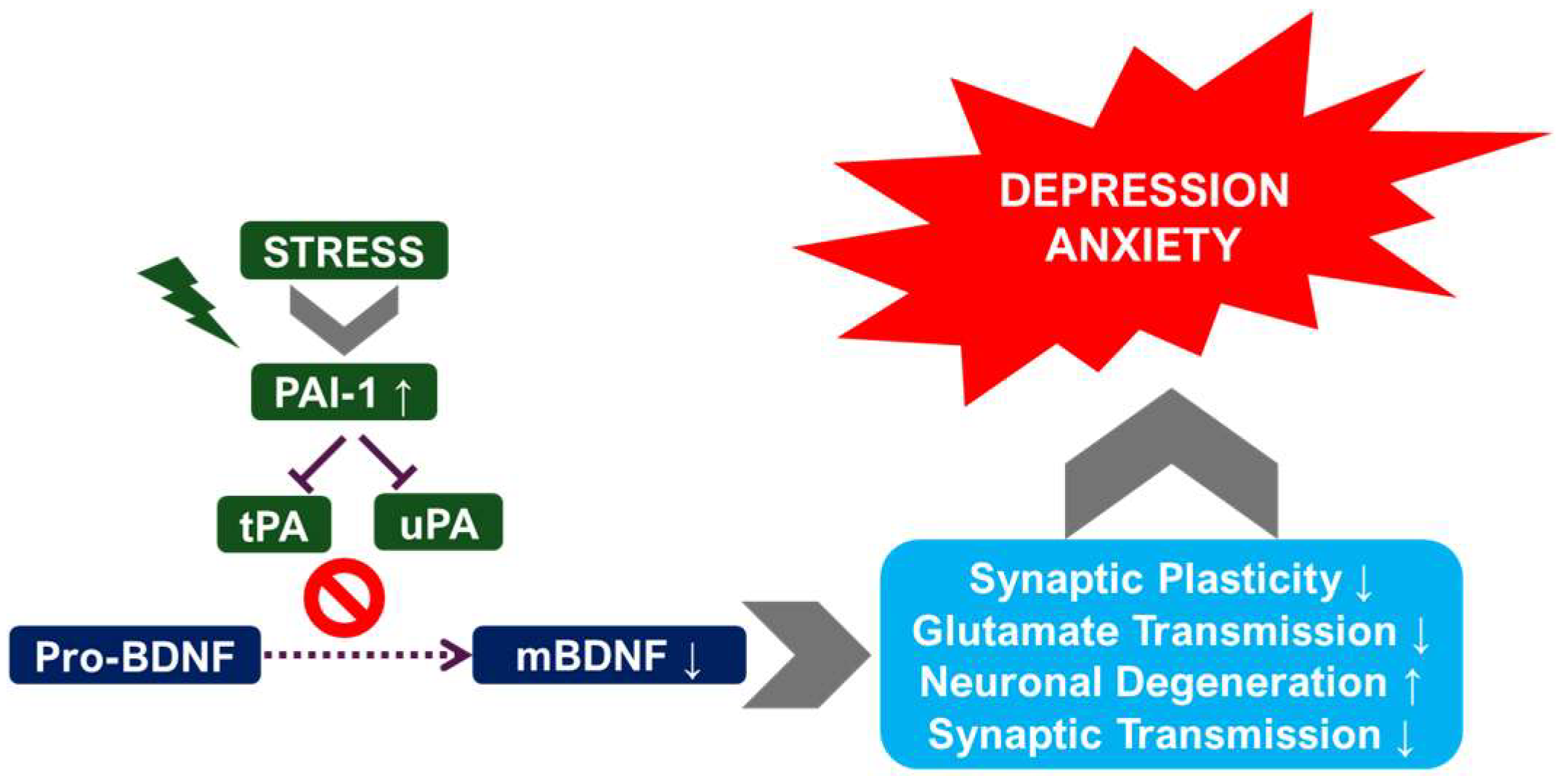
Depression is associated with reduced levels of BDNF in several brain regions, including the hippocampus [34,35,36]. BDNF plays a pivotal role in neuronal survival, growth, and synaptic plasticity [37,38,39]. Reduced BDNF levels contribute to the neuronal atrophy and impaired synaptic plasticity observed in depression. The PAS may interact with BDNF signaling pathways, potentially influencing BDNF expression and activity [10,40]. uPA and tPA may modulate BDNF levels either directly or indirectly through their effects on synaptic plasticity [41]. For instance, alterations in tPA activity could affect the processing of proBDNF to mature BDNF [41], impacting BDNF’s neurotrophic effects on hippocampal neurons [42]. The disruption of this intricate balance could contribute to the neurobiological changes underlying depression. The observed alterations in dendritic spine density and synaptic function in depression may also be influenced by uPA and tPA activity [42,43], further highlighting the complexity of their roles (Figure 3).
The interplay between tPA and BDNF represents a significant area of investigation in depression research [12,44]. BDNF, a key neurotrophin, plays an important role in neuronal survival, synaptic plasticity, and neurogenesis. It exists in two main forms, both of which are active: proBDNF (precursor) and BDNF. tPA plays an important role in the conversion of proBDNF to BDNF [12,22,44]. Preclinical studies have consistently demonstrated that alterations in the tPA/BDNF pathway are implicated in the pathogenesis of depression [44,45]. Specifically, reduced serum levels of tPA, impairing the conversion of proBDNF to BDNF, have been observed in depressed animals [44]. This imbalance can lead to impaired synaptic plasticity, reduced neurogenesis, and increased neuronal apoptosis, which are neuropathological features of depression. Further research is underway to fully understand the complex interplay between tPA, BDNF, and proBDNF in different brain regions and their contributions to the development and maintenance of depressive symptoms.

2.3.2. Clinical Studies: Investigating uPA and tPA in Human Depression

A number of clinical studies have investigated the relationship between serum levels and functional activity of PAS components and their association with major depressive disorder (MDD) in human populations [12,44]. These studies aim to evaluate whether alterations in these proteins could serve as reliable biomarkers or provide mechanistic insights into the pathogenesis of depression. While findings across studies vary, a recurring observation is the interplay between decreased protein levels and decreased enzymatic activity, which together may contribute to functional impairment of the tPA/BDNF axis. For instance, lower serum levels of tPA have been reported in depressed individuals [44], which may reflect a reduction in the enzymatic conversion of proBDNF to mature BDNF (BDNF), an essential step for supporting synaptic plasticity, neurogenesis, and neuronal survival.
Further supporting this pathway, several studies have shown a decreased BDNF/proBDNF ratio in patients with depression, suggesting impaired neurotrophin maturation and reduced neurotrophic signaling [12,44]. This biochemical imbalance mirrors preclinical findings and is increasingly recognized as a molecular hallmark of depressive pathology. In parallel, elevated levels of PAI-1, an endogenous inhibitor of both uPA and tPA, have been observed in patients with MDD, as shown in a recent systematic review and meta-analysis [46]. By binding to and inhibiting uPA and tPA, increased PAI-1 levels effectively suppress their catalytic activity, thereby disrupting downstream processes such as ECM remodeling, synaptic reorganization, and BDNF activation.
These overlapping mechanisms suggest that decreased PAS protein levels and decreased enzymatic activity represent two sides of the same coin, collectively leading to a dysfunctional neuroplasticity response in depression. However, despite these promising associations, the clinical literature remains inconclusive. Discrepancies among studies may arise from variability in sample size, patient heterogeneity (e.g., age, sex, medication status, comorbidities), depression subtypes (e.g., melancholic vs. atypical), and laboratory methods used to quantify protein levels and activity. Moreover, many of the available data are cross-sectional in nature, limiting the ability to infer causality or temporal dynamics [47,48,49].
To advance the field, future research should prioritize well-powered longitudinal studies that follow patients across different stages of illness and treatment [50,51,52]. Such designs will be critical to determine whether PAS component levels can predict depression onset, monitor disease progression, or serve as early indicators of treatment response. Additionally, stratifying patients by depression subtype or inflammatory profile may help clarify the role of the tPA/BDNF pathway in specific subpopulations. Ultimately, a deeper understanding of how PAS dysregulation affects neurobiological resilience may inform novel therapeutic strategies targeting the enzymatic machinery involved in neurotrophin activation.

2.3.3. Treatment Response and the Plasminogen System

The potential link between the PAS and antidepressant treatment response has also been a focus of clinical research [44,45]. Some studies suggest that changes in PAS component levels may be associated with treatment outcome, indicating a potential role for the PAS in mediating the therapeutic effects of antidepressants. For example, a study investigating the response to paroxetine treatment in Alzheimer’s disease patients with depressive symptoms found an association between a specific SERPINE1 polymorphism (rs1799889, which affects PAI-1 expression) and treatment response [45]. This suggests that genetic variations influencing PAI-1 levels may influence the effectiveness of antidepressant treatment. Studies examining the changes in PAS component levels before, during, and after antidepressant treatment may shed light on the temporal dynamics of these changes and their relationship to treatment efficacy. Furthermore, investigating whether specific antidepressants differentially affect the PAS could provide important insights into the mechanisms of action of the different classes of antidepressants.

2.4. The Role of Inflammation and the uPA/tPA System in Depression

2.4.1. Neuroinflammation and the Plasminogen System

The involvement of neuroinflammation in the pathogenesis of depression has gained considerable traction in recent years [12,53]. Neuroinflammation refers to the activation of immune cells within the CNS, leading to the release of inflammatory cytokines and other mediators that can damage neurons and disrupt brain function. The PAS is intricately linked to inflammatory processes, with uPA playing a significant role in mediating the migration and activation of immune cells [21,54]. In the context of depression, chronic low-grade inflammation within the brain may affect the expression and activity of uPA and tPA, contributing to the development and maintenance of depressive symptoms. For example, elevated levels of inflammatory cytokines can influence the expression of PAI-1, leading to an imbalance in the PAS and impaired ECM remodeling. This imbalance can disrupt synaptic plasticity and neuronal survival, contributing to the neuropathological changes observed in depression.
Moreover, systemic inflammation has been shown to disrupt the integrity of the blood–brain barrier (BBB), allowing for peripheral cytokines and immune mediators to enter the CNS and exacerbate neuroinflammatory responses. Disruption of the BBB can occur via multiple mechanisms, including tight junction breakdown, endothelial dysfunction, and cytokine-mediated permeability changes [55,56]. Inflammatory insults, such as LPS, TNF-α, and IL-6 can trigger these changes, heightening the sensitivity of the CNS to peripheral immune signals and amplifying PAS dysregulation [57,58]. This compromised barrier function enables greater trafficking of inflammatory cytokines, immune cells, and neurotoxic molecules, which may further suppress tPA and uPA activity via PAI-1 upregulation, impair BDNF maturation, and disrupt synaptic function. Ultimately, these changes contribute to a feed-forward loop of neuroinflammation, synaptic loss, and depressive pathology.
Further research is needed to elucidate the complex interplay between neuroinflammation, the PAS, and the development of depressive disorders. Understanding the precise molecular mechanisms involved may well lead to the development of novel anti-inflammatory therapies for depression.

2.4.2. Inflammatory Markers and Treatment Response

Clinical research is increasingly exploring the relationship between inflammatory markers and the response to antidepressant treatment [59]. Some studies suggest that individuals with elevated levels of inflammatory markers may exhibit a poorer response to antidepressant medication [59]. This indicates that inflammation may influence the effectiveness of antidepressant treatment, possibly by interfering with the mechanisms through which antidepressants exert their therapeutic effects. This suggests that targeting inflammation, potentially through indirect modulation of the PAS, could be a promising therapeutic strategy for improving treatment outcomes in depression. Further studies will explore potential strategies for combining anti-inflammatory therapies with antidepressants to enhance treatment efficacy. It is important to investigate whether specific inflammatory markers are more strongly associated with treatment response than others and to explore the potential interaction effects between inflammatory markers and genetic factors.

2.5. Therapeutic Implications and Future Directions

2.5.1. Targeting the Plasminogen System for Depression Treatment

The accumulating evidence from both preclinical and clinical studies suggests that manipulating the uPA and tPA system may offer therapeutic potential for depression [11]. However, significant challenges remain in translating these findings into effective clinical treatments. These would include identifying specific therapeutic targets within the PAS (e.g., uPA, tPA, PAI-1) and developing safe and effective interventions for depression that can selectively modulate their activity. This would involve developing drugs that specifically inhibit or enhance the activity of uPA, tPA, or PAI-1 or targeting the upstream signaling pathways that regulate their expression. Preclinical studies would test the efficacy and safety of these potential interventions in animal models of depression, followed by well-designed clinical trials to evaluate their effectiveness in human populations.

2.5.2. Biomarker Discovery and Diagnostic Applications

The discovery of reliable biomarkers for depression is a major factor in improving diagnosis, predicting treatment response, and identifying individuals at high risk of developing depression [12,44]. Studies examining serum levels of uPA, tPA, PAI-1, and BDNF have shown them to be promising potential biomarkers. Validating these biomarkers and determining their clinical utility are goals that are being enthusiastically pursued. This includes large-scale studies comparing biomarker levels in depressed individuals and healthy controls as well as longitudinal studies to assess the stability and predictive value of these biomarkers over time. The development of accurate and reliable diagnostic tools based on these biomarkers would significantly improve the early detection and management of depression. Naturally, this would lead to earlier intervention and improved treatment outcomes.

2.5.3. Limitations and Future Research Directions

The current literature on the role of uPA and tPA in depression presents several methodological challenges that require addressing in subsequent studies. These include inconsistencies in study designs (e.g., cross-sectional vs. longitudinal), variations in sample characteristics (age, sex, severity of depression, comorbid conditions), and differences in the methods used to measure uPA, tPA, PAI-1, and BDNF. Future studies should aim to standardize methods and use larger, more homogenous samples to improve the reliability and generalizability of the findings. This includes using well-validated assays for measuring PAS components and controlling for potential confounding factors, such as age, sex, and medication use. Careful consideration of pre-analytical variables, such as blood collection and processing techniques, is important in minimizing artifacts and ensuring accurate measurements.
While the evidence suggests a link between uPA/tPA dysregulation and depression, the precise mechanisms underlying this relationship are still being elucidated. Accordingly, this requires investigating the interactions between PAS, neuroinflammation, synaptic plasticity, and neurotransmitter systems in depression. Studies using advanced techniques, such as in vivo imaging and genetic manipulation, can help unravel the complex interplay between these factors. Investigating the role of specific signaling pathways downstream of uPA and tPA activation is also key in the understanding of their contribution to depression-related neuropathology.
Bridging the gap between preclinical and clinical findings is essential for translating research into effective clinical treatments for depression. Clearly, validating preclinical findings in human populations and investigating the clinical utility of modulating the plasminogen system for depression treatment are extremely important. This includes conducting well-designed clinical trials to test the efficacy and safety of potential interventions targeting the PAS. It is also important to investigate whether different clinical subtypes of depression respond differently to interventions targeting the PAS. Accordingly, this might involve stratifying patients based on clinical characteristics or biomarkers to identify subgroups that could benefit most from such treatments.

2.5.4. Conclusion: The Plasminogen System—A Promising Avenue for Depression Research

This comprehensive literature review has highlighted the accumulating evidence implicating the uPA and tPA system in the pathogenesis of depression. Preclinical studies using animal models have provided valuable insights into the alterations in the expression and activity of these proteins in response to stress and in association with depression-like behaviors. Clinical studies have offered some support for these findings but highlight the need for more research. Methodological limitations, including variations in study designs and measurement techniques, have contributed to the lack of definitive conclusions. Future research will benefit from standardizing methods, using larger sample sizes, and elucidating the precise mechanisms underlying the relationship between the PAS and depression. Targeting the plasminogen system through biomarker discovery and the development of novel therapeutic interventions represents a significant and promising avenue for future research in depression. The potential to improve diagnosis, predict treatment response, and develop novel therapies makes this area of research highly significant for improving the lives of individuals affected by depression.

3. Roles of uPA and tPA in Anxiety Disorders and PTSD

In this section, we examine the roles of uPA and tPA in anxiety disorders, including generalized anxiety disorder (GAD) and post-traumatic stress disorder (PTSD), based on both preclinical and clinical studies. Emerging evidence suggests the involvement of the PAS in a wider range of physiological and pathophysiological processes, including modulation of mood and cognitive functions [60]. We analyze the existing literature, comparing and contrasting findings, highlighting areas of agreement and disagreement, and identifying research gaps.

3.1. uPA’s Role in Anxiety and PTSD

3.1.1. Preclinical Studies

Preclinical research using animal models has yielded significant insights into uPA’s potential role in modulating anxiety and stress-related behaviors. We demonstrated that hippocampal uPA overexpression in rats mitigated stress-induced anxiety- and depression-like behaviors [60]. This effect was correlated with increased levels of BDNF in the hippocampus, suggesting a potential mechanism through which uPA exerts its anxiolytic effects. The study employed several behavioral tests, including the marble burying test, open field test, elevated plus maze test, sucrose splash test, tail suspension test, and forced swim test [60], providing a comprehensive assessment of anxiety- and depression-related behaviors. The strongly positive correlation between hippocampal uPA levels and BDNF, together with the negative correlation between uPA mRNA and anxiety/depression parameters, further supports uPA’s involvement in mood regulation [60]. However, the precise mechanisms underlying uPA’s action in the hippocampus and its interaction with BDNF continue to be elicited.
Other preclinical studies have investigated uPA’s role in different contexts. For instance, research on uPA in the context of intracerebral hemorrhage (ICH) [61] has shown that uPA can ameliorate oedema and improve outcomes in rat models of ICH [61]. While not directly addressing anxiety or PTSD, these findings highlight uPA’s broader impact on brain function and the potential for its therapeutic manipulation in various neurological conditions. The study [61] also noted the upregulation of BBB protein expression following uPA treatment, suggesting potential benefits in conditions where BBB integrity is compromised, a factor that could indirectly influence anxiety and stress responses. Further research is needed to explore the potential interplay between uPA’s effects on the BBB and its impact on anxiety- and stress-related behaviors.

3.1.2. Clinical Studies

Relatively few clinical studies have directly investigated uPA’s role in anxiety and PTSD. The paucity of clinical data in this area represents a significant research gap. The preclinical findings regarding uPA’s anxiolytic and antidepressant effects [60] warrant further investigations with human subjects. Clinical trials will assess the efficacy and safety of uPA-based therapies for anxiety and PTSD. Such trials should consider various factors, including dosage, administration route, and potential side effects, as well as patient characteristics like age, gender, and severity of the disorder. The correlation observed in animal models between uPA and BDNF [60] suggests a potential biomarker for treatment response in humans.

3.2. tPA’s Role in Anxiety and PTSD

3.2.1. Preclinical Studies

Preclinical research on tPA’s role in anxiety and PTSD is limited compared to the research on uPA. While tPA is primarily known for its role in fibrinolysis and its use in stroke treatment [62,63,64,65,66], some studies suggest it may have neuroprotective effects [67]. Yepes proposed that tPA, in addition to its proteolytic function, may interact with N-methyl-D-aspartate receptors (NMDARs) and activate the mammalian target of the rapamycin (mTOR) pathway, leading to increased glucose uptake in neurons and adaptation to metabolic stress [67]. This non-proteolytic function of tPA could have implications for neuronal survival and function in conditions involving stress and anxiety. However, the link between this neuroprotective mechanism and anxiety/PTSD is still being investigated.
Moreover, tPA’s role in the context of stroke, while not directly related to anxiety or PTSD, could indirectly influence these conditions. Studies showing that thrombus permeability influences the efficacy of tPA treatment in ischemic stroke [66] highlight the complexity of tPA’s effects on the brain. Indirectly, the impact of stroke on mood and cognitive function could contribute to anxiety and PTSD development, and this warrants further investigation. The use of tPA in stroke treatment also presents potential side effects, including hemorrhagic transformation [68], which could further complicate the assessment of tPA’s role in anxiety and stress-related conditions.

3.2.2. Clinical Studies

There are few clinical studies examining tPA’s direct role in anxiety and PTSD. The majority of clinical research on tPA focuses on its use as a thrombolytic agent in acute ischemic stroke [62,63,64,65,66,69,70,71,72,73]. Although some studies suggest sex-specific differences in tPA’s effectiveness in stroke [69,71], these findings are not directly applicable to anxiety or PTSD. The potential neuroprotective effects of tPA [67] in the context of stress and anxiety require further investigation through well-designed clinical trials.

3.3. Plasminogen Activator Inhibitor-1 (PAI-1) and Anxiety Disorders

PAI-1 is the primary physiological inhibitor of both tPA and uPA [74]. Although there is limited direct evidence linking PAI-1 to anxiety and PTSD, its involvement in inflammatory processes [74,75,76] and its influence on cellular behavior suggest a potential indirect role. While PAI-1 itself is not a proinflammatory molecule, inflammatory states are associated with elevated PAI-1 levels due to cytokine-mediated transcriptional activation. Specifically, the PAI-1 promoter is responsive to IL-6, TNF-α, and TGF-β, all of which are elevated in chronic inflammation [77,78]. Chronic inflammation is associated with various mental health disorders [74], and this cytokine-driven upregulation of PAI-1 may indirectly contribute to the development or worsening of anxiety and PTSD. Further research is needed to explore this potential link, including studies investigating PAI-1 levels in patients with anxiety disorders and PTSD. The complexity of PAI-1’s actions, including its interaction with vitronectin and the urokinase receptor (uPAR) [74], further complicates the understanding of its potential role in anxiety and PTSD.

3.4. Comparative Analysis of uPA and tPA

While both uPA and tPA belong to the PAS and are involved in fibrinolysis, their roles in anxiety and PTSD appear to differ. Preclinical studies have provided more evidence suggesting an anxiolytic role for uPA, particularly in the hippocampus [60], whereas the evidence for tPA’s direct involvement in anxiety and PTSD is less robust. tPA’s primarily known for its role in fibrinolysis and its clinical use in stroke treatment [62,63,64,65,66,69,70,71,72,73], while its potential neuroprotective effects [67] could indirectly impact anxiety and PTSD. The limited clinical data for both uPA and tPA in anxiety and PTSD highlight a significant research gap. Future studies could compare the effects of uPA and tPA on anxiety- and stress-related behaviors, both preclinically and clinically, to better understand their individual contributions and potential synergistic effects.

3.5. Limitations and Futur Directions

This section highlights significant gaps in our understanding of uPA’s and tPA’s roles in anxiety and PTSD. The majority of existing research focuses on preclinical studies using animal models, which may not fully translate to human conditions. Clinical studies are limited, particularly regarding uPA’s role. Furthermore, the complex interplay between the PAS, inflammation, and the neurobiological mechanisms underlying anxiety and PTSD is at an early investigative stage. Ideally, future research should prioritize the following:
  • Conducting well-designed clinical trials to investigate the efficacy and safety of uPA-based therapies for anxiety and PTSD.
  • Exploring the potential neuroprotective effects of tPA in the context of stress and anxiety, considering both its proteolytic and non-proteolytic functions.
  • Investigating the role of PAI-1 in anxiety and PTSD, focusing on its potential contribution to inflammation and its interaction with uPA and tPA.
  • Developing more sophisticated animal models that better reflect the complexity of human anxiety disorders and PTSD.
  • Employing multi-modal approaches that integrate behavioral, neuroimaging, and molecular data to understand the mechanisms underlying the effects of uPA and tPA on anxiety and stress-related behaviors.

3.6. Conclusion

While the existing literature suggests a potential role for uPA and tPA in anxiety and PTSD, primarily based on preclinical findings, significant research gaps remain. Further investigation is crucial to translate the promising preclinical data into effective clinical therapies. A comprehensive understanding of the complex interactions within the PAS and its influence on the neurobiological underpinnings of anxiety and PTSD is essential for the development of novel and targeted treatments for these debilitating disorders. Future research should prioritize clinical trials and multi-modal approaches to address these research gaps.

4. Summary of Key Findings

This review demonstrates the significant involvement of uPA and tPA in the pathogenesis of several mood disorders, including anxiety and depression (Table 1). While the precise mechanisms vary across disorders, consistent themes emerge, including alterations in BDNF levels, impaired synaptic plasticity, and contributions to neuroinflammation. The evidence suggests that uPA and tPA are not merely bystanders in these conditions but rather active participants, modulating key neurobiological processes. The findings highlight the complexity of these systems and the interconnectedness of various biological pathways in these conditions. However, it is important to note that the promising research in this area that aims to fully elucidate the precise roles of uPA and tPA in each disorder is still evolving.

5. General Conclusion

The findings discussed in this review have significant therapeutic implications. Modulation of uPA and tPA activity or expression could represent a novel therapeutic target for mood disorders. The development of such therapeutic strategies presents several challenges. The complexity of the uPA/tPA system, its involvement in multiple physiological processes, and its varied roles across different disorders necessitate a cautious and nuanced approach. Further research is crucial to delineate the precise mechanisms by which uPA and tPA contribute to each disorder, identify specific biomarkers of dysregulation, and develop targeted therapeutic interventions with minimal side effects. Future studies should focus on understanding the interactions between uPA/tPA and other signaling pathways, such as BDNF and inflammatory cascades, to develop more effective treatments for mood disorders. The development of specific inhibitors or activators of uPA and tPA, tailored to specific disorders and pathways, is a promising avenue for future research.

Author Contributions

A.B.: Conceptualization, literature search, data analysis, writing original draft, and visualization (section organization). S.S.: Writing, review & editing, critical discussion, supervision, and final manuscript revision for clarity and coherence. All authors have read and agreed to the published version of the manuscript.

Funding

This research received no external funding.

Conflicts of Interest

The authors declare no conflicts of interest.

Abbreviations

The following abbreviations are used in this manuscript:
ALIAcute Lung Injury
BBBBlood–Brain Barrier
BDNFBrain-Derived Neurotrophic Factor
CKDChronic Kidney Disease
CNSCentral Nervous System
CRSChronic Restraint Stress
CUMSChronic Unpredictable Mild Stress
ECMExtracellular Matrix
FDPFibrin Degradation Products
GADGeneralized Anxiety Disorder
ICHIntracerebral Hemorrhage
LTDLong-Term Depression
LTPLong-Term Potentiation
mBDNFMature Brain-Derived Neurotrophic Factor
MDDMajor Depressive Disorder
mTORMammalian Target of Rapamycin
NMDARN-Methyl-D-Aspartate Receptor
PAI-1Plasminogen Activator Inhibitor-1
PASPlasminogen Activator System
proBDNFPrecursor Brain-Derived Neurotrophic Factor
PTSDPost-Traumatic Stress Disorder
SDSSocial Defeat Stress
SERPINE1Serpin Family E Member 1 (gene encoding PAI-1)
TBITraumatic Brain Injury
tPATissue-Type Plasminogen Activator
uPAUrokinase-Type Plasminogen Activator

References

  1. Bahi, A.; Dreyer, J.L. Overexpression of plasminogen activators in the nucleus accumbens enhances cocaine-, amphetamine- and morphine-induced reward and behavioral sensitization. Genes Brain Behav. 2008, 7, 244–256. [Google Scholar] [CrossRef] [PubMed]
  2. Hedhli, N.; Falcone, D.J.; Huang, B.; Cesarman-Maus, G.; Kraemer, R.; Zhai, H.; Tsirka, S.E.; Santambrogio, L.; Hajjar, K.A. The annexin A2/S100A10 system in health and disease: Emerging paradigms. J. Biomed. Biotechnol. 2012, 2012, 406273. [Google Scholar] [CrossRef] [PubMed]
  3. Jänicke, F.; Schmitt, M.; Graeff, H. Clinical relevance of the urokinase-type and tissue-type plasminogen activators and of their type 1 inhibitor in breast cancer. Semin. Thromb. Hemost. 1991, 17, 303–312. [Google Scholar] [CrossRef] [PubMed]
  4. Andrade-Gordon, P.; Strickland, S. Interaction of heparin with plasminogen activators and plasminogen: Effects on the activation of plasminogen. Biochemistry 1986, 25, 4033–4040. [Google Scholar] [CrossRef]
  5. Brodsky, S.; Chen, J.; Lee, A.; Akassoglou, K.; Norman, J.; Goligorsky, M.S. Plasmin-dependent and -independent effects of plasminogen activators and inhibitor-1 on ex vivo angiogenesis. Am. J. Physiol. Heart Circ. Physiol. 2001, 281, H1784–H1792. [Google Scholar] [CrossRef]
  6. España, F.; Estellés, A.; Fernández, P.J.; Gilabert, J.; Sánchez-Cuenca, J.; Griffin, J.H. Evidence for the regulation of urokinase and tissue type plasminogen activators by the serpin, protein C inhibitor, in semen and blood plasma. Thromb. Haemost. 1993, 70, 989–994. [Google Scholar] [CrossRef]
  7. Higazi, A.A.; El-Haj, M.; Melhem, A.; Horani, A.; Pappo, O.; Alvarez, C.E.; Muhanna, N.; Friedman, S.L.; Safadi, R. Immunomodulatory effects of plasminogen activators on hepatic fibrogenesis. Clin. Exp. Immunol. 2008, 152, 163–173. [Google Scholar] [CrossRef]
  8. Ismail, A.A.; Shaker, B.T.; Bajou, K. The Plasminogen-Activator Plasmin System in Physiological and Pathophysiological Angiogenesis. Int. J. Mol. Sci. 2021, 23, 337. [Google Scholar] [CrossRef]
  9. Feng, Q.; Liu, K.; Liu, Y.X.; Byrne, S.; Ockleford, C.D. Plasminogen activators and inhibitors are transcribed during early macaque implantation. Placenta 2001, 22, 186–199. [Google Scholar] [CrossRef]
  10. Hsu, C.D.; Tsai, S.J. The tissue plasminogen activator/plasmin system may act through cleavage of pro-BDNF to increase risk of substance abuse. CNS Spectr. 2010, 15, 350. [Google Scholar] [CrossRef]
  11. Mennesson, M.; Revest, J.M. Glucocorticoid-Responsive Tissue Plasminogen Activator (tPA) and Its Inhibitor Plasminogen Activator Inhibitor-1 (PAI-1): Relevance in Stress-Related Psychiatric Disorders. Int. J. Mol. Sci. 2023, 24, 4496. [Google Scholar] [CrossRef]
  12. Li, H.; Zhao, M.; Jiang, C.; Zhao, H.; Wu, C.; Li, Y.; Zhang, S.; Xu, P.; Mou, T.; Xu, Y.; et al. Elevated Plasma Levels of Mature Brain-Derived Neurotrophic Factor in Major Depressive Disorder Patients with Higher Suicidal Ideation. Brain Sci. 2023, 13, 1223. [Google Scholar] [CrossRef]
  13. Melchor, J.P.; Strickland, S. Tissue plasminogen activator in central nervous system physiology and pathology. Thromb. Haemost. 2005, 93, 655–660. [Google Scholar] [CrossRef]
  14. Qiao, H.; Li, M.X.; Xu, C.; Chen, H.B.; An, S.C.; Ma, X.M. Dendritic Spines in Depression: What We Learned from Animal Models. Neural Plast. 2016, 2016, 8056370. [Google Scholar] [CrossRef]
  15. Zhu, Y.; Chao, C.; Duan, X.; Cheng, X.; Liu, P.; Su, S.; Duan, J.; Dong, T.T.; Tsim, K.W. Kai-Xin-San series formulae alleviate depressive-like behaviors on chronic mild stressed mice via regulating neurotrophic factor system on hippocampus. Sci. Rep. 2017, 7, 1467. [Google Scholar] [CrossRef]
  16. Bansal, Y.; Codeluppi, S.A.; Banasr, M. Astroglial Dysfunctions in Mood Disorders and Rodent Stress Models: Consequences on Behavior and Potential as Treatment Target. Int. J. Mol. Sci. 2024, 25, 6357. [Google Scholar] [CrossRef] [PubMed]
  17. Gyles, T.M.; Nestler, E.J.; Parise, E.M. Advancing preclinical chronic stress models to promote therapeutic discovery for human stress disorders. Neuropsychopharmacol. Off. Publ. Am. Coll. Neuropsychopharmacol. 2024, 49, 215–226. [Google Scholar] [CrossRef] [PubMed]
  18. Lopez, J.; Bagot, R.C. Defining Valid Chronic Stress Models for Depression With Female Rodents. Biol. Psychiatry 2021, 90, 226–235. [Google Scholar] [CrossRef]
  19. Schöner, J.; Heinz, A.; Endres, M.; Gertz, K.; Kronenberg, G. Post-traumatic stress disorder and beyond: An overview of rodent stress models. J. Cell. Mol. Med. 2017, 21, 2248–2256. [Google Scholar] [CrossRef]
  20. Tran, I.; Gellner, A.K. Long-term effects of chronic stress models in adult mice. J. Neural Transm. 2023, 130, 1133–1151. [Google Scholar] [CrossRef]
  21. Cunningham, O.; Campion, S.; Perry, V.H.; Murray, C.; Sidenius, N.; Docagne, F.; Cunningham, C. Microglia and the urokinase plasminogen activator receptor/uPA system in innate brain inflammation. Glia 2009, 57, 1802–1814. [Google Scholar] [CrossRef] [PubMed]
  22. Hoirisch-Clapauch, S. Mechanisms affecting brain remodeling in depression: Do all roads lead to impaired fibrinolysis? Mol. Psychiatry 2022, 27, 525–533. [Google Scholar] [CrossRef] [PubMed]
  23. Lang, U.E.; Borgwardt, S. Molecular mechanisms of depression: Perspectives on new treatment strategies. Cell. Physiol. Biochem. Int. J. Exp. Cell. Physiol. Biochem. Pharmacol. 2013, 31, 761–777. [Google Scholar] [CrossRef] [PubMed]
  24. Leuner, B.; Shors, T.J. Stress, anxiety, and dendritic spines: What are the connections? Neuroscience 2013, 251, 108–119. [Google Scholar] [CrossRef]
  25. Métivier, L.; Vivien, D.; Goy, R.; Agin, V.; Bui, E.; Benbrika, S. Plasminogen Activator Inhibitor-1 in the Pathophysiology of Late Life Depression. Int. J. Geriatr. Psychiatry 2024, 39, e70015. [Google Scholar] [CrossRef]
  26. Clemente, J.C.; Manasson, J.; Scher, J.U. The role of the gut microbiome in systemic inflammatory disease. BMJ (Clin. Res. Ed.) 2018, 360, j5145. [Google Scholar] [CrossRef]
  27. Coroneos, C.J.; Selber, J.C.; Offodile, A.C., 2nd; Butler, C.E.; Clemens, M.W. US FDA Breast Implant Postapproval Studies: Long-term Outcomes in 99,993 Patients. Ann. Surg. 2019, 269, 30–36. [Google Scholar] [CrossRef]
  28. Du, J.; Lv, H.; Dou, X.; Cao, Z. Nuclear Factor κB/MicroRNA-155 Upregulates the Expression Pattern of Cytokines in Regulating the Relapse of Chronic Sinusitis with Nasal Polyps and the Underlying Mechanism of Glucocorticoid. Med. Sci. Monit. Int. Med. J. Exp. Clin. Res. 2020, 26, e923618. [Google Scholar] [CrossRef]
  29. Hotamisligil, G.S. Inflammation, metaflammation and immunometabolic disorders. Nature 2017, 542, 177–185. [Google Scholar] [CrossRef]
  30. Longino, E.S.; Labby, A.B.; Wu, J.; Chapurin, N.; Li, P.; Chandra, R.K.; Turner, J.H.; Chowdhury, N.I. Association of cytokine profile with prior treatment failure and revision surgery in chronic rhinosinusitis. Int. Forum Allergy Rhinol. 2023, 13, 5–14. [Google Scholar] [CrossRef]
  31. Rohleder, N. Stress and inflammation—The need to address the gap in the transition between acute and chronic stress effects. Psychoneuroendocrinology 2019, 105, 164–171. [Google Scholar] [CrossRef]
  32. Kang, S.; Tanaka, T.; Inoue, H.; Ono, C.; Hashimoto, S.; Kioi, Y.; Matsumoto, H.; Matsuura, H.; Matsubara, T.; Shimizu, K.; et al. IL-6 trans-signaling induces plasminogen activator inhibitor-1 from vascular endothelial cells in cytokine release syndrome. Proc. Natl. Acad. Sci. USA 2020, 117, 22351–22356. [Google Scholar] [CrossRef]
  33. Sawdey, M.S.; Loskutoff, D.J. Regulation of murine type 1 plasminogen activator inhibitor gene expression in vivo. Tissue specificity and induction by lipopolysaccharide, tumor necrosis factor-alpha, and transforming growth factor-beta. J. Clin. Investig. 1991, 88, 1346–1353. [Google Scholar] [CrossRef]
  34. Colucci-D’Amato, L.; Speranza, L.; Volpicelli, F. Neurotrophic Factor BDNF, Physiological Functions and Therapeutic Potential in Depression, Neurodegeneration and Brain Cancer. Int. J. Mol. Sci. 2020, 21, 7777. [Google Scholar] [CrossRef] [PubMed]
  35. Esalatmanesh, S.; Kashani, L.; Akhondzadeh, S. Effects of Antidepressant Medication on Brain-derived Neurotrophic Factor Concentration and Neuroplasticity in Depression: A Review of Preclinical and Clinical Studies. Avicenna J. Med. Biotechnol. 2023, 15, 129–138. [Google Scholar] [CrossRef] [PubMed]
  36. Xu, W.; Yao, X.; Zhao, F.; Zhao, H.; Cheng, Z.; Yang, W.; Cui, R.; Xu, S.; Li, B. Changes in Hippocampal Plasticity in Depression and Therapeutic Approaches Influencing These Changes. Neural Plast. 2020, 2020, 8861903. [Google Scholar] [CrossRef] [PubMed]
  37. Bawari, S.; Tewari, D.; Argüelles, S.; Sah, A.N.; Nabavi, S.F.; Xu, S.; Vacca, R.A.; Nabavi, S.M.; Shirooie, S. Targeting BDNF signaling by natural products: Novel synaptic repair therapeutics for neurodegeneration and behavior disorders. Pharmacol. Res. 2019, 148, 104458. [Google Scholar] [CrossRef]
  38. Kojima, M.; Mizui, T. BDNF Propeptide: A Novel Modulator of Synaptic Plasticity. Vitam. Horm. 2017, 104, 19–28. [Google Scholar]
  39. Numakawa, T.; Kajihara, R. Neurotrophins and Other Growth Factors in the Pathogenesis of Alzheimer’s Disease. Life 2023, 13, 647. [Google Scholar] [CrossRef]
  40. Chen, S.; Jiang, H.; Liu, Y.; Hou, Z.; Yue, Y.; Zhang, Y.; Zhao, F.; Xu, Z.; Li, Y.; Mou, X.; et al. Combined serum levels of multiple proteins in tPA-BDNF pathway may aid the diagnosis of five mental disorders. Sci. Rep. 2017, 7, 6871. [Google Scholar] [CrossRef]
  41. Segawa, M.; Morinobu, S.; Matsumoto, T.; Fuchikami, M.; Yamawaki, S. Electroconvulsive seizure, but not imipramine, rapidly up-regulates pro-BDNF and t-PA, leading to mature BDNF production, in the rat hippocampus. Int. J. Neuropsychopharmacol. 2013, 16, 339–350. [Google Scholar] [CrossRef]
  42. Yeh, C.M.; Huang, C.C.; Hsu, K.S. Prenatal stress alters hippocampal synaptic plasticity in young rat offspring through preventing the proteolytic conversion of pro-brain-derived neurotrophic factor (BDNF) to mature BDNF. J. Physiol. 2012, 590, 991–1010. [Google Scholar] [CrossRef]
  43. Pawlak, R.; Rao, B.S.; Melchor, J.P.; Chattarji, S.; McEwen, B.; Strickland, S. Tissue plasminogen activator and plasminogen mediate stress-induced decline of neuronal and cognitive functions in the mouse hippocampus. Proc. Natl. Acad. Sci. USA 2005, 102, 18201–18206. [Google Scholar] [CrossRef]
  44. Jiang, H.; Chen, S.; Li, C.; Lu, N.; Yue, Y.; Yin, Y.; Zhang, Y.; Zhi, X.; Zhang, D.; Yuan, Y. The serum protein levels of the tPA-BDNF pathway are implicated in depression and antidepressant treatment. Transl. Psychiatry 2017, 7, e1079. [Google Scholar] [CrossRef] [PubMed]
  45. Fang, Y.; Zhang, L.; Zeng, Z.; Lian, Y.; Jia, Y.; Zhu, H.; Xu, Y. Promoter polymorphisms of SERPINE1 are associated with the antidepressant response to depression in Alzheimer’s disease. Neurosci. Lett. 2012, 516, 217–220. [Google Scholar] [CrossRef] [PubMed]
  46. Elsayed, M.; Mohamed, K.A.; Dardeer, K.T.; Zaafar, D.K.; Hassanin, S.O.; Abdelnaby, R.; Schönfeldt-Lecuona, C. Serum plasminogen activator inhibitor-1 levels in patients with major depressive disorder vs. healthy controls: A systematic review and meta-analysis. Trends Psychiatry Psychother. 2023, 45, e20230338. [Google Scholar] [CrossRef] [PubMed]
  47. Kesmodel, U.S. Cross-sectional studies—What are they good for? Acta Obstet. Gynecol. Scand. 2018, 97, 388–393. [Google Scholar] [CrossRef]
  48. Levin, K.A. Study design III: Cross-sectional studies. Evid.-Based Dent. 2006, 7, 24–25. [Google Scholar] [CrossRef]
  49. Mann, C.J. Observational research methods. Research design II: Cohort, cross sectional, and case-control studies. Emerg. Med. J. 2003, 20, 54–60. [Google Scholar] [CrossRef]
  50. Glymour, M.M.; Manly, J.J. Lifecourse social conditions and racial and ethnic patterns of cognitive aging. Neuropsychol. Rev. 2008, 18, 223–254. [Google Scholar] [CrossRef]
  51. Lenhart, O. The effects of health shocks on labor market outcomes: Evidence from UK panel data. Eur. J. Health Econ. HEPAC Health Econ. Prev. Care 2019, 20, 83–98. [Google Scholar] [CrossRef]
  52. Little, R.J.; D’Agostino, R.; Cohen, M.L.; Dickersin, K.; Emerson, S.S.; Farrar, J.T.; Frangakis, C.; Hogan, J.W.; Molenberghs, G.; Murphy, S.A.; et al. The prevention and treatment of missing data in clinical trials. N. Engl. J. Med. 2012, 367, 1355–1360. [Google Scholar] [CrossRef]
  53. Ulbrich, P.; Khoshneviszadeh, M.; Jandke, S.; Schreiber, S.; Dityatev, A. Interplay between perivascular and perineuronal extracellular matrix remodelling in neurological and psychiatric diseases. Eur. J. Neurosci. 2021, 53, 3811–3830. [Google Scholar] [CrossRef]
  54. Li, C.; Ma, Y.; Fei, F.; Zheng, M.; Li, Z.; Zhao, Q.; Du, J.; Liu, K.; Lu, R.; Zhang, S. Critical role and its underlying molecular events of the plasminogen receptor, S100A10 in malignant tumor and non-tumor diseases. J. Cancer 2020, 11, 826–836. [Google Scholar] [CrossRef]
  55. Banks, W.A.; Erickson, M.A. The blood-brain barrier and immune function and dysfunction. Neurobiol. Dis. 2010, 37, 26–32. [Google Scholar] [CrossRef] [PubMed]
  56. Varatharaj, A.; Galea, I. The blood-brain barrier in systemic inflammation. Brain Behav. Immun. 2017, 60, 1–12. [Google Scholar] [CrossRef] [PubMed]
  57. Erickson, M.A.; Banks, W.A. Neuroimmune Axes of the Blood-Brain Barriers and Blood-Brain Interfaces: Bases for Physiological Regulation, Disease States, and Pharmacological Interventions. Pharmacol. Rev. 2018, 70, 278–314. [Google Scholar] [CrossRef] [PubMed]
  58. Zlokovic, B.V. The blood-brain barrier in health and chronic neurodegenerative disorders. Neuron 2008, 57, 178–201. [Google Scholar] [CrossRef]
  59. Myung, W.; Lim, S.W.; Woo, H.I.; Park, J.H.; Shim, S.; Lee, S.Y.; Kim, D.K. Serum Cytokine Levels in Major Depressive Disorder and Its Role in Antidepressant Response. Psychiatry Investig. 2016, 13, 644–651. [Google Scholar] [CrossRef]
  60. Bahi, A.; Dreyer, J.L. Hippocampal Viral-Mediated Urokinase Plasminogen Activator (uPA) Overexpression Mitigates Stress-Induced Anxiety and Depression in Rats by Increasing Brain-Derived Neurotrophic Factor (BDNF) Levels. Biomolecules 2024, 14, 1603. [Google Scholar] [CrossRef]
  61. Tan, Q.; Chen, Q.; Niu, Y.; Feng, Z.; Li, L.; Tao, Y.; Tang, J.; Yang, L.; Guo, J.; Feng, H.; et al. Urokinase, a promising candidate for fibrinolytic therapy for intracerebral hemorrhage. J. Neurosurg. 2017, 126, 548–557. [Google Scholar] [CrossRef]
  62. Alexandrov, A.V. Ultrasound enhancement of fibrinolysis. Stroke 2009, 40 (Suppl. S3), S107–S110. [Google Scholar] [CrossRef]
  63. Bor-Seng-Shu, E.; Nogueira Rde, C.; Figueiredo, E.G.; Evaristo, E.F.; Conforto, A.B.; Teixeira, M.J. Sonothrombolysis for acute ischemic stroke: A systematic review of randomized controlled trials. Neurosurg. Focus 2012, 32, E5. [Google Scholar] [CrossRef]
  64. Daffertshofer, M.; Gass, A.; Ringleb, P.; Sitzer, M.; Sliwka, U.; Els, T.; Sedlaczek, O.; Koroshetz, W.J.; Hennerici, M.G. Transcranial low-frequency ultrasound-mediated thrombolysis in brain ischemia: Increased risk of hemorrhage with combined ultrasound and tissue plasminogen activator: Results of a phase II clinical trial. Stroke 2005, 36, 1441–1446. [Google Scholar] [CrossRef]
  65. Panagiotou, S.; Saha, S. Therapeutic benefits of nanoparticles in stroke. Front. Neurosci. 2015, 9, 182. [Google Scholar] [CrossRef]
  66. Santos, E.M.; Dankbaar, J.W.; Treurniet, K.M.; Horsch, A.D.; Roos, Y.B.; Kappelle, L.J.; Niessen, W.J.; Majoie, C.B.; Velthuis, B.; Marquering, H.A. Permeable Thrombi Are Associated With Higher Intravenous Recombinant Tissue-Type Plasminogen Activator Treatment Success in Patients With Acute Ischemic Stroke. Stroke 2016, 47, 2058–2065. [Google Scholar] [CrossRef]
  67. Yepes, M. Tissue-type plasminogen activator is a neuroprotectant in the central nervous system. Front. Cell. Neurosci. 2015, 9, 304. [Google Scholar] [CrossRef]
  68. García-Yébenes, I.; García-Culebras, A.; Peña-Martínez, C.; Fernández-López, D.; Díaz-Guzmán, J.; Negredo, P.; Avendaño, C.; Castellanos, M.; Gasull, T.; Dávalos, A.; et al. Iron Overload Exacerbates the Risk of Hemorrhagic Transformation After tPA (Tissue-Type Plasminogen Activator) Administration in Thromboembolic Stroke Mice. Stroke 2018, 49, 2163–2172. [Google Scholar] [CrossRef]
  69. Chauhan, A.; Moser, H.; McCullough, L.D. Sex differences in ischaemic stroke: Potential cellular mechanisms. Clin. Sci. 2017, 131, 533–552. [Google Scholar] [CrossRef]
  70. Marei, H.E.; Hasan, A.; Rizzi, R.; Althani, A.; Afifi, N.; Cenciarelli, C.; Caceci, T.; Shuaib, A. Potential of Stem Cell-Based Therapy for Ischemic Stroke. Front. Neurol. 2018, 9, 34. [Google Scholar] [CrossRef]
  71. Sohrabji, F.; Park, M.J.; Mahnke, A.H. Sex differences in stroke therapies. J. Neurosci. Res. 2017, 95, 681–691. [Google Scholar] [CrossRef] [PubMed]
  72. Wichaiyo, S.; Parichatikanond, W.; Rattanavipanon, W. Glenzocimab: A GPVI (Glycoprotein VI)-Targeted Potential Antiplatelet Agent for the Treatment of Acute Ischemic Stroke. Stroke 2022, 53, 3506–3513. [Google Scholar] [CrossRef] [PubMed]
  73. Young, A.R.; Ali, C.; Duretête, A.; Vivien, D. Neuroprotection and stroke: Time for a compromise. J. Neurochem. 2007, 103, 1302–1309. [Google Scholar] [CrossRef] [PubMed]
  74. Eddy, A.A.; Fogo, A.B. Plasminogen activator inhibitor-1 in chronic kidney disease: Evidence and mechanisms of action. J. Am. Soc. Nephrol. 2006, 17, 2999–3012. [Google Scholar] [CrossRef]
  75. Liu, C.; Ma, Y.; Su, Z.; Zhao, R.; Zhao, X.; Nie, H.G.; Xu, P.; Zhu, L.; Zhang, M.; Li, X.; et al. Meta-Analysis of Preclinical Studies of Fibrinolytic Therapy for Acute Lung Injury. Front. Immunol. 2018, 9, 1898. [Google Scholar] [CrossRef]
  76. Mukhopadhyay, S.; Johnson, T.A.; Duru, N.; Buzza, M.S.; Pawar, N.R.; Sarkar, R.; Antalis, T.M. Fibrinolysis and Inflammation in Venous Thrombus Resolution. Front. Immunol. 2019, 10, 1348. [Google Scholar] [CrossRef]
  77. Kusuyama, J.; Bandow, K.; Ohnishi, T.; Amir, M.S.; Shima, K.; Semba, I.; Matsuguchi, T. CXCL13 is a differentiation- and hypoxia-induced adipocytokine that exacerbates the inflammatory phenotype of adipocytes through PHLPP1 induction. Biochem. J. 2019, 476, 3533–3548. [Google Scholar] [CrossRef]
  78. Su, H.; Lin, X.; Paredong, A.; Yao, C.; Zhang, Y.; Geng, M.; Guan, Y.; Gong, L.; Jiang, F.; Lv, Q.; et al. IL-6/GATA2/SERPINE1 pathway is implicated in regulating cellular senescence after acute kidney injury. Front. Mol. Biosci. 2025, 12, 1538526. [Google Scholar] [CrossRef]
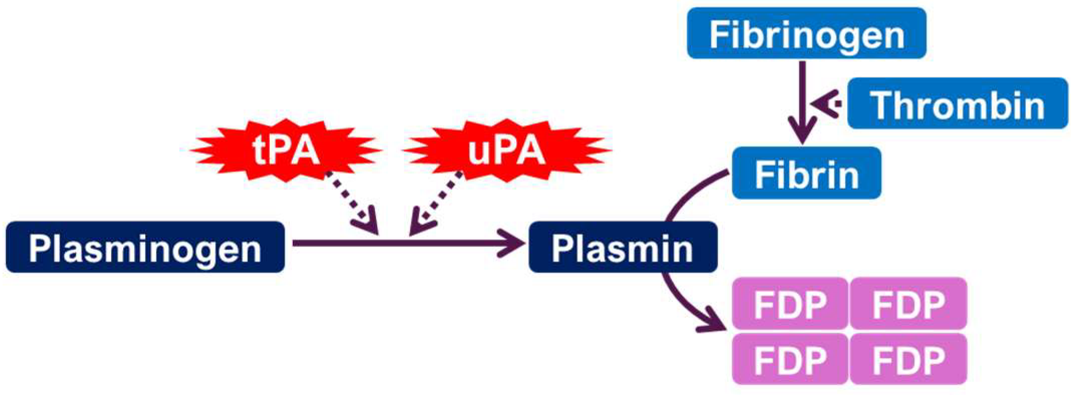
Figure 1. The plasminogen activating system. tPA: tissue-type plasminogen activator. uPA: urokinase-type plasminogen activator. FDP: fibrin degradation products.
Ijms 26 06899 g001
Figure 2. The role of the plasminogen activating system and BDNF maturation in neuronal survival, neuroplasticity, and synaptogenesis.
Ijms 26 06899 g002
Figure 3. Alteration of functional BDNF and depression. ↑ increase; ↓ decrease.
Ijms 26 06899 g003
Table 1. Summary of preclinical and clinical studies linking tPA, uPA, and PAI-1 to mood disorders, with associated brain regions, effects, and behavioral or clinical outcomes. ↑ increase; ↓ decrease.
ComponentStudy TypeSpecies/SubjectMood Disorder TypeEffect DirectionBrain Region (If Known)Behavioral/Clinical MarkerRef
tPAPreclinicalMouseDepression↓ tPA linked to ↓ BDNF, impaired synaptic plasticityHippocampusBDNF levels, behavior tests[44]
tPAPreclinicalRatDepressionImpaired proBDNF to BDNF conversionHippocampusBDNF processing[41]
tPAClinicalHumanDepression↓ tPA levels in MDD patientsSerumELISA, clinical scales[12]
tPAClinicalHumanDepression↓ BDNF/proBDNF ratio correlates with ↓ tPASerumBDNF ratio, clinical diagnosis[44]
tPAPreclinicalRatStroke-linked anxietyPossible neuroprotection via mTOR and NMDARsGlobal brainGlucose uptake, neuroprotection[67]
uPAPreclinicalMouseDepressionReduced uPA & blunted stress responseAmygdala, hippocampusBehavioral tests[21]
uPAPreclinicalRatAnxiety/DepressionuPA overexpression with ↓ anxiety and ↑ BDNFHippocampusBDNF levels, behavioral tests[60]
uPAClinicalHumanDepressionNot directly studied but inferred roleSerumNA[12]
PAI-1PreclinicalMouseDepression↑ PAI-1 with ↓ tPA/uPA activityHippocampusPlasmin activity, behavioral outcomes[21]
PAI-1ClinicalHumanDepression↑ PAI-1 in MDD patientsSerumMeta-analysis data[46]
PAI-1ClinicalHumanDepression (AD + depression)SERPINE1 polymorphism affects response to SSRIsGenetic/SerumGenotyping, clinical outcomes[45]
PAI-1ClinicalHumanAnxiety/PTSD↑ PAI-1 via cytokines IL-6, TNF-α, TGF-βEndothelium/CNSCytokine assays, inferred role[78]
Disclaimer/Publisher’s Note: The statements, opinions and data contained in all publications are solely those of the individual author(s) and contributor(s) and not of MDPI and/or the editor(s). MDPI and/or the editor(s) disclaim responsibility for any injury to people or property resulting from any ideas, methods, instructions or products referred to in the content.