Review

Innovative Peptide Therapeutics in the Pipeline: Transforming Cancer Detection and Treatment

by
Yanyamba Nsereko
1,
Amy Armstrong
1,
Fleur Coburn
1 and
Othman Al Musaimi
1,2,3,*
1
School of Pharmacy, Newcastle University, Newcastle upon Tyne NE1 7RU, UK
2
Department of Chemical Engineering, Imperial College London, London SW7 2AZ, UK
3
Orthogonal Peptides Limited, London SW7 2AZ, UK
*
Author to whom correspondence should be addressed.
Int. J. Mol. Sci. 2025, 26(14), 6815; https://doi.org/10.3390/ijms26146815
Submission received: 10 June 2025 / Revised: 5 July 2025 / Accepted: 11 July 2025 / Published: 16 July 2025
(This article belongs to the Special Issue Peptides as Biochemical Tools and Modulators of Biological Activity)

Abstract

Cancer remains a leading global health burden, profoundly affecting patient survival and quality of life. Current treatments—including chemotherapy, radiotherapy, immunotherapy, and surgery—are often limited by toxicity or insufficient specificity. Conventional chemotherapy, for instance, indiscriminately attacks rapidly dividing cells, causing severe side effects. In contrast, peptide-based therapeutics offer a paradigm shift, combining high tumour-targeting precision with minimal off-target effects. Their low immunogenicity, multi-pathway modulation capabilities, and adaptability for diagnostics and therapy make them ideal candidates for advancing oncology care. Innovative peptide platforms now enable three transformative applications: (1) precision molecular diagnostics (e.g., 18F-PSMA-1007 for prostate cancer detection), (2) targeted therapies (e.g., BT5528 and SAR408701 targeting tumour-specific antigens), and (3) theranostic systems (e.g., RAYZ-8009 and 177Lu-FAP-2286 integrating imaging and radiotherapy). Despite their promise, peptides face challenges like metabolic instability and short half-lives. Recent advances in structural engineering (e.g., cyclization and D-amino acid incorporation) and delivery systems (e.g., nanoparticles and PEGylation) have significantly enhanced their clinical potential. This review highlights peptide-based agents in development, showcasing their ability to improve early cancer detection, reduce metastasis, and enhance therapeutic efficacy with fewer adverse effects. Examples like CLP002 underscore their role in personalised medicine. By overcoming current limitations, peptide drugs are poised to redefine cancer management, offering safer, more effective alternatives to conventional therapies. Their integration into clinical practice could mark a critical milestone in achieving precision oncology.

1. Introduction

Cancer is a leading global cause of death, marked by uncontrolled cell growth and proliferation [1]. This pathological progression can result in metastatic dissemination to distant organs, significantly complicating therapeutic interventions [1]. Current treatment modalities, including radiotherapy, chemotherapy [2,3], immunotherapy [4], and molecular-targeted therapy, aim to suppress tumour progression [1,5,6]. While surgical resection is effective for localised solid tumours, incomplete removal may necessitate repeated procedures, potentially increasing patient morbidity and mortality risk [7]. A critical limitation of conventional therapies, particularly chemotherapy, is their poor cellular selectivity [5]. These treatments indiscriminately target rapidly dividing cells—both malignant and healthy populations in tissues such as hematopoietic systems, gastrointestinal epithelium, and hair follicles—resulting in substantial systemic toxicity and adverse effects [5].
Peptides play a pivotal role in targeted cancer therapy by specifically binding to overexpressed receptors or molecules within the tumour microenvironment (TME) [8,9]. They function either as therapeutic agents themselves or as targeted delivery vehicles for cytotoxic payloads that induce DNA damage or inhibit cell division [8,9]. Compared to conventional chemotherapy, peptide-based therapies demonstrate significantly fewer side effects due to their high target specificity [8,9,10]. This enhanced precision not only improves treatment efficacy but also substantially enhances patient quality of life by minimising off-target toxicity.
Peptides are biologically active polymers composed of up to 50 amino acids, with molecular weights ranging from 500 to 5000 Da [5]. Beyond their structural properties, peptides play a significant role in biomedical applications, including diagnostics [11], immunomodulation [12], and pharmaceutical development [13,14]. In oncology, peptides serve as versatile agents with theranostic, diagnostic, and therapeutic potential, enabling precise tumour targeting and treatment [9]. Their utility extends beyond cancer therapy, with applications in diverse medical fields such as urology, respiratory medicine, pain management, metabolic and cardiovascular disorders, and antimicrobial therapy (Figure 1) [15,16,17].
To minimise systemic toxicity and enhance cellular specificity, increasing research efforts are being directed toward boosting peptide-based targeted therapies, particularly antibody–drug conjugates (ADCs) and peptide–drug conjugates (PDCs) [18,19]. These advanced therapeutic modalities aim to improve treatment efficacy while reducing off-target effects, ultimately leading to better patient outcomes and quality of life [20,21,22].
Peptide-based therapeutics have demonstrated clinical efficacy across a spectrum of malignancies, including hepatocellular carcinoma, advanced metastatic cancers, hormone-responsive cancers (such as breast and prostate cancer), non-small cell lung cancer (NSCLC), and various solid tumours (Table 1) [9]. These targeted therapies offer promising alternatives to conventional treatments, particularly for tumours with specific molecular markers.
This review provides a systematic analysis of (i) the classification of therapeutic peptides, (ii) approved peptide-based drugs, and (iii) investigational candidates under clinical development. For each category, we evaluate molecular mechanisms of action, structural features, biological targets, routes of administration, therapeutic monitoring criteria, and adverse effect profiles. A dedicated focus on structure-activity relationships (SAR) bridges molecular design with clinical utility, underscoring their relevance across diverse therapeutic areas. Illustrative examples from Table 1 are discussed to highlight mechanistic insights and translational challenges in peptide drug delivery.

2. Peptide Classes

Since 1989, several distinct classes of therapeutic peptides have been developed for oncological applications. These include gonadotropin-releasing hormone (GnRH) analogues, somatostatin analogues (SSAs), PSMA-targeting peptides, ADCs, PDCs, and peptide receptor radionuclide therapy (PRRT), among others [9]. Moreover, certain peptide therapeutics like pegulicianine (Lumisight) demonstrate unique mechanisms of action that do not conform to these established classifications, representing the novel categories of peptide-based agents [7,64].
Peptides combat cancer through multiple mechanisms, such as acting as receptor agonists or antagonists to alter signalling pathways [65], inhibiting enzymes essential for tumour survival, and conjugating with radionuclides for diagnostic imaging or targeted radiotherapy [66,67]. Their versatility—particularly in theranostic applications where a single peptide can both diagnose (via radiolabelled imaging) [66,67] and treat (through targeted radiation or drug delivery)—makes them highly valuable in precision oncology [9,68]. For example, SSAs not only activate tumour-suppressive pathways but also deliver radioactive isotopes for detecting and treating neuroendocrine tumours. Similarly, RGD-based peptides selectively target tumour vasculature for dual imaging and therapeutic effects [69,70].
Thanks to their multifunctionality, high specificity, and low toxicity, peptides have become essential tools in modern cancer therapy, especially for overcoming drug resistance and enabling personalised treatments via targeted delivery systems [9,71]. Ongoing advancements in peptide conjugates that combine diagnostic and therapeutic functions are expected to expand their clinical applications in oncology [20,68,72]. Furthermore, peptides are increasingly recognised as potent anticancer agents due to their precision in disrupting critical tumour-driving pathways, low toxicity, and ability to target protein–protein interactions (PPIs) [73,74], enzymes [75], and receptor signalling [65]—key mechanisms that sustain cancer progression [9]. Unlike conventional small-molecule drugs, peptides can effectively “decode” cancer’s complex molecular signalling network.

2.1. GnRH Analogues (Agonists and Antagonists)

Gonadotropin-releasing hormone (GnRH) stimulates pituitary release of luteinising hormone (LH) and follicle-stimulating hormone (FSH), regulating gonadal function and reproduction [76,77]. Its overexpression in various cancers—including breast and prostate—makes it a compelling therapeutic target [78].
GnRH-R1, a GPCR overexpressed in reproductive cancers, enhances tumour-selective drug delivery due to its high affinity for PDCs and inherent antiproliferative effects [18,79,80]. In contrast, GnRH-R2 is largely considered a nonfunctional pseudogene in humans, questioning its role in cancer therapy [81,82]. Further research is needed to clarify its biological relevance.
Several GnRH agonists and antagonists have received FDA approval since 1989 (Table 2) [9]. As a key hypothalamic decapeptide, GnRH plays a pivotal role in regulating gonadal steroidogenesis and reproductive function through the hypothalamic–pituitary–gonadal axis [76]. Notably, numerous malignancies—including both reproductive and non-reproductive cancers—demonstrate significant overexpression of the GnRH receptor (Figure 2) [83].
This pathophysiological characteristic has been strategically exploited for targeted therapy, wherein GnRH analogues bind to these overexpressed receptors to modulate downstream hormonal signalling pathways [84,85]. The therapeutic effect is achieved through either agonistic (chronic receptor desensitisation) or antagonistic (immediate receptor blockade) mechanisms of action, depending on the clinical context.
Degarelix (Firmagon®), a second-generation GnRH antagonist, received FDA approval in 2008 for the treatment of advanced prostate cancer [86]. This synthetic decapeptide demonstrates unique physicochemical properties derived from its strategic amino acid composition [86]. The molecule contains hydrophobic residues (including D-ureidoalkyl modifications) that facilitate (i) non-covalent interactions with plasma membranes, (ii) binding to hydrophobic carrier proteins, and protection from proteolytic degradation [87]. Degarelix also has hydrophilic moieties that enable (i) extensive hydrogen bonding networks, (ii) ionic interactions with aqueous environments, and (iii) enhanced water solubility (Figure 3) [87]. This amphipathic design results in superior pharmacokinetic properties, including prolonged circulation time due to reduced renal clearance and remarkable resistance to enzymatic degradation [87]. The molecular architecture of degarelix exemplifies rational peptide drug design, optimising both stability and bioavailability for clinical applications.
Degarelix exerts its therapeutic effect through competitive antagonism of GnRH receptors in the anterior pituitary [86]. This binding immediately suppresses the secretion of LH and FSH, resulting in rapid chemical castration through profound reduction in testosterone production [86]. The consequent androgen deprivation leads to apoptosis of androgen-dependent prostate cancer cells and subsequent tumour regression [86]. Degarelix is administered via monthly subcutaneous injections and demonstrates a favourable pharmacokinetic profile. However, its clinical use is associated with characteristic side effects, including vasomotor symptoms (hot flashes, 56% incidence), injection site reactions (35–40% incidence), and metabolic alterations (weight gain, 5–10% incidence) [86].
While no GnRH peptide analogues are currently in clinical trials for cancer therapy, three non-peptide GnRH receptor antagonists—elagolix, relugolix, and linzagolix—are undergoing clinical trials for endometriosis-associated moderate-to-severe pain. These small-molecule antagonists demonstrate the continued therapeutic potential of targeting the GnRH pathway, though their current applications remain outside oncology [88]. Their clinical success in gynaecologic conditions may inform future cancer drug development, particularly hormone-sensitive malignancies.

2.2. Somatostatin (SST) Analogues

SST is a cyclic 14-amino acid neuropeptide that functions as a pleiotropic inhibitory hormone, primarily expressed in the central nervous system (CNS), pancreatic islets, and gastrointestinal mucosa (Figure 4) [89,90].
This multifunctional hormone exerts broad physiological inhibition, including the suppression of hypothalamic–pituitary hormones, the regulation of gastrointestinal secretions (gastrin and gastric acid), and the modulation of pancreatic endocrine function through insulin and glucagon regulation [89,90]. Beyond its endocrine roles, SST demonstrates significant therapeutic potential via three key mechanisms: (i) anti-proliferative effects through cell cycle arrest, (ii) anti-inflammatory properties via cytokine modulation, and (iii) analgesic actions through nociceptive pathway regulation [89]. These diverse biological effects are mediated through five G protein-coupled receptor subtypes (SSTR1–5) that show tissue-specific distribution patterns [89,91]. Notably, SSTR2 has emerged as the predominant mediator of both hormonal suppression and anti-tumorigenic effects, making it a particularly valuable therapeutic target [90]. The clinical relevance of these molecular properties in combating cancer is evidenced by the FDA approval of two synthetic SST analogues between 1998 and 2025 (Table 3), marking an important translation of basic research into therapeutic applications. The structural and functional complexity of SST and its receptor system continues to inform the development of targeted therapies for various endocrine and neoplastic conditions.
Octreotide (Sandostatin) is a synthetic cyclic octapeptide mimicking endogenous somatostatin, featuring a Cys2–Cys7 disulfide bridge (Figure 5). Octreotide’s high affinity for SSTRs also makes it valuable for PRRT in neuroendocrine tumours [92]. Octreotide inhibits multiple hormones including growth hormone, insulin, glucagon, and various gastrointestinal peptides [93].
Clinically, it is used to treat acromegaly, thyrotrophinomas, carcinoid syndrome, VIPomas, and severe diarrhoea [93]. Administered subcutaneously, octreotide exhibits prolonged activity compared to natural somatostatin [94]. Common adverse effects include gastrointestinal disturbances (nausea, diarrhoea, steatorrhea), injection site reactions, glucose dysregulation, and cardiovascular effects (bradycardia and arrhythmia) [93]. Cholelithiasis occurs due to reduced gallbladder motility [93].
Lanreotide (Somatuline®) is a synthetic octapeptide analogue of SST, characterised by a stabilising disulfide bridge that enhances its metabolic stability (Figure 6) [9,95]. Approved by the FDA in 2007, it is clinically used for the management of NETs due to its high binding affinity for SSTR2 and SSTR5 [96,97,98].
Upon receptor binding, lanreotide exerts its antitumour effects through multiple mechanisms: (i) inhibition of growth-promoting hormones and peptides, leading to reduced tumour proliferation; (ii) induction of cell cycle arrest; and (iii) suppression of growth hormone (GH) secretion in the pituitary [98,99]. Lanreotide is administered via deep subcutaneous injection, it is associated with a well-defined adverse effect profile, including gastrointestinal disturbances (nausea, 10–20%), musculoskeletal pain (5–15%), cardiovascular effects (hypertension, 5–10%), metabolic alterations (hyperglycaemia, 10–25%), and biliary complications (cholelithiasis, 15–30%) [99].
While not currently applied in oncology, recent advances in SSTR modulation demonstrate its broad therapeutic potential. Paltusotine, the first oral SST2 agonist (in development for acromegaly), showcases how receptor-subtype selectivity and improved pharmacokinetics can enhance treatment efficacy and compliance [100]. Similarly, the SST2-selective agonist CRN02481 effectively controls pathological insulin secretion in hyperinsulinism, proving that targeted SSTR modulation can regulate aberrant endocrine signalling [101]. These successes provide valuable mechanistic insights for oncology applications, particularly for neuroendocrine SSTR overexpression, which is well-documented [97,102]. The demonstrated ability to (1) achieve receptor-subtype specificity, (2) develop non-peptide agonists, and (3) modulate pathogenic secretory pathways offers a strategic blueprint for developing novel cancer therapies targeting SSTR pathways.

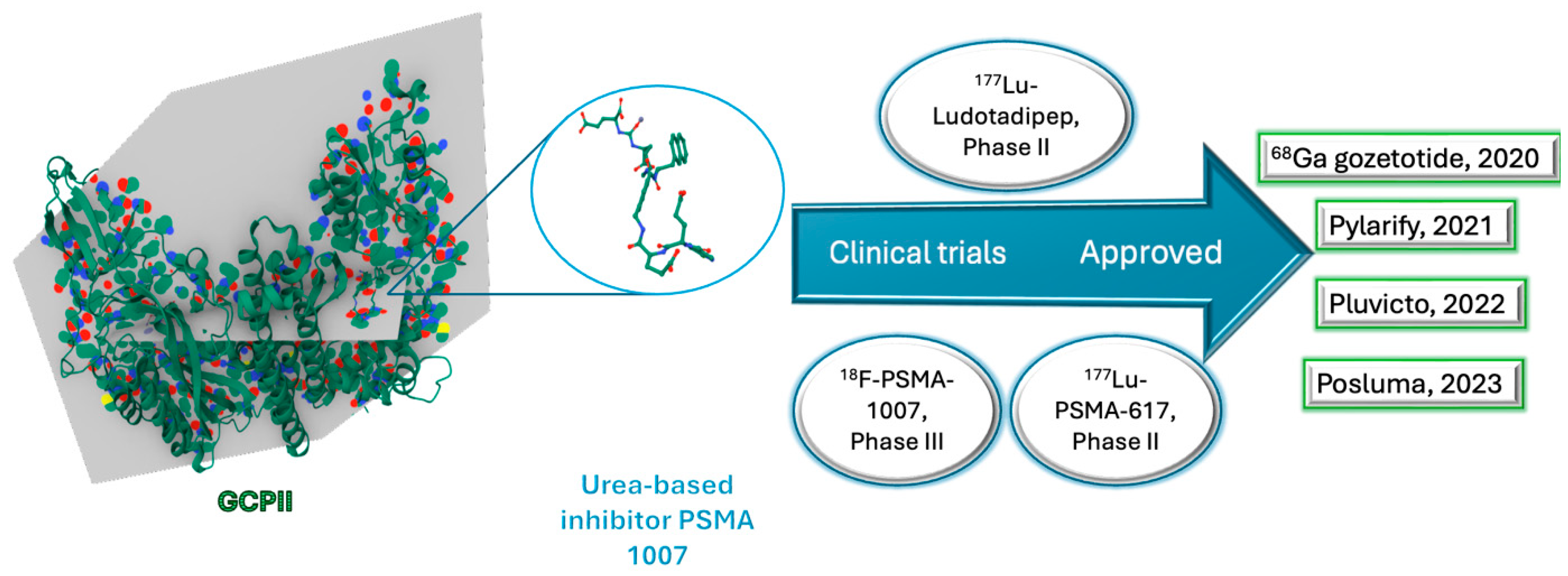
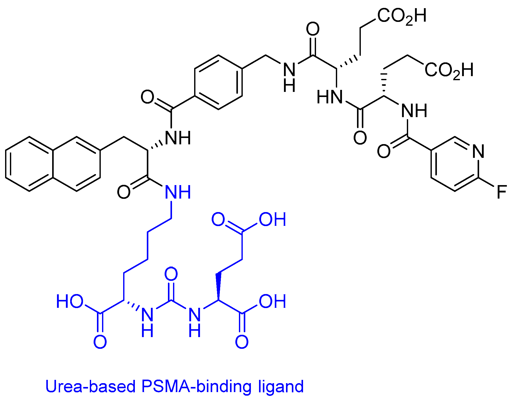
2.3. Prostate-Specific Membrane Antigen (PSMA) Peptide Antagonist

PSMA is a 100 kDa type II transmembrane glycoprotein, composed of 750 amino acids, which functions as a glutamate carboxypeptidase (GCPII) [103]. It plays a dual physiological role in (i) glutamatergic neurotransmission via hydrolysis of N-acetylaspartylglutamate (NAAG) and (ii) intestinal folate absorption through hydrolysis of polyglutamated folates [103]. While PSMA is constitutively expressed in non-malignant tissues—including prostate epithelium, salivary glands, lacrimal glands, and renal proximal tubules—it is markedly overexpressed in prostate cancer cells and the neovasculature of various solid tumours [103]. Moreover, PSMA plays a role in prostate carcinogenesis and disease progression [103]. This overexpression correlates with disease progression, making PSMA a key biomarker and therapeutic target in oncology [103,104].
PSMA-targeting agents exploit this differential expression through selective binding to PSMA-positive cells, enabling both diagnostic imaging and targeted radioligand therapy [105]. Since 2020, the FDA has approved four PSMA-based agents for clinical use (Table 4) [9], reflecting rapid advancements in theranostic applications. Notably, [18F]PSMA-1007—a next-generation PSMA inhibitor currently in clinical development—exhibits improved pharmacokinetic properties for PET imaging, including reduced renal excretion and enhanced tumour uptake [23].
All GCPII inhibitors feature three key components: (i) a P1′ moiety binding the S1′ pocket, (ii) a zinc-chelating group replacing the scissile bond, and (iii) an effector moiety interacting with the S1 pocket, with classification based on zinc-binding groups [106,107]. The amphipathic pharmacophore pocket (8 × 8 × 8 Å), formed by residues including Phe209, Asn257, and Tyr700, exhibits limited plasticity due to structural constraints (fixed 8 Å width between Phe209 and Leu428) but contains a flexible glutarate sensor [106,107]. It preferentially binds C-terminal glutamate via polar and hydrophobic interactions, explaining why most substrates and clinical inhibitors are glutamate-derived, including those currently undergoing clinical trials (Figure 7) [108,109].
The arene-binding site (Trp541/Arg463/Arg511) enhances inhibitor affinity through π-stacking when accessible [109]. Zinc coordination primarily involves Cys/His/Asp/Glu residues (96% of cases) [109]. The dynamic non-pharmacophore pocket forms a 20 Å-deep entrance funnel (8 Å narrow base) lined by Ser454-Tyr552 and an arginine patch (Arg463/534/536), with Arg463 located on β-strand β13 and Arg534/536 on antiparallel β14 [107].
Lutetium (177Lu) vipivotide tetraxetan (Pluvicto®) is a targeted radioligand therapy comprising three key components: (i) a PSMA-binding urea-based ligand, (ii) a DOTA (1,4,7,10-tetraazacyclododecane-1,4,7,10-tetraacetic acid) chelator, and (iii) the β-emitting radionuclide 177Lu (Figure 8) [9,96].
Pluvicto was approved in 2022 to treat metastatic mCRPC with PSMA expression [105]. Pluvicto selectively delivers cytotoxic radiation to PSMA-positive tumour cells and their microenvironment [105]. The emitted β particles induce DNA double-strand breaks in targeted cells and adjacent tumour stroma (bystander effect), resulting in irreversible cellular damage and apoptosis [111]. Pluvicto is administered intravenously and has side effects such as decreased levels of lymphocytes, leukocytes, platelets, calcium, sodium, and haemoglobin [111].

18Fluorine-PSMA-1007

18F-PSMA-1007 is an emerging radiopharmaceutical diagnostic agent comprising a PSMA-targeting ligand conjugated to the positron-emitting isotope fluorine-18 (18F) (Figure 9) [112]. Currently under clinical investigation, this compound shows particular promise for early-stage prostate cancer detection, demonstrating superior sensitivity for stage I disease compared to conventional imaging techniques [112].
Its diagnostic mechanism relies on selective binding to overexpressed PSMA receptors in prostate cancer cells, enabling precise tumour localisation through 18F-mediated PET imaging [23]. While preliminary studies suggest potential theranostic applications, further research is required to fully characterise its therapeutic utility [23]. A notable diagnostic consideration is the compound’s PSMA-specificity, which—while advantageous for tumour targeting—may lead to false-positive interpretations due to physiological PSMA expression in other tissues including bladder transitional cell carcinoma, renal cell carcinoma, and colonic carcinoma [112]. This underscores the importance of correlating imaging findings with clinical and histopathological data to ensure accurate diagnosis. The agent’s high tumour-to-background ratio offers significant advantages in detecting low-volume and metastatic disease, though clinicians must remain cognizant of its limitations in specificity [112].
18F-PSMA-1007 exhibits superior pharmacokinetic properties compared to 68Ga-PSMA agents, most notably its extended 110-min half-life [23]. This prolonged half-life enhances diagnostic accuracy by allowing for delayed imaging windows, which improves tumour-to-background contrast and increases detection rates [23]. The compound’s favourable physical characteristics—including low positron energy (0.635 MeV) and high positron yield (97%)—provide exceptional spatial resolution, enabling precise identification of small lesions (<5 mm) on PET-CT imaging [23]. Furthermore, 18F-PSMA-1007 demonstrates remarkable specificity (85–92%) and selectivity for locoregional lymph node metastases, significantly improving diagnostic confidence in staging and restaging prostate cancer [112]. These combined attributes make 18F-PSMA-1007 particularly valuable for detecting oligometastatic disease and guiding treatment decisions [112].
18F-PSMA-1007 demonstrates unique pharmacokinetic advantages, including predominant hepatobiliary excretion that minimises urinary tract activity, thereby significantly improving diagnostic accuracy for pelvic lymph node metastases [23,112].
Administered intravenously, this radiotracer achieves optimal imaging quality within 2 h post-injection—a faster timeframe compared to the 3 h requirement for 68Ga-PSMA-11 [112]. The rapid target-to-background ratio optimisation enables earlier high-resolution PET imaging, with enhanced detection of small nodal metastases due to reduced obscuration by bladder activity. This improved temporal and spatial resolution makes 18F-PSMA-1007 particularly valuable for precise staging of prostate cancer, especially in the prostatic bed and pelvic nodal regions where conventional PSMA PET tracers face limitations due to urinary excretion artefacts [23,112].
Prior to 18F-PSMA-1007 administration, comprehensive patient evaluation is required, including an assessment of contrast allergies, vital signs (blood pressure and respiratory rate), and laboratory parameters (biochemical, haematological, and urinalysis) [113]. Renal function is particularly critical, as the radiopharmaceutical is contraindicated in patients with an estimated glomerular filtration rate (eGFR) below 40 mL/min/1.73 m2 [113]. Clinical trial data demonstrate an excellent safety profile for 18F-PSMA-1007, with no significant adverse effects reported in Phase III studies [113]. Additionally, its cost-effectiveness enhances accessibility for patients [23].
However, diagnostic limitations exist, and false-negative results frequently occur in liver metastases due to high physiological hepatobiliary excretion of the tracer, while false-positive findings may arise from nonspecific bone uptake [112]. These challenges highlight the importance of complementary imaging approaches. Although discordant results were observed in clinical trials, emerging evidence suggests that combined PET/MRI may improve diagnostic accuracy compared to either modality alone [112].

2.4. Peptide Receptor Radionuclide Therapy (PRRT)

PRRT represents a precision oncology approach that selectively delivers cytotoxic radiation to tumour cells through the molecular targeting of overexpressed receptors [114]. This therapeutic strategy primarily exploits the high expression of SSTR2 in NETs, utilising either β-emitting radionuclides (e.g., 177Lu) or α-emitters (e.g., 225Ac) to induce DNA damage in malignant cells while sparing healthy tissue [115]. PRRT demonstrates exceptional clinical tolerability, with a favourable toxicity profile that enhances patient safety during treatment [66,114].
The FDA approved four diagnostic and five theranostic PRRT agents between 1999 and 2025 (Table 5) [67], with several promising candidates currently in development. Notable investigational agents include the following: RAYZ-8009 (DOTA-RYZ-GPC3), targeting glypican-3 in hepatocellular carcinoma [60], and 177Lu-FAP-2286, directed against fibroblast activation protein (FAP) in stromal-rich tumours [62]. These therapies show particular efficacy in managing metastatic NETs, where they address the critical need for targeted treatment options in advanced disease [116].
64Cu-DOTATATE (Detectnet™) is a radiolabelled somatostatin analogue comprising three key structural components: (i) a targeting peptide sequence, (ii) the macrocyclic chelator DOTA (1,4,7,10-tetraazacyclododecane-1,4,7,10-tetraacetic acid), and (iii) the positron-emitting copper-64 (64Cu) radionuclide (Figure 10) [9,114].
The molecule’s stability is enhanced by a disulfide bridge that constrains its conformational flexibility. Approved by the FDA in 2020 for SSTR PET imaging, Detectnet specifically binds to cells overexpressing SSTR2 with high affinity [117]. The 64Cu radionuclide decays via β+ emission (0.653 MeV), enabling high-resolution PET imaging with favourable dosimetry [117].

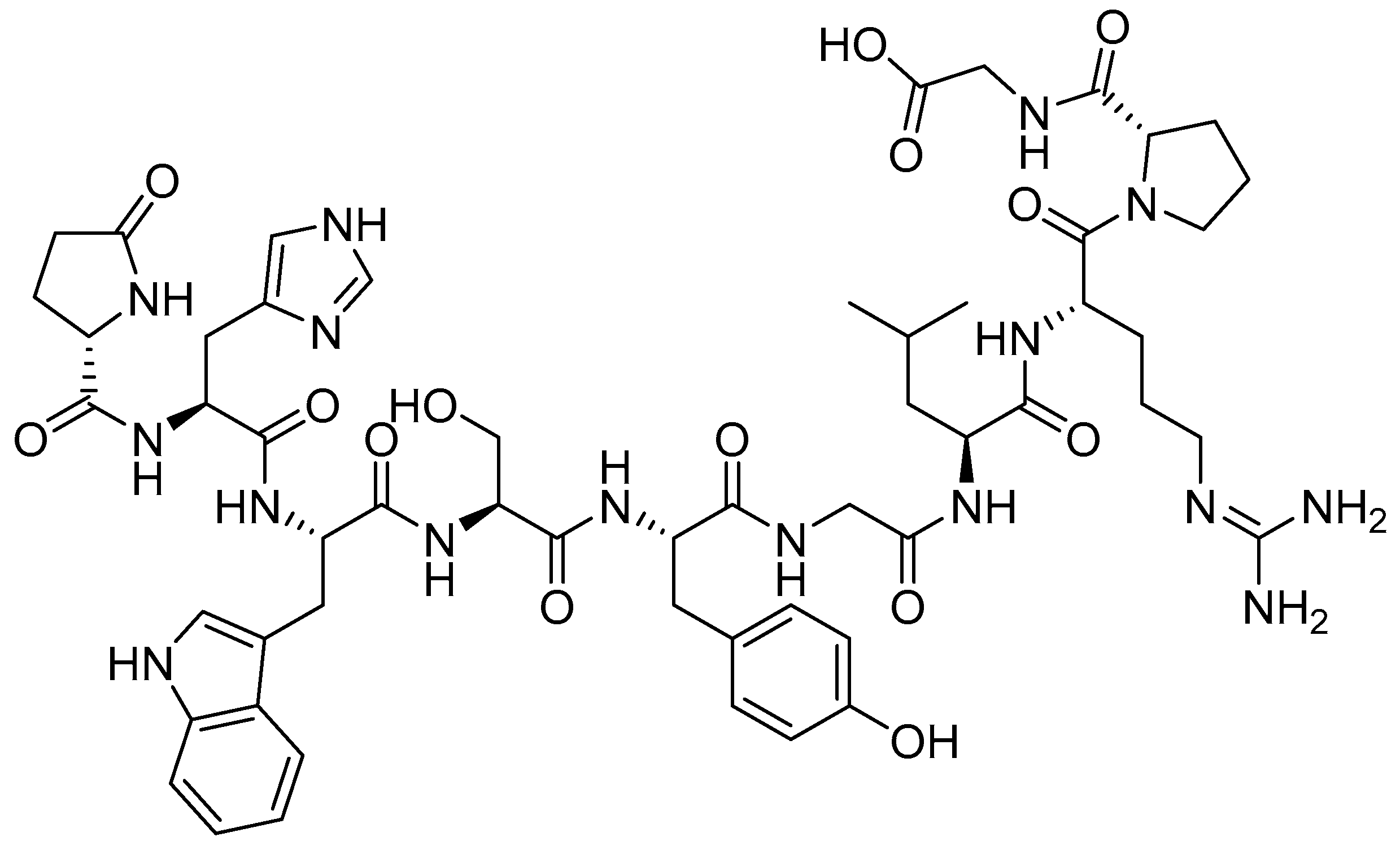
2.4.1. RAYZ-8009 (DOTA-RYZ-GPC3)

Liver cancer ranks as the sixth most commonly diagnosed malignancy and the third leading cause of cancer-related mortality globally [60,118]. A promising molecular target in hepatocellular carcinoma (HCC) is glypican-3 (GPC3), a cell-surface heparan sulfate proteoglycan that shows minimal expression in normal tissues but significant overexpression in 70–80% of HCC cases [60]. The novel theranostic agent DOTA-RYZ-GPC3 (RAYZ-8009) represents a breakthrough in PRRT, combining a GPC3-targeting macrocyclic peptide with a radiometal chelator for both diagnostic and therapeutic applications [60]. Preclinical studies demonstrate RAYZ-8009’s exceptional cross-species binding affinity for GPC3 across humans, canines, cynomolgus monkeys, and murine models [119].
Comparative preclinical evaluation of 177Lu-RAYZ-8009 (β-emitter) and 225Ac-RAYZ-8009 (α-emitter) revealed distinct therapeutic advantages: 225Ac-RAYZ-8009 delivers high-linear energy transfer (LET) α-particles (5800 keV), exhibits short tissue penetration (40–100 μm), causes clustered DNA double-strand breaks, and demonstrates superior tumour cell kill efficiency per decay [60]. 177Lu-RAYZ-8009 shows more pronounced tumour growth inhibition in HepG2 xenograft models, benefits from longer β-particle path length (0.2–2 mm), and enables crossfire effect for heterogeneous tumour targeting [60]. These findings highlight the complementary potential of α- versus β-emitting radiopharmaceuticals in precision oncology, with α-emitters offering superior cytotoxicity for isolated tumour cells and β-emitters providing better volumetric radiation for larger tumour masses.
PeptiDream pharmaceutical company has an ongoing preclinical trial for 225Ac/68Ga-GPC3 (RYZ 801/RYZ 811). RYZ 811 is a theranostic agent which has the same peptide binder and linker as RYZ 801 but a different radionuclide, 68Ga [118]. In total, 47 patients with HCC used RYZ 811, and 90% of them had an uptake of RYZ 811 into the tumour cells [118]. Initial clinical data demonstrated no serious adverse effects in treated patients, supporting the favourable safety profile of RYZ-811 [118]. However, the small cohort size limits the statistical power of these findings, underscoring the need for larger-scale trials to confirm safety and establish more robust efficacy endpoints. Additionally, while preliminary results are promising, further mechanistic studies are required to fully characterise RYZ-811’s diagnostic potential, including its targeting specificity, biodistribution patterns, and detection sensitivity in relevant disease states. These investigations should ideally employ standardised imaging protocols and incorporate comparator agents to validate diagnostic performance.

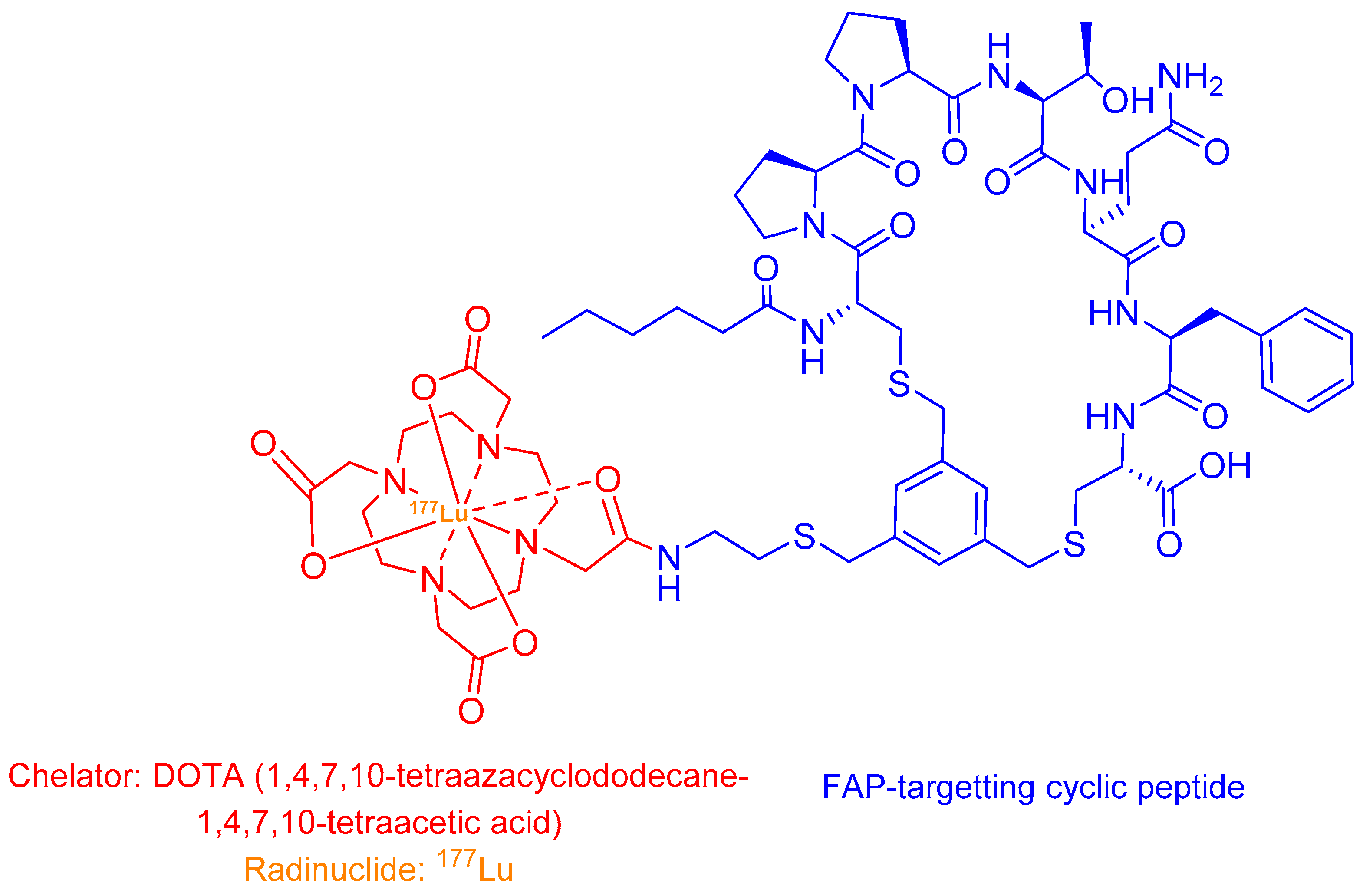
2.4.2. 177Lu-FAP-2286

FAP is a type II transmembrane serine protease exhibiting dual enzymatic activity as both a dipeptidyl peptidase and endopeptidase [120]. This 97 kDa glycoprotein, composed of 760 amino acid residues, belongs to the prolyl oligopeptidase family and demonstrates negligible expression in normal adult tissues [120]. The FAP structure comprises three distinct regions: (i) a short N-terminal cytoplasmic domain (6 amino acids), (ii) a hydrophobic transmembrane segment (20 amino acids), and (iii) a substantial extracellular portion (736 amino acids) containing the catalytic machinery [120]. The extracellular domain features two functionally critical subdomains: an 8-bladed β-propeller that regulates substrate access and a C-terminal α/β-hydrolase domain housing the catalytic triad (Ser624, Asp702, His734) responsible for proteolytic activity [120].
FAP is selectively overexpressed on cancer-associated fibroblasts (CAFs) in 80–95% of epithelial tumours and certain mesenchymal malignancies, where it plays a multifaceted role in tumour progression [121,122]. FAP-positive CAFs drive tumorigenesis and metastasis through four key mechanisms: (i) promoting extracellular matrix (ECM) remodelling via its collagenolytic activity, (ii) stimulating angiogenesis to support tumour vasculature, (iii) establishing an immunosuppressive TME through cytokine secretion, and (iv) facilitating intracellular signalling pathways that enhance tumour cell survival [123]. This transmembrane protease shows particularly high expression in aggressive cancers including pancreatic ductal adenocarcinoma (>90%), mesothelioma (85–90%), colorectal carcinoma (70–80%), head and neck squamous cell carcinoma (60–75%), and salivary gland tumours (50–60%) [120,124]. The tumour-specific expression pattern of FAP, combined with its central role in maintaining the pro-tumorigenic stroma, makes it an exceptionally promising target for both diagnostic imaging and stromal-targeted therapies, especially in treatment-resistant, stroma-rich cancers where traditional approaches often fail [120].
177Lu-FAP-2286 is a novel theranostic agent comprising a FAP-targeting cyclic peptide conjugated to a radionuclide chelator (Figure 11) [123]. This compound selectively binds to FAP-expressing cells, delivering cytotoxic β-radiation (177Lu) for therapy while enabling γ-imaging for treatment monitoring [122,124].
Currently in Phase 1/2 trials, 177Lu-FAP-2286 shows particular promise for advanced metastatic sarcomas and other FAP-rich malignancies [16,125]. The diagnostic counterpart, 68Ga-FAP-2286, serves a dual purpose: (i) quantifying FAP expression via PET/CT to identify eligible patients, and (ii) providing a baseline for comparative therapeutic response assessment when followed by 177Lu-FAP-2286 [123]. This sequential theranostic approach allows for personalised dose optimisation and real-time treatment efficacy evaluation, addressing a critical need in stromal-targeted radionuclide therapy.
177Lu-FAP-2286 demonstrates superior tumour retention due to the extended half-life (6.65 days) of lutetium-177, enabling prolonged radiation exposure to FAP-positive malignancies [123]. This pharmacokinetic advantage enhances both diagnostic sensitivity and therapeutic efficacy, as evidenced by robust tumour uptake across diverse cancer types including pancreatic, breast, and sarcomas [123,124]. Clinical data reveal three key benefits compared to its 68Ga counterpart: (i) enhanced therapeutic profile, with higher tumour-to-background ratios (3.5:1 vs. 2.1:1 at 24 h), (ii) improved safety, grade 1–2 adverse events only (vs. Grade 3 in 12% with 68Ga-FAP-2286), and better clinical efficacy, 62% reduction in metastatic burden (RECIST 1.1), 45% objective tumour size reduction, and 78% pain score improvement [123].
These outcomes correlate with significantly higher patient-reported satisfaction scores (p < 0.01), positioning 177Lu-FAP-2286 as a transformative theranostic agent for FAP-expressing cancers [123]. The compound’s dual capacity for precise tumour localisation and effective cytoreduction—coupled with its favourable tolerability—underscores its potential to address unmet needs in advanced metastatic disease [123].
177Lu-FAP-2286 is administered as a weekly intravenous infusion over six weeks, with continuous monitoring of vital signs (blood pressure, heart rate) and symptom progression throughout treatment [123]. While most clinical studies report only mild (Grade 1–2) adverse events, one trial observed Grade 3 haematologic toxicities—notably leukopenia and pancytopenia—in 9% of participants [123]. The current evidence base remains limited by two critical constraints: (i) an extremely small patient cohort (n = 11, with advanced metastatic sarcoma), which precludes meaningful statistical analysis of efficacy or safety endpoints, and (ii) the absence of a standardised dose-escalation strategy, leaving the maximum tolerated dose (MTD) and optimal therapeutic window undefined [123]. These limitations underscore the necessity for expanded Phase 2 trials incorporating larger, histologically stratified patient populations (n ≥ 50), protocol-defined dose optimisation (e.g., 3 + 3 design), and correlative biomarker studies to establish predictive response parameters. The compound’s promising preliminary safety profile nevertheless supports continued clinical development as a targeted therapeutic for FAP-expressing malignancies [123].
Recent advances in FAP-targeted radiopharmaceuticals include promising preclinical candidates like [137La]La-FAP-2286, a novel theranostic analogue currently under investigation [126]. This compound demonstrates the growing interest in FAP as a diagnostic and therapeutic target across various tumour types, building upon the clinical validation of other FAP-directed agents [126]. The theranostic approach exemplified by [137La]La-FAP-2286 offers potential advantages for both tumour detection and treatment monitoring in FAP-expressing malignancies, though further characterisation of its binding affinity, pharmacokinetics, and therapeutic efficacy remains necessary [126].

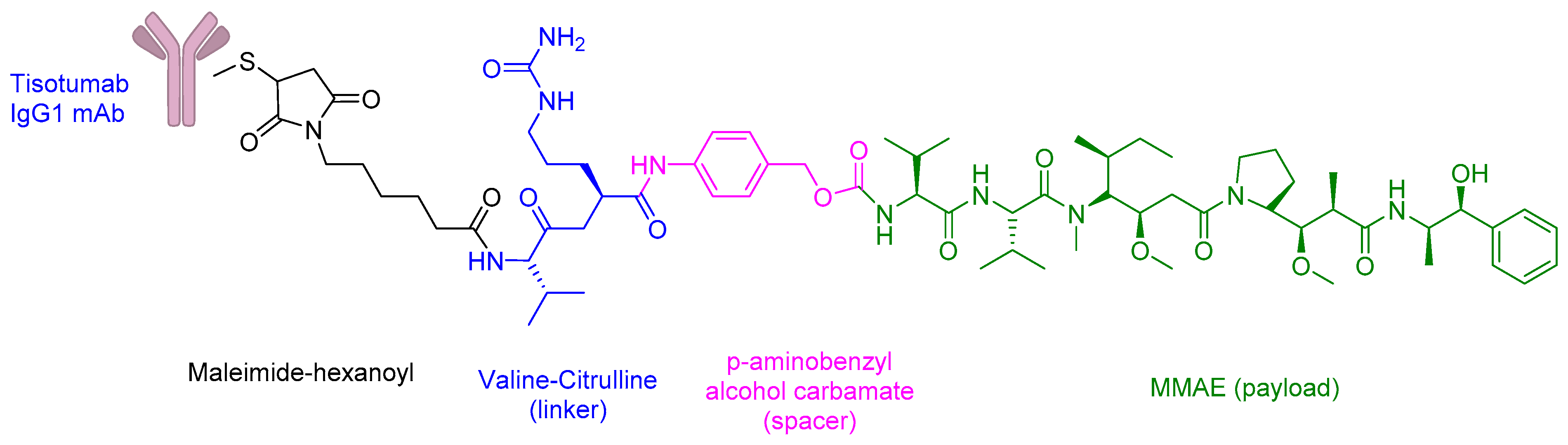
2.5. Antibody–Drug Conjugates (ADCs)

ADCs represent a targeted therapeutic platform comprising three key components: (i) a tumour antigen-specific monoclonal antibody, (ii) a chemically stable linker designed to withstand systemic circulation, and (iii) a potent cytotoxic payload (typically 100–1000× more potent than conventional chemotherapy) [1,125,127,128]. These engineered bioconjugates selectively bind overexpressed tumour-associated receptors, enabling precise delivery of their cytotoxic payload directly to malignant cells while minimising off-target effects [1,127]. The FDA has accelerated approval of seven ADCs between 2019 and 2021 for various haematological and solid tumours (Table 6) [9], with several promising candidates like the CEACAM5-targeting SAR408701 currently in late-stage clinical development [31]. This expanding therapeutic class demonstrates how rational drug design can improve the therapeutic index of highly cytotoxic agents through molecular targeting.
Tisotumab Vedotin-Tftv (TIVDAK) is an FDA-approved ADC with (i) Tisotumab IgG1 monoclonal antibody, (ii) a protease-cleavable valine-citrulline linker, (iii) monomethyl auristatin E (MMAE) payload, which is a potent inhibitor of cell division and microtubule-disrupting agent (Figure 12) [129,130].
TIVDAK has an exceptional antitumour activity and is indicated for recurrent or metastatic cervical cancer [131]. TIVDAK contains a monoclonal antibody that acts against the tissue factor (TF) and a cytotoxic payload, MMAE [130]. The TF is a transmembrane glycoprotein, and it is involved in the initiation of the extrinsic coagulation pathway [130,132]. Additionally, the TF exhibits cell signalling properties, such as the activation of protease-activated receptor 2 (PAR2), which further causes gene transcription, cell survival, and cytoskeletal changes [133]. The TF is expressed on the surface of cancer cells, including cervical cancer, ovarian cancer, NSCLC, breast cancer, head and neck squamous cell carcinoma, and glioblastoma, and is involved in tumour growth, angiogenesis, and metastases [131,134,135].
TIVDAK binds to TF with a high affinity and interferes with the PAR2 pathway [133]. Additionally, TV binds to TF, leading to the formation of a TIVDAK-TF complex [130]. The complex is internalised and transported to the lysosome [130]. The linker is then enzymatically cleaved, and MMAE is released [130]. MMAE binds to the tubulin and disrupts the microtubule polymerisation [130]. This further causes G2/M cell cycle arrest and apoptosis [130]. Additionally, TIVDAK can also provide a bystander effect where its cell permeability property induces the release of MMAE to the neighbouring dividing cells, which in turn causes cell death [129].
TIVDAK is administered intravenously, and its adverse events include nausea (54%), alopecia (39.3%), conjunctivitis (30%), fatigue (26.1%), and dry eye (23%) [130]. Grade 3 or worse treatment-related adverse events include neutropenia, fatigue, ulcerative keratitis, anaemia, and peripheral neuropathy [130]. Additionally, other side effects include haemorrhage, embryo foetal toxicity, and life-threatening Stevens–Johnson Syndrome [130].
Zynlonta® (loncastuximab tesirine-lpyl) is an FDA-approved ADC with three structurally optimised components: (i) a humanised IgG1κ monoclonal antibody targeting CD19, (ii) a protease-cleavable valine-alanine (Val-Ala) dipeptide linker (the sole peptide element in the construct, and (3) a potent pyrrolobenzodiazepine (PBD) dimer payload (SG3199) capable of inducing DNA interstrand crosslinks (Figure 13) [60,136]. Indeed, this drug exemplifies how peptides can serve solely as linkers in ADCs.
This ADC is specifically indicated for relapsed/refractory large B-cell lymphoma (R/R LBCL), including DLBCL not otherwise specified, HGBCL, and DLBCL arising from low-grade lymphoma [136]. The polybrominated biphenyls (PBBs) warhead’s unique mechanism—forming irreversible DNA adducts that block replication—provides exceptional cytotoxicity against malignant B-cells, while the Val-Ala linker ensures stable plasma circulation and tumour-specific payload release via cathepsin cleavage [60].

SAR408701

Carcinoembryonic antigen-related cell adhesion molecule 5 (CEACAM5) is a glycosylphosphatidylinositol (GPI)-anchored glycoprotein (180–200 kDa) that plays critical roles in cell adhesion and intracellular signalling [31]. Structurally, it mediates homotypic and heterotypic interactions through its immunoglobulin-like domains, influencing tumour progression and metastasis [31]. CEACAM5 is markedly overexpressed (≥50% of tumour cells) in epithelial-derived carcinomas, with the highest prevalence in gastrointestinal malignancies (70–90% of colorectal, 50–70% of gastric, and 40–60% of pancreatic cancers) [31]. It is also frequently upregulated in NSCLC (30–50%), genitourinary tumours (40–60% of bladder cancers), and a subset of breast carcinomas (15–30%, particularly triple-negative subtypes) [83]. Beyond adhesion, CEACAM5 activates pro-tumorigenic signalling pathways, including PI3K/AKT-mediated cell survival, SRC-family kinase-dependent motility, and Wnt/β-catenin modulation, making it a compelling biomarker and therapeutic target in epithelial cancers [31].
In 2023, the investigational antibody–drug conjugate SAR408701 (tusamitab revtamab) advanced to Phase III clinical trials for CEACAM5-positive NSCLC, marking a significant milestone in targeted therapy development (Figure 14) [137].
This novel anti-CEACAM5 maytansinoid conjugate comprises three key components: (i) a humanised monoclonal antibody specifically targeting CEACAM5-expressing epithelial tumours, (ii) a cleavable sulfo-SPDB linker designed for tumour-selective payload release, and (iii) the potent cytotoxic agent DM4 (a maytansine derivative) [83]. The DM4 payload exerts its antimitotic effect through two complementary mechanisms: (a) binding to tubulin at the vinca domain with picomolar affinity and (b) inducing catastrophic microtubule network disruption (Figure 15) [31].
This dual-action mechanism leads to sustained G2/M phase cell cycle arrest and subsequent apoptosis in CEACAM5-overexpressing malignancies [31]. The ADC’s design leverages CEACAM5’s tumour-restricted expression pattern (minimal in normal tissues) to maximise therapeutic index while minimising off-target toxicity—a critical advantage in treating advanced NSCLC where conventional chemotherapy often shows limited efficacy and significant adverse effects [31].
Preclinical evaluation of SAR408701 demonstrated high binding specificity (KD = 0.5–2 nM) for CEACAM5, with potent dose-dependent antitumour activity in both in vitro and in vivo models, achieving 70–90% tumour growth inhibition in CEACAM5-expressing xenografts through its dual mechanism of antibody-mediated targeting and DM4-induced microtubule disruption [31]. While the ADC showed a strong linear dose–response relationship (p < 0.001), therapeutic doses ≥100 mg/m2 induced significant toxicities, including haematologic adverse events (Grade 3/4 leukopenia [33%], thrombocytopenia [15%], neutropenia [20%]) and non-haematologic effects (Grade 2/3 asthenia [45%], peripheral neuropathy [18%], and keratopathy [22%]) [137].These findings highlight the critical balance required between SAR408701’s promising antitumour efficacy and its manageable but clinically significant toxicity profile, underscoring the importance of careful dose optimisation in ongoing Phase III trials for CEACAM5-positive malignancies.

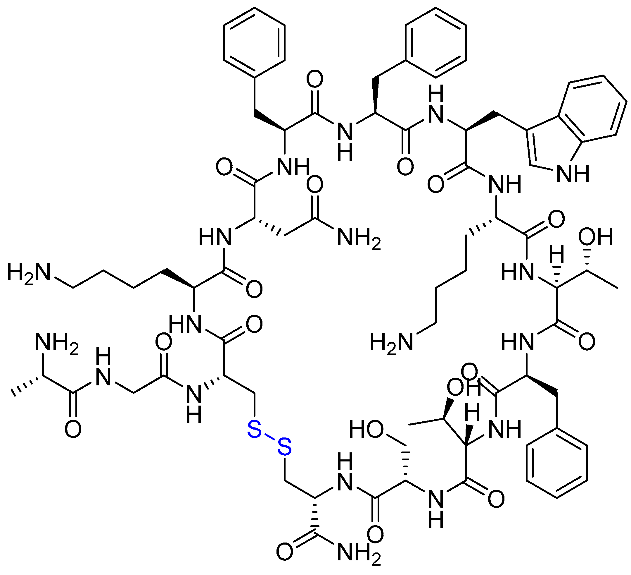
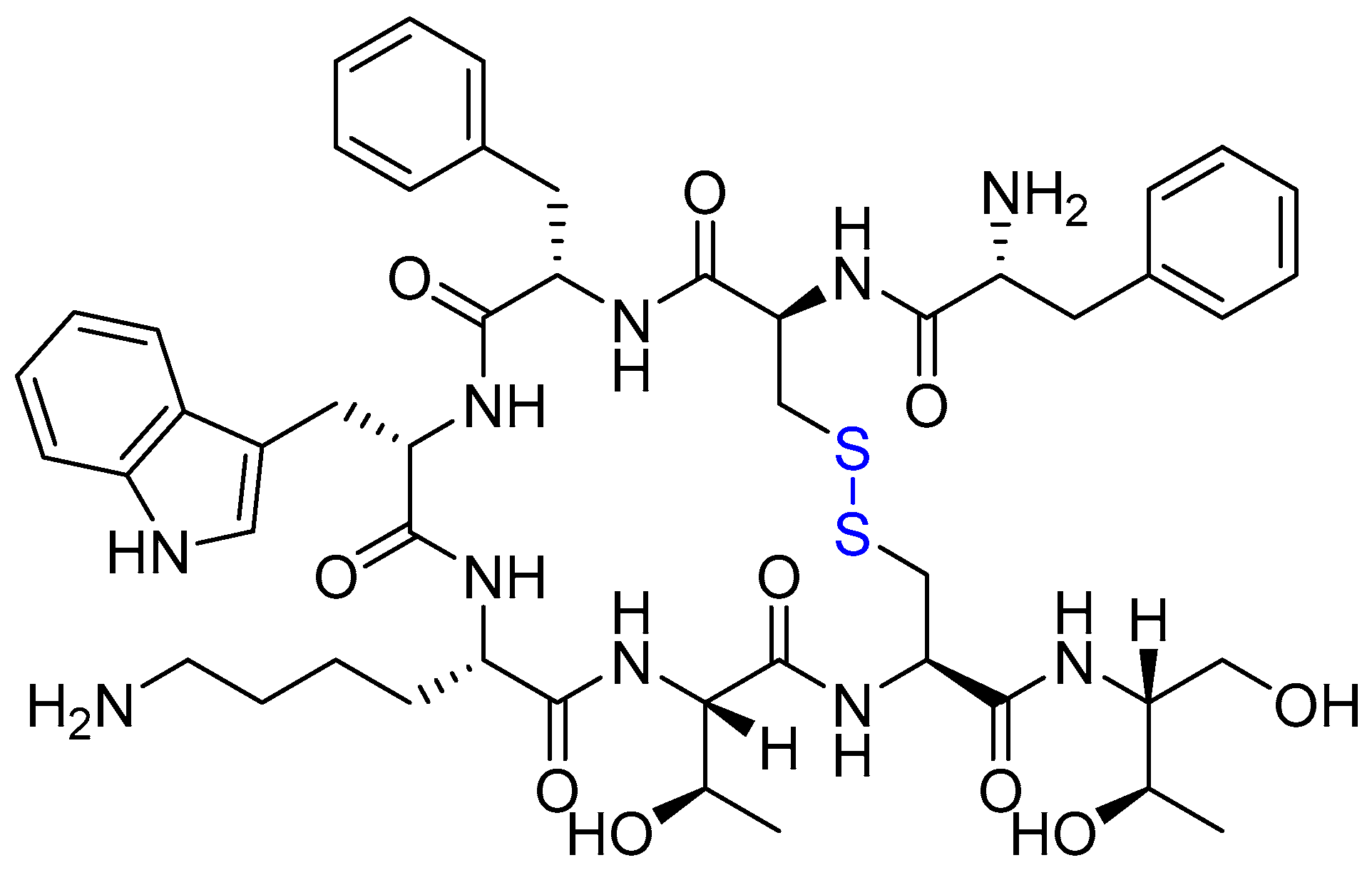
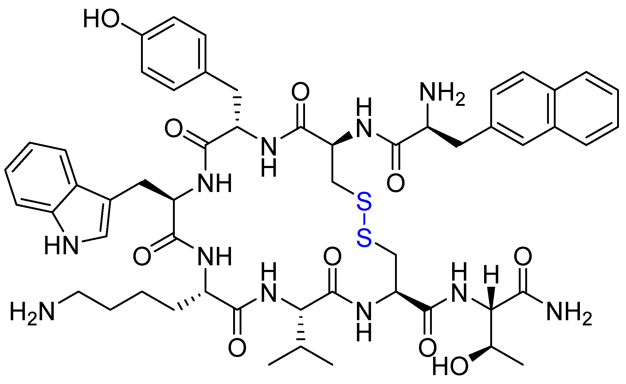
2.6. Peptide–Drug Conjugates (PDCs)

PDCs represent an emerging class of targeted therapeutics with several advantages over traditional ADCs, including higher selectivity, improved tumour penetration, and reduced systemic toxicity [18,72]. PDCs are broadly categorised into two groups: (1) cell-penetrating peptides (CPPs), which facilitate intracellular delivery, and (2) cell-targeting peptides (CTPs), which bind overexpressed receptors on cancer cells [72]. Structurally, PDCs replace the monoclonal antibody component of ADCs with a smaller peptide (2–20 kDa vs. ~160 kDa for ADCs), enabling deeper tumour stroma penetration and enhanced cellular uptake [138]. Additionally, their low immunogenicity minimises the risk of immune-related adverse effects, a common limitation of ADCs [8,18]. However, PDCs face challenges such as rapid renal clearance due to their short half-life [18,21]. To address this, structural modifications—such as increasing peptide negative charge or incorporating albumin-binding motifs—have been employed to delay glomerular filtration and prolong circulation time [8].
The first FDA-approved PDC, 177Lu-DOTATATE (Lutathera®), was introduced in 2018 to treat somatostatin receptor-positive gastroenteropancreatic NETs (GEP-NETs) [8]. Administered intravenously, it leverages peptide-mediated targeting for precise radionuclide delivery [139]. Another early PDC, melphalan flufenamide (Pepaxto®), was withdrawn from the US market due to insufficient clinical benefit and safety concerns [140]. However, it remains approved in the EU/UK under the brand name Pepaxti® in combination with dexamethasone for relapsed multiple myeloma [141]. Despite these setbacks, multiple PDCs remain in clinical development, demonstrating their potential as next-generation targeted therapies [18].

Bicycle Toxin Conjugate (BTC) BT5528

Solid tumours such as ovarian cancer, glioma, and triple-negative breast cancer exhibit significant overexpression of ephrin type-A receptor 2 (EphA2) receptors compared to healthy tissues [15,33]. Targeting this biomarker, BT5528 represents an innovative BTC composed of three key elements: (1) an EphA2-binding bicycle peptide, (2) a protease-sensitive valine-citrulline linker, and (3) the microtubule-disrupting payload MMAE [13]. Currently in Phase II trials, BT5528’s compact molecular structure (~10 kDa) enables superior tumour penetration compared to bulkier ADCs (~150 kDa), enhancing targeted drug delivery [13]. The conjugate exerts cytotoxicity through two distinct mechanisms: extracellular cleavage by tumour-associated proteases releases free MMAE into the microenvironment (inducing bystander effects), while receptor-mediated internalisation facilitates intracellular payload release, triggering mitotic arrest via tubulin polymerisation inhibition (Figure 16) [13].
This dual-action design capitalises on EphA2’s tumour-selective expression, minimising off-target effects while maximising antineoplastic activity [62]. Preliminary data demonstrate high receptor specificity and potent efficacy, though further clinical evaluation will establish its therapeutic index [62].
BT5528 demonstrates favourable metabolic stability in both preclinical and clinical settings, albeit with a relatively short plasma half-life (~15–20 h) that necessitates biweekly intravenous administration [13]. The recommended Phase II dose (RP2D) of 6.5 mg/m2 every two weeks achieves optimal therapeutic exposure, demonstrating significant antitumour activity with measurable tumour regression in EphA2-positive malignancies [118]. While generally well-tolerated at this dose, BT5528 exhibits characteristic dose-limiting toxicities including gastrointestinal effects (Grade 2/3 vomiting [25%], diarrhoea [30%], nausea [35%]), haematologic complications (neutropenia [Grade 3/4: 20%], anaemia [Grade 2: 15%]), and systemic symptoms (fatigue [Grade 2: 40%], alopecia [15%]) [15]. Notably, 18% of patients develop peripheral neuropathy—a class effect of MMAE-based conjugates—which typically manifests as Grade 1/2 sensory disturbances but may require dose modifications at higher grades [15]. These findings underscore the importance of maintaining the RP2D to balance efficacy with manageable toxicity in ongoing clinical development.

2.7. Other Classes

2.7.1. Lumisight (Pegulicianine) and Lumicell Direct Visualisation System (DVS)

The FDA recently approved two innovative diagnostic systems for intraoperative tumour margin assessment: Lumisight™ (pegulicianine) and the Lumicell™ Direct Visualisation System (DVS) [16,31] This integrated theranostic platform combines three key components: (1) the fluorescent optical agent pegulicianine (a PDC that selectively accumulates in malignant tissue), (2) a proprietary handheld imaging device with 1 mm spatial resolution, and (3) artificial intelligence-driven detection software that provides real-time, patient-specific tumour probability mapping [7]. Designed for use during breast-conserving surgery, the system enables surgeons to identify residual tumour deposits (<0.1 cm3) with 92% sensitivity and 85% specificity in clinical trials, significantly reducing the need for repeat lumpectomies [7]. The FDA approval was based on multicentre trial data demonstrating a 50% reduction in positive margin rates compared to standard palpation-guided resection [31].
Lumisight™ is an activatable fluorescent probe with a sophisticated molecular design featuring three key components: (1) a GGRK peptide substrate cleavable by tumour-associated proteases (matrix metalloproteinase (MMP)-2/9 and cathepsins B/L), (2) a near-infrared Cy5 fluorophore (λexem = 649/670 nm), and (3) a QSY21 quencher molecule linked via polyethylene glycol (PEG) spacers (Figure 17) [16]. In its intact state, the probe remains optically inactive due to fluorescence resonance energy transfer (FRET) between the Cy5-QSY21 pair.
Upon encountering the proteolytic TME, enzymatic cleavage at the GGRK site releases two fluorescent fragments: (i) fragment 2: Cy5-PEG (emitting detectable NIR signal), and (ii) fragment 3: QSY21-PEG-peptide (quenched) (Figure 18) [16].
This tumour-selective activation mechanism achieves >100-fold fluorescence enhancement in malignant versus normal tissue, enabling real-time visualisation of residual tumour deposits with submillimetre precision during oncologic surgery [16]. The design capitalises on the 10–100× higher activity of MMPs and cathepsins in tumours compared to healthy parenchyma, ensuring exceptional target-to-background ratios [16].
Lumisight™ is administered intravenously 2–4 h preoperatively, where tumour-associated proteases activate its fluorescence specifically in malignant tissue [31]. During lumpectomy, surgeons use the Lumicell™ DVS to scan the resection cavity with a handheld probe (1 mm resolution), detecting residual tumour deposits ≥0.1 mm3 via real-time AI analysis of the fluorescent signal—a process that reduces positive margin rates by 50% and repeat surgeries by 85%, yielding USD 15,000–USD 20,000 cost savings per patient [31]. While demonstrating high patient satisfaction (92%) and minimal severe adverse effects (<1% Grade ≥3 adverse events), the system requires careful screening for PEG hypersensitivity (0.3% incidence) and contrast allergies (1.2% cross-reactivity risk) [60]. Its primary limitation stems from diagnostic inaccuracy in the pivotal trial (N = 357), with 43% false positives (inflammatory tissue) and 8% false negatives (micrometastases), prompting ongoing refinements in imaging algorithms and protease activity thresholds to improve specificity [60]. Despite these challenges, the technology represents a significant advance in precision oncology by enabling immediate, intraoperative margin assessment—a critical factor in reducing local recurrence rates from 5–10% to <2% at 5 years [31].

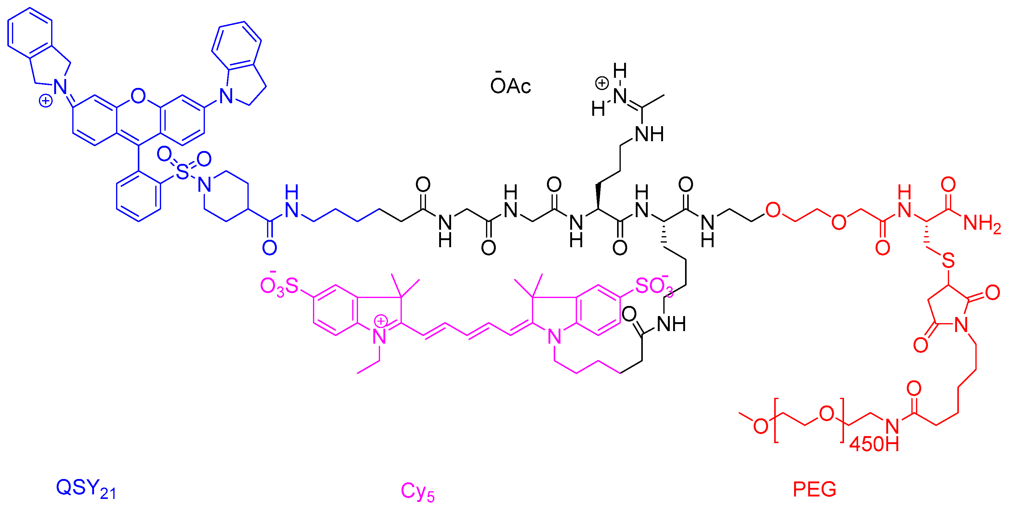
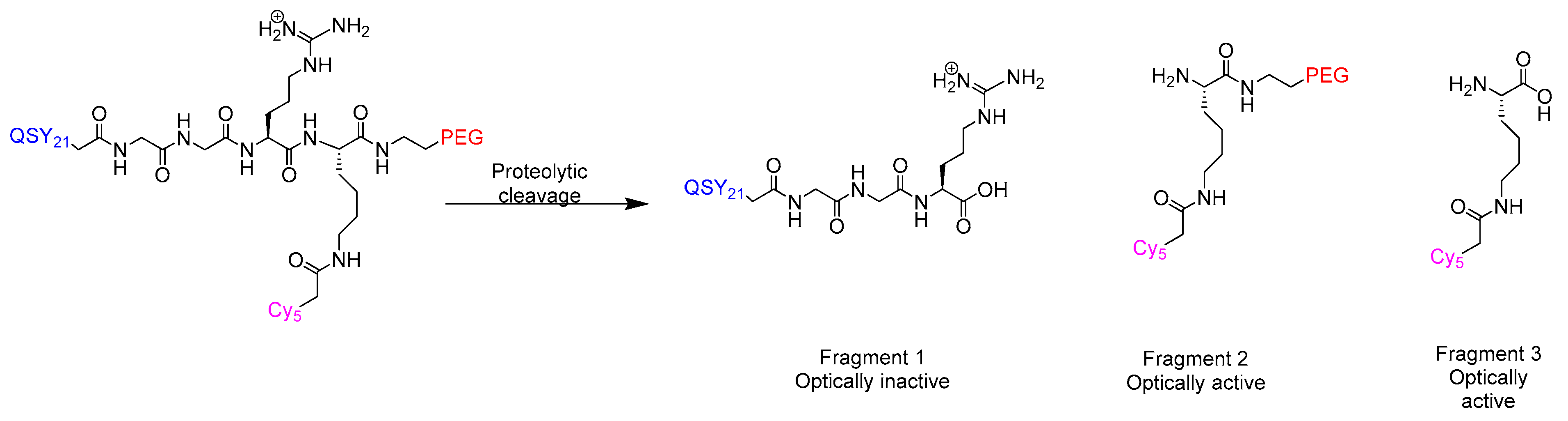
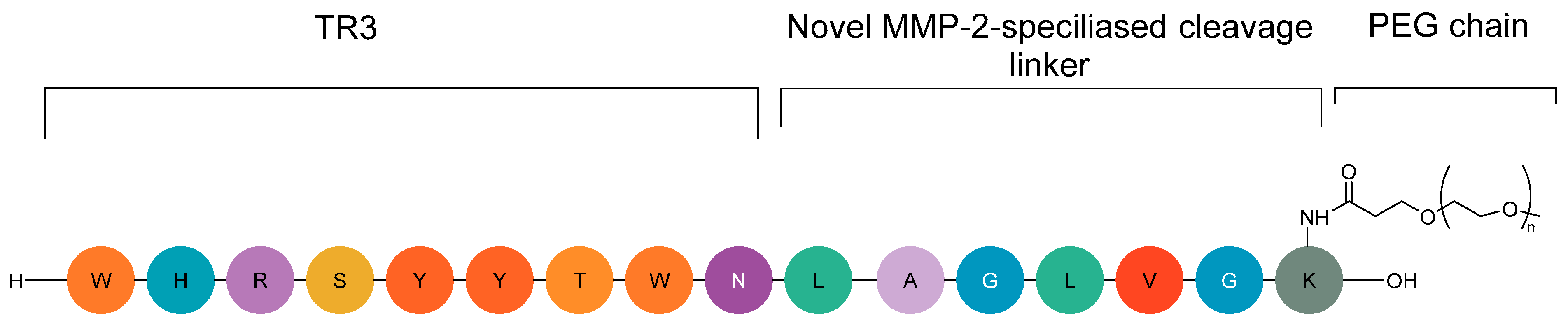
2.7.2. CLP002 (TR3-M-NP) Peptide in Immunotherapy

Immunotherapy has revolutionised cancer treatment by modulating immune responses, primarily through checkpoint inhibitors targeting pathways like CTLA-4 and PD-1/PD-L1, which normally maintain self-tolerance by regulating T-cell activation [142]. While antibody-based inhibitors (e.g., ipilimumab, nivolumab) have shown remarkable clinical success, their systemic activity often triggers severe immune-related adverse events (irAEs), including pneumonitis, hepatitis, and endocrine dysfunction, due to indiscriminate immune activation [142]. The TME exacerbates immunosuppression by upregulating PD-L1 and MMP-2, which silence CD8+ T-cells and facilitate immune evasion [142].
To address these limitations, peptide-based tumour-activated inhibitors like CLP002 (TR3-M-NP) have been developed—a 9-amino-acid anti-PD-L1 peptide conjugated to a PEG chain that forms protective nanoparticles in circulation (Figure 19) [46,142].
This design leverages MMP-22 overexpression in tumours: upon cleavage, TR3-M binds PD-L1 with high specificity, blocking PD-1/PD-L1 interactions between cancer cells and peripheral blood mononuclear cells (PBMCs) to restore antitumour immunity [142]. Unlike antibodies, CLP002’s low molecular weight enhances tumour penetration while minimising off-target effects in PD-L1-expressing healthy tissues (e.g., heart and lungs), thereby reducing immune-related adverse events (irAE) risks [142]. Despite its promising mechanism, no CLP002-derived agents are currently in clinical development, highlighting an unmet need for smarter immunotherapies that combine precision with reduced toxicity.

3. Obstacles and Opportunities

Despite their promise, peptide therapeutics face clinical challenges including poor stability, limited permeability, rapid clearance, and potential resistance [143]. While exhibiting unfavourable pharmacokinetics compared to small molecules [73], peptides offer superior target specificity and reduced off-target effects. Current limitations in membrane permeability and efficacy are being addressed through structural modifications (cyclisation, hydrophobic tuning) and advanced delivery approaches (cell-penetrating peptides, nanocarriers) to accelerate their clinical development [143,144]. Successful examples include cyclic peptides like cyclosporin A (30% oral bioavailability), the cyclic peptide octreotide, which shows improved stability against enzymatic degradation [9] and oral semaglutide (Rybelsus®), which maintains efficacy despite requiring 70–140× higher doses than injectable counterparts due to low bioavailability (0.4–1.0%) [145,146].
Tuvia et al. developed an oral suspension (OS) that enhances octreotide absorption by reversibly modulating intestinal tight junctions [147]. Preclinical studies in monkeys confirmed its safety, showing temporary restructuring of tight junction proteins without compromising barrier function [147]. In human trials, oral octreotide/OS achieved comparable pharmacokinetics and growth hormone suppression to subcutaneous injections—a key therapeutic benchmark for acromegaly [148]. This formulation offers a potential non-invasive alternative, potentially improving long-term patient compliance [148].
D-peptides present unique advantages for disrupting cancer-related PPIs, demonstrating high selectivity [149], resistance to proteolytic degradation [150,151,152], and a favourable safety profile [153]. A notable example is the D-peptide NMTP-5, which reactivates p53 by inhibiting MDM2 [154].

4. Conclusions

Peptide-based anticancer therapies are revolutionising oncology by merging precision targeting, potent tumour suppression, and reduced toxicity compared to conventional treatments. Innovations such as PRRT (e.g., 177Lu-DOTATATE for NETs), PDCs (e.g., BT5528 for solid tumours), and theranostics (e.g., 225Ac/68Ga-RYZ-801/811 for HCC) enable simultaneous diagnosis and therapy, while diagnostic peptides like Lumisight™ and 18F-PSMA-1007 enhance early detection and surgical precision. With five peptide drugs already in Phase III trials (out of 42 reviewed), these therapies demonstrate strong clinical promise—often outperforming traditional benchmarks in efficacy and safety.
However, challenges remain, including rapid clearance (requiring PEGylation or cyclisation), delivery hurdles (necessitating microneedles or nanoparticle formulations), and high costs—highlighted by setbacks like Pepaxto®’s withdrawal. Cost-effectiveness is critical, as seen with Lumisight™, which reduces expenses by USD 15,000–USD 20,000 per patient while improving satisfaction. Additionally, mechanistic studies are still needed to optimise agents like RYZ-811, ensuring precise biodistribution and sensitivity in stromal-targeted therapy.
The next-generation peptides are already delivering improved efficacy, safety, and survival, while the success of non-peptide drugs in trials may inspire peptide-based alternatives as preferred modalities. By addressing current limitations through advanced engineering, robust trials, and cost-reduction strategies, peptide therapies could redefine personalised oncology, offering tailored, effective, and patient-centred care with the potential to significantly reduce global cancer mortality.

Author Contributions

All authors contributed to the literature review, manuscript preparation, and critical revision of the final version. All authors have read and approved the final manuscript for publication.

Funding

This research received no external funding.

Institutional Review Board Statement

Not applicable.

Informed Consent Statement

Not applicable.

Data Availability Statement

Data sharing is not applicable.

Conflicts of Interest

Author Othman Al Musaimi was employed by the company Orthogonal Peptides Limited. The remaining authors declare that the research was conducted in the absence of any commercial or financial relationships that could be construed as a potential conflict of interest.

References

  1. Dickerson, H.; Diab, A.; Al Musaimi, O. Epidermal Growth Factor Receptor Tyrosine Kinase Inhibitors in Cancer: Current Use and Future Prospects. Int. J. Mol. Sci. 2024, 25, 10008. [Google Scholar] [CrossRef]
  2. Karati, D.; Mahadik, K.R.; Trivedi, P.; Kumar, D. Alkylating Agents, the Road Less Traversed, Changing Anticancer Therapy. Anti-Cancer Agents Med. Chem. 2022, 22, 1478–1495. [Google Scholar] [CrossRef] [PubMed]
  3. Stanton, R.A.; Gernert, K.M.; Nettles, J.H.; Aneja, R. Drugs that target dynamic microtubules: A new molecular perspective. Med. Res. Rev. 2011, 31, 443–481. [Google Scholar] [CrossRef] [PubMed]
  4. Zaidi, N.; Jaffee, E.M. Immunotherapy transforms cancer treatment. J. Clin. Investig. 2019, 129, 46–47. [Google Scholar] [CrossRef] [PubMed]
  5. Lamb, H.O.; Benfield, A.H.; Henriques, S.T. Peptides as innovative strategies to combat drug resistance in cancer therapy. Drug Discov. Today 2024, 29, 104206. [Google Scholar] [CrossRef] [PubMed]
  6. Lee, Y.T.; Tan, Y.J.; Oon, C.E. Molecular targeted therapy: Treating cancer with specificity. Eur. J. Pharmacol. 2018, 834, 188–196. [Google Scholar] [CrossRef] [PubMed]
  7. Lumisight Approval. 2024. Available online: https://lumicell.com/the-science-propelling-lumicell/ (accessed on 10 July 2025).
  8. Thundimadathil, J. Cancer treatment using peptides: Current therapies and future prospects. J. Amino Acids 2012, 2012, 967347. [Google Scholar] [CrossRef] [PubMed]
  9. Al Musaimi, O. Peptide Therapeutics: Unveiling the Potential against Cancer—A Journey through 1989. Cancers 2024, 16, 1032. [Google Scholar] [CrossRef] [PubMed]
  10. Pérez-Herrero, E.; Fernández-Medarde, A. Advanced targeted therapies in cancer: Drug nanocarriers, the future of chemotherapy. Eur. J. Pharm. Biopharm. 2015, 93, 52–79. [Google Scholar] [CrossRef] [PubMed]
  11. Reubi, J.C. Peptide receptors as molecular targets for cancer diagnosis and therapy. Endocr. Rev. 2003, 24, 389–427. [Google Scholar] [CrossRef]
  12. Hancock, R.E.; Haney, E.F.; Gill, E.E. The immunology of host defence peptides: Beyond antimicrobial activity. Nat. Rev. Immunol. 2016, 16, 321–334. [Google Scholar] [CrossRef] [PubMed]
  13. Jensen, S.M.; Potts, G.K.; Ready, D.B.; Patterson, M.J. Specific MHC-I Peptides Are Induced Using PROTACs. Front. Immunol. 2018, 9, 2697. [Google Scholar] [CrossRef] [PubMed]
  14. de la Torre, B.G.; Albericio, F. The Pharmaceutical Industry in 2024: An Analysis of the FDA Drug Approvals from the Perspective of Molecules. Molecules 2025, 30, 482. [Google Scholar] [CrossRef] [PubMed]
  15. Sharma, K.; Sharma, K.K.; Sharma, A.; Jain, R. Peptide-based drug discovery: Current status and recent advances. Drug Discov. Today 2023, 28, 103464. [Google Scholar] [CrossRef] [PubMed]
  16. Wang, L.; Wang, N.; Zhang, W.; Cheng, X.; Yan, Z.; Shao, G.; Wang, X.; Wang, R.; Fu, C. Therapeutic peptides: Current applications and future directions. Signal Transduct. Target Ther. 2022, 7, 48. [Google Scholar] [CrossRef] [PubMed]
  17. Al Musaimi, O. FDA’s stamp of approval: Unveiling peptide breakthroughs in cardiovascular diseases, ACE, HIV, CNS, and beyond. J. Pept. Sci. 2024, 30, e3627. [Google Scholar] [CrossRef] [PubMed]
  18. Armstrong, A.; Coburn, F.; Nsereko, Y.; Al Musaimi, O. Peptide–Drug Conjugates: A New Hope for Cancer. J. Pept. Sci. 2025, 31, e70040. [Google Scholar] [CrossRef] [PubMed]
  19. Wang, D.; Yin, F.; Li, Z.; Zhang, Y.; Shi, C. Current progress and remaining challenges of peptide–drug conjugates (PDCs): Next generation of antibody-drug conjugates (ADCs)? J. Nanobiotechnol. 2025, 23, 305. [Google Scholar] [CrossRef] [PubMed]
  20. Wang, M.; Liu, J.; Xia, M.; Yin, L.; Zhang, L.; Liu, X.; Cheng, Y. Peptide-drug conjugates: A new paradigm for targeted cancer therapy. Eur. J. Med. Chem. 2024, 265, 116119. [Google Scholar] [CrossRef] [PubMed]
  21. Fu, C.; Yu, L.; Miao, Y.; Liu, X.; Yu, Z.; Wei, M. Peptide-drug conjugates (PDCs): A novel trend of research and development on targeted therapy, hype or hope? Acta Pharm. Sin. B 2023, 13, 498–516. [Google Scholar] [CrossRef] [PubMed]
  22. Joubert, N.; Beck, A.; Dumontet, C.; Denevault-Sabourin, C. Antibody-Drug Conjugates: The Last Decade. Pharmaceuticals 2020, 13, 245. [Google Scholar] [CrossRef] [PubMed]
  23. Werner, R.A.; Derlin, T.; Lapa, C.; Sheikbahaei, S.; Higuchi, T.; Giesel, F.L.; Behr, S.; Drzezga, A.; Kimura, H.; Buck, A.K.; et al. (18)F-Labeled, PSMA-Targeted Radiotracers: Leveraging the Advantages of Radiofluorination for Prostate Cancer Molecular Imaging. Theranostics 2020, 10, 1–16. [Google Scholar] [CrossRef] [PubMed]
  24. 68Ga-NTA-476 Imaging in Prostate Cancer. 2025. Available online: https://clin.larvol.com/trial-detail/ACTRN12623001157662 (accessed on 10 July 2025).
  25. Kelly, W.K.; Srinivas, S.; Maly, J.; Kilari, D.; Hussain, A.; Saraiya, B.; Siddiqui, B.A.; Monk, P.; Chatta, G.S.; Narayan, V.; et al. Phase 1/2 study of REGN4336 alone or in combination with cemiplimab or nezastomig in patients (pts) with metastatic castration-resistant prostate cancer (mCRPC). J. Clin. Oncol. 2025, 43 (Suppl. 5), TPS295. [Google Scholar] [CrossRef]
  26. First Human Trial of Targeting MDM2/MDMX PET Imaging. 2024. Available online: https://clin.larvol.com/trial-detail/NCT06443762 (accessed on 10 July 2025).
  27. Kenny, L.M.; Gilbert, F.J.; Gopalakrishnan, G.; Aravind, P.; Barwick, T.; Patel, N.; Hiscock, D.R.; Boros, I.; Kealey, S.; Aigbirhio, F.I.; et al. The HERPET study: Imaging HER2 expression in breast cancer with the novel PET tracer [18F]GE-226, a first-in-patient study. J. Clin. Oncol. 2022, 40 (Suppl. 16), 3069. [Google Scholar] [CrossRef]
  28. Liu, J.; Li, Y.; Lian, X.; Zhang, C.; Feng, J.; Tao, H.; Wang, Z. Potential target within the tumor microenvironment—MT1-MMP. Front. Immunol. 2025, 16, 1517519. [Google Scholar] [CrossRef] [PubMed]
  29. de Valk, K.S.; Deken, M.M.; Handgraaf, H.J.M.; Bhairosingh, S.S.; Bijlstra, O.D.; van Esdonk, M.J.; Terwisscha van Scheltinga, A.G.T.; Valentijn, A.; March, T.L.; Vuijk, J.; et al. First-in-Human Assessment of cRGD-ZW800-1, a Zwitterionic, Integrin-Targeted, Near-Infrared Fluorescent Peptide in Colon Carcinoma. Clin. Cancer Res. 2020, 26, 3990–3998. [Google Scholar] [CrossRef] [PubMed]
  30. Chen, Z.; Fu, F.; Li, F.; Zhu, Z.; Yang, Y.; Chen, X.; Jia, B.; Zheng, S.; Huang, C.; Miao, W. Comparison of [(99m)Tc]3PRGD(2) Imaging and [(18)F]FDG PET/CT in Breast Cancer and Expression of Integrin α(v)β(3) in Breast Cancer Vascular Endothelial Cells. Mol. Imaging. Biol. 2018, 20, 846–856. [Google Scholar] [CrossRef] [PubMed]
  31. Decary, S.; Berne, P.F.; Nicolazzi, C.; Lefebvre, A.M.; Dabdoubi, T.; Cameron, B.; Rival, P.; Devaud, C.; Prades, C.; Bouchard, H.; et al. Preclinical Activity of SAR408701: A Novel Anti-CEACAM5-maytansinoid Antibody-drug Conjugate for the Treatment of CEACAM5-positive Epithelial Tumors. Clin. Cancer Res. 2020, 26, 6589–6599. [Google Scholar] [CrossRef] [PubMed]
  32. Emons, G.; Gorchev, G.; Harter, P.; Wimberger, P.; Stähle, A.; Hanker, L.; Hilpert, F.; Beckmann, M.W.; Dall, P.; Gründker, C.; et al. Efficacy and safety of AEZS-108 (LHRH agonist linked to doxorubicin) in women with advanced or recurrent endometrial cancer expressing LHRH receptors: A multicenter phase 2 trial (AGO-GYN5). Int. J. Gynecol. Cancer 2014, 24, 260–265. [Google Scholar] [CrossRef] [PubMed]
  33. Bennett, G.; Brown, A.; Mudd, G.; Huxley, P.; Van Rietschoten, K.; Pavan, S.; Chen, L.; Watcham, S.; Lahdenranta, J.; Keen, N. MMAE Delivery Using the Bicycle Toxin Conjugate BT5528. Mol. Cancer Ther. 2020, 19, 1385–1394. [Google Scholar] [CrossRef] [PubMed]
  34. Kumthekar, P.; Tang, S.C.; Brenner, A.J.; Kesari, S.; Piccioni, D.E.; Anders, C.; Carrillo, J.; Chalasani, P.; Kabos, P.; Puhalla, S.; et al. ANG1005, a Brain-Penetrating Peptide-Drug Conjugate, Shows Activity in Patients with Breast Cancer with Leptomeningeal Carcinomatosis and Recurrent Brain Metastases. Clin. Cancer Res. 2020, 26, 2789–2799. [Google Scholar] [CrossRef] [PubMed]
  35. McKean, M.; Bendell, J.C.; Petrylak, D.P.; Powles, T.B.; Sonpavde, G.P.; Dickson, A.; Dosunmu, L.; Hennessy, M.G.; Jeffrey, P.; Rigby, M.; et al. 599TiP BT8009-100 phase I/II study of the safety, pharmacokinetics, & preliminary clinical activity of BT8009 in patients with Nectin-4 expressing advanced malignancies. Ann. Oncol. 2020, 31, S500–S501. [Google Scholar] [CrossRef]
  36. Lorusso, P.; Meric-Bernstam, F.; Hafez, N.; Rodriguez Rivera, I.I.; Tripathy, D.; Wilks, S.; Pearson, P.; Needle, M.N.; Tolcher, A.W. 669P CBX-12-101: Final results of a phase I study of CBX-12, a peptide drug conjugate (PDC) in patients (pts) with metastatic solid tumors. Ann. Oncol. 2024, 35, S525. [Google Scholar] [CrossRef]
  37. Gong, J.; Hu, X.; Zhang, J.; Du, Y.; Huang, R.; Teng, Y.; Tan, W.; Shen, L. Phase Ia study of CBP-1008, a bi-specific ligand drug conjugate targeting FRα and TRPV6, in patients with advanced solid tumors. J. Clin. Oncol. 2021, 39 (Suppl. 15), 3077. [Google Scholar] [CrossRef]
  38. Pagliaro, L.; Marchesini, M.; Roti, G. Targeting oncogenic Notch signaling with SERCA inhibitors. J. Hematol. Oncol. 2021, 14, 8. [Google Scholar] [CrossRef] [PubMed]
  39. Hartrampf, P.E.; Weinzierl, F.X.; Buck, A.K.; Rowe, S.P.; Higuchi, T.; Seitz, A.K.; Kübler, H.; Schirbel, A.; Essler, M.; Bundschuh, R.A.; et al. Matched-pair analysis of [(177)Lu]Lu-PSMA I&T and [(177)Lu]Lu-PSMA-617 in patients with metastatic castration-resistant prostate cancer. Eur. J. Nucl. Med. Mol. Imaging 2022, 49, 3269–3276. [Google Scholar] [CrossRef] [PubMed]
  40. Chavda, V.P.; Solanki, H.K.; Davidson, M.; Apostolopoulos, V.; Bojarska, J. Peptide-Drug Conjugates: A New Hope for Cancer Management. Molecules 2022, 27, 7232. [Google Scholar] [CrossRef] [PubMed]
  41. Gregorc, V.; Gaafar, R.M.; Favaretto, A.; Grossi, F.; Jassem, J.; Polychronis, A.; Bidoli, P.; Tiseo, M.; Shah, R.; Taylor, P.; et al. NGR-hTNF in combination with best investigator choice in previously treated malignant pleural mesothelioma (NGR015): A randomised, double-blind, placebo-controlled phase 3 trial. Lancet Oncol. 2018, 19, 799–811. [Google Scholar] [CrossRef] [PubMed]
  42. Zhu, Y.S.; Tang, K.; Lv, J. Peptide-drug conjugate-based novel molecular drug delivery system in cancer. Trends Pharmacol. Sci. 2021, 42, 857–869. [Google Scholar] [CrossRef] [PubMed]
  43. Demeule, M.; Charfi, C.; Currie, J.C.; Zgheib, A.; Danalache, B.A.; Béliveau, R.; Marsolais, C.; Annabi, B. The TH1902 Docetaxel Peptide-Drug Conjugate Inhibits Xenografts Growth of Human SORT1-Positive Ovarian and Triple-Negative Breast Cancer Stem-like Cells. Pharmaceutics 2022, 14, 1910. [Google Scholar] [CrossRef] [PubMed]
  44. Charfi, C.; Demeule, M.; Currie, J.C.; Larocque, A.; Zgheib, A.; Danalache, B.A.; Ouanouki, A.; Béliveau, R.; Marsolais, C.; Annabi, B. New Peptide-Drug Conjugates for Precise Targeting of SORT1-Mediated Vasculogenic Mimicry in the Tumor Microenvironment of TNBC-Derived MDA-MB-231 Breast and Ovarian ES-2 Clear Cell Carcinoma Cells. Front. Oncol. 2021, 11, 760787. [Google Scholar] [CrossRef] [PubMed]
  45. Han, I.H.; Choi, I.; Choi, H.; Kim, S.; Jeong, C.; Yang, J.; Cao, Y.; Choi, J.; Lee, H.; Shin, J.S.; et al. Conformation-sensitive targeting of CD18 depletes M2-like tumor-associated macrophages resulting in inhibition of solid tumor progression. J. Immunother. Cancer 2025, 13, e011422. [Google Scholar] [CrossRef] [PubMed] [PubMed Central]
  46. Zhao, Z.; Fetse, J.; Mamani, U.F.; Guo, Y.; Li, Y.; Patel, P.; Liu, Y.; Lin, C.Y.; Li, Y.; Mustafa, B.; et al. Development of a peptide-based tumor-activated checkpoint inhibitor for cancer immunotherapy. Acta Biomater. 2025, 193, 484–497. [Google Scholar] [CrossRef] [PubMed]
  47. Tanada, M.; Tamiya, M.; Matsuo, A.; Chiyoda, A.; Takano, K.; Ito, T.; Irie, M.; Kotake, T.; Takeyama, R.; Kawada, H.; et al. Development of Orally Bioavailable Peptides Targeting an Intracellular Protein: From a Hit to a Clinical KRAS Inhibitor. J. Am. Chem. Soc. 2023, 145, 16610–16620. [Google Scholar] [CrossRef] [PubMed]
  48. Mahalingam, D.; Harb, W.; Patnaik, A.; Ulahannan, S.; Mahdi, H.; Ahluwalia, M.; Patel, M.; Dowlati, A.; Bullock, A.; Wen, P.; et al. 374 A first-in-human Phase 1/2 open label trial evaluating the safety, pharmacology, and preliminary efficacy of VT1021 in subjects with advanced solid tumors. J. Immunother. Cancer 2020, 8 (Suppl. 3), A228. [Google Scholar] [CrossRef]
  49. Guerlavais, V.; Sawyer, T.K.; Carvajal, L.; Chang, Y.S.; Graves, B.; Ren, J.G.; Sutton, D.; Olson, K.A.; Packman, K.; Darlak, K.; et al. Discovery of Sulanemadlin (ALRN-6924), the First Cell-Permeating, Stabilized α-Helical Peptide in Clinical Development. J. Med. Chem. 2023, 66, 9401–9417. [Google Scholar] [CrossRef] [PubMed]
  50. Zhou, X.; Singh, M.; Sanz Santos, G.; Guerlavais, V.; Carvajal, L.A.; Aivado, M.; Zhan, Y.; Oliveira, M.M.S.; Westerberg, L.S.; Annis, D.A.; et al. Pharmacologic Activation of p53 Triggers Viral Mimicry Response Thereby Abolishing Tumor Immune Evasion and Promoting Antitumor Immunity. Cancer Discov. 2021, 11, 3090–3105. [Google Scholar] [CrossRef] [PubMed]
  51. Bekaii-Saab, T.; Wei, L.; Wesolowski, R.; Ahn, D.; Wu, C.; Lustberg, M.; Mortazavi, A.; Ramaswamy, B.; Overholser, J.; Kaumaya, P. A Phase Ib of a combination of two chimeric (Trastuzumab-like and Pertuzumab-like) HER-2 B cell peptide vaccine emulsified in ISA 720 and nor-MDP adjuvant in patients with advanced solid tumors [abstract]. In: Proceedings of the American Association for Cancer Research Annual Meeting 2019; 2019 Mar 29-Apr 3; Atlanta, GA. Philadelphia (PA): AACR. Cancer Res. 2019, 79 (Suppl. 13), Abstract nr CT017. [Google Scholar]
  52. Hackenbruch, C.; Bauer, J.; Heitmann, J.S.; Maringer, Y.; Nelde, A.; Denk, M.; Zieschang, L.; Kammer, C.; Federmann, B.; Jung, S.; et al. FusionVAC22_01: A phase I clinical trial evaluating a DNAJB1-PRKACA fusion transcript-based peptide vaccine combined with immune checkpoint inhibition for fibrolamellar hepatocellular carcinoma and other tumor entities carrying the oncogenic driver fusion. Front. Oncol. 2024, 14, 1367450. [Google Scholar] [CrossRef] [PubMed]
  53. Tong, Y.; Chen, Z.; Wu, J.; Huang, Q.; He, Y.; Shang, H.; Xia, D.; Peng, E.; Wang, Z.; Liang, X.; et al. METTL3 promotes an immunosuppressive microenvironment in bladder cancer via m6A-dependent CXCL5/CCL5 regulation. J. Immunother. Cancer 2025, 13, e011108. [Google Scholar] [CrossRef] [PubMed]
  54. Ohio State University Comprehensive Cancer Center. BZLF1 Peptide Vaccine (OSU-2131) with QS-21 for the Prevention of Epstein-Barr Virus Related Cancer in Patients Awaiting Solid Organ Transplants. 2024. Available online: https://clin.larvol.com/trial-detail/NCT06741072 (accessed on 10 July 2025).
  55. Alsalloum, A.; Shevchenko, J.A.; Sennikov, S. NY-ESO-1 antigen: A promising frontier in cancer immunotherapy. Clin. Transl. Med. 2024, 14, e70020. [Google Scholar] [CrossRef] [PubMed]
  56. Schoen, R.E.; Boardman, L.A.; Cruz-Correa, M.; Bansal, A.; Kastenberg, D.; Hur, C.; Dzubinski, L.; Kaufman, S.F.; Rodriguez, L.M.; Richmond, E.; et al. Randomized, Double-Blind, Placebo-Controlled Trial of MUC1 Peptide Vaccine for Prevention of Recurrent Colorectal Adenoma. Clin. Cancer Res. 2023, 29, 1678–1688. [Google Scholar] [CrossRef] [PubMed]
  57. ProteinQure to Present Data on PQ203, a Novel Peptide-Drug Conjugate for Triple Negative Breast Cancer, at 2024 San Antonio Breast Cancer Symposium. 2024. Available online: https://www.biospace.com/press-releases/proteinqure-to-present-data-on-pq203-a-novel-peptide-drug-conjugate-for-triple-negative-breast-cancer-at-2024-san-antonio-breast-cancer-symposium (accessed on 10 July 2025).
  58. Dmello, C.; Brenner, A.; Piccioni, D.; Wen, P.Y.; Drappatz, J.; Mrugala, M.; Lewis, L.D.; Schiff, D.; Fadul, C.E.; Chamberlain, M.; et al. Phase II trial of blood–brain barrier permeable peptide-paclitaxel conjugate ANG1005 in patients with recurrent high-grade glioma. Neuro-Oncol. Adv. 2024, 6, vdae186. [Google Scholar] [CrossRef] [PubMed]
  59. Fu, S.; Hirte, H.; Welch, S.; Ilenchuk, T.T.; Lutes, T.; Rice, C.; Fields, N.; Nemet, A.; Dugourd, D.; Piha-Paul, S.; et al. First-in-human phase I study of SOR-C13, a TRPV6 calcium channel inhibitor, in patients with advanced solid tumors. Investig. New Drugs 2017, 35, 324–333. [Google Scholar] [CrossRef] [PubMed]
  60. Lin, F.; Clift, R.; Ehara, T.; Yanagida, H.; Horton, S.; Noncovich, A.; Guest, M.; Kim, D.; Salvador, K.; Richardson, S.; et al. Peptide Binder to Glypican-3 as a Theranostic Agent for Hepatocellular Carcinoma. J. Nucl. Med. 2024, 65, 586–592. [Google Scholar] [CrossRef] [PubMed]
  61. Memorial Sloan Kettering Cancer Center. Phase I, Open-Label Study of the Safety and Dosimetry of a 3-Dose Regimen of Escalating Doses of 177Lu-DOTA-EB-TATE in Adult Patients with Advanced, Well-Differentiated Neuroendocrine Tumors. Available online: https://www.mskcc.org/cancer-care/clinical-trials/21-362 (accessed on 10 July 2025).
  62. Novartis. A Study of 177Lu-FAP-2286 in Advanced Solid Tumors. 2025. Available online: https://www.novartis.com/clinicaltrials/study/nct04939610 (accessed on 10 July 2025).
  63. Zoptarelin Doxorubicin Fails to Improve Survival in Phase III Endometrial Cancer Trial. 2017. Available online: https://www.targetedonc.com/view/zoptarelin-doxorubicin-fails-to-improve-survival-in-phase-iii-endometrial-cancer-trial (accessed on 10 July 2025).
  64. Al Musaimi, O.; AlShaer, D.; de la Torre, B.G.; Albericio, F. 2024 FDA TIDES (Peptides and Oligonucleotides) Harvest. Pharmaceuticals 2025, 18, 291. [Google Scholar] [CrossRef] [PubMed]
  65. Thomas, M.K.; Nikooienejad, A.; Bray, R.; Cui, X.; Wilson, J.; Duffin, K.; Milicevic, Z.; Haupt, A.; Robins, D.A. Dual GIP and GLP-1 Receptor Agonist Tirzepatide Improves Beta-cell Function and Insulin Sensitivity in Type 2 Diabetes. J. Clin. Endocrinol. Metab. 2021, 106, 388–396. [Google Scholar] [CrossRef] [PubMed]
  66. Harris, P.E.; Zhernosekov, K. The evolution of PRRT for the treatment of neuroendocrine tumors; What comes next? Front. Endocrinol. 2022, 13, 941832. [Google Scholar] [CrossRef] [PubMed]
  67. Feijtel, D.; de Jong, M.; Nonnekens, J. Peptide Receptor Radionuclide Therapy: Looking Back, Looking Forward. Curr. Top. Med. Chem. 2020, 20, 2959–2969. [Google Scholar] [CrossRef] [PubMed]
  68. Burkett, B.J.; Bartlett, D.J.; McGarrah, P.W.; Lewis, A.R.; Johnson, D.R.; Berberoğlu, K.; Pandey, M.K.; Packard, A.T.; Halfdanarson, T.R.; Hruska, C.B.; et al. A Review of Theranostics: Perspectives on Emerging Approaches and Clinical Advancements. Radiol. Imaging Cancer 2023, 5, e220157. [Google Scholar] [CrossRef] [PubMed]
  69. Cheng, X.; Li, C.; Hong, H.; Zhou, Z.; Wu, Z. Macrocyclic RGD-Peptides with High Selectivity for αvβ3 Integrin in Cancer Imaging and Therapy. RSC Med. Chem. 2025. [Google Scholar] [CrossRef] [PubMed]
  70. Du, H.; Cui, C.; Wang, L.; Liu, H.; Cui, G. Novel Tetrapeptide, RGDF, Mediated Tumor Specific Liposomal Doxorubicin (DOX) Preparations. Mol. Pharm. 2011, 8, 1224–1232. [Google Scholar] [CrossRef] [PubMed]
  71. Al Musaimi, O. Lasso Peptides Realm: Insights and Applications. Peptides 2024, 182, 171317. [Google Scholar] [CrossRef] [PubMed]
  72. Heh, E.; Allen, J.; Ramirez, F.; Lovasz, D.; Fernandez, L.; Hogg, T.; Riva, H.; Holland, N.; Chacon, J. Peptide Drug Conjugates and Their Role in Cancer Therapy. Int. J. Mol. Sci. 2023, 24, 829. [Google Scholar] [CrossRef] [PubMed]
  73. Nhàn, N.T.T.; Yamada, T.; Yamada, K.H. Peptide-Based Agents for Cancer Treatment: Current Applications and Future Directions. Int. J. Mol. Sci. 2023, 24, 12931. [Google Scholar] [CrossRef] [PubMed]
  74. de Castro, G.V.; Worm, D.J.; Grabe, G.J.; Rowan, F.C.; Haggerty, L.; de la Lastra, A.L.; Popescu, O.; Helaine, S.; Barnard, A. Characterization of the Key Determinants of Phd Antitoxin Mediated Doc Toxin Inactivation in Salmonella. ACS Chem. Biol. 2022, 17, 1598–1606. [Google Scholar] [CrossRef] [PubMed]
  75. Wang, H.; Dawber, R.S.; Zhang, P.; Walko, M.; Wilson, A.J.; Wang, X. Peptide-based inhibitors of protein-protein interactions: Biophysical, structural and cellular consequences of introducing a constraint. Chem. Sci. 2021, 12, 5977–5993. [Google Scholar] [CrossRef] [PubMed]
  76. Hurine, J.A.; Lambalk, C.B. Gonadotropin-releasing-hormone-receptor antagonists. Lancet 2001, 358, 1793–1803. [Google Scholar] [CrossRef] [PubMed]
  77. Carolsfeld, J.; Powell, J.F.; Park, M.; Fischer, W.H.; Craig, A.G.; Chang, J.P.; Rivier, J.E.; Sherwood, N.M. Primary structure and function of three gonadotropin-releasing hormones, including a novel form, from an ancient teleost, herring. Endocrinology 2000, 141, 505–512. [Google Scholar] [CrossRef] [PubMed]
  78. Olberg, D.E.; Hausner, S.H.; Bauer, N.; Klaveness, J.; Indrevoll, B.; Andressen, K.W.; Dahl, M.; Levy, F.O.; Sutcliffe, J.L.; Haraldsen, I. Radiosynthesis of high affinity fluorine-18 labeled GnRH peptide analogues: In vitro studies and in vivo assessment of brain uptake in rats. MedChemComm 2015, 6, 708–714. [Google Scholar] [CrossRef]
  79. Gründker, C.; Emons, G. The Role of Gonadotropin-Releasing Hormone in Cancer Cell Proliferation and Metastasis. Front. Endocrinol. 2017, 8, 187. [Google Scholar] [CrossRef] [PubMed]
  80. Hawryłkiewicz, A.; Ptaszyńska, N. Gemcitabine Peptide-Based Conjugates and Their Application in Targeted Tumor Therapy. Molecules 2021, 26, 364. [Google Scholar] [CrossRef] [PubMed]
  81. Cheng, C.K.; Leung, P.C. Molecular biology of gonadotropin-releasing hormone (GnRH)-I, GnRH-II, and their receptors in humans. Endocr. Rev. 2005, 26, 283–306. [Google Scholar] [CrossRef] [PubMed]
  82. Limonta, P.; Moretti, R.M.; Montagnani Marelli, M.; Motta, M. The biology of gonadotropin hormone-releasing hormone: Role in the control of tumor growth and progression in humans. Front. Neuroendocrinol. 2003, 24, 279–295. [Google Scholar] [CrossRef] [PubMed]
  83. Olberg, D.E.; Bauer, N.; Andressen, K.W.; Hjørnevik, T.; Cumming, P.; Levy, F.O.; Klaveness, J.; Haraldsen, I.; Sutcliffe, J.L. Brain penetrant small molecule (18)F-GnRH receptor (GnRH-R) antagonists: Synthesis and preliminary positron emission tomography imaging in rats. Nucl. Med. Biol. 2016, 43, 478–489. [Google Scholar] [CrossRef] [PubMed]
  84. Al-Inany, H.G.; Youssef, M.A.; Ayeleke, R.O.; Brown, J.; Lam, W.S.; Broekmans, F.J. Gonadotrophin-releasing hormone antagonists for assisted reproductive technology. Cochrane Database Syst. Rev. 2016, 4, Cd001750. [Google Scholar] [CrossRef] [PubMed]
  85. Markatos, C.; Biniari, G.; Chepurny, O.G.; Karageorgos, V.; Tsakalakis, N.; Komontachakis, G.; Vlata, Z.; Venihaki, M.; Holz, G.G.; Tselios, T.; et al. Cytotoxic Activity of Novel GnRH Analogs Conjugated with Mitoxantrone in Ovarian Cancer Cells. Molecules 2024, 29, 4127. [Google Scholar] [CrossRef] [PubMed]
  86. Degarelix Drug Label. Available online: https://www.accessdata.fda.gov/drugsatfda_docs/nda/2008/022201s000_Lbl.pdf (accessed on 10 July 2025).
  87. Guryanov, I.; Orlandin, A.; Viola, A.; Biondi, B.; Badocco, D.; Formaggio, F.; Ricci, A.; Cabri, W. Overcoming Chemical Challenges in the Solid-Phase Synthesis of High-Purity GnRH Antagonist Degarelix. Part 1. Org. Process Res. Dev. 2019, 23, 2746–2753. [Google Scholar] [CrossRef]
  88. Ali, M.; Raslan, M.; Ciebiera, M.; Zaręba, K.; Al-Hendy, A. Current approaches to overcome the side effects of GnRH analogs in the treatment of patients with uterine fibroids. Expert Opin. Drug Saf. 2022, 21, 477–486. [Google Scholar] [CrossRef] [PubMed]
  89. Zhao, W.; Han, S.; Qiu, N.; Feng, W.; Lu, M.; Zhang, W.; Wang, M.; Zhou, Q.; Chen, S.; Xu, W.; et al. Structural insights into ligand recognition and selectivity of somatostatin receptors. Cell Res. 2022, 32, 761–772. [Google Scholar] [CrossRef] [PubMed]
  90. Theodoropoulou, M.; Stalla, G.K. Somatostatin receptors: From signaling to clinical practice. Front. Neuroendocrinol. 2013, 34, 228–252. [Google Scholar] [CrossRef] [PubMed]
  91. Patel, Y.C. Molecular pharmacology of somatostatin receptor subtypes. J. Endocrinol. Investig. 1997, 20, 348–367. [Google Scholar] [CrossRef] [PubMed]
  92. Bergsma, H.; van Vliet, E.I.; Teunissen, J.J.; Kam, B.L.; de Herder, W.W.; Peeters, R.P.; Krenning, E.P.; Kwekkeboom, D.J. Peptide receptor radionuclide therapy (PRRT) for GEP-NETs. Best Pract. Res. Clin. Gastroenterol. 2012, 26, 867–881. [Google Scholar] [CrossRef] [PubMed]
  93. Octreotide Approval Letter and Drug Label. Available online: https://www.accessdata.fda.gov/drugsatfda_docs/nda/98/021008a_appltr_prntlbl.pdf (accessed on 10 July 2025).
  94. Mycapssa Drug Label. Available online: https://www.accessdata.fda.gov/drugsatfda_docs/nda/2020/208232Orig1s000AdminCorres.pdf (accessed on 10 July 2025).
  95. McCafferty, R.; Fawzy, R. Chapter 38—Miscellaneous Hormones. In Side Effects of Drugs Annual; Ray, S.D., Ed.; Elsevier: Amsterdam, The Netherlands, 2017; pp. 447–455. [Google Scholar]
  96. Ladrière, T.; Faudemer, J.; Levigoureux, E.; Peyronnet, D.; Desmonts, C.; Vigne, J. Safety and Therapeutic Optimization of Lutetium-177 Based Radiopharmaceuticals. Pharmaceutics 2023, 15, 1240. [Google Scholar] [CrossRef] [PubMed]
  97. Stueven, A.K.; Kayser, A.; Wetz, C.; Amthauer, H.; Wree, A.; Tacke, F.; Wiedenmann, B.; Roderburg, C.; Jann, H. Somatostatin Analogues in the Treatment of Neuroendocrine Tumors: Past, Present and Future. Int. J. Mol. Sci. 2019, 20, 3049. [Google Scholar] [CrossRef] [PubMed]
  98. Lanreotide Approval Letter. 2007. Chrome-Extension. Available online: https://www.accessdata.fda.gov/drugsatfda_docs/nda/2007/022074s000_Approv.pdf (accessed on 10 July 2025).
  99. Lanreotide Drug Label. 2007. Available online: https://www.accessdata.fda.gov/drugsatfda_docs/label/2021/215395s000lbl.pdf (accessed on 10 July 2025).
  100. Zhao, J.; Wang, S.; Markison, S.; Kim, S.H.; Han, S.; Chen, M.; Kusnetzow, A.K.; Rico-Bautista, E.; Johns, M.; Luo, R.; et al. Discovery of Paltusotine (CRN00808), a Potent, Selective, and Orally Bioavailable Non-peptide SST2 Agonist. ACS Med. Chem. Lett. 2023, 14, 66–74. [Google Scholar] [CrossRef] [PubMed]
  101. Juliana, C.A.; Chai, J.; Arroyo, P.; Rico-Bautista, E.; Betz, S.F.; De León, D.D. A selective nonpeptide somatostatin receptor 5 agonist effectively decreases insulin secretion in hyperinsulinism. J. Biol. Chem. 2023, 299, 104816. [Google Scholar] [CrossRef] [PubMed]
  102. Rogoza, O.; Megnis, K.; Kudrjavceva, M.; Gerina-Berzina, A.; Rovite, V. Role of Somatostatin Signalling in Neuroendocrine Tumours. Int. J. Mol. Sci. 2022, 23, 1447. [Google Scholar] [CrossRef] [PubMed]
  103. Gourni, E.; Henriksen, G. Metal-Based PSMA Radioligands. Molecules 2017, 22, 523. [Google Scholar] [CrossRef] [PubMed]
  104. Ghosh, A.; Heston, W.D. Tumor target prostate specific membrane antigen (PSMA) and its regulation in prostate cancer. J. Cell Biochem. 2004, 91, 528–539. [Google Scholar] [CrossRef] [PubMed]
  105. Aggarwal, S.; Singh, P.; Topaloglu, O.; Isaacs, J.T.; Denmeade, S.R. A dimeric peptide that binds selectively to prostate-specific membrane antigen and inhibits its enzymatic activity. Cancer Res. 2006, 66, 9171–9177. [Google Scholar] [CrossRef] [PubMed]
  106. Davis, M.I.; Bennett, M.J.; Thomas, L.M.; Bjorkman, P.J. Crystal structure of prostate-specific membrane antigen, a tumor marker and peptidase. Proc. Natl. Acad. Sci. USA 2005, 102, 5981–5986. [Google Scholar] [CrossRef] [PubMed]
  107. Barinka, C.; Hlouchova, K.; Rovenska, M.; Majer, P.; Dauter, M.; Hin, N.; Ko, Y.S.; Tsukamoto, T.; Slusher, B.S.; Konvalinka, J.; et al. Structural basis of interactions between human glutamate carboxypeptidase II and its substrate analogs. J. Mol. Biol. 2008, 376, 1438–1450. [Google Scholar] [CrossRef] [PubMed]
  108. Mesters, J.R.; Barinka, C.; Li, W.; Tsukamoto, T.; Majer, P.; Slusher, B.S.; Konvalinka, J.; Hilgenfeld, R. Structure of glutamate carboxypeptidase II, a drug target in neuronal damage and prostate cancer. EMBO J. 2006, 25, 1375–1384. [Google Scholar] [CrossRef] [PubMed]
  109. Barinka, C.; Starkova, J.; Konvalinka, J.; Lubkowski, J. A high-resolution structure of ligand-free human glutamate carboxypeptidase II. Acta Crystallogr. Sect. F Struct. Biol. Cryst. Commun. 2007, 63 Pt 3, 150–153. [Google Scholar] [CrossRef] [PubMed]
  110. Barinka, C.; Byun, Y.; Dusich, C.L.; Banerjee, S.R.; Chen, Y.; Castanares, M.; Kozikowski, A.P.; Mease, R.C.; Pomper, M.G.; Lubkowski, J. Interactions between human glutamate carboxypeptidase II and urea-based inhibitors: Structural characterization. J. Med. Chem. 2008, 51, 7737–7743. [Google Scholar] [CrossRef] [PubMed]
  111. Pluvicto Drug Label. Available online: https://www.accessdata.fda.gov/drugsatfda_docs/label/2022/215833s000lbl.pdf (accessed on 10 July 2025).
  112. Foley, R.W.; Redman, S.L.; Graham, R.N.; Loughborough, W.W.; Little, D. Fluorine-18 labelled prostate-specific membrane antigen (PSMA)-1007 positron-emission tomography-computed tomography: Normal patterns, pearls, and pitfalls. Clin. Radiol. 2020, 75, 903–913. [Google Scholar] [CrossRef] [PubMed]
  113. Olivier, P.; Giraudet, A.L.; Skanjeti, A.; Merlin, C.; Weinmann, P.; Rudolph, I.; Hoepping, A.; Gauthé, M. Phase III Study of (18)F-PSMA-1007 Versus (18)F-Fluorocholine PET/CT for Localization of Prostate Cancer Biochemical Recurrence: A Prospective, Randomized, Crossover Multicenter Study. J. Nucl. Med. 2023, 64, 579–585. [Google Scholar] [CrossRef] [PubMed]
  114. Basu, S.; Parghane, R.V.; Kamaldeep Chakrabarty, S. Peptide Receptor Radionuclide Therapy of Neuroendocrine Tumors. Semin. Nucl. Med. 2020, 50, 447–464. [Google Scholar] [CrossRef] [PubMed]
  115. Rosar, F.; Kochems, N.; Bartholomä, M.; Maus, S.; Stemler, T.; Linxweiler, J.; Khreish, F.; Ezziddin, S. Renal Safety of [(177)Lu]Lu-PSMA-617 Radioligand Therapy in Patients with Compromised Baseline Kidney Function. Cancers 2021, 13, 3095. [Google Scholar] [CrossRef] [PubMed]
  116. Reubi, J.C.; Schar, J.C.; Waser, B.; Wenger, S.; Heppeler, A.; Schmitt, J.S.; Macke, H.R. Affinity profiles for human somatostatin receptor subtypes SST1-SST5 of somatostatin radiotracers selected for scintigraphic and radiotherapeutic use. Eur. J. Nucl. Med. 2000, 27, 273–282. [Google Scholar] [CrossRef] [PubMed]
  117. Detectnet Drug Label. 2020. Available online: https://www.accessdata.fda.gov/drugsatfda_docs/label/2020/213227s000lbl.pdf (accessed on 16 January 2021).
  118. PeptiDream Announces Initial Public Offering of RayzeBio and Update on GPC3 Program. 2023. Available online: https://uk.marketscreener.com/quote/stock/PEPTIDREAM-INC-13370541/news/PeptiDream-Announces-Initial-Public-Offering-of-RayzeBio-and-Update-on-GPC3-Program-44886778/ (accessed on 10 July 2025).
  119. de la Torre, B.G.; Albericio, F. The Pharmaceutical Industry in 2022: An Analysis of FDA Drug Approvals from the Perspective of Molecules. Molecules 2023, 28, 1038. [Google Scholar] [CrossRef] [PubMed]
  120. Kelly, T.; Huang, Y.; Simms, A.E.; Mazur, A. Fibroblast activation protein-α: A key modulator of the microenvironment in multiple pathologies. Int. Rev. Cell Mol. Biol. 2012, 297, 83–116. [Google Scholar] [CrossRef] [PubMed]
  121. A Study of 177Lu-FAP-2286 in Advanced Solid Tumors (LuMIERE) Study Details Tab Study Overview. 2023. Available online: https://clinicaltrials.gov/about-site/disclaimer (accessed on 10 July 2025).
  122. McConathy, J.; Dhawan, M.; Goenka, A.; Moy, R.; Menda, Y.; Chasen, B.; Khushman, M.; Mintz, A.; Zakharia, Y.; Sunderland, J.; et al. 177Lu-FAP-2286 in patients with advanced or metastatic solid tumors: Initial data from a phase 1/2 study investigating safety, pharmacokinetics, dosimetry, and preliminary antitumor activity (LuMIERE). J. Nucl. Med. 2022, 63 (Suppl. 2), 2271. [Google Scholar] [CrossRef]
  123. Banihashemian, S.S.; Akbari, M.E.; Pirayesh, E.; Divband, G.; Abolhosseini Shahrnoy, A.; Nami, R.; Mazidi, S.M.; Nasiri, M. Feasibility and therapeutic potential of [(177)Lu]Lu-FAPI-2286 in patients with advanced metastatic sarcoma. Eur. J. Nucl. Med. Mol. Imaging 2024, 52, 237–246. [Google Scholar] [CrossRef] [PubMed]
  124. Clovis Oncology Highlights Updated LuMIERE Phase 1 Data of Targeted Radiotherapy Candidate FAP-2286 at the 35th Annual EANM Congress. 2022. Available online: https://www.businesswire.com/news/home/20221017005267/en/Clovis-Oncology-Highlights-Updated-LuMIERE-Phase-1-Data-of-Targeted-Radiotherapy-Candidate-FAP-2286-at-the-35th-Annual-EANM-Congress (accessed on 10 July 2025).
  125. Dorywalska, M.; Dushin, R.; Moine, L.; Farias, S.E.; Zhou, D.; Navaratnam, T.; Lui, V.; Hasa-Moreno, A.; Casas, M.G.; Tran, T.T.; et al. Molecular Basis of Valine-Citrulline-PABC Linker Instability in Site-Specific ADCs and Its Mitigation by Linker Design. Mol. Cancer Ther. 2016, 15, 958–970. [Google Scholar] [CrossRef] [PubMed]
  126. Shirpour, A.; Hadadi, A.; Zolghadri, S.; Vosoughi, S.; Rajabifar, S. Preclinical evaluation of [13xLa]La-FAP-2286 as a novel theranostic agent for tumors expressing fibroblast activation protein. Sci. Rep. 2025, 15, 7475. [Google Scholar] [CrossRef] [PubMed]
  127. Diamantis, N.; Banerji, U. Antibody-drug conjugates--an emerging class of cancer treatment. Br. J. Cancer 2016, 114, 362–367. [Google Scholar] [CrossRef] [PubMed]
  128. Dal Corso, A.; Cazzamalli, S.; Gebleux, R.; Mattarella, M.; Neri, D. Protease-Cleavable Linkers Modulate the Anticancer Activity of Noninternalizing Antibody-Drug Conjugates. Bioconjugate Chem. 2017, 28, 1826–1833. [Google Scholar] [CrossRef] [PubMed]
  129. Markham, A. Tisotumab Vedotin: First Approval. Drugs 2021, 81, 2141–2147. [Google Scholar] [CrossRef] [PubMed]
  130. TIVDAK Drug Label. Available online: https://www.accessdata.fda.gov/drugsatfda_docs/label/2021/761208Orig1s000lbledt.pdf (accessed on 10 July 2025).
  131. Bogani, G.; Coleman, R.L.; Vergote, I.; Raspagliesi, F.; Lorusso, D.; Monk, B.J. Tisotumab vedotin in recurrent or metastatic cervical cancer. Curr. Probl. Cancer 2023, 47, 100952. [Google Scholar] [CrossRef] [PubMed]
  132. de Bono, J.S.; Concin, N.; Hong, D.S.; Thistlethwaite, F.C.; Machiels, J.P.; Arkenau, H.T.; Plummer, R.; Jones, R.H.; Nielsen, D.; Windfeld, K.; et al. Tisotumab vedotin in patients with advanced or metastatic solid tumours (InnovaTV 201): A first-in-human, multicentre, phase 1-2 trial. Lancet Oncol. 2019, 20, 383–393. [Google Scholar] [CrossRef] [PubMed]
  133. Alley, S.C.; Harris, J.R.; Cao, A.; Heuvel EG-vd Velayudhan, J.; Satijn, D.; Verploegen, S.; Dominguez, T.; Breij, E.C. Abstract 221: Tisotumab vedotin induces anti-tumor activity through MMAE-mediated, Fc-mediated, and Fab-mediated effector functions in vitro. Cancer Res. 2019, 79 (Suppl. 13), 221. [Google Scholar] [CrossRef]
  134. de Goeij, B.E.; Satijn, D.; Freitag, C.M.; Wubbolts, R.; Bleeker, W.K.; Khasanov, A.; Zhu, T.; Chen, G.; Miao, D.; van Berkel, P.H.; et al. High turnover of tissue factor enables efficient intracellular delivery of antibody-drug conjugates. Mol. Cancer Ther. 2015, 14, 1130–1140. [Google Scholar] [CrossRef] [PubMed]
  135. de Bono, J.S.; Harris, J.R.; Burm, S.M.; Vanderstichele, A.; Houtkamp, M.A.; Aarass, S.; Riisnaes, R.; Figueiredo, I.; Nava Rodrigues, D.; Christova, R.; et al. Systematic study of tissue factor expression in solid tumors. Cancer Rep. 2023, 6, e1699. [Google Scholar] [CrossRef] [PubMed]
  136. Wang, Y.; Fan, S.; Zhong, W.; Zhou, X.; Li, S. Development and Properties of Valine-Alanine based Antibody-Drug Conjugates with Monomethyl Auristatin E as the Potent Payload. Int. J. Mol. Sci. 2017, 18, 1860. [Google Scholar] [CrossRef] [PubMed]
  137. CEACAM5-Directed Tusamitamab Ravtansine Progresses to Phase 3 Trial in NSCLC. 2023. Available online: https://www.onclive.com/view/ceacam5-directed-tusamitamab-ravtansine-progresses-to-phase-3-trial-in-nsclc (accessed on 10 July 2025).
  138. Hoppenz, P.; Els-Heindl, S.; Beck-Sickinger, A.G. Peptide-Drug Conjugates and Their Targets in Advanced Cancer Therapies. Front. Chem. 2020, 8, 571. [Google Scholar] [CrossRef] [PubMed]
  139. Hennrich, U.; Kopka, K. Lutathera®: The First FDA- and EMA-Approved Radiopharmaceutical for Peptide Receptor Radionuclide Therapy. Pharmaceuticals 2019, 12, 114. [Google Scholar] [CrossRef] [PubMed]
  140. Olivier, T.; Prasad, V. The approval and withdrawal of melphalan flufenamide (melflufen): Implications for the state of the FDA. Transl. Oncol. 2022, 18, 101374. [Google Scholar] [CrossRef] [PubMed]
  141. Pepaxti Drug Approval Letter. 2022. Available online: https://www.pharmaceutical-technology.com/news/mhra-oncopeptides-multiple-myeloma-drug/#:~:text=MHRA%20grants%20marketing%20authorisation%20for,or%20following%20the%20last%20treatment (accessed on 10 July 2025).
  142. Bashir, B.; Wang, J.S.; Falchook, G.; Fontana, E.; Arkenau, H.T.; Carter, L.; Galot, R.; Basu, B.; Greystoke, A.; Subbiah, V.; et al. Results From First-in-Human Phase I Dose-Escalation Study of a Novel Bicycle Toxin Conjugate Targeting EphA2 (BT5528) in Patients With Advanced Solid Tumors. J. Clin. Oncol. 2024, 42, 3443–3452. [Google Scholar] [CrossRef] [PubMed]
  143. Al Musaimi, O.; Lombardi, L.; Williams, D.R.; Albericio, F. Strategies for Improving Peptide Stability and Delivery. Pharmaceuticals 2022, 15, 1283. [Google Scholar] [CrossRef] [PubMed]
  144. Liu, M.; Li, C.; Pazgier, M.; Li, C.; Mao, Y.; Lv, Y.; Gu, B.; Wei, G.; Yuan, W.; Zhan, C.; et al. D-peptide inhibitors of the p53-MDM2 interaction for targeted molecular therapy of malignant neoplasms. Proc. Natl. Acad. Sci. USA 2010, 107, 14321–14326. [Google Scholar] [CrossRef] [PubMed]
  145. Drucker, D.J. Advances in oral peptide therapeutics. Nat. Rev. Drug Discov. 2020, 19, 277–289. [Google Scholar] [CrossRef] [PubMed]
  146. Andreadis, P.; Karagiannis, T.; Malandris, K.; Avgerinos, I.; Liakos, A.; Manolopoulos, A.; Bekiari, E.; Matthews, D.R.; Tsapas, A. Semaglutide for type 2 diabetes mellitus: A systematic review and meta-analysis. Diabetes Obes. Metab. 2018, 20, 2255–2263. [Google Scholar] [CrossRef] [PubMed]
  147. Tuvia, S.; Pelled, D.; Marom, K.; Salama, P.; Levin-Arama, M.; Karmeli, I.; Idelson, G.H.; Landau, I.; Mamluk, R. A novel suspension formulation enhances intestinal absorption of macromolecules via transient and reversible transport mechanisms. Pharm. Res. 2014, 31, 2010–2021. [Google Scholar] [CrossRef] [PubMed]
  148. Tuvia, S.; Atsmon, J.; Teichman, S.L.; Katz, S.; Salama, P.; Pelled, D.; Landau, I.; Karmeli, I.; Bidlingmaier, M.; Strasburger, C.J.; et al. Oral octreotide absorption in human subjects: Comparable pharmacokinetics to parenteral octreotide and effective growth hormone suppression. J. Clin. Endocrinol. Metab. 2012, 97, 2362–2369. [Google Scholar] [CrossRef] [PubMed]
  149. Liu, M.; Li, X.; Xie, Z.; Xie, C.; Zhan, C.; Hu, X.; Shen, Q.; Wei, X.; Su, B.; Wang, J.; et al. D-Peptides as Recognition Molecules and Therapeutic Agents. Chem. Rec. 2016, 16, 1772–1786. [Google Scholar] [CrossRef] [PubMed]
  150. Welch, B.D.; Francis, J.N.; Redman, J.S.; Paul, S.; Weinstock, M.T.; Reeves, J.D.; Lie, Y.S.; Whitby, F.G.; Eckert, D.M.; Hill, C.P.; et al. Design of a potent D-peptide HIV-1 entry inhibitor with a strong barrier to resistance. J. Virol. 2010, 84, 11235–11244. [Google Scholar] [CrossRef] [PubMed]
  151. Liu, M.; Pazgier, M.; Li, C.; Yuan, W.; Li, C.; Lu, W. A left-handed solution to peptide inhibition of the p53-MDM2 interaction. Angew. Chem. Int. Ed. Engl. 2010, 49, 3649–3652. [Google Scholar] [CrossRef] [PubMed]
  152. Maxian, T.; Gerlitz, L.; Riedl, S.; Rinner, B.; Zweytick, D. Effect of L- to D-Amino Acid Substitution on Stability and Activity of Antitumor Peptide RDP215 against Human Melanoma and Glioblastoma. Int. J. Mol. Sci. 2021, 22, 8469. [Google Scholar] [CrossRef] [PubMed]
  153. Zhang, S.; Sun, X.; Liu, W.; Wu, J.; Wu, Y.; Jiang, S.; Wang, X.; Gao, X.; Zuo, Q.; Zhang, H.; et al. Determining the Multivalent Effects of d-Peptide-Based Radiotracers. Chem. Biomed. Imaging 2025, 3, 180–190. [Google Scholar] [CrossRef] [PubMed]
  154. Zhou, Y.; Chen, Y.; Tan, Y.; Hu, R.; Niu, M.-M. An NRP1/MDM2-Targeted D-Peptide Supramolecular Nanomedicine for High-Efficacy and Low-Toxic Liver Cancer Therapy. Adv. Healthc. Mater. 2021, 10, 2002197. [Google Scholar] [CrossRef] [PubMed]
Figure 1. Total number of FDA-approved peptide-based therapeutic applications between 1989 and 2025.
Ijms 26 06815 g001
Figure 2. GnRH chemical structure. First amino acid is Glp; pyroglutamic acid (also known as PCA, 5-oxoproline, pidolic acid).
Ijms 26 06815 g002
Figure 3. Chemical structure of degarelix (Firmagon). Blue, hydrophobic residues; black, hydrophilic residues.
Ijms 26 06815 g003
Figure 4. Chemical structure of somatostatin cyclic peptide. Blue, disulfide bridge between Cys3 and Cys14.
Ijms 26 06815 g004
Figure 5. Chemical structure of octreotide. Blue, disulfide bridge.
Ijms 26 06815 g005
Figure 6. Chemical structure of lanreotide. Blue, disulfide bridge.
Ijms 26 06815 g006
Figure 7. Human glutamate carboxypeptidase II (GCPII) in complex with a urea-based inhibitor PSMA 1007. PDB file; 5O5T [110]. Blue oval, in clinical trials; green rectangle, FDA approved.
Ijms 26 06815 g007
Figure 8. The chemical structure of Pluvicto.
Ijms 26 06815 g008
Figure 9. Chemical structure of 18 F-PSMA-1007.
Ijms 26 06815 g009
Figure 10. Chemical structure of 64Cu DOTATATE (Detectnet).
Ijms 26 06815 g010
Figure 11. The chemical structure of 177Lu-FAP-2286.
Ijms 26 06815 g011
Figure 12. Chemical structure of TIVDAK.
Ijms 26 06815 g012
Figure 13. Chemical structure of Zynlota.
Ijms 26 06815 g013
Figure 14. The chemical structure of SAR408701.
Ijms 26 06815 g014
Figure 15. SAR408701 mechanism of action. Humanised IgG1 CEACAM5-specific monoclonal antibody binds to CEACAM5. Once SAR408701 is internalised, the SPDB linker is cleaved, thus releasing DM4. DM4 disassembles the microtubules, thus interrupting mitosis.
Ijms 26 06815 g015
Figure 16. BT5528 mechanism of action. BT5528 targets EphA2, internalises and releases MMAE via protease cleavage, inhibiting tubulin polymerisation and causing mitotic arrest. Extracellular cleavage also releases MMAE into the TME, enabling bystander effects on neighbouring cells.
Ijms 26 06815 g016
Figure 17. The chemical structure of lumisight. Blue, QSY21; pink, Cy5; red, PEG.
Ijms 26 06815 g017
Figure 18. Lumisight cleavage mechanism. Lumisight is a prodrug activated by tumour-associated cathepsins and MMPs, cleaving it into fluorescent fragments (650/675 nm) while the quencher (QSY21) keeps the intact molecule non-fluorescent.
Ijms 26 06815 g018
Figure 19. Chemical structure of CLP002.
Ijms 26 06815 g019
Table 1. Different anticancer peptide therapeutics in the pipeline.
#Clinical Trials IDDrugIndicationsTherapeutic TargetPhaseRef
Diagnostic
1NCT0672366518F-PSMA-1007Prostate cancerPSMAIII[23]
2NCT0652044968Ga-NTA-476Prostate cancerPSMAI[24]
3NCT05125016REGN4336Metastatic castration-resistant prostate cancer (mCRPC)PSMA/CD3I/II[25]
4NCT06443762[68Ga] MDM2/MDMXPositron emission tomography (PET) imagingMDM2/MDMXI[26]
5NCT03827317[18F]GE-226HER2 in breast cancerHER2N/A[27]
6NCT06719856MMP14 (MT1-MMP) Targeting Bicyclic Peptide ProbePET imaging in solid tumoursMMP14 (MT1-MMP)N/A[28]
7NCT05518071FLUOPANCFluorescence-guided surgery of pancreatic and bileduct tumoursIntegrinII[29]
8NCT0274216899mTc-3PRGD2PET in breast cancerIntegrinN/A[30]
Therapeutic
9NCT02187848SAR408701NSCLCCEACAM5II[31]
10NCT00569257 *AEZS-108Endometrial and ovarian cancer LHRH receptorTerminated[32]
11NCT04180371BT5528Solid tumoursEphA2 receptorI/II[33]
12NCT02048059ANG1005Brain metastases from breast cancer LRP1 III[34]
13NCT03486730BT1718Solid tumoursMT1-MMP proteinI/II[21]
14NCT06225596BT8009Advanced or metastatic urothelial cancerNectin-4II/III[35]
15NCT06730100CBX-12Solid tumours, Platinum-resistant and refractory ovarian cancerNot applicableII[36]
16NCT04740398CBP-1008Solid tumour FRα, TRPV6I[37]
17NCT04928612CBP-1018Lung tumourFOLR1, PSMAI[21]
18NCT02082691G-202Hepatocellular carcinoma PSMAII[38]
19NCT06972628177Lu-PSMA-617Prostate cancerPSMAII[39]
20NCT05579184177Lu-LudotadipepProstate cancerPSMAII[21]
21NCT05465590 **MB1707Solid tumour CXCR4Withdrawn[40]
22NCT01098266NGR015Malignant pleural mesotheliomapAPNIII[41]
23NCT02936323PEN-221Lung cancer SSTR2II[42]
24NCT04706962TH1902Triple negative breast cancerSORT1I[43,44]
25NCT06400160TB511Advanced solid tumoursM2 macrophagesI/II[45]
26NCT05597917tTF-NGRSoft tissue sarcomaCD13, αvβ3 integrinIII[21]
27N/ACLP002Different cancer typesPD-L1II[46]
28NCT05012618LUNA18Solid tumoursGTPases like KRASI[47]
29NCT03364400VT1021Solid tumoursCD36 and CD47I/II[48]
30NCT02264613Sulanemadlin (ALRN-6924)Chemoprotection agentMDM2 and MDM4N/A[49,50]
31NCT06949410HER2 VaccineLocally advanced breast cancerHER2I[51]
32NCT06789198Peptide Vaccine For fibrolamellar hepatocellular carcinoma patients and other tumour entities carrying the driver fusion DNAJB1-PRKACA (FusionVAC22_02)DNAJB1-PRKACA fusion transcriptI[52]
33NCT06762925METTL3 Peptide InhibitorsEnhancing anti-tumour immune response by reshaping the TMECXCL5/CCL5N/A[53]
34NCT06741072BZLF1 Peptide VaccinePrevention of Epstein–Barr virus related cancer in patients awaiting solid organ transplantsThe promoters of early EBV lytic genesIb[54]
35NCT05479045New York oesophageal squamous cell carcinoma 1 (NY-ESO-1) peptide vaccineAnti-PD1 resistance in patients with platinum-refractory stage III/IV ovarian cancer (OC)NY-ESO-1II[55]
36NCT01720836MUC1 peptide vaccineNSCLCCell surface mucinI/II[56]
37N/APQ203 proteinqureTriple negative breast cancer (TNBC)sortillin receptorIa/b[57]
38NCT01967810ANG1005High-grade glioma (HGG)MicrotubulesII[58]
39NCT03784677SOR-C13Advanced solid tumoursMicrotubulesI[59]
Theranostic
40NCT06726161RAYZ-8009/811Hepatocellular carcinomaGPC3I/1b[60]
41NCT06991738177Lu-DOTA-EB-TATEAdult patients with metastatic, radioactive iodine non-responsive oncocytic (Hurthle cell) thyroid cancerneuroendocrine tumours (NETs)I/II[61]
42NCT04939610177Lu-FAP-2286Advanced metastatic cancerFAPI/II[62]
CXCR4, C-X-C chemokine receptor 4; CEACAM5, carcinoembryonic antigen-related cell adhesion molecule 5; EphA2, ephrin type-A receptor 2; FAP, fibroblast activation protein; FOLR1, folate receptor alpha; FRα, folate receptor alpha; GPC3, glypican-3; LHRH, luteinizing hormone-releasing hormone; LRP1, low density lipoprotein receptor-related 1; mCRPC, castration-resistant prostate cancer; MT1-MMP, membrane type 1 matrix metalloprotease; NSCLC, non-small cell lung cancer; pAPN, porcine aminopeptidase N; PD-L1, programmed death-ligand 1; PSMA, prostate specific membrane antigen; SORT1, sortillin; SSTR2, somatostatin receptor 2; TRPV6, transient receptor vanilloid subfamily member 6; tTF, truncated tissue factor. * AEZS-108, The study was terminated because it did not demonstrate significant improvement in progression-free survival (PFS) compared to doxorubicin alone [63]; ** MB1707, the study was withdrawn due to a lack of supporting data for efficacy, safety, and commercial viability.
Table 2. GnRH agonists and antagonists that have been approved by the FDA from 1989–2025.
GnRH AnaloguesIndicationApproval Year
Agonists
Goserelin (Zoladex)Localised prostate cancer1989
Leuprolide (Lupron)Advanced prostate cancer and central precocious puberty1995
Nafarelin (Synarel)Endometriosis1998
Trelstar (triptorelin)Prostate cancer2000
Histrelin (Supprelin LA)Advanced prostate cancer and central precocious puberty (CPP) in children2007
Antagonist
Ganirelix (Antagon)Prevent premature LH surges or ovulation in women undergoing fertility treatment of controlled ovarian hyperstimulation1999
Cetrorelix (cetrolide)Prevent premature ovulation as part of controlled ovarian stimulation treatment2000
Abarelix (Plenaxis)Palliative treatment of advanced prostate cancer2003
Degarelix (Firmagon)Advanced prostate cancer2008
LH, luteinising hormone.
Table 3. Somatostatin analogues approved by the FDA from 1998–2025.
Somatostatin AnaloguesIndicationApproval Year
Octreotide (Sandostatin)To treat acromegaly and alleviate symptoms associated with metastatic carcinoid tumours1998
Lanreotide (Somatuline)NETs2007
Table 4. PSMA antagonists approved by the FDA from 2020 to 2025.
PSMA AntagonistIndicationApproval Year
68Ga-PSMA-11 (68Ga gozetotide)Diagnosis of prostate cancer2020
Piflufolastat F-18 (Pylarify)To detect and monitor prostate cancer2021
Lutetium 177Lu vipivotide tetraxetan (Pluvicto)To treat adults with a specific type of advanced prostate cancer called PSMA-positive mCRPC2022
Flotufolastat F-18 (Posluma)To reveal a more precise image of prostate cancer2023
Table 5. PRRT analogues approved by the FDA from 1999–2025.
PRRTIndicationApproval Year
Depreotide (Neotect)To identify SSTR-bearing pulmonary masses in patients presenting with pulmonary lesions on computed tomography and/or chest x-ray who have known malignancy or who are highly suspect for malignancy1999
68Ga-DOTATATE (Netspot)For diagnosing and staging NETs2016
177Lu-DOTATATE (Lutathera)To treat NETs2018
68Ga-DOTATOCDiagnosis and staging of NETs2019
64Cu-DOTATATE (Detectnet)To aid locating and identifying SSTR-positive NETs in adult patients2020
SSTRs, somatostatin receptors; NETs, neuroendocrine tumours.
Table 6. ADCs approved by the FDA from 2019 to 2025.
ADCsIndicationApproval Year
Enfortumab Vedotin-Ejfv (Padcev)Treating adults with urothelial cancer2019
Polatuzumab vedotin-piiq (Polivy)To treat adults with diffuse large B-cell lymphoma (DLBCL), a blood cancer affecting white blood cells, in patients whose cancer has not been treated before2019
Fam-trastuzumab deruxtecan-nxki (Enhertu)Treating adults with HER2-positive breast cancer that is metastatic (has spread to other parts of the body) or cannot be removed by surgery2019
Belantamab Mafodotin-Blmf (Blenrep)Treatment for multiple myeloma, a cancer of the bone marrow2020
Tisotumab Vedotin-Tftv (TIVDAK)To treat adults with cervical cancer when the disease has worsened during or after previous systemic (whole-body) treatment2021
Loncastuximab Tesirine-Lpyl (Zynlonta)Treatment of adult patients with relapsed or refractory diffuse large B-cell lymphoma (DLBCL) and high-grade B-cell lymphoma (HGBL), after two or more lines of systemic therapy2021
Telisotuzumab vedotin-tllv (Emrelis)To treat locally advanced or metastatic, non-squamous non-small cell lung cancer (NSCLC) with high c-Met protein overexpression after prior systemic therapy2025
DLBCL, diffuse large B-cell lymphoma; HGBCL, high-grade B-cell lymphoma; NSCLC, non-small-cell lung cancer.
Disclaimer/Publisher’s Note: The statements, opinions and data contained in all publications are solely those of the individual author(s) and contributor(s) and not of MDPI and/or the editor(s). MDPI and/or the editor(s) disclaim responsibility for any injury to people or property resulting from any ideas, methods, instructions or products referred to in the content.

