
Prevalence, Risk Factors and Potential Protective Strategies for Hypomagnesemia in Kidney Transplant Recipients

Abstract

1. Introduction
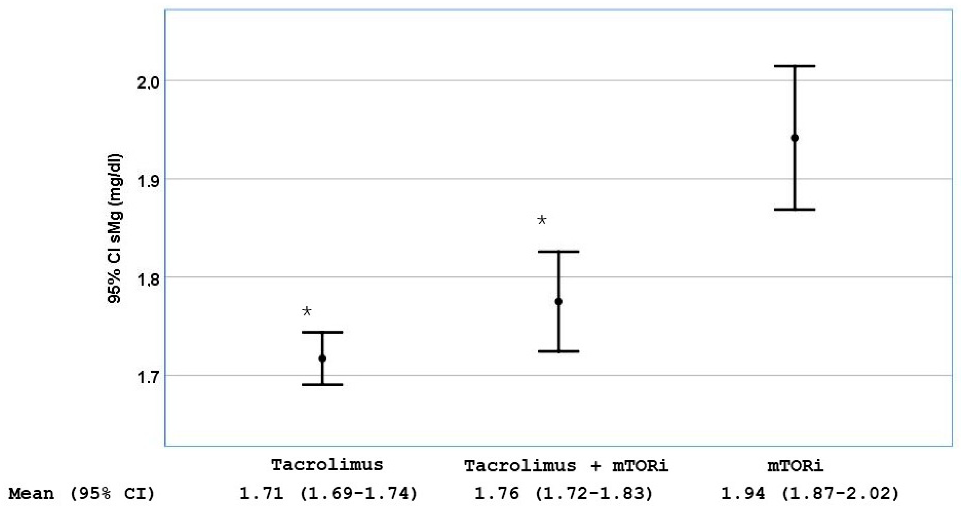
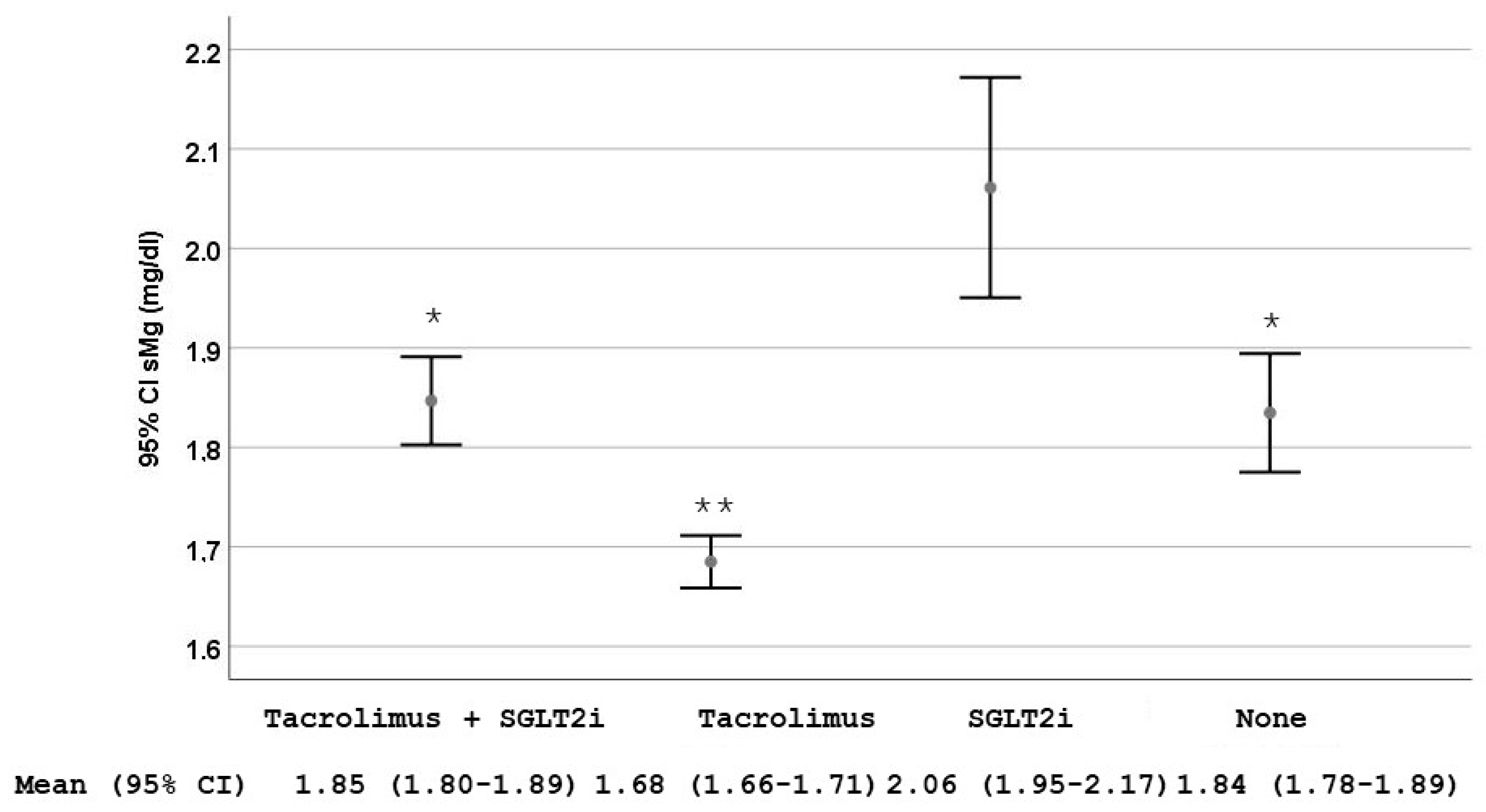
2. Results
3. Discussion
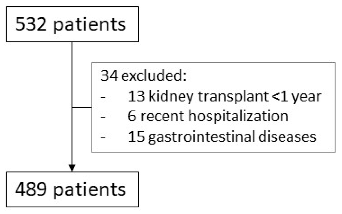
4. Materials and Methods
Statistical Analysis
5. Conclusions
Author Contributions
Funding
Institutional Review Board Statement
Informed Consent Statement
Data Availability Statement
Conflicts of Interest
Abbreviations
| ACEI | Angiotensin-converting enzyme inhibitor |
| ARB | Angiotensin receptor blocker |
| ATP | Adenosine triphosphate |
| CAV | Cardiac allograft vasculopathy |
| CVD | Cardiovascular disease |
| DCT | Distal convoluted tubule |
| DM | Diabetes mellitus |
| EGF | Epidermal growth factor |
| eGFR | Estimated glomerular filtration rate |
| HT | Heart transplant |
| KTR | Kidney transplant recipients |
| Mg2+ | Magnesium |
| MRA | Mineralocorticoid receptor antagonists |
| mTORi | Mammalian target of rapamycin inhibitors |
| PPIs | Proton pump inhibitors |
| PTH | Parathyroid hormone |
| ROS | Reactive oxygen species |
| SGLT2i | Sodium-glucose cotransporter-2 inhibitors |
| TRPM6 | Transient Receptor Potential Melastatin 6 |
| TRPM7 | Transient Receptor Potential cation channel subfamily M member 7 |
References
- Hariharan, S.; Israni, A.K.; Danovitch, G. Long-Term Survival after Kidney Transplantation. N. Engl. J. Med. 2021, 385, 729–743. [Google Scholar] [CrossRef] [PubMed]
- Garnier, A.S.; Duveau, A.; Planchais, M.; Subra, J.F.; Sayegh, J.; Augusto, J.F. Serum Magnesium after Kidney Transplantation: A Systematic Review. Nutrients 2018, 10, 729. [Google Scholar] [CrossRef] [PubMed]
- Beilhack, G.; Lindner, G.; Funk, G.C.; Monteforte, R.; Schwarz, C. Electrolyte disorders in stable renal allograft recipients. Swiss Med. Wkly. 2020, 150, w20366. [Google Scholar] [CrossRef]
- Odler, B.; Deak, A.T.; Pregartner, G.; Riedl, R.; Bozic, J.; Trummer, C.; Prenner, A.; Söllinger, L.; Krall, M.; Höflechner, L.; et al. Hypomagnesemia Is a Risk Factor for Infections after Kidney Transplantation: A Retrospective Cohort Analysis. Nutrients 2021, 13, 1296. [Google Scholar] [CrossRef]
- Duni, A.; Koutlas, V.; Tsitouridis, A.; Tzalavra, E.; Oikonomaki, T.; Kitsos, A.; Rapsomanikis, K.P.; Alekos, J.; Tatsis, V.; Pappas, C.; et al. Longitudinal Assessment of Electrolyte Disorders in a Cohort of Chronic Stable Kidney Transplant Recipients. Transplant. Proc. 2021, 53, 2786–2792. [Google Scholar] [CrossRef]
- Touyz, R.M.; de Baaij, J.H.F.; Hoenderop, J.G.J. Magnesium Disorders. N. Engl. J. Med. 2024, 390, 1998–2009. [Google Scholar] [CrossRef]
- Wynne, Z.; Falat, C. Disorders of Calcium and Magnesium. Emerg. Med. Clin. N. Am. 2023, 41, 833–848. [Google Scholar] [CrossRef]
- Matias, P.; Ávila, G.; Ferreira, A.C.; Laranjinha, I.; Ferreira, A. Hypomagnesemia: A potential underlooked cause of persistent vitamin D deficiency in chronic kidney disease. Clin. Kidney J. 2023, 16, 1776–1785. [Google Scholar] [CrossRef]
- De Waele, L.; Van Gaal, P.J.; Abramowicz, D. Electrolytes disturbances after kidney transplantation. Acta Clin. Belg. 2019, 74, 48–52. [Google Scholar] [CrossRef]
- Douwes, R.M.; Vinke, J.S.J.; Gomes-Neto, A.W.; Ayerdem, G.; van Hassel, G.; Berger, S.P.; Touw, D.J.; Blokzijl, H.; Bakker, S.J.L.; de Borst, M.H.; et al. Type of proton-pump inhibitor and risk of iron deficiency in kidney transplant recipients—Results from the Transplant Lines Biobank and Cohort Study. Transpl. Int. 2021, 34, 2305–2316. [Google Scholar] [CrossRef]
- Costello, R.B.; Elin, R.J.; Rosanoff, A.; Wallace, T.C.; Guerrero-Romero, F.; Hruby, A.; Lutsey, P.L.; Nielsen, F.H.; Rodriguez-Moran, M.; Song, Y.; et al. Perspective: The Case for an Evidence-Based Reference Interval for Serum Magnesium: The Time Has Come. Adv. Nutr. 2016, 7, 977–993. [Google Scholar] [CrossRef] [PubMed]
- Siddiqui, R.W.; Nishat, S.M.H.; Alzaabi, A.A.; Alzaabi, F.M.; Al Tarawneh, D.J.; Al Tarawneh, Y.J.; Khan, A.; Khan, M.A.M.; Siddiqui, T.W.; Siddiqui, S.W. The Connection Between Magnesium and Heart Health: Understanding Its Impact on Cardiovascular Wellness. Cureus 2024, 16, e72302. [Google Scholar] [CrossRef] [PubMed]
- Panta, R.; Regmi, S. Role of Magnesium, Effects of Hypomagnesemia, and Benefits of Magnesium Supplements in Cardiovascular and Chronic Kidney Diseases. Cureus 2024, 16, e64404. [Google Scholar] [CrossRef]
- Racca, V.; Scaglione, A.; De Maria, R.; Panzarino, C.; Santangelo, M.A.; Cipriani, M. Hypomagnesemia after heart transplantation or left ventricular assist device implant for end-stage heart failure. Clin. Transplant. 2020, 34, e13902. [Google Scholar] [CrossRef]
- Miles, C.D.; Westphal, S.G. Electrolyte Disorders in Kidney Transplantation. Clin. J. Am. Soc. Nephrol. 2020, 15, 412–414. [Google Scholar] [CrossRef]
- Stefanelli, L.F.; Alessi, M.; Bertoldi, G.; Rossato, V.; Di Vico, V.; Nalesso, F.; Calò, L.A. Calcineurin-Inhibitor-Induced Hypomagnesemia in Kidney Transplant Patients: A Monocentric Comparative Study between Sucrosomial Magnesium and Magnesium Pidolate Supplementation. J. Clin. Med. 2023, 12, 752. [Google Scholar] [CrossRef]
- Higgins, R.M.; Hart, P.; Lam, F.T.; Kashi, H. Conversion from tacrolimus to cyclosporin in stable renal transplant patients: Safety, metabolic changes, and pharmacokinetic comparison. Transplantation 2000, 70, 199–202. [Google Scholar]
- Minutolo, R.; De Nicola, L.; Mallamaci, F.; Zoccali, C. Thiazide diuretics are back in CKD: The case of chlorthalidone. Clin. Kidney J. 2022, 16, 41–51. [Google Scholar] [CrossRef]
- Zitt, E.; Woess, E.; Mayer, G.; Lhotta, K. Effect of cinacalcet on renal electrolyte handling and systemic arterial blood pressure in kidney transplant patients with persistent hyperparathyroidism. Transplantation 2011, 92, 883–889. [Google Scholar] [CrossRef]
- Andoh, T.F.; Burdmann, E.A.; Fransechini, N.; Houghton, D.C.; Bennett, W.M. Comparison of acute rapamycin nephrotoxicity with cyclosporine and FK506. Kidney Int. 1996, 50, 1110–1117. [Google Scholar] [CrossRef]
- Sánchez-Fructuoso, A.I.; Santín Cantero, J.M.; Pérez Flores, I.; Valero San Cecilio, R.; Calvo Romero, N.; Vilalta Casas, R. Changes in magnesium and potassium homeostasis after conversion from a calcineurin inhibitor regimen to an mTOR inhibitor-based regimen. Transplant. Proc. 2010, 42, 3047–3049. [Google Scholar] [CrossRef] [PubMed]
- Sánchez Fructuoso, A.I.; Bedia Raba, A.; Banegas Deras, E.; Vigara Sánchez, L.A.; Valero San Cecilio, R.; Franco Esteve, A.; Cruzado Vega, L.; Gavela Martínez, E.; González Garcia, M.E.; Saurdy Coronado, P.; et al. Sodium-glucose cotransporter-2 inhibitor therapy in kidney transplant patients with type 2 or post-transplant diabetes: An observational multicentre study. Clin. Kidney J. 2023, 16, 1022–1034. [Google Scholar] [CrossRef]
- Zhang, J.; Huan, Y.; Leibensperger, M.; Seo, B.; Song, Y. Comparative Effects of Sodium-Glucose Cotransporter 2 Inhibitors on Serum Electrolyte Levels in Patients with Type 2 Diabetes: A Pairwise and Network Meta-Analysis of Randomized Controlled Trials. Kidney360 2022, 3, 477–487. [Google Scholar] [CrossRef] [PubMed]
- Secondulfo, C.; Vecchione, N.; Russo, D.; Hamzeh, S.; Iacuzzo, C.; Apicella, L.; Di Pietro, R.A.; Pisani, A.; Amicone, M.; Cirillo, M.; et al. Impact of SGLT2 Inhibitors on Magnesium in Kidney Transplant Patients with and Without Diabetes. Int. J. Mol. Sci. 2025, 26, 2904. [Google Scholar] [CrossRef]
- Shah, C.V.; Sparks, M.A.; Lee, C.T. Sodium/Glucose Cotransporter 2 Inhibitors and Magnesium Homeostasis: A Review. Am. J. Kidney Dis. 2024, 83, 648–658. [Google Scholar] [CrossRef]
- Song, C.C.; Brown, A.; Winstead, R.; Yakubu, I.; Demehin, M.; Kumar, D.; Gupta, G. Early initiation of sodium-glucose linked transporter inhibitors (SGLT-2i) and associated metabolic and electrolyte outcomes in diabetic kidney transplant recipients. Endocrinol. Diabetes Metab. 2020, 4, e00185. [Google Scholar] [CrossRef]
- Ray, E.C.; Boyd-Shiwarski, C.R.; Liu, P.; Novacic, D.; Cassiman, D. SGLT2 Inhibitors for Treatment of Refractory Hypomagnesemia: A Case Report of 3 Patients. Kidney Med. 2020, 2, 359–364. [Google Scholar] [CrossRef] [PubMed]
- Gommers, L.M.; Hoenderop, J.G.; Bindels, R.J.; de Baaij, J.H. Hypomagnesemia in Type 2 Diabetes: A Vicious Circle? Diabetes 2016, 65, 3–13. [Google Scholar] [CrossRef]
- Van Laecke, S.; Van Biesen, W.; Verbeke, F.; De Bacquer, D.; Peeters, P.; Vanholder, R. Posttransplantation hypomagnesemia and its relation with immunosuppression as predictors of new-onset diabetes after transplantation. Am. J. Transplant. 2009, 9, 2140–2149. [Google Scholar] [CrossRef]
- Huang, J.W.; Famure, O.; Li, Y.; Kim, S.J. Hypomagnesemia and the Risk of New-Onset Diabetes Mellitus after Kidney Transplantation. J. Am. Soc. Nephrol. 2016, 27, 1793–1800. [Google Scholar] [CrossRef]
- Kao, W.H.; Folsom, A.R.; Nieto, F.J.; Mo, J.P.; Watson, R.L.; Brancati, F.L. Serum and dietary magnesium and the risk for type 2 diabetes mellitus: The Atherosclerosis Risk in Communities Study. Arch. Intern. Med. 1999, 159, 2151–2159. [Google Scholar] [CrossRef] [PubMed]
- Larsson, S.C.; Wolk, A. Magnesium intake and risk of type 2 diabetes: A meta-analysis. J. Intern. Med. 2007, 262, 208–214. [Google Scholar] [CrossRef]
- Ishimura, E.; Okuno, S.; Yamakawa, T.; Inaba, M.; Nishizawa, Y. Serum magnesium concentration is a significant predictor of mortality in maintenance hemodialysis patients. Magnes. Res. 2007, 20, 237–244. [Google Scholar] [PubMed]
- Tam, M.; Gómez, S.; González-Gross, M.; Marcos, A. Possible roles of magnesium on the immune system. Eur. J. Clin. Nutr. 2003, 57, 1193–1197. [Google Scholar] [CrossRef] [PubMed]
- Levey, A.S.; Stevens, L.A.; Schmid, C.H.; Zhang, Y.L.; Castro AF3rd Feldman, H.I.; Kusek, J.W.; Eggers, P.; Van Lente, F.; Greene, T.; CKD-EPI (Chronic Kidney Disease Epidemiology Collaboration); et al. A new equation to estimate glomerular filtration rate. Ann. Intern. Med. 2009, 150, 604–612, Erratum in Ann. Intern. Med. 2011, 155, 408. [Google Scholar] [CrossRef]
- von Elm, E.; Altman, D.G.; Egger, M.; Pocock, S.J.; Gøtzsche, P.C.; Vandenbroucke, J.P.; STROBE Initiative. The Strengthening the Reporting of Observational Studies in Epidemiology (STROBE) statement: Guidelines for reporting observational studies. J. Clin. Epidemiol. 2008, 61, 344–349. [Google Scholar] [CrossRef]


| Variable | Serum Magnesium Level | p | |
|---|---|---|---|
| Low (≤1.7 mg/dL) (N = 248) | Normal (≥1.8 mg/dL) (N = 241) | ||
| Recipient age, mean (SD) | 66.4 (14.1) | 62.9 (13.2) | 0.131 |
| Recipient gender (male) N (%) | 160 (49.8%) | 161 (50%) | 0.594 |
| Diabetes mellitus N (%) | 123 (49.4%) | 126 (50.6%) | 0.553 |
| Months post-transplantation, median (IQR) | 119.3 (46–99) | 113 (60–172) | 0.618 |
| eFGR, median (IQR) | 45.7 (34.0–63.2) | 43.5 (31.8–57.5) | 0.497 |
| Serum magnesium (mg/dL), mean (SD) | 1.56 (0.14) | 1.96 (0.17) | <0.001 |
| Serum calcium (mg/dL), median (IQR) | 9.7 (9.4–10.0) | 9.8 (9.4–10.1) | 0.186 |
| Serum phosphorus, median (IQR) | 3.3 (3.0–3.7) | 3.5 (3.0–3.9 | 0.002 |
| 25 OH vitamin D (ng/mL), median (IQR) (N = 465) | 25 (18–33) | 25 (18–33) | 0.958 |
| PTH (pg/mL), median (IQR) (N = 469) | 98.7 (67.3–140.3) | 100 (71.4–141.0) | 0.818 |
| Magnesium fractional excretion (%) (N = 443) | 7.23 (4.80–9.99) | 7.18 (5.05–10.15) | 0.722 |
| Urinary magnesium (mg/day), median (IQR) (N = 443) | 630 (424–937) | 768 (560–1039) | 0.003 |
| Urinary magnesium/creatinine ratio (mg/g) (N = 443) | 56.9 (37.2–75.0) | 66.5 (47.3–89.3) | <0.001 |
| Variable | Serum Magnesium Level | p | |
|---|---|---|---|
| Low (≤1.7 mg/dL) (N = 248) | Normal (≥1.8 mg/dL) (N = 241) | ||
| Tacrolimus treatment N (%) | 217 (54.4%) | 182 (45.6%) | 0.001 |
| Tacrolimus levels (ng/mL) | 8.22 (2.76) | 7.68 (2.01) | 0.030 |
| Cyclosporine treatment N (%) | 16 (42.1%) | 21 (56.8%) | 0.344 |
| Cyclosporine levels (ng/mL) | 97.8 (29.7) | 94.2 (26.3) | 0.698 |
| Mycophenolate treatment N (%) | 190 (52.6) | 171 (47.4) | 0.155 |
| Prednisone treatment N (%) | 134 (50.6) | 131 (49.4) | 0.943 |
| Azathioprine treatment N (%) | 9 (60%) | 6 (40%) | 0.465 |
| mTOR inhibitors treatment N (%) | 52 (39.4%) | 80 (60.6%) | 0.002 |
| Loop diuretics, treatment N (%) | 22 (46.8%) | 25 (53.2%) | 0.573 |
| Thiazide diuretics treatment N (%) | 44 (62.9%) | 26 (37.1%) | 0.028 |
| Cholecalciferol treatment | 152 (51.7%) | 142 (48.3%) | 0.593 |
| Calcitriol treatment N (%) | 3 (30.3%) | 7 (70%) | 0.186 |
| Paricalcitol treatment N (%) | 25 (53.2%) | 22 (46.8%) | 0.721 |
| Cinacalcet treatment N (%) | 49 (63.6%) | 28 (36.4%) | 0.013 |
| Proton pump inhibitors treatment N (%) | 173 (50.7%) | 168 (49.3%) | 0.991 |
| Denosumab treatment N (%) | 12 (66.7%) | 6 (33.3) | 0.168 |
| ACEI treatment N (%) | 83 (56.1%) | 65 (43.9%) | 0.118 |
| ARA2 treatment N (%) | 127 (51%) | 122 (49.0%) | 0.897 |
| MRA treatment N (%) | 30 (45.5%) | 36 (54.5) | 0.358 |
| iSGLT2 treatment N (%) | 26 (26.0%) | 94 (74.0%) | <0.001 |
| Oral Mg2+ supplement | 110 (75.9%) | 35 (24.1%) | <0.001 |
| Variable | OR (PIC) | p |
|---|---|---|
| Age | 0.97 (0.96–0.99) | 0.001 |
| Months post-transplantation | 1.005 (1.003–1.008) | <0.001 |
| Diabetes mellitus No Si | 1 1.94 (1.22–3.08) | 0.005 |
| iSGLT2 treatment No Yes | 6.18 (3.64–10.5) 1 | <0.001 |
| Tacrolimus treatment No Yes | 1 2.91 (1.62–5.22) | <0.001 |
| Thiazide treatment No Yes | 1 2.23 (1.21–4.08) | 0.010 |
| Cinacalcet treatment No Yes | 1 2.31 (1.29–4.13) | 0.005 |
| IMTOR treatment No Yes | 1.61 (1.02–2.62) 1 | 0.042 |
| Serum phosphate * <3.7 mg/dL ≥3.7 mg/dL (p75) | 1.99 (1.29–3.05) 1 | 0.002 |
| Serum calcium * ≤10 mg/dL >10 mg/dL (p75) | 1.75 (1.11–2.78) 1 | 0.017 |
Disclaimer/Publisher’s Note: The statements, opinions and data contained in all publications are solely those of the individual author(s) and contributor(s) and not of MDPI and/or the editor(s). MDPI and/or the editor(s) disclaim responsibility for any injury to people or property resulting from any ideas, methods, instructions or products referred to in the content. |
© 2025 by the authors. Licensee MDPI, Basel, Switzerland. This article is an open access article distributed under the terms and conditions of the Creative Commons Attribution (CC BY) license (https://creativecommons.org/licenses/by/4.0/).
Share and Cite
Riaza Ortiz, C.; Fernández Fernández, C.; Pujol Pujol, M.; Muñiz Rincón, M.; Aiffil Meneses, A.S.; Pérez Flores, I.M.; Calvo Romero, N.; Moreno de la Higuera, M.Á.; Rodríguez Cubillo, B.; Ramos Corral, R.; et al. Prevalence, Risk Factors and Potential Protective Strategies for Hypomagnesemia in Kidney Transplant Recipients. Int. J. Mol. Sci. 2025, 26, 6528. https://doi.org/10.3390/ijms26136528
Riaza Ortiz C, Fernández Fernández C, Pujol Pujol M, Muñiz Rincón M, Aiffil Meneses AS, Pérez Flores IM, Calvo Romero N, Moreno de la Higuera MÁ, Rodríguez Cubillo B, Ramos Corral R, et al. Prevalence, Risk Factors and Potential Protective Strategies for Hypomagnesemia in Kidney Transplant Recipients. International Journal of Molecular Sciences. 2025; 26(13):6528. https://doi.org/10.3390/ijms26136528
Chicago/Turabian StyleRiaza Ortiz, Cristina, Carlos Fernández Fernández, Marina Pujol Pujol, María Muñiz Rincón, Arianne Sofía Aiffil Meneses, Isabel María Pérez Flores, Natividad Calvo Romero, María Ángeles Moreno de la Higuera, Beatriz Rodríguez Cubillo, Raquel Ramos Corral, and et al. 2025. "Prevalence, Risk Factors and Potential Protective Strategies for Hypomagnesemia in Kidney Transplant Recipients" International Journal of Molecular Sciences 26, no. 13: 6528. https://doi.org/10.3390/ijms26136528
APA StyleRiaza Ortiz, C., Fernández Fernández, C., Pujol Pujol, M., Muñiz Rincón, M., Aiffil Meneses, A. S., Pérez Flores, I. M., Calvo Romero, N., Moreno de la Higuera, M. Á., Rodríguez Cubillo, B., Ramos Corral, R., & Sánchez Fructuoso, A. I. (2025). Prevalence, Risk Factors and Potential Protective Strategies for Hypomagnesemia in Kidney Transplant Recipients. International Journal of Molecular Sciences, 26(13), 6528. https://doi.org/10.3390/ijms26136528