Article

High Prevalence of TERT and CTNNB1 Mutations in Brazilian HCC Tissues: Insights into Early Detection and Risk Stratification

by
Thaís Barbosa Ferreira Sant’Anna
1,
Mariana Leonardo Terra
1,
Jose Junior França de Barros
1,
Leonardo Alexandre de Souza Ruivo
1,
Arlete Fernandes
2,
Maria Dirlei Ferreira de Souza Begnami
3,4,
Vera Lucia Nunes Pannain
2,
Antônio Hugo José Fróes Marques Campos
5,6,
Otacilio da Cruz Moreira
1 and
Natalia Motta de Araujo
1,*
1
Laboratory of Molecular Virology and Parasitology, Oswaldo Cruz Institute, Oswaldo Cruz Foundation, Rio de Janeiro 21040-900, Brazil
2
Department of Pathology, Clementino Fraga Filho University Hospital, Federal University of Rio de Janeiro, Rio de Janeiro 21941-617, Brazil
3
Department of Anatomic Pathology, A.C.Camargo Cancer Center, São Paulo 01509-010, Brazil
4
Sírio-Libanês Institute of Education and Research, Sírio-Libanês Hospital, São Paulo 01308-050, Brazil
5
A.C.Camargo Biobank, A.C.Camargo Cancer Center, São Paulo 01509-010, Brazil
6
D’Or Institute for Research and Education, IDOr, São Paulo 01401-002, Brazil
*
Author to whom correspondence should be addressed.
Int. J. Mol. Sci. 2025, 26(13), 6503; https://doi.org/10.3390/ijms26136503
Submission received: 9 June 2025 / Revised: 30 June 2025 / Accepted: 4 July 2025 / Published: 6 July 2025

Abstract

Hepatocellular carcinoma (HCC) is a major cause of cancer-related mortality worldwide, but its molecular drivers remain underexplored in Latin American populations. This study investigated the prevalence, co-occurrence, and tissue distribution of somatic mutations in the TERT promoter (C228T/C250T) and CTNNB1 exon 3, as well as TERT gene expression, in liver tissues from Brazilian patients. A total of 85 samples (42 HCC, 21 cirrhosis, and 22 hepatitis) were analysed using Sanger sequencing and RT-qPCR. TERT promoter mutations were detected in 47.6% of HCC tissues, including C228T (45.2%) and C250T (2.4%), and were also present in 19% of cirrhotic tissues but absent in hepatitis samples, supporting their emergence in early hepatocarcinogenesis. CTNNB1 exon 3 mutations occurred in 17.2% of HCCs and significantly co-occurred with TERTp mutations (66.7%, p = 0.0485), although the number of CTNNB1-mutated cases was limited. TERT expression was significantly upregulated in HCC tissues regardless of mutation status (p < 0.001). This is the first study to characterise these mutations in Brazilian HCC patients, highlighting the C228T mutation as a promising biomarker for early detection and molecular surveillance in cirrhotic individuals. Despite the genetic admixture of the studied population, the mutational patterns were comparable to those reported in more homogeneous populations, reinforcing the global relevance of these molecular alterations.

1. Introduction

Hepatocellular carcinoma (HCC) is the most common type of liver cancer, accounting for approximately 90% of primary liver malignancies. Due to its aggressive nature and poor prognosis, the global incidence of HCC closely mirrors patient mortality, representing a major public health challenge [1,2,3]. Currently, liver cancer is the sixth most common cancer and the third leading cause of cancer-related deaths worldwide [4,5]. Despite advances in medical treatment, HCC prognosis remains unfavourable, with a 5-year survival rate below 20%, primarily due to late diagnosis [2]. Early detection is crucial to improving patient outcomes, yet this remains challenging, particularly in populations with limited healthcare access or inadequate screening protocols.
Globally, HCC arises from a complex interplay of genetic factors, environmental exposures, viral infections, and chronic liver diseases [6]. Major risk factors include chronic hepatitis B virus (HBV) or hepatitis C virus (HCV) infections, chronic alcohol consumption, and metabolic dysfunction-associated steatotic liver disease (MASLD), formerly known as non-alcoholic fatty liver disease (NAFLD), frequently accompanied by obesity and type 2 diabetes. These risk factors promote chronic hepatic inflammation, leading to repeated cycles of hepatocyte injury, regeneration, and eventual cirrhosis, observed in 80–90% of HCC cases [1].
Among the diverse genetic alterations associated with HCC, somatic mutations in the telomerase reverse transcriptase promoter (TERTp) and in the β-catenin-encoding CTNNB1 gene are particularly noteworthy [7,8]. TERTp mutations (notably C228T, also referred to as −124C > T, and C250T, also referred to as −146C > T) increase telomerase activity, critical for tumour cell immortality [9]. A recent comprehensive review revealed that, globally, TERTp mutations occur in approximately 49.2% of HCC cases, with markedly higher frequencies observed in HCV-associated tumours (66.2%) compared to HBV-associated cases (31.6%) [10]. Additionally, mutations in CTNNB1 exon 3, mainly amino acid substitutions at residues S29, D32, S33, G34, S37, T41, and S45, disrupt key phosphorylation sites, stabilizing β-catenin and thereby activating canonical Wnt/β-catenin signalling pathways that drive cellular proliferation and tumour progression [11]. CTNNB1 exon 3 mutations, present in 23.1% of HCC cases worldwide, are similarly more prevalent in HCV-related HCC (30.7%) than in HBV-related tumours (12.8%) [10]. There is evidence of concordance between TERTp and CTNNB1 mutations in HCC, suggesting a cooperative role in hepatocarcinogenesis, as studies indicate that their co-occurrence may promote tumour development and progression [12,13].
In Brazil, 10,598 deaths were attributed to HCC in 2022 [14], and an estimated 65% of cases are linked to chronic HCV infection [15]. Combined with the country’s pronounced admixed ancestry (European, African and Indigenous), this epidemiological profile creates a unique setting to examine how viral aetiology and host genetics shape the mutational landscape of HCC. Despite extensive international documentation, the prevalence and significance of TERTp and CTNNB1 mutations remain largely unexplored among Brazilian patients.
Therefore, this study aims to analyse the prevalence of TERTp (C228T and C250T) and CTNNB1 exon 3 mutations and evaluate TERT expression in tumour and non-tumour liver tissues from Brazilian patients, providing novel insights into the molecular pathogenesis of HCC in this genetically heterogeneous population.

2. Results

2.1. TERTp and CTNNB1 Exon 3 Mutations in Tumour Tissue Samples and Their Correlation with Clinicopathological Characteristics

Among the 42 HCC tumour tissue samples analysed, the TERTp C228T mutation was identified in 19 cases (45.2%). Patients harbouring this mutation were predominantly aged ≥60 years (68.4%), male (57.9%), with tumours < 5 cm in size (42.1%), and moderately differentiated (63.2%). Regarding underlying liver disease, 42.1% of TERT-mutated cases were associated with HCV infection, while no cases were linked to HBV. No statistically significant associations were found between TERT mutation status and clinicopathological variables (p > 0.05; Table 1).
Somatic mutations in exon 3 of CTNNB1 were detected in 5 of the 29 HCC tumour samples (17.2%). These mutations were exclusively observed in patients aged ≥60 years (100%), predominantly male (60%), with tumours < 5 cm in size (60%), and moderate histological differentiation (60%). Most CTNNB1-mutated cases were associated with non-viral aetiologies (80%), while only 20% were linked to HCV infection and none were linked to HBV. Again, no statistically significant differences were observed between CTNNB1 mutation status and the evaluated clinicopathological parameters (Table 1). It should be noted that the absence of statistically significant associations may be influenced by the small number of cases in each aetiological subgroup, which limits the power of the statistical analysis.

2.2. TERTp and CTNNB1 Exon 3 Mutations Across All Samples and Their Correlation with Liver Disease Tissue Types

Among the 85 liver tissue samples analysed, the TERTp C250T mutation was identified in only one case (1.2%), corresponding to HCC tumour tissue; the paired adjacent non-tumour tissue from this patient did not harbour the mutation. In contrast, the C228T mutation in the TERTp was detected in 23 of the 85 samples (27.1%), with distribution across tissue types as follows: 19 of 42 HCC tissues (45.2%), 4 of 21 cirrhotic tissues (19%), and none of the 22 hepatitis tissues. Statistically significant differences were observed between HCC and hepatitis tissues (p = 0.0001), hepatitis and cirrhotic tissues (p = 0.0485), and tumour versus non-tumour tissues overall (p = 0.0005). No statistically significant difference was found between HCC and cirrhotic tissues (p = 0.0542), though a trend was noted (Table 2). Overall, among the 42 HCC tissue samples analysed, TERTp mutations at positions C228T and C250T were identified in 47.6% of the samples (n = 19 and n = 1, respectively).
Somatic mutations in exon 3 of the CTNNB1 gene were assessed in 67 liver tissue samples, revealing six mutated cases (9%). These included five HCC tissues, with amino acid substitutions at S29A (n = 1), S29F (n = 1), D32V (n = 1), and D32G (n = 2), and one cirrhotic tissue harbouring a T41P substitution. The overall mutation frequency was 17.2% (5/29) in HCC tissues, 5.6% (1/18) in cirrhotic tissues, and 0% in hepatitis tissues. Although CTNNB1 exon 3 mutations appeared more frequent with advancing liver disease, no statistically significant correlation was observed across tissue types (p > 0.05; Table 2).

2.3. TERTp and CTNNB1 Exon 3 Mutations in Paired Samples and Their Correlation with Liver Disease Tissue Types

Analysis of paired tumour and adjacent non-tumour liver tissues revealed distinct mutation patterns. For the TERTp C228T mutation, 12 out of 26 patients (46.2%) exhibited the mutation exclusively in tumour tissue, while no mutations were detected solely in non-tumour tissue (p = 0.0001; Table 3). In addition, two patients (7.7%) exhibited the C228T mutation in both tumour and non-tumour tissues, while 12 patients (46.2%) had no detectable mutation in either tissue. Similarly, assessment of CTNNB1 exon 3 mutations in paired samples showed that 4 of 24 patients (16.7%) harboured mutations only in tumour tissue, with no cases presenting mutations restricted to non-tumour tissue (p = 0.1092; Table 3). Additionally, one patient (4.2%) exhibited mutations in both tumour and non-tumour tissues, while 19 patients (79.2%) had no detectable mutation in either tissue.
These findings underscore that both TERTp and CTNNB1 mutations preferentially localise to malignant tissue, although only TERTp mutations demonstrated a statistically significant enrichment when comparing paired tumour and non-tumour samples.

2.4. Association Between TERTp and CTNNB1 Exon 3 Mutations

Analysis of the association between TERTp C228T and CTNNB1 exon 3 mutations revealed a notable overlap. Among the six samples harbouring CTNNB1 exon 3 mutations, four (66.7%) also carried the C228T mutation in the TERTp. In contrast, only 13 of the 54 (24.1%) CTNNB1 wild-type samples exhibited TERTp C228T. This association was statistically significant (p = 0.0485; Table 4).

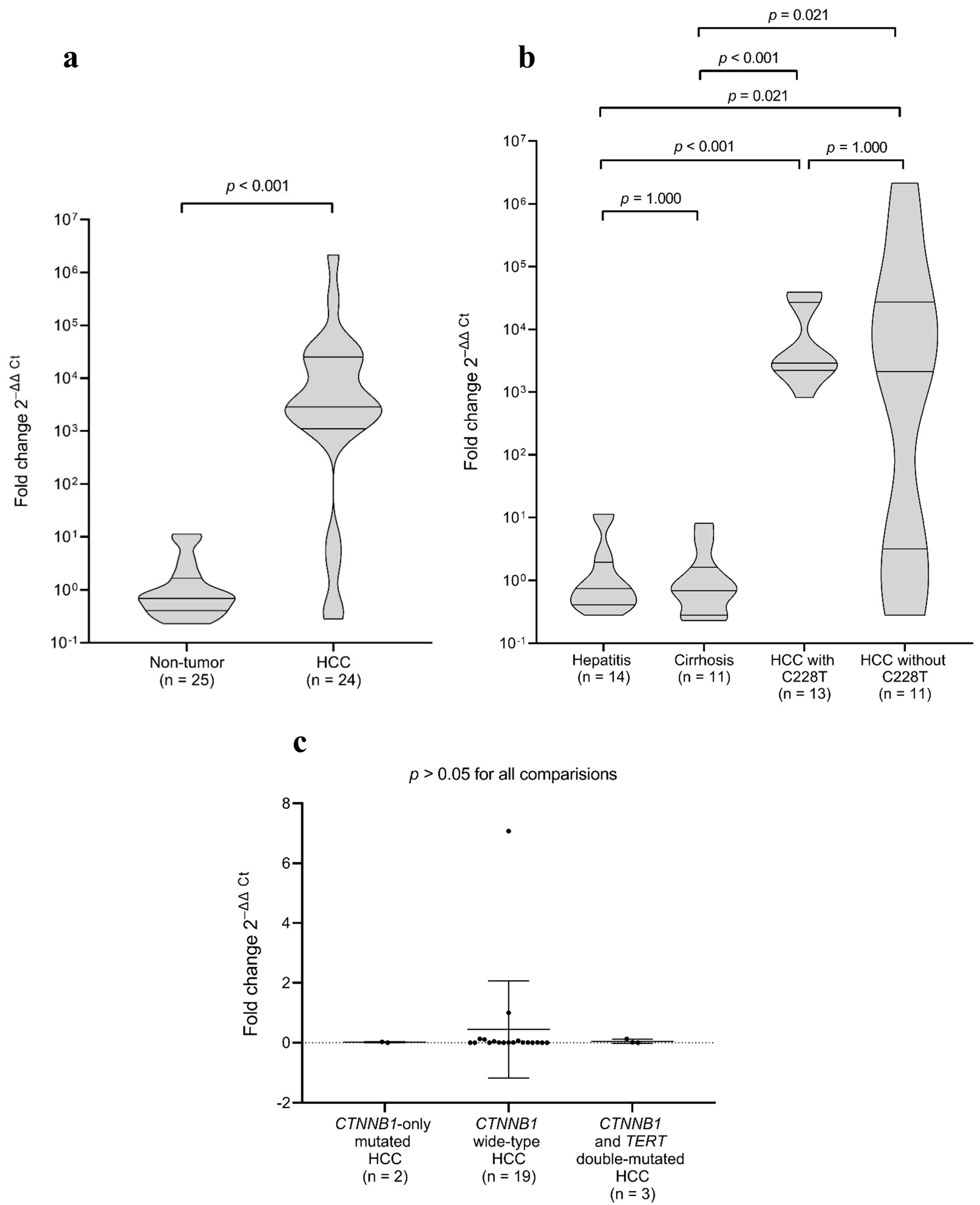
2.5. TERT Gene Expression in Tumour and Non-Tumour Samples

TERT expression was significantly elevated in HCC tissues compared to non-tumour liver tissue samples diagnosed histologically as cirrhosis or hepatitis (p < 0.001; Figure 1a). No significant difference in TERT expression was observed between HCC samples harbouring the C228T mutation and those without this mutation (p = 1.000), providing evidence that TERT overexpression is a consistent feature of HCC, independent of C228T mutation status (Figure 1b). Additionally, the comparison between cirrhotic and hepatitis tissues revealed no significant difference in TERT expression levels (p = 1.000; Figure 1b). Further analysis showed no significant differences in TERT expression among HCC tissues with CTNNB1 mutations only, wild-type CTNNB1, and those with both CTNNB1 and TERTp mutations (p > 0.05; Figure 1c), suggesting that CTNNB1 mutation status did not influence TERT expression levels in HCC tissues.

3. Discussion

In this study, we investigated the prevalence of TERTp and CTNNB1 exon 3 mutations, as well as TERT gene expression, in HCC, cirrhotic, and hepatitis tissues from Brazilian patients. Our findings confirm and expand upon existing data, providing important insights into the molecular landscape of HCC in Latin American patients, a population underrepresented in global genomic studies.
TERTp and CTNNB1 mutations have been reported to be significantly associated with older patient age, male sex, and smaller tumours (<5 cm) [12,16]. Although no statistically significant associations were found in our study, we also observed a higher frequency of these mutations in male and older patients, as well as in smaller, moderately differentiated tumours, aligning with the previous literature. In addition, TERTp mutations were frequent in HCV-related HCCs (42.1%) but absent in HBV-related cases. This pattern aligns with international data showing that TERTp mutations are strongly associated with HCV infection and are less common in HBV-related tumours, as confirmed by large multicentre studies [10,13,17]. The higher prevalence of TERTp mutations in HCV-associated HCC supports the hypothesis that chronic inflammation and oxidative stress from HCV infection promote genomic instability, favouring TERTp mutations [18]. In contrast, HBV-associated HCC shows more variable TERTp mutation rates, likely influenced by HBV genotypes, integration patterns, and cofactors such as aflatoxin exposure and metabolic disorders [19,20]. Although TERT promoter mutations are less frequent in HBV-related HCC compared to HCV-related cases, activation of the TERT gene in HBV infection can occur through alternative mechanisms, particularly via HBV DNA integration into the TERT locus. This integration may lead to aberrant TERT expression and has been reported as a frequent early event in HBV-associated hepatocarcinogenesis [20,21]. Regarding CTNNB1 exon 3 mutations, they were markedly more prevalent in non-viral HCCs in our study, with a frequency of 80%. Consistently, CTNNB1 mutations are well documented as being more frequent in non-viral HCCs (e.g., alcohol-related and MASLD) than in viral-related cases [12,22]. While CTNNB1 mutations are key drivers of hepatocarcinogenesis, their prognostic value in established HCC remains controversial. Some studies link these mutations to less aggressive tumours, lower alpha-fetoprotein levels, and better differentiation [23,24,25], while others associate them with worse outcomes, such as vascular invasion, or find no significant impact on survival [26,27]. Although our findings did not reach statistical significance, likely due to sample size limitations, they reinforce the notion of distinct molecular pathways driving hepatocarcinogenesis according to underlying aetiology, underscoring the importance of considering viral status when interpreting the mutational landscape of HCC.
The 47.6% prevalence of TERTp mutations among the analysed Brazilian HCC cases is highly consistent with the 49.2% reported in a global series of 4133 HCC samples [10]. Our findings are also comparable to those from the USA (44.3%) [28], Germany (47.4%) [29], and Italy (50.4%) [30], despite slight regional differences. The C228T mutation in the TERTp was identified in 45.2% of HCC tissues, while the C250T mutation was detected in only one case (2.4%). This predominance of C228T over C250T is consistent with prior studies conducted across different geographic regions, where C228T accounts for approximately 90% of all TERTp mutations [10]. These data reinforce the notion that the C228T mutation generates a highly accessible binding site for ETS/TCF transcription factors, promoting telomerase activation more effectively than C250T [31]. Notably, we found that the TERTp C228T mutation was present in 19% of cirrhotic tissues, contrasting with earlier findings by Nault et al. (2013, 2014) [12,32], who reported no TERTp mutations in non-tumourous cirrhotic tissues of different aetiologies. This pattern of progressive increase, from 0% in hepatitis to 19% in cirrhosis and 45.2% in HCC, supports the hypothesis that TERTp C228T arises early in hepatocarcinogenesis, potentially within cirrhotic nodules prior to overt malignancy. These findings highlight the potential utility of the TERTp C228T mutation as a biomarker for enhanced surveillance and risk stratification in cirrhotic patients, warranting further investigation in longitudinal cohorts. It is worth noting that all tissue samples from this study underwent rigorous histopathological review to ensure accurate classification and exclude microscopic tumour infiltration, strengthening the validity of TERTp C228T detection in non-tumour cirrhotic tissues. While the clinical implementation of TERTp mutations screening is not yet established, our finding of C228T in nearly one-fifth of cirrhotic samples suggests a promising avenue for molecular surveillance in high-risk individuals. If validated in larger, prospective studies, TERTp mutational status could be incorporated into risk models and potentially guide personalised HCC monitoring strategies.
Regarding CTNNB1 exon 3, mutations were identified in 17.2% of HCC tissue samples, a frequency comparable to the 23.1% prevalence reported in a global series of 5276 HCC cases [10]. This frequency is in line with previous reports from Iran (18.1%) [33], Taiwan (18.3%) [34], and France (19%) [35]. As expected, these mutations were nearly absent in cirrhotic and hepatitis tissues, corroborating the idea that CTNNB1 mutations typically arise later in the carcinogenic cascade and are rare in precursor lesions [32,36].
TERT gene expression analysis revealed robust overexpression in HCC tissues compared to cirrhotic and hepatitis tissues (p < 0.001), independent of C228T mutation status. This aligns with findings from Nault et al. (2020), who demonstrated that although TERT is markedly upregulated in HCC, TERTp mutations are not the sole drivers of this overexpression, implicating additional regulatory mechanisms [37]. Consistent with this, we observed no significant difference in TERT expression between cirrhotic and hepatitis tissues, reinforcing that TERT upregulation is primarily linked to malignant transformation rather than to chronic liver injury alone. Furthermore, TERT expression did not vary significantly among HCC tissues stratified by CTNNB1 mutation status, suggesting that β-catenin activation does not directly influence TERT transcription in this context. Our study also confirmed a significant association between CTNNB1 and TERTp mutations, with 66.7% of CTNNB1-mutated HCCs also carrying the C228T mutation (p = 0.0485). While based on a small number of CTNNB1-mutated cases, the statistically significant co-occurrence with TERTp mutations supports previous reports [12,13,37,38] and suggests a potential synergistic role in hepatocarcinogenesis, which should be explored further in larger cohorts.
Beyond their role in tumourigenesis, TERTp and CTNNB1 exon 3 mutations hold significant prognostic value. TERTp mutations correlate with aggressive HCC phenotypes, including reduced progression-free survival and increased recurrence rates after resection, independent of tumour stage or liver function [12,39]. Conversely, CTNNB1-mutated HCCs often exhibit distinct clinicopathological features (e.g., well-differentiated histology), though their prognostic impact remains debated [27,38]. Therapeutically, these mutations represent emerging targets for personalised approaches. Preclinical evidence suggests that TERTp-mutant HCCs may be more sensitive to Polo-like kinase 1 (PLK1) inhibitors due to telomere maintenance vulnerabilities and aberrant cell cycle regulation [40]. In contrast, CTNNB1-mutant tumours, characterised by an immune-excluded microenvironment and resistance to PD-1 blockade [41], may benefit from therapies targeting the Wnt/β-catenin pathway, such as the small-molecule inhibitor PRI-724, currently under investigation [42]. Integrating mutational profiling with these emerging therapies could refine risk stratification and expand treatment options for molecularly defined HCC subsets.
This study has several limitations. First, the relatively small sample size, particularly in subgroup analyses (e.g., CTNNB1-mutated HCCs), may limit statistical power and generalisability. Second, the samples were obtained from two tertiary centres in Southeastern Brazil, which may not fully capture the nationwide heterogeneity of HCC cases. Third, the use of Sanger sequencing, while robust for hotspot detection, has limited sensitivity for low-frequency variants, potentially underestimating mutation prevalence in heterogeneous tumour samples. Fourth, while we profiled mutational and gene expression patterns, functional assays were beyond the scope of this study and should be pursued in future research. Finally, due to the retrospective design of this study and incomplete long-term clinical records, we were unable to correlate TERTp and CTNNB1 mutation status with patient survival, recurrence rates, or treatment outcomes. The absence of these clinical endpoints limits our ability to assess the prognostic value of these mutations in Brazilian HCC patients. Future prospective studies with standardised follow-up protocols are warranted to evaluate the impact of these molecular alterations on disease progression and therapeutic response. Despite these limitations, this is the first study to investigate TERTp and CTNNB1 mutations in Brazilian HCC patients, providing valuable and novel insights into the molecular landscape of HCC in this population.
Importantly, while our findings reaffirm key international trends, they also underscore the need for larger, multicentric studies in Brazil, given the country’s unique viral and ethnic background. Despite the limited sample size, the mutation frequencies and patterns observed in this admixed population were largely consistent with global data, reinforcing the broad relevance of these molecular markers. Nevertheless, it remains unclear whether genetic ancestry could subtly influence the mutational landscape or modulate the interplay between tumour drivers in hepatocarcinogenesis. Future studies incorporating ancestry-informative markers and larger, geographically diverse cohorts will be essential to investigate potential ancestry-related differences, ensuring that biomarker-based surveillance strategies are equitable and effective across populations.

4. Materials and Methods

4.1. Study Population

Liver tissue specimens, either cryopreserved (n = 50) or formalin-fixed paraffin-embedded (FFPE) (n = 35), were obtained from two institutions in Brazil: the A.C.Camargo Biobank at A.C.Camargo Cancer Center, São Paulo State, and the Department of Pathology at Clementino Fraga Filho University Hospital, Rio de Janeiro State. Demographic and clinicopathological data (age, sex, histology and underlying liver disease aetiology) were abstracted from the corresponding medical records when available. However, for a small subset of cases, especially among older FFPE samples, complete clinical data were not retrievable due to limitations in archived records.
For analysis of the C228T and C250T hotspot mutations in TERTp, we examined 85 liver-tissue samples collected from 59 patients with HCC. Histopathological review classified each specimen into one of three disease stages: HCC tissue (n = 42), cirrhotic non-tumour tissue (n = 21), or hepatitis tissue without cirrhosis (n = 22). Paired tumour and adjacent non-tumour samples (cirrhosis or hepatitis) were available for 26 of these patients. Somatic mutations in exon 3 of CTNNB1 were assessed in 67 liver specimens from 43 of the above patients. This subset comprised 29 HCC samples, 18 cirrhotic tissues, and 20 hepatitis tissues; paired tumour/non-tumour material was analysed from 24 patients. The smaller number of samples analysed for CTNNB1 reflects differences in amplification efficiency, as a greater proportion of tissue specimens yielded high-quality PCR products for TERTp than for CTNNB1. All tissue samples were independently reviewed by experienced pathologists to ensure accurate histopathological classification. Particular attention was given to the distinction between tumour and adjacent non-tumour tissues, especially cirrhotic nodules, to prevent misclassification due to microscopic tumour infiltration. Only samples with well-demarcated histological features and no evidence of tumour cell contamination were included in the non-tumour groups.
All participants provided written informed consent for tissue collection, banking, and research use. The study was approved by the Ethics Committees of Clementino Fraga Filho University Hospital (approval 139/10), A.C.Camargo Cancer Center (2.485.580), and the Oswaldo Cruz Institute/FIOCRUZ (2.296.938).

4.2. DNA Extraction and PCR Amplification

DNA was isolated from cryopreserved liver tissue on the QIAsymphony platform (QIAGEN, Hilden, Germany) with the BC400-V7 protocol. DNA from FFPE tissue was extracted with the QIAamp DNA FFPE Tissue Kit (QIAGEN, Hilden, Germany) according to the manufacturer’s instructions.
To detect the C228T and C250T mutations in TERTp, a 270-bp (base pair) fragment was amplified with primers flanking both hotspots (forward 5′-GCCGGGCTCCCAGTGGATTCG-3′; reverse 5′-GCTTCCCACGTGCGCAGCAGGA-3′). Each 25 µL reaction contained 2 µL of genomic DNA and 1 U of Platinum Taq DNA Polymerase (Invitrogen, Waltham, MA, USA). Cycling parameters were as follows: 95 °C for 15 min; 42 cycles of 95 °C for 15 s, 63 °C for 15 s, and 72 °C for 45 s; followed by a final extension at 72 °C for 5 min, as previously described [43].
For exon 3 of CTNNB1 (427 bp), PCR employed primers 5′-CCTGGCTATCATTCT GCTTTTC-3′ (forward) and 5′-TCAAAACTGCATTCTGACTTTCA-3′ (reverse) under the same reagent concentrations. Cycling conditions were as follows: 94 °C for 2 min; 35 cycles of 94 °C for 30 s, 58 °C for 30 s, and 72 °C for 1 min; final extension at 72 °C for 10 min, according to a published protocol [44].
Negative controls were included in both the extraction and PCR steps to monitor contamination. Amplicons were resolved on 1.5% agarose gels and visualised under UV illumination.

4.3. Nucleotide Sequencing

PCR products were purified using the QIAquick PCR Purification Kit (QIAGEN, Hilden, Germany) according to the manufacturer’s instructions. The TERTp and exon 3 of the CTNNB1 gene were sequenced via direct Sanger sequencing using the BigDye Terminator v3.1 Cycle Sequencing Kit (Applied Biosystems, Waltham, MA, USA). Sequencing reactions were analysed on an ABI 3730xl automated sequencer (Applied Biosystems).
Sequence data were processed and analysed using MEGA11: Molecular Evolutionary Genetics Analysis version 11 [45]. The analysis focused on the TERTp hotspot mutations C250T and C228T, whereas amino acid substitutions at residues S29, D32, S33, G34, S37, T41, and S45 were assessed in CTNNB1 exon 3.

4.4. RNA Extraction and TERT Reverse Transcription Quantitative PCR (RT-qPCR)

To evaluate the expression profile of the TERT gene, real-time PCR was performed to quantify TERT mRNA levels in the cryopreserved tissue samples. RNA extraction was automated using the QIAsymphony system (QIAGEN, Hilden, Germany) and the miRNA CT 400 V8 protocol from the QIAsymphony RNA Kit (QIAGEN, Hilden, Germany). In this protocol, total RNA was treated with the DNase enzyme provided in the kit during the purification process. The total RNA concentration was determined using a NanoDrop, and normalization was performed accordingly. RNA was reverse transcribed for first-strand cDNA synthesis using the SuperScript™ IV First-Strand Synthesis System (Invitrogen, Waltham, MA, USA) and random primers. Reverse transcription was performed by incubating the reaction mixture at 55 °C for 10 min, followed by inactivation at 80 °C for 10 min, according to the manufacturer’s instructions.
TERT expression was assessed using the TaqMan Applied Biosystems Gene Expression Assay (Hs00972656_m1) (Thermo Fisher Scientific, Waltham, MA, USA), and gene expression was normalised to the internal control 18S rRNA (Hs99999901_s1) and GAPDH (Hs99999905_m1) (Thermo Fisher Scientific, Waltham, MA, USA). RT-qPCR was performed under the following conditions: 50 °C for 2 min, initial denaturation at 95 °C for 10 min, followed by 45 cycles of 95 °C for 15 s and 60 °C for 1 min. The relative quantification of TERT mRNA was calculated using the 2−ΔΔCt method [46], using ExpressionSuite v1.3 software (Applied Biosystems, Waltham, MA, USA). No reverse transcriptase (-RT) controls were loaded in parallel to the samples.

4.5. Statistical Analysis

Fisher’s exact test was used to assess associations between tissue type, clinicopathological variables, and the presence of TERTp C228T or CTNNB1 mutations (R software version 4.4.1, R Foundation for Statistical Computing, Vienna, Austria). TERT expression was compared between HCC and non-tumour tissues using the Mann–Whitney U test and across hepatitis, cirrhosis, and HCC groups (stratified by mutation status) using the Kruskal–Wallis test followed by Dunn’s post hoc test. For multiple comparisons by CTNNB1 mutation status, we employed one-way ANOVA with Holm–Sidak post hoc tests. Tests were chosen based on data distribution. TERT expression assays were run in technical duplicates. Additional statistical analyses were conducted in SigmaPlot version 14.0 (Systat Software, San Jose, CA, USA) and GraphPad Prism version 8.0.1 (GraphPad, La Jolla, CA, USA), with p < 0.05 considered statistically significant.

5. Conclusions

In conclusion, this study provides novel insights into the molecular landscape of HCC in Brazilian patients, demonstrating a high prevalence of TERTp mutations, particularly C228T, not only in tumour tissues but also in non-tumourous cirrhotic tissues. These findings strongly support the early emergence of TERTp mutations in hepatocarcinogenesis and highlight their potential as biomarkers for molecular surveillance and risk stratification in cirrhotic individuals, especially in regions with high HCV burden. Moreover, the significant co-occurrence of TERTp and CTNNB1 exon 3 mutations suggests a possible synergistic role in tumour progression, pointing to distinct molecular pathways that could be exploited for therapeutic intervention. Despite the genetically admixed background of the Brazilian population, the mutational profiles observed were consistent with those in more homogeneous populations, reinforcing the broader applicability of these molecular alterations across diverse ethnic groups. Future studies involving larger, multicentric cohorts and functional analyses are essential to validate these findings, determine their prognostic value, and explore their utility in guiding personalised strategies for HCC detection, surveillance, and treatment.

Author Contributions

Conceptualisation, T.B.F.S. and N.M.d.A.; formal analysis, T.B.F.S., M.L.T. and L.A.d.S.R.; funding acquisition, N.M.d.A.; methodology, T.B.F.S., M.L.T., J.J.F.d.B., L.A.d.S.R. and A.F.; project administration, N.M.d.A.; resources, V.L.N.P., M.D.F.d.S.B. and A.H.J.F.M.C.; validation, T.B.F.S., O.d.C.M. and N.M.d.A.; writing—original draft, T.B.F.S. and N.M.d.A.; writing—review and editing: all authors. All authors have read and agreed to the published version of the manuscript.

Funding

This research was funded by the Fundação Carlos Chagas Filho de Amparo à Pesquisa do Estado do Rio de Janeiro (FAPERJ), grant number E-26/210.585/2024, and the Conselho Nacional de Desenvolvimento Científico e Tecnológico (CNPq), grant number 309649/2022-6. The APC was funded by the Oswaldo Cruz Institute.

Institutional Review Board Statement

The study was conducted in accordance with the Declaration of Helsinki and approved by the Ethics Committees of Clementino Fraga Filho University Hospital (approval 139/10), A.C.Camargo Cancer Center (2.485.580), and the Oswaldo Cruz Institute/FIOCRUZ (2.296.938).

Informed Consent Statement

Informed consent was obtained from all subjects involved in the study.

Data Availability Statement

The data supporting the findings of this study are available from the corresponding author upon reasonable request. These data are not publicly available due to privacy and ethical restrictions.

Acknowledgments

The authors thank the DNA Sequencing Platform (RPT01A-VPPCB/FIOCRUZ) of the Oswaldo Cruz Foundation for technical support.

Conflicts of Interest

The authors declare no conflicts of interest.

Abbreviations

The following abbreviations are used in this manuscript:
bpbase pair
CTNNB1catenin beta 1
FFPEformalin-fixed paraffin-embedded
HBVhepatitis B virus
HCChepatocellular carcinoma
HCVhepatitis C virus
MASLDmetabolic dysfunction-associated steatotic liver disease
NAFLDnon-alcoholic fatty liver disease
PLK1polo-like kinase 1
RT-qPCRreverse transcription quantitative PCR
TERTptelomerase reverse transcriptase promoter

References

  1. El-Serag, H.B. Hepatocellular carcinoma. N. Engl. J. Med. 2011, 365, 1118–1127. [Google Scholar] [CrossRef] [PubMed]
  2. Llovet, J.M.; Kelley, R.K.; Villanueva, A.; Singal, A.G.; Pikarsky, E.; Roayaie, S.; Lencioni, R.; Koike, K.; Zucman-Rossi, J.; Finn, R.S. Hepatocellular carcinoma. Nat. Rev. Dis. Primers 2021, 7, 6. [Google Scholar] [CrossRef]
  3. Villanueva, A. Hepatocellular Carcinoma. N. Engl. J. Med. 2019, 380, 1450–1462. [Google Scholar] [CrossRef] [PubMed]
  4. Ferlay, J.E.M.; Lam, F.; Laversanne, M.; Colombet, M.; Mery, L.; Piñeros, M.; Znaor, A.; Soerjomataram, I.; Bray, F. Global Cancer Observatory: Cancer Today. Lyon, France: International Agency for Research on Cancer. Available online: https://gco.iarc.who.int/media/globocan/factsheets/cancers/11-liver-and-intrahepatic-bile-ducts-fact-sheet.pdf (accessed on 18 January 2025).
  5. Sung, H.; Ferlay, J.; Siegel, R.L.; Laversanne, M.; Soerjomataram, I.; Jemal, A.; Bray, F. Global Cancer Statistics 2020: GLOBOCAN Estimates of Incidence and Mortality Worldwide for 36 Cancers in 185 Countries. CA Cancer J. Clin. 2021, 71, 209–249. [Google Scholar] [CrossRef]
  6. Zucman-Rossi, J.; Villanueva, A.; Nault, J.C.; Llovet, J.M. Genetic Landscape and Biomarkers of Hepatocellular Carcinoma. Gastroenterology 2015, 149, 1226–1239.e4. [Google Scholar] [CrossRef]
  7. Kotiyal, S.; Evason, K.J. Exploring the Interplay of Telomerase Reverse Transcriptase and beta-Catenin in Hepatocellular Carcinoma. Cancers 2021, 13, 4202. [Google Scholar] [CrossRef]
  8. Schulze, K.; Nault, J.C.; Villanueva, A. Genetic profiling of hepatocellular carcinoma using next-generation sequencing. J. Hepatol. 2016, 65, 1031–1042. [Google Scholar] [CrossRef]
  9. Horn, S.; Figl, A.; Rachakonda, P.S.; Fischer, C.; Sucker, A.; Gast, A.; Kadel, S.; Moll, I.; Nagore, E.; Hemminki, K.; et al. TERT promoter mutations in familial and sporadic melanoma. Science 2013, 339, 959–961. [Google Scholar] [CrossRef] [PubMed]
  10. Terra, M.L.; Sant’Anna, T.B.F.; de Barros, J.J.F.; de Araujo, N.M. Geographic and Viral Etiology Patterns of TERT Promoter and CTNNB1 Exon 3 Mutations in Hepatocellular Carcinoma: A Comprehensive Review. Int. J. Mol. Sci. 2025, 26, 2889. [Google Scholar] [CrossRef]
  11. Gao, C.; Wang, Y.; Broaddus, R.; Sun, L.; Xue, F.; Zhang, W. Exon 3 mutations of CTNNB1 drive tumorigenesis: A review. Oncotarget 2018, 9, 5492–5508. [Google Scholar] [CrossRef]
  12. Nault, J.C.; Mallet, M.; Pilati, C.; Calderaro, J.; Bioulac-Sage, P.; Laurent, C.; Laurent, A.; Cherqui, D.; Balabaud, C.; Zucman-Rossi, J. High frequency of telomerase reverse-transcriptase promoter somatic mutations in hepatocellular carcinoma and preneoplastic lesions. Nat. Commun. 2013, 4, 2218. [Google Scholar] [CrossRef] [PubMed]
  13. Totoki, Y.; Tatsuno, K.; Covington, K.R.; Ueda, H.; Creighton, C.J.; Kato, M.; Tsuji, S.; Donehower, L.A.; Slagle, B.L.; Nakamura, H.; et al. Trans-ancestry mutational landscape of hepatocellular carcinoma genomes. Nat. Genet. 2014, 46, 1267–1273. [Google Scholar] [CrossRef]
  14. INCA. Instituto Nacional do Câncer, Ministério da Saúde. Tipos de Câncer. Câncer de Fígado. Available online: https://www.gov.br/inca/pt-br/assuntos/cancer/tipos/figado (accessed on 5 May 2025).
  15. Paranagua-Vezozzo, D.C.; Ono, S.K.; Alvarado-Mora, M.V.; Farias, A.Q.; Cunha-Silva, M.; Franca, J.I.; Alves, V.A.; Sherman, M.; Carrilho, F.J. Epidemiology of HCC in Brazil: Incidence and risk factors in a ten-year cohort. Ann. Hepatol. 2014, 13, 386–393. [Google Scholar] [CrossRef]
  16. Nault, J.C.; Ningarhari, M.; Rebouissou, S.; Zucman-Rossi, J. The role of telomeres and telomerase in cirrhosis and liver cancer. Nat. Rev. Gastroenterol. Hepatol. 2019, 16, 544–558. [Google Scholar] [CrossRef]
  17. Chianchiano, P.; Pezhouh, M.K.; Kim, A.; Luchini, C.; Cameron, A.; Weiss, M.J.; He, J.; Voltaggio, L.; Oshima, K.; Anders, R.A.; et al. Distinction of intrahepatic metastasis from multicentric carcinogenesis in multifocal hepatocellular carcinoma using molecular alterations. Hum. Pathol. 2018, 72, 127–134. [Google Scholar] [CrossRef]
  18. Machida, K. HCV and tumor-initiating stem-like cells. Front. Physiol. 2022, 13, 903302. [Google Scholar] [CrossRef]
  19. An, P.; Xu, J.; Yu, Y.; Winkler, C.A. Host and Viral Genetic Variation in HBV-Related Hepatocellular Carcinoma. Front. Genet. 2018, 9, 261. [Google Scholar] [CrossRef]
  20. Tu, T.; Budzinska, M.A.; Shackel, N.A.; Urban, S. HBV DNA Integration: Molecular Mechanisms and Clinical Implications. Viruses 2017, 9, 75. [Google Scholar] [CrossRef]
  21. Sung, W.K.; Zheng, H.; Li, S.; Chen, R.; Liu, X.; Li, Y.; Lee, N.P.; Lee, W.H.; Ariyaratne, P.N.; Tennakoon, C.; et al. Genome-wide survey of recurrent HBV integration in hepatocellular carcinoma. Nat. Genet. 2012, 44, 765–769. [Google Scholar] [CrossRef]
  22. Kawai-Kitahata, F.; Asahina, Y.; Tanaka, S.; Kakinuma, S.; Murakawa, M.; Nitta, S.; Watanabe, T.; Otani, S.; Taniguchi, M.; Goto, F.; et al. Comprehensive analyses of mutations and hepatitis B virus integration in hepatocellular carcinoma with clinicopathological features. J. Gastroenterol. 2016, 51, 473–486. [Google Scholar] [CrossRef] [PubMed]
  23. Lu, G.; Lin, J.; Song, G.; Chen, M. Prognostic significance of CTNNB1 mutation in hepatocellular carcinoma: A systematic review and meta-analysis. Aging 2023, 15, 9759–9778. [Google Scholar] [CrossRef]
  24. Mao, T.L.; Chu, J.S.; Jeng, Y.M.; Lai, P.L.; Hsu, H.C. Expression of mutant nuclear beta-catenin correlates with non-invasive hepatocellular carcinoma, absence of portal vein spread, and good prognosis. J. Pathol. 2001, 193, 95–101. [Google Scholar] [CrossRef]
  25. Wang, Z.; Sheng, Y.Y.; Gao, X.M.; Wang, C.Q.; Wang, X.Y.; Lu, X.U.; Wei, J.W.; Zhang, K.L.; Dong, Q.Z.; Qin, L.X. beta-catenin mutation is correlated with a favorable prognosis in patients with hepatocellular carcinoma. Mol. Clin. Oncol. 2015, 3, 936–940. [Google Scholar] [CrossRef] [PubMed]
  26. Khalaf, A.M.; Fuentes, D.; Morshid, A.I.; Burke, M.R.; Kaseb, A.O.; Hassan, M.; Hazle, J.D.; Elsayes, K.M. Role of Wnt/beta-catenin signaling in hepatocellular carcinoma, pathogenesis, and clinical significance. J. Hepatocell. Carcinoma 2018, 5, 61–73. [Google Scholar] [CrossRef]
  27. Rebouissou, S.; Franconi, A.; Calderaro, J.; Letouze, E.; Imbeaud, S.; Pilati, C.; Nault, J.C.; Couchy, G.; Laurent, A.; Balabaud, C.; et al. Genotype-phenotype correlation of CTNNB1 mutations reveals different ss-catenin activity associated with liver tumor progression. Hepatology 2016, 64, 2047–2061. [Google Scholar] [CrossRef] [PubMed]
  28. Killela, P.J.; Reitman, Z.J.; Jiao, Y.; Bettegowda, C.; Agrawal, N.; Diaz, L.A., Jr.; Friedman, A.H.; Friedman, H.; Gallia, G.L.; Giovanella, B.C.; et al. TERT promoter mutations occur frequently in gliomas and a subset of tumors derived from cells with low rates of self-renewal. Proc. Natl. Acad. Sci. USA 2013, 110, 6021–6026. [Google Scholar] [CrossRef] [PubMed]
  29. Quaas, A.; Oldopp, T.; Tharun, L.; Klingenfeld, C.; Krech, T.; Sauter, G.; Grob, T.J. Frequency of TERT promoter mutations in primary tumors of the liver. Virchows Arch. 2014, 465, 673–677. [Google Scholar] [CrossRef]
  30. Pezzuto, F.; Izzo, F.; Buonaguro, L.; Annunziata, C.; Tatangelo, F.; Botti, G.; Buonaguro, F.M.; Tornesello, M.L. Tumor specific mutations in TERT promoter and CTNNB1 gene in hepatitis B and hepatitis C related hepatocellular carcinoma. Oncotarget 2016, 7, 54253–54262. [Google Scholar] [CrossRef]
  31. Hafezi, F.; Bercoff, D.P. The Solo Play of TERT Promoter Mutations. Cells 2020, 9, 749. [Google Scholar] [CrossRef]
  32. Nault, J.C.; Calderaro, J.; Tommaso, L.D.; Balabaud, C.; Zafrani, E.S.; Bioulac-Sage, P.; Roncalli, M.; Zucman-Rossi, J. Telomerase reverse transcriptase promoter mutation is an early somatic genetic alteration in the transformation of premalignant nodules in hepatocellular carcinoma on cirrhosis. Hepatology 2014, 60, 1983–1992. [Google Scholar] [CrossRef]
  33. Javanmard, D.; Najafi, M.; Babaei, M.R.; Niya, M.H.K.; Esghaei, M.; Panahi, M.; Tameshkel, F.S.; Tavakoli, A.; Jazayeri, S.M.; Ghaffari, H.; et al. Investigation of CTNNB1 gene mutations and expression in hepatocellular carcinoma and cirrhosis in association with hepatitis B virus infection. Infect. Agent. Cancer 2020, 15, 37. [Google Scholar] [CrossRef] [PubMed]
  34. Lu, L.C.; Shao, Y.Y.; Lee, Y.H.; Hsieh, M.S.; Hsiao, C.H.; Lin, H.H.; Kao, H.F.; Ma, Y.Y.; Yen, F.C.; Cheng, A.L.; et al. beta-catenin (CTNNB1) mutations are not associated with prognosis in advanced hepatocellular carcinoma. Oncology 2014, 87, 159–166. [Google Scholar] [CrossRef]
  35. Laurent-Puig, P.; Legoix, P.; Bluteau, O.; Belghiti, J.; Franco, D.; Binot, F.; Monges, G.; Thomas, G.; Bioulac-Sage, P.; Zucman-Rossi, J. Genetic alterations associated with hepatocellular carcinomas define distinct pathways of hepatocarcinogenesis. Gastroenterology 2001, 120, 1763–1773. [Google Scholar] [CrossRef]
  36. Park, J.Y.; Park, W.S.; Nam, S.W.; Kim, S.Y.; Lee, S.H.; Yoo, N.J.; Lee, J.Y.; Park, C.K. Mutations of beta-catenin and AXIN I genes are a late event in human hepatocellular carcinogenesis. Liver Int. 2005, 25, 70–76. [Google Scholar] [CrossRef] [PubMed]
  37. Nault, J.C.; Martin, Y.; Caruso, S.; Hirsch, T.Z.; Bayard, Q.; Calderaro, J.; Charpy, C.; Copie-Bergman, C.; Ziol, M.; Bioulac-Sage, P.; et al. Clinical Impact of Genomic Diversity From Early to Advanced Hepatocellular Carcinoma. Hepatology 2020, 71, 164–182. [Google Scholar] [CrossRef]
  38. Schulze, K.; Imbeaud, S.; Letouze, E.; Alexandrov, L.B.; Calderaro, J.; Rebouissou, S.; Couchy, G.; Meiller, C.; Shinde, J.; Soysouvanh, F.; et al. Exome sequencing of hepatocellular carcinomas identifies new mutational signatures and potential therapeutic targets. Nat. Genet. 2015, 47, 505–511. [Google Scholar] [CrossRef] [PubMed]
  39. Nault, J.C.; Galle, P.R.; Marquardt, J.U. The role of molecular enrichment on future therapies in hepatocellular carcinoma. J. Hepatol. 2018, 69, 237–247. [Google Scholar] [CrossRef]
  40. Tang, Q.; Hu, G.; Sang, Y.; Chen, Y.; Wei, G.; Zhu, M.; Chen, M.; Li, S.; Liu, R.; Peng, Z. Therapeutic targeting of PLK1 in TERT promoter-mutant hepatocellular carcinoma. Clin. Transl. Med. 2024, 14, e1703. [Google Scholar] [CrossRef]
  41. de Galarreta, M.R.; Bresnahan, E.; Molina-Sanchez, P.; Lindblad, K.E.; Maier, B.; Sia, D.; Puigvehi, M.; Miguela, V.; Casanova-Acebes, M.; Dhainaut, M.; et al. beta-Catenin Activation Promotes Immune Escape and Resistance to Anti-PD-1 Therapy in Hepatocellular Carcinoma. Cancer Discov. 2019, 9, 1124–1141. [Google Scholar] [CrossRef]
  42. Gabata, R.; Harada, K.; Mizutani, Y.; Ouchi, H.; Yoshimura, K.; Sato, Y.; Kitao, A.; Kimura, K.; Kouji, H.; Miyashita, T.; et al. Anti-tumor Activity of the Small Molecule Inhibitor PRI-724 Against beta-Catenin-activated Hepatocellular Carcinoma. Anticancer. Res. 2020, 40, 5211–5219. [Google Scholar] [CrossRef]
  43. Scott, G.A.; Laughlin, T.S.; Rothberg, P.G. Mutations of the TERT promoter are common in basal cell carcinoma and squamous cell carcinoma. Mod. Pathol. 2014, 27, 516–523. [Google Scholar] [CrossRef] [PubMed]
  44. Lee, S.E.; Chang, S.H.; Kim, W.Y.; Lim, S.D.; Kim, W.S.; Hwang, T.S.; Han, H.S. Frequent somatic TERT promoter mutations and CTNNB1 mutations in hepatocellular carcinoma. Oncotarget 2016, 7, 69267–69275. [Google Scholar] [CrossRef] [PubMed]
  45. Tamura, K.; Stecher, G.; Kumar, S. MEGA11: Molecular Evolutionary Genetics Analysis Version 11. Mol. Biol. Evol. 2021, 38, 3022–3027. [Google Scholar] [CrossRef] [PubMed]
  46. Livak, K.J.; Schmittgen, T.D. Analysis of relative gene expression data using real-time quantitative PCR and the 2(-Delta Delta C(T)) Method. Methods 2001, 25, 402–408. [Google Scholar] [CrossRef]
Figure 1. TERT gene expression according to tissue type and mutational status. (a) TERT mRNA expression levels in HCC tissues (n = 24) compared to non-tumour (cirrhosis and hepatitis) liver tissues (n = 25). TERT expression was significantly elevated in HCC tissues relative to non-tumour tissues (p < 0.001). (b) Comparison of TERT expression between HCC tissues harbouring the TERTp C228T mutation (n = 13) and those without the mutation (n = 11). No significant difference was observed between mutated and non-mutated HCCs (p = 1.000). Additionally, TERT expression between cirrhotic and hepatitis tissues was compared, showing no significant difference (p = 1.000). (c) TERT expression in HCC tissues stratified by CTNNB1 mutation status: CTNNB1-mutated only, wild-type for CTNNB1, and harbouring both CTNNB1 and TERT mutations. No statistically significant differences were detected among the groups (p > 0.05). TERT expression was measured through RT-qPCR using TaqMan assays, normalised to 18S rRNA and GAPDH. Relative quantification was calculated using the 2−ΔΔCt method. Statistical significance was determined as p < 0.05.
Ijms 26 06503 g001
Table 1. Correlation between the presence of TERTp and CTNNB1 exon 3 mutations in tumour tissue samples and the clinicopathological characteristics of patients.
VariablesTERTp
C228T
p-ValueCTNNB1
Exon3 Mutations
p-Value
Negative
n = 23 (%)
Positive
n = 19 (%)
Negative
n = 24 (%)
Positive
n = 5 (%)
Age (years) 0.7080 0.2981
≥6011 (47.8)13 (68.4) 15 (62.5)5 (100)
<606 (26.1)4 (21.1)6 (25)0
Unknown6 (26.1)2 (10.5)3 (12.5)0
Sex 0.4384 1
Male13 (56.5)11 (57.9) 15 (62.5)3 (60)
Female4 (17.4)6 (31.6)6 (25)2 (40)
Unknown6 (26.1)2 (10.5)3 (12.5)0
Aetiology 0.07872 1
HBV3 (13)0 00
HCV4 (17.4)8 (42.1)6 (25)1 (20)
Non-viral16 (69.6)11 (57.9)18 (75)4 (80)
Tumour size 0.4795 0.6146
≥5 cm10 (43.5)7 (36.8) 12 (50)2 (40)
<5 cm6 (26.1)8 (42.1)7 (29.2)3 (60)
Unknown7 (30.4)4 (21.1)5 (20.8)0
Tumour differentiation 0.7854 0.3377
Well01 (5.3) 1 (4.2)0
Moderately14 (60.9)12 (63.2)17 (70.8)3 (60)
Poorly2 (8.7)1 (5.3)01 (20)
Unknown7 (30.4)5 (26.3)6 (25)1 (20)
Table 2. Correlation between TERTp and CTNNB1 exon 3 mutation frequencies and liver disease tissue types across all samples.
MutationTissue Type (n, %)p-Value
Non-TumourTumourTumour X Non-TumourHepatitis X CirrhosisHCC X HepatitisHCC X Cirrhosis
HepatitisCirrhosisHCC
TERTp
C228T
0/22 (0)4/21 (19)19/42 (45.2)0.00050.04850.00010.0542
CTNNB1
exon 3
0/20 (0)1/18 (5.6)5/29 (17.2)0.07760.47370.07040.3839
Table 3. Correlation between TERTp and CTNNB1 exon 3 mutation frequencies and liver disease tissue types in paired samples.
MutationPaired Tissue (n, %)p-Value
Tumour 1Non-Tumour 1
TERTp C228T12/26 (46.2)0/26 (0)0.0001
CTNNB1 exon 34/24 (16.7)0/24 (0)0.1092
1 Mutations detected exclusively in this tissue type.
Table 4. Co-occurrence of TERTp C228T and CTNNB1 exon 3 mutations.
MutationCTNNB1 Mutations PositiveCTNNB1 Mutations Negativep-Value
TERTp C228T positive4/6 (66.7%)13/54 (24.1%)0.0485
TERTp C228T negative2/6 (33.3%)41/54 (75.9%)
Disclaimer/Publisher’s Note: The statements, opinions and data contained in all publications are solely those of the individual author(s) and contributor(s) and not of MDPI and/or the editor(s). MDPI and/or the editor(s) disclaim responsibility for any injury to people or property resulting from any ideas, methods, instructions or products referred to in the content.

