Article

Genome-Wide Identification and Expression Analysis of Zona Pellucida (ZP) Gene Family in Cynoglossus semilaevis

1
College of Life Science, Qingdao University, Qingdao 266071, China
2
State Key Laboratory of Mariculture Biobreeding and Sustainable Goods, Yellow Sea Fisheries Research Institute, Chinese Academy of Fishery Sciences, Qingdao 266071, China
3
Laboratory for Marine Fisheries Science and Food Production Processes, Qingdao Marine Science and Technology Center, Qingdao 266237, China
*
Author to whom correspondence should be addressed.
These authors contributed equally to this work.
Int. J. Mol. Sci. 2025, 26(11), 5346; https://doi.org/10.3390/ijms26115346
Submission received: 17 April 2025 / Revised: 23 May 2025 / Accepted: 30 May 2025 / Published: 2 June 2025
(This article belongs to the Section Molecular Genetics and Genomics)

Abstract

The Chinese tongue sole (Cynoglossus semilaevis) is a commercially important mariculture species; however, its fertilization and hatching rates under artificial conditions remain relatively low. Zona pellucida proteins (ZPs), which mediate sperm–egg binding, were previously identified as differentially expressed genes between newly differentiated ovaries and testes in C. semilaevis. In this study, we identified 25 ZPs of C. semilaevis through genomic analysis and classified them into five subfamilies. All genes possessed a conserved ZP domain, characteristic of the gene family from mammals to teleosts. Among them, nine genes were highly expressed in ovary cells, with the expression levels increasing during ovarian development, while another three genes were predominantly expressed in liver cells. Protein–protein interaction analysis predicted that 12 ZPs interacted with key reproductive regulators such as Gdf9, Arid4a, Arid4b, and Rbl, which were involved in steroidogenesis, sperm–egg recognition, and folliculogenesis. Functional analyses using RNA interference revealed that Cszpc7-1 knockdown in ovarian cells led to the downregulation of cyp19a, esr2, bmp15, and adamts-1, while the expression of rbl, gnas, adgrl1, and adgrl2 was upregulated. In contrast, Cszpax1 knockdown resulted in decreased expression of cyp19a, foxl2, arid4a, and zeb1, along with upregulation of arid4b, ogg1, and gdf9. These results suggested that ZP genes might contribute to ovarian homeostasis by regulating steroid hormone synthesis, follicular development, and ovulation. This study contributed to a deeper understanding of the reproductive mechanisms of C. semilaevis and provided evolutionary insights into the functional divergence of the ZP gene family across teleosts.

1. Introduction

In recent years, flatfish aquaculture has expanded rapidly, showing significant growth potential. However, suboptimal fertilization and hatching rates in artificial systems remain a major challenge, limiting production efficiency. For Paralichthys lethostigma, fertilization and hatching rates under artificial conditions are 33.30 ± 2.18% and 53.00 ± 3.00%, respectively—far lower than the 82.1 ± 1.3% and 86.1 ± 1.5% rates observed in natural environments [1]. Similarly, Scophthalmus maximus exhibits fertilization rates consistently below 50% in aquaculture settings [2]. These reproductive inefficiencies hinder stock production and constrain the sustainable development of commercial flatfish farming.
Fertilization is a complex process in which sperm recognition of the zona pellucida (ZP) is a critical step. The ZP, a glycoprotein-rich structure surrounding the oocyte, is composed of specialized proteins known as zona pellucida proteins (ZPs). Since the first instance of identification and characterization of ZP proteins was described in mice [3,4], ZP proteins have since been characterized in various vertebrates, including humans [5], chickens [6], and medaka [7]. Genomic analyses reveal considerable interspecies variations in ZP gene copy numbers. For instance, fish genomes contain varying numbers of ZP genes, with 21 in zebrafish Danio rerio, 20 in medaka Oryzias latipes, and 19 in Nile tilapia Oreochromis niloticus [8]. Unlike mammals, fish exhibit a greater expansion of ZPB, ZPC, and ZPAX subfamilies, reflecting lineage-specific diversification.
ZP proteins play essential roles in sperm–egg recognition, the induction of the acrosome reaction, and the protection of fertilized eggs and early embryos [9]. Most functional studies on ZP genes have been conducted in mammals. In mice, ZP1 contributes to the structural integrity of the ZP matrix by linking ZP fibers [10], while ZP2 is involved in secondary sperm binding following the acrosome reaction [11]. Mutations in ZP3 disrupt oocyte meiosis by reducing the percentage of MII oocytes, indicating its role in germinal vesicle breakdown [12].
Compared to mammals, research on the ZP gene family in teleosts remains limited, with most studies focusing on gene expression patterns and regulatory factors. In teleost fish, ZP genes are predominantly expressed in the ovary, although some exhibit high expression in the liver. In medaka, three ZP genes are liver-expressed, while the rest are ovary-expressed [13]. Similarly, in Nile tilapia, zpb2b and zpc2 are expressed in the liver and contain estrogen response elements (EREs) in their promoter regions [8]. Beyond their structural role in the egg envelope, ZP genes also perform additional functions in teleosts. For example, ovary-expressed zp3a in rare minnow influences egg adhesiveness and buoyancy [14]. In Antarctic notothenioid fishes, ZP proteins exhibit unique melting-promoting activity, enhancing egg survival in freezing conditions [15].
The Chinese tongue sole (Cynoglossus semilaevis) is a major marine aquaculture species in China, prized for its high nutritional value and desirable taste. Its commercial farming has expanded rapidly; however, reproductive challenges persist, as males exhibit weaker reproductive capacity and females produce lower-quality eggs, leading to fertilization and hatching rates below 50% [16]. A previous study identified differentially expressed genes between testes and ovaries of C. semilaevis, with several ZP genes displaying female-biased expression [17]. A systematic exploration of ZP genes in C. semilaevis was conducted through genomic survey, phylogenetic classification, and expression dynamics analysis. Additionally, the effect of ZP knockdown on genes involved in oogenesis, ovary follicle development, and ovulation was examined. Additionally, the effect of ZP genes knockdown on other genes involved in oogenesis, ovary follicle development, and ovulation was examined.

2. Results

2.1. Identification and Characterization of ZP Genes

Through domain-based screening, 25 ZP genes containing the characteristic ZP domain were identified in C. semilaevis (including Cszpax1, Cszpax2, Cszpax3-1, Cszpax3-2, Cszpax4, Cszpax5, Cszpb1a, Cszpb1b, Cszpb1c, Cszpb1d, Cszpb2a, Cszpb2b, Cszpb2c, Cszpc2, Cszpc3, Cszpc4a, Cszpc4b, Cszpc4c, Cszpc5-1, Cszpc5-2, Cszpc6, Cszpc7-1, Cszpc7-2, Cszpc8, and Cszpd). As shown in Table 1, ZP family members were located on nine different autosomes. These ZP genes exhibited open reading frame (ORF) sequences ranging from 642 bp to 3282 bp, encoding proteins of 214–1094 amino acids. MWs spanned 23.67–123.50 kDa, with pIs varying from 4.67 to 9.09.

2.2. Phylogenetic Analysis of ZP Family

As shown in Figure 1, five distinct subclusters (ZPA, ZPAX, ZPB, ZPC, and ZPD subfamilies) were strikingly observed. Within each subfamily, amino acid sequences from different four teleost species clustered together, while the evolutionary relationships among mammals, amphibians, reptiles, and other species are shown in Appendix A Figure A1. Interestingly, the same subfamily genes from nine species were clustered in the same subcluster completely. Noteworthily, teleost fish possessed all ZP subfamilies except for ZPA. Among these subfamilies, ZPD maintained a single copy across various teleost species, whereas the ZPAX, ZPB, and ZPC subfamilies had undergone expansion. All ZP genes in C. semilaevis were embedded with the teleost clade, revealing a highly close relationship with other fish species.

2.3. Gene Structural Features of ZP Family

The ZP gene family displays remarkable structural diversity, with individual genes containing between 1 and 24 introns interspersed with 2–25 exons. (Figure 2A).
As shown in Figure 2B, all proteins shared a common ZP domain, which was typically located at the C-terminus of the polypeptide. However, the specialized trefoil domain was exclusively present in the ZPB subtype.
Fifteen conserved motifs were identified in the ZP genes of C. semilaevis, demonstrating significant sequence conservation across the gene family. (Figure 2B). Three conserved motifs (Motif3, Motif4, and Motif1) existed in almost all members. In addition, the type and number of conserved motifs showed higher similarity between the same subfamily, suggesting that members of the same subfamily may have similar functions. Some members have unique motifs, suggesting functional variability between subfamilies.

2.4. Syntenic Analysis of ZP Genes in Teleost

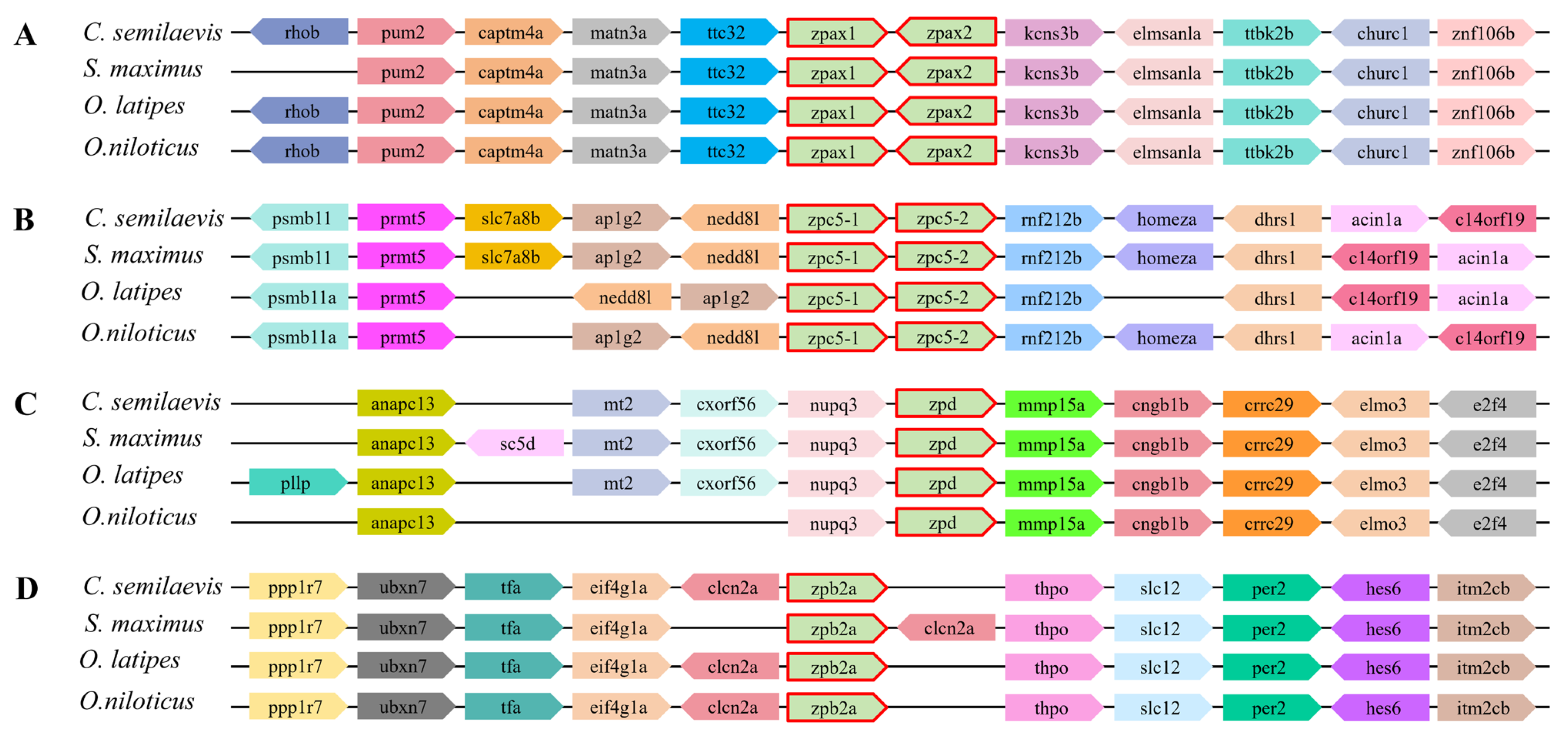
To further investigate the conservation and genomic location of ZP genes with its neighboring genes, a synteny analysis was conducted between ZP genes of C. semilaevis and other teleost fish, including O. niloticus, S. maximus, and O. latipes. The results showed a high degree of conservatism (Figure 3). One to two genes from the four subfamilies of C. semilaevis were selected for investigation.
Similar upstream and downstream genes were present in the four fish species. In detail, the zpax1 gene and zpax2 gene arranged in tandem on the chromosome in C. semilaevis and the other three selected teleost fish species (Figure 3A), as well as zpc5-1 and zpc5-2 (Figure 3B). Noteworthily, there were some inversions that appeared in the adjacent linear genes of zpc5-1 and zpc5-2. For example, nedd8l and aplg2 were inverted in O. niloticus, and there was an inversion between c14orf19 and acin1a in S. maximus and O. latipes. Moreover, several genes were missing or inserted upstream and downstream of these ZP genes in some fish species, especially for zpd and zpb2a (Figure 3C,D). These phenomena might indicate the evolutionary conservation between different species.

2.5. Protein–Protein Interaction (PPI) Network Analysis

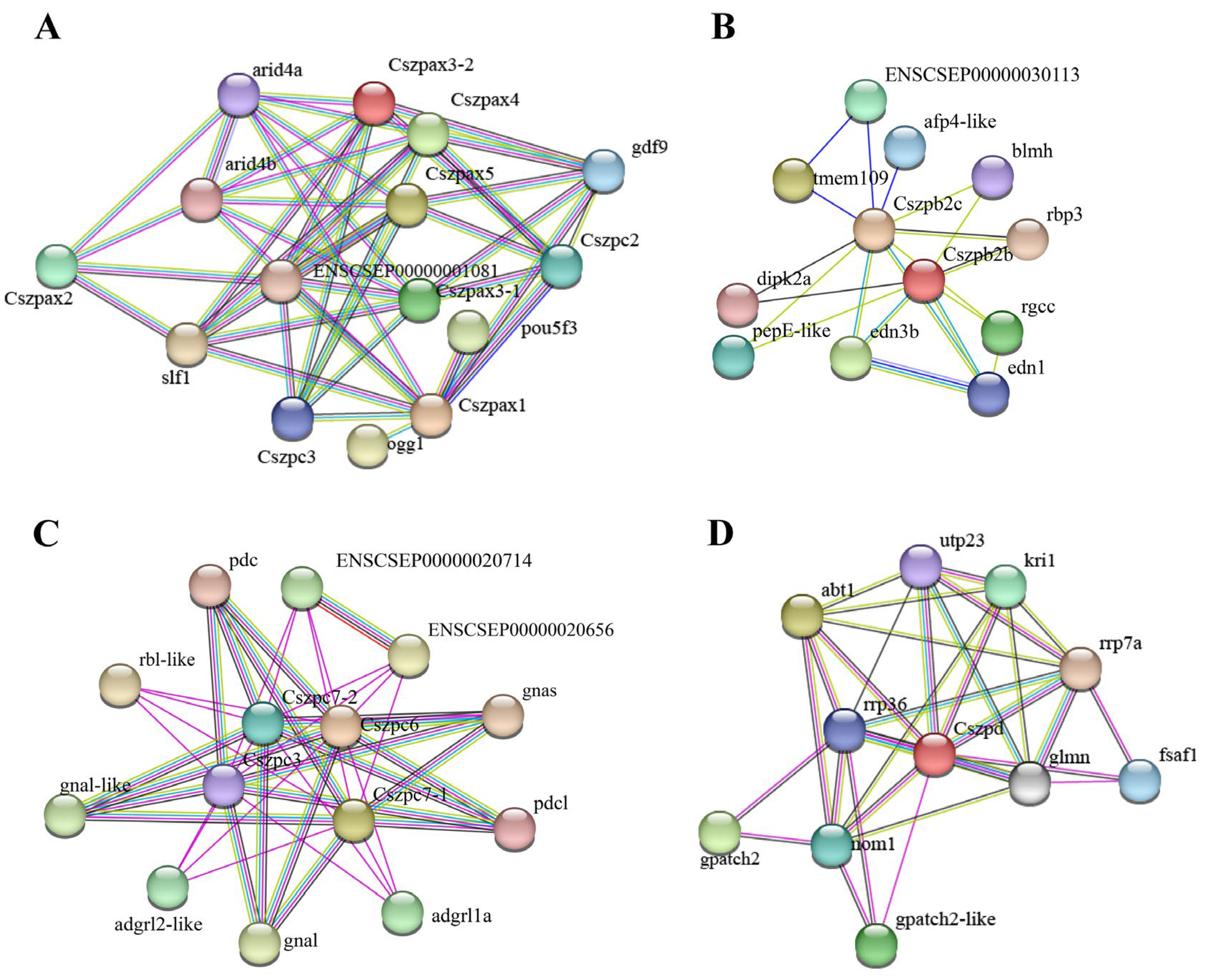
To better understand the function and interaction relationships of ZP genes in C. semilaevis, PPI network diagrams for each subfamily were constructed (Figure 4). Based on string analysis, there were 39 functional partners that built a PPI network together with CsZPs, including 9 proteins for Zpax, 10 for Zpb, 10 for Zpc, and 10 for Zpd. Seven proteins involved in cell proliferation and cycle regulation were identified within the CsZP-associated PPI networks, including Arid4a and Arid4b in the Zpax-associated network; Dipk2a and Edn1 in the Zpb-associated network; and Gpatch, Nom1, and Glmn in the Zpd-associated network. Gdf9 proteins were the partners for Zpax only. The oxidative stress factors Ogg1 and Slf1 were found to be associated exclusively with the Zpax network, and the activator Abt1 may be related to the transcriptional regulation of Zpd.

2.6. Expression Patterns of ZP Genes in C. semilaevis

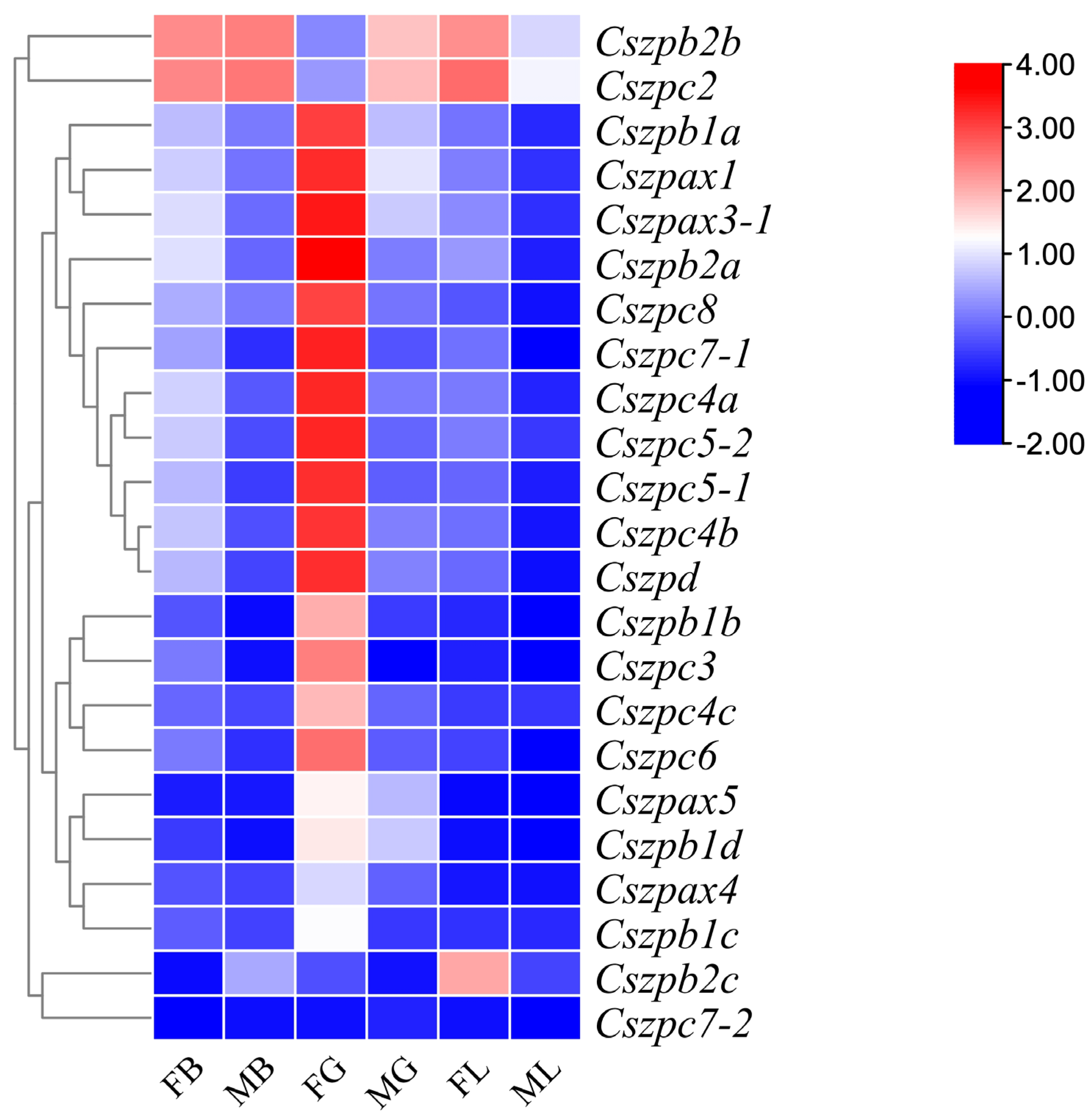
Comparative transcriptome analysis showed that most ZP genes exhibited significant differences in mRNA abundance across the brain, gonad, and liver tissues 1.5 years post-hatch (yph) C. semilaevis (Figure 5). Thirteen genes (Cszpax1, Cszpax3-1, Cszpb1a, Cszpb2a, Cszpc3, Cszpc4a, Cszpc4b, Cszpc5-1, Cszpc5-2, Cszpc6, Cszpc7-1, Cszpc8, and Cszpd) displayed gonad-biased expression exclusively in females, with negligible expression in all male tissues. Cszpb2c displayed liver-specific expression. In contrast, Cszpb2b and Cszpc2 were highly expressed in the female liver while also showing moderate expression in both male and female brains. Notably, Cszpc7-2 transcripts were undetectable across all examined tissues under the experimental conditions.
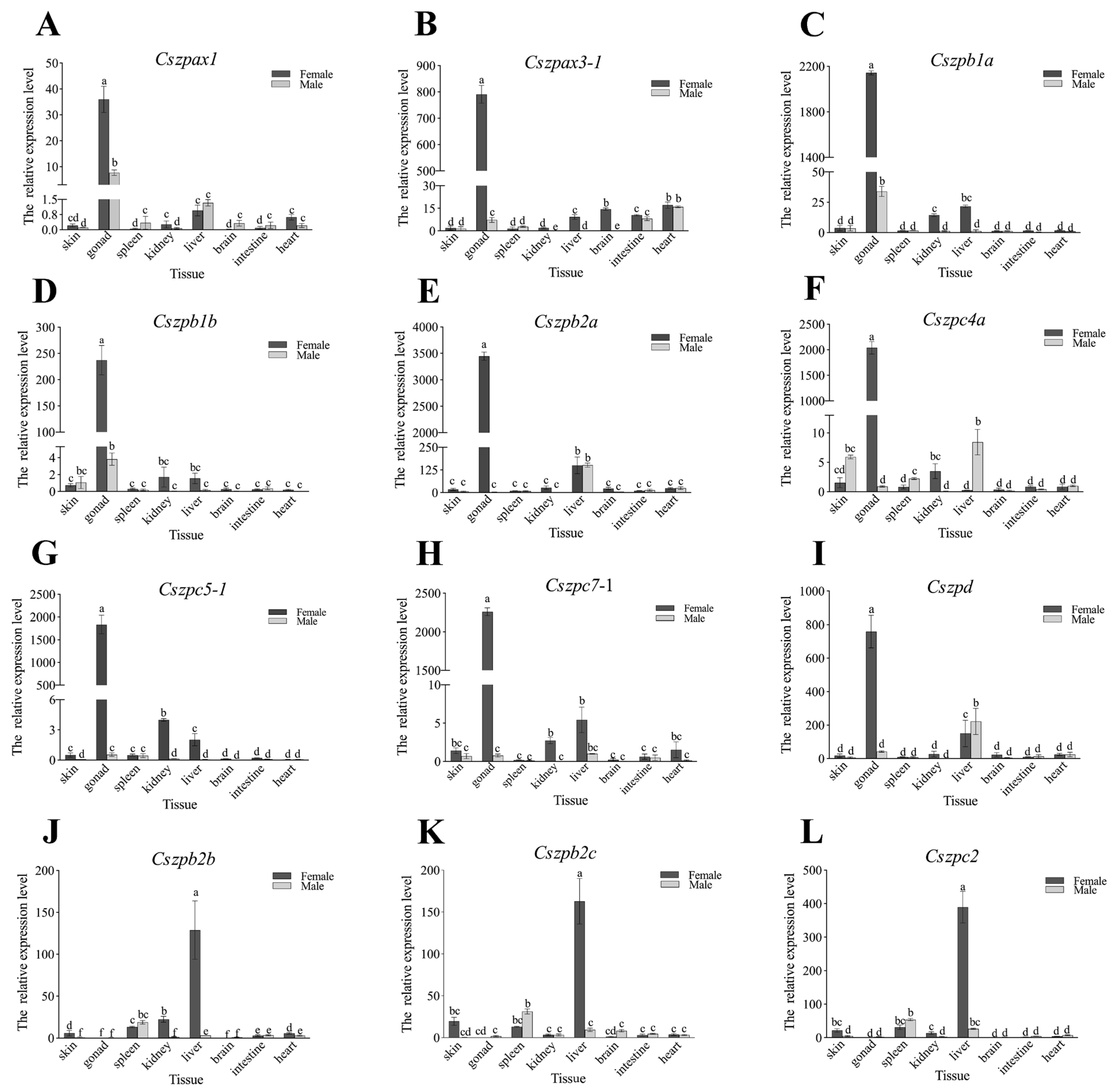
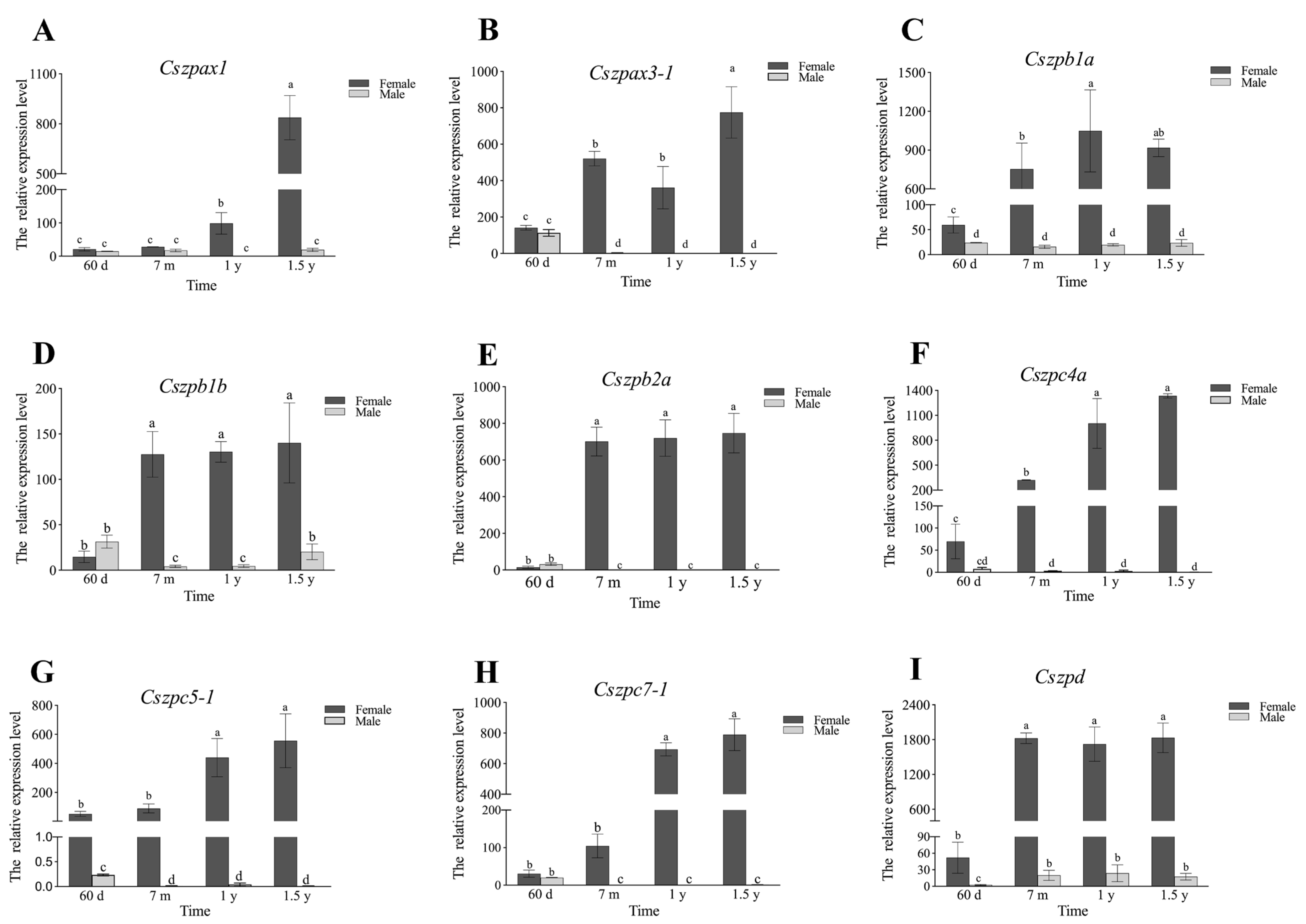
qPCR analysis further validated the transcriptomic findings (Figure 6). Nine ovary-enriched genes exhibited predominant expression exclusively in the ovaries 1.5 yph in female gonads (>100-fold higher) compared to other tissues. Conversely, three liver-and brain-enriched genes showed exceptionally high expression in the livers of 1.5 yph females (>100-fold elevation), while their expression levels in the brain samples remained comparatively low. We subsequent examined the expression trends of the nine ovary-enriched genes in gonads at different developmental stages (Figure 7). The results showed that Cszpax1, Cszpax3-1, Cszpb1b, Cszpb2a, Cszpc4a, and Cszpc7-1 did not exhibit sexual-biased expression in 60-day post-hatch (dph) individuals but displayed significantly higher expression in ovaries compared to testes from the 7-month post-hatch (mph) to 1 yph individuals. The expressions of almost all genes reached their peaks in the ovaries at 1.5 yph.
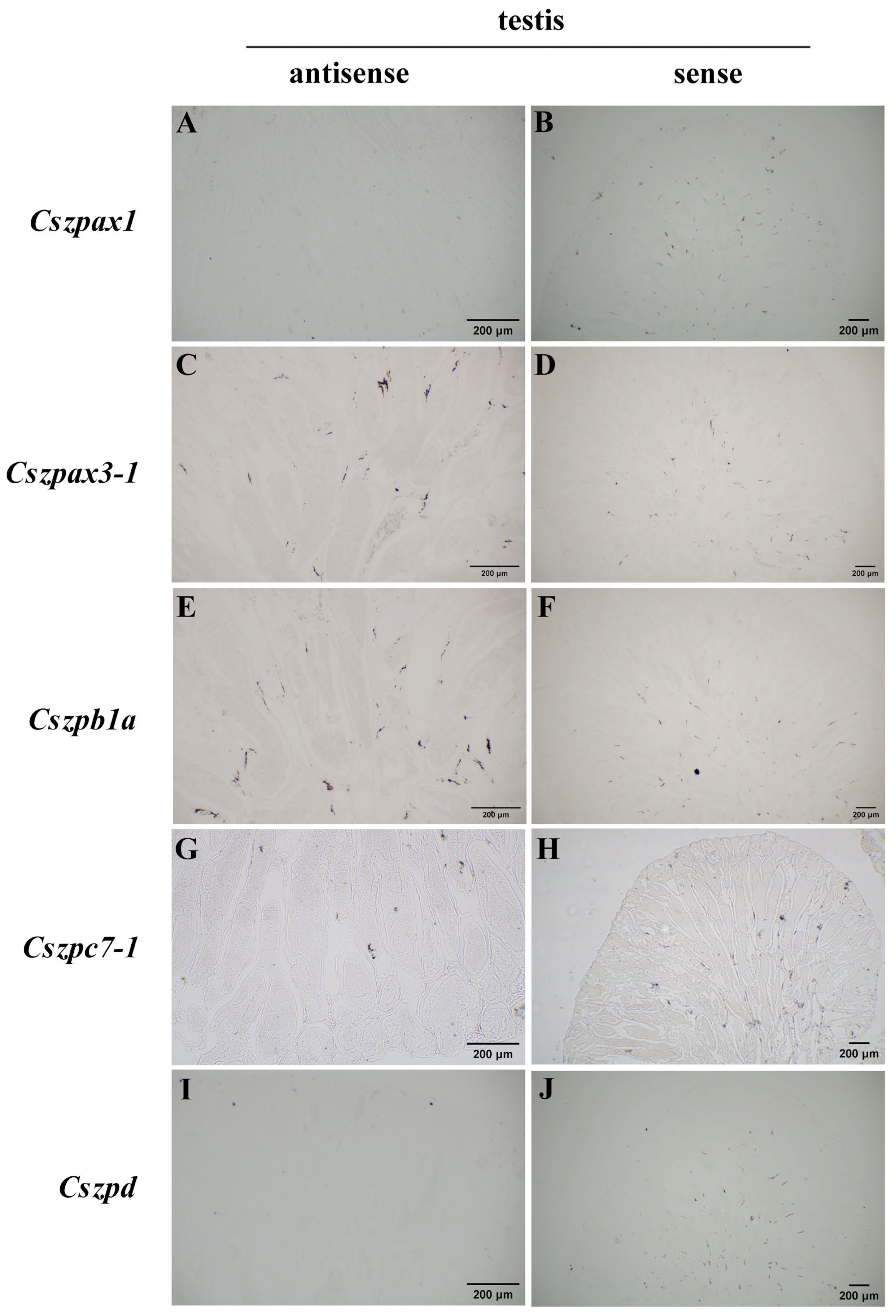
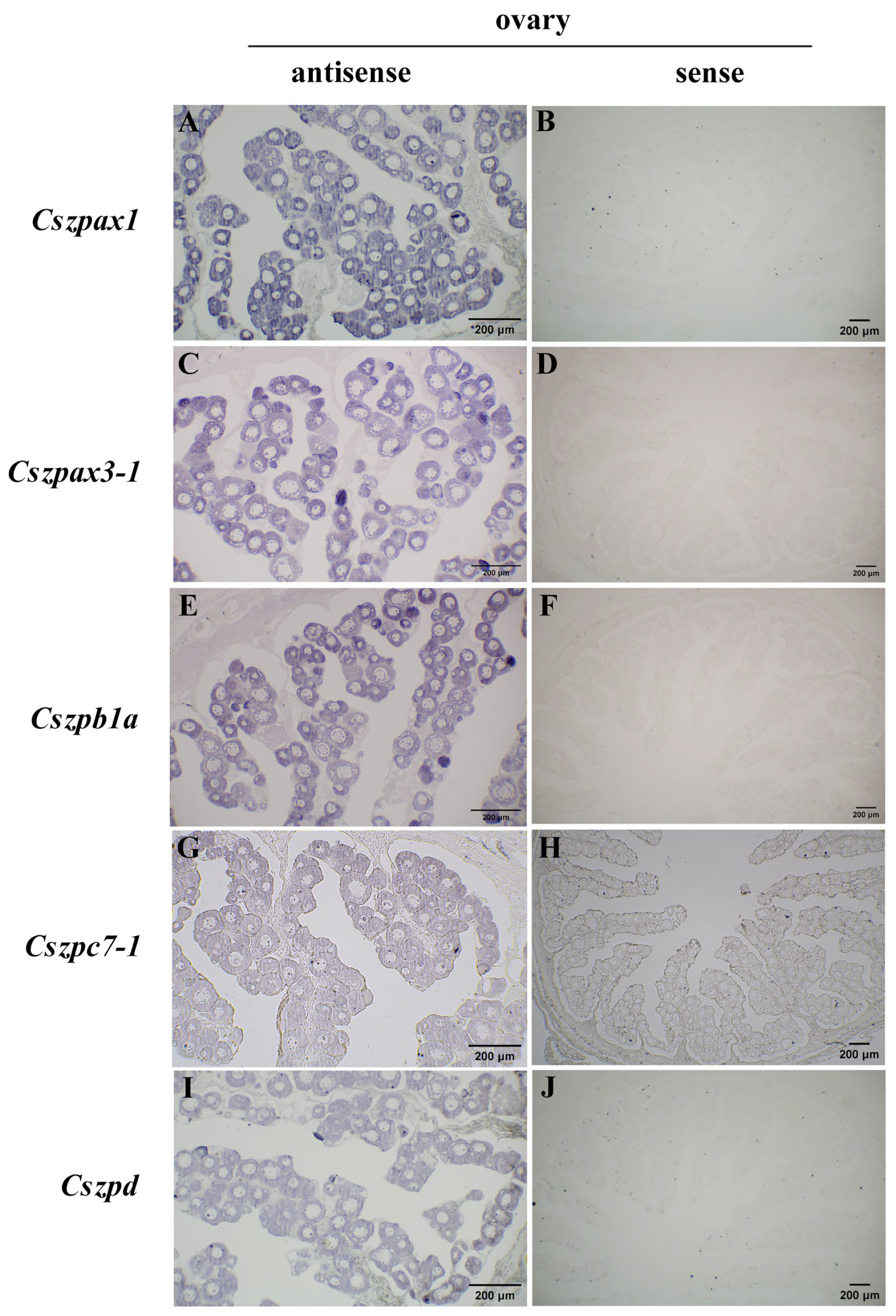
In the ovaries of 1.5 yph individuals, we detected strong hybridization signals for Cszpax1, Cszpax3-1, Cszpb1a, Cszpc7-1, and Cszpd genes, while no detectable signals were observed in the testes (Figure 8). This result is consistent with transcriptomic and qPCR results, and these genes exhibited expression throughout all developmental stages of oocytes within the ovaries.

2.7. Knockdown Effects of Cszpax1, Cszpc7-1 on CO

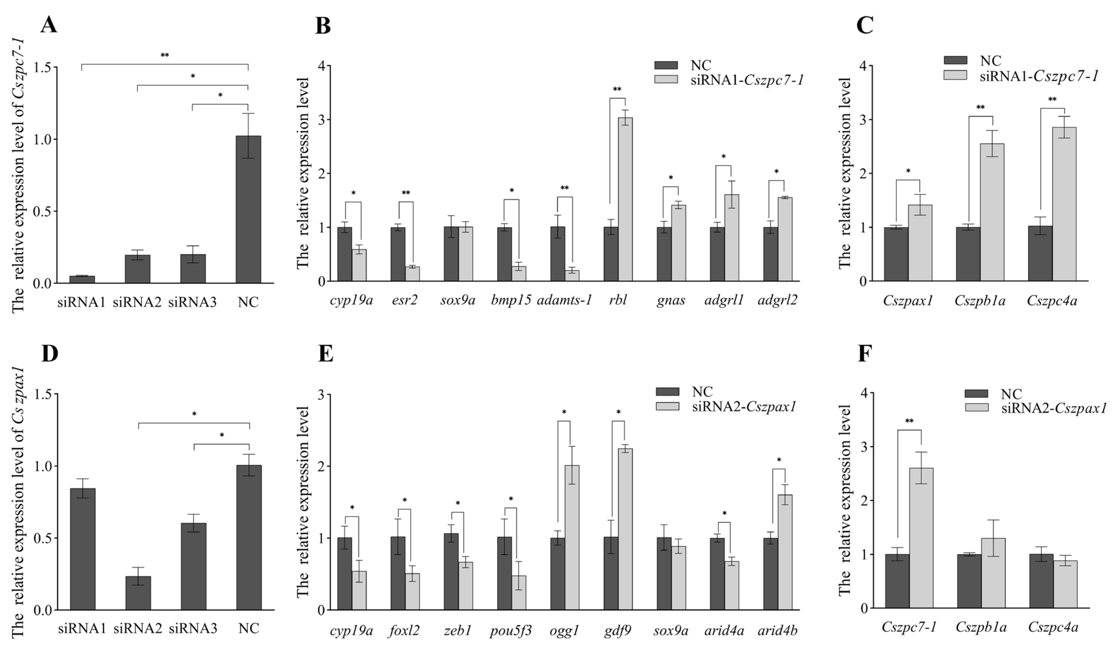
Three siRNA targeting specific sites of Cszpax1 and Cszpc7-1 were designed to knockdown gene expression in CO. The expression levels of Cszpax1 and Cszpc7-1 decreased significantly by 80% and 60%, with siRNA1-Cszpc7 and siRNA2-Cszpax1 showing the best knockdown effects (Figure 9). In the subsequent RNA interference (RNAi) experiments, the expressions of genes associated with sex determination and ovarian development were affected. Following Cszpc7-1 knockdown, the expression levels of cyp19a, esr2, adamts-1, and bmp15 were significantly downregulated, while the expressions of rbl, gnas, adgrl1, and adgrl2 were markedly upregulated. After Cszpax1 knockdown, the expression levels of foxl2, zeb1, arid4a, and cyp19a were markedly downregulated. In contrast, the expression of ogg1, gdf9, and arid4b was notably upregulated. However, sox9a gene expression did not chance significantly. Additionally, Cszpc7-1 knockdown resulted in upregulation of other ZP genes such as Cszpb1a, Cszpax1, and Cszpc4a, while Cszpax1 knockdown only led to a significant increase in Cszpc7-1 expression.

3. Discussion

In teleosts, zona pellucida genes contribute not only to sperm-egg recognition but to even broader functions, regulating egg adhesiveness and buoyancy [14], as well as enhancing egg antifreeze properties during cold conditions [15]. All known ZP genes contain a conserved ZP domain at the C-terminus, which is likely critical for the biological function of ZP proteins. Through genome-wide screening for candidate sequences containing the ZP domain and subsequent phylogenetic analysis, we identified 25 ZP genes in the C. semilaevis genome, including seven ZPB genes, eleven ZPC genes, one ZPD gene, and six ZPAX genes. Fish proteins have evolved significantly faster than their mammalian homologues due to the presence of genome duplication events in fish [18,19]. Gene duplication led to the creation of the ancestors of the various subfamilies of ZP, and thus, fish ZP genes tend to be multiply replicated [20], with the ZPC gene being abundantly replicated in zebrafish and medaka [21].
Interestingly, the C. semilaevis genome lacks ZPA genes, which is consistent with findings in other teleosts [20,22,23]. Compared to the human genome, the ZPB, ZPC, and ZPAX subfamilies have undergone significant expansion in teleosts but still beyond a high degree of evolutionary conservation. The C. semilaevis genome contains seven ZPB genes, with six of them (excluding Cszpb1c) possessing a trefoil domain. This unique three-loop structure, stabilized by disulfide bonds between six conserved cysteine residues, functions as a structural component of the zona pellucida rather than as a sperm receptor [24]. While the chicken genome contains only two ZPAX genes, teleost genomes have a larger number, with six ZPAX genes identified in C. semilaevis. Similarly, the ZPC subfamily has also expanded in C. semilaevis, reaching a total of eleven genes. Goudet et al. proposed that species relying on external fertilization might require a larger repertoire of ZP genes [21]. Compared to mammals, fish eggs have a larger radius and are released in large quantities, which may necessitate a greater number of egg envelope proteins [25]. Moreover, teleost fish eggs are exposed to more challenging conditions, and the egg envelope hardens during fertilization, providing mechanical protection against external pressures and bacterial infections [26,27]. These environmental and reproductive pressures likely drove the expansion of the ZP gene family in teleosts, with multiple ZP genes playing critical roles in fertilization and embryonic development.
Previous study revealed nine ZP genes with ovary-biased expression and three ZP genes with liver-biased expression in the C. semilaevis transcriptome [17]. In this study, qPCR results confirmed that nine ovary-expressed ZP genes were upregulated hundreds of times compared with their expressions in other examined tissues. Further, Cszpb1a, Cszpc5-1, and Cszpd already exhibited female-biased expression at the early stage of gonadal differentiation (60 dph). As development progressed and the gonads matured, all nine genes accumulated in the ovaries of 1.5 yph individuals, with consistent expression in oocytes at all developmental stages. These findings indicate that ovary-specific ZP genes play a crucial role in ovarian development and maturation. In medaka, zpb2b, zpb2c, and zpc2 are highly expressed in the liver [7,28]. Similarly, in tilapia, zpb2b and zpc2 exhibit predominant expression in the liver [8]. The zpb2b, zpb2c, and zpc2 in C. semilaevis, which cluster phylogenetically with these genes, also show significant liver expression. This suggests that during evolution, the teleost has developed lineage-specific adaptations in ZP gene expression, with some species expressing ZP genes in either the ovary or liver. Salmon eggs have a relatively thick chorion (30~40 μm), and their ZP genes are expressed in both the liver and ovary [29]. In contrast, zebrafish eggs have a much thinner chorion (~5 μm), which may require fewer ZP products, leading to the exclusive ovarian expression of ZP genes [30]. Although C. semilaevis also has a relatively thin chorion, the open marine environment differs significantly from freshwater habitats. Most marine fish produce pelagic eggs with oil droplets and lack parental egg-guarding behaviors. Additionally, marine fish generally have higher fecundity than freshwater species, necessitating the production of more egg envelope proteins. Zeb1 is known to regulate germ cell mitosis and gametogenesis in mice [31]. In C. semilaevis, zeb1 is significantly enriched in the pseudotemporal differentiation trajectory, suggesting its potential to promote gamete maturation through chromatin remodeling or epigenetic modifications [32]. The knockdown of Cszpax1 led to a reduction in zeb1 expression, which may affect oocyte maturation and ovulation, although the precise mechanism requires further investigation.
To construct the interaction networks for CsZps, we employed PPI network analysis and RNAi. When Cszpc7-1 expression was reduced, the expression of rbl, gnas, adgrl1, and adgrl2 was upregulated. Rbl proteins in fish eggs prevent polysperm entry by associating with cortical granules [33,34]. Upregulated rbl expression following Cszpc7-1 knockdown implies the involvement of the ZP gene in cortical-granule-mediated polysperm blockade. G protein-coupled receptors (GPCRs) are closely related to oocyte maturation in mammals and fish [35,36,37]. As members of GPCRs, gnas, adgrl1, and adgrl2 were found to be upregulated with Cszpc7-1 knockdown, suggesting that Cszpc7-1 might participate in oocyte maturation via GPCR signaling regulation. When Cszpax1 expression was reduced, the expression levels of arid4a and pou5f3 were downregulated. In contrast, the expression levels of ogg1, gdf9, and arid4b was upregulated. The ARID gene family, which encodes transcriptional regulators, plays essential roles in cell differentiation and proliferation [38]. Arid4a−/− Arid4b+/− mice exhibited reduced fertility, Sertoli cell dysfunction, and spermatogenic failure [39]. Pou5f3 (oct4), a potential key regulator of mouse oocyte development [40], plays a post-embryonic role during the maturation of gonads and gametes in medaka [41]. Gdf9 has been reported to play roles in ovarian folliculogenesis and ovulation in mammalian species [42]. It also regulates C. semilaevis reproduction and growth via the Smad signaling pathway [43]. Ogg1, the first enzyme in the base excision repair pathway, is involved in protecting oocytes against oxidative stress post-fertilization [44]. Taken together, the knockdown effects of Cszpc7-1 and Cszpax1 genes confirm their PPI network and suggest their potential function in oocyte development and protection.
On another hand, we examined the expression of a number of genes associated with sex determination and ovarian development. When Cszpc7-1 expression was reduced, the expression levels of several genes involved in ovary development, including cyp19a, esr2, bmp15, and adamts-1, also decreased. Similarly, when Cszpax1 expression was reduced, numerous ovarian-development-related genes including cyp19a, foxl2, and zeb1, were downregulated, while the spermatogenesis-related gene sox9a remained unaffected. Aromatase cyp19a, cooperating with foxl2, plays a crucial role in the biosynthesis of gonadal steroids, as well as ovarian differentiation oocyte maturation and in teleosts [45,46,47]. In zebrafish, the loss of foxl2 leads to cyp19a downregulation, reduced estrogen levels, and oocyte apoptosis [48].In addition, esr2, a member of the estrogen receptor family, is regulated by cyp19a and binds to estradiol (E2) to facilitate oocyte maturation and ovulation [49]. In C. semilaevis, the downregulation of foxl2, cyp19a, and esr2 with CsZP knockdown implies that CsZPs might maintain ovarian function through estrogen synthesis and signaling pathways. Bmp15, a member of the TGF-β superfamily, collaborates with gdf9 to regulate folliculogenesis and steroidogenesis in mammals. Knockout of these two genes in mice leads to reduced fertility or even infertility [50]. Adamts-1 is critical for follicular growth and ovulation in mice, and its deficiency lowers ovulation rates and fertilization efficiency [51,52,53]. Our results reveal the potential function of CsZP genes in oocyte development and fertilization, which is consistent with mammalian studies demonstrating the essential role of ZP genes in these processes, where their knockouts leads to infertility or impaired folliculogenesis [12,54,55,56,57].
We also investigated the effects of Cszpax1 and Cszpc7-1 knockdown on the expression of other ZP genes and found a compensatory expression effect between Cszpax1 and Cszpc7-1. This suggests that ovarian cells may respond to ZP gene knockdown by modulating gene expression levels to mitigate or counteract the effects of RNAi interference on the ZP gene interaction network.

4. Materials and Methods

4.1. Animal Euthanasia and Ethics Statement

In this study, fish were anesthetized using MS-222 (Solarbio, Beijing, China)(120 mg/L) to minimize pain. All experimental adhered to the guidelines established by the Institutional Animal Care and Use Committee of the Yellow Sea Fisheries Research Institute.

4.2. Fish and Sample Collection

Fish samples in this study were collected from Haiyang Aquaculture Base (Haiyang, China). Genetic sex determination was performed by excising tall fin clips from each individual and subjecting them to PCR-based assays using established sex-specific primers (Table A1) [58]. After dissection, tissues were collected from four female and four male 1.5 yph C. semilaevis, including skin, gonad, spleen, kidney, liver, brain, intestine, and heart. Additionally, gonadal tissues from four different developmental stages (including 60 dph, 7 mph, 1 yph, and 1.5 yph) were collected. These tissues were flash-frozen in liquid nitrogen and maintained at −80 °C. Total RNA was isolated from tissue samples using TRIzol reagent (Invitrogen, Carlsbad, CA, USA) and then reverse transcribed to cDNA using ReverTra Ace qPCR RT Master Mix (Toyobo, Osaka, Japan) and stored at −20 °C. Gonadal tissues of 1.5 yph fish were also fixed in 4% paraformaldehyde (Solarbio, Beijing, China) at 4 °C for 24 h, embedded in paraffin, and sectioned for in situ hybridization (ISH) analysis.

4.3. Identification of ZP Genes in C. semilaevis Genome

To identify all ZP gene members, the data corresponding to ZP domain (PF00100) were downloaded from the PFAM protein family database (version 35, EMBL-EBI, Hinxton, Cambridgeshire, UK) as a seed [59]. Then, HMMER software (version 3.0, HMMER.org, Howard Hughes Medical Institute, Chevy Chase, MA, USA) was employed to construct a Hidden Markov Model (HMM) [60], which was used to identify the superfamily members from C. semilaevis genome. The amino acid and nucleotide sequences of the homologous ZP genes from human (H. sapiens), chicken (Gallus gallus), turtle (Pelodiscus sinensis), Xenopus (X. tropicalis), Coelacanth (Latimeria chalumnae), Nile tilapia (O. niloticus), zebrafish (D. rerio), and medaka (O. latipes) were downloaded from the National Center for Biotechnology Information (NCBI) database (https://www.ncbi.nlm.nih.gov/, accessed on 7 October 2023) and Ensembl databases (http://asia.ensembl.org/index.html, accessed on 19 October 2023), and the relevant information was listed in Table A2. C. semilaevis ZP gene candidates were captured in the transcriptome and genome databases using the BLAST+ software (version 2.12.0, National Center for Biotechnology Information, Bethesda, MA, USA) [61,62] against the homologous ZP gene sequences from other species, with a cutoff E-value of 1 × 10⁻¹⁰e−10. The final ZP genes of C. semilaevis were confirmed by combining the results from the HMMER and BLAST searches.

4.4. Sequence and Evolutionary Analysis

To obtain the phylogenetic relationships of ZP genes in teleost fish, ZP gene family sequences from various species were used (Table A2). Multiple sequence alignment (MSA) (version 7.505, Computational Biology Research Center, AIST, Tokyo, Japan) was conducted using MAFFT with default parameters to align the amino acid sequences [63]. Then, a phylogenetic tree was constructed using the maximum likelihood method via IQ-TREE 2 software (version 2.4.0, Center for Integrative Bioinformatics Vienna, Vienna, Austria) [64]. Next, the tree file generated by IQ-TREE was uploaded to iTol (version 5, European Molecular Biology Laboratory, Heidelberg, Germany) for visualization and customization [65]. The exon/intron gene structures of C. semilaevis sequence were analyzed using Gene structure display server 2.0 (GSDS 2.0, version 2.0, Center for Bioinformatics, Peking University, Beijing, China) [66]. The conserved structural domains and motifs were characterized using PFAM database and MEME program (version 5.4.1, MEME Suite, National Institutes of Health, Bethesda, MA, USA) [67], followed by visualization using the TBtools software (version 2.142, South China Agricultural University, Guangzhou, China) [68]. The chromosomal positions of sequences were obtained from the NCBI database. The molecular masses (MWs) and theoretical isoelectric points (pIs) were predicted by ExPASy (https://web.expasy.org/protparam/, accessed on 19 October 2023).

4.5. Synteny Analysis

Synteny analysis of ZP genes in teleost fish, including C. semilaevis, O. niloticus, S. maximus, O. latipes, was performed. The genomic annotation information was obtained from the Ensembl databases. Syntenic regions near the ZP genes were identified using the Genomicus database (version 100.01, Institut de Biologie de l’École Normale Supérieure, Paris, France) following the method previously described by Zhu et al. [69,70]. For each ZP gene, five neighboring genes upstream and five downstream genes were selected for comparison.

4.6. Protein–Protein Interaction Prediction

Based on the homology of C. semiaevis, the Search Tool for The Retrieval of Interacting Genes/Proteins (STRING) database (version 11.5, European Molecular Biology Laboratory, Heidelberg, Germany) was used to predict the potential role partners of the proteins with medium confidence (0.40) [71].

4.7. Heatmap Plotting with RNA-Seq Data

We utilized previous RNA-seq data of the gonad, brain, and liver samples from 1.5 yph fish [17] to understand the mRNA distribution of ZP genes in C. semilaevis. The heatmap was generated using the TBtools software to visualize the mRNA expression profile.

4.8. Quantitative Real-Time (qPCR) Analysis

The primers used for qPCR are listed in Table A1. Actin was selected as the internal reference gene. Reactions were completed using THUNDERBIRD™ Next SYBR® qPCR Mix (Toyobo, Osaka, Japan) and 7500 Fast Real-Time PCR system (ABI, Los Angeles, CA, USA). Amplification conditions were 95 °C for 30 s, 95 °C for 5 s, 60 °C for 34 s, and 40 cycles. Each set of samples was repeated four times. The experimental data were obtained using the 2−ΔΔCt method [72]. Data were analyzed by one-way ANOVA using SPSS version 20.0 (IBM, Armonk, NY, USA), followed by Tukey’s post hoc test for multiple comparisons. A p-value < 0.05 was considered statistically significant.

4.9. In Situ Hybridization Analysis

The primers used in this experiment to amplify fragments of Cszpax1, Cszpax3-1, Cszpb1a, Cszpc7-1, and Cszpd are listed in Table A1. T7/SP6 RNA polymerase promoters were introduced for in vitro synthesis of antisense/sense probe. After synthesized of the probes using the digoxigenin (DIG) RNA labeling kit (Roche Diagnostics, Nutley, NJ, USA), the probes were purified were purified using the lithium chloride (LiCl) (Sigma, Darmstadt, Germany) precipitation method for the next hybridization. In situ hybridization procedure followed the protocol outlined by Zhu et al. [73]. Signals were detected using the BCIP/NBT substrate (Roche, Mannheim, Germany) and pictures were acquired with a Nikon ECLIPSE 80i microscope (Nikon, Tokyo, Japan).

4.10. Cell Culture and siRNA Transfection Analysis

The C. semilaevis ovarian (CO) cells were cultured in L-15 medium with the following components required for cell growth: EGF (Beyotime, Shanghai, China) (5 ng/mL), bFGF (Beyotime, Shanghai, China) (5 ng/mL), LIF (Beyotime, Shanghai, China) (5 ng/mL), 2% penicillin–streptomycin–amphotericin B mixed solution (Solarbio, Beijing, China), β-Mercaptoethanol (27.5 µg/mL) (Vwr, Radnor, PA, USA), and 20% fetal bovine serum (FBS) (Gibco, Grand Island, NY, USA), and they were maintained in a 24 °C incubator without CO2. When the cell density reached 70–80%, the cells were inoculated in 12-well cell culture plates for transfection.
Three specific small interfering RNA (siRNA) sites of Cszpc7-1 and Cszpax1 were designed by Sangon (Shanghai, China) (Table A1). Targeted siRNAs and negative control (NC) samples were transfected into CO by using RiboFECT™ CP Transfection Kit (Ribobio, Guangzhou, China). Cy3-siRNA (Ribobio, Guangzhou, China) was transfected into CO at the same time for the valuation of transfection efficiency. Cells were harvest by TRIzol, and the subsequent qPCR assays were conducted as mentioned above. The expression levels of Cszpc7-1, Cszpax1, and other sex- and hormone-related genes (including foxl2, cyp19a, sox9a, bmp15, esr2, adamts-1, rbl, gnas, adgrl1, adgrl2, pou5f3, gdf9, ogg1, arid4a, arid4b, and zeb1) were examined with the primers listed in Table A1.

5. Conclusions

In conclusion, we identified 25 genes in the C. semilaevis ZP gene family, all harboring a conserved ZP domain. We analyzed the sequences and phylogeny of ZP genes and predicted their potential protein–protein interaction partners. Furthermore, we investigated the spatiotemporal expression patterns of 12 ZP genes across eight different tissues, including nine ovary-expressed and three liver-expressed members. Additionally, we analyzed the gonadal expression profiles of nine ovary-specific ZP genes during different developmental stages. Cszpax1- and Cszpc7-1-specific RNAi indicated that ZP genes might play crucial roles in oogenesis, follicular development, and ovulation, suggesting that their function is conserved and analogous to the role of ZP genes in mammals. Collectively, these findings provide valuable information for better understanding the evolution and functional diversification of ZP genes family in teleosts.

Author Contributions

Conceptualization, Z.C. and N.W.; methodology, Z.C.; validation, K.Z. and H.Y.; formal analysis, C.G.; investigation, K.Z. and Z.C.; resources, N.W. and M.Z.; writing—original draft preparation, K.Z.; writing—review and editing, Z.C., Z.S. and X.L.; project administration, S.C.; funding acquisition, S.C. and X.L. All authors have read and agreed to the published version of the manuscript.

Funding

This work was supported by the National Natural Science Foundation of China (32230107); Central Public-interest Scientific Institution Basal Research Fund, YSFRI, CAFS (20603022024006); the Shandong Key R&D Program for Academician teams in Shandong (2023ZLYS02); the earmarked fund for China Agriculture Research System (CARS-47-G03); Tianshan Scholar Climbing Project of Shandong Province, China; and The Innovative Team Project of Chinese Academy of Fishery Sciences (2023TD20).

Institutional Review Board Statement

The animal experiment was inspected and approved by the Institutional Animal Care and Use Committee at the Yellow Sea Fisheries Research Institute, CAFS (Approve No.: YSFRI-2022023).

Informed Consent Statement

Not applicable.

Data Availability Statement

The original contributions presented in this study are included in the article. Further inquiries can be directed to the corresponding author.

Conflicts of Interest

The authors declare no conflicts of interest.

Abbreviations

The following abbreviations are used in this manuscript:
cyp19acytochrome p450 19a
foxl2forkhead box L2
esr2estrogen receptor 2
bmp15bone morphogenetic protein 15
adamts-1ADAM metallopeptidase with thrombospondin type 1 motif 1
sox9aSRY-box transcription factor 9a
zeb1zinc finger E-box bingding homeobox 1
rblrhamnose-binding lectin
gnasguanine nucleotide binding protein, alpha stimulating
gdf9growth differentiation factor 9
pou5f3POU class 5 homeobox 3
adgrl1adhesion G protein-coupled receptor L1
adgrl2adhesion G protein-coupled receptor L2
gnalguanine nucleotide-binding protein G(s) subunit alpha-like
ogg18-oxoguanine DNA glycosylase 1
arid4aAT-rich interaction domain 4A
arid4bAT-rich interaction domain 4B

Appendix A

Figure A1. Phylogenetic tree of ZPs were constructed using all ZP protein sequences from the following species: C. semilaevis (Cs), H. sapiens (Hs), D. rerio (Dr), O. niloticus (On), G. gallus (Gg), X. troicalis (Xt), L. chalumnae (Lc), P. sinensis (Ps), and O. latipes (Ol). The five subclusters are represented in different colors. CsZPs are marked with red stars.
Ijms 26 05346 g0a1
Figure A2. The spatial expression patterns of ZP genes of C. semilaevis at 1.5 yph. (A) Antisense probe for Cszpax1 in testis. (B) Sense probe for Cszpax1 in testis. (C) Antisense probe for Cszpax3-1 in testis. (D) Sense probe for Cszpax3-1 in testis. (E) Antisense probe for Cszpb1a in testis. (F) Sense probe for Cszpb1a in testis. (G) Antisense probe for Cszpc7-1 in testis. (H) Sense probe for Cszpc7-1 in testis. (I) Antisense probe for Cszpd in testis. (J) Sense probe for Cszpd in testis. Cszpax1, Cszpax3-1, Cszpb1a, Cszpc7-1, and Cszpd genes with no significant hybridization signals were observed in male gonads. Scale bars = 200 μm.
Ijms 26 05346 g0a2
Table A1. The primers used in the present study.
PrimerPurposeSequences (5′→3′)
zpb2a-q-FqPCRGCACCAGGGATGGACAGTTT
zpb2a-q-RqPCRAGGAGGGGTCATTAGGGTCC
zpb1b-q-FqPCRCTCGGTGTGGTTGCTTCTCT
zpb1b-q-RqPCRCTTGGGAGTGGGAAGTGGTC
zpb1a-q-FqPCRCCCAAAAGCAACACTTCGGG
zpb1a-q-RqPCRGGGTCTAAAGGGTTGGCACA
zpax1-q-FqPCRTGTCCAGGAGCACACGTTTT
zpax1-q-RqPCRAGAGGCAGGAAGTAGACCGT
zpd-q-FqPCRATTGCCATCAGAGCCGTTCA
zpd-q-RqPCRCTTGGAGACGCTGGACATGT
zpax3-1-q-FqPCRCCGTATGGACAGCCTGACTC
zpax3-1-q-RqPCRCCATGATGACCACCCAGCTT
zpc4a-q-FqPCRTCAGTACCGGCCCTTTTGTC
zpc4a-q-RqPCRCCCTCATTACAGCAGCACCA
zpc5-1-q-FqPCRAATGGAGGAGTGTGTGGCAG
zpc5-1-q-RqPCRATGGCAGATGAGTGGTAGCG
zpc7-1-q-FqPCRAACCGGAACAAACCCACAGT
zpc7-1-q-RqPCRACAACTGGTCCCACTCTTGC
β-actin-FqPCRTCAATTCTCCACGAACCA
β-actin-RqPCRCTTACACAGCGAGCAACC
cyp19a-q-FqPCRGGTGAGGATGTGACCCAGTGT
cyp19a-q-RqPCRACGGGCTGAAATCGCAAG
foxl2-q-FqPCRGAGAGGAAGGGCAACTACTGGA
foxl2-q-RqPCRTGGTTGGAAGTGCGTGGG
sox9a-q-FqPCRAAGAACCACACAGATCAAGACAGA
sox9a-q-RqPCRTAGTCATACTGTGCTCTGGTGATG
bmp15-q-FqPCRCGGACGGAAGAACTAAACA
bmp15-q-RqPCRTCATACTGGACCTGGAACG
esr2-q-FqPCRGATTAGGAGAAGGTGGAGAAGG
esr2-q-RqPCRGGTAACCAGAGGCATAGTCGTG
zeb1-q-FqPCRGCTCCTCTTCAAGGCACAGT
zeb1-q-RqPCRGACGTTACCATCCACTGCCA
adamts-1-q-FqPCRCTGGCCTCAAACCCTACCTG
adamts-1-q-RqPCRGGCCTCGCTCCTCTTCATAC
zpax1-siRNA-1siRNACCAGAGUCCUUCAGAUAUA
zpax1-siRNA-2siRNACGACUGUGCCGGCAAUCUA
zpax1-siRNA-3siRNACAGACAAGCAGUGGUUUAA
zpc7-1-siRNA-1siRNAGGCUGUGUAGCUACGUUAA
zpc7-1-siRNA-2siRNACGCUGGAGAUCGUCAUCAA
zpc7-1-siRNA-3siRNACGCGAUUGUUGUCGGGCUA
zpb1a-SP6ISHATTTAGGTGACACTATAGAATCGAGGCTACGAGGTGGATT
zpb1a-T7ISHTAATACGACTCACTATAGGGGCTCATAAACACGGGCTCCA
zpd-SP6ISHATTTAGGTGACACTATAGAATGTGGCAGGTGTAGGACTCT
zpd-T7ISHTAATACGACTCACTATAGGGATCTGAATGGTGGCGTCGAG
zpax3-1-SP6ISHATTTAGGTGACACTATAGAAGAGCGTCTGACACCAGAACT
zpax3-1-T7ISHTAATACGACTCACTATAGGGAGCTTGAGTCTTCGTGCTGAA
zpc7-1-SP6ISHATTTAGGTGACACTATAGAACCTTGAAAATGGGTGCCTGC
zpc7-1-T7ISHTAATACGACTCACTATAGGGCCTCAACCTGGTCTTGAGGTC
zpax1-SP6ISHATTTAGGTGACACTATAGAATCTGTTGAATATACAGTCCCAGAGG
zpax1-T7ISHTAATACGACTCACTATAGGGAGCTGAAGTGTGTGCGGTTA
sex-FGender
detection
CCTAAATGATGGATGTAGATTCTGTC
sex-RGender
detection
GATCCAGAGAAAATAAACCCAGG
gdf9-q-FqPCRACGCTCACCTTCTGAGTTCC
gdf9-q-RqPCRAAGATCATGACGCTCAGGGG
adgrl1-q-FqPCRGTGGAAAGGCCGTGTCCTAA
adgrl1-q-RqPCRCTATTTGGCTGACCCACGGT
rbl-q-FqPCRGACGCCGTGATCAGACTACC
rbl-q-RqPCRACATACGTAGGCCACCTCCA
gnas-q-FqPCRTGCACGCTACACTACACCTG
gnas-q-RqPCRATGTTCTCTGTGTCGACCGC
pou5f3-q-FqPCRGCCAGAGTCGGCCATATGAT
pou5f3-q-RqPCRTAGAGGATGCTCGGGTCTGG
adgrl1-q-FqPCRCGGATGCACGAGCTTATCCT
adgrl1-q-RqPCRAGATGGGACAGGCAATGTGG
adgrl2-q-FqPCRAACGCACTCGCAACATTGTC
adgrl2-q-RqPCRTCACCCATAAGCCCCTCTCA
ogg1-q-FqPCRGGCCAAACATGCGGTACTTT
ogg1-q-RqPCRCGTGCCACATTTCCTTTCGT
arid4b-q-FqPCRCTGTGGCTGGAACGTCAGAT
arid4b-q-RqPCRGAGCGATGGACACGGTTAGT
arid4a-q-FqPCRCCTCCTCCATGTCATCGTCG
arid4a-q-RqPCRCGAGGCTTCTGGGACTTTGT
Table A2. List of species used in this study.
SpeciesGeneAccession No
Cynoglossus semilaevisCszpax1XP_024912764.1
Cszpax2XP_024912783.1
Cszpax3-1XP_008319235.1
Cszpax3-2XP_024915769.1
Cszpax4XP_008310879.2
Cszpax5XP_008319357.1
Cszpb1aXP_008329736.1
Cszpb1bXP_008328541.1
Cszpb1cXP_024920423.1
Cszpb1dXP_008315308.1
Cszpb2aXP_008332246.1
Cszpb2bXP_024912115.1
Cszpb2cXP_008310180.2
Cszpc2XP_016888570.1
Cszpc3XP_008323734.1
Cszpc4aXP_008314339.1
Cszpc4bXP_024914509.1
Cszpc4cXP_008325694.1
Cszpc5-1XP_008332968.3
Cszpc5-2NP_001284511.2
Cszpc6XP_008332190.1
Cszpc7XP_008327912.1
Cszpc8XP_008306734.1
Cszpc9XP_008325416.3
CszpdXP_008309362.1
Danio rerioDrzpax1ENSDARG00000069251
Drzpax2ENSDARG00000017188
Drzpax4ENSDARG00000079034
Drzpb2a-1ENSDARG00000090237
Drzpb2a-2ENSDARG00000091409
Drzpb2a-3ENSDARG00000086352
Drzpb2a-4ENSDARG00000086522
Drzpb2bENSDARG00000055415
Drzpb2cENSDARG00000004898
DrzpdENSDARG00000051959
Drzpc1-1ENSDARG00000042129
Drzpc1-2ENSDARG00000042130
Drzpc2ENSDARG00000090768
Drzpc3ENSDARG00000016908
Drzpc4a-1ENSDARG00000040081
Drzpc4a-2ENSDARG00000070178
Drzpc4bENSDARG00000092919
Drzpc5-1ENSDARG00000038720
Drzpc5-2ENSDARG00000054313
Drzpc6ENSDARG00000039828
Oreochromis niloticusOnzpax1ENSONIP00000007688
Onzpax2ENSONIP00000007675
Onzpax3ENSONIP00000008459
Onzpax4ENSONIP00000006775
Onzpb1aXP_003452556.1
Onzpb1bENSONIP00000010516
Onzpb2aENSONIP00000012346
Onzpb2bENSONIP00000018582
Onzpb2cENSONIP00000018726
OnzpdENSONIP00000012896
Onzpc1ENSONIP00000004983
Onzpc2ENSONIP00000018727
Onzpc3ENSONIP00000015344
Onzpc4ENSONIP00000002777
Onzpc5-1ENSONIP00000019557
Onzpc5-2ENSONIP00000019559
Onzpc6ENSONIP00000012513
Onzpc7ENSONIP00000002749
Onzpc8ENSONIP00000013206
Oryzias latipesOlzpax1ENSORLG00000012471
Olzpax2ENSORLG00000012527
Olzpax3-1ENSORLG00000006604
Olzpax3-2ENSORLG00000006621
Olzpax4ENSORLG00000018174
Olzpax5ENSORLG00000006629
Olzpb2aENSORLG00000016580
Olzpb2bENSORLG00000010880
Olzpb2cENSORLG00000010086
OlzpdENSORLG00000015608
Olzpc1ENSORLG00000020441
Olzpc2ENSORLG00000010134
Olzpc3ENSORLG00000005414
Olzpc5-1ENSORLG00000012273
Olzpc5-2ENSORLG00000012249
Olzpc6ENSORLG00000016064
Olzpc7-1ENSORLG00000009853
Olzpc7-2ENSORLG00000009867
Olzpc8aENSORLG00000006875
Olzpc8bENSORLG00000015033
Pelodiscus sinensisPszpaENSPSIG00000004165
PszpdENSPSIG00000004205
Pszpb1ENSPSIG00000005870
Pszpb2a-1ENSPSIG00000005091
Pszpb2a-2ENSPSIG00000014055
Pszpb2bENSPSIG00000006328
Pszpc1ENSPSIG00000010174
Pszpc2ENSPSIG00000013197
Pszpc3ENSPSIG00000016070
Pszpax1ENSPSIG00000011815
Pszpax2ENSPSIG00000011721
Xenopus tropicalisXtzpaENSXETG00000016704
XtzpdENSXETG00000013767
Xtzpb1ENSXETG00000015911
Xtzpb2ENSXETG00000011855
Xtzpc1ENSXETG00000019465
Xtzpc2ENSXETG00000025433
Xtzpax1ENSXETG00000015572
Xtzpax2ENSXETG00000014953
Gallus gallusGgzpaENSGALP00000038302
Ggzpb1CAC16087.1
Ggzpb2ENSGALP00000032884
Ggzpc1ENSGALP00000002636
Ggzpc2-1ENSGALP00000002356
Ggzpc2-2ENSGALP00000002368
Ggzpax1ENSGALP00000026515
Ggzpax2ENSGALP00000026528
GgzpdBAD13713.2
Homo sapiensHszpaENSG00000103310
Hszpb1ENSG00000149506
Hszpb2ENSG00000116996
HszpcENSG00000188372
Latimeria chalumnaeLczpaENSLACG00000005253
Lczpb1ENSLACG00000005027
Lczpb2a-1ENSLACG00000008179
Lczpb2a-2ENSLACG00000007163
Lczpb2a-3ENSLACG00000005832
Lczpb2a-4ENSLACG00000004264
Lczpb2bENSLACG00000011054
Lczpb2cENSLACG00000007972
Lczpc1ENSLACG00000001864
Lczpc2-1ENSLACG00000000590
Lczpc2-2ENSLACG00000001956
Lczpc2-3ENSLACG00000002494
Lczpc2-4ENSLACG00000004624
Lczpc5ENSLACG00000007941
LczpdENSLACG00000002854
Lczpax1ENSLACG00000015100
Lczpax2ENSLACG00000006540

References

  1. Xu, S.; Tian, Z.; Zhu, H. Comparison of Induced and Natural Spawning in Southern Flounder Paralichthys lethostigma. Fish. Sci. 2009, 28, 3. [Google Scholar]
  2. Huang, L.; Liang, M.; Zhang, H.; Qu, J.; Wang, X.; Zheng, K. The effect of dietary vitamin A level on reproductive performance of broodstock Scophthamus maximus. Prog. Fish. Sci. 2013, 34, 62–70. [Google Scholar]
  3. Bleil, J.D.; Wassarman, P.M. Structure and function of the zona pellucida: Identification and characterization of the proteins of the mouse oocyte’s zona pellucida. Dev. Biol. 1980, 76, 185–202. [Google Scholar] [CrossRef] [PubMed]
  4. Bleil, J.D.; Wassarman, P.M. Mammalian sperm-egg interaction: Identification of a glycoprotein in mouse egg zonae pellucidae possessing receptor activity for sperm. Cell 1980, 20, 873–882. [Google Scholar] [CrossRef]
  5. Lefièvre, L.; Conner, S.J.; Salpekar, A.; Olufowobi, O.; Ashton, P.; Pavlovic, B.; Lenton, W.; Afnan, M.; Brewis, I.A.; Monk, M.; et al. Four zona pellucida glycoproteins are expressed in the human. Hum. Reprod. 2004, 19, 1580–1586. [Google Scholar] [CrossRef]
  6. Smith, J.; Paton, I.R.; Hughes, D.C.; Burt, D.W. Isolation and mapping the chicken zona pellucida genes: An insight into the evolution of orthologous genes in different species. Mol. Reprod. Dev. 2005, 70, 133–145. [Google Scholar] [CrossRef]
  7. Murata, K.; Sasaki, T.; Yasumasu, S.; Iuchi, I.; Enami, J.; Yasumasu, I.; Yamagami, K. Cloning of cDNAs for the precursor protein of a low-molecular-weight subunit of the inner layer of the egg envelope (chorion) of the fish Oryzias latipes. Dev. Biol. 1995, 167, 9–17. [Google Scholar] [CrossRef]
  8. Wu, T.; Cheng, Y.; Liu, Z.; Tao, W.; Zheng, S.; Wang, D. Bioinformatic analyses of zona pellucida genes in vertebrates and their expression in Nile tilapia. Fish. Physiol. Biochem. 2018, 44, 435–449. [Google Scholar] [CrossRef]
  9. Qi, H.; Williams, Z.; Wassarman, P.M. Secretion and assembly of zona pellucida glycoproteins by growing mouse oocytes microinjected with epitope-tagged cDNAs for mZP2 and mZP3. Mol. Biol. Cell 2002, 13, 530–541. [Google Scholar] [CrossRef]
  10. Greve, J.M.; Wassarman, P.M. Mouse egg extracellular coat is a matrix of interconnected filaments possessing a structural repeat. J. Mol. Biol. 1985, 181, 253–264. [Google Scholar] [CrossRef]
  11. Bleil, J.D.; Greve, J.M.; Wassarman, P.M. Identification of a secondary sperm receptor in the mouse egg zona pellucida: Role in maintenance of binding of acrosome-reacted sperm to eggs. Dev. Biol. 1988, 128, 376–385. [Google Scholar] [CrossRef] [PubMed]
  12. Gao, L.L.; Zhou, C.X.; Zhang, X.L.; Liu, P.; Jin, Z.; Zhu, G.Y.; Ma, Y.; Li, J.; Yang, Z.X.; Zhang, D. ZP3 is Required for Germinal Vesicle Breakdown in Mouse Oocyte Meiosis. Sci. Rep. 2017, 7, 41272. [Google Scholar] [CrossRef] [PubMed]
  13. Yokokawa, R.; Watanabe, K.; Kanda, S.; Nishino, Y.; Yasumasu, S.; Sano, K. Egg envelope formation of medaka Oryzias latipes requires ZP proteins originating from both the liver and ovary. J. Biol. Chem. 2023, 299, 104600. [Google Scholar] [CrossRef] [PubMed]
  14. Cao, Y.-Q.; Wang, Y.-X.; Zhao, Y.; Zhang, J.; He, X.; Xie, P.; Chen, J.; Sun, Y.-H. Transfer of the zp3a gene results in changes in egg adhesiveness and buoyancy in transgenic zebrafish. Zool. Res. 2023, 44, 259–268. [Google Scholar] [CrossRef]
  15. Cao, L.; Huang, Q.; Wu, Z.; Cao, D.-d.; Ma, Z.; Xu, Q.; Hu, P.; Fu, Y.; Shen, Y.; Chan, J.; et al. Neofunctionalization of zona pellucida proteins enhances freeze-prevention in the eggs of Antarctic notothenioids. Nat. Commun. 2016, 7, 12987. [Google Scholar] [CrossRef]
  16. Li, Y.; Liu, Y.; Fan, C.; Wang, T.; Chen, C.; Li, Z.; Li, Q.; Chen, S. Study on the method of quick cultivation of female parental half-smooth tongue sole (Cynoglossus semilaevis) with nereid. J. Shanghai Ocean Univ. 2013, 22, 7. [Google Scholar]
  17. Wang, N.; Yang, Q.; Wang, J.; Shi, R.; Li, M.; Gao, J.; Xu, W.; Yang, Y.; Chen, Y.; Chen, S. Integration of Transcriptome and Methylome Highlights the Roles of Cell Cycle and Hippo Signaling Pathway in Flatfish Sexual Size Dimorphism. Front. Cell Dev. Biol. 2021, 9, 743722. [Google Scholar] [CrossRef]
  18. Jaillon, O.; Aury, J.-M.; Brunet, F.; Petit, J.-L.; Stange-Thomann, N.; Mauceli, E.; Bouneau, L.; Fischer, C.; Ozouf-Costaz, C.; Bernot, A.; et al. Genome duplication in the teleost fish Tetraodon nigroviridis reveals the early vertebrate proto-karyotype. Nature 2004, 431, 946–957. [Google Scholar] [CrossRef] [PubMed]
  19. Kasahara, M.; Naruse, K.; Sasaki, S.; Nakatani, Y.; Qu, W.; Ahsan, B.; Yamada, T.; Nagayasu, Y.; Doi, K.; Kasai, Y.; et al. The medaka draft genome and insights into vertebrate genome evolution. Nature 2007, 447, 714–719. [Google Scholar] [CrossRef]
  20. Conner, S.J.; Hughes, D.C. Analysis of fish ZP1/ZPB homologous genes—Evidence for both genome duplication and species-specific amplification models of evolution. Reproduction 2003, 126, 347–352. [Google Scholar] [CrossRef]
  21. Goudet, G.; Mugnier, S.; Callebaut, I.; Monget, P. Phylogenetic analysis and identification of pseudogenes reveal a progressive loss of zona pellucida genes during evolution of vertebrates. Biol. Reprod. 2008, 78, 796–806. [Google Scholar] [CrossRef] [PubMed]
  22. Sano, K.; Kawaguchi, M.; Yoshikawa, M.; Iuchi, I.; Yasumasu, S. Evolution of the teleostean zona pellucida gene inferred from the egg envelope protein genes of the Japanese eel, Anguilla japonica. FEBS J. 2010, 277, 4674–4684. [Google Scholar] [CrossRef] [PubMed]
  23. Sun, Y.; Yu, H.; Zhang, Q.; Qi, J.; Zhong, Q.; Chen, Y.; Li, C. Molecular characterization and expression pattern of two zona pellucida genes in half-smooth tongue sole (Cynoglossus semilaevis). Comp. Biochem. Physiol. B Biochem. Mol. Biol. 2010, 155, 316–321. [Google Scholar] [CrossRef] [PubMed]
  24. Braun, B.C.; Ringleb, J.; Waurich, R.; Viertel, D.; Jewgenow, K. Functional role of feline zona pellucida protein 4 trefoil domain: A sperm receptor or structural component of the domestic cat zona pellucida? Reprod. Domest. Anim. 2009, 44 (Suppl. 2), 234–238. [Google Scholar] [CrossRef]
  25. Sano, K.; Kawaguchi, M.; Katano, K.; Tomita, K.; Inokuchi, M.; Nagasawa, T.; Hiroi, J.; Kaneko, T.; Kitagawa, T.; Fujimoto, T.; et al. Comparison of Egg Envelope Thickness in Teleosts and its Relationship to the Sites of ZP Protein Synthesis. J. Exp. Zool. B Mol. Dev. Evol. 2017, 328, 240–258. [Google Scholar] [CrossRef]
  26. Yamagami, K.; Hamazaki, T.S.; Yasumasu, S.; Masuda, K.; Iuchi, I. Molecular and cellular basis of formation, hardening, and breakdown of the egg envelope in fish. Int. Rev. Cytol. 1992, 136, 51–92. [Google Scholar]
  27. Kudo, S. Enzymes responsible for the bactericidal effect in extracts of vitelline and fertilisation envelopes of rainbow trout eggs. Zygote 2000, 8, 257–265. [Google Scholar] [CrossRef]
  28. Murata, K.; Sugiyama, H.; Yasumasu, S.; Iuchi, I.; Yasumasu, I.; Yamagami, K. Cloning of cDNA and estrogen-induced hepatic gene expression for choriogenin H, a precursor protein of the fish egg envelope (chorion). Proc. Natl. Acad. Sci. USA 1997, 94, 2050–2055. [Google Scholar] [CrossRef]
  29. Hyllner, S.J.; Westerlund, L.; Olsson, P.E.; Schopen, A. Cloning of rainbow trout egg envelope proteins: Members of a unique group of structural proteins. Biol. Reprod. 2001, 64, 805–811. [Google Scholar] [CrossRef]
  30. Wang, H.; Gong, Z. Characterization of two zebrafish cDNA clones encoding egg envelope proteins ZP2 and ZP3. Biochim. Biophys. Acta 1999, 1446, 156–160. [Google Scholar] [CrossRef]
  31. Hasuwa, H.; Ueda, J.; Ikawa, M.; Okabe, M. miR-200b and miR-429 function in mouse ovulation and are essential for female fertility. Science 2013, 341, 71–73. [Google Scholar] [CrossRef] [PubMed]
  32. Liu, X.; Huang, Y.; Tan, F.; Wang, H.Y.; Chen, J.Y.; Zhang, X.; Zhao, X.; Liu, K.; Wang, Q.; Liu, S.; et al. Single-Cell Atlas of the Chinese Tongue Sole (Cynoglossus semilaevis) Ovary Reveals Transcriptional Programs of Oogenesis in Fish. Front. Cell Dev. Biol. 2022, 10, 828124. [Google Scholar] [CrossRef] [PubMed]
  33. Nosek, J.; Krajhanzl, A.; Kocourek, J. Studies on lectins. LVII. Immunofluorescence localization of lectins present in fish ovaries. Histochemistry 1983, 79, 131–139. [Google Scholar] [CrossRef] [PubMed]
  34. Dong, C.-H.; Yang, S.-T.; Yang, Z.-A.; Zhang, L.; Gui, J.-F. A C-type lectin associated and translocated with cortical granules during oocyte maturation and egg fertilization in fish. Dev. Biol. 2004, 265, 341–354. [Google Scholar] [CrossRef]
  35. Zhou, J.; Sun, S.; Li, R.; Xu, H.; Li, M.; Li, Z. Transcriptome analysis of Schizothorax oconnori (Cypriniformes: Cyprinidae) oocytes: The role of K(+) in promoting yolk globule fusion and regulating oocyte maturation. Fish. Physiol. Biochem. 2024, 50, 435–448. [Google Scholar] [CrossRef]
  36. Mukherjee, D.; Majumder, S.; Roy Moulik, S.; Pal, P.; Gupta, S.; Guha, P.; Kumar, D. Membrane receptor cross talk in gonadotropin-, IGF-I-, and insulin-mediated steroidogenesis in fish ovary: An overview. Gen. Comp. Endocrinol. 2017, 240, 10–18. [Google Scholar] [CrossRef]
  37. França, M.M.; Mendonca, B.B. Genetics of ovarian insufficiency and defects of folliculogenesis. Best. Pract. Res. Clin. Endocrinol. Metab. 2022, 36, 101594. [Google Scholar] [CrossRef]
  38. Ren, D.; Wei, X.; Lin, L.; Yuan, F.; Bi, Y.; Guo, Z.; Liu, L.; Ji, L.; Yang, X.; Han, K.; et al. A novel heterozygous missense variant of the ARID4A gene identified in Han Chinese families with schizophrenia-diagnosed siblings that interferes with DNA-binding activity. Mol. Psychiatry 2022, 27, 2777–2786. [Google Scholar] [CrossRef]
  39. Wu, R.C.; Jiang, M.; Beaudet, A.L.; Wu, M.Y. ARID4A and ARID4B regulate male fertility, a functional link to the AR and RB pathways. Proc. Natl. Acad. Sci. USA 2013, 110, 4616–4621. [Google Scholar] [CrossRef]
  40. Zuccotti, M.; Merico, V.; Sacchi, L.; Bellone, M.; Brink, T.C.; Bellazzi, R.; Stefanelli, M.; Redi, C.A.; Garagna, S.; Adjaye, J. Maternal Oct-4 is a potential key regulator of the developmental competence of mouse oocytes. BMC Dev. Biol. 2008, 8, 97. [Google Scholar] [CrossRef]
  41. Sánchez-Sánchez, A.V.; Camp, E.; García-España, A.; Leal-Tassias, A.; Mullor, J.L. Medaka Oct4 is expressed during early embryo development, and in primordial germ cells and adult gonads. Dev. Dyn. 2010, 239, 672–679. [Google Scholar] [CrossRef] [PubMed]
  42. Paulini, F.; Melo, E.O. The role of oocyte-secreted factors GDF9 and BMP15 in follicular development and oogenesis. Reprod. Domest. Anim. 2011, 46, 354–361. [Google Scholar] [CrossRef] [PubMed]
  43. Shi, R.; Li, X.; Cheng, P.; Yang, Q.; Chen, Z.; Chen, S.; Wang, N. Characterization of growth differentiation factor 9 and bone morphogenetic factor 15 in Chinese tongue sole (Cynoglossus semilaevis): Sex-biased expression pattern and promoter regulation. Theriogenology 2022, 182, 119–128. [Google Scholar] [CrossRef]
  44. Lord, T.; Aitken, R.J. Fertilization stimulates 8-hydroxy-2′-deoxyguanosine repair and antioxidant activity to prevent mutagenesis in the embryo. Dev. Biol. 2015, 406, 1–13. [Google Scholar] [CrossRef] [PubMed]
  45. Young, M.; McPhaul, M.J. A steroidogenic factor-1-binding site and cyclic adenosine 3′,5′-monophosphate response element-like elements are required for the activity of the rat aromatase promoter in rat Leydig tumor cell lines. Endocrinology 1998, 139, 5082–5093. [Google Scholar] [CrossRef]
  46. Shao, C.; Liu, G.; Liu, S.; Liu, C.; Chen, S. Characterization of the cyp19a1a gene from a BAC sequence in half-smooth tongue sole (Cynoglossus semilaevis) and analysis of its conservation among teleosts. Acta Oceanol. Sin. 2013, 32, 35–43. [Google Scholar] [CrossRef]
  47. Zhang, C.; He, Q.; Cheng, H.; Li, L.; Ruan, X.; Duan, X.; Huang, F.; Yang, H.; Zhang, H.; Shi, H.; et al. Transcription factors foxl2 and foxl3 regulate cyp19a1a and cyp11b in orange-spotted grouper (Epinephelus coioides). Aquac. Rep. 2022, 25, 101243. [Google Scholar] [CrossRef]
  48. Yang, Y.J.; Wang, Y.; Li, Z.; Zhou, L.; Gui, J.F. Sequential, Divergent, and Cooperative Requirements of Foxl2a and Foxl2b in Ovary Development and Maintenance of Zebrafish. Genetics 2017, 205, 1551–1572. [Google Scholar] [CrossRef]
  49. Xu, X.; Wang, X.; Zhou, L.; Liu, Q.; Li, J. Changes of estrogen, aromatase and estrogen receptor during oogenesis and gestation of Sebastes schlegelii. Oceanol. Limnol. Sin. 2021, 52, 1236–1243. [Google Scholar]
  50. Dong, J.; Albertini, D.F.; Nishimori, K.; Kumar, T.R.; Lu, N.; Matzuk, M.M. Growth differentiation factor-9 is required during early ovarian folliculogenesis. Nature 1996, 383, 531–535. [Google Scholar] [CrossRef]
  51. Curry, T.E., Jr. ADAMTS1 and versican: Partners in ovulation and fertilization. Biol. Reprod. 2010, 83, 505–506. [Google Scholar] [CrossRef] [PubMed]
  52. Shozu, M.; Minami, N.; Yokoyama, H.; Inoue, M.; Kurihara, H.; Matsushima, K.; Kuno, K. ADAMTS-1 is involved in normal follicular development, ovulatory process and organization of the medullary vascular network in the ovary. J. Mol. Endocrinol. 2005, 35, 343–355. [Google Scholar] [CrossRef] [PubMed]
  53. Brown, H.M.; Dunning, K.R.; Robker, R.L.; Pritchard, M.; Russell, D.L. Requirement for ADAMTS-1 in extracellular matrix remodeling during ovarian folliculogenesis and lymphangiogenesis. Dev. Biol. 2006, 300, 699–709. [Google Scholar] [CrossRef] [PubMed]
  54. Rankin, T.L.; O’Brien, M.; Lee, E.; Wigglesworth, K.; Eppig, J.; Dean, J. Defective zonae pellucidae in Zp2-null mice disrupt folliculogenesis, fertility and development. Development 2001, 128, 1119–1126. [Google Scholar] [CrossRef]
  55. Liu, W.; Li, K.; Bai, D.; Yin, J.; Tang, Y.; Chi, F.; Zhang, L.; Wang, Y.; Pan, J.; Liang, S.; et al. Dosage effects of ZP2 and ZP3 heterozygous mutations cause human infertility. Hum. Genet. 2017, 136, 975–985. [Google Scholar] [CrossRef]
  56. Rankin, T.; Talbot, P.; Lee, E.; Dean, J. Abnormal zonae pellucidae in mice lacking ZP1 result in early embryonic loss. Development 1999, 126, 3847–3855. [Google Scholar] [CrossRef]
  57. Lamas-Toranzo, I.; Fonseca Balvís, N.; Querejeta-Fernández, A.; Izquierdo-Rico, M.J.; González-Brusi, L.; Lorenzo, P.L.; García-Rebollar, P.; Avilés, M.; Bermejo-Álvarez, P. ZP4 confers structural properties to the zona pellucida essential for embryo development. eLife 2019, 8, e48904. [Google Scholar] [CrossRef]
  58. Liu, Y.; Chen, S.; Gao, F.; Meng, L.; Hu, Q.; Song, W.; Shao, C.; Lv, W. SCAR-transformation of Sex-specific SSR Marker and Its Application inHalf-smooth Tongue Sole (Cynoglossus semiliaevis). J. Agric. Biotechnol. 2014, 22, 787–792. [Google Scholar]
  59. Mistry, J.; Chuguransky, S.; Williams, L.; Qureshi, M.; Salazar Gustavo, A.; Sonnhammer, E.L.L.; Tosatto, S.C.E.; Paladin, L.; Raj, S.; Richardson, L.J.; et al. Pfam: The protein families database in 2021. Nucleic Acids Res. 2020, 49, D412–D419. [Google Scholar] [CrossRef]
  60. Eddy, S.R. Hidden Markov models. Curr. Opin. Struct. Biol. 1996, 6, 361–365. [Google Scholar] [CrossRef]
  61. Altschul, S.F.; Gish, W.; Miller, W.; Myers, E.W.; Lipman, D.J. Basic local alignment search tool. J. Mol. Biol. 1990, 215, 403–410. [Google Scholar] [CrossRef] [PubMed]
  62. Camacho, C.; Coulouris, G.; Avagyan, V.; Ma, N.; Papadopoulos, J.; Bealer, K.; Madden, T.L. BLAST+: Architecture and applications. BMC Bioinform. 2009, 10, 421. [Google Scholar] [CrossRef] [PubMed]
  63. Katoh, K.; Standley, D.M. MAFFT: Iterative Refinement and Additional Methods. In Multiple Sequence Alignment Methods; Russell, D.J., Ed.; Humana Press: Totowa, NJ, USA, 2014; pp. 131–146. [Google Scholar]
  64. Minh, B.Q.; Schmidt, H.A.; Chernomor, O.; Schrempf, D.; Woodhams, M.D.; von Haeseler, A.; Lanfear, R. IQ-TREE 2: New Models and Efficient Methods for Phylogenetic Inference in the Genomic Era. Mol. Biol. Evol. 2020, 37, 1530–1534. [Google Scholar] [CrossRef] [PubMed]
  65. Letunic, I.; Bork, P. Interactive Tree Of Life (iTOL) v5: An online tool for phylogenetic tree display and annotation. Nucleic Acids Res. 2021, 49, W293–W296. [Google Scholar] [CrossRef]
  66. Hu, B.; Jin, J.; Guo, A.-Y.; Zhang, H.; Luo, J.; Gao, G. GSDS 2.0: An upgraded gene feature visualization server. Bioinformatics 2014, 31, 1296–1297. [Google Scholar] [CrossRef]
  67. Bailey, T.L.; Boden, M.; Buske, F.A.; Frith, M.; Grant, C.E.; Clementi, L.; Ren, J.; Li, W.W.; Noble, W.S. MEME Suite: Tools for motif discovery and searching. Nucleic Acids Res. 2009, 37 (Suppl. 2), W202–W208. [Google Scholar] [CrossRef]
  68. Chen, C.; Chen, H.; Zhang, Y.; Thomas, H.R.; Frank, M.H.; He, Y.; Xia, R. TBtools: An Integrative Toolkit Developed for Interactive Analyses of Big Biological Data. Mol. Plant 2020, 13, 1194–1202. [Google Scholar] [CrossRef]
  69. Louis, A.; Muffato, M.; Roest Crollius, H. Genomicus: Five genome browsers for comparative genomics in eukaryota. Nucleic Acids Res. 2012, 41, D700–D705. [Google Scholar] [CrossRef]
  70. Zhu, Y.; Li, S.; Su, B.; Xue, T.; Cao, M.; Li, C. Genome-wide identification, characterization, and expression of the Toll-like receptors in Japanese flounder (Paralichthys olivaceus). Aquaculture 2021, 545, 737127. [Google Scholar] [CrossRef]
  71. Szklarczyk, D.; Kirsch, R.; Koutrouli, M.; Nastou, K.; Mehryary, F.; Hachilif, R.; Gable, A.L.; Fang, T.; Doncheva, N.T.; Pyysalo, S.; et al. The STRING database in 2023: Protein–protein association networks and functional enrichment analyses for any sequenced genome of interest. Nucleic Acids Res. 2022, 51, D638–D646. [Google Scholar] [CrossRef]
  72. Livak, K.J.; Schmittgen, T.D. Analysis of Relative Gene Expression Data Using Real-Time Quantitative PCR and the 2−ΔΔCT Method. Methods 2001, 25, 402–408. [Google Scholar] [CrossRef]
  73. Zhu, Y.; Cui, Z.; Yang, Y.; Xu, W.; Shao, C.; Fu, X.; Li, Y.; Chen, S. Expression analysis and characterization of dmrt2 in Chinese tongue sole (Cynoglossus semilaevis). Theriogenology 2019, 138, 1–8. [Google Scholar] [CrossRef]
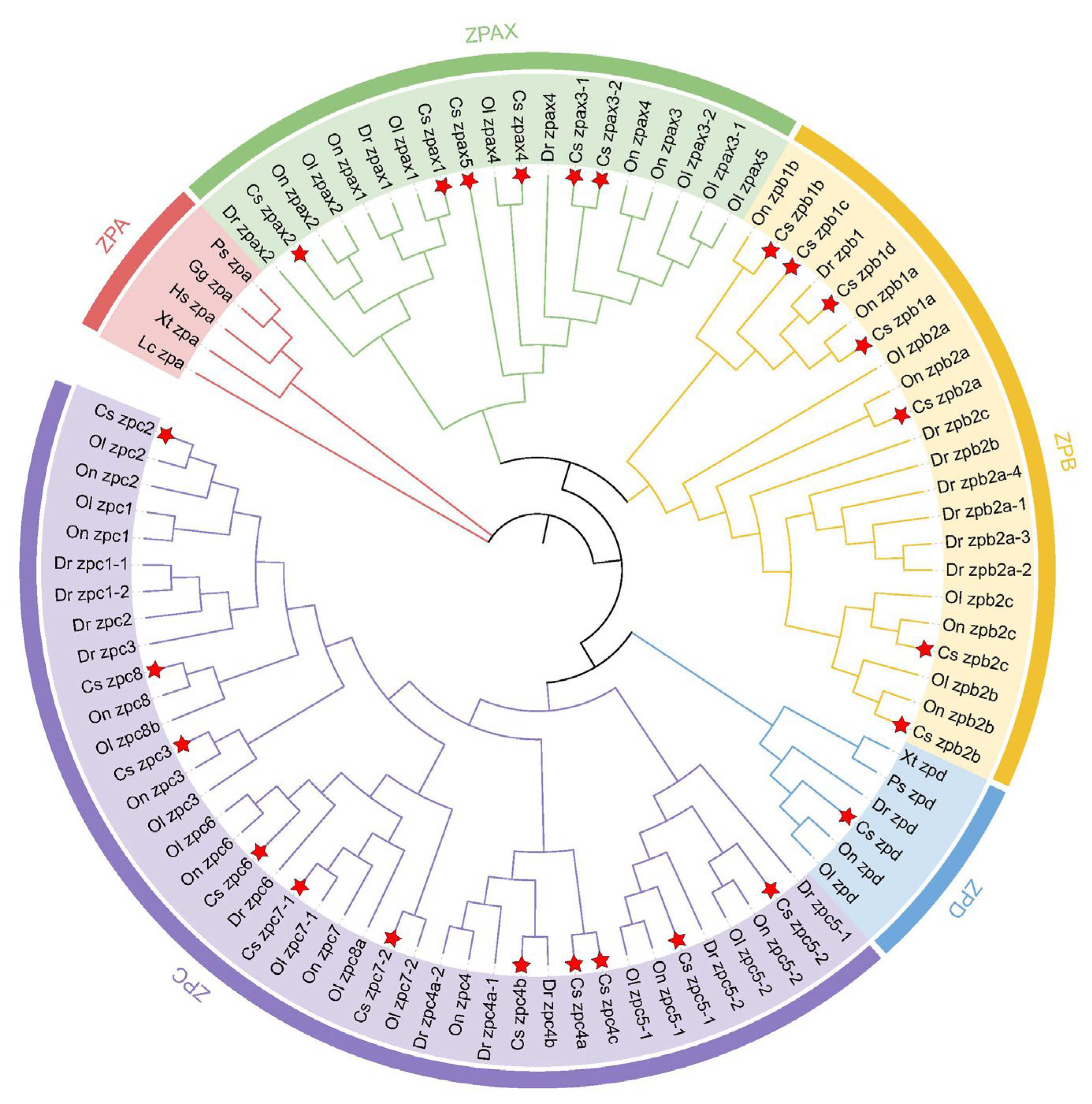
Figure 1. Phylogenetic analysis of ZP genes. Different colors in the circle represent different subclusters (red for ZPA, green for ZPAX, yellow for ZPB, blue for ZPD, purple for ZPC). Phylogenetic tree for nine species: C. semilaevis (Cs), Homo sapiens (Hs), D. rerio (Dr), O. niloticus (On), Gallus gallus (Gg), X. troicalis (Xt), Latimeria chalumnae (Lc), Pelodiscus sinensis (Ps), and O. latipes (Ol). The five subclusters are represented in different colors. CsZPs were marked with red stars.
Ijms 26 05346 g001
Figure 2. Gene structures and conserved domains of ZP family members. (A) Gene structures. The yellow and blue rectangles indicate the CDS and UTR regions; the gray line represents the introns. (B) The conserved domains of ZP family members. Different colored and shapes indicate different domains and motifs. Horizontal gray bars indicate amino acid sequences with no predictive functional domains. Protein domains are shown relative to the length of the position in the amino acid sequences.
Ijms 26 05346 g002
Figure 3. Synteny analysis of ZP gene in selected teleosts. (A) Synteny analysis of zpax1 and zpax2 genes. (B) Synteny analysis of zpc5-1 and zpc5-2 genes. (C) Synteny analysis of zpd genes. (D) Synteny analysis of zpb2a genes. The colored pentagons indicate different genes, and the direction of each pentagon indicates the gene direction. The empty space indicates a region with other genes or the absence of the gene in the genome.
Ijms 26 05346 g003
Figure 4. The predicted ZP genes functional partners using the protein–protein interaction method (PPI). (A) PPI analysis of ZPAX. (B) PPI analysis of ZPB. (C) PPI analysis of ZPC. (D) PPI analysis of ZPD.
Ijms 26 05346 g004
Figure 5. Heatmap of ZP family members; mRNA abundances in three different tissues of healthy male and female C. semilaevis. FB−female brain; MB−male brain; FG−female gonad; MG−male gonad; FL−female liver; ML−male liver. The expression levels were quantified as FPKM based on RNA-Seq. Gene expression levels are color coded from low (blue) to high (red). Each row represents one gene (listed on the right). Note: Only 23 of the 25 gene family members are shown in the heatmap because Cszpax2 and Cszpax3-1 were not detected in the transcriptome data, likely due to very low expression levels.
Ijms 26 05346 g005
Figure 6. Relative expression levels of ZP genes in different tissue of male and female C. semilaevis. (A) Cszpax1. (B) Cszpax3-1. (C) Cszpb1a. (D) Cszpb1b. (E) Cszpb2a. (F) Cszpc4a. (G) Cszpc5-1. (H) Cszpc7-1. (I) Cszpd. (J) Cszpb2b. (K) Cszpb2c. (L) Cszpc2. The brown and gray separately represent female and male. Bars with different letters indicate statistically significant differences (p < 0.05).
Ijms 26 05346 g006
Figure 7. Relative expression levels of ZP genes in the gonadal tissues of male and female C. semilaevis at different times. (A) Cszpax1. (B) Cszpax3-1. (C) Cszpb1a. (D) Cszpb1b. (E) Cszpb2a. (F) Cszpc4a. (G) Cszpc5-1. (H) Cszpc7-1. (I) Cszpd. The brown and gray separately represent female and male. Bars with different letters indicate statistically significant differences (p < 0.05).
Ijms 26 05346 g007
Figure 8. The spatial expression patterns of ZP genes of C. semilaevis at 1.5 yph. (A) Antisense probe for Cszpax1 in ovary. (B) Sense probe for Cszpax1 in ovary. (C) Antisense probe for Cszpax3-1 in ovary. (D) Sense probe for Cszpax3-1 in ovary. (E) Antisense probe for Cszpb1a in ovary. (F) Sense probe for Cszpb1a in ovary. (G) Antisense probe for Cszpc7-1 in ovary. (H) Sense probe for Cszpc7-1 in ovary. (I) Antisense probe for Cszpd in ovary. (J) Sense probe for Cszpd in ovary. Strong hybridization signals of Cszpax1, Cszpax3-1, Cszpb1a, Cszpc7-1, and Cszpd were specifically localized in the oocytes of female gonadal tissues. Scale bars = 200 μm.
Ijms 26 05346 g008
Figure 9. The knockdown effects of Cszpc7-1 and Cszpax1 siRNA in CO cells. (AC). Knockdown efficiency of Cszpc7-1 and its effects on sex-related genes and other ZP genes. (DF). Knockdown efficiency of Cszpax1 and its effects on sex-related genes and other ZP genes. A p-value less than 0.05 was considered to indicate a significant difference between the two groups and indicated by *. (*—p < 0.05; **—p < 0.01).
Ijms 26 05346 g009
Table 1. Sequence feature of ZP family members.
NameGene IDGene Length (bp) ORF Length (bp)Amino Acid Length (aa) MW (kDa)pIChrLocationNo. of ExonsNo. of Introns
Cszpax110338091858502850950103.73 4.8475,983,260–5,989,1092120
Cszpax21124872473614166255461.72 5.0375,977,607–5,981,2201312
Cszpax3-110338663014,745241280491.44 5.19123,962,096–
23,976,840
2524
Cszpax3-21124877263896240080089.83 5.44123,966,848–23,970,7431918
Cszpax4103380637841932821094123.50 6.6771,154,026–1,162,4442019
Cszpax51033867305305211870679.67 5.78123,976,991–23,982,2951615
Cszpb1a1033942782910146148754.12 4.67189,161,710–9,164,619109
Cszpb1b1033933743542159953359.41 8.841712,633,779–12,637,3201211
Cszpb1c10339339713,95930331011110.60 5.321712813072–12,827,0301615
Cszpb1d10338379211,22632431081120.00 6.199,156,026–9,167,2511817
Cszpb2a1033960712286127842647.19 5.39204,011,277–4,013,56287
Cszpb2b1033801894402193864672.52 5.79611,810,105–11,814,50687
Cszpb2c1033801372580147949352.91 7.34611,208,465–11,211,04487
Cszpc21033801302718134444848.56 5.35611,212,333–11,215,05087
Cszpc31033898915953145548554.01 6.111417,172,577–17,178,5291211
Cszpc4a1033830753615190863670.82 6.1912,597,477–2,601,09198
Cszpc4b1033840132723128142747.76 8.65913,824,630–13,827,35298
Cszpc4c1033911904981174958364.97 5.7914,104,869–4,109,84987
Cszpc5-11033966002058106835639.68 8.182011,811,312–11,813,36998
Cszpc5-2103396601215393931334.77 5.442011,814,022–11,816,174109
Cszpc61033960375493140146752.55 9.09203,517,755–3,523,24798
Cszpc7-11033929292522155751956.42 4.98176,833,291–6,835,812109
Cszpc7-2103391061270294831634.97 6.41132,117,282–32,119,98387
Cszpc8103377641109664221423.67 6.5942,417,479–2,418,57421
Cszpd1033795544431121840645.21 6.5562,033,303–2,037,733109
Disclaimer/Publisher’s Note: The statements, opinions and data contained in all publications are solely those of the individual author(s) and contributor(s) and not of MDPI and/or the editor(s). MDPI and/or the editor(s) disclaim responsibility for any injury to people or property resulting from any ideas, methods, instructions or products referred to in the content.