Article

Resveratrol Attenuates CSF Markers of Neurodegeneration and Neuroinflammation in Individuals with Alzheimer’s Disease

1
Laboratory for Dementia and Parkinsonism, Translational Neurotherapeutics Program, Department of Neurology, Georgetown University Medical Center, Washington, DC 20057, USA
2
Interdisciplinary Program in Neuroscience, Georgetown University Medical Center, Washington, DC 20057, USA
3
Memory Disorders Program, Department of Neurology, Georgetown University, Washington, DC 20057, USA
*
Author to whom correspondence should be addressed.
Int. J. Mol. Sci. 2025, 26(11), 5044; https://doi.org/10.3390/ijms26115044
Submission received: 9 May 2025 / Revised: 16 May 2025 / Accepted: 20 May 2025 / Published: 23 May 2025
(This article belongs to the Special Issue Molecular Advances in Neurologic and Neurodegenerative Disorders)

Abstract

Alzheimer’s disease (AD) is a progressive neurodegenerative disorder that is characterized by amyloid-beta (Aβ) accumulation and neuroinflammation. A previous multicenter, phase 2, double-blind, placebo-controlled trial randomized 179 participants into placebo or resveratrol over 52 weeks. Sub-analysis of CSF biomarkers of neuronal damage, inflammation, and microglial activity was performed in a subset of patients treated with a placebo (n = 21) versus resveratrol (n = 30). Markers of neuronal damage, including neuron-specific enolase and hyperphosphorylated neurofilaments, were reduced. Microglial activation was measured via a triggering receptor expressed on myeloid cells (TREM)-2 at baseline and after resveratrol treatment. Resveratrol significantly reduced CSF TREM2 levels and decreased inflammation and tissue damage, including matrix metalloprotease (MMP)-9. Cathepsin D, a lysosomal marker of autophagy, was reduced in the resveratrol group compared with placebo, while angiogenin, a marker of vascular angiogenesis, was increased. These data suggest that resveratrol may exert anti-inflammatory and neuroprotective effects in AD by reducing CSF TREM2 and other markers of neuronal damage. Further research is needed to assess the significance of these biomarker changes on clinical outcomes in patients with neurodegenerative diseases.

1. Introduction

Alzheimer’s disease (AD) is the most common form of dementia, affecting millions of people worldwide. One of the pathological hallmarks of AD is accumulation of amyloid-beta (Aβ) plaques and neurofibrillary tangles composed of hyperphosphorylated tau in the brain. These events are associated with chronic neuroinflammation [1], which may promote disease progression and severity. Microglia serve as the resident immune cells of the brain, and their activation is crucial for clearing cellular debris, including Aβ. However, dysregulated microglial activity has been implicated in the progression of AD, as overactivated microglia can contribute to chronic inflammation and exacerbate neuronal damage [2]. There is a growing interest in the role of microglial cells, which play a key role in modulating the inflammatory response in AD [3]. Given the complex interplay between microglial activity, Aβ aggregation, and neuroinflammation, it is imperative to explore therapeutic modulators of these pathways.
Resveratrol is a sirtuin-1 (SIRT1) activator that stimulates mitochondrial biogenesis [4,5,6,7,8,9,10] and regulates autophagy [11,12]. Resveratrol facilitates mitophagy [13], which is necessary for mitochondrial dynamics, which is deficient in neurodegenerative diseases [14,15]. Resveratrol also inhibits the activation of the NLRP3 inflammasome (also known as nucleotide-binding domain, leucine-rich repeat, and pyrin domain-containing protein 3) and lowers inflammation [16,17]. The various roles of resveratrol to inhibit the NLRP3 inflammasome, promote autophagy, and maintain mitochondrial integrity [18] suggest that resveratrol is a therapeutic candidate that has strong neuroprotective effects in AD and other neurodegenerative diseases [17,19].
A previous phase 2, multicenter, placebo-controlled, clinical trial investigated the effects of resveratrol on biomarkers for cognitive and functional outcomes in patients diagnosed with mild-to-moderate AD [20]. Post hoc analysis of cerebrospinal fluid (CSF) samples from these patients showed that treatment with resveratrol stabilized CSF Aβ levels and reduced markers of neuroinflammation, including matrix metalloprotease (MMP)-9, and regulated microglial activity [21]. This report further investigates the role of resveratrol on autophagy via the lysosomal marker cathepsin D and microglial activation via a triggering receptor expressed on myeloid cells (TREM-2), as well as other CSF biomarkers of neuronal and vascular damage in AD patients.

2. Results

2.1. Resveratrol Reduces CSF Markers of Cell Death

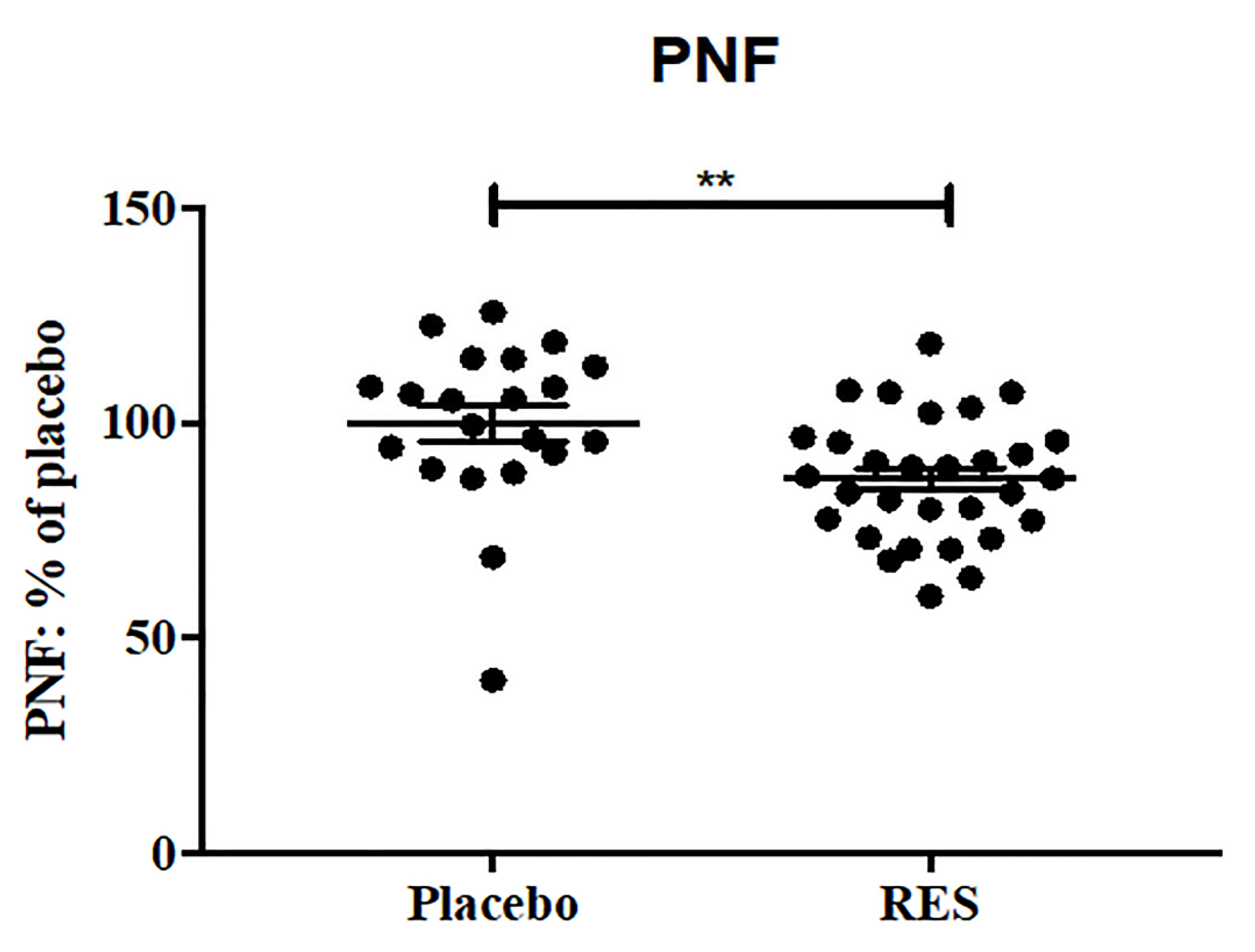
We first studied the effects of resveratrol treatment on molecular markers of cell death and autophagy in the CSF of AD patients. At baseline, there were no significant differences in CSF levels of neuron-specific enolase (NSE) or cathepsin D between the placebo and resveratrol groups (Figure 1a, Table 1). After 52 weeks, the resveratrol group showed a significant reduction in CSF NSE levels compared with baseline (Figure 1a). Notably, NSE levels in the resveratrol group were reduced by 40% compared with the placebo group at 52 weeks (Figure 1a). NSE is a well-established biomarker of cerebral tissue damage and is commonly used to assess stroke severity [22,23,24,25,26]. In addition, CSF levels of cathepsin D were significantly lower in the resveratrol group at 52 weeks compared with both baseline and the placebo group (Figure 1b, Table 1). Cathepsin D, a lysosomal proteinase, is highly expressed in senile plaques and typically elevated in the CSF of AD patients [27,28,29]. The effects of resveratrol on phosphorylated neurofilaments (PNF) were also measured (Table 1, Figure 2). The percentages of PNF at 52weeks are expressed relative to baseline because the PNF standards were not reliable. At week 52, PNF was significantly reduced in the resveratrol group compared with placebo.

2.2. CSF Biomarker of Angiogenesis and TREM2

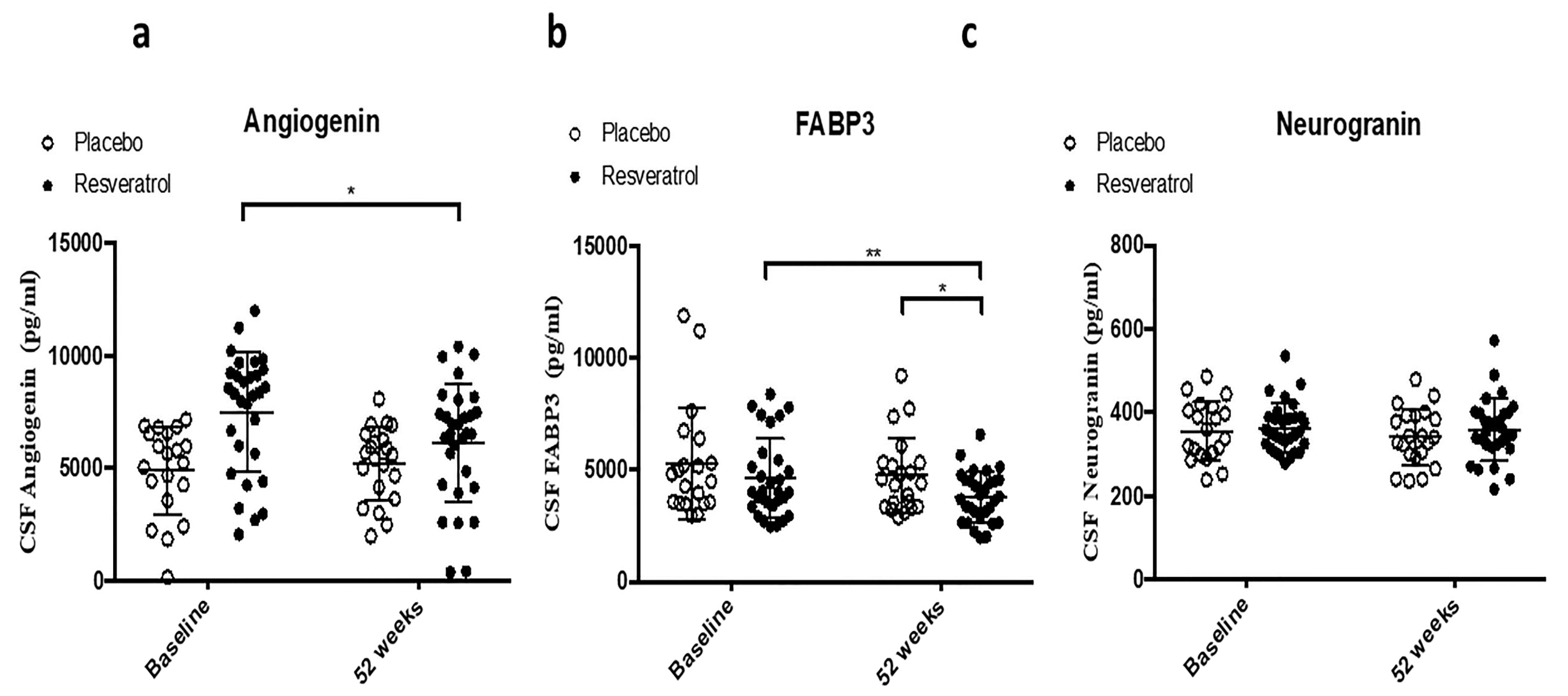
We then investigated the effects of resveratrol treatment on molecular markers of inflammation and angiogenesis in the CSF of AD patients. The levels of angiogenin, a biomarker of angiogenesis, significantly decreased within the resveratrol group by 52 weeks (Figure 3a, Table 1). Baseline levels of fatty acid binding protein 3 (FABP3) (Figure 3b, Table 1) and neurogranin (Figure 3c, Table 1) in the CSF were similar between the placebo and resveratrol groups. Over 52 weeks, FABP3 levels were significantly reduced in the resveratrol group compared with both baseline and the placebo group at 52 weeks (Figure 3b). No significant changes were observed in CSF neurogranin levels across groups (Figure 3c).

2.3. CSF Biomarkers of Matrix Metalloproteases as Dual Markers of Inflammation and BBB Integrity

We assessed the effects of resveratrol treatment on the CSF levels of MMPs and tissue inhibitor of metalloproteinases (TIMPs) as markers of possible vascular damage and neuroinflammation. At baseline, CSF levels of MMP-9 and TREM2 were not significantly different between the placebo and resveratrol groups (Table 1, Figure 4). After 52 weeks, MMP-9 levels were significantly reduced in the resveratrol group compared with both baseline and the placebo group (Figure 4a, Table 1). Similarly, TREM2 levels were significantly lower in the resveratrol group compared with the placebo group at 52 weeks (Figure 4b, Table 1).
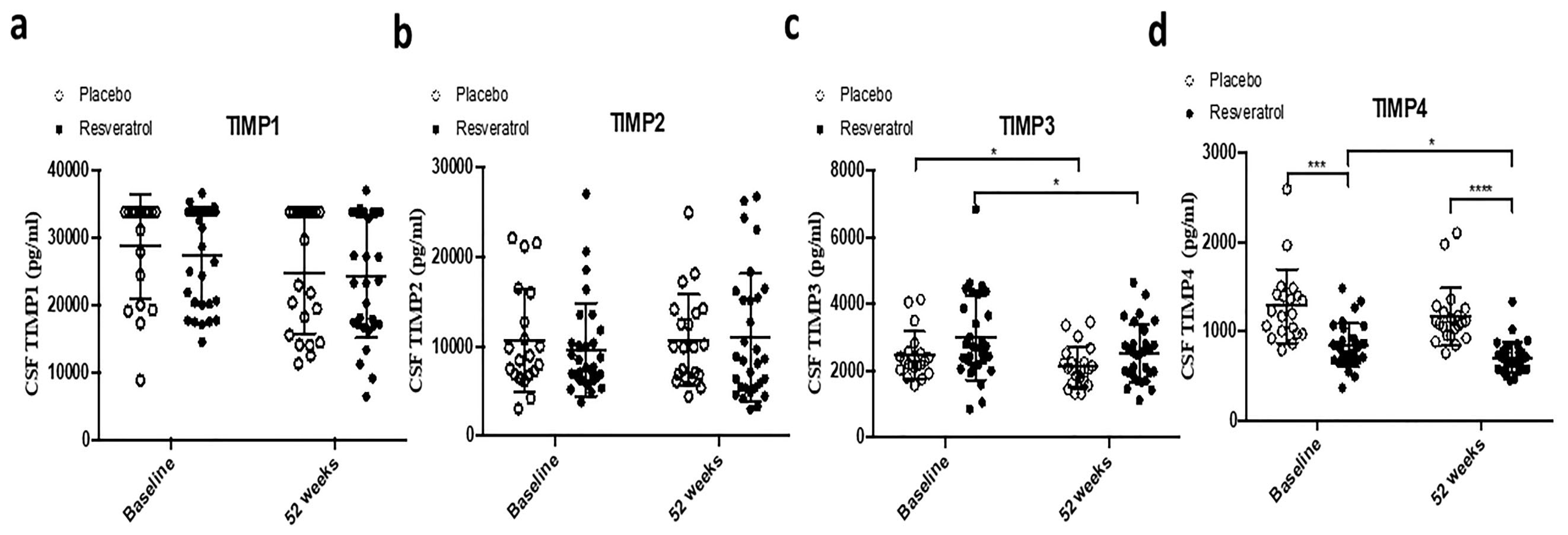
TIMPs 1, 2, 3, and 4 were also measured. No significant differences in TIMP-1, TIMP-2, or TIMP-3 levels were observed between groups at baseline (Figure 5a–c, Table 1). By 52 weeks, TIMP-3 levels were significantly reduced in both the resveratrol and placebo groups compared with baseline (Figure 5c). TIMP-4 levels, which were significantly different at baseline between the two groups, showed a significant reduction within the resveratrol group by 52 weeks (Figure 5d).

3. Discussion

Resveratrol decreases aging-dependent cognitive decline and pathology in animal models of AD [30]. Preclinical studies have shown that resveratrol can reduce Aβ aggregation, protect neurons from oxidative stress, and attenuate neuroinflammation and its ability to cross the blood–brain barrier (BBB), which further supports its potential as a treatment for neurodegeneration [20,31,32]. Resveratrol can also be relevant in chronic diseases and longevity in humans [33,34]. A randomized, placebo-controlled, double-blind, multicenter, 52-week trial of resveratrol in individuals with mild-to-moderate AD reported its safety and tolerability [20]. Doses increased in 500 mg increments every 13 weeks, but no differences between the resveratrol- and placebo-treated groups were found in laboratory tests, vital signs, physical examinations, or neurologic examinations. Further ad hoc analysis in an enriched population with a pre-specified biomarker, including low CSF Aβ42 levels, showed that CSF Aβ and microglial activity were significantly reduced after 52 weeks of resveratrol treatment [21]. The sub-analysis also suggested that biomarker changes are associated with functional improvement in activities of daily living in AD patients [21]. Cathepsin D is a lysosomal protein that contributes to autophagy in many neurodegenerative diseases, and its levels are elevated in the CSF of patients with AD, suggesting it could be a potential biomarker of autophagy [35,36]. Our data show a significant decrease in cathepsin D after 1 year of treatment with resveratrol. In addition to resveratrol effects on lysosomal clearance and regulation of microglial function, a decrease in markers of axonal damage, NSE [22,37], and phosphorylated neurofilaments further suggests a neuroprotective role of resveratrol. Neurogranin is a protein primarily found in dendritic spines and contributes to synaptic plasticity, and its levels are elevated in the CSF of AD patients compared with controls [38], but no change in CSF levels of neurogranin was observed with resveratrol treatment. The role of cathepsin D is complex, and its elevated levels in AD may indicate a compensatory effect to regulate autophagy, but the reduction in biomarkers of axonal damage together with cathepsin D suggest regulation of autophagy and attenuation of neuronal damage.
Overactivated microglia contribute to chronic inflammation and exacerbate neuronal damage [2]. Our data indicate a significant decrease in CSF levels of TREM2 after resveratrol treatment. TREM2 is a transmembrane receptor that binds to various ligands and contributes to microglia clearance of cellular debris, including Aβ plaques, thereby regulating microglial function and clearance [39]. Soluble TREM2 (sTREM2) is released from cell membranes and binds Aβ to prevent its aggregation, suggesting a protective role against AD [40]. Carriers of TREM2 mutations exhibit increased amyloid deposition and accelerated cognitive decline [40], perhaps due to a dysregulated microglial response to Aβ [41]. In fact, TREM2 is upregulated in response to Aβ accumulation, and it facilitates microglial clearance of amyloid plaques. However, sustained TREM2 activation can exacerbate inflammatory responses and tissue damage [42]. TREM2 has gained attention due to its involvement in various neurodegenerative diseases, including AD, multiple sclerosis, amyotrophic lateral sclerosis (ALS), and Parkinson’s disease (PD). TREM2 promotes microglial survival, migration, and phagocytosis of apoptotic cells, making it a critical regulator of neuroinflammatory processes in the brain [43]. Given the beneficial interplay between TREM2, microglial activity, and neuroinflammation, it is beneficial to explore the potential of resveratrol as a common therapeutic modulator of these pathways. TREM2-targeted therapies could provide a novel approach to balancing microglial activation, reducing harmful inflammation, and promoting toxic protein clearance.
Tissue inhibitors of metalloproteinases (TIMPs) are a family of proteins that inhibit the activity of MMPs to prevent the degradation of the extracellular matrix (ECM) and prevent tissue damage and inflammation [44]. Resveratrol reduces CSF levels of both TIMP-3 and TIMP-4, as well as MMP-9, consistent with our previous data [21], suggesting potential reduction of inflammation in AD patients. The role of TIMPs and MMPs, such as MMP-9, is critical in both inflammation and vascular changes, including angiogenesis in neurodegeneration [45]. Angiogenin is a ribonuclease, which plays a crucial role in angiogenesis and formation of new blood vessels in stress responses, and has been linked to various diseases, including neurodegeneration [38]. Our data show a decrease in TIMPs and MMP-9, suggesting reduced vascular and ECM damage, while the increase in the level of angiogenin may contribute to potential vascular remodeling after resveratrol treatment. However, the manifestation of changes in TIMPs and MMPs is varied in different diseases and different tissues. For example, elevated levels of MMP-2, MMP-3, MMP-10, TIMP-1, and TIMP-2 were detected in the CSF of patients with delirium who had pre-existing dementia [46]. Conversely, MMP-2, MMP-12, and TIMP-1 were higher in acute trauma (e.g., hip fracture) compared with healthy controls or patients without dementia. In a totally unrelated condition such as pyramidal syndrome or progressive weakness and stiffness of the lower limbs due to tropical spastic paraparesis and viral infection (inflammation), TIMP-2, TIMP-3, and TIMP-4 levels were increased compared with healthy controls, while TIMP-1 was only increased in the presence of inflammation, and MMP-3 and MMP-9 levels were significantly increased in the presence or absence of inflammation [47]. MMPs as a family of proteinase can play critical roles in acute and chronic neuroinflammation, as well as blood vessel integrity and BBB permeability [48,49]. The variable levels of MMPs and/or TIMPs may be due to their proteolytic substrates that may also depend on the tissue as well as inflammatory conditions, blood vessel status, and specific processing stage of the proteolytic activity of these molecules, thus providing differential read-out and relationship in different conditions.
Resveratrol could also be a candidate for the treatment of traumatic brain injury (TBI), which is a major cause of death and disability [50], and individuals who experience TBI are at increased risk for several neurological disorders, such as PD, ALS, MS Lewy body dementia (LBD), and AD [51]. The potential efficacy of resveratrol to treat TBI has been demonstrated in preclinical TBI models [52]. Resveratrol attenuates the level of molecular markers of damage, including malondialdehyde (MDA), glutathione (GSH), nitric oxide (NO), and xanthine oxidase (XO) [53], therefore providing a neuroprotective strategy against brain damage. Resveratrol was also shown to halt cognitive deficits that are mediated via p38 activation and subsequent activation of the nrf2 (nuclear factor 1 erythroid nuclear factor 2) pathway in TBI rats [54]. Resveratrol was also shown to lower neuroinflammation and enhance long-term hippocampal functioning in models of TBI [55]. Aside from these specific examples in TBI models, resveratrol has neuroprotective effects in many disease conditions [20,21,32,56], and it is imperative to explore the role of resveratrol as a preventative strategy in neurodegeneration.
Resveratrol’s neuroprotective effects are mediated through multiple mechanisms, including the activation of SIRT1, a protein deacetylase involved in cellular stress responses. In addition, resveratrol exerts anti-inflammatory effects by inhibiting the nuclear factor kappa-light-chain-enhancer of activated B cells (NF-κB) signaling pathway, a central regulator of inflammation [57,58]. By targeting these molecular pathways, resveratrol may help reduce chronic inflammation that contributes to the progression of AD. ABP3 (also known as heart-type fatty acid binding protein) transports fatty acids to the mitochondria, facilitating their use for energy production and regulation of energy balance by controlling fatty acid metabolism [59] and mitochondrial autophagy [60]. Biologically, resveratrol stimulates mitochondrial biogenesis [4,5,6,7,8] and regulates mitochondrial dynamics via autophagy [11,12]. Resveratrol plays a role in parkin-related mitophagy [13], which is necessary for mitochondrial dynamics, but it is deficient in neurodegenerative diseases, including PD and AD [14,15].
PD is the second most common neurodegenerative disorder causing motor and non-motor symptoms, loss of dopamine (DA), and aggregation of misfolded alpha-synuclein [61,62,63]. There is evidence of the presence of oxidative stress and mitochondrial damage in the brains of patients with PD [64,65,66]. Specific gene mutations that cause PD can cause oxidative stress and mitochondrial dysfunction in the familial and the sporadic forms of PD [64,65,66]. PD-linked familial gene mutations, like PTEN-induced putative kinase 1 (PINK1), DJ-1, alpha-synuclein, leucine-rich repeat kinase 2 (Lrrk2), and parkin, express proteins that interact with mitochondrial proteins, oxidative stress, and free radicals [67,68,69]. The SIRT1 activator resveratrol promotes mitochondrial biogenesis [4,5,6,7,8] and regulates mitochondrial dynamics via mitophagy [11,12]. The role of mitochondrial energy metabolism, including production of adenosine triphosphate (ATP) and nicotinamide adenine dinucleotide (NAD+), plays a critical role in the etiology and pathogenesis of PD [64,65,66], therefore optimizing a therapeutic option that enhances energy metabolism and may counteract alpha-synuclein aggregation and attenuate inflammation [67,68,69]. ATP and NAD+ are deficient in PD and several other neurodegenerative diseases [70], and mitochondria produce ATP to meet energy demands, regulate homeostasis, and lower reactive oxygen species that cause oxidative stress and loss of DA in PD [71,72,73]. Dopaminergic neurons seem to be vulnerable to the toxic events that occur during the neurodegenerative process of PD, and mitochondrial energy metabolism and integrity are central to these pathways [74]. Mitochondrial ATP production is deficient in muscles of PD patients [75]. Resveratrol may improve energy metabolism and have the potential to slow PD progression [76,77]. Mitochondria are essential for neuronal function, and enhancement of glycolysis increases ATP levels and may lead to novel therapeutic options for PD [78].
Friedreich’s ataxia (FRDA) is another autosomal-recessive inherited neurodegenerative disease, and the most common ataxia caused by mutation in the FXN gene, resulting in deficiency of the mitochondrial protein frataxin. Resveratrol increases frataxin expression in vitro and in vivo in models of FRDA. A 1.5- to 2-fold increase in frataxin protein expression was observed in lymphoblasts and fibroblasts derived from individuals with FRDA, and a 200 mg/kg subcutaneous RSV resulted in a 1.5-fold increase in human frataxin protein in the brain of humanized FRDA (YG8R) mice [79]. An open-label study in 24 individuals with FRDA showed clinical improvements via composite Friedreich’s ataxia endpoints and benefits to speech and hearing following treatment with resveratrol, as well as a reduction in plasma F2-isoprostanes, a marker of oxidative stress, compared with baseline [80]. Mucopolysaccharidosis type I (MPS I) is an autosomal-recessive disease that presents with chronic, progressive, multisystem, lysosomal storage caused by deficiency or lack of activity of the α-L-iduronidase (IDUA) enzyme [81,82,83,84], and resveratrol was shown to upregulate IDUA activity [85], providing a potential treatment option for MPSI.
There is a critical need to develop new disease-modifying therapies for neurodegenerative diseases, including AD and PD. One promising candidate for such modulation is resveratrol, a naturally occurring polyphenol. Resveratrol is an antioxidant, anti-inflammatory, and neuroprotective agent, and it has been explored as a potential therapeutic agent for neurodegenerative diseases due to its multifaceted biological roles [34,86].

4. Materials and Methods

4.1. Patient Demographics

With the Alzheimer’s Disease Cooperative Study (ADCS), a randomized, placebo-controlled, double-blind, multi-site, phase 2 trial of resveratrol in individuals with mild-to-moderate dementia due to AD was previously completed [20]. The study drug was pure, synthetic, resveratrol powder (encapsulated) versus a matching placebo. Concomitant use of FDA-approved medications for AD (e.g., cholinesterase inhibitors) was allowed. Participants (total N = 179) were randomized to placebo or resveratrol with 500 mg orally once daily (with a dose increase of 500 mg increments every 13 weeks, ending with 1000 mg twice daily). The total treatment duration was 52 weeks. A total of N = 56 participants completed the trial in the resveratrol arm, and N = 48 completed week 52 in the placebo. The primary outcomes were safety and tolerability and resveratrol effects on AD biomarkers (Aβ40 and Aβ42, CSF Aβ40, Aβ42, tau, and phospho-tau181) and volumetric MRI (primary outcomes). Clinical outcomes (secondary) were also examined. Detailed pharmacokinetics were obtained in a subset (n = 15) at baseline and at weeks 13, 26, 39, and 52. Resveratrol entered the brain, as its major metabolites were detected in plasma and CSF. Weight loss was observed in the resveratrol group. Compared with a decline found in the placebo group, plasma Aβ40 and CSF Aβ40 levels were stabilized by resveratrol. In the subset of individuals with biomarker-confirmed AD (baseline Aβ42 < 600 ng/mL), resveratrol treatment also stabilized CSF Aβ42. Brain volume loss was increased by resveratrol treatment (3 versus 1%), suggesting a potent anti-inflammatory effect [21].

4.2. Multiplex Xmap ELISA

Xmap technology uses magnetic microspheres that are internally coded with two fluorescent dyes. Through precise combinations of these two dyes, multiple proteins are simultaneously measured within a sample. All samples, including placebo and resveratrol at baseline and 52 weeks, were analyzed in parallel using the same reagents. A total of 25 μL of human CSF or plasma was incubated overnight at 4 °C with 25 μL of a mixed bead solution containing MMP-1, -2, -7, -9, and -10 (Millipore Cat# HMMP1MAG-55K, Rockville, MD, USA), MMP-3, -12, and -13. (Millipore Cat# HMMP2MAG-55K), tissue inhibitor of metalloproteases TIMP-1 and -2 (Millipore Cat# HTIMP1MAG-54K), TIMP-1, -2, -3, and -4. (Millipore Cat# HTIMP2MAG-54K), Cathepsin D (Millipore Cat# HNDG3MAG-36K), neuron-specific enolase -NSE (Millipore Cat# HNDIMAG-39K), and a panel including angiogenin, FABP3 (fatty acid binding protein 3), TREM 2, and neurogranin (Millipore Cat# HNS2MAG-95K). After washing, samples were incubated with 25 μL of detection antibody solution for 1.5 h at room temperature. Streptavidin-phycoerythrin (25 μL) was added to each well containing the 25 μL of detection antibody solution. Samples were then washed and suspended in 100 μL of sheath fluid. Samples were then run on MAGPIX with Xponent software 4.3 (Millipore 40-012, Luminex200TM System). The median fluorescent intensity (MFI) data was analyzed using a 5-parameter logistic or spline curve-fitting method for calculating analyte concentrations in samples.

4.3. Standard Protocol Approvals, Registrations, and Patient Consent

This study was conducted in accordance with Good Clinical Practice guidelines. Informed consent was obtained from participants and study partners. The study was conducted under local institutional review board supervision, under Food and Drug Administration IND 104205, and registered at ClinicalTrials.gov (NCT01504854).

4.4. Statistical Analysis

The outcomes measured here are all exploratory, post hoc analyses. Data are summarized as raw values, range as appropriate, and mean ± SD for N = 21 in the placebo group and N = 30 in the resveratrol group. All graphs and statistical analyses were performed in Graph Pad Prism Software version 7.0 (Graph Pad Prism Software, Inc., La Jolla, CA. USA). For baseline comparison between the two treatment arms, unpaired t-tests assuming both equal and unequal variances and Wilcoxon rank sum tests, were performed to compare biomarkers and clinical variables. Paired t-tests were performed within groups at baseline versus 52 weeks of treatment, and unpaired t-tests were performed for comparison of placebo and resveratrol treatments.

Author Contributions

M.H. processed all samples. S.B. and C.M. wrote the manuscript. R.S.T. was the principal investigator of the resveratrol trials. X.L. curated the data and reviewed the manuscript. C.M. supervised the processing of biofluid samples and finalized the manuscript. All authors have read and agreed to the published version of the manuscript.

Funding

Funding for this study was provided by Georgetown University to Charbel Moussa.

Institutional Review Board Statement

This study was conducted in accordance with Good Clinical Practice guidelines. Informed consent was obtained from participants and study partners. This study was conducted under local institutional-review-board supervision, under Food and Drug Administration IND 104205, and registered at ClinicalTrials.gov (NCT01504854). The work presented in this manuscript conforms to all rigorous and ethical standards for research conduct, data collection and analysis, and data dissemination.

Informed Consent Statement

Not applicable.

Data Availability Statement

The final data generated and analyzed, materials, and all interpretations are available to the scientific and non-scientific community upon request. No datasets are deposited in a repository, and there is no web link to any datasets.

Acknowledgments

The authors acknowledge the Alzheimer’s Disease Cooperative Study (ADCS) for the donation of the CSF samples from AD participants in the randomized, placebo-controlled, double-blind, multi-site, phase 2 trial of resveratrol in individuals with mild-to-moderate dementia.

Conflicts of Interest

All other authors declare no conflicts of interest.

References

  1. Heneka, M.T.; Carson, M.J.; Khoury, E.J.; Landreth, G.E.; Brosseron, F.; Feinstein, D.L.; Jacobs, A.H.; Wyss-Coray, T.; Vitorica, J.; Ransohoff, R.M.; et al. Neuroinflammation in Alzheimer’s disease. Lancet Neurol. 2015, 14, 388–405. [Google Scholar] [CrossRef] [PubMed]
  2. Hansen, D.V.; Hanson, J.E.; Sheng, M. Microglia in Alzheimer’s disease. J. Cell Biol. 2018, 217, 459–472. [Google Scholar] [CrossRef] [PubMed]
  3. Colonna, M.; Wang, Y. TREM2 variants: New keys to decipher Alzheimer disease pathogenesis. Nat. Rev. Neurosci. 2016, 17, 201–207. [Google Scholar] [CrossRef]
  4. Yang, R.; Dong, H.; Jia, S.; Yang, Z. Resveratrol as a modulatory of apoptosis and autophagy in cancer therapy. Clin. Transl. Oncol. 2022, 24, 1219–1230. [Google Scholar] [CrossRef]
  5. Yang, X.; Jiang, T.; Wang, Y.; Guo, L. The Role and Mechanism of SIRT1 in Resveratrol-regulated Osteoblast Autophagy in Osteoporosis Rats. Sci. Rep. 2019, 9, 18424. [Google Scholar] [CrossRef]
  6. D’Arcy, M.S. A review of the chemopreventative and chemotherapeutic properties of the phytochemicals berberine, resveratrol and curcumin, and their influence on cell death via the pathways of apoptosis and autophagy. Cell Biol. Int. 2020, 44, 1781–1791. [Google Scholar] [CrossRef]
  7. Rubinsztein, D.C.; Marino, G.; Kroemer, G. Autophagy and aging. Cell 2011, 146, 682–695. [Google Scholar] [CrossRef]
  8. Wang, J.; Huang, P.; Pan, X.; Xia, C.; Zhang, H.; Zhao, H.; Yuan, Z.; Liu, J.; Meng, C.; Liu, F. Resveratrol reverses TGF-beta1-mediated invasion and metastasis of breast cancer cells via the SIRT3/AMPK/autophagy signal axis. Phytother. Res. 2023, 37, 211–230. [Google Scholar] [CrossRef] [PubMed]
  9. Lagouge, M.; Argmann, C.; Gerhart-Hines, Z.; Meziane, H.; Lerin, C.; Daussin, F.; Messadeq, N.; Milne, J.; Lambert, P.; Elliott, P.; et al. Resveratrol improves mitochondrial function and protects against metabolic disease by activating SIRT1 and PGC-1alpha. Cell 2006, 127, 1109–1122. [Google Scholar] [CrossRef]
  10. Canto, C.; Gerhart-Hines, Z.; Feige, J.N.; Lagouge, M.; Noriega, L.; Milne, J.C.; Elliott, P.J.; Puigserver, P.; Auwerx, J. AMPK regulates energy expenditure by modulating NAD+ metabolism and SIRT1 activity. Nature 2009, 458, 1056–1060. [Google Scholar] [CrossRef]
  11. Lin, F.; Zhang, S.; Zhu, X.; Lv, Z. Autophagy-related 7 proteindependent autophagy mediates resveratrol-caused upregulation of mitochondrial biogenesis and steroidogenesis in aged Leydig cell. Mol. Biol. Rep. 2023, 51, 28. [Google Scholar]
  12. Tao, G.; Wang, X.; Wang, J.; Ye, Y.; Zhang, M.; Lang, Y.; Ding, S. Dihydro-resveratrol ameliorates NLRP3 inflammasome-mediated neuroinflammation via Bnip3-dependent mitophagy in Alzheimer’s disease. Br. J. Pharmacol. 2025, 182, 1005–1024. [Google Scholar] [CrossRef] [PubMed]
  13. Wu, D.; Zhang, H.; Li, F.; Liu, S.; Wang, Y.; Zhang, Z.; Wang, J.; Wu, Q. Resveratrol alleviates acute lung injury in mice by promoting Pink1/Parkin-related mitophagy and inhibiting NLRP3 inflammasome activation. Biochim. Biophys. Acta Gen. Subj. 2024, 1868, 130612. [Google Scholar] [CrossRef] [PubMed]
  14. Cen, X.; Zhang, M.; Zhou, M.; Ye, L.; Xia, H. Mitophagy Regulates Neurodegenerative Diseases. Cells 2021, 10, 1876. [Google Scholar] [CrossRef]
  15. Li, A.; Zhang, S.; Li, J.; Liu, K.; Huang, F.; Liu, B. Metformin and resveratrol inhibit Drp1-mediated mitochondrial fission and prevent ER stress-associated NLRP3 inflammasome activation in the adipose tissue of diabetic mice. Mol. Cell. Endocrinol. 2016, 434, 36–47. [Google Scholar] [CrossRef]
  16. Li, M.; Wu, X.; An, P.; Dang, H.; Liu, Y.; Liu, R. Effects of resveratrol on autophagy and the expression of inflammasomes in a placental trophoblast oxidative stress model. Life Sci. 2020, 256, 117890. [Google Scholar] [CrossRef]
  17. Mo, Y.; Sun, Y.Y.; Liu, K.Y. Autophagy and inflammation in ischemic stroke. Neural Regen. Res. 2020, 15, 1388–1396. [Google Scholar]
  18. Chang, Y.P.; Ka, S.M.; Hsu, W.H.; Chen, A.; Chao, L.K.; Lin, C.C.; Hsieh, C.C.; Chen, M.C.; Chiu, H.W.; Ho, C.L.; et al. Resveratrol inhibits NLRP3 inflammasome activation by preserving mitochondrial integrity and augmenting autophagy. J. Cell. Physiol. 2015, 230, 1567–1579. [Google Scholar] [CrossRef]
  19. Zhang, X.; Wang, Z.; Li, X.; Chen, J.; Yu, Z.; Li, X.; Sun, C.; Hu, L.; Wu, M.; Liu, L. Polydatin protects against atherosclerosis by activating autophagy and inhibiting pyroptosis mediated by the NLRP3 inflammasome. J. Ethnopharmacol. 2023, 309, 116304. [Google Scholar] [CrossRef]
  20. Turner, R.S.; Thomas, R.G.; Craft, S.; van Dyck, C.H.; Mintzer, J.; Reynolds, B.A.; Brewer, J.B.; Rissman, R.A.; Raman, R.; Aisen, P.S. A randomized, double-blind, placebo-controlled trial of resveratrol for Alzheimer disease. Neurology 2015, 85, 1383–1391. [Google Scholar] [CrossRef]
  21. Moussa, C.; Hebron, M.; Huang, X.; Ahn, J.; Rissman, R.A.; Aisen, P.S.; Turner, R.S. Resveratrol regulates neuro-inflammation and induces adaptive immunity in Alzheimer’s disease. J. Neuroinflamm. 2017, 14, 1. [Google Scholar] [CrossRef] [PubMed]
  22. Isgro, M.A.; Bottoni, P.; Scatena, R. Neuron-Specific Enolase as a Biomarker: Biochemical and Clinical Aspects. Adv. Exp. Med. Biol. 2015, 867, 125–143. [Google Scholar] [PubMed]
  23. Vos, P.E.; Jacobs, B.; Andriessen, T.M.; Lamers, K.J.; Borm, G.F.; Beems, T.; Edwards, M.; Rosmalen, C.F.; Vissers, J.L. GFAP and S100B are biomarkers of traumatic brain injury: An observational cohort study. Neurology 2010, 75, 1786–1793. [Google Scholar] [CrossRef] [PubMed]
  24. Meric, E.; Gunduz, A.; Turedi, S.; Cakir, E.; Yandi, M. The prognostic value of neuron-specific enolase in head trauma patients. J. Emerg. Med. 2010, 38, 297–301. [Google Scholar] [CrossRef]
  25. Guzel, A.; Er, U.; Tatli, M.; Aluclu, U.; Ozkan, U.; Duzenli, Y.; Satici, O.; Guzel, E.; Kemaloglu, S.; Ceviz, A.; et al. Serum neuron-specific enolase as a predictor of short-term outcome and its correlation with Glasgow Coma Scale in traumatic brain injury. Neurosurg. Rev. 2008, 31, 439–444; discussion 444–445. [Google Scholar] [CrossRef]
  26. Brea, D.; Sobrino, T.; Blanco, M.; Cristobo, I.; Rodriguez-Gonzalez, R.; Rodriguez-Yanez, M.; Moldes, O.; Agulla, J.; Leira, R.; Castillo, J. Temporal profile and clinical significance of serum neuron-specific enolase and S100 in ischemic and hemorrhagic stroke. Clin. Chem. Lab. Med. 2009, 47, 1513–1518. [Google Scholar] [CrossRef]
  27. Cataldo, A.M.; Paskevich, P.A.; Kominami, E.; Nixon, R.A. Lysosomal hydrolases of different classes are abnormally distributed in brains of patients with Alzheimer disease. Proc. Natl. Acad. Sci. USA 1991, 88, 10998–11002. [Google Scholar] [CrossRef]
  28. Cataldo, A.M.; Barnett, J.L.; Berman, S.A.; Li, J.; Quarless, S.; Bursztajn, S.; Lippa, C.; Nixon, R.A. Gene expression and cellular content of cathepsin D in Alzheimer’s disease brain: Evidence for early up-regulation of the endosomal-lysosomal system. Neuron 1995, 14, 671–680. [Google Scholar] [CrossRef]
  29. Schwagerl, A.L.; Mohan, P.S.; Cataldo, A.M.; Vonsattel, J.P.; Kowall, N.W.; Nixon, R.A. Elevated levels of the endosomal-lysosomal proteinase cathepsin D in cerebrospinal fluid in Alzheimer disease. J. Neurochem. 1995, 64, 443–446. [Google Scholar] [CrossRef]
  30. Tome-Carneiro, J.; Larrosa, M.; Gonzalez-Sarrias, A.; Tomas-Barberan, F.A.; Garcia-Conesa, M.T.; Espin, J.C. Resveratrol and clinical trials: The crossroad from in vitro studies to human evidence. Curr. Pharm. Des. 2013, 19, 6064–6093. [Google Scholar] [CrossRef]
  31. Baur, J.A.; Sinclair, D.A. Therapeutic potential of resveratrol: The in vivo evidence. Nat. Rev. Drug Discov. 2006, 5, 493–506. [Google Scholar] [CrossRef] [PubMed]
  32. Sawda, C.; Moussa, C.; Turner, R.S. Resveratrol for Alzheimer’s disease. Ann. N.Y. Acad. Sci. 2017, 1403, 142–149. [Google Scholar] [CrossRef]
  33. Zhou, D.D.; Luo, M.; Huang, S.Y.; Saimaiti, A.; Shang, A.; Gan, R.Y.; Li, H.B. Effects and Mechanisms of Resveratrol on Aging and Age-Related Diseases. Oxid. Med. Cell. Longev. 2021, 2021, 9932218. [Google Scholar] [CrossRef]
  34. Zhang, L.X.; Li, C.X.; Kakar, M.U.; Khan, M.S.; Wu, P.F.; Amir, R.M.; Dai, D.F.; Naveed, M.; Li, Q.Y.; Saeed, M.; et al. Resveratrol (RV): A pharmacological review and call for further research. Biomed. Pharmacother. 2021, 143, 112164. [Google Scholar] [CrossRef]
  35. Ntais, C.; Polycarpou, A.; Ioannidis, J.P. Meta-analysis of the association of the cathepsin D Ala224Val gene polymorphism with the risk of Alzheimer’s disease: A HuGE gene-disease association review. Am. J. Epidemiol. 2004, 159, 527–536. [Google Scholar] [CrossRef]
  36. Lowry, J.R.; Klegeris, A. Emerging roles of microglial cathepsins in neurodegenerative disease. Brain Res. Bull. 2018, 139, 144–156. [Google Scholar] [CrossRef] [PubMed]
  37. Heckler, I.; Venkataraman, I. Phosphorylated neurofilament heavy chain: A potential diagnostic biomarker in amyotrophic lateral sclerosis. J. Neurophysiol. 2022, 127, 737–745. [Google Scholar] [CrossRef] [PubMed]
  38. Loveland, A.B.; Koh, C.S.; Ganesan, R.; Jacobson, A.; Korostelev, A.A. Structural mechanism of angiogenin activation by the ribosome. Nature 2024, 630, 769–776. [Google Scholar] [CrossRef]
  39. Li, Y.; Xu, H.; Wang, H.; Yang, K.; Luan, J.; Wang, S. TREM2: Potential therapeutic targeting of microglia for Alzheimer’s disease. Biomed. Pharmacother. 2023, 165, 115218. [Google Scholar] [CrossRef]
  40. Guerreiro, R.; Wojtas, A.; Bras, J.; Carrasquillo, M.; Rogaeva, E.; Majounie, E.; Cruchaga, C.; Sassi, C.; Kauwe, J.S.; Younkin, S.; et al. TREM2 variants in Alzheimer’s disease. N. Engl. J. Med. 2013, 368, 117–127. [Google Scholar] [CrossRef]
  41. Ulland, T.K.; Colonna, M. TREM2—A key player in microglial biology and Alzheimer disease. Nat. Rev. Neurol. 2018, 14, 667–675. [Google Scholar] [CrossRef] [PubMed]
  42. Jiang, T.; Yu, J.T.; Zhu, X.C.; Tan, L. TREM2 in Alzheimer’s disease. Mol. Neurobiol. 2016, 53, 3876–3887. [Google Scholar] [CrossRef] [PubMed]
  43. Jay, T.R.; Miller, C.M.; Cheng, P.J.; Graham, L.C.; Bemiller, S.; Broihier, M.L.; Xu, G.; Margevicius, D.; Karlo, J.C.; Sousa, G.L.; et al. TREM2 deficiency eliminates TREM2+ inflammatory macrophages and ameliorates pathology in Alzheimer’s disease mouse models. J. Exp. Med. 2017, 214, 2873–2889. [Google Scholar] [CrossRef] [PubMed]
  44. Murphy, G. Tissue inhibitors of metalloproteinases. Genome Biol. 2011, 12, 233. [Google Scholar] [CrossRef]
  45. Cabral-Pacheco, G.A.; Garza-Veloz, I.; la Rosa, C.C.-D.; Ramirez-Acuna, J.M.; Perez-Romero, B.A.; Guerrero-Rodriguez, J.F.; Martinez-Avila, N.; Martinez-Fierro, M.L. The Roles of Matrix Metalloproteinases and Their Inhibitors in Human Diseases. Int. J. Mol. Sci. 2020, 21, 9739. [Google Scholar] [CrossRef]
  46. Aksnes, M.; Schibstad, M.H.; Chaudhry, F.A.; Neerland, B.E.; Caplan, G.; Saltvedt, I.; Eldholm, R.S.; Myrstad, M.; Edwin, T.H.; Persson, K.; et al. Differences in metalloproteinases and their tissue inhibitors in the cerebrospinal fluid are associated with delirium. Commun. Med. 2024, 4, 124. [Google Scholar] [CrossRef]
  47. Kettlun, A.M.; Cartier, L.; Garcia, L.; Collados, L.; Vasquez, F.; Ramirez, E.; Valenzuela, M.A. TIMPs and MMPs expression in CSF from patients with TSP/HAM. Life Sci. 2003, 72, 2863–2876. [Google Scholar] [CrossRef]
  48. Rosenberg, G.A. Neurological diseases in relation to the blood-brain barrier. J. Cereb. Blood Flow Metab. 2012, 32, 1139–1151. [Google Scholar] [CrossRef]
  49. Rosenberg, G.A. Matrix metalloproteinases in neuroinflammation. Glia 2002, 39, 279–291. [Google Scholar] [CrossRef]
  50. Malec, J.F.; Brown, A.W.; Leibson, C.L.; Flaada, J.T.; Mandrekar, J.N.; Diehl, N.N.; Perkins, P.K. The mayo classification system for traumatic brain injury severity. J. Neurotrauma 2007, 24, 1417–1424. [Google Scholar] [CrossRef]
  51. Ghiam, M.K.; Patel, S.D.; Hoffer, A.; Selman, W.R.; Hoffer, B.J.; Hoffer, M.E. Drug Repurposing in the Treatment of Traumatic Brain Injury. Front. Neurosci. 2021, 15, 635483. [Google Scholar] [CrossRef] [PubMed]
  52. Weber, J.T. Experimental models of repetitive brain injuries. Prog. Brain Res. 2007, 161, 253–261. [Google Scholar] [PubMed]
  53. Ates, O.; Cayli, S.; Altinoz, E.; Gurses, I.; Yucel, N.; Sener, M.; Kocak, A.; Yologlu, S. Neuroprotection by resveratrol against traumatic brain injury in rats. Mol. Cell. Biochem. 2007, 294, 137–144. [Google Scholar] [CrossRef]
  54. Shi, Z.; Qiu, W.; Xiao, G.; Cheng, J.; Zhang, N. Resveratrol Attenuates Cognitive Deficits of Traumatic Brain Injury by Activating p38 Signaling in the Brain. Med. Sci. Monit. 2018, 24, 1097–1103. [Google Scholar] [CrossRef]
  55. Atalay, T.; Gulsen, I.; Colcimen, N.; Alp, H.H.; Sosuncu, E.; Alaca, I.; Ak, H.; Ragbetli, M.C. Resveratrol Treatment Prevents Hippocampal Neurodegeneration in a Rodent Model of Traumatic Brain Injury. Turk. Neurosurg. 2017, 27, 924–930. [Google Scholar]
  56. Rahman, M.H.; Akter, R.; Bhattacharya, T.; Abdel-Daim, M.M.; Alkahtani, S.; Arafah, M.W.; Al-Johani, N.S.; Alhoshani, N.M.; Alkeraishan, N.; Alhenaky, A.; et al. Resveratrol and Neuroprotection: Impact and Its Therapeutic Potential in Alzheimer’s Disease. Front. Pharmacol. 2020, 11, 619024. [Google Scholar] [CrossRef] [PubMed]
  57. Vingtdeux, V.; Giliberto, L.; Zhao, H.; Chandakkar, P.; Wu, Q.; Simon, J.E.; Janle, E.M.; Lobo, J.; Ferruzzi, M.G.; Davies, P.; et al. AMP-activated protein kinase signaling activation by resveratrol modulates amyloid-beta peptide metabolism. J. Biol. Chem. 2010, 285, 9100–9113. [Google Scholar] [CrossRef]
  58. Shakibaei, M.; Csaki, C.; Nebrich, S.; Mobasheri, A. Resveratrol inhibits IL-1β-induced inflammatory signaling and apoptosis in human articular chondrocytes: Potential for use as a therapeutic agent for osteoarthritis. Biochem. Pharmacol. 2009, 77, 676–687. [Google Scholar]
  59. Zamzam, A.; Syed, M.H.; Rotstein, O.D.; Eikelboom, J.; Klein, D.J.; Singh, K.K.; Abdin, R.; Qadura, M. Validating fatty acid binding protein 3 as a diagnostic and prognostic biomarker for peripheral arterial disease: A three-year prospective follow-up study. eClinicalMedicine 2023, 55, 101766. [Google Scholar] [CrossRef]
  60. Zhong, F.F.; Wei, B.; Bao, G.X.; Lou, Y.P.; Wei, M.E.; Wang, X.Y.; Xiao, X.; Tian, J.J. FABP3 Induces Mitochondrial Autophagy to Promote Neuronal Cell Apoptosis in Brain Ischemia-Reperfusion Injury. Neurotox. Res. 2024, 42, 35. [Google Scholar] [CrossRef]
  61. Spillantini, M.G.; Schmidt, M.L.; Lee, V.M.; Trojanowski, J.Q.; Jakes, R.; Goedert, M. Alpha-synuclein in Lewy bodies. Nature 1997, 388, 839–840. [Google Scholar] [CrossRef] [PubMed]
  62. Spillantini, M.G.; Crowther, R.A.; Jakes, R.; Hasegawa, M.; Goedert, M. alpha-Synuclein in filamentous inclusions of Lewy bodies from Parkinson’s disease and dementia with lewy bodies. Proc. Natl. Acad. Sci. USA 1998, 95, 6469–6473. [Google Scholar] [CrossRef]
  63. Goedert, M. Alpha-synuclein and neurodegenerative diseases. Nat. Rev. Neurosci. 2001, 2, 492–501. [Google Scholar] [CrossRef] [PubMed]
  64. Schapira, A.H. Mitochondria in the aetiology and pathogenesis of Parkinson’s disease. Lancet Neurol. 2008, 7, 97–109. [Google Scholar] [CrossRef]
  65. Vives-Bauza, C.; de Vries, R.L.; Tocilescu, M.A.; Przedborski, S. Is there a pathogenic role for mitochondria in Parkinson’s disease? Park. Relat Disord. 2009, 3, S241–S244. [Google Scholar] [CrossRef]
  66. Gautier, C.A.; Corti, O.; Brice, A. Mitochondrial dysfunctions in Parkinson’s disease. Rev. Neurol. 2014, 170, 339–343. [Google Scholar] [CrossRef]
  67. Cabreira, V.; Massano, J.J. [Parkinson’s Disease: Clinical Review and Update]. Acta Med. Port. 2019, 32, 661–670. [Google Scholar] [CrossRef]
  68. Armstrong, M.J.; Okun, M.S. Diagnosis and Treatment of Parkinson Disease: A Review. JAMA 2020, 323, 548–560. [Google Scholar] [CrossRef] [PubMed]
  69. Olanow, C.W.; Obeso, J.A.; Stocchi, F. Drug insight: Continuous dopaminergic stimulation in the treatment of Parkinson’s disease, Nature clinical practice. Neurology 2006, 2, 382–392. [Google Scholar]
  70. Alqahtani, T.; Deore, S.L.; Kide, A.A.; Shende, B.A.; Sharma, R.; Chakole, R.D.; Nemade, L.S.; Kale, N.K.; Borah, S.; Deokar, S.S.; et al. Mitochondrial dysfunction and oxidative stress in Alzheimer’s disease, and Parkinson’s disease, Huntington’s disease and Amyotrophic Lateral Sclerosis-An updated review. Mitochondrion 2023, 71, 83–92. [Google Scholar] [CrossRef]
  71. Mahadevan, H.M.; Hashemiaghdam, A.; Ashrafi, G.; Harbauer, A.B. Mitochondria in Neuronal Health: From Energy Metabolism to Parkinson’s Disease. Adv. Biol. 2021, 5, e2100663. [Google Scholar] [CrossRef] [PubMed]
  72. Liu, X.L.; Wang, Y.D.; Yu, X.M.; Li, D.W.; Li, G.R. Mitochondria-mediated damage to dopaminergic neurons in Parkinson’s disease. Int. J. Mol. Med. 2018, 41, 615–623. [Google Scholar] [CrossRef] [PubMed]
  73. Dragicevic, E.; Schiemann, J.; Liss, B. Dopamine midbrain neurons in health and Parkinson’s disease: Emerging roles of voltage-gated calcium channels and ATP-sensitive potassium channels. Neuroscience 2015, 284, 798–814. [Google Scholar] [CrossRef] [PubMed]
  74. Trinh, D.; Israwi, A.R.; Arathoon, L.R.; Gleave, J.A.; Nash, J.E. The multi-faceted role of mitochondria in the pathology of Parkinson’s disease. J. Neurochem. 2021, 156, 715–752. [Google Scholar] [CrossRef]
  75. Mischley, L.K.; Shankland, E.; Liu, S.Z.; Bhayana, S.; Fox, D.J.; Marcinek, D.J. ATP and NAD+ Deficiency in Parkinson’s Disease. Nutrients 2023, 15, 943. [Google Scholar] [CrossRef]
  76. Siddique, A.H.H.; Kale, P.P. Importance of glucose and its metabolism in neurodegenerative disorder, as well as the combination of multiple therapeutic strategies targeting alpha-synuclein and neuroprotection in the treatment of Parkinson’s disease. Rev. Neurol. 2024, 180, 736–753. [Google Scholar] [CrossRef]
  77. Ribeiro, G.; Rodrigues, F.R.; Pasqualotto, E.; Dantas, J.M.; Di Luca, D.G. Exposure to Glycolysis-Enhancing Drugs and Risk of Parkinson’s Disease: A Meta-Analysis. J. Park. Dis. 2024, 14, 1237–1242. [Google Scholar] [CrossRef]
  78. Schultz, J.L.; Brinker, A.N.; Xu, J.; Ernst, S.E.; Tayyari, F.; Rauckhorst, A.J.; Liu, L.; Uc, E.Y.; Taylor, E.B.; Simmering, J.E.; et al. A pilot to assess target engagement of terazosin in Parkinson’s disease. Park. Relat. Disord. 2022, 94, 79–83. [Google Scholar] [CrossRef]
  79. Li, L.; Voullaire, L.; Sandi, C.; Pook, M.A.; Ioannou, P.A.; Delatycki, M.B.; Sarsero, J.P. Pharmacological screening using an FXN-EGFP cellular genomic reporter assay for the therapy of Friedreich ataxia. PLoS ONE 2013, 8, e55940. [Google Scholar] [CrossRef]
  80. Yiu, E.M.; Tai, G.; Peverill, R.E.; Lee, K.J.; Croft, K.D.; Mori, T.A.; Scheiber-Mojdehkar, B.; Sturm, B.; Praschberger, M.; Vogel, A.P.; et al. An open-label trial in Friedreich ataxia suggests clinical benefit with high-dose resveratrol, without effect on frataxin levels. J. Neurol. 2015, 262, 1344–1353. [Google Scholar] [CrossRef]
  81. Campos, D.; Monaga, M. Mucopolysaccharidosis type I: Current knowledge on its pathophysiological mechanisms. Metab. Brain Dis. 2012, 27, 121–129. [Google Scholar] [CrossRef] [PubMed]
  82. Muenzer, J.; Wraith, J.E.; Clarke, L.A.; International Consensus Panel on Management and Treatment of Mucopolysaccharidosis I. Management and treatment guidelines. Pediatrics 2009, 123, 19–29. [Google Scholar] [CrossRef] [PubMed]
  83. Giugliani, R.; Federhen, A.; Rojas, M.V.; Vieira, T.; Artigalas, O.; Pinto, L.L.; Azevedo, A.C.; Acosta, A.; Bonfim, C.; Lourenco, C.M.; et al. Mucopolysaccharidosis I, and VI: Brief review and guidelines for treatment. Genet. Mol. Biol. 2010, 33, 589–604. [Google Scholar] [CrossRef] [PubMed]
  84. Saito, S.; Ohno, K.; Maita, N.; Sakuraba, H. Structural and clinical implications of amino acid substitutions in alpha-L-iduronidase: Insight into the basis of mucopolysaccharidosis type I. Mol. Genet. Metab. 2014, 111, 107–112. [Google Scholar] [CrossRef]
  85. Rintz, E.; Ziemian, M.; Kobus, B.; Gaffke, L.; Pierzynowska, K.; Wegrzyn, G. Synergistic effects of resveratrol and enzyme replacement therapy in the Mucopolysaccharidosis type I. Biochem. Pharmacol. 2024, 229, 116467. [Google Scholar] [CrossRef]
  86. Pannu, N.; Bhatnagar, A. Resveratrol: From enhanced biosynthesis and bioavailability to multitargeting chronic diseases. Biomed. Pharmacother. 2019, 109, 2237–2251. [Google Scholar] [CrossRef]
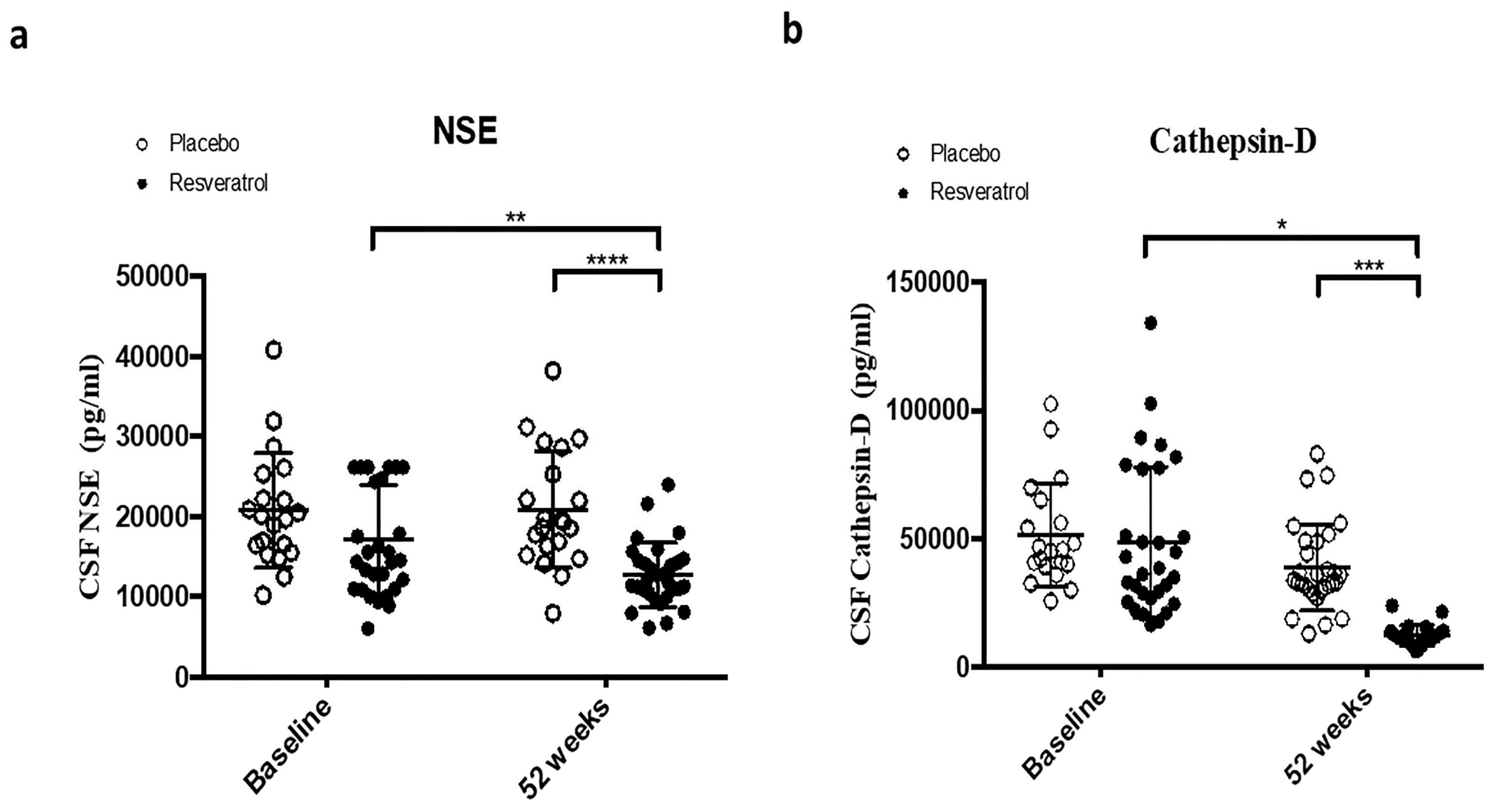
Figure 1. ELISA measurements show the concentrations of (a) Neuron specific enolase (NSE) and (b) Cathepsin-D that are significantly reduced in the CSF of patients treated with resveratrol (n = 30) compared to placebo (n = 21) ta baseline and 52 weeks. Mean ± SD, p-values and statistical methods are listed in Table 1.
Ijms 26 05044 g001
Figure 2. ELISA measurement of phosphorylated neurofilaments (PNF) concentrations expressed as percentages normalized to placebo group shows a significant reduction in PNF in the CSF of patients treated with placebo (n = 21) compared to resveratrol (RES, n = 30) for 52 weeks. Mean ± SD, p values and statistical methods are listed in Table 1.
Ijms 26 05044 g002
Figure 3. ELISA concentrations of (a) Angiogenin, (b) Fatty Acid Binding Protein 3 (FABP3), and (c) Neurogranin in the CSF of patients treated with placebo (N = 21) or resveratrol (N = 30) for 52 weeks. Mean ± SD, p values and statistical methods are listed in Table 1.
Ijms 26 05044 g003
Figure 4. ELISA measurement shows concentrations of (a) matrix metalloprotease (MMP)-9 is significantly reduced in the CSF of patients treated with resveratrol (N = 30) compared to placebo (N = 21) for 52 weeks. (b) Triggering Receptor Expressed on Myeloid Cells (TREM)-2 is significantly reduced in the CSF of patients treated with resveratrol (N = 30) compared to placebo (N = 21) for 52 weeks. Mean ± SD, p values and statistical methods are listed in Table 1.
Ijms 26 05044 g004
Figure 5. ELISA measurements of concentrations of (a) Tissue Inhibitor of Metalloproteinase (TIMP)-1, (b) TIMP2, (c) TIMP3, and (d) TIMP4 in the CSF of patients treated with placebo (N = 21) or resveratrol (N = 30) for 52 weeks. Mean ± SD, p values and statistical methods are listed in Table 1.
Ijms 26 05044 g005
Table 1. Summary of statistical tests of null changes between baseline and 52 weeks and tests of null differences at baseline and at 52 weeks using all detected molecules in CSF of patients treated with placebo (N = 21) or resveratrol (N = 30). Statistical significance: * p < 0.05, ** p < 0.01, *** p < 0.001, and **** p < 0.0001.
AnalytesBaseline 52 Weeks Baseline 52 Weeks
Paired t-TestWilcoxon Signed-Rank TestBaseline: Active vs. Placebo 52 Weeks: Active vs. Placebo
PlaceboResveratrolPlaceboActiveUnpaired t-Test (Unequal)Unpaired t-Test (Equal)Unpaired t-Test (Unequal)Unpaired t-Test (Equal)
NSE0.85940.0051 **0.33710.0047 **0.0387 *0.0773<0.0001 ****<0.0001 ****
Cathepsin D0.20530.09710.11530.07010.68630.70710.0002 ***0.0001 ***
TREM20.36220.27540.11530.17460.27550.2430.0023 **0.0048 **
Angiogenin0.52840.0437 *0.44920.0249 *0.0002 ***0.0005 ***0.06360.1581
FABP30.22160.0066 **0.2520.0027 **0.31110.27620.164 *0.0205 *
Neurogranin0.39550.7770.21520.22740.66840.6560.20130.41
p-NFNANANANANANA0.0131 *0.0081 **
MMP-20.16920.67590.2290.8067NANANANA
MMP-90.36210.0003 ***0.37780.0004 ***>0.9999>0.99990.0092 **0.0058 **
MMP-100.0640.25270.08430.1502NANANANA
TIMP-10.11770.13680.09460.06980.52330.51870.82940.8293
TIMP-20.94520.35680.89830.67020.49650.48670.85170.8600
TIMP-30.0275 *0.0371 *0.0153 *0.0323 *0.36130.36920.05040.0688
TIMP-40.09680.0126 *0.05830.0086 **0.0002 ***<0.0001 ****<0.0001 ****<0.0001 ****
Disclaimer/Publisher’s Note: The statements, opinions and data contained in all publications are solely those of the individual author(s) and contributor(s) and not of MDPI and/or the editor(s). MDPI and/or the editor(s) disclaim responsibility for any injury to people or property resulting from any ideas, methods, instructions or products referred to in the content.