Article

Macrophage-Derived Factors with the Potential to Contribute to the Pathogenicity of HIV-1 and HIV-2: Roles of M-CSF and CXCL7

1
Division of Pharmaceutical Quality Research Four (DPQR-IV), Office of Pharmaceutical Quality Research, Center for Drug Evaluation and Research, U.S. Food and Drug Administration, 10903 New Hampshire Avenue, Silver Spring, MD 20993, USA
2
Division of Pharmaceutical Quality Research Three (DPQR-III), Office of Pharmaceutical Quality Research, Center for Drug Evaluation and Research, U.S. Food and Drug Administration, 10903 New Hampshire Avenue, Silver Spring, MD 20993, USA
3
Laboratory of Molecular Virology, Division of Emerging and Transfusion Transmitted Diseases (DETTD), Center for Biologics Evaluation and Research (CBER), U.S. Food and Drug Administration, 10903 New Hampshire Avenue, Silver Spring, MD 20993, USA
*
Authors to whom correspondence should be addressed.
These authors contributed equally to this work.
Deceased.
Int. J. Mol. Sci. 2025, 26(11), 5028; https://doi.org/10.3390/ijms26115028
Submission received: 29 March 2025 / Revised: 9 May 2025 / Accepted: 21 May 2025 / Published: 23 May 2025
(This article belongs to the Special Issue Advanced Research on Immune Cells and Cytokines (2nd Edition))

Abstract

Human immunodeficiency virus (HIV) type 2 (HIV-2) is less pathogenic than HIV-1. However, the factors responsible for the differences in pathogenicity are still not well defined. To investigate this issue, we performed infection of primary human monocyte-derived macrophages (MDMs) with individual HIV-1 or HIV-2 strains and compared the levels of M-CSF, a cytokine shown to promote HIV-1 infection and replication in our previous studies, and CXCL7, a chemokine identified as being expressed at levels correlated with HIV type by our preliminary gene-expression analysis. We tested several HIV-2 isolates able to replicate in human MDMs and observed that all of them induced the production of M-CSF at high levels similar to those previously established for HIV-1 infection. In addition, the production of M-CSF in MDMs infected with HIV-1 or HIV-2 isolates correlated with the extent of virus replication. In contrast to M-CSF, the chemokine CXCL7 was differentially expressed between MDMs infected with HIV-1 or HIV-2 isolates, as revealed by qPCR and ELISA testing. Together, these results suggest that M-CSF induction may play similar roles in promoting the replication of HIV-1 and HIV-2, while differential regulation of chemokine expression may be an important factor contributing to the differential pathogenicity of the two HIV subtypes.

1. Introduction

Acquired immunodeficiency syndrome (AIDS) can develop following infection with HIV-1 [1,2] or HIV-2 [3,4,5] or following co-infection with HIV-1 and HIV-2 [6,7,8]. Both HIV-1 and HIV-2 arose following several zoonotic cross-species transmissions of simian immunodeficiency viruses (SIV), which include the chimpanzee virus SIVcpz, the predecessor of HIV-1 groups M and N; the gorilla virus SIVgor for HIV-1 groups O and P; and the sooty mangabey virus SIVsmm for HIV-2 groups A through I [3,9,10]. Although their genomic organization and clinical features are similar [9,11], HIV-2 infections are predominantly restricted to West Africa, occur less frequently, and are generally less pathogenic than HIV-1 infections [4,7,8,11]. The progression to AIDS in HIV-2-infected individuals is also reduced and/or delayed, with a corresponding mortality rate about two-thirds lower than for HIV-1. Intriguingly, there are observations suggesting that HIV-2 and HIV-1 co-infection may prolong the time to progression to AIDS compared to HIV-1 mono-infection [12,13], although this correlation was not confirmed in all related studies [14]. HIV-2 also has lower transmission rates, and while HIV-1 infection is associated with a steady decline in CD4+ T-lymphocyte counts, in HIV-2 infection, the decline is much slower and viremia levels are lower at all stages of the disease [15,16]. The immune response to HIV infection may be an important factor in determining the clinical course of disease [17]. More efficient innate and adaptive anti-HIV-2 immune responses appear to contribute to the observed differences from HIV-1 [3,6,8,11,17,18,19]. Cytokines are an integral part of the immune response, and a more in-depth understanding of their effects and patterns in HIV-1 and HIV-2 infections may provide an opportunity to better understand the mechanisms that play a role in virus control [17,20].
HIV initially infects CD4+ memory T cells and uses CCR5 as the co-receptor for entry [17,21,22]. An exponential burst of virus replication follows, and, around the time of peak viremia, significant depletion of CD4+ mucosal T cells occurs. In addition to CD4+ T lymphocytes, cells of the monocyte/macrophage lineage contribute to HIV pathogenesis at multiple points during the course of disease. Macrophages are among the first cells infected with HIV-1 [1,22,23,24]. There is also evidence that resident macrophages in multiple anatomical sites, such as the central nervous system (CNS), lymph nodes, and gut lymphoid tissue, are targets for HIV during acute infection and that the HIV proviral DNA may be integrated into the host-cell genome [25,26,27,28,29]. Macrophages are long-lived cells that are relatively resistant to the cytopathic effects of HIV-1 and can therefore function as a reservoir for HIV and thus as a vector for dissemination of the virus to other susceptible cells, including CD4+ T-lymphocytes [30,31,32,33,34,35]. Furthermore, HIV-infected macrophages may persist during highly active antiretroviral therapy, thus hampering virus eradication, which may thus require the development of more macrophage-specific anti-HIV therapies [25,26,28,29,36,37]. Additionally, macrophages are regulatory cells that control the pace and intensity of disease progression via the production of cytokines during the course of innate and adaptive immune responses [24,38]. Infection of macrophages by HIV-1 is clearly an important component of AIDS pathogenesis and contributes to comorbidities during HIV infection [28,34,35,38,39]. Similarly to HIV-1, HIV-2 can infect macrophages as well as T lymphocytes and utilize chemokine co-receptors for viral entry [40,41]. HIV-2 strains are generally more promiscuous than HIV-1 strains in their use of co-receptors, but CCR5 and CXCR4 appear to still be the major co-receptors for infection, and it is not clear whether use of the other co-receptors is relevant in vivo [16,42,43,44].
Systemic cytokine/chemokine release has been observed following HIV-1 infection [17,20,45]. Characterization of the cytokines/chemokines that can be induced by HIV-1 and HIV-2 is therefore important to understanding viral pathogenesis. We have previously reported that M-CSF is endogenously produced by human macrophages infected with HIV-1, as is reflected by increases at both the mRNA and protein levels [46]. Exogenous M-CSF is capable of rendering macrophages more susceptible to HIV-1 infection, most likely through its ability to enhance expression of the HIV-1 receptor and co-receptor (CD4 and CCR5, respectively) [47,48,49]. Although several reports document the ability of HIV-2 to infect human macrophages [40,50,51], in-depth studies on the impact of HIV-2 on the production of cytokines by infected macrophages have not been performed. Thus, compared to HIV-1, the role of macrophages in the HIV-2 disease process is not well delineated.
Chemokines are a group of small, soluble proteins with chemoattractant and immunomodulatory activities that belong to the cytokine family. Recruitment of macrophages and T cells to the infection site(s) by chemokines, induced following HIV infection, facilitates expansion of an ongoing infection and the establishment of virus reservoirs [52,53,54]. Recently, we reported that expression of the β-chemokine CCL2 is increased in HIV-1-infected human MDMs but decreased in HIV-2-infected MDMs when compared to uninfected controls [41]. The decrease in CCL2 expression following HIV-2 infection occurs at both the mRNA and protein levels. However, it remains to be investigated whether additional chemokine(s) can also be differentially regulated following HIV-1 or HIV-2 infection.
Our current study assessed the ability of primary or minimally passaged HIV-2 isolates to infect human MDMs in vitro and compared the expression levels of M-CSF and CXCL7 in macrophages following infection with individual HIV-1 or HIV-2 isolates.

2. Results

2.1. HIV-2 Isolates Use Various Co-Receptors for Infection

Our previous studies showed that M-CSF production can be induced by HIV-1 infection of human MDMs [46,55]. We next examined whether HIV-2 infection of MDMs could also induce M-CSF production. To this end, we used several minimally passaged HIV-2 isolates reported to use different co-receptors for their infection (Table 1) [56]. Using a GHOST cell assay [57,58], we confirmed that the HIV-2 B3-B4 and HIV-2 B7-B9 isolates utilize CCR5 as their co-receptor. The M-tropic HIV-2 Rod isolate uses CXCR4 as its co-receptor, while the HIV-2 B2 and HIV-2 B5 isolates could potentially employ CCR5, CXCR4, CCR1, and CCR2b as co-receptors (Table 1 and Figure 1).

2.2. MDMs Infected with HIV-1 and HIV-2 Isolates Produce Similar Levels of M-CSF

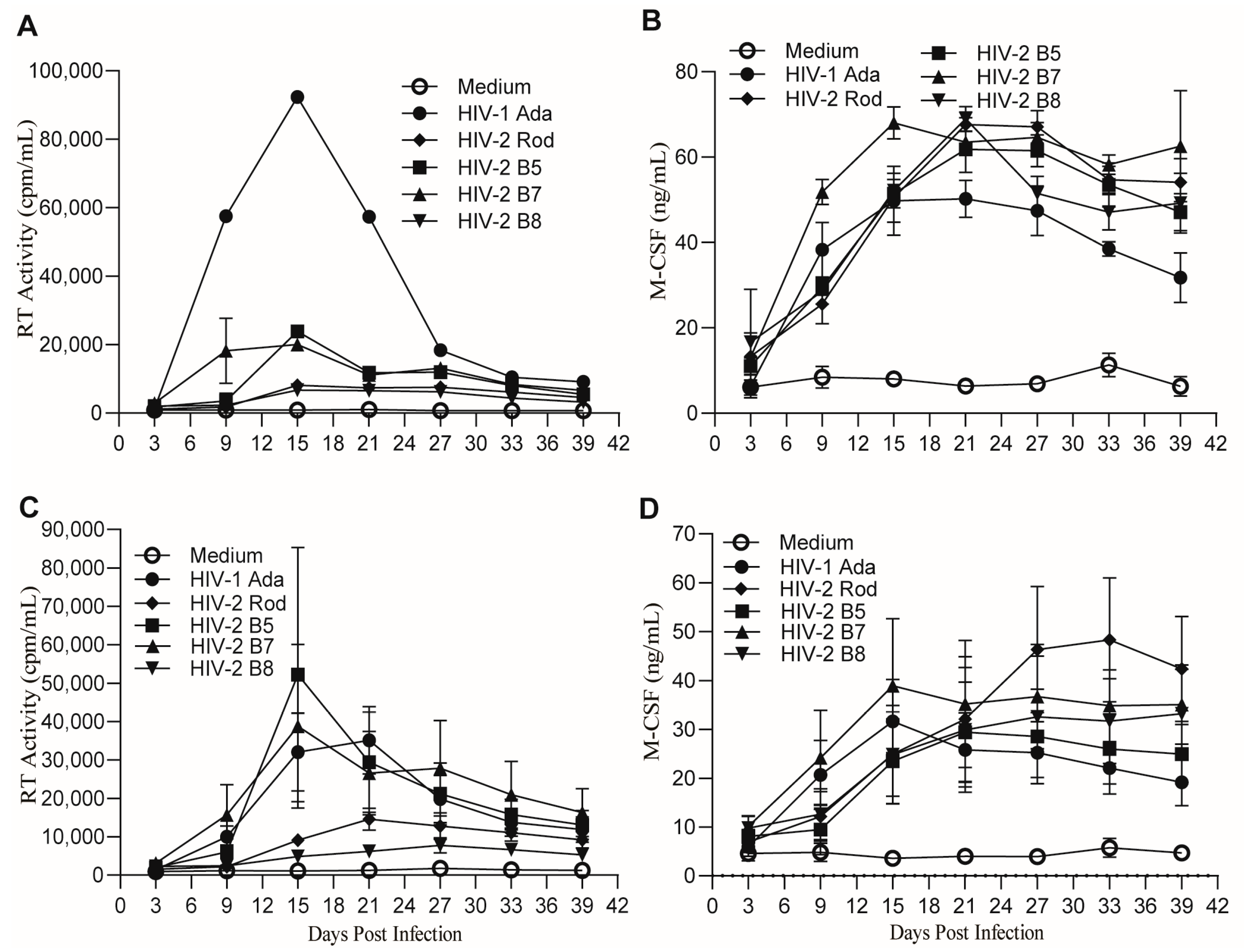
Initially, monocytes from one healthy donor were differentiated into macrophages, infected with the pleiotropic HIV-2 isolate B5, the CXCR4-tropic isolate Rod, and the CCR5-tropic HIV-2 isolates B7 and B8 and cultured for 39 days. The lab-adapted HIV-1 strain Ada served as a control. Measurement of the HIV RT activities in the culture supernatants collected from day 3 to day 39 at 6-day intervals indicated that all HIV isolates replicated in human MDMs, although their replication capabilities were different (Figure 2A). HIV-1 Ada exhibited the most robust replication, while HIV-2 isolates B8 and Rod showed low levels of replication in the human MDMs (Figure 2A). Interestingly, MDMs infected with the various HIV isolates all produced M-CSF at similar levels (Figure 2B). To further corroborate these observations, we repeated the time-course study by performing infection of MDMs derived from four additional healthy donors using the same HIV isolates. Results from the repeated experiments confirmed that the HIV isolates replicated in human MDMs to different degrees, while their replication induced the production of similar amounts of M-CSF (Figure 2C,D).
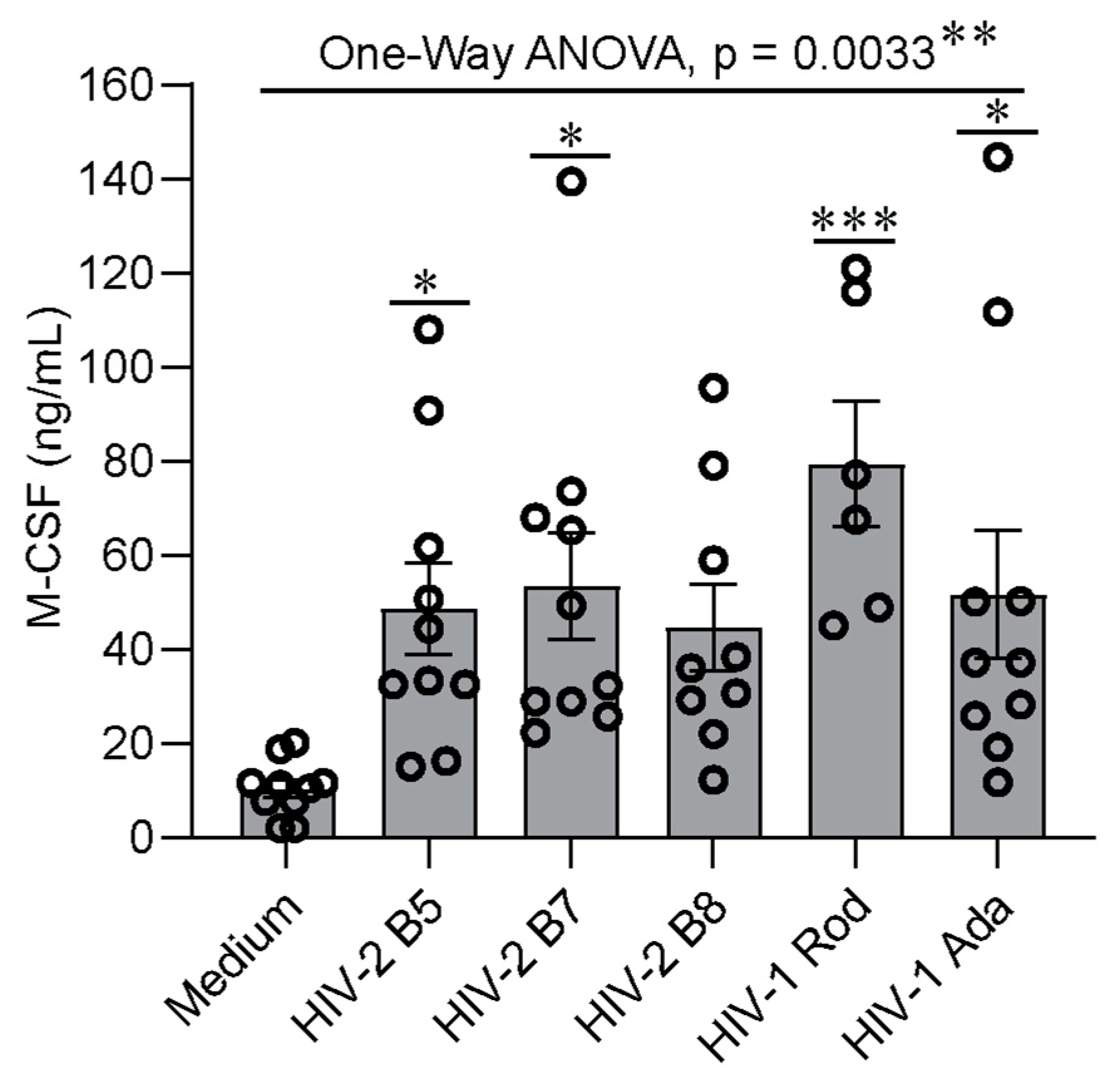
Further statistical analyses of M-CSF production at the time of peak virus replication in MDMs from 6–10 donors using one-way ANOVA with Dunnett’s test for multiple testing corrections indicated that the average levels of M-CSF expression were significantly different between the uninfected and the five HIV-infected groups (p = 0.0033) (Figure 3). Dunnett’s test for multiple comparisons, with the uninfected group as a control, indicated that MDMs infected with HIV-2 B5, B7, or Rod or with HIV-1 Ada produced significantly higher amounts of M-CSF than uninfected cells (Figure 3). In contrast, when cells infected with HIV-1 Ada were used as the control group for multiple comparisons, no significant difference was observed between HIV-1 Ada and each of the HIV-2 virus isolates. Of note, additional HIV isolates, including HIV-2 B4 and the minimally passaged HIV-1 isolates 92UG024 and BCF03, were also used for infection of MDMs from some, but not all, of the donors due to the limited size of the virus stocks. Like HIV-1 Ada, HIV-1 92UG024 and HIV-1 BCF03 also infected and replicated robustly in MDMs, which led to induction of M-CSF production (Supplementary Table S1). Statistical analysis of the data from infections with the additional individual HIV isolates by one-way ANOVA with Dunnett’s test for multiple comparisons indicated that M-CSF production by infected MDMs did not differ significantly across HIV isolates (Supplementary Figure S1). Taken together, these results indicate that infection of human MDMs with both HIV-1 and HIV-2 can induce M-CSF production.
The kinetics and the amounts of M-CSF production varied among the HIV isolates and donors tested. The amount of M-CSF production (Figure 2B,D) did not always correlate with the level of virus replication (Figure 2A,C). The amounts of M-CSF were often substantial (over 2-fold) even when HIV-2 replication was relatively low (HIV-2 B8 and Rod, Figure 2C). This observation is consistent with our previous studies using HIV-1 whereby maximal M-CSF production could be observed even at lower thresholds of virus replication [46]. However, for each HIV isolate, the replication kinetics correlated with M-CSF production by MDMs during the infection process (Figure 4). The correlation coefficients of HIV-1 Ada and HIV-2 B5, B7, B8, and Rod isolates were 0.788 ± 0.003, 0.722 ± 0.007, 0.770 ± 0.004, 0.629 ± 0.011, and 0.671 ± 0.013, respectively (Figure 4F). These data indicate a moderate (R = 0.5–0.7) to high (R = 0.7–0.9) correlation between the degree of HIV virus replication and M-CSF production.

2.3. MDMs Infected with HIV-1 and HIV-2 Isolates Produce Different Levels of CXCL7

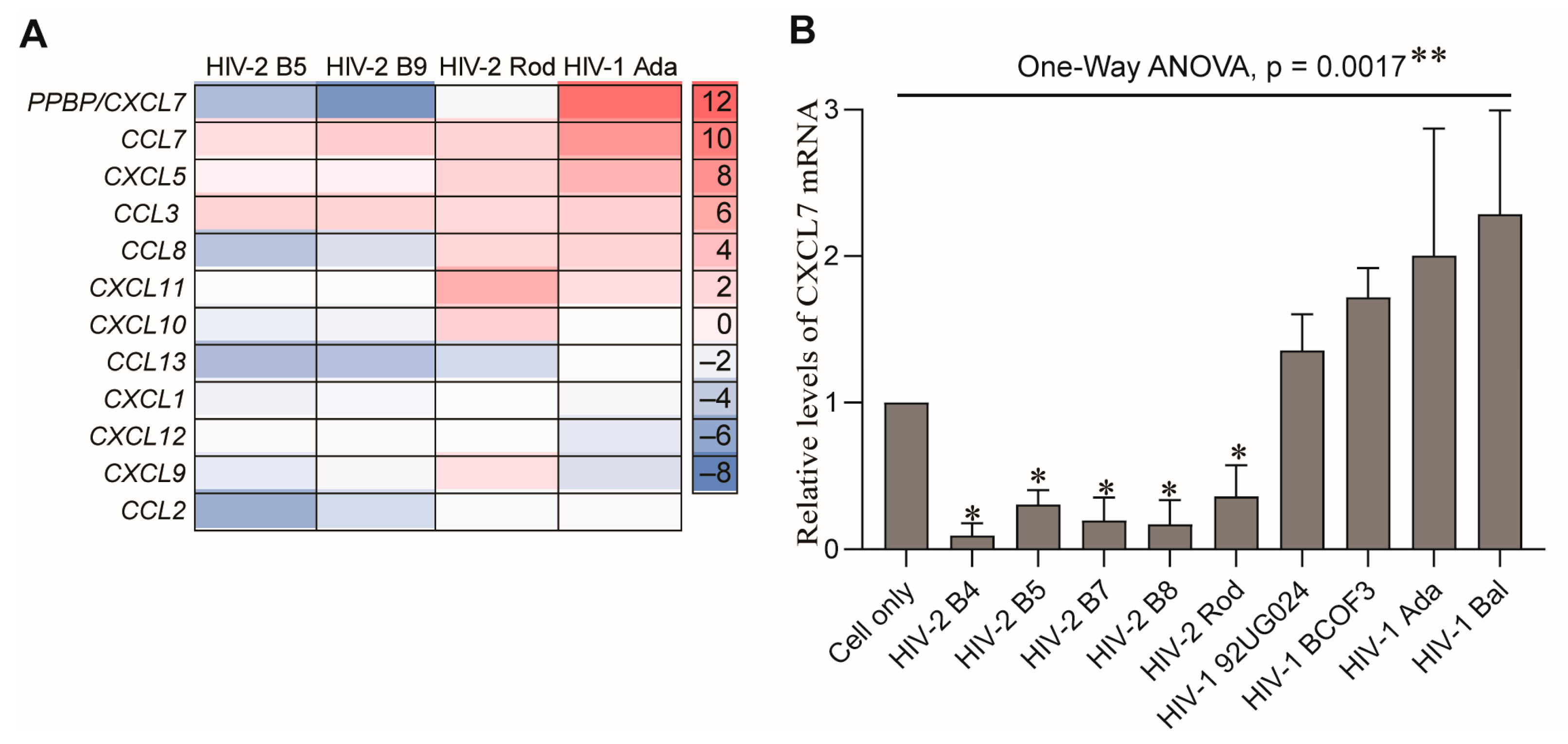
The similar abilities of HIV-1 and HIV-2 isolates to induce M-CSF production following their infection of MDMs suggest that M-CSF is unlikely to be a factor associated with their different pathogenicity. Recently, we revealed that infection of MDMs with HIV-1 or HIV-2 differentially regulates production of the β-chemokine, CCL2. To further investigate whether other chemokines can also be differentially regulated during infection with HIV-1 or HIV-2, we compared the expression profiles of 12 chemokines in MDMs following infection with HIV-1 Ada or HIV-2 B5, B9, or Rod isolates by Affymetrix microarray, using cell samples prepared at the peak of viral replication. Among the 12 chemokines, the pro-platelet basic protein (PPBP) gene was identified as upregulated in HIV-1-infected MDMs but downregulated in HIV-2-infected MDMs (Figure 5A). The protein encoded by PPBP is a platelet-derived growth factor that belongs to the CXC chemokine family and can be cleaved to produce the chemokine, CXCL7 [59,60]. The differences in regulation of PPBP/CXCL7 gene expression were confirmed in MDMs infected with HIV-1 Ada or Bal or HIV-2 B5, B7, B8, or Rod isolates by qPCR (Figure 5B).
To further corroborate this finding, we next measured the concentrations of CXCL7 protein in HIV-infected MDM culture supernatants that were collected from day 6 to day 36 at 6-day intervals by ELISA. Similarly to M-CSF production, the kinetics and the amounts of CXCL7 production varied among the HIV isolates and donors tested, and the amounts of CXCL7 production did not always correlate with the amount of virus replication (Figure 6A,B). However, when compared with uninfected MDMs, MDMs infected with HIV-1 secreted higher amounts of CXCL7, while MDMs infected with HIV-2 isolates produced lower amounts of CXCL7 during the course of infection (Figure 6B). Statistical analysis of CXCL7 production at the time of peak virus replication in MDMs from the six to eight donors by one-way ANOVA using Dunnett’s test for multiple comparisons indicated that the relative amounts of CXCL7 produced by MDMs infected with individual HIV isolates were significantly different (p < 0.0001) (Figure 6C). Multiple comparisons by Dunnett’s test using the HIV-1 Ada group as a control indicated that MDMs infected with individual HIV-2 B5, B7, or B8 isolates produced significantly lower amounts of CXCL7 (Figure 6C). In addition, statistical analysis of the CXCL7 levels in the supernatants harvested from infection of MDMs with additional HIV isolates (described in Section 2.2) further indicates that MDMs produced different levels of CXCL7 following infection with HIV-1 or HIV-2 (Supplementary Figure S2). Together, these results demonstrated that HIV-1 and HIV-2 infection of human MDMs differentially regulate production of the chemokine CXCL7.

3. Discussion

In the current study, we established that in vitro infection of primary human MDMs with HIV-1 or HIV-2 induced the production of similar levels of the cytokine M-CSF but had different effects on CXCL7 chemokine expression. The increased M-CSF production was consistent with and further supports previous observations regarding the role of M-CSF in HIV-1 infection [46,48,49,55,61]. However, induction of M-CSF production does not appear to be a universal feature of MDMs responding to virus infections. Other viruses capable of replicating in human MDMs, including measles and respiratory syncytial viruses, as shown in one of our previous studies, did not enhance the production of M-CSF but did have the capacity to induce the production of pro-inflammatory cytokines or chemokines [55]. In contrast, M-CSF and IL-34, which facilitate macrophage survival and differentiation and mediate their biological activity via binding to and signaling through the M-CSF receptor (c-fms/CD115), are produced at high levels in chronic hepatitis C virus infection [62]. Although Epstein-Barr virus (EBV) has not been reported to induce production of M-CSF, it expresses an early gene product (BARF1) that contributes to viral pathogenesis by forming a complex with M-CSF, causing a reduction in M-CSF biological activity [63]. In addition, Kaposi’s sarcoma herpes virus (KSHV) has been shown to encode proteins capable of interfering with the development of macrophages mediated by M-CSF that are essential for their differentiation and survival [64]. These findings indicate the complex role that M-CSF may play in the pathogenesis of different viruses via increases in its production or interference with its biological activity.
Although the ability of HIV-2 to infect human macrophages has previously been reported [40], detailed investigations regarding the effect of this virus on M-CSF production by MDMs have not been conducted. Using the minimally passaged HIV-2 isolates B5, B7, B8, and Rod, we found that all tested HIV-2 isolates had the capacity to replicate in human MDMs (Figure 1, Figure 2 and Figure 6) and induce the production of M-CSF (Figure 2 and Figure 3). The levels of M-CSF were increased compared to those in uninfected MDMs and comparable to those induced by HIV-1 infection but did not always correlate with the level of virus replication (Figure 2). We also observed that even low levels of HIV replication were capable of inducing M-CSF expression. Furthermore, we found that for minimally passaged HIV-2 isolates, the co-receptor used appeared to have little impact on the ability of the virus to induce M-CSF production (Figure 2 and Figure 3). The complex mechanism(s) of HIV-induced M-CSF production in MDMs is still largely undefined. Our early studies revealed that virion binding to MDMs alone did not induce production of M-CSF and that HIV-1 binding, entry, and replication were all required [46]. In addition to HIV-1 replication, cell-to-cell interactions between macrophages and lymphocytes may further augment M-CSF production [65,66]. Our preliminary attempts to identify the HIV-1 gene(s) responsible for induction of M-CSF in human MDMs using molecularly cloned deletion mutants showed that removal of HIV-1 Vpr, Vpr/Vpu, or Vif reduced virus replication, but the impact on M-CSF production could not be definitively ascertained when the results from several human donors were compared, most likely because impaired replication of the mutated viruses failed to induce adequate levels of M-CSF for comparison. Although it was shown that HIV-1 Nef may affect the production of several pro-inflammatory cytokines and chemokines, no meaningful impact on the induction of M-CSF was reported [67,68,69]. However, there is evidence that Nef may interfere with M-CSF receptor signaling via activation of Hck tyrosine kinase [70].
M-CSF promotes HIV infection of macrophages via multiple mechanisms [61]. These include increased expression of CD4, CCR5, and CXCR4 receptors to facilitate virus entry [49,71,72,73]; reduced susceptibility of HIV-1 entry to inhibition by β-chemokines such as CCL5 (RANTES) [74]; enhancement of viral gene expression [71]; offsetting of the effects of cytokines like IL-32 that suppress HIV-1 replication [65]; and promotion of the ontogeny and survival of macrophages, contributing to both the number and longevity of the infected cells. Our findings that M-CSF production was a common feature of HIV-1 and HIV-2 infection imply a greater role of M-CSF in the establishment and maintenance of lentivirus infection, including a role in the re-activation of virus reservoirs [75], and the above effects may be associated with the ability of M-CSF to facilitate macrophage differentiation into an M2 phenotype [65,66,75]. However, the M2 phenotype encompasses multiple subtypes, as macrophage differentiation may be affected by a range of cytokines and maturation factors. Furthermore, the macrophage population in vivo likely includes a spectrum of cells with features that place them between the M1 or M2 phenotypes induced in vitro [52]. Being aware of the potential deficiencies of any in vitro model in mimicking real life scenarios, we used pooled human serum from multiple donors in an attempt to more adequately replicate the cytokine milieu naturally encountered by monocytes and generate cells resembling the macrophage population encountered by HIV-1 and/or HIV-2 in vivo [76].
It is still not well understood why HIV-2 is less pathogenic than HIV-1. The similar behavior of HIV-1 and HIV-2 in the induction of M-CSF excludes this cytokine as a potential factor contributing to the observed differences in their pathogenicity. Acute HIV infection also induces production of chemokines (CCL2, CCL3, CCL4, and CCL5), which may recruit uninfected monocytes/macrophages and/or CD4+ T lymphocytes to the sites of virus replication, thus promoting the spread of infection and/or the establishment of virus reservoirs [20,77,78]. Recently, we revealed that CCL2 expression was increased in HIV-1-infected MDMs but decreased in HIV-2-infected MDMs compared to uninfected controls [41]. The significance of CCL2 as a factor associated with the severity of HIV infection was further supported by the observation that CCL2 expression was enhanced in viremic (>100,000 RNA copies/mL) compared to aviremic (<50 RNA copies/mL) HIV-1-positive individuals [79].
In this study, we identified CXCL7 as an additional chemokine that is differentially regulated in human MDMs infected with HIV-1 versus HIV-2. CXCL7, also known as neutrophil activating peptide 2 (NAP2), is an ELR+ chemokine originally identified in activated platelets and later found to be expressed in a variety of cells, including monocytes/macrophages [60,80,81,82]. CXCL7 is synthesized as a pro-platelet basic protein (PPBP) precursor and undergoes proteolytic cleavage to generate the active chemokine [60,83]. The potential mechanisms responsible for the differential regulation of CXCL7 during HIV-1 and HIV-2 infection remain to be elucidated. The observed changes in the mRNA levels of the CXCL7 precursor PPBP (Figure 5) suggest that differences in NF-κB regulation during HIV-1 and HIV-2 infection might play a role [84].
CXCL7 is potent chemotactic agent and appears to be an important factor in tumorigenesis and inflammation that acts by recruiting cells of the immune system, although the precise mechanisms underlying CXCL7’s pleotropic cellular effects remain to be elucidated [60,85,86]. At present, there is very limited information regarding the potential role of CXCL7 in the pathogenesis of HIV infection. Our pilot study suggests that CXCL7 may play a pivotal role by attracting lymphocytes (Supplementary Figure S3) to the sites of virus replication, thus enhancing the spread of infection. Our hypothesis is consistent with the observations of Landrø et al., who found that CXCL7 plasma levels in vivo were significantly higher in HIV-infected (presumably HIV-1-infected) patients before and during highly active antiretroviral therapy (HAART) compared to healthy controls [87]. Another study also reported increased CXCL7 levels in HIV-1 infected individuals regardless of their clinical status, but the difference from uninfected controls was not statistically significant [88]. Significantly increased CXCL7 levels were also found in a small group of long-term non-progressors (LTNP) compared to healthy controls [89]. Unfortunately, this study did not include patients with typical or accelerated progression to AIDS. The study also revealed that the LTNP had high titers of HIV-1 neutralizing antibodies, which likely offset the infection-promoting effect of CXCL7 [89].
Based on the release of cytokines and chemokines following acute HIV infection, we have proposed a model wherein chemokines produced by HIV-infected macrophages attract uninfected monocytes/macrophages and/or lymphocytes to the sites of virus replication. M-CSF secreted by the infected cells then renders the incoming monocytes/macrophages more susceptible to infection by facilitating cell differentiation, enhancing expression of CD4, CCR5, and/or CXCR4 and potentially affecting virus replication after entry. Reduced expression of CCL2 and CXCL7 chemokines following HIV-2 infection may impair recruitment of macrophages and/or T cells to the sites of virus replication and hence impede virus spread and/or establishment of viral reservoirs. In addition, decreased CCL2 and CXCL7 expression may also contribute to the less pronounced activation of the immune system during HIV-2 infection compared to HIV-1 infection [16]. Whether or not CXCL7 has an additional role in regulating HIV pathogenesis, as by impacting activation of the coagulation system, remains to be determined.
In conclusion, our current and previous [41] studies reveal that infection of MDMs with HIV-2 induced the production of similar levels of M-CSF but lower levels of CXCL7 and CCL2 compared to MDMs infected with HIV-1. These findings imply that M-CSF may play a similar role in supporting HIV-1 and HIV-2 infection and subsequent virus replication, while differential regulation of certain chemokines following infection with HIV-1 and HIV-2 may correlate with their distinct pathogenicity. Finally, revealing the cytokine patterns during HIV-1 and/or HIV-2 infection of primary human macrophages may provide important information to support the development of better in vitro assays and thereby facilitate the characterization and quality testing of new potential anti-HIV therapies.

4. Materials and Methods

4.1. Monocyte Isolation and Culture

Leukapheresis blood units from healthy HIV-seronegative donors were obtained from the Department of Transfusion Medicine (DTM) under an Institutional Review Board (IRB)-approved protocol at the National Institutes of Health. A categorical exemption is in place for experimental studies performed by CDER/FDA researchers using existing deidentified blood donors who provided written informed consent according to the international ethical guidelines for biomedical research involving human subjects; these samples were anonymized prior to being sent to the FDA. Peripheral blood mononuclear cells (PBMC) were isolated from human blood following leukapheresis of HIV-1-seronegative donors and subsequent density gradient centrifugation; monocytes were purified by countercurrent centrifugal cell elutriation, as previously described [46]. Over 90% of the cells in the elutriated monocyte fraction were CD14+, as determined by flow cytometry analysis, and showed ≥95% viability (trypan blue exclusion test). Monocytes were differentiated in culture for 7–10 days at 37 °C in 5% CO2 at a concentration of 2 × 10⁶ cells/mL, 2.0 mL per well, in six-well tissue-culture plates (Costar, Cambridge, MA, USA) using DMEM (Invitrogen, Carlsbad, CA, USA) complete medium containing 10% pooled human serum (PHS), 2 mM L-glutamine, 1 mM sodium pyruvate, and penicillin (50 units/mL)/streptomycin (50 μg/mL) (Invitrogen) to generate MDMs. The PHS was generated in house from plasma from multiple non-transfused, healthy, HIV-seronegative male donors obtained from the NIH DTM.

4.2. HIV Infection of Human Monocyte-Derived Macrophages

After differentiation, MDMs were harvested by scraping and then replated into 24-well tissue-culture plates (Nunc, Inc., Naperville, IL, USA), at a concentration of 0.5 × 10⁶ cells/mL, 1.5 mL per well. After 24 to 48 h, MDMs were infected with individual HIV-1 Ada, Bal, 92UG024, and BCF03 isolates as previously described [46] or with primary isolates of HIV-2 that had been minimally passaged in human PBMC. HIV-1 Bal was purchased from Advanced Biotechnologies (Columbia, MD, USA). HIV-1 Ada was obtained through the AIDS Research and Reference Reagent Program (ARRRP), NIAID, NIH, from Dr. H. Gendelman [47,90], then expanded in human macrophages prior to purification by ultracentrifugation and cryopreservation (Advanced Biotechnologies) [46]. HIV-1 92UG024 (Cata#1649) and HIV-1 BCF03 (Cata#3335) are minimally passaged HIV-1 isolates that were obtained from the AIDS Research and Reference Reagent Repository. HIV-2 B2-B5 and HIV-2 B7-B9 were described previously [56], and the stocks of these HIV-2 isolates were expanded as previously described [46]. An early PBMC-passaged, non-cloned isolate of HIV-2 Rod that was obtained from Dr. F. Clavel in 1987 [91] and cryopreserved was cultured in human MDMs to generate the virus stock used in these studies. Input virus for the primary HIV-2 isolates was equalized based on reverse transcriptase (RT) activity using 0.1 cpm/cell or HIV-2 p26 (5 ng/well). HIV-1 input was also normalized based on RT activity. Virus adsorption was carried out for 4 h at 37 °C. Cells were then washed, and fresh medium was added. Every 3 days post-infection, 80% of the culture medium was collected, stored at −80 °C, and replaced with fresh medium. Subsequently, supernatants were assessed for cytokine/chemokine at 6-day intervals.

4.3. GHOST Cell Assay for Determination of HIV Co-Receptor Usage

Parental GHOST cells and GHOST cell lines expressing CCR1, CCR2b, CCR3, CCR4 CXCR4, CCR8, Bonzo and CD4 were obtained through the NIH AIDS Research and Reference Reagent Program, Division of AIDS, NIAID, NIH [58]. The GHOST-CCR5 cell line was obtained from Dr. Nathaniel Landau [57,92]. These GHOST cell lines were used to determine co-receptor usage by HIV-2 isolates. Briefly, cells were seeded at a concentration of 105 cells/well in a Costar 24-well tissue-culture plate in MEM medium (Quality Biological, Inc., Gaithersburg, MD, USA) prior to the day of infection. Subsequently, these cells were infected with HIV-2 B2-5, and B7-9, as well as HIV-2 Rod (5 ng p26 per well); incubated at 37 °C/5% CO2 for 2 h; washed thoroughly; and cultured in MEM medium supplemented with 10% FBS (Hyclone/Thermo Scientific; Logan, UT, USA); 10 μg/mL puromycin (Sigma; St. Louis, MO, USA); 100 μg/mL hygromycin (Invitrogen; Carlsbad, CA, USA); 500 μg/mL G418; penicillin/streptomycin; and L-glutamine (Quality Biological, Inc.). Culture supernatants were harvested on days 4, 8, and 12; HIV-2 virus replication was determined by measuring p26 antigen levels using the SIV p27 antigen ELISA kit (Zeptometrix Corporation; Buffalo, NY, USA) as described previously [93] after we confirmed that this ELISA kit cross-reacts with HIV-2 p26 antigen.

4.4. Reverse Transcriptase Assay

Progression of virus replication in MDMs infected with HIV-1 or HIV-2 was monitored by measuring RT activity. The RT assay is a 3H-based modification of the method described by Hoffman, et al. [46]. Values shown reflect the average of duplicate samples (cpm/25 μL) that differed by not more than 15%.

4.5. M-CSF Bioassay

M-CSF biological activity was determined by measuring proliferation of the M-CSF- dependent murine cell line, M-NFS-60, as previously described [46,94,95]. An Infinite F50 plate reader (Tecan, Durham, NC, USA) was used to measure absorbance. The limit of detection for the M-CSF bioassay is 1.5 ng/mL. Confirmation of M-CSF identity or verification of the concentration was performed early in these studies using neutralizing anti-M-CSF antibodies or an M-CSF-specific ELISA (R&D Systems, Minneapolis, MN, USA) [46].

4.6. CXCL7 ELISA

CXCL7 concentrations in the supernatants collected from cultures of HIV-infected MDMs throughout the time course of infections were measured using ELISA kits (Cat# EA100464) purchased from Origene (Gaithersburg, MD, USA), following the manufacturer’s instructions.

4.7. Quantitative PCR

Total RNA was extracted from cells by TRIzol reagent (Invitrogen, Carlabad, CA, USA), and 2 μg RNA was reverse transcribed to cDNA using Omniscript (Qiagen Technology, Germantown, MD, USA) and random-hexamer primer methods. CXCL7 primers and the probe were purchased from Applied Biosystems (Cat# Hs00234077, Forster City, CA, USA). Each 30 μL volume of the PCR reaction included 2 μL 2× diluted cDNA (equal 100 ng RNA), 300 nM sense and antisense primers, 200 nM 6′-Fam labeled fluorescence target probe, and 1.5 μL 20× glyceraldehyde-3-phosphate dehydrogenase (GAPDH) endogenous control. Amplification of an endogenous control was performed in the same wells to standardize the level of expression of the target RNA. The CT data from real-time PCR was used to determine ΔCT and ΔΔCT for the relative amount of the target mRNA. The formula 2−ΔΔCT was used to calculate the level of CXCL7 gene expression. The ΔCT as obtained by subtracting the GAPDH CT value from the CXCL7 gene CT value. The ΔΔCT was obtained by subtracting the control ΔCT from target ΔCT.

4.8. Statistical Analysis

Statistical analysis of data from more than two groups was performed using the one-way ANOVA with Dunnett’s multiple comparisons test that is built into Prisms GraphPad Version 10 (Dotmatics, Boston, MA, USA). Statistical analysis of data between two groups was performed using Student’s t-test. The statistical significance of any difference was determined by a p-value < 0.05.

Supplementary Materials

The following supporting information can be downloaded at: https://www.mdpi.com/article/10.3390/ijms26115028/s1.

Author Contributions

Conceptualization, K.A.C., C.G., J.K. and T.A.G.; methodology, C.G., J.K., W.O., T.A.G., K.F., C.S.R.L., F.S., M.P., A.M., L.T., T.S. and K.A.C.; formal analysis, C.G., W.O., J.K., T.A.G., A.M. and K.A.C.; investigation, K.A.C., C.G., J.K., W.O., T.A.G., K.F., C.S.R.L., F.S., M.P., L.T. and T.S.; data curation, K.A.C., W.O., C.G. and T.S.; writing—original draft preparation, C.G. and W.O.; writing—review and editing, W.O., K.A.C. and T.S.; supervision, K.A.C.; funding acquisition, K.A.C. All authors have read and agreed to the published version of the manuscript.

Funding

This work was supported by FDA internal funding.

Institutional Review Board Statement

Ethical review and approval were waived for this study, given that leukapheresis blood packs from healthy donors were obtained under an institutional review board-approved protocol at the National Institutes of Health and anonymized samples were provided to our laboratory.

Informed Consent Statement

Informed consent was obtained from all subjects involved in the study.

Data Availability Statement

The data presented in this study are available in this article in the main figures or tables.

Acknowledgments

We thank Indira Hewlett for her input and advice regarding HIV-2 and the late Owen Woods for the expansion of HIV-2 isolates for use in this study. We thank Shamus Keeler and Zhaohua Zhou for thoughtful review of the manuscript. This work was supported by FDA internal funding.

Conflicts of Interest

The authors declare that they have no conflicts of interest with the contents of this article.

References

  1. Kedzierska, K.; Crowe, S.M. The role of monocytes and macrophages in the pathogenesis of HIV-1 infection. Curr. Med. Chem. 2002, 9, 1893–1903. [Google Scholar] [CrossRef] [PubMed]
  2. Cohen, M.S.; Hellmann, N.; Levy, J.A.; DeCock, K.; Lange, J. The spread, treatment, and prevention of HIV-1: Evolution of a global pandemic. J. Clin. Investig. 2008, 118, 1244–1254. [Google Scholar] [CrossRef] [PubMed]
  3. Reeves, J.D.; Doms, R.W. Human immunodeficiency virus type 2. J. Gen. Virol. 2002, 83 Pt 6, 1253–1265. [Google Scholar] [CrossRef] [PubMed]
  4. de Silva, T.I.; Cotten, M.; Rowland-Jones, S.L. HIV-2: The forgotten AIDS virus. Trends Microbiol. 2008, 16, 588–595. [Google Scholar] [CrossRef]
  5. Campbell-Yesufu, O.T.; Gandhi, R.T. Update on human immunodeficiency virus (HIV)-2 infection. Clin. Infect. Dis. 2011, 52, 780–787. [Google Scholar] [CrossRef]
  6. Acchioni, C.; Sandini, S.; Acchioni, M.; Sgarbanti, M. Co-Infections and Superinfections between HIV-1 and Other Human Viruses at the Cellular Level. Pathogens 2024, 13, 349. [Google Scholar] [CrossRef]
  7. Williams, A.; Menon, S.; Crowe, M.; Agarwal, N.; Biccler, J.; Bbosa, N.; Ssemwanga, D.; Adungo, F.; Moecklinghoff, C.; Macartney, M.; et al. Geographic and Population Distributions of Human Immunodeficiency Virus (HIV)-1 and HIV-2 Circulating Subtypes: A Systematic Literature Review and Meta-analysis (2010–2021). J. Infect. Dis. 2023, 228, 1583–1591. [Google Scholar] [CrossRef]
  8. Esbjörnsson, J.; Jansson, M.; Jespersen, S.; Månsson, F.; Hønge, B.L.; Lindman, J.; Medina, C.; da Silva, Z.J.; Norrgren, H.; Medstrand, P.; et al. HIV-2 as a model to identify a functional HIV cure. AIDS Res. Ther. 2019, 16, 24. [Google Scholar] [CrossRef]
  9. Pandrea, I.; Sodora, D.L.; Silvestri, G.; Apetrei, C. Into the wild: Simian immunodeficiency virus (SIV) infection in natural hosts. Trends Immunol. 2008, 29, 419–428. [Google Scholar] [CrossRef]
  10. Sauter, D.; Kirchhoff, F. Key Viral Adaptations Preceding the AIDS Pandemic. Cell Host Microbe 2019, 25, 27–38. [Google Scholar] [CrossRef]
  11. Leligdowicz, A.; Rowland-Jones, S. Tenets of protection from progression to AIDS: Lessons from the immune responses to HIV-2 infection. Expert Rev. Vaccines 2008, 7, 319–331. [Google Scholar] [CrossRef] [PubMed]
  12. Esbjörnsson, J.; Månsson, F.; Kvist, A.; Isberg, P.E.; Biague, A.J.; da Silva, Z.J.; Jansson, M.; Fenyö, E.M.; Norrgren, H.; Medstrand, P. Increased survival among HIV-1 and HIV-2 dual-infected individuals compared to HIV-1 single-infected individuals. AIDS 2014, 28, 949–957. [Google Scholar] [CrossRef] [PubMed]
  13. Esbjörnsson, J.; Månsson, F.; Kvist, A.; Isberg, P.E.; Nowroozalizadeh, S.; Biague, A.J.; da Silva, Z.J.; Jansson, M.; Fenyö, E.M.; Norrgren, H.; et al. Inhibition of HIV-1 disease progression by contemporaneous HIV-2 infection. N. Engl. J. Med. 2012, 367, 224–232. [Google Scholar] [CrossRef] [PubMed]
  14. Prince, P.D.; Matser, A.; van Tienen, C.; Whittle, H.C.; Schim van der Loeff, M.F. Mortality rates in people dually infected with HIV-1/2 and those infected with either HIV-1 or HIV-2: A systematic review and meta-analysis. AIDS 2014, 28, 549–558. [Google Scholar] [CrossRef]
  15. Ter Schiphorst, E.; Hansen, K.C.; Holm, M.; Hønge, B.L. Mother-to-child HIV-2 transmission: Comparison with HIV-1 and evaluation of factors influencing the rate of transmission. A systematic review. Trans. R. Soc. Trop. Med. Hyg. 2022, 116, 399–408. [Google Scholar] [CrossRef]
  16. Visseaux, B.; Le Hingrat, Q.; Damond, F.; Charpentier, C.; Descamps, D. Physiopathology of HIV-2 infection. Virologie 2019, 23, 277–291. [Google Scholar] [CrossRef]
  17. McMichael, A.J.; Borrow, P.; Tomaras, G.D.; Goonetilleke, N.; Haynes, B.F. The immune response during acute HIV-1 infection: Clues for vaccine development. Nat. Rev. Immunol. 2010, 10, 11–23. [Google Scholar] [CrossRef]
  18. Azevedo-Pereira, J.M.; Santos-Costa, Q. HIV Interaction With Human Host: HIV-2 As a Model of a Less Virulent Infection. AIDS Rev. 2016, 18, 44–53. [Google Scholar]
  19. Vidya Vijayan, K.K.; Karthigeyan, K.P.; Tripathi, S.P.; Hanna, L.E. Pathophysiology of CD4+ T-Cell Depletion in HIV-1 and HIV-2 Infections. Front. Immunol. 2017, 8, 580. [Google Scholar] [CrossRef]
  20. Melchjorsen, J.; Sørensen, L.N.; Paludan, S.R. Expression and function of chemokines during viral infections: From molecular mechanisms to in vivo function. J. Leukoc. Biol. 2003, 74, 331–343. [Google Scholar] [CrossRef]
  21. Berger, E.A.; Murphy, P.M.; Farber, J.M. Chemokine receptors as HIV-1 coreceptors: Roles in viral entry, tropism, and disease. Annu. Rev. Immunol. 1999, 17, 657–700. [Google Scholar] [CrossRef] [PubMed]
  22. Salazar-Gonzalez, J.F.; Salazar, M.G.; Keele, B.F.; Learn, G.H.; Giorgi, E.E.; Li, H.; Decker, J.M.; Wang, S.; Baalwa, J.; Kraus, M.H.; et al. Genetic identity, biological phenotype, and evolutionary pathways of transmitted/founder viruses in acute and early HIV-1 infection. J. Exp. Med. 2009, 206, 1273–1289. [Google Scholar] [CrossRef]
  23. Wodarz, D.; Lloyd, A.L.; Jansen, V.A.; Nowak, M.A. Dynamics of macrophage and T cell infection by HIV. J. Theor. Biol. 1999, 196, 101–113. [Google Scholar] [CrossRef] [PubMed]
  24. Kedzierska, K.; Crowe, S.M.; Turville, S.; Cunningham, A.L. The influence of cytokines, chemokines and their receptors on HIV-1 replication in monocytes and macrophages. Rev. Med. Virol. 2003, 13, 39–56. [Google Scholar] [CrossRef] [PubMed]
  25. Kumar, A.; Abbas, W.; Herbein, G. HIV-1 latency in monocytes/macrophages. Viruses 2014, 6, 1837–1860. [Google Scholar] [CrossRef]
  26. Brown, D.; Mattapallil, J.J. Gastrointestinal tract and the mucosal macrophage reservoir in HIV infection. Clin. Vaccine Immunol. 2014, 21, 1469–1473. [Google Scholar] [CrossRef]
  27. Wallet, C.; De Rovere, M.; Van Assche, J.; Daouad, F.; De Wit, S.; Gautier, V.; Mallon, P.W.G.; Marcello, A.; Van Lint, C.; Rohr, O.; et al. Microglial Cells: The Main HIV-1 Reservoir in the Brain. Front. Cell Infect. Microbiol. 2019, 9, 362. [Google Scholar] [CrossRef]
  28. Joseph, J.; Daley, W.; Lawrence, D.; Lorenzo, E.; Perrin, P.; Rao, V.R.; Tsai, S.Y.; Varthakavi, V. Role of macrophages in HIV pathogenesis and cure: NIH perspectives. J. Leukoc. Biol. 2022, 112, 1233–1243. [Google Scholar] [CrossRef]
  29. Tang, Y.; Chaillon, A.; Gianella, S.; Wong, L.M.; Li, D.; Simermeyer, T.L.; Porrachia, M.; Ignacio, C.; Woodworth, B.; Zhong, D.; et al. Brain microglia serve as a persistent HIV reservoir despite durable antiretroviral therapy. J. Clin. Investig. 2023, 133, e167417. [Google Scholar] [CrossRef]
  30. Siliciano, R.F. Latency and reservoirs for HIV-1. AIDS 1999, 13, S49–S58. [Google Scholar]
  31. Cohen, D.E.; Walker, B.D. Human immunodeficiency virus pathogenesis and prospects for immune control in patients with established infection. Clin. Infect. Dis. 2001, 32, 1756–1768. [Google Scholar] [CrossRef] [PubMed]
  32. Balestra, E.; Perno, C.F.; Aquaro, S.; Panti, S.; Bertoli, A.; Piacentini, M.; Forbici, F.; D’Arrigo, R.; Calio, R.; Garaci, E. Macrophages: A crucial reservoir for human immunodeficiency virus in the body. J. Biol. Regul. Homeost. Agents 2001, 15, 272–276. [Google Scholar] [PubMed]
  33. Tan, J.; Sattentau, Q.J. The HIV-1-containing macrophage compartment: A perfect cellular niche? Trends Microbiol. 2013, 21, 405–412. [Google Scholar] [CrossRef]
  34. Machado Andrade, V.; Stevenson, M. Host and Viral Factors Influencing Interplay between the Macrophage and HIV-1. J. Neuroimmune Pharmacol. 2019, 14, 33–43. [Google Scholar] [CrossRef] [PubMed]
  35. Hendricks, C.M.; Cordeiro, T.; Gomes, A.P.; Stevenson, M. The Interplay of HIV-1 and Macrophages in Viral Persistence. Front. Microbiol. 2021, 12, 646447. [Google Scholar] [CrossRef]
  36. Abbas, W.; Tariq, M.; Iqbal, M.; Kumar, A.; Herbein, G. Eradication of HIV-1 from the macrophage reservoir: An uncertain goal? Viruses 2015, 7, 1578–1598. [Google Scholar] [CrossRef]
  37. Khan, T.; Mayuresh Patkar, M.; Momin, M.; Omri, A. Macrophage targeted nanocarrier delivery systems in HIV therapeutics. Expert Opin. Drug Deliv. 2020, 17, 903–918. [Google Scholar] [CrossRef]
  38. Teer, E.; Joseph, D.E.; Glashoff, R.H.; Faadiel Essop, M. Monocyte/Macrophage-Mediated Innate Immunity in HIV-1 Infection: From Early Response to Late Dysregulation and Links to Cardiovascular Diseases Onset. Virol. Sin. 2021, 36, 565–576. [Google Scholar] [CrossRef]
  39. Herskovitz, J.; Gendelman, H.E. HIV and the Macrophage: From Cell Reservoirs to Drug Delivery to Viral Eradication. J. Neuroimmune Pharmacol. 2019, 14, 52–67. [Google Scholar] [CrossRef]
  40. Valentin, A.; Albert, J.; Fenyö, E.M.; Asjö, B. Dual tropism for macrophages and lymphocytes is a common feature of primary human immunodeficiency virus type 1 and 2 isolates. J. Virol. 1994, 68, 6684–6689. [Google Scholar] [CrossRef]
  41. Gao, C.; Ouyang, W.; Kutza, J.; Grimm, T.A.; Fields, K.; Lankford, C.S.R.; Schwartzkopff, F.; Paciga, M.; Stantchev, T.; Tiffany, L.; et al. Macrophage-Derived Factors with the Potential to Contribute to Pathogenicity of HIV-1 and HIV-2: Role of CCL-2/MCP-1. Viruses 2023, 15, 2160. [Google Scholar] [CrossRef]
  42. Edinger, A.L.; Clements, J.E.; Doms, R.W. Chemokine and orphan receptors in HIV-2 and SIV tropism and pathogenesis. Virology 1999, 260, 211–221. [Google Scholar] [CrossRef] [PubMed]
  43. Morner, A.; Bjorndal, A.; Leandersson, A.C.; Albert, J.; Bjorling, E.; Jansson, M. CCR5 or CXCR4 is required for efficient infection of peripheral blood mononuclear cells by promiscuous human immunodeficiency virus type 2 primary isolates. AIDS Res. Hum. Retroviruses 2002, 18, 193–200. [Google Scholar] [CrossRef]
  44. Azevedo-Pereira, J.M.; Santos-Costa, Q.; Moniz-Pereira, J. HIV-2 infection and chemokine receptors usage—Clues to reduced virulence of HIV-2. Curr. HIV Res. 2005, 3, 3–16. [Google Scholar] [CrossRef] [PubMed]
  45. Stacey, A.R.; Norris, P.J.; Qin, L.; Haygreen, E.A.; Taylor, E.; Heitman, J.; Lebedeva, M.; DeCamp, A.; Li, D.; Grove, D.; et al. Induction of a striking systemic cytokine cascade prior to peak viremia in acute human immunodeficiency virus type 1 infection, in contrast to more modest and delayed responses in acute hepatitis B and C virus infections. J. Virol. 2009, 83, 3719–3733. [Google Scholar] [CrossRef] [PubMed]
  46. Gruber, M.F.; Weih, K.A.; Boone, E.J.; Smith, P.D.; Clouse, K.A. Endogenous macrophage CSF production is associated with viral replication in HIV-1-infected human monocyte-derived macrophages. J. Immunol. 1995, 154, 5528–5535. [Google Scholar] [CrossRef]
  47. Gendelman, H.E.; Orenstein, J.M.; Martin, M.A.; Ferrua, C.; Mitra, R.; Phipps, T.; Wahl, L.A.; Lane, H.C.; Fauci, A.S.; Burke, D.S.; et al. Efficient isolation and propagation of human immunodeficiency virus on recombinant colony-stimulating factor 1-treated monocytes. J. Exp. Med. 1988, 167, 1428–1441. [Google Scholar] [CrossRef]
  48. Kalter, D.C.; Nakamura, M.; Turpin, J.A.; Baca, L.M.; Hoover, D.L.; Dieffenbach, C.; Ralph, P.; Gendelman, H.E.; Meltzer, M.S. Enhanced HIV replication in macrophage colony-stimulating factor-treated monocytes. J. Immunol. 1991, 146, 298–306. [Google Scholar] [CrossRef]
  49. Bergamini, A.; Perno, C.F.; Dini, L.; Capozzi, M.; Pesce, C.D.; Ventura, L.; Cappannoli, L.; Falasca, L.; Milanese, G.; Caliò, R.; et al. Macrophage colony-stimulating factor enhances the susceptibility of macrophages to infection by human immunodeficiency virus and reduces the activity of compounds that inhibit virus binding. Blood 1994, 84, 3405–3412. [Google Scholar] [CrossRef]
  50. Hattori, N.; Michaels, F.; Fargnoli, K.; Marcon, L.; Gallo, R.C.; Franchini, G. The human immunodeficiency virus type 2 vpr gene is essential for productive infection of human macrophages. Proc. Natl. Acad. Sci. USA 1990, 87, 8080–8084. [Google Scholar] [CrossRef]
  51. Marchant, D.; Neil, S.J.; McKnight, A. Human immunodeficiency virus types 1 and 2 have different replication kinetics in human primary macrophage culture. J. Gen. Virol. 2006, 87 Pt 2, 411–418. [Google Scholar] [CrossRef] [PubMed]
  52. Wong, M.E.; Jaworowski, A.; Hearps, A.C. The HIV Reservoir in Monocytes and Macrophages. Front. Immunol. 2019, 10, 1435. [Google Scholar] [CrossRef] [PubMed]
  53. Khanal, S.; Schank, M.; El Gazzar, M.; Moorman, J.P.; Yao, Z.Q. HIV-1 Latency and Viral Reservoirs: Existing Reversal Approaches and Potential Technologies, Targets, and Pathways Involved in HIV Latency Studies. Cells 2021, 10, 475. [Google Scholar] [CrossRef] [PubMed]
  54. Vellas, C.; Martres, D.; Requena, M.; Nayrac, M.; Collercandy, N.; Latour, J.; Barange, K.; Alric, L.; Martin-Blondel, G.; Izopet, J.; et al. Compartmentalized HIV-1 reservoir in intestinal monocytes/macrophages on antiretroviral therapy. J. Infect. Dis. 2025, 231, 611–621. [Google Scholar] [CrossRef]
  55. Kutza, J.; Crim, L.; Feldman, S.; Hayes, M.P.; Gruber, M.; Beeler, J.; Clouse, K.A. Macrophage colony-stimulating factor antagonists inhibit replication of HIV-1 in human macrophages. J. Immunol. 2000, 164, 4955–4960. [Google Scholar] [CrossRef]
  56. Heredia, A.; Vallejo, A.; Soriano, V.; Epstein, J.S.; Hewlett, I.K. Chemokine receptors and HIV-2. AIDS 1997, 11, 1198–1199. [Google Scholar] [CrossRef]
  57. Deng, H.; Liu, R.; Ellmeier, W.; Choe, S.; Unutmaz, D.; Burkhart, M.; Di Marzio, P.; Marmon, S.; Sutton, R.E.; Hill, C.M.; et al. Identification of a major co-receptor for primary isolates of HIV-1. Nature 1996, 381, 661–666. [Google Scholar] [CrossRef]
  58. Mörner, A.; Björndal, A.; Albert, J.; Kewalramani, V.N.; Littman, D.R.; Inoue, R.; Thorstensson, R.; Fenyö, E.M.; Björling, E. Primary human immunodeficiency virus type 2 (HIV-2) isolates, like HIV-1 isolates, frequently use CCR5 but show promiscuity in coreceptor usage. J. Virol. 1999, 73, 2343–2349. [Google Scholar] [CrossRef]
  59. Schenk, B.I.; Petersen, F.; Flad, H.D.; Brandt, E. Platelet-derived chemokines CXC chemokine ligand (CXCL)7, connective tissue-activating peptide III, and CXCL4 differentially affect and cross-regulate neutrophil adhesion and transendothelial migration. J. Immunol. 2002, 169, 2602–2610. [Google Scholar] [CrossRef]
  60. Wu, Q.; Tu, H.; Li, J. Multifaceted Roles of Chemokine C-X-C Motif Ligand 7 in Inflammatory Diseases and Cancer. Front. Pharmacol. 2022, 13, 914730. [Google Scholar] [CrossRef]
  61. Haine, V.; Fischer-Smith, T.; Rappaport, J. Macrophage colony-stimulating factor in the pathogenesis of HIV infection: Potential target for therapeutic intervention. J. Neuroimmune Pharmacol. 2006, 1, 32–40. [Google Scholar] [CrossRef] [PubMed]
  62. Preisser, L.; Miot, C.; Le Guillou-Guillemette, H.; Beaumont, E.; Foucher, E.D.; Garo, E.; Blanchard, S.; Frémaux, I.; Croué, A.; Fouchard, I.; et al. IL-34 and macrophage colony-stimulating factor are overexpressed in hepatitis C virus fibrosis and induce profibrotic macrophages that promote collagen synthesis by hepatic stellate cells. Hepatology 2014, 60, 1879–1890. [Google Scholar] [CrossRef] [PubMed]
  63. Shim, A.H.; Chang, R.A.; Chen, X.; Longnecker, R.; He, X. Multipronged attenuation of macrophage-colony stimulating factor signaling by Epstein-Barr virus BARF1. Proc. Natl. Acad. Sci. USA 2012, 109, 12962–12967. [Google Scholar] [CrossRef] [PubMed]
  64. Gilardini Montani, M.S.; Falcinelli, L.; Santarelli, R.; Romeo, M.A.; Granato, M.; Faggioni, A.; Cirone, M. Kaposi Sarcoma Herpes Virus (KSHV) infection inhibits macrophage formation and survival by counteracting Macrophage Colony-Stimulating Factor (M-CSF)-induced increase of Reactive Oxygen Species (ROS), c-Jun N-terminal kinase (JNK) phosphorylation and autophagy. Int. J. Biochem. Cell Biol. 2019, 114, 105560. [Google Scholar]
  65. Osman, A.; Bhuyan, F.; Hashimoto, M.; Nasser, H.; Maekawa, T.; Suzu, S. M-CSF inhibits anti-HIV-1 activity of IL-32, but they enhance M2-like phenotypes of macrophages. J. Immunol. 2014, 192, 5083–5089. [Google Scholar] [CrossRef]
  66. Robinson, T.O.; Zhang, M.; Ochsenbauer, C.; Smythies, L.E.; Cron, R.Q. CD4 regulatory T cells augment HIV-1 expression of polarized M1 and M2 monocyte derived macrophages. Virology 2017, 504, 79–87. [Google Scholar] [CrossRef]
  67. Mangino, G.; Percario, Z.A.; Fiorucci, G.; Vaccari, G.; Acconcia, F.; Chiarabelli, C.; Leone, S.; Noto, A.; Horenkamp, F.A.; Manrique, S.; et al. HIV-1 Nef induces proinflammatory state in macrophages through its acidic cluster domain: Involvement of TNF alpha receptor associated factor 2. PLoS ONE 2011, 6, e22982. [Google Scholar] [CrossRef]
  68. Rossi, F.W.; Prevete, N.; Rivellese, F.; Lobasso, A.; Napolitano, F.; Granata, F.; Selleri, C.; de Paulis, A. HIV-1 Nef promotes migration and chemokine synthesis of human basophils and mast cells through the interaction with CXCR4. Clin. Mol. Allergy 2016, 14, 15. [Google Scholar] [CrossRef]
  69. Aiello, A.; Giannessi, F.; Percario, Z.A.; Fecchi, K.; Arenaccio, C.; Leone, S.; Carollo, M.; D’Aversa, E.; Chaperot, L.; Gambari, R.; et al. HIV-1 Nef Protein Affects Cytokine and Extracellular Vesicles Production in the GEN2.2 Plasmacytoid Dendritic Cell Line. Viruses 2021, 14, 74. [Google Scholar] [CrossRef]
  70. Suzu, S.; Harada, H.; Matsumoto, T.; Okada, S. HIV-1 Nef interferes with M-CSF receptor signaling through Hck activation and inhibits M-CSF bioactivities. Blood 2005, 105, 3230–3237. [Google Scholar] [CrossRef]
  71. Oravecz, T.; Pall, M.; Roderiquez, G.; Gorrell, M.D.; Ditto, M.; Nguyen, N.Y.; Boykins, R.; Unsworth, E.; Norcross, M.A. Regulation of the receptor specificity and function of the chemokine RANTES (regulated on activation, normal T cell expressed and secreted) by dipeptidyl peptidase IV (CD26)-mediated cleavage. J. Exp. Med. 1997, 186, 1865–1872. [Google Scholar] [CrossRef] [PubMed]
  72. Lee, B.; Sharron, M.; Montaner, L.J.; Weissman, D.; Doms, R.W. Quantification of CD4, CCR5, and CXCR4 levels on lymphocyte subsets, dendritic cells, and differentially conditioned monocyte-derived macrophages. Proc. Natl. Acad. Sci. USA 1999, 96, 5215–5220. [Google Scholar] [CrossRef]
  73. Schwartzkopff, F.; Grimm, T.A.; Lankford, C.S.; Fields, K.; Wang, J.; Brandt, E.; Clouse, K.A. Platelet factor 4 (CXCL4) facilitates human macrophage infection with HIV-1 and potentiates virus replication. Innate Immun. 2009, 15, 368–379. [Google Scholar] [CrossRef] [PubMed]
  74. Amzazi, S.; Ylisastigui, L.; Bakri, Y.; Rabehi, L.; Gattegno, L.; Parmentier, M.; Gluckman, J.C.; Benjouad, A. The inhibitory effect of RANTES on the infection of primary macrophages by R5 human immunodeficiency virus type-1 depends on the macrophage activation state. Virology 1998, 252, 96–105. [Google Scholar] [CrossRef] [PubMed]
  75. Blanco, A.; Coronado, R.A.; Arun, N.; Ma, K.; Dar, R.D.; Kieffer, C. Monocyte to macrophage differentiation and changes in cellular redox homeostasis promote cell type-specific HIV latency reactivation. Proc. Natl. Acad. Sci. USA 2024, 121, e2313823121. [Google Scholar] [CrossRef]
  76. Sattentau, Q.J.; Stevenson, M. Macrophages and HIV-1: An Unhealthy Constellation. Cell Host Microbe 2016, 19, 304–310. [Google Scholar] [CrossRef]
  77. Fantuzzi, L.; Canini, I.; Belardelli, F.; Gessani, S. HIV-1 gp120 stimulates the production of beta-chemokines in human peripheral blood monocytes through a CD4-independent mechanism. J. Immunol. 2001, 166, 5381–5387. [Google Scholar] [CrossRef]
  78. Fantuzzi, L.; Conti, L.; Gauzzi, M.C.; Eid, P.; Del Cornò, M.; Varano, B.; Canini, I.; Belardelli, F.; Gessani, S. Regulation of chemokine/cytokine network during in vitro differentiation and HIV-1 infection of human monocytes: Possible importance in the pathogenesis of AIDS. J. Leukoc. Biol. 2000, 68, 391–399. [Google Scholar] [CrossRef]
  79. Ansari, A.W.; Bhatnagar, N.; Dittrich-Breiholz, O.; Kracht, M.; Schmidt, R.E.; Heiken, H. Host chemokine (C-C motif) ligand-2 (CCL2) is differentially regulated in HIV type 1 (HIV-1)-infected individuals. Int. Immunol. 2006, 18, 1443–1451. [Google Scholar] [CrossRef]
  80. Yan, J.; Zhao, Q.; Wang, J.; Tian, X.; Wang, J.; Xia, X.; Ott, M.; Rao, G.; Heimberger, A.B.; Li, S. FGL2-wired macrophages secrete CXCL7 to regulate the stem-like functionality of glioma cells. Cancer Lett. 2021, 506, 83–94. [Google Scholar] [CrossRef]
  81. Wang, Y.H.; Shen, C.Y.; Lin, S.C.; Kuo, W.H.; Kuo, Y.T.; Hsu, Y.L.; Wang, W.C.; Lin, K.T.; Wang, L.H. Monocytes secrete CXCL7 to promote breast cancer progression. Cell Death Dis. 2021, 12, 1090. [Google Scholar] [CrossRef] [PubMed]
  82. Smith, N.L.; Bromley, M.J.; Denning, D.W.; Simpson, A.; Bowyer, P. Elevated levels of the neutrophil chemoattractant pro-platelet basic protein in macrophages from individuals with chronic and allergic aspergillosis. J. Infect. Dis. 2015, 211, 651–660. [Google Scholar] [CrossRef] [PubMed]
  83. Brandt, E.; Petersen, F.; Ludwig, A.; Ehlert, J.E.; Bock, L.; Flad, H.D. The beta-thromboglobulins and platelet factor 4: Blood platelet-derived CXC chemokines with divergent roles in early neutrophil regulation. J. Leukoc. Biol. 2000, 67, 471–478. [Google Scholar] [CrossRef] [PubMed]
  84. Heusinger, E.; Kirchhoff, F. Primate Lentiviruses Modulate NF-κB Activity by Multiple Mechanisms to Fine-Tune Viral and Cellular Gene Expression. Front. Microbiol. 2017, 8, 198. [Google Scholar] [CrossRef]
  85. Unver, N.; Esendagli, G.; Yilmaz, G.; Guc, D. CXCL7-induced macrophage infiltration in lung tumor is independent of CXCR2 expression: CXCL7-induced macrophage chemotaxis in LLC tumors. Cytokine 2015, 75, 330–337. [Google Scholar] [CrossRef]
  86. Zhou, C.; Gao, Y.; Ding, P.; Wu, T.; Ji, G. The role of CXCL family members in different diseases. Cell Death Discov. 2023, 9, 212. [Google Scholar] [CrossRef]
  87. Landrø, L.; Ueland, T.; Otterdal, K.; Frøland, S.S.; Aukrust, P. Persistently raised plasma levels of platelet-derived inflammatory mediators in HIV-infected patients during highly active anti-retroviral therapy. J. Thromb. Haemost. 2011, 9, 1075–1077. [Google Scholar] [CrossRef]
  88. Jacobs, E.S.; Keating, S.M.; Abdel-Mohsen, M.; Gibb, S.L.; Heitman, J.W.; Inglis, H.C.; Martin, J.N.; Zhang, J.; Kaidarova, Z.; Deng, X.; et al. Cytokines Elevated in HIV Elite Controllers Reduce HIV Replication In Vitro and Modulate HIV Restriction Factor Expression. J. Virol. 2017, 91, e02051. [Google Scholar] [CrossRef]
  89. Hao, Y.; Bai, G.; Wang, J.; Zhao, L.; Sutherland, K.; Cai, J.; Cao, C. Identifiable biomarker and treatment development using HIV-1 long term non-progressor sera. BMC Immunol. 2015, 16, 25. [Google Scholar] [CrossRef]
  90. Gendelman, H.E.; Orenstein, J.M.; Baca, L.M.; Weiser, B.; Burger, H.; Kalter, D.C.; Meltzer, M.S. The macrophage in the persistence and pathogenesis of HIV infection. AIDS 1989, 3, 475–495. [Google Scholar] [CrossRef]
  91. Clavel, F.; Guetard, D.; Brun-Vezinet, F.; Chamaret, S.; Rey, M.A.; Santos-Ferreira, M.O.; Laurent, A.G.; Dauguet, C.; Katlama, C.; Rouzioux, C.; et al. Isolation of a new human retrovirus from West African patients with AIDS. Science 1986, 233, 343–346. [Google Scholar] [CrossRef] [PubMed]
  92. Landau, N.R.; Littman, D.R. Packaging system for rapid production of murine leukemia virus vectors with variable tropism. J. Virol. 1992, 66, 5110–5113. [Google Scholar] [CrossRef] [PubMed]
  93. Devadas, K.; Biswas, S.; Haleyurgirisetty, M.; Ragupathy, V.; Wang, X.; Lee, S.; Hewlett, I. Identification of Host Micro RNAs That Differentiate HIV-1 and HIV-2 Infection Using Genome Expression Profiling Techniques. Viruses 2016, 8, 121. [Google Scholar] [CrossRef] [PubMed]
  94. Nakoinz, I.; Lee, M.T.; Weaver, J.F.; Ralph, P. Differentiation of the IL-3-dependent NFS-60 cell line and adaption to growth in macrophage colony-stimulating factor. J. Immunol. 1990, 145, 860–864. [Google Scholar] [CrossRef]
  95. Mosmann, T. Rapid colorimetric assay for cellular growth and survival: Application to proliferation and cytotoxicity assays. J. Immunol. Methods 1983, 65, 55–63. [Google Scholar] [CrossRef]
Figure 1. Co-receptors used by HIV-2 isolates for infection. GHOST cell lines expressing the coreceptors CCR5 (A); CD4, CCR5, or CXCR4 (B); CCR4, CCR5, CXCR4, CCR1, CCR2b, CCR3, CCR8, or Bonzo (C,D) were infected with the HIV-2 isolates indicated in the figure. HIV-2 p26 antigen levels in supernatants harvested from HIV-2-infected GHOST cells were measured at the time points indicated using an SIV p27 ELISA kit that also detects HIV-2 p26 antigen.
Ijms 26 05028 g001
Figure 2. Time course of virus replication and M-CSF production following infection of MDMs with HIV-1 or HIV-2 isolates. MDMs differentiated for 10 days in complete DMEM containing 10% PHS were harvested, replated, and infected with the HIV-1 isolate Ada, the pleiotropic HIV-2 isolate B5, the CXCR4-tropic HIV-2 Rod, or the CCR5-tropic HIV-2 isolates B7 or B8. Culture supernatants were collected from day 3 to day 39 at 3-day intervals, and cultures were provided with fresh medium following supernatant collection. Harvested supernatants were assessed every 6 days for virus replication by measuring RT activity (A,C), and M-CSF biological activity (B,D) by measuring proliferation of the M-CSF-dependent murine cell line M-NFS-60. Data shown in (A,B) are results for HIV virus replication and M-CSF production from representative donor #5. Data shown in (B,D) are results from five donors (Mean ± SEM, n = 5).
Ijms 26 05028 g002
Figure 3. M-CSF production by MDMs is increased following infection with HIV-1 or HIV-2 strains. Supernatants were harvested from MDMs that were uninfected (Medium) or MDMs that were infected in parallel with individual HIV-2 isolates (B5, B7, B8, Rod, or HIV-1 Ada) at the time of peak viral replication. The levels of M-CSF present in the supernatants were measured by M-CSF-dependent proliferation of M-NFS-60 cells. Each symbol represents one individual donor. The asterisks on the long bar line indicate significant differences between the uninfected and the five HIV-1 and HIV-2 infection groups (**, p < 0.005, n = 6–10), as was analyzed using one-way ANOVA. The asterisks on the short bar lines indicate significant differences (*, p < 0.05 and ***, p < 0.0005) between the HIV-infected group under the bar lines and the uninfected control group, as assessed using Dunnett’s test for multiple comparisons.
Ijms 26 05028 g003
Figure 4. Correlation between HIV virus replication and M-CSF production. RT activities and M-CSF levels in the culture supernatants collected from day 3 to day 39 at 6-day intervals were used for generating the scatterplots and correlation coefficients by Excel. Panels (AE) show the scatterplots and the correlation coefficients (R) of HIV-1 Ada and HIV-2 isolates B5, B7, B8, and Rod from one representative donor. Data shown in Panel (F) are correlation coefficients of the HIV isolates from 4–5 donors, with each symbol representing one individual donor.
Ijms 26 05028 g004
Figure 5. Differential regulation of CXCL7 (PPBP) gene expression in MDMs infected with HIV-1 or HIV-2. (A) The expression profiles of chemokine genes in MDMs infected with HIV-1 Ada or HIV-2 B5, B9, or Rod was analyzed by Affymetrix array using cell samples collected on day 15 post-infection. (B) The expression levels of CXCL7 mRNA in MDMs infected with HIV-1 Ada, 92UG024, BCF03, or Bal or HIV-2 B4, B5, B7, B8, or Rod isolates were measured by qPCR. The expression levels of CXCL7 mRNA were examined using cells harvested on day 15 post-infection, normalized by the levels of GAPDH mRNA and presented as the amount relative to uninfected cells (Cell only). Statistical analysis was performed using one-way ANOVA with Dunnett’s test for multiple testing corrections. The asterisks on the long bar line indicate a significant difference between the uninfected group and the nine HIV-1 and HIV-2-infected groups (**, p < 0.005, n = 3), as found using one-way ANOVA. The asterisks on the short bar lines indicate significant differences (*, p < 0.05) between the HIV infected group under the bar lines and the HIV-1 Ada control group, as assessed using Dunnett’s test for multiple comparisons.
Ijms 26 05028 g005
Figure 6. Differential CXCL7 production by MDMs following infection with HIV-1 or HIV-2. (A,B) Time course of virus replication and CXCL7 production following infection of MDMs with HIV-1 or HIV-2 isolates. MDMs differentiated for 10 days in DMEM containing 10% PHS were harvested, replated, and infected with the indicated HIV isolates. RT activities (A) and CXCL7 levels (B) of the culture supernatants collected from day 6 to day 36 were assessed at 6-day intervals. Data shown are results from three donors (Mean ± SEM, n = 3). (C) The concentrations of CXCL7 in the supernatants harvested from MDMs infected with the indicated HIV isolates at the peak viral-replication time points were determined by ELISA. The concentrations of CXCL7 in the supernatants of each infection group were normalized to the expression levels of CXCL7 in the supernatants harvested from uninfected MDMs and presented as relative CXCL7 levels. Each symbol represents an individual donor. Data are shown as the mean ± SEM (n = 6–8). The asterisks on the long bar line indicate a significant difference among the five HIV-infection groups (p < 0.0001, n = 6–8), as analyzed using one-way ANOVA. The asterisks on the short bar lines indicate significant differences (***, p < 0.0005, ****, p < 0.0001) between the HIV-infection groups under the bar lines and the HIV-1 Ada control group, as assessed using Dunnett’s test for multiple comparisons.
Ijms 26 05028 g006
Table 1. HIV-2 isolates from Portugal used in this study.
IsolateOriginCo-Receptor 1
HIV-2 B2PortugalCXCR4, CCR5, CCR1, CCR2B
HIV-2 B3PortugalCCR5
HIV-2 B4PortugalCCR5
HIV-2 B5PortugalCXCR4, CCR5, CCR1, CCR2B
HIV-2 B7PortugalCCR5
HIV-2 B8PortugalCCR5
HIV-2 B9PortugalCCR5
1 Co-receptor: Chemokine receptor that acts in concert with CD4 to enable HIV–cell fusion.
Disclaimer/Publisher’s Note: The statements, opinions and data contained in all publications are solely those of the individual author(s) and contributor(s) and not of MDPI and/or the editor(s). MDPI and/or the editor(s) disclaim responsibility for any injury to people or property resulting from any ideas, methods, instructions or products referred to in the content.