Article

Integration Analysis of Hair Follicle Transcriptome and Proteome Reveals the Mechanisms Regulating Wool Fiber Diameter in Angora Rabbits

Anhui Provincial Key Laboratory of Livestock and Poultry Product Safety Engineering, Institute of Animal Husbandry and Veterinary Medicine, Anhui Academy of Agricultural Sciences, Hefei 230031, China
*
Author to whom correspondence should be addressed.
These authors contributed equally to this work.
Int. J. Mol. Sci. 2024, 25(6), 3260; https://doi.org/10.3390/ijms25063260
Submission received: 2 February 2024 / Revised: 7 March 2024 / Accepted: 12 March 2024 / Published: 13 March 2024
(This article belongs to the Section Molecular Biology)

Abstract

:
Fiber diameter is an important characteristic that determines the quality and economic value of rabbit wool. This study aimed to investigate the genetic determinants of wool fiber diameter through an integration analysis using transcriptomic and proteomic datasets from hair follicles of coarse and fine wool from Angora rabbits. Using a 4D label-free technique, we identified 423 differentially expressed proteins (DEPs) in hair follicles of coarse and fine wool in Angora rabbits. Eighteen DEPs were examined using parallel reaction monitoring, which verified the reliability of our proteomic data. Functional enrichment analysis revealed that a set of biological processes and signaling pathways related to wool growth and hair diameter were strongly enriched by DEPs with fold changes greater than two, such as keratinocyte differentiation, skin development, epidermal and epithelial cell differentiation, epidermis and epithelium development, keratinization, and estrogen signaling pathway. Association analysis and protein–protein interaction network analysis further showed that the keratin (KRT) family members, including KRT77, KRT82, KRT72, KRT32, and KRT10, as well as CASP14 and CDSN, might be key factors contributing to differences in fiber diameter. Our results identified DEPs in hair follicles of coarse and fine wool and promoted understanding of the molecular mechanisms underlying wool fiber diameter variation among Angora rabbits.

1. Introduction

Animal fibers are extensively utilized in, and serve as an important agricultural commodity for, global textile industries [1,2,3]. Rabbit fibers, being one of the most favored natural fibers worldwide, possess significant economic value within the textile industry. The annual wool yield of rabbit wool ranks third among animal fibers globally, following sheep wool and mohair [4]. Fiber diameter stands out as a primary economic trait determining the quality of rabbit wool and influencing profit returns for wool producers [5,6,7]. Given its high heritability (0.59), fiber diameter is greatly influenced by genetic factors [8]. Over the past two decades, with an increase in the demand for fine wool from Angora rabbits, the cultivation of rabbits with fine wool has become a priority in Angora rabbit breeding. Therefore, comprehending the genetic mechanisms underlying fiber diameter in Angora rabbits holds paramount importance in enhancing the economic value of rabbit wool and facilitating advancements in fine-wool Angora rabbit breeding.
Wool fibers are derived from hair follicles, which are embedded in the skin of animals, through intricate biological processes [9,10]. Rabbit fibers can be categorized into three types: guard, awn, and down hairs [4]. Guard hairs are coarse fibers with a diameter of 50–60 μm and originate from the central primary hair follicles. Down hairs are fine fibers with a diameter of 15 μm and arise from secondary hair follicles [4,9,11,12]. The diameters of coarse fibers and primary hair follicles significantly exceed those of fine fibers and secondary hair follicles [13]. Fiber diameter is not determined by embryonic hair follicle precursors, but rather by specific genes expressed during the formation of the follicle [14]. In our previous study, we analyzed the fiber diameter of coarse and fine wool as well as the morphology and transcriptome of hair follicles from coarse and fine fibers in Angora rabbits [13]. The morphology of the secondary hair follicle plays a crucial role in determining wool quality [15]. Wool growth is regulated by various molecules and signaling pathways, including fibroblast growth factor (FGF), epidermal growth factor (EGF), insulin-like growth factor 1 (IGF1), as well as Sonic Hedgehog (Shh) and MAPK signaling [16,17,18]. Polymorphisms in FGF5, slit guidance ligand 3 (SLIT3), and zinc finger protein 280B (ZNF280B) have been associated with variations in wool fiber diameter [19,20,21]. Proteins constitute the fundamental components of hair fibers, with wool primarily consisting of proteins that account for 90–95% of the fiber [22,23]. Wool comprises two major types of proteins: intermediate filament keratins (KRTs) and keratin-associated proteins (KAPs) [24]. The KRT and KAP families comprise major structural proteins that contribute to cuticle structure and fiber characteristics, thereby determining the quality of the fiber structure and characteristics of general hair in mammals, as well as enabling a greater understanding of these properties [1,25]. Wool growth involves both spatially and temporally dependent aggregation and expression of keratins [26]. Previous studies have revealed the contribution of lipids and carbohydrates to the composition and structure of wool [25,27], which are also present in dissected hair follicles [28].
In recent years, proteomics has been widely used in all areas of animal sciences, including the investigation of wool protein composition and its relationship with wool quality [2,29,30,31]. For instance, the characterization of skin protein profiles has been employed to explore the genetic mechanisms underlying wool production and traits in sheep of different sexes [32,33]. Through an integrated analysis of transcriptomic and proteomic data, Liu et al. [34] revealed the potential significance of the collagen alpha family proteins in goat hair follicle development and wool bending. Zhao et al. [35] systematically investigated the genetic determinants influencing fiber diameter in Tibetan cashmere goats using integrated proteomic and transcriptomic datasets. While numerous studies have focused on sheep and goats, limited information is available regarding hair follicle proteomes in Angora rabbits with coarse or fine wool. Therefore, this study aims to conduct a comparative analysis of hair follicle proteomes between coarse and fine wool of Angora rabbits using a 4D label-free quantitative approach along with an integrative analysis of transcriptome and proteome data.

2. Results

2.1. Identification of Proteins in Hair Follicles of Coarse and Fine Wool

To gain a comprehensive understanding of the proteome profiles potentially associated with differences in fiber fineness, we employed a label-free proteomic approach to investigate proteomes of hair follicles from coarse and fine wools of Angora rabbits. The results revealed that out of 1,167,434 secondary spectra, 83,708 spectra were successfully matched. A total of 2275 proteins were identified, with quantification performed for 1431 proteins (Table 1). The molecular weights of these proteins were mainly in the 10- to 70-kDa range (Figure S1A). In addition, the distribution of the number of peptides in proteins showed that most proteins contained more than two specific peptides (Figure S1B). Consequently, the identification of proteins demonstrated the accuracy and reliability of both the quantitative and qualitative results and provided the basis for the selection of differential proteins.

2.2. Identification of Differential Proteins in Hair Follicles of Coarse and Fine Wool

To explore protein differences in the hair follicles between coarse and fine wool, we compared the DEPs between the two groups. DEPs were identified using fold change > 1.5 or <0.67 and p < 0.05 as screening criteria, resulting in the identification of 423 significant DEPs in hair follicles of coarse and fine wool samples. In comparison to the coarse (control) group, 151 proteins were upregulated and 272 proteins were downregulated in the fine (experimental) group (Figure 1A). Among the 423 DEPs, the KRT protein family, including KRT82, KRT77, KRT72, KRT32, KRT73, KRT84, KRT7, KRT26, KRT10, KRT16, KRT15, KRT28, KRT40, and KRT1, were identified (Table 2). Of these, eleven and three proteins were upregulated and downregulated in the fine wool group, respectively. The volcano plot in Figure 1 reveals the distribution of, and changes in, all DEPs from the coarse and fine wool groups, where these 14 KRT proteins were labeled (Figure 1B). These results demonstrate that the KRT protein family is a potentially important regulator that determines the structure of fine fibers in rabbits.

2.3. Functional Enrichment Analysis of DEPs from Hair Follicles of Coarse and Fine Wool

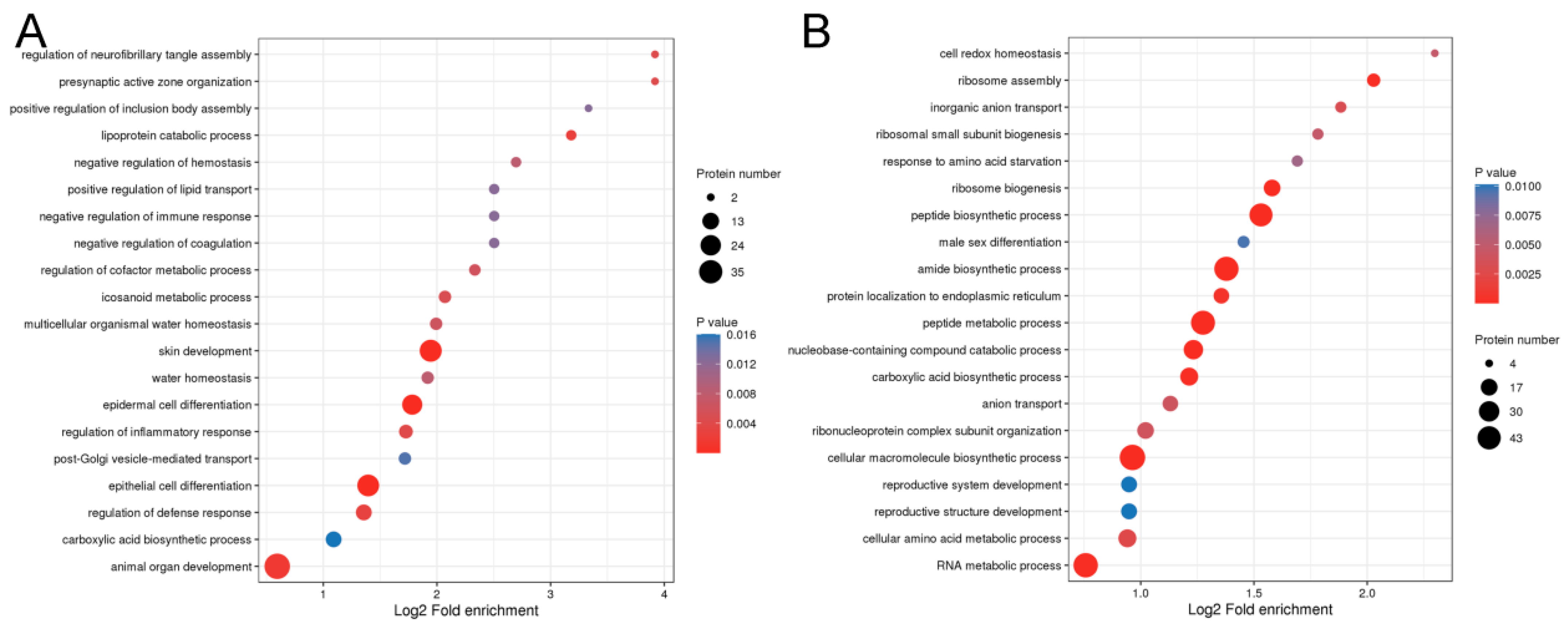
To investigate the potential function of DEPs in fiber fineness in rabbits, we conducted functional enrichment analysis on the genes encoding DEPs (Figure 2). Gene ontology (GO) analysis discriminated between upregulated and downregulated proteins. As shown in Figure 2A, the upregulated proteins were significantly enriched in epithelial cell differentiation (29 DEPs), epidermal cell differentiation (23 DEPs), skin development (30 DEPs), positive regulation of lipid transport (3 DEPs), and lipoprotein catabolic process (3 DEPs). However, the downregulated proteins were mainly involved in common structural or functional processes (Figure 2B). We further divided the DEPs into four groups (Q1, Q2, Q3, and Q4) according to fold change, as shown in Figure S2. GO classification enrichment was then performed for each group to explore correlations in the function of proteins with different fold changes. GO terms related to biological processes were enriched by DEPs with different fold changes (Figure 3). Proteins with fold changes > 2 were strongly enriched in keratinocyte differentiation, skin development, epidermal cell differentiation, epidermal development, epithelium development, epithelial cell differentiation, and keratinization. However, the other DEPs, with a fold change < 2, were weakly enriched in biological processes related to hair fibers.
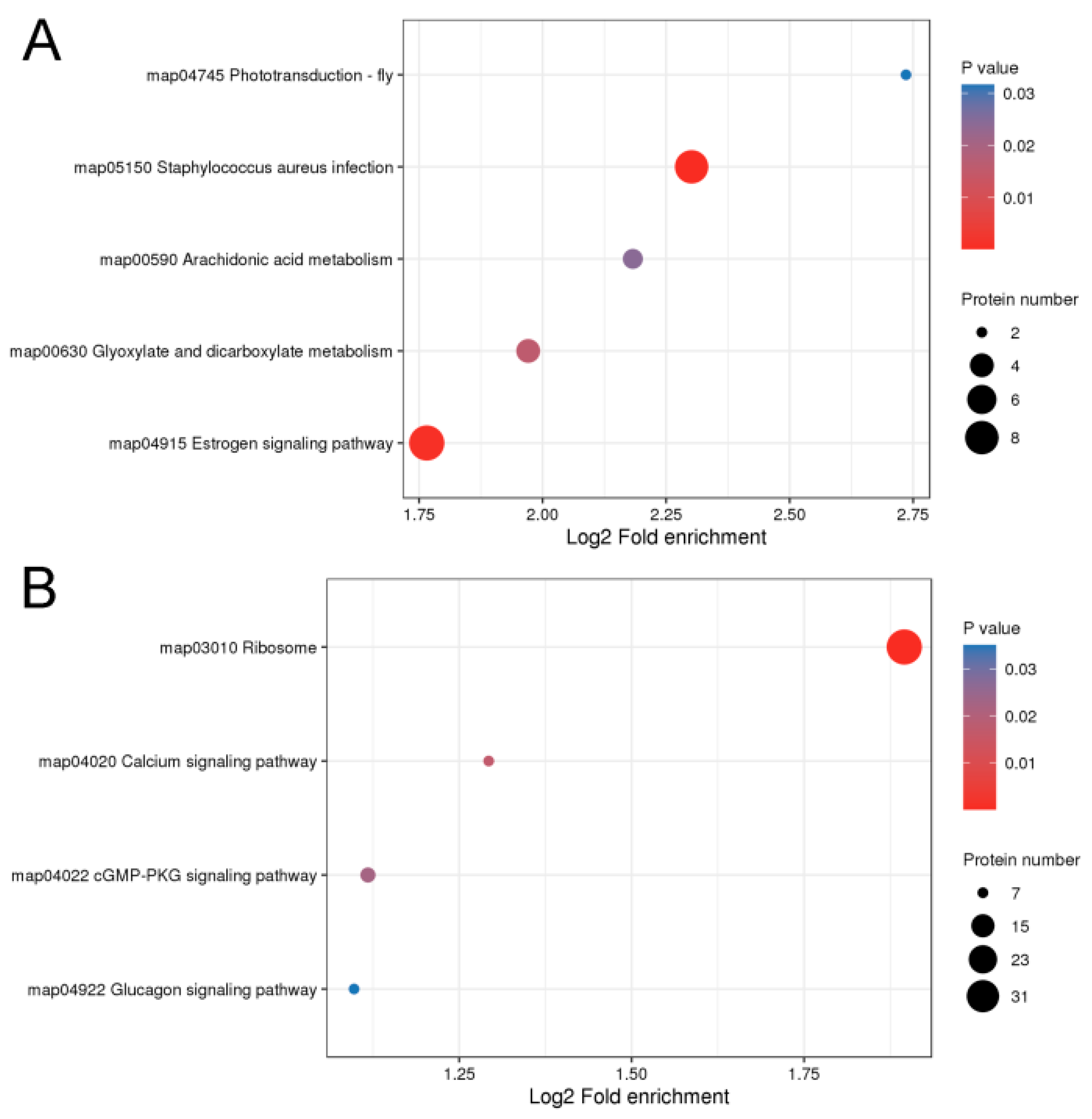
Kyoto Encyclopedia of Genes and Genomes (KEGG) enrichment analysis was conducted to further characterize the biological functions of the identified DEPs (Figure 4). The upregulated proteins were involved in the phototransduction-fly (CALML5 and ACTB) and estrogen signaling pathways (KRT26, KRT10, CALML5, KRT32, KRT15, KRT28, and KRT40), and the downregulated proteins were involved in the ribosome, calcium, and cGMP-dependent protein kinase or protein kinase G (cGMP-PKG) signaling pathways. In particular, the estrogen signaling pathway was strongly enriched by upregulated proteins (KRT10, KRT32, KRT15, KRT28, and KRT40) with a fold change > 2 (Figure S3). These findings showed that the upregulated proteins, especially proteins with a fold change > 2, were important in determining fiber diameter in rabbits.

2.4. Integrated Analysis of Transcriptome and Proteome of Hair Follicles from Coarse and Fine Wool

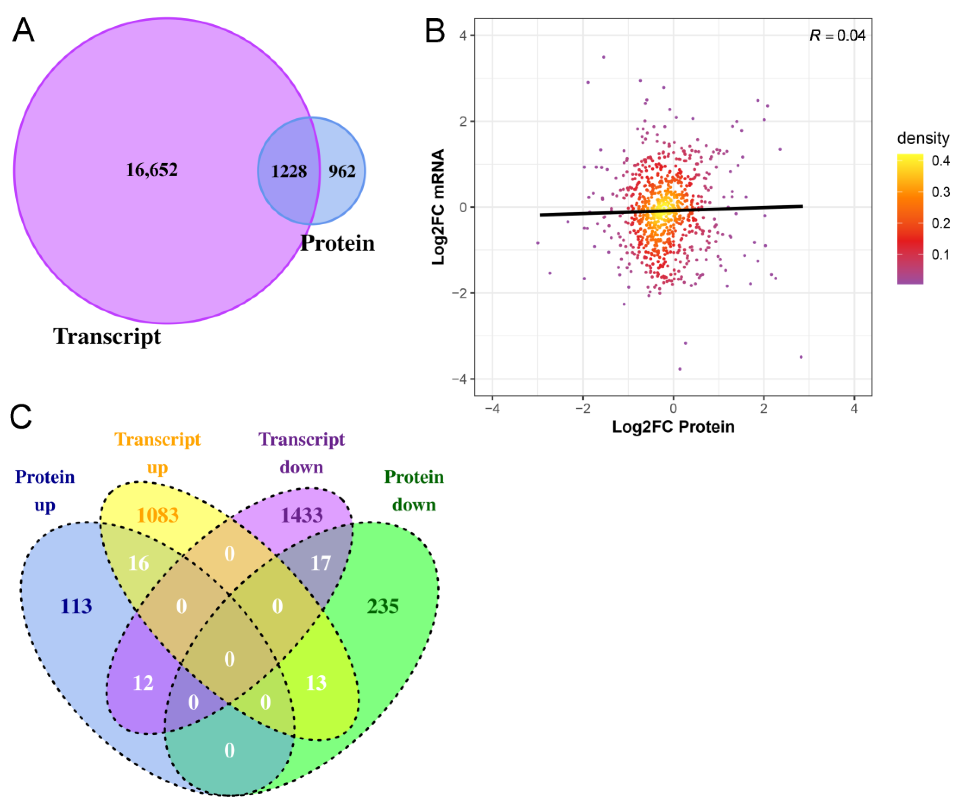
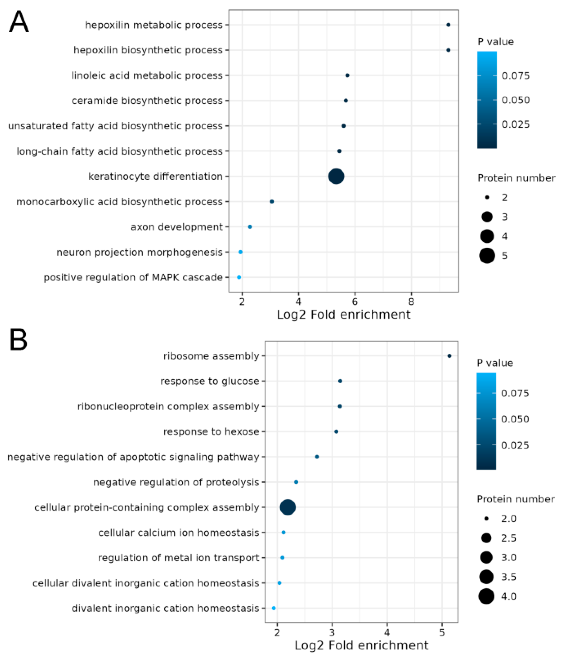
The transcriptome data of the hair follicles of coarse and fine wool from our previous study were used for integrated analyses [13]. A total of 1228 genes were quantified at both the transcriptomic and proteomic levels (Figure 5A). Subsequently, the correlation between gene expression and protein expression was examined (Figure 5B). The Pearson correlation coefficient was found to be very low (0.04), indicating that mRNA and protein expression levels in Angora rabbit hair follicles were not always positively correlated. Furthermore, the changing trends of all DEPs at both the transcriptomic and proteomic levels were analyzed. Based on mRNA and protein expression levels, a total of 33 proteins (Table S1), including 16 upregulated and 17 downregulated proteins, exhibited concordant trends at both the transcriptomic and proteomic levels (Figure 5C). Thirteen DEPs showed increased expression at the transcriptomic level but decreased expression at the proteomic level, while twelve DEPs displayed downregulation at the transcriptomic level but upregulation at the proteomic level. Notably, KRT72, KRT73, KRT77, KRT82, and KRT32 were upregulated at both the transcriptomic and proteomic levels. GO analysis demonstrated that keratinocyte differentiation (KRT73, KRT77, KRT82, KRT72, and KRT32), unsaturated fatty acid biosynthetic processes (ALOX12B and ALOXE3), and long-chain fatty acid biosynthetic processes (ALOX12B and ALOXE3) were significantly enriched in proteins upregulated at both the transcriptomic and proteomic levels (Figure 6A, Table S2). Proteins that were downregulated at the transcriptomic and proteomic levels were not involved in regulating hair follicle development (Figure 6B, Table S3).

2.5. Protein-Protein Interaction (PPI) Networks

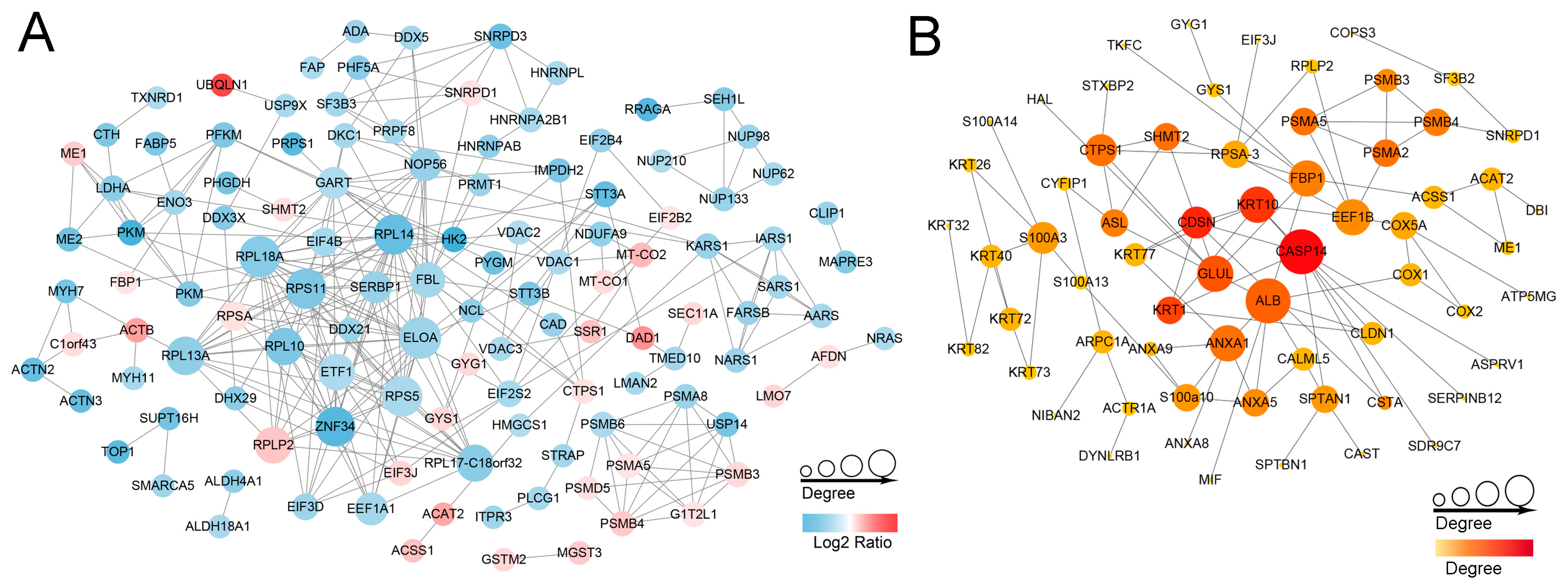
We further investigated the mechanism of DEPs involved in regulating the fineness of hair fibers and revealed functional interactions among the DEPs. PPI network analysis of the DEPs in the STRING database was performed. Sequential visualization was performed using Cytoscape software (version 3.8.2). In total, 120 proteins formed a tightly connected network with 284 edges (Figure 7A). Among these 120 DEPs, 30 were upregulated, while 90 were downregulated. The hub nodes, including ELOA, RPL18A, RPS11, RPS5, RPL14, ZNF34, RPL13A, and RPL10, exhibited more than 40 connections and all belonged to ribosomal proteins that showed downregulation in the ribosome pathway. Additionally, we analyzed the upregulated proteins and the relationships between them are shown in Figure 7B. Hub proteins such as CASP14, GLUL, KRT10, CDSN, and ALB, all with high degree values, were identified.

2.6. Validation of Proteins via Parallel Reaction Monitoring (PRM)

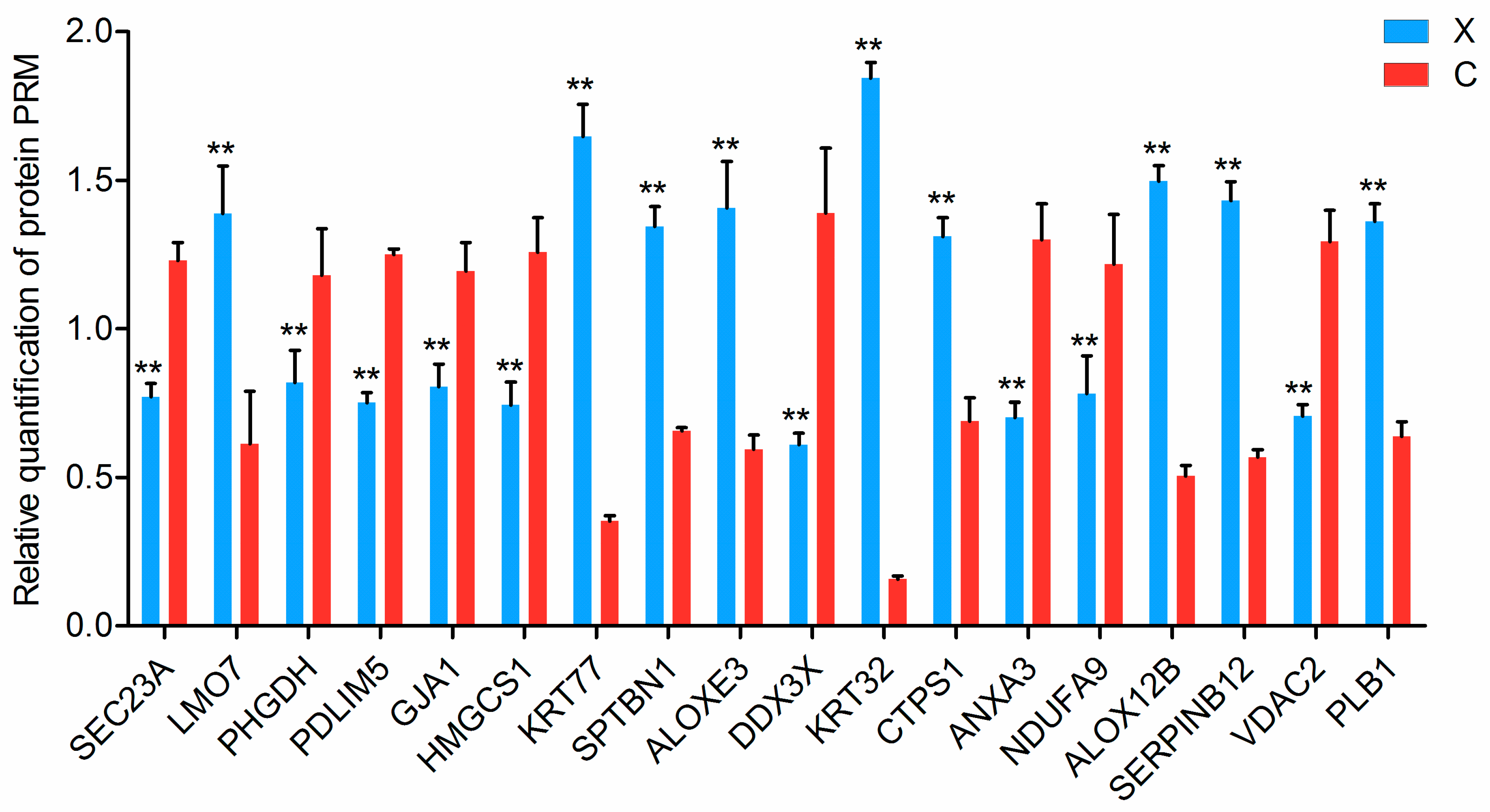
According to the integrated analysis results, we selected and assessed the 33 proteins that exhibited consistent differential expression patterns at both the transcriptomic and proteomic levels in coarse and fine wool samples. Nineteen proteins were further validated using PRM-targeted proteomics. The results showed that 18 proteins were successfully quantified (Figure 8, Table S4). The relative abundance level of all of the 18 proteins (SEC23A, LMO7, PHGDH, PDLIM5, GJA1, HMGCS1, KRT77, SPTBN1, ALOXE3, DDX3X, KRT32, CTPS1, ANXA3, NDUFA9, ALOX12B, SERPINB12, VDAC2, and PLB1) showed similar upregulation or downregulation trends in both 4D label-free-based proteomics and PRM, confirming the reliability of our proteomics data.

3. Discussion

Rabbit hair is characterized by its antistatic properties, softness, glossiness, and durability, and as a result, it is usually chosen to produce luxury textile materials [4]. It is also the core of the third-largest animal fiber industry worldwide [36]. Fiber diameter has important commercial value in fiber production and greatly influences the quality of rabbit hair. In addition, fiber diameter determines the processing performance and end use of wool, as well as its market price [37,38].
In our study, we performed a proteome-wide analysis of hair follicles from coarse and fine wool of Angora rabbits and identified a set of functional proteins involved in regulating the diameter of the wool fibers. The present study showed that 423 proteins (151 upregulated and 272 downregulated) were differentially expressed in hair follicles of coarse and fine wool. The GO enrichment analysis revealed that many upregulated proteins were involved in epithelial and epidermal cell differentiation, skin development, positive regulation of lipid transport, and lipoprotein catabolism. In particular, the upregulated proteins with fold changes > 2 exhibited significant enrichment in biological processes related to hair follicle development, such as keratinocyte differentiation, skin development, epidermal cell differentiation, epidermal and epithelial development, epithelial cell differentiation, and keratinization. Conversely, only a limited number of downregulated proteins in the fine wool group participated in the above biological processes to regulate fiber structure. Consequently, we speculate that the upregulated proteins in the fine wool group, particularly those displaying fold changes > 2, played pivotal roles in determining the structure of fine fibers in Angora rabbits. Skin development and epidermal and epithelial cell differentiation were significantly enriched by KRTAP24-1, KRT82, KRT77, KRT72, KRT10, KRT32, KRT15, KRT28, and KRT40, all with fold changes > 2. KRT and KAP proteins are the main structural proteins of wool, forming a matrix between the keratin intermediate filaments in the fiber [39,40]; they also play a role in determining hair fiber quality in mammals [41]. It has been reported that the caprine KRTAP24-1 gene serves as a marker for fine cashmere fiber by influencing its diameter [6]. KRT10 is expressed early during the differentiation of keratinocytes and is essential for epidermal integrity [42]. KRT77, a member of the type II keratin family, plays an important role in epidermal and coat formation [43]. Furthermore, differential abundance of KRT40 has been observed between cashmere and guard fiber in sheep and goats [1]. Collectively, these findings highlight the significance of these differentially expressed KRT proteins as important structural components that may contribute to variations in hair diameter among Angora rabbits.
The KEGG-enriched upregulated proteins mainly fall into the phototransduction-fly and estrogen signaling pathways, while the downregulated proteins are mainly involved in the ribosome, calcium, and cGMP-PKG signaling pathways. The phototransduction-fly signaling pathway is a photoperiodic pathway involved in regulating biological processes [44]. Red light can promote the growth of rabbit hair and the development of secondary hair follicles, increase the percentage of fine hair, and reduce the fineness of fine hair; green light improves the fineness of coarse hair and hinders hair follicle development in rabbits [45]. It is worth noting that the phototransduction-fly pathway is enriched by upregulated proteins (CALML5 and ACTB), with the CALML5 gene being an essential epidermal regulator required for late epidermal differentiation [46]. Therefore, it can be inferred that the phototransduction-fly pathway plays a crucial role in regulating rabbit hair quality and hair growth. Estrogen is an important modulator of skin physiology and hair growth [47,48]. Proteins with fold changes > 2 (KRT10, KRT32, KRT15, KRT28, and KRT40) were strongly enriched in the estrogen signaling pathway. These proteins are important structural proteins in wool. This finding suggests that the estrogen signaling pathway is involved in regulating the structural components of fine wool in Angora rabbits. Both our present study and previous research have shown substantial enrichment of DEPs and DEGs related to ribosome pathways between coarse and fine wool hair follicles [13]. Similarly, ribosome pathways were also significantly enriched among DEGs from two groups of Gansu alpine Merino sheep with different wool fiber diameters [49]. A defect in ribosomes has been shown to alter cell fate from a hair cell to a non-hair cell [50,51]. Therefore, we hypothesize that the phototransduction-fly signaling pathway, along with estrogen and ribosome pathways, potentially play vital roles in controlling hair fineness in Angora rabbits. Calcium and cGMP-PKG are key signal transduction pathways involved not only in regulating Ca2+ concentration but also in maintaining ion homeostasis by balancing potassium, sodium, and anions in organisms [52,53,54,55]. This indicates that a stronger regulation of ion homeostasis occurs in the hair follicles of coarse wool.
Association analysis of the transcriptome and proteome is an efficient strategy for understanding the principles of gene expression control and can provide more comprehensive gene expression information [56]. Correlation analysis revealed that 1228 genes were expressed at both the transcriptomic and proteomic levels in hair follicles between coarse and fine wool. However, our study demonstrated a low correlation (0.04) between transcriptomic and proteomic profiles. Among these, only 33 differentially expressed proteins exhibited consistent trends at both the transcriptional and translational levels. Previous studies have also reported a limited correlation between the transcriptome and proteome [57,58]. This discrepancy may arise from the fact that changes in mRNA abundance do not necessarily translate directly into corresponding alterations in protein levels. Genetic variability that does not alter transcription levels may alter protein functions and thus potentially alter phenotypes [59]. Moreover, it is important to consider that individual genes typically generate diverse transcripts and proteoforms [60,61], which can lead to variations in relative protein expression levels and potentially functional proteins, based on their respective mRNA abundance [62,63].
In addition, GO terms associated with hair growth were enriched in proteins upregulated at the transcriptomic and proteomic levels, such as in keratinocyte differentiation, unsaturated fatty acid biosynthetic processes, and long-chain fatty acid biosynthetic processes, which directly or indirectly regulate hair structures. For instance, keratinocytes play important roles in follicle induction and hair growth [64,65]. Unsaturated fatty acid biosynthesis is also involved in hair metabolism [66]. Biosynthesis of long-chain fatty acids is highly important for skin function as it serves as a structural component that is not bound to membranes; it contributes to the development and maintenance of both hair and skin function [67]. The upregulated proteins, including KRT73, KRT77, KRT82, KRT72, KRT32, ALOX12B, and ALOXE3, were involved in these biological processes, indicating their significant contribution to the difference in diameter between coarse and fine wool observed in Angora rabbits.
Finally, PPI analysis of the upregulated proteins showed that the hub nodes CASP14, KRT10, and CDSN were associated with wool traits and hair growth. For instance, CASP14 and KRT10 have been implicated in keratinocyte differentiation [42,68]. CDSN is expressed in the epidermis and inner root sheath of hair follicles and plays a key role in hair physiology [69]. We subsequently verified these results using PRM-targeted proteomics and further demonstrated the reliability of our data.

4. Materials and Methods

4.1. Animals and Sample Collection

Twelve healthy 1-year-old female Angora rabbits from the Animal Husbandry Institute of the Anhui Academy of Agriculture Sciences were used in this study. The Angora rabbits were divided into four groups. Each group contained three rabbits from the same parent that were raised under the same conditions (ambient temperature and natural photoperiod) and fed based on appetite, with drinking water supplied using an automatic water feeder. The coarse and fine wool fibers of each Angora rabbit were pulled from the roots from the back of the body in the spring, when wool fiber grew to 73 days after shaving. The hair follicles of coarse and fine wool were cut and placed in a 2.0 mL freezing tube, snap-frozen in liquid nitrogen, and then stored at −80 °C for subsequent experiments.

4.2. Hair Follicle Protein Extraction and Tryptic Digestion

The hair follicle samples of coarse and fine wool fibers were ground into powder with liquid nitrogen and then transferred to centrifuge tubes. Four volumes of lysis buffer (8 M urea and 1% protease inhibitor cocktail) were added to the powder, followed by three treatments with a high-intensity ultrasonic processor (Scientz). The remaining debris was removed by centrifugation at 12,000× g at 4 °C for 10 min. The supernatant was collected, and the protein concentration was determined using a Bicinchoninic Acid Assay (BCA) kit and then normalized to 1 mg/mL. Then, protein samples from each group were mixed into one biological replicate. Finally, four biological replicates were obtained from the coarse and fine wool groups, respectively.
The protein solution was reduced using 5 mM dithiothreitol for 30 min at 56 °C and alkylated with 11 mM iodoacetamide for 15 min at room temperature in darkness. The protein sample was diluted with 100 mM triethylammonium bicarbonate (TEAB) to a urea concentration of <2 M. Trypsin solution was then added at a trypsin-to-protein mass ratio of 1:50 for the first digestion overnight, and at a ratio of 1:100 for the second 4 h digestion.

4.3. Liquid Chromatography Tandem Mass Spectrometry Analysis

The tryptic peptides were dissolved in solvent A (0.1% formic acid, 2% acetonitrile/water), directly loaded onto a home-made reverse-phase analytical column (length, 25 cm; diameter, 75/100 μm). The gradient consisted of increasing solvent B (0.1% formic acid, 90% acetonitrile) from 6% to 24% over 70 min, from 24% to 35% over 14 min, then to 80% over 3 min, remaining at 80% for a further 3 min, at a constant flow rate of 450 nL/min on a nanoElute UHPLC system (Bruker Daltonics, Billerica, MA, USA). The separated peptides were injected into the capillary ion source for ionization and then into the timsTOF Pro mass spectrometer (Bruker Daltonics, Billerica, MA, USA) for analysis. The ion source voltage was set at 1.60 kV and the peptide parent ions and their secondary fragments were detected and analyzed using a high-resolution TOF. The mass spectrometry scan range was set to 100–1700 m/z. Data acquisition was conducted using the parallel accumulation serial fragmentation (PASEF) mode. The 10-fold PASEF mode acquisition of mother ions was performed after first-level mass spectrum acquisition, using a cycle window time of 1.17 s. For secondary spectra with charge numbers of zero to five, the dynamic exclusion was set at 30 s.

4.4. Data and Bioinformatics Analysis

The resulting liquid chromatography-tandem mass spectrometry (LC-MS/MS) data were processed using the MaxQuant search engine version 1.6.15.0. Search parameter settings were as follows: database for Oryctolagus_cuniculus_9986_PR_20220422 (41,459 entries). Trypsin/P is designated as an enzyme that allows up to two deletions. The mass tolerance for the precursor ions was set to 20 ppm in the first search and to 5 ppm in the main search. The mass tolerance for the fragment ions was set to 0.02 Da. Carbamidomethylation on Cys was specified as a fixed modification, and acetylation at the protein N-terminus and oxidation on Met were specified as variable modifications. The false discovery rate (FDR) was adjusted to <1%, and the minimum score of modified peptides was set to >40. Proteins with a fold change > 1.5 or < 0.67 and statistical significance (p < 0.05) were considered DEPs.
The protein functions were analyzed based on searches against the gene ontology (GO; https://geneontology.org/, accessed on 21 December 2021) and Kyoto Encyclopedia of Genes and Genomes (KEGG; www.kegg.jp/feedback/copyright.html, accessed on 17 September 2021) databases [70]. A p value < 0.05 was considered significantly different for GO terms and pathways. A protein-protein interaction (PPI) network was constructed with a confidence score ≥ 0.7 using STRING version 11.0 [71] and visualized and analyzed using Cytoscape software version 3.8.2 (http://www.cytoscape.org/, accessed on 15 July 2023). The hub genes were identified using the plug-in cytoHubba in Cytoscape software. Correlation analysis of genes and proteins was performed using the R function core test. The transcriptome data from the hair follicles of coarse and fine wool were from our previous study [13].

4.5. Validation of Protein Expression Levels Using Parallel Reaction Monitoring

Protein extraction and digestion were performed as described above. Proteins (200 μg) from each sample [33] were prepared for parallel reaction monitoring (PRM) detection. The mobile phase was run at a constant flow rate of 1000 L/min in gradient mode. Mobile phase A contained 0.1% formic acid and 2% acetonitrile, while mobile phase B contained 0.1% formic acid and 90% acetonitrile. The liquid phase gradient setting was as follows: from 0 to 40 min, the gradient was 6% to 20% B; from 40 to 52 min, the gradient was 20% to 30% B; from 52 to 56 min, the gradient was 30% to 80% B; and from 56 to 60 min, the gradient was 80% B. The flow rate was maintained at 450 nL/min. After isolation, the peptides were subjected to an NSI source followed by tandem mass spectrometry (MS/MS) in Q Exactive HF-X (Thermo, Grand Island, NY, USA). The electrospray voltage applied was 2.1 kV. A primary mass spectrometry scan range was set at 440–1265 m/z, with a resolution of 30,000. Automatic gain control (AGC) was set at 3 × 10⁶, and the maximum ion implantation time (MIT) was 50 ms. For the secondary mass spectrometry Orbitrap scan, the resolution was set at 17,500, AGC was set at 2 × 10⁵, and MIT was set at 220 ms. The isolation window for MS/MS was set at 1.4 m/z.
The resulting MS data were processed using Skyline version 3.6 software. For the peptide settings, the enzyme was set as trypsin [KR/P], and the maximum missed cleavage was set as two. The peptide length was set as seven to twenty-five, and fixed modification was set as the carbamidomethyl on Cys. The transition settings were as follows: precursor charges were set as two and three, ion charges were set as one, and ion types were set as b and y. The product ions were set from ion three to the last ion, and the ion-match tolerance was set as 0.02 Da. The FDR for protein identification was set at 1%.

4.6. Statistical Analysis

The data were expressed as means ± standard deviation and determined using GraphPad Prism 5 (GraphPad Software, Inc., La Jolla, CA, USA) in this study. A student’s t-test was employed to analyze differences between two groups. The threshold of p < 0.05 was considered statistically significant.

5. Conclusions

In summary, we characterized the differences in protein abundance between hair follicles of coarse and fine wool from Angora rabbits. Through integrated analysis of transcriptome and proteome, we identified functional and regulatory networks. Our findings revealed proteins, such as KRTAP24-1, KRT10, KRT32, KRT15, KRT28, KRT40, KRT73, KRT77, KRT82, KRT72, CASP14, and CDSN, which may be associated with wool fiber diameter. This study provides valuable transcriptomic and proteomic expression datasets that contribute to our understanding of the regulatory mechanisms underlying the differences in fiber diameters between coarse and fine wool in Angora rabbits. Further studies are warranted to elucidate the regulatory mechanisms of candidate functional proteins on wool fiber diameter.

Supplementary Materials

The following supporting information can be downloaded at https://www.mdpi.com/article/10.3390/ijms25063260/s1.

Author Contributions

H.Z., D.H. and H.D. conceived and designed the study. H.D. and D.H. performed sample collection. Y.W. conducted statistical and bioinformatics analyses. H.D. and X.W. prepared figures and tables. H.D. wrote and revised the manuscript. All authors have read and agreed to the published version of the manuscript.

Funding

The work was supported by the earmarked fund for China Agriculture Research System (Grant No.: CARS-43-A-4), the Provincial Key R&D Program of Anhui Province (Grant No.: 202104a06020032), Discipline Leading Talent Plan of Anhui Academy of Agricultural Sciences (Grant No.: LJRC-202107), Anhui Provincial Key Laboratory of Livestock and Poultry Product Safety Engineering Project (Grant No.: XMT2022-8).

Institutional Review Board Statement

The present study was carried out in strict accordance with relevant guidelines and regulations determined by the Ministry of Agriculture of the People’s Republic of China. All experimental protocols were approved by the Ethics Committee of Anhui Academy of Agricultural Sciences (AAAS2022-19).

Informed Consent Statement

Not applicable.

Data Availability Statement

The data are available from the corresponding authors upon reasonable request.

Acknowledgments

We are thankful to Yongxin Yang for their skillful assistance.

Conflicts of Interest

The authors declare no conflicts of interest.

References

  1. Zhang, C.; Qin, Q.; Liu, Z.; Xu, X.; Lan, M.; Xie, Y.; Wang, Z.; Li, J.; Liu, Z. Identification of the key proteins associated with different hair types in sheep and goats. Front. Genet. 2022, 13, 993192. [Google Scholar] [CrossRef] [PubMed]
  2. Plowman, J.E.; Harland, D.P.; Campos, A.; Rocha, E.S.S.; Thomas, A.; Vernon, J.A.; van Koten, C.; Hefer, C.; Clerens, S.; de Almeida, A.M. The wool proteome and fibre characteristics of three distinct genetic ovine breeds from Portugal. J. Proteom. 2020, 225, 103853. [Google Scholar] [CrossRef] [PubMed]
  3. Zheng, Y.Y.; Sheng, S.D.; Hui, T.Y.; Yue, C.; Sun, J.M.; Guo, D.; Guo, S.L.; Li, B.J.; Xue, H.L.; Wang, Z.Y.; et al. An Integrated Analysis of Cashmere Fineness lncRNAs in Cashmere Goats. Genes 2019, 10, 266. [Google Scholar] [CrossRef] [PubMed]
  4. Dorozynska, K.; Maj, D. Rabbits—Their domestication and molecular genetics of hair coat development and quality. Anim. Genet. 2021, 52, 10–20. [Google Scholar] [CrossRef] [PubMed]
  5. Miao, C.; Yang, Y.; Li, S.; Guo, Y.; Shui, W.; Cao, Q. Discrimination and quantification of homologous keratins from goat and sheep with dual protease digestion and PRM assays. J. Proteom. 2018, 186, 38–46. [Google Scholar] [CrossRef] [PubMed]
  6. Wang, J.; Zhou, H.; Luo, Y.; Zhao, M.; Gong, H.; Hao, Z.; Hu, J.; Hickford, J. Variation in the Caprine KAP24-1 Gene Affects Cashmere Fibre Diameter. Animals 2019, 9, 15. [Google Scholar] [CrossRef]
  7. Zhao, J.; Qin, H.; Xin, J.; Liu, N.; Han, R.; Perez-Campo, F.M.; Li, H. Discovery of genes and proteins possibly regulating mean wool fibre diameter using cDNA microarray and proteomic approaches. Sci. Rep. 2020, 10, 7726. [Google Scholar] [CrossRef]
  8. Safari, E.; Fogarty, N.M.; Gilmour, A.R. A review of genetic parameter estimates for wool, growth, meat and reproduction traits in sheep. Livest. Prod. Sci. 2005, 92, 271–289. [Google Scholar] [CrossRef]
  9. Galbraith, H. Fundamental hair follicle biology and fine fibre production in animals. Animal 2010, 4, 1490–1509. [Google Scholar] [CrossRef]
  10. Yue, Y.J.; Liu, J.B.; Yang, M.; Han, J.L.; Guo, T.T.; Guo, J.; Feng, R.L.; Yang, B.H. De novo assembly and characterization of skin transcriptome using RNAseq in sheep (Ovis aries). Genet. Mol. Res. 2015, 14, 1371–1384. [Google Scholar] [CrossRef]
  11. Ibraheem, M.; Galbraith, H.; Scaife, J.; Ewen, S. Growth of secondary hair follicles of the Cashmere goat in vitro and their response to prolactin and melatonin. J. Anat. 1994, 185, 135–142. [Google Scholar] [PubMed]
  12. Zhu, B.; Xu, T.; Yuan, J.; Guo, X.; Liu, D. Transcriptome sequencing reveals differences between primary and secondary hair follicle-derived dermal papilla cells of the Cashmere goat (Capra hircus). PLoS ONE 2013, 8, e76282. [Google Scholar] [CrossRef] [PubMed]
  13. Huang, D.; Ding, H.; Wang, Y.; Cheng, G.; Wang, X.; Leng, T.; Zhao, H. Hair Follicle Transcriptome Analysis Reveals Differentially Expressed Genes That Regulate Wool Fiber Diameter in Angora Rabbits. Biology 2023, 12, 445. [Google Scholar] [CrossRef]
  14. Tian, Y.Z.; Usman, T.; Tian, K.C.; Di, J.; Huang, X.X.; Xu, X.M.; Tulafu, H.; Wu, W.W.; Fu, X.F.; Bai, Y.; et al. Comparative study of 13 candidate genes applying multi-reference normalization to detect the expression of different fineness in skin tissues of wool sheep. Genet. Mol. Res. 2017, 16. [Google Scholar] [CrossRef]
  15. Li, C.; He, X.; Wu, Y.; Li, J.; Zhang, R.; An, X.; Yue, Y. Single-Cell Transcriptome Sequence Profiling on the Morphogenesis of Secondary Hair Follicles in Ordos Fine-Wool Sheep. Int. J. Mol. Sci. 2024, 25, 584. [Google Scholar] [CrossRef]
  16. He, X.; Chao, Y.; Zhou, G.; Chen, Y. Fibroblast growth factor 5-short (FGF5s) inhibits the activity of FGF5 in primary and secondary hair follicle dermal papilla cells of cashmere goats. Gene 2016, 575, 393–398. [Google Scholar] [CrossRef] [PubMed]
  17. Liu, G.; Liu, C.; Zhang, Y.; Sun, H.; Yang, L.; Bai, L.; Gao, S. Hair Follicle Development of Rex Rabbits Is Regulated Seasonally by Wnt10b/β-Catenin, TGFβ-BMP, IGF1, and EGF Signaling Pathways. Animals 2023, 13, 3742. [Google Scholar] [CrossRef]
  18. Rishikaysh, P.; Dev, K.; Diaz, D.; Qureshi, W.M.; Filip, S.; Mokry, J. Signaling involved in hair follicle morphogenesis and development. Int. J. Mol. Sci. 2014, 15, 1647–1670. [Google Scholar] [CrossRef]
  19. Fatima, N.; Jia, L.; Liu, B.; Li, L.; Bai, L.; Wang, W.; Zhao, S.; Wang, R.; Liu, E. A homozygous missense mutation in the fibroblast growth factor 5 gene is associated with the long-hair trait in Angora rabbits. BMC Genom. 2023, 24, 298. [Google Scholar] [CrossRef]
  20. Zhao, H.; Hu, R.; Li, F.; Yue, X. Five SNPs within the FGF5 Gene Significantly Affect Both Wool Traits and Growth Performance in Fine-Wool Sheep (Ovis aries). Front. Genet. 2021, 12, 732097. [Google Scholar] [CrossRef]
  21. Yue, L.; Lu, Z.; Guo, T.; Liu, J.; Yuan, C.; Yang, B. Association of SLIT3 and ZNF280B Gene Polymorphisms with Wool Fiber Diameter. Animals 2023, 13, 3552. [Google Scholar] [CrossRef] [PubMed]
  22. McLaren, R.J.; Rogers, G.R.; Davies, K.P.; Maddox, J.F.; Montgomery, G.W. Linkage mapping of wool keratin and keratin-associated protein genes in sheep. Mamm. Genome 1997, 8, 938–940. [Google Scholar] [CrossRef] [PubMed]
  23. Plowman, J.E.; Deb-Choudhury, S.; Clerens, S.; Thomas, A.; Cornellison, C.D.; Dyer, J.M. Unravelling the proteome of wool: Towards markers of wool quality traits. J. Proteom. 2012, 75, 4315–4324. [Google Scholar] [CrossRef] [PubMed]
  24. Powell, B.C.; Rogers, G.E. The role of keratin proteins and their genes in the growth, structure and properties of hair. Exs 1997, 78, 59–148. [Google Scholar] [PubMed]
  25. Koehn, H.; Clerens, S.; Deb-Choudhury, S.; Morton, J.D.; Dyer, J.M.; Plowman, J.E. The proteome of the wool cuticle. J. Proteome Res. 2010, 9, 2920–2928. [Google Scholar] [CrossRef] [PubMed]
  26. Bawden, C.S.; Powell, B.C.; Walker, S.K.; Rogers, G.E. Expression of a wool intermediate filament keratin transgene in sheep fibre alters structure. Transgenic Res. 1998, 7, 273–287. [Google Scholar] [CrossRef]
  27. Clerens, S.; Cornellison, C.D.; Deb-Choudhury, S.; Thomas, A.; Plowman, J.E.; Dyer, J.M. Developing the wool proteome. J. Proteom. 2010, 73, 1722–1731. [Google Scholar] [CrossRef]
  28. Plowman, J.E.; Harland, D.P.; Ganeshan, S.; Woods, J.L.; van Shaijik, B.; Deb-Choudhury, S.; Thomas, A.; Clerens, S.; Scobie, D.R. The proteomics of wool fibre morphogenesis. J. Struct. Biol. 2015, 191, 341–351. [Google Scholar] [CrossRef]
  29. Almeida, A.M.; Bassols, A.; Bendixen, E.; Bhide, M.; Ceciliani, F.; Cristobal, S.; Eckersall, P.D.; Hollung, K.; Lisacek, F.; Mazzucchelli, G.; et al. Animal board invited review: Advances in proteomics for animal and food sciences. Animal 2015, 9, 1–17. [Google Scholar] [CrossRef]
  30. Plowman, J.; Thomas, A.; Perloiro, T.; Clerens, S.; de Almeida, A.M. Characterisation of white and black merino wools: A proteomics study. Animal 2019, 13, 659–665. [Google Scholar] [CrossRef]
  31. Li, Y.; Zhou, G.; Zhang, R.; Guo, J.; Li, C.; Martin, G.; Chen, Y.; Wang, X. Comparative proteomic analyses using iTRAQ-labeling provides insights into fiber diversity in sheep and goats. J. Proteom. 2018, 172, 82–88. [Google Scholar] [CrossRef]
  32. Guo, T.; Han, J.; Yuan, C.; Liu, J.; Niu, C.; Lu, Z.; Yue, Y.; Yang, B. Comparative proteomics reveals genetic mechanisms underlying secondary hair follicle development in fine wool sheep during the fetal stage. J. Proteom. 2020, 223, 103827. [Google Scholar] [CrossRef]
  33. Zhang, Y.; Guo, Y.; Tang, H.; Wang, X.; Wang, L.; Zhou, P. Proteomics-based screening of differentially expressed proteins in skin of Chinese merino fine sheep (JunKen type) of different gender. Sheng Wu Gong Cheng Xue Bao 2022, 38, 3925–3939. [Google Scholar] [PubMed]
  34. Liu, Y.; Ding, Y.; Liu, Z.; Chen, Q.; Li, X.; Xue, X.; Pu, Y.; Ma, Y.; Zhao, Q. Integration Analysis of Transcriptome and Proteome Reveal the Mechanisms of Goat Wool Bending. Front. Cell Dev. Biol. 2022, 10, 836913. [Google Scholar] [CrossRef]
  35. Zhao, B.; Wu, C.; Sammad, A.; Ma, Z.; Suo, L.; Wu, Y.; Fu, X. The fiber diameter traits of Tibetan cashmere goats are governed by the inherent differences in stress, hypoxic, and metabolic adaptations: An integrative study of proteome and transcriptome. BMC Genom. 2022, 23, 191. [Google Scholar] [CrossRef]
  36. Niranjan, S.K.; Sharma, S.R.; Gowane, G.R. Estimation of Genetic Parameters for Wool Traits in Angora Rabbit. Asian Austral. J. Anim. 2011, 24, 1335–1340. [Google Scholar] [CrossRef]
  37. Rafat, S.A.; de Rochambeau, H.; Brims, M.; Thebault, R.G.; Deretz, S.; Bonnet, M.; Allain, D. Characteristics of Angora rabbit fiber using optical fiber diameter analyzer. J. Anim. Sci. 2007, 85, 3116–3122. [Google Scholar] [CrossRef]
  38. Chen, Y.; Bao, Z.; Liu, M.; Li, J.; Dai, Y.; Wang, F.; Zhang, X.; Zhai, P.; Zhao, B.; Wu, X. Promoter Methylation Changes in KRT17: A Novel Epigenetic Marker for Wool Production in Angora Rabbit. Int. J. Mol. Sci. 2022, 23, 6077. [Google Scholar] [CrossRef] [PubMed]
  39. Li, S.; Xi, Q.; Zhao, F.; Wang, J.; He, Z.; Hu, J.; Liu, X.; Luo, Y. A highly polymorphic caprine keratin-associated protein gene identified and its effect on cashmere traits. J. Anim. Sci. 2021, 99, skab233. [Google Scholar] [CrossRef] [PubMed]
  40. Hearle, J.W. A critical review of the structural mechanics of wool and hair fibres. Int. J. Biol. Macromol. 2000, 27, 123–138. [Google Scholar] [CrossRef]
  41. Seki, Y.; Yokohama, M.; Wada, K.; Fujita, M.; Kotani, M.; Nagura, Y.; Kanno, M.; Nomura, K.; Amano, T.; Kikkawa, Y. Expression analysis of the type I keratin protein keratin 33A in goat coat hair. Anim. Sci. J. 2011, 82, 773–781. [Google Scholar] [CrossRef]
  42. Maytin, E.V.; Lin, J.C.; Krishnamurthy, R.; Batchvarova, N.; Ron, D.; Mitchell, P.J.; Habener, J.F. Keratin 10 gene expression during differentiation of mouse epidermis requires transcription factors C/EBP and AP-2. Dev. Biol. 1999, 216, 164–181. [Google Scholar] [CrossRef] [PubMed]
  43. Zhang, M.; Wu, D.; Ahmed, Z.; Liu, X.; Chen, J.; Ma, J.; Wang, M.; Liu, J.; Zhang, J.; Huang, B.; et al. The genetic secrets of adaptation: Decoding the significance of the 30-bp insertion in the KRT77 gene for Chinese cattle. Anim. Biotechnol. 2023, 34, 3847–3854. [Google Scholar] [CrossRef]
  44. Cai, P.; Zhang, W.; Jiang, S.; Xiong, Y.; Yuan, H.; Gao, Z.; Gao, X.; Ma, C.; Zhou, Y.; Gong, Y.; et al. Insulin-like Androgenic Gland Hormone Induced Sex Reversal and Molecular Pathways in Macrobrachium nipponense: Insights into Reproduction, Growth, and Sex Differentiation. Int. J. Mol. Sci. 2023, 24, 14306. [Google Scholar] [CrossRef] [PubMed]
  45. Wang, J. Effect of Different LED Light on Hair Follicle Structure and Fur Quality of Angora Rabbits and Its Mechanism. Master’s Thesis, Nanjing Agricultural University, Nanjing, China, 2017. [Google Scholar]
  46. Sun, B.K.; Boxer, L.D.; Ransohoff, J.D.; Siprashvili, Z.; Qu, K.; Lopez-Pajares, V.; Hollmig, S.T.; Khavari, P.A. CALML5 is a ZNF750- and TINCR-induced protein that binds stratifin to regulate epidermal differentiation. Genes Dev. 2015, 29, 2225–2230. [Google Scholar] [CrossRef]
  47. Ohnemus, U.; Uenalan, M.; Inzunza, J.; Gustafsson, J.A.; Paus, R. The hair follicle as an estrogen target and source. Endocr. Rev. 2006, 27, 677–706. [Google Scholar] [CrossRef]
  48. Ohnemus, U.; Uenalan, M.; Conrad, F.; Handjiski, B.; Mecklenburg, L.; Nakamura, M.; Inzunza, J.; Gustafsson, J.A.; Paus, R. Hair cycle control by estrogens: Catagen induction via estrogen receptor (ER)-alpha is checked by ER beta signaling. Endocrinology 2005, 146, 1214–1225. [Google Scholar] [CrossRef] [PubMed]
  49. Yue, Y.; Guo, T.; Liu, J.; Guo, J.; Yuan, C.; Feng, R.; Niu, C.; Sun, X.; Yang, B. Exploring Differentially Expressed Genes and Natural Antisense Transcripts in Sheep (Ovis aries) Skin with Different Wool Fiber Diameters by Digital Gene Expression Profiling. PLoS ONE 2015, 10, e129249. [Google Scholar] [CrossRef]
  50. Kawa, D. Twist of Fate: Ribosomal Stress Reprograms Root Hair Patterning. Plant Cell 2020, 32, 2079–2080. [Google Scholar] [CrossRef]
  51. Wang, W.; Ryu, K.H.; Bruex, A.; Barron, C.; Schiefelbein, J. Molecular Basis for a Cell Fate Switch in Response to Impaired Ribosome Biogenesis in the Arabidopsis Root Epidermis. Plant Cell 2020, 32, 2402–2423. [Google Scholar] [CrossRef]
  52. Gao, Y.; Li, T.; Sun, Q.; Huo, B. Gradient fluid shear stress regulates migration of osteoclast precursors. Cell Adh. Migr. 2019, 13, 183–191. [Google Scholar] [CrossRef]
  53. Wen, X.; Yang, M.; Zhou, K.; Huang, J.; Fan, X.; Zhang, W.; Luo, J. Transcriptomic and proteomic analyses reveal the common and unique pathway(s) underlying different skin colors of leopard coral grouper (Plectropomus leopardus). J. Proteom. 2022, 266, 104671. [Google Scholar] [CrossRef] [PubMed]
  54. Ellis, D.Z.; Nathanson, J.A.; Sweadner, K.J. Carbachol inhibits Na(+)-K(+)-ATPase activity in choroid plexus via stimulation of the NO/cGMP pathway. Am. J. Physiol. Cell Physiol. 2000, 279, C1685–C1693. [Google Scholar] [CrossRef] [PubMed]
  55. Wolin, M.S.; Ahmad, M.; Gupte, S.A. Oxidant and redox signaling in vascular oxygen sensing mechanisms: Basic concepts, current controversies, and potential importance of cytosolic NADPH. Am. J. Physiol. Lung Cell Mol. Physiol. 2005, 289, L159–L173. [Google Scholar] [CrossRef]
  56. Buccitelli, C.; Selbach, M. mRNAs, proteins and the emerging principles of gene expression control. Nat. Rev. Genet. 2020, 21, 630–644. [Google Scholar] [CrossRef] [PubMed]
  57. Ghazalpour, A.; Bennett, B.; Petyuk, V.A.; Orozco, L.; Hagopian, R.; Mungrue, I.N.; Farber, C.R.; Sinsheimer, J.; Kang, H.M.; Furlotte, N.; et al. Comparative analysis of proteome and transcriptome variation in mouse. PLoS Genet. 2011, 7, e1001393. [Google Scholar] [CrossRef]
  58. Pascal, L.E.; True, L.D.; Campbell, D.S.; Deutsch, E.W.; Risk, M.; Coleman, I.M.; Eichner, L.J.; Nelson, P.S.; Liu, A.Y. Correlation of mRNA and protein levels: Cell type-specific gene expression of cluster designation antigens in the prostate. BMC Genom. 2008, 9, 246. [Google Scholar] [CrossRef]
  59. Ng, P.C.; Henikoff, S. SIFT: Predicting amino acid changes that affect protein function. Nucleic Acids Res. 2003, 31, 3812–3814. [Google Scholar] [CrossRef]
  60. Aebersold, R.; Agar, J.N.; Amster, I.J.; Baker, M.S.; Bertozzi, C.R.; Boja, E.S.; Costello, C.E.; Cravatt, B.F.; Fenselau, C.; Garcia, B.A.; et al. How many human proteoforms are there? Nat. Chem. Biol. 2018, 14, 206–214. [Google Scholar] [CrossRef]
  61. Salovska, B.; Zhu, H.; Gandhi, T.; Frank, M.; Li, W.; Rosenberger, G.; Wu, C.; Germain, P.L.; Zhou, H.; Hodny, Z.; et al. Isoform-resolved correlation analysis between mRNA abundance regulation and protein level degradation. Mol. Syst. Biol. 2020, 16, e9170. [Google Scholar] [CrossRef]
  62. Lundberg, E.; Fagerberg, L.; Klevebring, D.; Matic, I.; Geiger, T.; Cox, J.; Algenas, C.; Lundeberg, J.; Mann, M.; Uhlen, M. Defining the transcriptome and proteome in three functionally different human cell lines. Mol. Syst. Biol. 2010, 6, 450. [Google Scholar] [CrossRef]
  63. de Godoy, L.M.; Olsen, J.V.; Cox, J.; Nielsen, M.L.; Hubner, N.C.; Frohlich, F.; Walther, T.C.; Mann, M. Comprehensive mass-spectrometry-based proteome quantification of haploid versus diploid yeast. Nature 2008, 455, 1251–1254. [Google Scholar] [CrossRef]
  64. Feng, X.; Coulombe, P.A. A role for disulfide bonding in keratin intermediate filament organization and dynamics in skin keratinocytes. J. Cell Biol. 2015, 209, 59–72. [Google Scholar] [CrossRef]
  65. Trueb, R.M. Pharmacologic interventions in aging hair. Clin. Interv. Aging 2006, 1, 121–129. [Google Scholar] [CrossRef]
  66. Tan, T.H.; Li, S.W.; Chang, C.W.; Chen, Y.C.; Liu, Y.H.; Ma, J.T.; Chang, C.P.; Liao, P.C. Rat Hair Metabolomics Analysis Reveals Perturbations of Unsaturated Fatty Acid Biosynthesis, Phenylalanine, and Arachidonic Acid Metabolism Pathways Are Associated with Amyloid-beta-Induced Cognitive Deficits. Mol. Neurobiol. 2023, 60, 4373–4395. [Google Scholar] [CrossRef] [PubMed]
  67. Westerberg, R.; Tvrdik, P.; Unden, A.B.; Mansson, J.E.; Norlen, L.; Jakobsson, A.; Holleran, W.H.; Elias, P.M.; Asadi, A.; Flodby, P.; et al. Role for ELOVL3 and fatty acid chain length in development of hair and skin function. J. Biol. Chem. 2004, 279, 5621–5629. [Google Scholar] [CrossRef] [PubMed]
  68. Eckhart, L.; Ban, J.; Fischer, H.; Tschachler, E. Caspase-14: Analysis of gene structure and mRNA expression during keratinocyte differentiation. Biochem. Biophys. Res. Commun. 2000, 277, 655–659. [Google Scholar] [CrossRef] [PubMed]
  69. Levy-Nissenbaum, E.; Betz, R.C.; Frydman, M.; Simon, M.; Lahat, H.; Bakhan, T.; Goldman, B.; Bygum, A.; Pierick, M.; Hillmer, A.M.; et al. Hypotrichosis simplex of the scalp is associated with nonsense mutations in CDSN encoding corneodesmosin. Nat. Genet. 2003, 34, 151–153. [Google Scholar] [CrossRef]
  70. Kanehisa, M.; Goto, S. KEGG: Kyoto encyclopedia of genes and genomes. Nucleic Acids Res. 2000, 28, 27–30. [Google Scholar] [CrossRef] [PubMed]
  71. Szklarczyk, D.; Gable, A.L.; Lyon, D.; Junge, A.; Wyder, S.; Huerta-Cepas, J.; Simonovic, M.; Doncheva, N.T.; Morris, J.H.; Bork, P.; et al. STRING v11: Protein-protein association networks with increased coverage, supporting functional discovery in genome-wide experimental datasets. Nucleic Acids Res. 2019, 47, D607–D613. [Google Scholar] [CrossRef] [PubMed]
Figure 1. Analysis of differentially expressed proteins in the hair follicles of coarse and fine wool from rabbits. (A) Number of upregulated and downregulated proteins. Orange, increased expression; green, decreased expression. (B) Volcano plot of differentially expressed proteins. C, coarse wool group; X, fine wool group; red, increased expression; green, decreased expression; black, none.
Ijms 25 03260 g001
Figure 2. Gene ontology analysis of differentially expressed proteins in the hair follicles of coarse and fine wool from rabbits. (A) Upregulated proteins. (B) Downregulated proteins. p < 0.05 was considered significantly different.
Ijms 25 03260 g002
Figure 3. Heatmap analysis of the differentially expressed proteins with different fold changes. Relevant functions in different Q groups were gathered together. Red, strong enrichment; blue, weak enrichment. Q1 < 0.5, 0.5 < Q2 < 0.667, 1.5 < Q3 < 2.0, and Q4 > 2.0.
Ijms 25 03260 g003
Figure 4. Enriched Kyoto Encyclopedia of Genes and Genomes pathways of the differentially expressed proteins in the hair follicles of coarse and fine wool. (A) Upregulated proteins. (B) Downregulated proteins. p < 0.05 was considered significantly different.
Ijms 25 03260 g004
Figure 5. Comparative analysis of the transcriptome and proteome of the hair follicles from coarse and fine wool. (A) Quantitative comparison of transcriptome and proteome. (B) Comparison of the expression between transcriptome and proteome. The x-axis and y-axis indicate the Log2 Fold Change (Log2FC) of the proteomic and transcriptomic profile, respectively. (C) Venn plot of up-regulated and down-regulated DEPs and differentially expressed genes (DEGs) detected at both the protein and transcript levels in comparison groups. The transcriptome data used in this analysis was taken from our previous study [13].
Ijms 25 03260 g005
Figure 6. Gene ontology analysis of proteins differentially expressed at the transcriptomic and proteomic levels. (A) Upregulated proteins. (B) Downregulated proteins.
Ijms 25 03260 g006
Figure 7. Protein–protein interaction networks of differentially expressed proteins in coarse (control) and fine (experimental) wool groups. (A) Differentially expressed proteins. Red, upregulated proteins; blue, downregulated proteins. A larger circle indicates a stronger interaction with more genes; a darker color indicates a larger fold change. (B) Upregulated proteins. Larger circle sizes and darker colors indicate stronger interactions with more genes.
Ijms 25 03260 g007
Figure 8. Verification of the differentially expressed proteins using PRM. X, hair follicle of the fine wool group; C, hair follicle of the coarse wool group. ** p < 0.01.
Ijms 25 03260 g008
Table 1. Summary of protein identification.
Total SpectraMatched SpectraPeptidesUnique PeptidesIdentified ProteinsQuantifiable Proteins
1,167,43483,70814,65512,46322751431
Table 2. Expression levels of differentially expressed KRT proteins between coarse and fine wool of Angora rabbits.
Protein AccessionGene NameX AverageC AverageX/C RatioX/C p ValueRegulation Type
G1SDF4KRT821.61024550.38975454.1311.10187 × 10−10Up
G1SHX4KRT840.6460351.353965250.4775.18384 × 10−10Down
G1SHZ4KRT70.52886651.471133250.3598.82596 × 10−8Down
G1SKE3KRT771.5790110.4209893.7511.05656 × 10−8Up
G1SUH1KRT721.46062150.53937852.7084.71087 × 10−10Up
G1SWB4KRT261.27936350.72063651.7757.79235 × 10−5Up
G1T1V0KRT101.578394250.421605753.7447.48266 × 10−9Up
G1T4P0KRT321.495854250.504145752.9675.87822 × 10−11Up
G1T4R6KRT160.749501751.250498250.5991.71792 × 10−8Down
G1T6X1KRT151.440775250.5592252.5763.67408 × 10−6Up
G1T8T1KRT281.408513250.59148652.3811.81837 × 10−9Up
G1T9T2KRT401.468482750.531517252.7631.43752 × 10−8Up
G1TDN8KRT731.258899250.741100751.6996.19365 × 10−8Up
G1U9I8KRT11.41894050.58105952.4423.43761 × 10−11Up
Disclaimer/Publisher’s Note: The statements, opinions and data contained in all publications are solely those of the individual author(s) and contributor(s) and not of MDPI and/or the editor(s). MDPI and/or the editor(s) disclaim responsibility for any injury to people or property resulting from any ideas, methods, instructions or products referred to in the content.