Article

Jozimine A2, a Dimeric Naphthylisoquinoline (NIQ) Alkaloid, Shows In Vitro Cytotoxic Effects against Leukemia Cells through NF-κB Inhibition

1
Department of Pharmaceutical Biology, Institute of Pharmaceutical and Biomedical Sciences, Johannes Gutenberg University, Staudinger Weg 5, 55128 Mainz, Germany
2
Division of Cancer Genome Research, German Cancer Research Center (DKFZ) Heidelberg, National Center for Tumor Diseases (NCT), NCT Heidelberg, a Partnership between DKFZ and University Hospital, 69120 Heidelberg, Germany
3
Institute of Organic Chemistry, University of Würzburg, Am Hubland, 97074 Würzburg, Germany
*
Author to whom correspondence should be addressed.
Int. J. Mol. Sci. 2024, 25(6), 3087; https://doi.org/10.3390/ijms25063087
Submission received: 14 February 2024 / Revised: 27 February 2024 / Accepted: 5 March 2024 / Published: 7 March 2024

Abstract

:
Naphthylisoquinoline (NIQ) alkaloids are rising as a promising class of secondary metabolites with pharmaceutical potential. NF-κB has already been recognized as a significant modulator of cancer proliferation and drug resistance. We have previously reported the mechanisms behind the cytotoxic effect of dioncophylline A, an NIQ monomer, in leukemia cells. In the current study, we have investigated the cytotoxic effect of jozimine A2, an NIQ dimer, on leukemia cells in comparison to a second, structurally unsymmetric dimer, michellamine B. To this end, molecular docking was applied to predict the binding affinity of the dimers towards NF-κB, which was then validated through microscale thermophoresis. Next, cytotoxicity assays were performed on CCRF-CEM cells and multidrug-resistant CEM/ADR5000 cells following treatment. Transcriptome analysis uncovered the molecular networks affected by jozimine A2 and identified the cell cycle as one of the major affected processes. Cell death modes were evaluated through flow cytometry, while angiogenesis was measured with the endothelial cell tube formation assay on human umbilical vein endothelial cells (HUVECs). The results indicated that jozimine A2 bound to NF-κB, inhibited its activity and prevented its translocation to the nucleus. In addition, jozimine A2 induced cell death through apoptosis and prevented angiogenesis. Our study describes the cytotoxic effect of jozimine A2 on leukemia cells and explains the interactions with the NF-κB signaling pathway and the anticancer activity.

Graphical Abstract

1. Introduction

A promising class of bioactive natural products are the naphthylisoquinoline (NIQ) alkaloids, such as dioncophylline A, isolated from tropical lianas of the Dioncophyllaceae and Ancistrocladaceae families [1,2]. We have recently shown that dioncophylline A and related NIQ alkaloids exhibit strong cytotoxicity in leukemia cells, mediated by NF-κB inhibition, angiogenesis suppression, G2/M cell cycle arrest, and autophagy induction [3].
Some of these structurally and biosynthetically unique secondary metabolites also occur as dimers, such as jozimine A2 (for the structure, see Figure 1A), which is the (twofold O-demethylated) dimer of dioncophylline A, with the two dioncophylline A moieties being connected by a chiral binaphthyl axis [4].
Michellamine B was isolated from Ancistrocladus likoko following a procedure described earlier [5], while jozimine A2 was obtained from Ancistrocladus ileboensis as reported in the literature [6]. The chromatographic and spectroscopic data of the two pure metabolites were in accordance with those reported previously [4,7]. Both compounds were fully soluble in DMSO.
In several cases, dimeric NIQs have proven enhanced activities as compared to their monomeric analogs in several anti-infective test systems (e.g., against HIV, Plasmodium) [1,2,4,6]. More recently, some of these dimers, such as jozimine A2, have demonstrated strong antitumoral activities against pancreatic and cervical cancer cells, making the testing of their antileukemic activity a rewarding task.
As different studies have shown, NF-κB modulates a plethora of cellular processes, including cell proliferation, angiogenesis, and apoptosis suppression, making it an important link in promoting inflammation, cancer proliferation, and drug resistance [8,9,10,11,12,13,14,15,16]. NF-κB is activated in an inflammatory milieu and regulates various pro-inflammatory cytokines (TNFα, IL-6, IL-1β) as well as STAT3, a transcription factor that activates proliferative and survival genes in cancer cells [10,17,18,19]. Additionally, by activating various apoptosis-suppressing genes, NF-κB prevents apoptosis and promotes cancer cell proliferation in solid tumors, leukemias, and lymphomas [11,20,21,22,23,24]. Interestingly, there is also a complex interplay between NF-κB and autophagy, another type of programmed cell death, as the regulation of both pathways is interdependent [25]. Both apoptosis and autophagy have become attractive targets in cancer treatment. However, while apoptosis activation is always desired in cancer treatment, autophagy has a much more complex role and can induce tumor suppression or tumor proliferation, depending on the tumor stage [25,26,27,28]. Due to increasing multidrug resistance in cancer, numerous studies are now using natural products to target NF-κB and induce apoptosis or autophagy, leading to cancer suppression [3,16,29,30,31,32,33]. Furthermore, many of these phytochemicals, such as curcumin, resveratrol, and lycopene, exert their anti-cancer activity not only by targeting NF-κB but also by controlling epigenetic modifications, which are key regulators of apoptosis and autophagy [30,32,33,34,35,36].
In this paper, we describe the anticancer potential of jozimine A2, a naphthylisoquinoline alkaloid, on drug-sensitive CCRF-CEM leukemia cells and provide valuable information regarding its mechanism of action and the difference in activity in comparison to a structurally related but different NIQ alkaloid dimer, namely michellamine B. By using bioinformatic tools, we uncover the binding site that leads to the inhibition of the NF-κB pathway in silico. In addition, Ingenuity Pathway Analysis (IPA)TM highlighted the
NF-κB pathway as the main network affected after treatment with jozimine A2. We further explored the mechanism by which jozimine A2 killed the cells. Moreover, jozimine A2 hampered the translocation of NF-κB to the nucleus, which led to the downregulation of NF-κB-dependent genes. Here, we report for the first time the mechanism of action of the anti-leukemic activity of jozimine A2.

2. Results

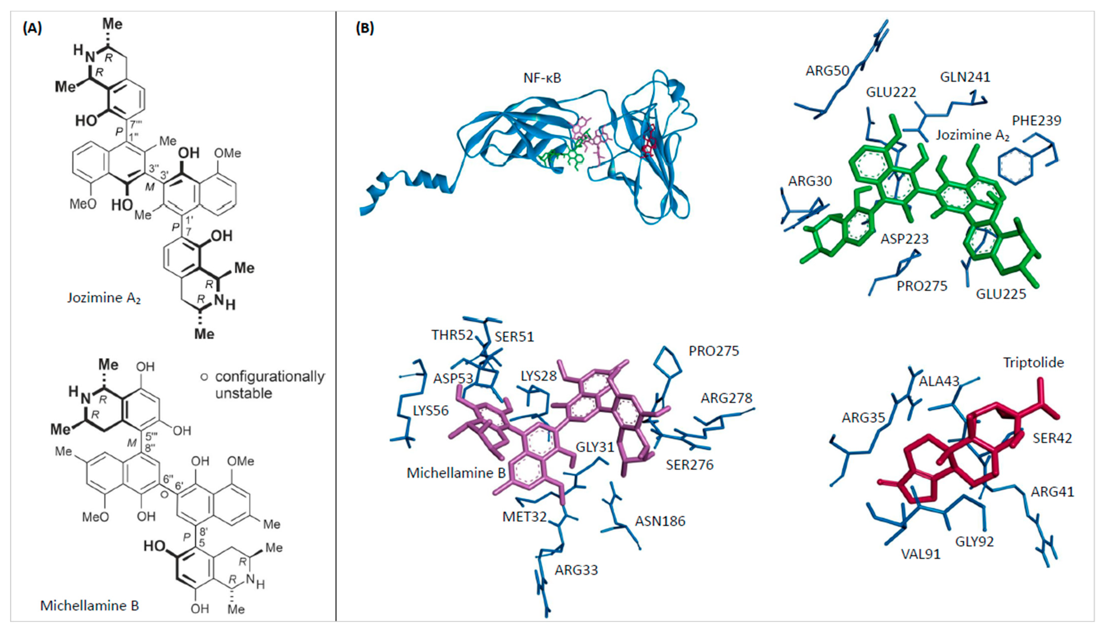
2.1. In Silico Binding of Jozimine A2 and Michellamine B to NF-κB

Molecular docking analysis, an integral technique in computer-aided drug discovery, was utilized to elucidate the binding interactions of jozimine A2 and michellamine B (for the structures, see Figure 1A) with the NF-κB protein. The calculated binding free energies of −11.11 ± 0.02 kcal/mol and −8.20 ± 0.00 kcal/mol for michellamine B and jozimine A2, respectively, demonstrated favorable predicted protein-ligand affinities (Table 1). However, as shown in Figure 1B, the analysis of the precise molecular docking conformations revealed distinct NF-κB binding sites for both natural products compared to the reference inhibitor triptolide. While binding in close proximity to one another, the specific amino acid residues involved in the predicted NF-κB complexes significantly differed between michellamine B and jozimine A2.
Both molecules investigated in this study have three biaryl axes as characteristic structural features. Depending on the degree of steric hindrance, such single bonds between aromatic ring systems can freely rotate—or can be rotationally hindered—and can then be an element of axial chirality (besides the likewise present stereogenic centers).
In the case of jozimine A2, all three axes are rotationally hindered; they are P-, M-, and P-configured. Any docking approach has to consider that this axial configuration does not change during the docking process. Indeed, the structure of jozimine A2 in the binding pocket has still retained its P,M,P configuration: M at the central axis and P at the two outer ones.
On the contrary, in the related dimeric alkaloid michellamine B, only the two outer axes are rotationally stable: one is M-configured and one is P, while the central axis, having much less steric hindrance from neighboring ortho-substituents, can freely rotate at room temperature, thus achieving a fast M,P-interconversion. Any docking procedure must recognize the stable axial arrays for the outer axes while accepting free rotation for the inner one. In the pocket, michellamine B indeed shows the expected axial configurations at the outer axes, once M and once P. The central axis is found to be P in the energetically lowest conformation, but there are both P- and M-configured geometries among the energetically next-higher species, clearly proving the procedure to accept free rotation at the central axis, fully in agreement with the experimental reality.
The current work is an illustrative example of the successful docking of a natural product possessing both configurationally stable and unstable biaryl axes.

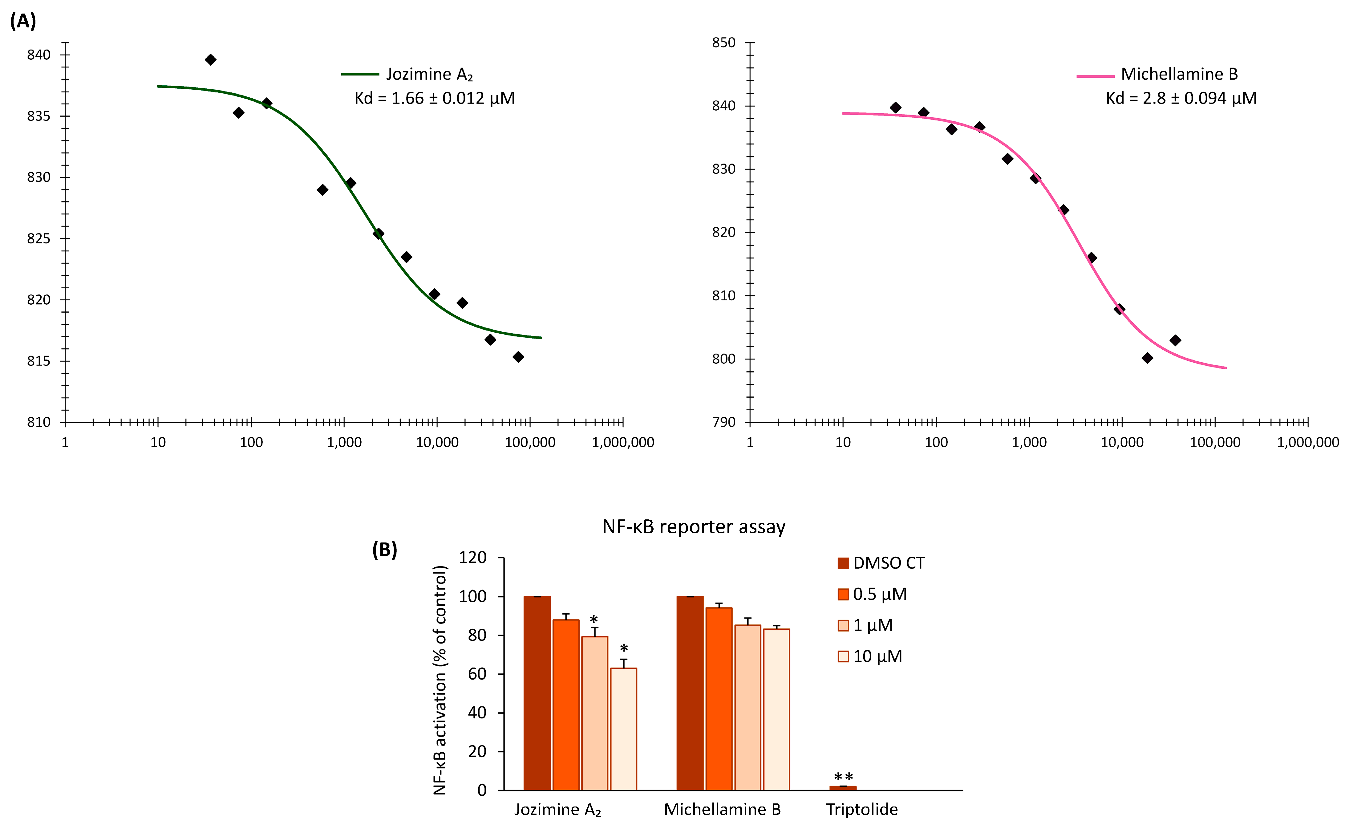
2.2. Jozimine A2 and Michellamine B Both Bind to NF-κB In Vitro, but Only Jozimine A2 Inhibits NF-κB Activity

Microscale thermophoresis (MST) was used to determine the affinity of jozimine A2 and michellamine B towards recombinant NF-κB proteins. This biophysical assay is often used to investigate the interactions between ligands and proteins [37] and represents a good way to confirm the in silico data. The MST analysis revealed that both jozimine A2 and michellamine B strongly bind to the NF-κB protein, with a dissociation constant (Kd) of 2.8 ± 0.094 µM for michellamine B and 1.66 ± 0.012 µM for jozimine A2 (Figure 2A). After both in silico (molecular docking) and in vitro (MST) assays showed the potential jozimine A2 and michellamine B have on NF-κB, the next step was to determine whether these compounds can inhibit NF-κB translocation. For this purpose, the reporter cell line HEK-BlueTM Null 1 was treated with increasing concentrations of the respective compounds. Interestingly, jozimine A2 significantly inhibited NF-κB activity in a dose-dependent manner by 40%, whereas michellamine B only weakly and not significantly decreased NF-κB activity in comparison to DMSO as the control (Figure 2B).

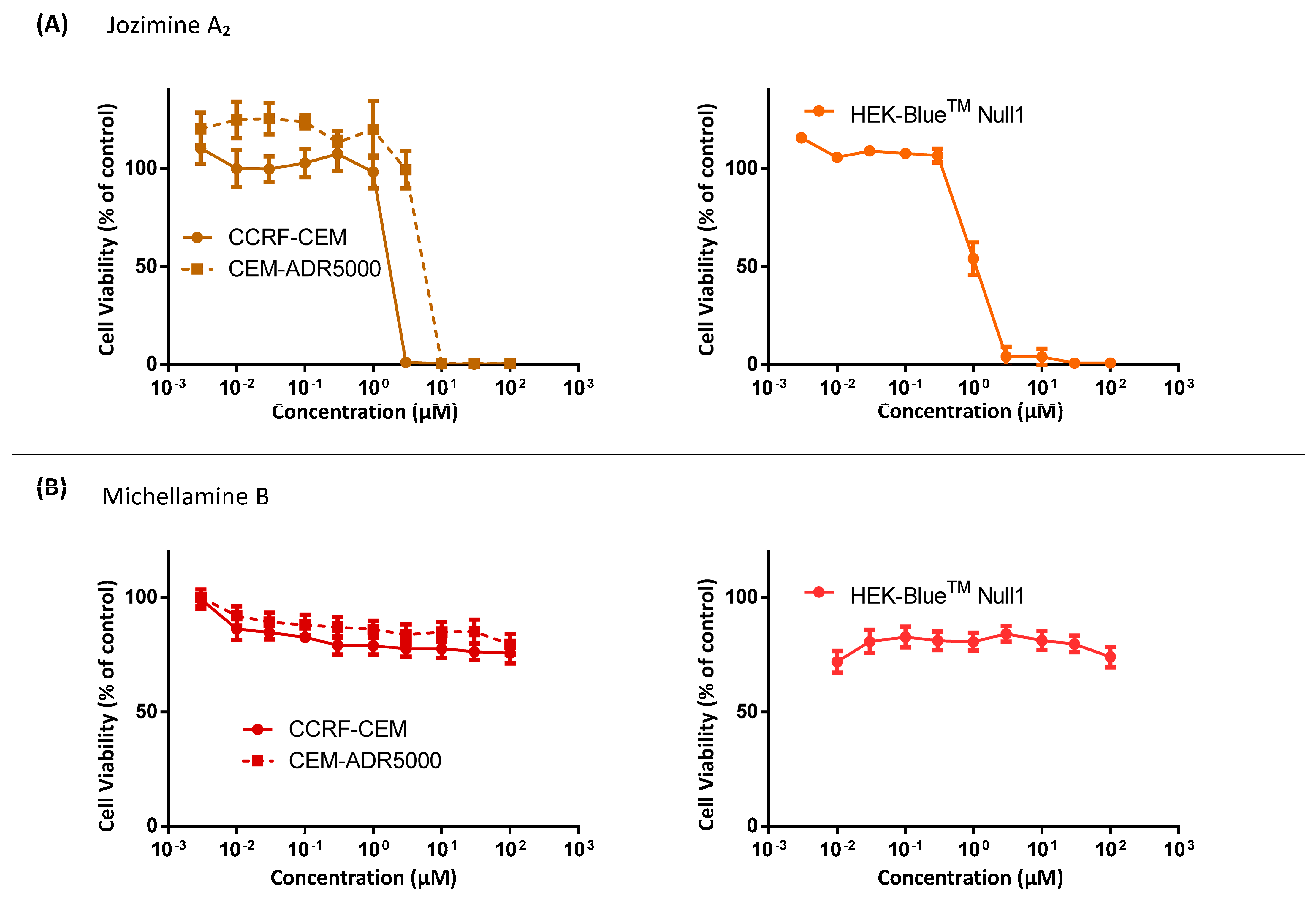
2.3. Cytotoxicity of Jozimine A2 and Michellamine B

The cytotoxic activity of jozimine A2 and michellamine B was evaluated in human CCRF-CEM and multidrug-resistant CEM/ADR5000 leukemia cells utilizing a resazurin reduction assay. Figure 3A demonstrates that jozimine A2 had potent cytotoxicity towards both CCRF-CEM (IC50 = 0.45 ± 0.00 μM) and CEM/ADR5000 (IC50 = 0.69 ± 0.05 μM) cell lines. The IC50 values are shown in Table 2. Conversely, michellamine B (Figure 3B) did not prevent cell growth in either cell line. Additionally, jozimine A2 suppressed the viability of HEK-BlueTM Null 1 cells with an IC50 of 0.54 ± 0.01 μM, affirming its broad cytotoxic efficacy across multiple human cell types. Because michellamine B did not show any cytotoxic effect, it was not further investigated.

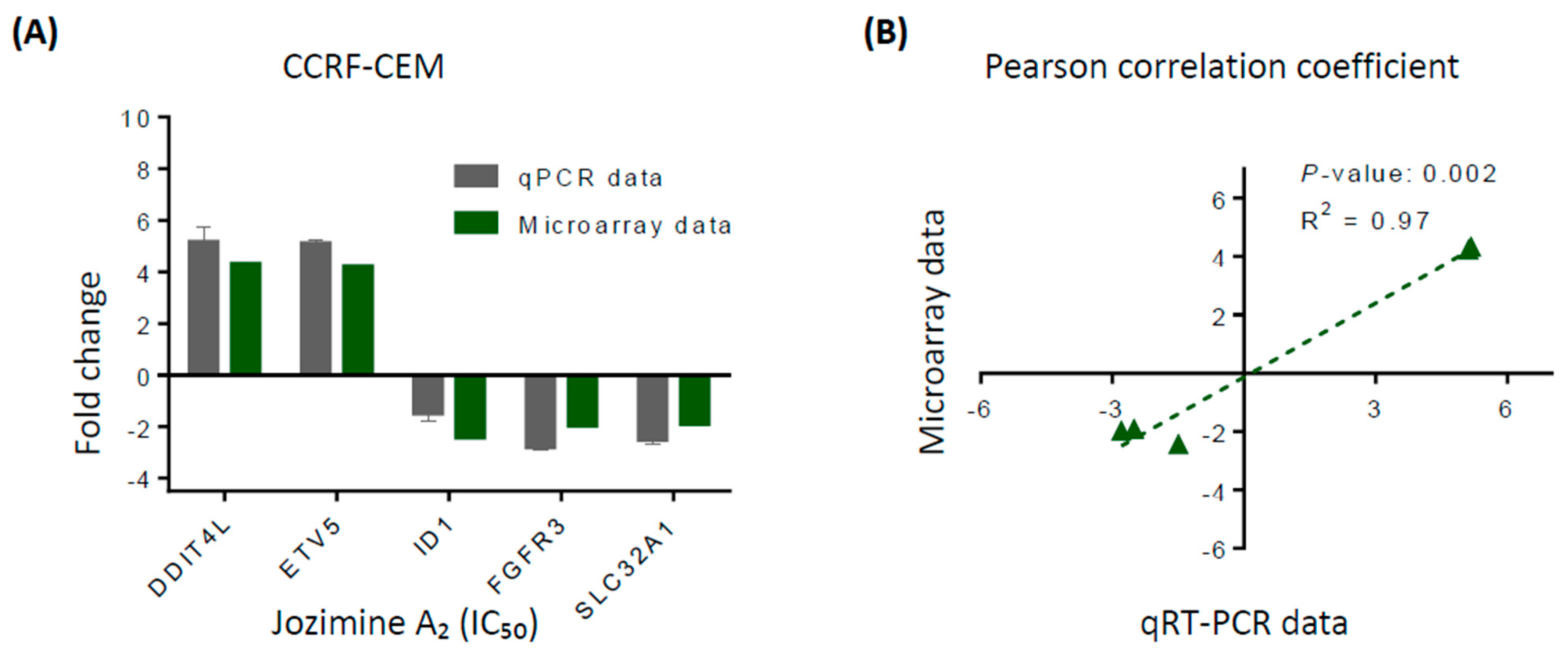
2.4. Validation of Microarray Analysis Using qRT-PCR

The top affected genes were used to confirm the microarray analyses by the qRT-PCR method. Treatment of CCRF-CEM cells with jozimine A2 with an IC50 concentration increased the expression of DDIT4L and ETV5 genes and decreased the expressions of ID1, FGFR3, and SLC32A1, as observed in the microarray analyses (Figure 4A). The correlation between the qPCR and microarray was calculated using the Pearson correlation test (r2 = 0.985) (Figure 4B).

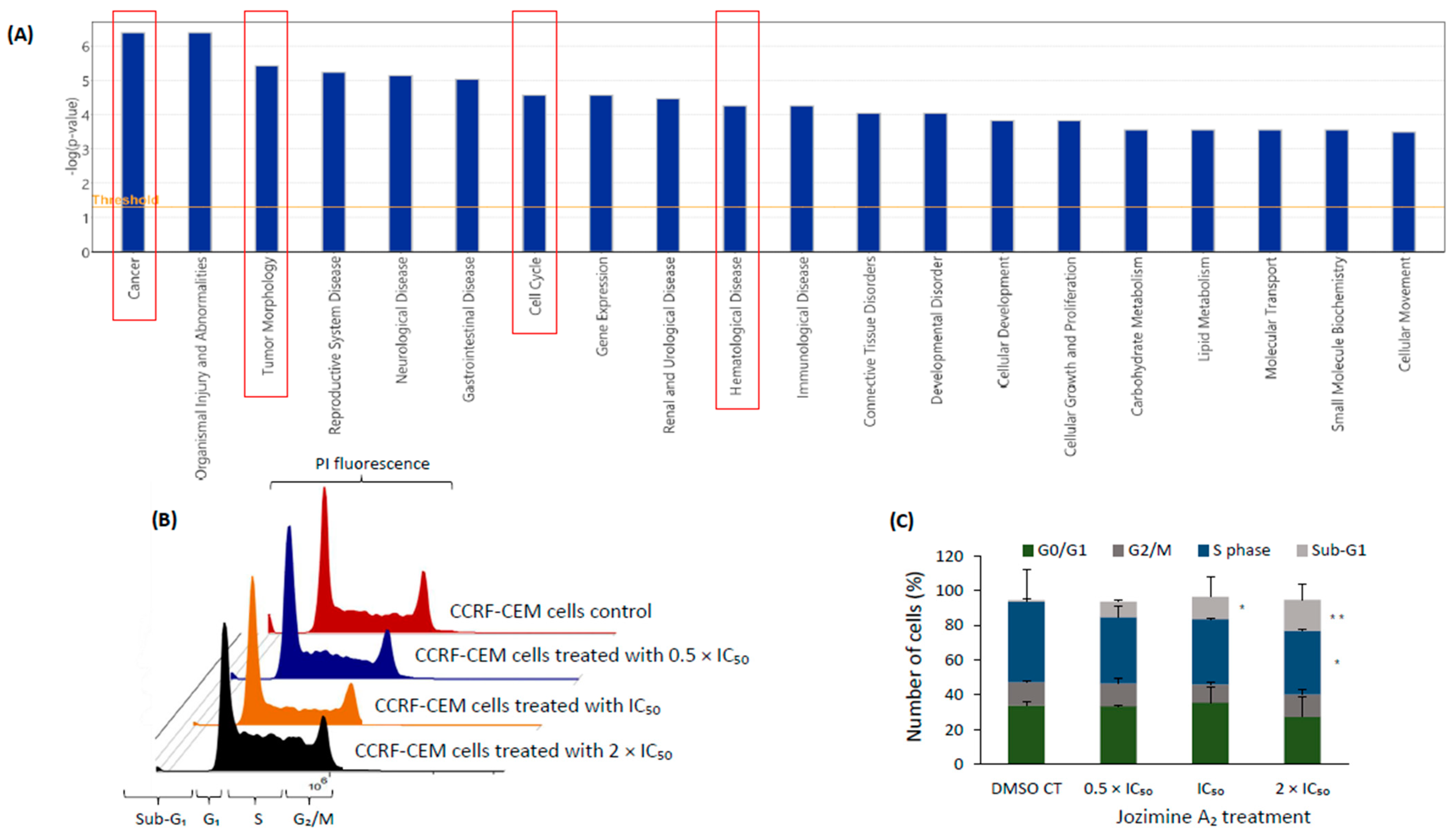
2.5. The Effect of Jozimine A2 on the Cell Cycle

The IPA analysis predicted that one of the major canonical pathways affected after treatment with jozimine A2 was the cell cycle, in addition to other diseases and functions such as tumor morphology, hematological diseases, and cancer (Figure 5A). Jozimine A2 induced the accumulation of cells in the sub-G1 phase in a dose-dependent manner. Already at a concentration of 0.5 × IC50, the sub-G1 phase increased to 8.94%, going to 12.93% at IC50 and reaching 17.85% at 2 × IC50. The cell population in the G0/G1 phase was 33.4% for 0.5 × IC50 and 35.55% for IC50 and decreased to 27.25% at 2 × IC50 concentration (Figure 5B,C).

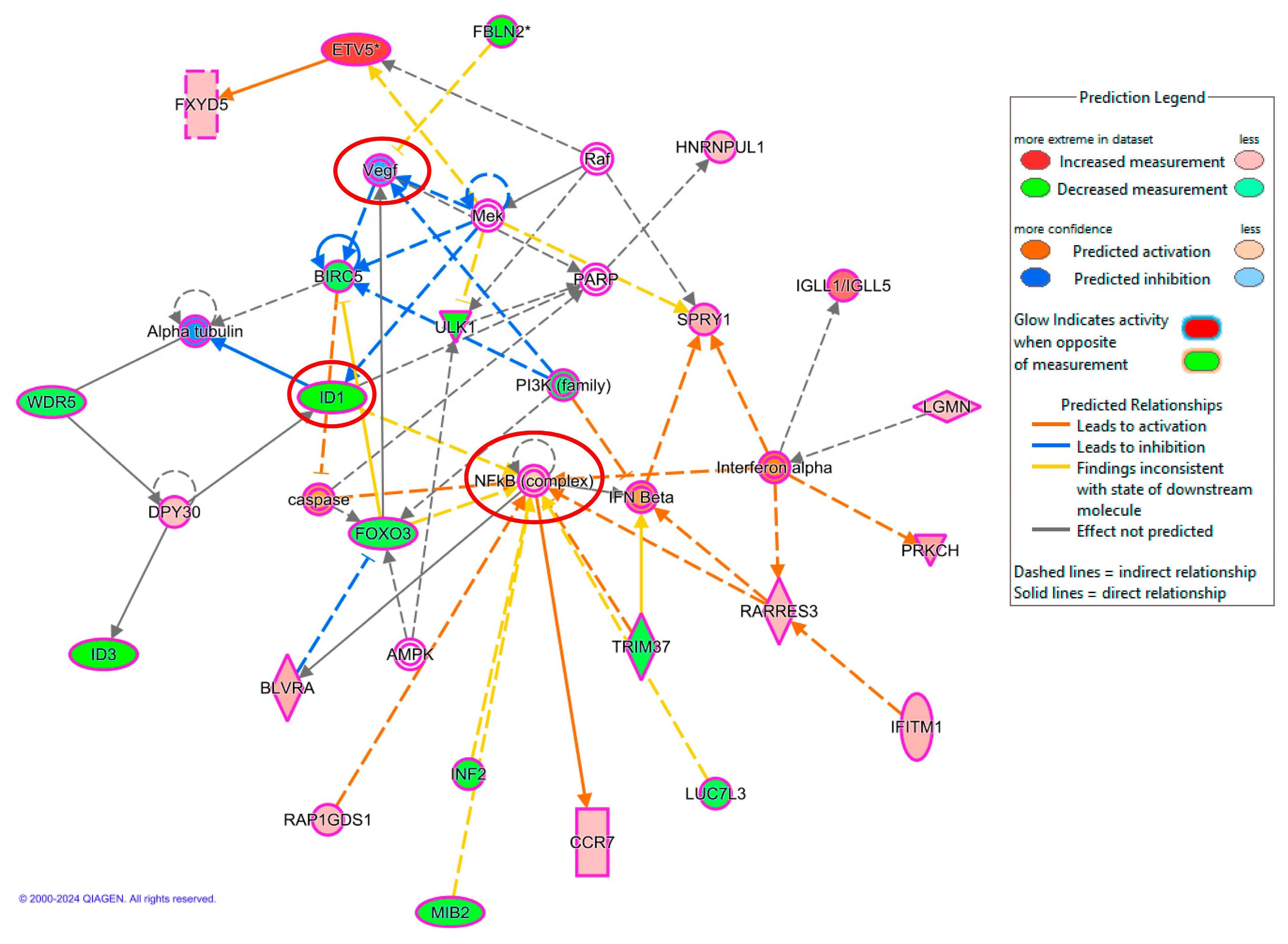
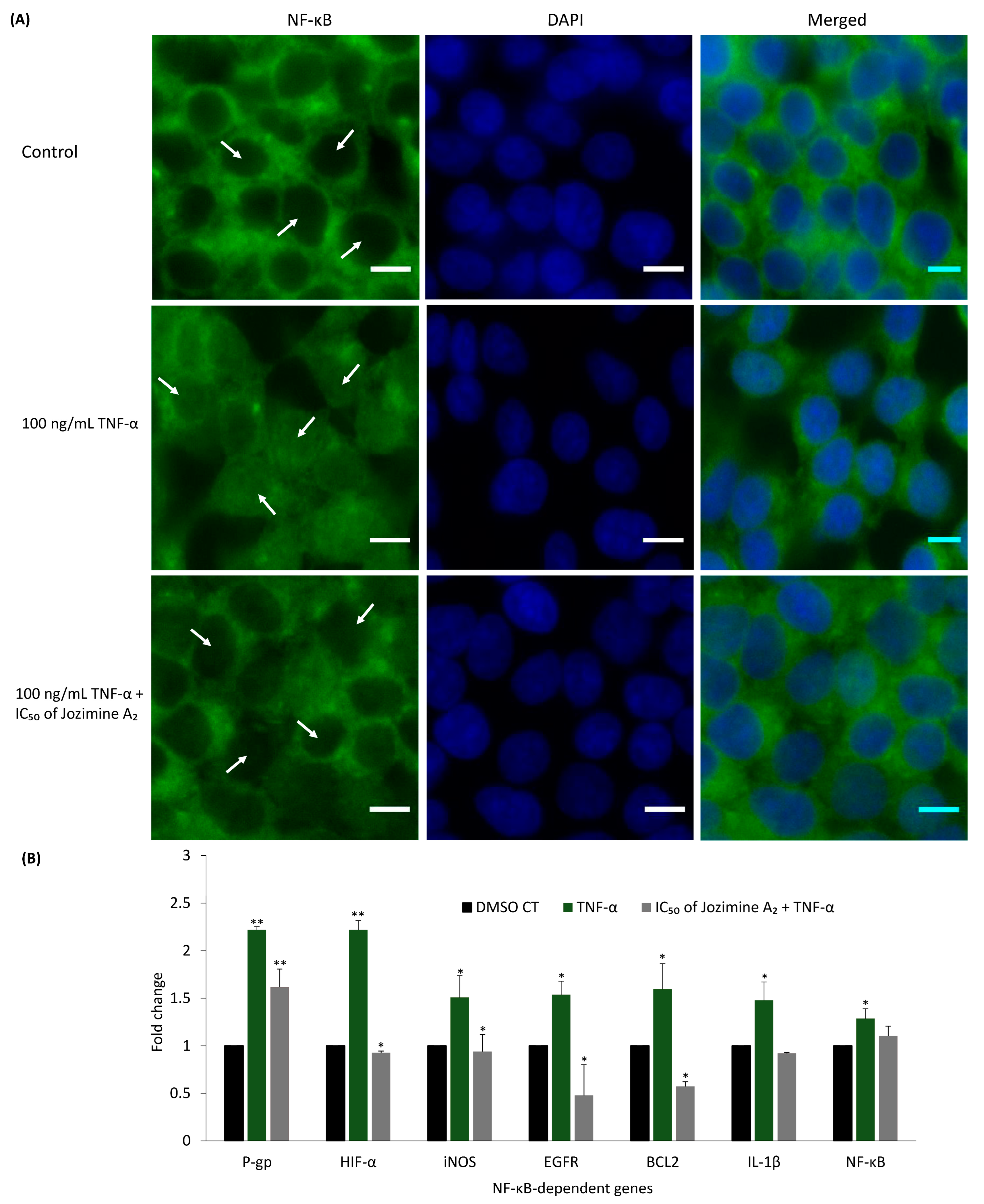
2.6. Jozimine A2 Prevents NF-κB Translocation and Suppresses the Expression of NF-κB-Dependent Genes

The IPA analysis showed that NF-κB is one of the networks most strongly affected by jozimine A2, with downregulated expression of genes related to tumor progression, such as ID1 and VEGF (Figure 6). Immunofluorescence was used to verify whether jozimine A2 could prevent NF-κB translocation to the nucleus following TNF-α activation. In DMSO-treated control cells, NF-κB was translocated to the nucleus following treatment with TNF-α (Figure 7A). However, incubation with jozimine A2 prevented the translocation despite the addition of TNF-α (Figure 7A). In addition to the immunofluorescence experiments, the qPCR results indicated that treatment with TNF-α increased the expression of NF-κB-dependent genes, such as HIF-α, iNOS, and IL-1β, compared to DMSO as the control, while treatment with TNF-α and jozimine A2 significantly decreased the expression of these genes (Figure 7B). Thus, jozimine A2 modulates the NF-κB pathway.

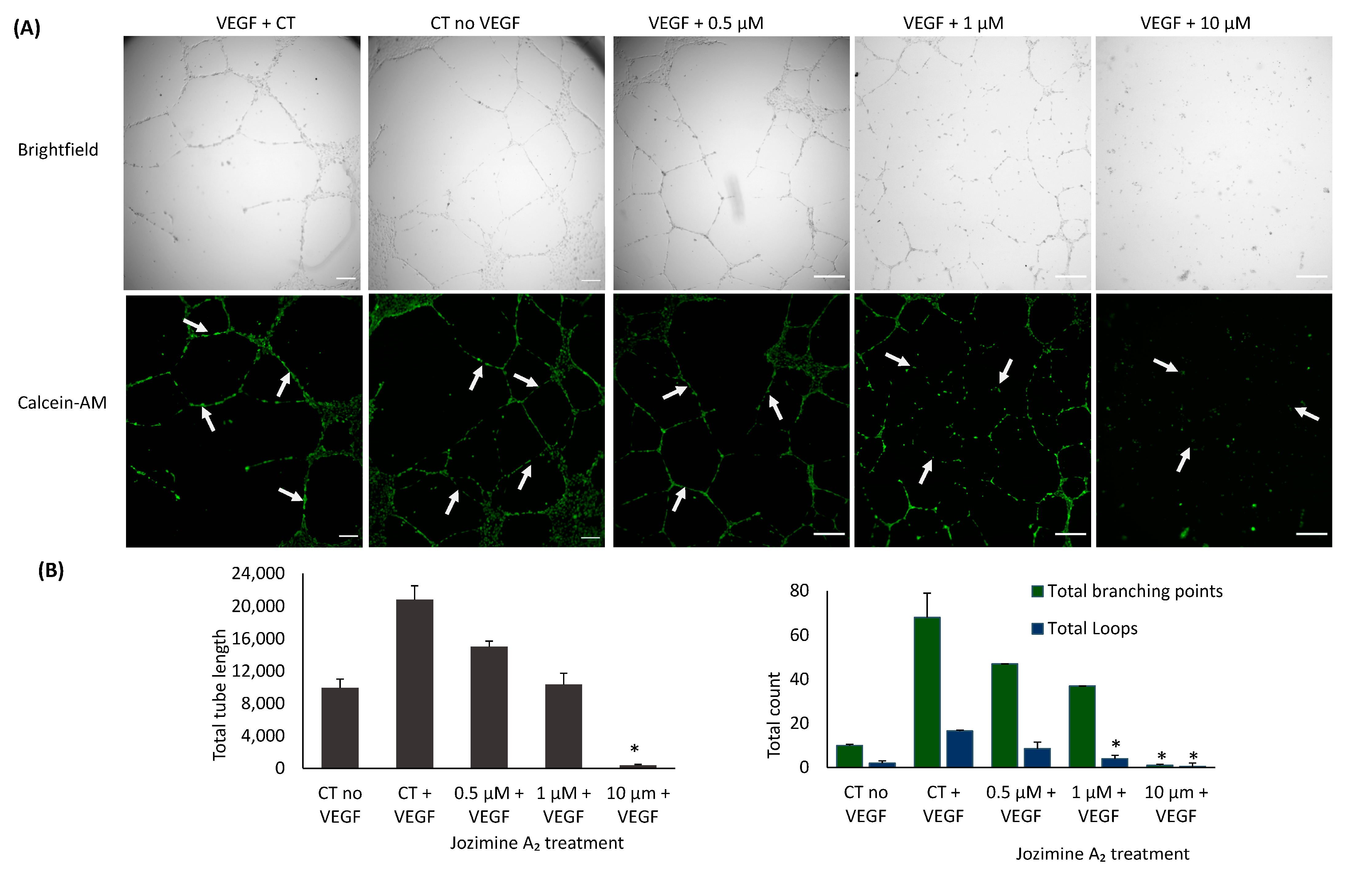
2.7. Suppression of Endothelial Tube Formation by Jozimine A2

Jozimine A2 was also investigated for its potential to prevent endothelial tube formation. HUVECs were incubated with increasing concentrations (0.5 µM, 1 µM, and 10 µM) of jozimine A2. As Figure 8 indicates, jozimine A2 dose-dependently suppressed endothelial tube formation by inhibiting both total branching points and tube length after VEGF induction. Angiogenesis suppression had also been predicted by IPA (Figure 6), as the same genes regulating NF-κB signaling are also related to angiogenesis, namely ID1 and VEGF, which were downregulated (Figure 6).

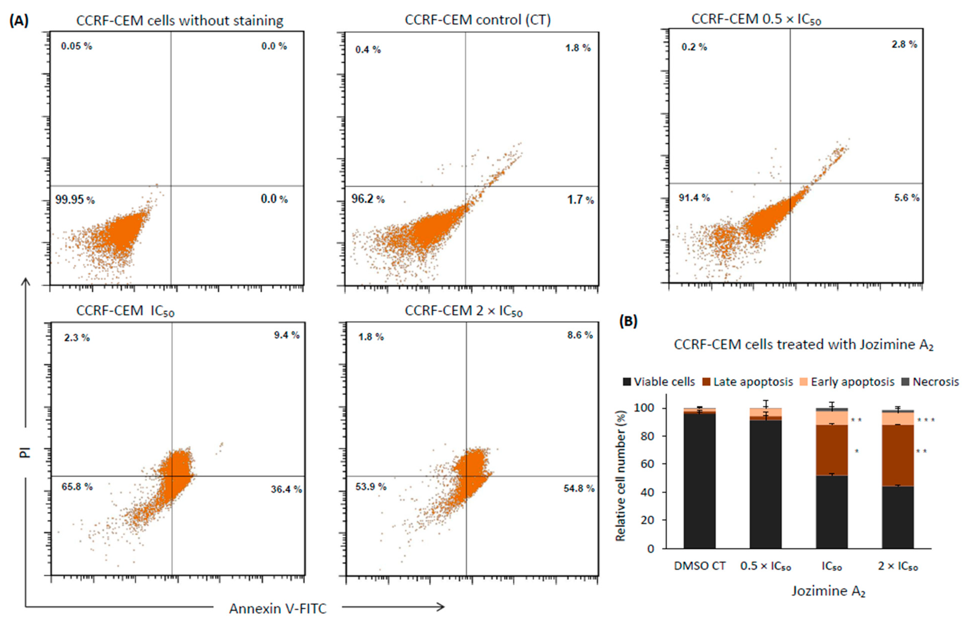
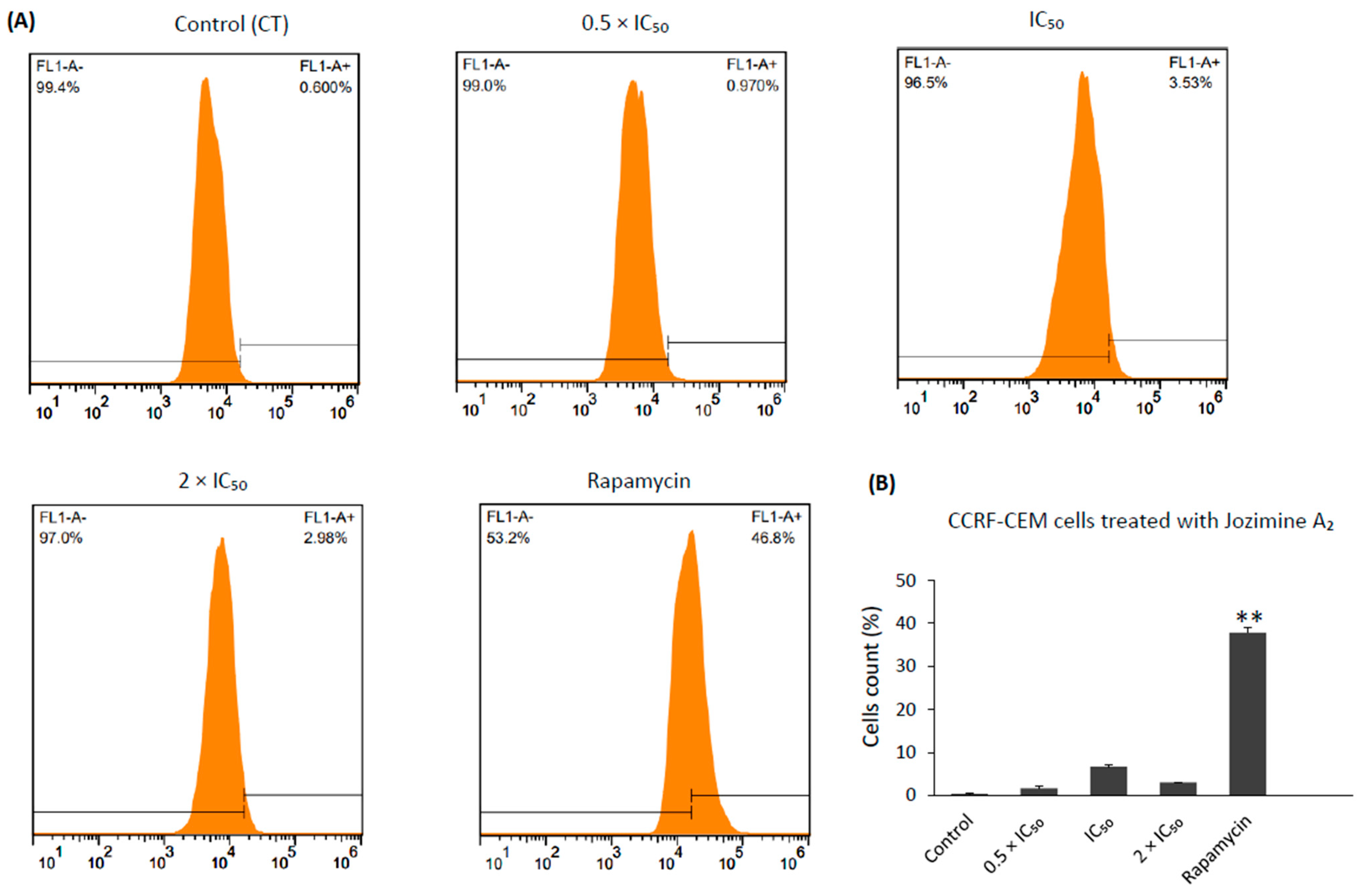
2.8. Jozimine A2 Induces Cell Death through Apoptosis by Autophagy

As the results from the cell cycle indicated an accumulation of apoptotic cells (sub-G1 phase), it seemed rewarding to understand how jozimine A2 induces cell death. As such, two different cell death mechanisms were investigated: apoptosis and autophagy. Jozimine A2 treatment dose-dependently triggered apoptotic cell death in CCRF-CEM cells after treatment for 48 h. Exposure to jozimine A2 induces early apoptosis already at 0.5 × IC50 (Figure 9A,B). Increasing levels of late apoptotic cells were measured at higher concentrations, IC50 (36.4%) and 2 × IC50 (54.8%), compared to the DMSO-treated control (1.7%) (Figure 9A,B). At higher concentrations, necrotic cell fractions appeared, namely 2.34% for IC50 and 1.8% at 2 × IC50, compared to the DMSO-treated control (0.4%) (Figure 9A,B).
On the other hand, autophagy was induced to a lesser extent. Jozimine A2 exerted autophagy at IC50 and 2 × IC50 to a percentage of 6.8 and 3.2 of the autophagic cells, respectively, compared to the untreated control from two independent experiments. However, treatment with rapamycin, which was used as a positive control, induced 37.8% of the autophagic cells (Figure 10). Thus, jozimine A2 preferentially induces apoptosis as the main cell death mechanism.

3. Discussion

Jozimine A2 and michellamine B are bioactive dimeric naphthylisoquinoline (NIQ) alkaloids produced by tropical Ancistrocladus lianas, with marked anti-plasmodial and anti-HIV activities, respectively [4,7]. The NIQ alkaloids are an emerging class of active secondary metabolites with increased pharmaceutical potential, as members of this class, depending on their individual structure, are active against various pathogens causing tropical diseases and against different cancer cells [4,5,38]. Additionally, jozimine A2 also showed increased anticancer activity towards fibrosarcoma (HT1080), multiple myeloma (MM.1S), human colon carcinoma (HT-29), and human pancreatic cancer (PANC-1) cells, making it a promising therapeutical candidate in the battle against cancer [38].
Recently, our group described how dioncophylline A, a related but monomeric NIQ alkaloid, can induce autophagy, leading to cell death in acute leukemia cells, and act as an NF-κB inhibitor [3]. Therefore, it was interesting to investigate whether the two NIQ alkaloid dimers, jozimine A2 and michellamine B, have similar activities as the monomeric dioncophylline A.
In the molecular docking experiments, both dimers showed high binding energies, particularly michellamine B (−11.1 kcal/mol), which was significantly stronger than for jozimine A2, with its still very good energy (−8.2 kcal/mol) towards NF-κB. This stronger binding for michellamine B is certainly due to the higher number of free OH groups, which may give rise to additional interactions and a higher polarity. Also advantageous for the high affinity of michellamine B might be the better flexibility of the molecule, which can thus more easily adapt its conformation to the geometry inside the pocket.
Despite all the similarities, there are some obvious differences between the two alkaloids. Firstly, concerning the functional groups: Jozimine A2 has two fewer free OH groups than michellamine B; thus it is more lipophilic. Secondly, regarding their molecular shape: because of the position of the outer axes of jozimine A2, the molecule is slimmer than the more compact michellamine B, which can influence its docking behavior. Lastly, from a stereochemical point of view, in jozimine A2, the two outer axes are both P-configured, while in michellamine B, one outer axis is M and one is P. The central axis in jozimine A2 is rotationally hindered, too, making it more rigid, while in michellamine B, the central axis is flexible, which makes it easier for the molecule to slip into a pocket in a docking process.
The promising results on NF-κB obtained from molecular docking and MST encouraged us to verify in vitro if the two dimers can prevent NF-κB translocation to the nucleus and check the cytotoxic potential of these new compounds on leukemia cells. Surprisingly, the in vitro behavior of the two alkaloids was found to be different, as the NF-κB reporter assay shows: Jozimine A2 significantly blocks NF-κB activity compared to michellamine B, which only displays a weak effect on NF-κB. Furthermore, the cytotoxicity assay revealed that jozimine A2 prevents cell proliferation in drug-sensitive cells (CCRF-CEM) with an excellent IC50 = 0.45 ± 0.00 μM, comparable to the standard doxorubicin, which has an IC50 = 0.14 μM, while the IC50 of michellamine B was not reached (>100 µM) [3]. Moreover, jozimine A2 also inhibited cell proliferation of multidrug-resistant CEM/ADR5000 leukemia cells at IC50 = 0.69 ± 0.05 μM. The higher flexibility, the larger polarity (and better water solubility), and the more compact structure do not seem to play a significant role in vitro. The higher lipophilicity of jozimine A2 should entail better membrane permeability and, thus, a higher availability of the agent in the cells. In addition to the NF-κB reporter assay, the immunofluorescence highlighted that jozimine A2 modulated the NF-κB pathway, not only by suppressing its activity but also by blocking its translocation to the nucleus. Given the necessity for new chemotherapeutic agents due to the increasing number of drug-resistant tumors, jozimine A2 has proven to be a valuable chemotherapeutic candidate [39,40]. Therefore, it was selected for further investigation, to decipher its underlying mechanism.
The IPA analysis identified “cancer”, “cell cycle”, “hematological diseases”, and “tumor morphology” among the top canonical pathways affected by the treatment with jozimine A2. According to the microarray analysis, the top deregulated genes after treatment with jozimine A2 were DDIT4L, ETV5, ID1, FGFR3, and SLC32A1. These results were confirmed using qPCR. DDIT4L is a p53-dependent DNA damage repair gene and an mTOR inhibitor, which has been identified as a potential biomarker in several cancers [41]. SLC32A1 is aberrantly methylated in pancreatic cancer and has been identified as an NF-κB-inducible gene [42,43]. FGFR3 expression has been correlated with tumor growth in different cancer types and is a known regulator of the NF-κB pathway [44,45]. Additionally, jozimine A2 does not only prevent translocation of NF-κB to the nucleus, but it also blocks tumor-progression genes such as ID1 and VEGF.
Given the positive results obtained, we decided to further investigate the mechanisms behind the anti-tumor activity of jozimine A2. Angiogenesis is an important factor in tumor growth and metastasis, and numerous drugs have been developed to inhibit angiogenesis [3,46,47]. Therefore, the next step was to investigate how jozimine A2 influences endothelial tube formation in vitro. As the microarray and qPCR analyses showed a downregulation of ID1 and VEGF, it was not surprising that jozimine A2 reduced endothelial tube formation and, consequently, angiogenesis. This is in accordance with our previous study, which described the potential of monomeric NIQ alkaloids to inhibit angiogenesis [3].
Although strong cytotoxic activities against various cancer types have been described for several NIQ alkaloids [3,26,27,28,29,41,42,43], the triggering mechanisms have not yet been deciphered. As this unique pharmacological class of natural products is emerging, there is still much to learn about the mechanisms of action of each individual structure. Given the results from the IPA analysis regarding the “cell cycle,” we decided to investigate the impact of jozimine A2 on the cell cycle. As the results indicated, jozimine A2 does not induce cell cycle arrest. However, in view of the observed accumulation of cell debris (sub-G1 phase), it seemed rewarding to investigate both apoptosis and autophagy as possible modes to induce cell death. Several studies have described apoptosis or autophagy as the main induced cell death mode in different cancer cells [3,5,27,28,29,48,49,50,51,52]. Our investigations show that jozimine A2 preferentially dose-dependently induces apoptosis in CCRF-CEM cells, as the number of late-apoptotic cells was significantly increased compared to the control. In comparison, autophagy was only weakly induced by 6% after treatment with jozimine A2, which was much lower than the positive control, rapamycin, which induced autophagy by 37%.

4. Materials and Methods

4.1. Molecular Docking

Jozimine A2 and michellamine B were obtained by isolation from Central African Ancistrocladus lianas [5,6]. The structures of jozimine A2 and michellamine B were formed manually on ChemDraw 19.0, https://perkinelmerinformatics.com/products/research/chemdraw, Massachusetts, USA (accessed on 1 February 2021), by using the template of the sdf structure of dioncophylline A 3D, which was downloaded from the PubChem database, https://pubchem.ncbi.nlm.nih.gov/compound/Dioncophylline-A Maryland, USA (accessed on 1 August 2022 ). After energy minimization, the compounds were saved as pdb files on Chem3D 19.0, https://perkinelmerinformatics.com/products/research/chemdraw, Massachusetts, USA (accessed on 1 February 2021), and converted to pdbqt files by PyRx. Triptolide as the positive control was also downloaded from PubChem and went through the same procedure. For jozimine A2, all three axes were rotationally hindered, while for michellamine B, the central axis was left freely rotating, and two outer axes were hindered on AutoDockTools 1.5.6., https://ccsb.scripps.edu/mgltools/, California, USA (accessed on 24 January 2021). The stereostructures of the two compounds were confirmed by one of the authors (G.B.).
The NF-κB p65/p50 heterodimer bound to the IκBα protein complex was obtained from the Protein Data Bank (PDB ID 1NFI) as a PDB file. The inhibitor IκBα chain, p50 chain, and heteroatoms were removed to isolate the p65 subunit. Polar hydrogens and Kollman charges were added to the p65 protein structure, which was then converted to a PDBQT file using AutoDockTools. A grid box centered at x = 3.348, y = 44.715, z = 17.684 with dimensions of 80 × 70 × 126 points and 0.4 Å spacing was generated around the protein. The gpf, glf, and dpf affinity map files were also prepared. Molecular docking using a Lamarckian algorithm was performed utilizing the supercomputing facilities at Johannes Gutenberg University Mainz, hpc.uni-mainz.de, Mainz, Germany (accessed on 30 August 2022). Docking runs totaling 250 iterations with a maximum of 2,500,000 energy evaluations were completed for each compound assessed.

4.2. Microscale Thermophoresis

Microscale thermophoresis (MST) was used to confirm the docking results. The human recombinant NF-κB protein was purchased from Sino Biological Europe GmbH (NF-κB cat. No. 12054-H09E, Sino Biological Europe GmbH, Eschborn, Germany), and the labeling was performed as previously described [53]. Serial dilutions from 300,000 to 100 nM of jozimine A2 and michellamine B were incubated for 30 min at room temperature (RT) with the labeled human recombinant NF-κB protein in a 1:1 ratio. Standard capillaries from NanoTemper Monolith™ NT (NanoTemper Technologies GmbH, Munich, Germany) were used to conduct the experiments. MST was run at 40% LED power and 10% MST power for jozimine A2 and 40% LED power and 40% MST power for michellamine B.

4.3. NF-κB Reporter Assay

1 × 10⁵ NF-κB SEAP reporter HEK-BlueTM Null 1 cells, acquired from InvivoGen (San Diego, CA, USA), were seeded on a 96-well plate and incubated for 24 h. Afterwards, the cells were treated with increasing concentrations (0.5 µM, 1 µM, and 10 µM) of jozimine A2 and michellamine B, respectively, for another 24 h. Tumor necrosis factor (TNF, 100 ng/mL, Thermofisher; Darmstadt, Germany) was added to activate the NF-κB pathway. Triptolide (1 µM, InvivoGen, San Diego, CA, USA) served as a positive control [52]. After adding pre-heated Quanti-Blue (InvivoGen, San Diego, CA, USA), the fluorescence signal was measured on an Infinite M2000 Tecan (Tecan, Crailsheim, Germany) at 630 nm. The experiment was performed in triplicate.

4.4. Cytotoxicity Assay

The cytotoxic effects of jozimine A2 and michellamine B were assessed using a resazurin reduction assay (Promega, Mannheim, Germany) and DMSO (dimethylsulfoxide) as a negative control. In this assay, living cells reduce non-fluorescent resazurin dye to fluorescent resorufin [54].
CCRF-CEM cells and multidrug-resistant CEM/ADR5000 cells were kindly provided by Prof. Axel Sauerbrey (Department of Pediatrics, University of Jena, Germany). The multidrug resistance phenotype of CEM/ADR5000 cells with resistance to anthracyclines, Vinca alkaloids, taxanes, and epipodophyllotoxines has been reported [55]. HEK-BlueTM Null 1 cells were purchased commercially (InvivoGen, San Diego, CA, USA).
All cells seeded in 96-well culture plates were treated with varying concentrations (0.003 µM to 100 µM) of the two compounds for 72 h (37 °C). HEK-BlueTM Null 1 cells were pre-incubated for 24 h to allow attachment prior to treatment. After treatment, 20 μL of 0.01% resazurin solution (Sigma-Aldrich, Schnelldorf, Germany) were added to each well and incubated for another 4 h (37 °C). Fluorescence was measured at excitation 544 nm/emission 590 nm on an Infinite M2000 Pro plate reader (Tecan, Crailsheim, Germany). Cell viability percentages were calculated by comparing the fluorescence of treated vs. untreated cells with a medium background subtracted. IC50 values were determined using nonlinear regression analysis relative to DMSO as the control. The assay was performed in triplicate with six replicate wells per concentration. IC50 values are expressed as the mean ± standard deviation.

4.5. Microarray Hybridization

Microarray hybridization expression analysis was carried out to identify the underlying mechanisms of jozimine A2 anticancer activity. Therefore, CCRF-CRM cells were incubated for 24 h with jozimine A2 at the IC50 concentration. The InviTrap® Spin Universal RNA Mini Kit (Invitek Molecular, Berlin, Germany) was used for the extraction of the total RNA using the protocol provided by the manufacturer. Microarray hybridization for both treated and untreated samples was conducted on Illumina Human HT-12 BeadChip arrays (Illumina, San Diego, CA, USA) at the Genomics and Proteomics Core Facility at the German Cancer Research Center (DKFZ, Heidelberg, Germany). Data were analyzed through the Chipster software (https://chipster.csc.fi), and deregulated genes were then further investigated using the Ingenuity Pathway Analysis (IPA) software (QIAGEN® Ingenuity Pathway Analysis Fall Release (2023)) as previously described [3,56].

4.6. Quantitative Real-Time PCR

After RNA was isolated by the above-described method, 1 µg of RNA was converted to complementary DNA (cDNA) using the Luna Script™ RT SuperMix Kit (E3010, New England Bio Labs, Darmstadt, Germany). To amplify the genes as per the manufacturer’s instructions, 5 × Hot Start Taq EvaGreen® qPCR Mix (no ROX) (Axon Labortechnik, Kaiserslautern, Germany) was used next. The NCBI/Primer-BLAST tool was used to design the primers, which were then purchased from Eurofins Genomics (Ebersberg, Germany). A list with the primers name and sequences can be found in Table 3. The sequence of the primers is the same that we used in our previously published work [3,57]. To conduct the qPCR in 40 cycles on CFX384™ (Bio-Rad, Munich, Germany), 384 well-plates were used. The running conditions were as follows: denaturation at 95 °C for 15 s, gradient annealing temperatures at 62–47 °C for 30 s, and a 1 min elongation step at 72 °C. The Cq values were calculated by Bio-Rad CFX Manager Software. The gene expression was then normalized using GAPDH as the control, and the fold change was calculated by applying the 2−ΔΔCt method [58].

4.7. Cell Cycle

CCRF-CEM cells (1 × 10⁶ cells/well) were seeded in a 6-well plate and treated with different concentrations (0.5 × IC50, IC50, 2 × IC50) of jozimine A2 or DMSO for 24 h. The cells were centrifuged at 350× g for 5 min at 10 °C and washed with ice-cold PBS. To fix the cell pellet, ice-cold ethanol 75% was added and the cells were kept at −20 °C for 3 h. The cells were then washed again with PBS and 5 µg RNAse A (Merck KgaA, Darmstadt, Germany) was added to the samples. Propidium iodide in PBS (PI, 50 μg/mL) (Merck KgaA, Darmstadt, Germany) was used for cell staining. The measurements were carried out in a BD AccuriTM C6 Flow Cytometer (BD Biosciences, Becton Drive, Franklin Lakes, NJ, USA) [3,59].

4.8. Immunofluorescence

After seeding in chamber slides (Nune), HEK-BlueTM Null 1 cells were incubated with jozimine A2 at 0.5 × IC50 concentration and triptolide, respectively, for 24 h. NF-κB activation was induced by adding 100 ng/mol TNF after treatment for 24 h. The cells were washed three times with PBS and fixed with 4% paraformaldehyde for 30 min at room temperature. The monolayer was then blocked for 1 h in 3% bovine serum albumin and Triton-X 100. After washing, the diluted anti-NF-κB antibody (1:200, D2E5, Cell Signaling, Frankfurt am Main, Germany) was added and incubated overnight at 4 °C. The slides were washed three times and incubated with the secondary antibody Alexa Fluor 488-conjugated anti-rabbit (1:700, Invitrogen, A11032, Darmstadt, Germany) for 1 h. Lastly, 2 μg/mL 4′,6-diamidino-2-phenylindole (DAPI) (Sigma-Aldrich) was added for staining, and the cells were mounted in Fluoromount-G® (SouthernBiotech, Birmingham, AL, USA) [3]. The images were acquired using an AF7000 Widefield Fluorescence Microscope (40 × magnification).

4.9. Endothelial Tube Formation

The tube formation assay was used to examine the potential of jozimine A2 for angiogenesis. Μ-Slide angiogenesis (ibidi GmbH, Martinsried, Germany) was coated with 10 μL Matrigel® (Corning GmbH, Kaiserslautern, Germany) and polymerized for 30 min. Next, human umbilical vein endothelial cells (HUVECs) (1 × 10⁴ cells/well), received from Dr. Ronald E. Unger (Repair-Lab, Institute of Pathology, University Medical Center of Johannes Gutenberg University Mainz, Mainz, Germany), were seeded on Matrigel® with 30 ng/mL VEGF (Promocell, Heidelberg, Germany). For the tube formation, jozimine A2 (0.5 µM, 1 µM, and 10 µM) or DMSO (control) was added to the cells and incubated for 16–18 h at 37 °C and 5% CO2. As a next step, the cells were stained with 6.25 μg/mL Calcein-AM (Cayman Chemical, Ann Arbor, MI, USA) for 30 min at RT and then rinsed three times with Hank’s balanced salt solution (HBSS) (Corning B. V. Life Sciences, Amsterdam, Niederland). The fluorescent images were obtained with the F7000 widefield fluorescence microscope (Microscopy & Histology Core Facility at IMB Mainz, Mainz, Germany). The tube analysis was carried out using Wimasis’ Image Analysis Platform (https://mywim.wimasis.com/ accessed on 1 March 2023) [60].

4.10. Apoptosis

Apoptosis was detected using the Annexin V-FITC/PI double-staining kit (Invitrogen, Life Technologies GmbH, Darmstadt, Germany) by flow cytometry. In a 6-well plate, CCRF-CEM cells (1 × 10⁶ cells/well) were seeded for 24 h. The cells were treated with increasing concentrations (0.5 × IC50, IC50, 2 × IC50) of jozimine A2 or DMSO (control) for 24 h and 48 h. As a next step, the cells were harvested and washed once with cold PBS and 1 × annexin binding buffer. After centrifugation, the cells were stained by adding 5 µL of Annexin V-FITC and propidium iodide (PI) and incubated at RT in the dark for 15 min. Apoptosis was detected by flow cytometry (BD AccuriTM C6, BD Biosciences, Becton Drive, Franklin Lakes, NJ, USA). The experiments were performed in triplicate.

4.11. Autophagy

The Autophagy Detection Kit (ab139484, Abcam, Cambridge, UK) was used to detect autophagy according to the manufacturer’s instructions. CCRF-CEM cells were exposed to various concentrations of jozimine A2 (0.5 × IC50, IC50, 2 × IC50) and rapamycin (0.5 µM) as a positive control for 24 h. Cells were washed twice using the 1 × assay buffer provided in the Autophagy Detection Kit and were then centrifuged. The cell pellets were resuspended in 250 µL cell culture medium supplemented with 5% FBS and without indicators. 250 µL medium containing green stain solution (from the Detection Kit) was added, and cells were incubated for 30 min at 37 °C. Finally, the cells were washed again and re-immersed in the assay buffer. The autophagy signal was detected on the BD AccuriTM C6 flow cytometer (BD Biosciences, Becton Drive, Franklin Lakes, USA) using a standard green (FL1) filter [3,59].

5. Conclusions

In conclusion, jozimine A2, a dimeric natural NIQ alkaloid, is a potential antitumor drug candidate through its cytotoxicity and NF-κB inhibition. Jozimine A2 bound to NF-κB both in silico and in vitro and inhibited its activity and translocation to the nucleus. Furthermore, jozimine A2 induced apoptosis in leukemia cells, as predicted by the IPA analysis and cell cycle investigation, and blocked angiogenesis by preventing endothelial tube formation.

Author Contributions

Conceptualization, M.D., G.B. and T.E.; Methodology, R.D., R.Y., S.M.K., G.B., T.E. and M.D.; Software, R.D., R.Y. and M.D.; Validation, R.D., R.Y. and M.D.; Formal Analysis, R.D., R.Y., S.M.K., G.B., T.E. and M.D.; Investigation, R.D., R.Y., S.M.K., G.B. and M.D.; Resources, G.B., S.M.K. and T.E.; Data Curation, G.B., T.E. and M.D.; Writing—Original Draft Preparation, R.D., R.Y., G.B. and M.D.; Writing—Review and Editing, R.D., R.Y., S.M.K., G.B., T.E. and M.D.; Visualization, M.D. and T.E.; Supervision, G.B., T.E. and M.D.; Project Administration, T.E. and M.D.; Funding Acquisition, T.E. All authors have read and agreed to the published version of the manuscript.

Funding

R.D. is grateful for the PhD stipend from the Deutsche Forschungsgemeinschaft (DFG, GRK2015/2).

Data Availability Statement

Data are contained within the article.

Acknowledgments

R.D. is grateful for a PhD stipend from the Deutsche Forschungsgemeinschaft (DFG, GRK2015/2). We thank the Microarray Unit of the Genomics and Proteomics Core Facility, German Cancer Research Center (DKFZ) Heidelberg, for providing excellent Expression Profiling services. We thank Shaimaa Fayez (University of Würzburg) for the isolation of dimeric NIQ alkaloids.

Conflicts of Interest

The authors declare no conflicts of interest.

References

  1. Feineis, D.; Bringmann, G. Asian Ancistrocladus Lianas as Creative Producers of Naphthylisoquinoline Alkaloids. In Progress in the Chemistry of Organic Natural Products; Springer: Berlin/Heidelberg, Germany, 2023; Volume 119. [Google Scholar]
  2. Lombe, B.K.; Feineis, D.; Bringmann, G. Dimeric Naphthylisoquinoline Alkaloids: Polyketide-Derived Axially Chiral Bioactive Quateraryls. Nat. Prod. Rep. 2019, 36, 1513–1545. [Google Scholar] [CrossRef]
  3. Yücer, R.; Fayez, S.; Feineis, D.; Klauck, S.M.; Shan, L.; Bringmann, G.; Efferth, T.; Dawood, M. Cytotoxicity of Dioncophylline A and Related Naphthylisoquinolines in Leukemia Cells, Mediated by NF-κB Inhibition, Angiogenesis Suppression, G2/M Cell Cycle Arrest, and Autophagy Induction. Phytomedicine 2023, 2023, 155267. [Google Scholar] [CrossRef]
  4. Bringmann, G.; Zhang, G.; Büttner, T.; Bauckmann, G.; Kupfer, T.; Braunschweig, H.; Brun, R.; Mudogo, V. Jozimine A2: The First Dimeric Dioncophyllaceae-Type Naphthylisoquinoline Alkaloid, with Three Chiral Axes and High Antiplasmodial Activity. Chem. A Eur. J. 2013, 19, 916–923. [Google Scholar] [CrossRef]
  5. Fayez, S.; Feineis, D.; Mudogo, V.; Seo, E.J.; Efferth, T.; Bringmann, G. Ancistrolikokine I and Further 5,8′-Coupled Naphthylisoquinoline Alkaloids from the Congolese Liana Ancistrocladus likoko and Their Cytotoxic Activities against Drug-Sensitive and Multidrug Resistant Human Leukemia Cells. Fitoterapia 2018, 129, 114–125. [Google Scholar] [CrossRef]
  6. Li, J.; Seupel, R.; Bruhn, T.; Feineis, D.; Kaiser, M.; Brun, R.; Mudogo, V.; Awale, S.; Bringmann, G. Jozilebomines A and B, Naphthylisoquinoline Dimers from the Congolese Liana Ancistrocladus ileboensis, with Antiausterity Activities against the PANC-1 Human Pancreatic Cancer Cell Line. J. Nat. Prod. 2017, 80, 2807–2817. [Google Scholar] [CrossRef]
  7. Boyd, M.R.; Hallock, Y.F.; Cardellina, J.H.; Manfredi, K.P.; Blunt, J.W.; McMahon, J.B.; Buckheit, R.W.; Bringmann, G.; Schäffer, M.; Cragg, G.M.; et al. Anti-HIV Michellamines from Ancistrocladus korupensis. J. Med. Chem. 1994, 37, 1740–1745. [Google Scholar] [CrossRef]
  8. Orlowski, R.Z.; Baldwin, A.S. NF-κB as a Therapeutic Target in Cancer. Trends. Mol. Med. 2002, 8, 385–389. [Google Scholar] [CrossRef] [PubMed]
  9. Staudt, L.M. Oncogenic Activation of NF-κB. Cold Spring Harb. Perspect. Biol. 2010, 2, a000109. [Google Scholar] [CrossRef] [PubMed]
  10. Karin, M. NF-κB as a Critical Link between Inflammation and Cancer. Cold Spring Harb. Perspect. Biol. 2009, 1, a000141. [Google Scholar] [CrossRef]
  11. Lin, A.; Karin, M. NF-κB in Cancer: A Marked Target. Semin. Cancer Biol. 2003, 13, 107–114. [Google Scholar] [CrossRef] [PubMed]
  12. Li, Y.; Zhao, B.; Peng, J.; Tang, H.; Wang, S.; Peng, S.; Ye, F.; Wang, J.; Ouyang, K.; Li, J.; et al. Inhibition of NF-κB Signaling Unveils Novel Strategies to Overcome Drug Resistance in Cancers. Drug Resist. Updates 2024, 73, 101042. [Google Scholar] [CrossRef]
  13. deGraffenried, L.A.; Chandrasekar, B.; Friedrichs, W.E.; Donzis, E.; Silva, J.; Hidalgo, M.; Freeman, J.W.; Weiss, G.R. NF-κB Inhibition Markedly Enhances Sensitivity of Resistant Breast Cancer Tumor Cells to Tamoxifen. Ann. Oncol. 2004, 15, 885–890. [Google Scholar] [CrossRef]
  14. Abdin, S.M.; Tolba, M.F.; Zaher, D.M.; Omar, H.A. Nuclear Factor-κB Signaling Inhibitors Revert Multidrug-Resistance in Breast Cancer Cells. Chem. Biol. Interact. 2021, 340, 109450. [Google Scholar] [CrossRef] [PubMed]
  15. Bentires-Alj, M.; Barbu, V.; Fillet, M.; Chariot, A.; Relic, B.; Jacobs, N.; Gielen, J.; Merville, M.P.; Bours, V. NF-κB Transcription Factor Induces Drug Resistance through MDR1 Expression in Cancer Cells. Oncogene 2003, 22, 90–97. [Google Scholar] [CrossRef] [PubMed]
  16. Radzka, J.; Łapińska, Z.; Szwedowicz, U.; Gajewska-Naryniecka, A.; Gizak, A.; Kulbacka, J. Alternations of NF-κB Signaling by Natural Compounds in Muscle-Derived Cancers. Int. J. Mol. Sci. 2023, 24, 11900. [Google Scholar] [CrossRef] [PubMed]
  17. Hoesel, B.; Schmid, J.A. The Complexity of NF-κB Signaling in Inflammation and Cancer. Mol. Cancer 2013, 12, 86. [Google Scholar] [CrossRef] [PubMed]
  18. Fan, Y.; Mao, R.; Yang, J. NF-κB and STAT3 Signaling Pathways Collaboratively Link Inflammation to Cancer. Protein Cell 2013, 4, 176–185. [Google Scholar] [CrossRef]
  19. Zhang, T.; Ma, C.; Zhang, Z.; Zhang, H.; Hu, H. NF-κB Signaling in Inflammation and Cancer. MedComm 2021, 2, 618–653. [Google Scholar] [CrossRef]
  20. Yu, L.; Li, L.; Medeiros, L.J.; Young, K.H. NF-κB Signaling Pathway and Its Potential as a Target for Therapy in Lymphoid Neoplasms. Blood Rev. 2017, 31, 77–92. [Google Scholar] [CrossRef]
  21. Bernal-Mizrachi, L.; Lovly, C.M.; Ratner, L. The Role of NF-κB-1 and NF-κB-2-Mediated Resistance to Apoptosis in Lymphomas. Proc. Natl. Acad. Sci. USA 2006, 103, 9220–9225. [Google Scholar] [CrossRef]
  22. Di Francesco, B.; Verzella, D.; Capece, D.; Vecchiotti, D.; Di Vito Nolfi, M.; Flati, I.; Cornice, J.; Di Padova, M.; Angelucci, A.; Alesse, E.; et al. NF-κB: A Druggable Target in Acute Myeloid Leukemia. Cancers 2022, 14, 3557. [Google Scholar] [CrossRef]
  23. Braun, T.; Carvalho, G.; Fabre, C.; Grosjean, J.; Fenaux, P.; Kroemer, G. Targeting NF-κB in Hematologic Malignancies. Cell Death Differ. 2006, 13, 748–758. [Google Scholar] [CrossRef]
  24. Zhou, J.; Ching, Y.Q.; Chng, W.J. Aberrant Nuclear Factor-Kappa B Activity in Acute Myeloid Leukemia: From Molecular Pathogenesis to Therapeutic Target. Oncotarget 2015, 6, 5490. [Google Scholar] [CrossRef]
  25. Trocoli, A.; Djavaheri-Mergny, M. The Complex Interplay between Autophagy and NF-κB Signaling Pathways in Cancer Cells. Am. J. Cancer Res. 2011, 1, 629. [Google Scholar]
  26. Ahmadi-Dehlaghi, F.; Mohammadi, P.; Valipour, E.; Pournaghi, P.; Kiani, S.; Mansouri, K. Autophagy: A Challengeable Paradox in Cancer Treatment. Cancer Med. 2023, 12, 11542–11569. [Google Scholar] [CrossRef]
  27. Luo, M.; Han, L. Autophagy Induction Sensitizes Cancer Cells to Anti-Cancer Drugs. Autophagy 2022, 19, 2393–2394. [Google Scholar] [CrossRef]
  28. Jung, H.J.; Seo, I.; Casciello, F.; Jacquelin, S.; Lane, S.W.; Suh, S.I.; Suh, M.H.; Lee, J.S.; Baek, W.K. The Anticancer Effect of Chaetocin Is Enhanced by Inhibition of Autophagy. Cell Death Dis. 2016, 7, e2098. [Google Scholar] [CrossRef] [PubMed]
  29. Pfeffer, C.M.; Singh, A.T.K. Apoptosis: A Target for Anticancer Therapy. Int. J. Mol. Sci. 2018, 19, 448. [Google Scholar] [CrossRef] [PubMed]
  30. Guan, C.; Zhou, X.; Li, H.; Ma, X.; Zhuang, J. NF-κB Inhibitors Gifted by Nature: The Anticancer Promise of Polyphenol Compounds. Biomed. Pharmacother. 2022, 156, 113951. [Google Scholar] [CrossRef] [PubMed]
  31. Das, R.; Mehta, D.K.; Dhanawat, M. Medicinal Plants in Cancer Treatment: Contribution of Nuclear Factor- Kappa B (NF-κB) Inhibitors. Mini-Rev. Med. Chem. 2022, 22, 1938–1962. [Google Scholar] [CrossRef] [PubMed]
  32. Ramasamy, T.S.; Ayob, A.Z.; Myint, H.H.L.; Thiagarajah, S.; Amini, F. Targeting Colorectal Cancer Stem Cells Using Curcumin and Curcumin Analogues: Insights into the Mechanism of the Therapeutic Efficacy. Cancer Cell Int. 2015, 15, 96. [Google Scholar] [CrossRef] [PubMed]
  33. Jobin, C.; Bradham, C.A.; Russo, M.P.; Juma, B.; Narula, A.S.; Brenner, D.A.; Sartor, R.B. Curcumin Blocks Cytokine-Mediated NF-κB Activation and Proinflammatory Gene Expression by Inhibiting Inhibitory Factor I-κB Kinase Activity. J. Immunol. 1999, 163, 3474–3483. [Google Scholar] [CrossRef] [PubMed]
  34. Fernandes, G.F.S.; Silva, G.D.B.; Pavan, A.R.; Chiba, D.E.; Chin, C.M.; Dos Santos, J.L. Epigenetic Regulatory Mechanisms Induced by Resveratrol. Nutrients 2017, 9, 1201. [Google Scholar] [CrossRef] [PubMed]
  35. Farhan, M.; Ullah, M.; Faisal, M.; Farooqi, A.; Sabitaliyevich, U.; Biersack, B.; Ahmad, A. Differential Methylation and Acetylation as the Epigenetic Basis of Resveratrol’s Anticancer Activity. Medicines 2019, 6, 24. [Google Scholar] [CrossRef] [PubMed]
  36. Xu, N.; Wang, L.; Fu, S.; Jiang, B. Resveratrol Is Cytotoxic and Acts Synergistically with NF-κB Inhibition in Osteosarcoma MG-63 Cells. Arch. Med. Sci. 2021, 17, 166. [Google Scholar] [CrossRef] [PubMed]
  37. Jerabek-Willemsen, M.; Wienken, C.J.; Braun, D.; Baaske, P.; Duhr, S. Molecular Interaction Studies Using Microscale Thermophoresis. Assay Drug Dev. Technol. 2011, 9, 342–353. [Google Scholar] [CrossRef]
  38. Awale, S.; Dibwe, D.F.; Balachandran, C.; Fayez, S.; Feineis, D.; Lombe, B.K.; Bringmann, G. Ancistrolikokine E3, a 5,8′-Coupled Naphthylisoquinoline Alkaloid, Eliminates the Tolerance of Cancer Cells to Nutrition Starvation by Inhibition of the Akt/MTOR/Autophagy Signaling Pathway. J. Nat. Prod. 2018, 81, 2282–2291. [Google Scholar] [CrossRef] [PubMed]
  39. Liu, X.; Yi, J.; Li, T.; Wen, J.; Huang, K.; Liu, J.; Wang, G.; Kim, P.; Song, Q.; Zhou, X. DRMref: Comprehensive Reference Map of Drug Resistance Mechanisms in Human Cancer. Nucleic Acids Res. 2024, 52, D1253–D1264. [Google Scholar] [CrossRef]
  40. Bukowski, K.; Kciuk, M.; Kontek, R. Mechanisms of Multidrug Resistance in Cancer Chemotherapy. Int. J. Mol. Sci. 2020, 21, 3233. [Google Scholar] [CrossRef]
  41. Welz, L.; Kakavand, N.; Hang, X.; Laue, G.; Ito, G.; Silva, M.G.; Plattner, C.; Mishra, N.; Tengen, F.; Ogris, C.; et al. Epithelial X-Box Binding Protein 1 Coordinates Tumor Protein P53-Driven DNA Damage Responses and Suppression of Intestinal Carcinogenesis. Gastroenterology 2022, 162, 223–237. [Google Scholar] [CrossRef] [PubMed]
  42. Wang, B.; Wei, H.; Prabhu, L.; Zhao, W.; Martin, M.; Hartley, A.V.; Lu, T. Role of Novel Serine 316 Phosphorylation of the P65 Subunit of NF-κB in Differential Gene Regulation. J. Biol. Chem. 2015, 290, 20336–20347. [Google Scholar] [CrossRef]
  43. Shimizu, H.; Horii, A.; Sunamura, M.; Motoi, F.; Egawa, S.; Unno, M.; Fukushige, S. Identification of Epigenetically Silenced Genes in Human Pancreatic Cancer by a Novel Method “Microarray Coupled with Methyl-CpG Targeted Transcriptional Activation” (MeTA-Array). Biochem. Biophys. Res. Commun. 2011, 411, 162–167. [Google Scholar] [CrossRef]
  44. Li, L.; Zhang, S.; Li, H.; Chou, H. FGFR3 Promotes the Growth and Malignancy of Melanoma by Influencing EMT and the Phosphorylation of ERK, AKT, and EGFR. BMC Cancer 2019, 19, 963. [Google Scholar] [CrossRef]
  45. Wu, W.B.; Chen, L.; Jia, G.Z.; Tang, Q.L.; Han, B.M.; Xia, S.J.; Jiang, Q.; Liu, H.T. Inhibition of FGFR3 Upregulates MHC-I and PD-L1 via TLR3/NF-κB Pathway in Muscle-Invasive Bladder Cancer. Cancer Med. 2023, 12, 15676–15690. [Google Scholar] [CrossRef]
  46. Rajabi, M.; Mousa, S.A. The Role of Angiogenesis in Cancer Treatment. Biomedicines 2017, 5, 34. [Google Scholar]
  47. Lugano, R.; Ramachandran, M.; Dimberg, A. Tumor Angiogenesis: Causes, Consequences, Challenges and Opportunities. Cell. Mol. Life Sci. 2020, 77, 1745–1770. [Google Scholar]
  48. Brünnert, D.; Seupel, R.; Goyal, P.; Bach, M.; Schraud, H.; Kirner, S.; Köster, E.; Feineis, D.; Bargou, R.C.; Schlosser, A.; et al. Ancistrocladinium A Induces Apoptosis in Proteasome Inhibitor-Resistant Multiple Myeloma Cells: A Promising Therapeutic Agent Candidate. Pharmaceuticals 2023, 16, 1181. [Google Scholar] [CrossRef]
  49. Stucki, M. Histone H2A.X Tyr142 Phosphorylation: A Novel SWItCH for Apoptosis? DNA Repair. 2009, 8, 873–876. [Google Scholar] [CrossRef] [PubMed]
  50. Sharifi, M.; Moridnia, A. Apoptosis-Inducing and Antiproliferative Effect by Inhibition of MiR-182-5p through the Regulation of CASP9 Expression in Human Breast Cancer. Cancer Gene Ther. 2017, 24, 75–82. [Google Scholar] [CrossRef]
  51. Li, L.; Liu, W.; Sun, Q.; Zhu, H.; Hong, M.; Qian, S. Decitabine Downregulates TIGAR to Induce Apoptosis and Autophagy in Myeloid Leukemia Cells. Oxid. Med. Cell Longev. 2021, 2021, 8877460. [Google Scholar] [CrossRef]
  52. Yinjun, L.; Jie, J.; Yungui, W. Triptolide Inhibits Transcription Factor NF-κB and Induces Apoptosis of Multiple Myeloma Cells. Leuk. Res. 2005, 29, 99–105. [Google Scholar] [CrossRef]
  53. Saeed, M.E.M.; Yücer, R.; Dawood, M.; Hegazy, M.E.F.; Drif, A.; Ooko, E.; Kadioglu, O.; Seo, E.J.; Kamounah, F.S.; Titinchi, S.J.; et al. In Silico and In Vitro Screening of 50 Curcumin Compounds as EGFR and NF-κB Inhibitors. Int. J. Mol. Sci. 2022, 23, 3966. [Google Scholar] [CrossRef] [PubMed]
  54. O’Brien, J.; Wilson, I.; Orton, T.; Pognan, F. Investigation of the Alamar Blue (Resazurin) Fluorescent Dye for the Assessment of Mammalian Cell Cytotoxicity. Eur. J. Biochem. 2000, 267, 5421–5426. [Google Scholar] [CrossRef] [PubMed]
  55. Efferth, T.; Konkimalla, V.B.; Wang, Y.F.; Sauerbrey, A.; Meinhardt, S.; Zintl, F.; Mattern, J.; Volm, M. Prediction of Broad Spectrum Resistance of Tumors towards Anticancer Drugs. Clin. Cancer Res. 2008, 14, 2405–2412. [Google Scholar] [CrossRef] [PubMed]
  56. Krämer, A.; Green, J.; Pollard, J.; Tugendreich, S. Causal Analysis Approaches in Ingenuity Pathway Analysis. Bioinformatics 2014, 30, 523–530. [Google Scholar] [CrossRef]
  57. Seo, E.J.; Khelifi, D.; Fayez, S.; Feineis, D.; Bringmann, G.; Efferth, T.; Dawood, M. Molecular Determinants of the Response of Cancer Cells towards Geldanamycin and Its Derivatives. Chem. Biol. Interact. 2023, 383, 110677. [Google Scholar] [CrossRef]
  58. Schmittgen, T.D.; Livak, K.J. Analysis of Relative Gene Expression Data Using Real-Time Quantitative PCR and the 2(-Delta Delta C(T)) Method. Methods 2001, 25, 402–408. [Google Scholar]
  59. Biosciences, B.D. White Paper The Essential Cell Analysis Tool The Essential Cell Analysis Tool BD Biosciences. 2013. Available online: https://www.bdbiosciences.com/content/dam/bdb/marketing-documents/Accuri-WP-Essential-Cell-Analysis-Tool.pdf (accessed on 1 March 2023).
  60. Wimasis WimTube: Tube Formation Assay Image Analysis Solution. Release 4.0. Available online: https://www.wimasis.com/citations (accessed on 26 February 2024).
Figure 1. The (A) chemical structures of jozimine A2 and michellamine B and the (B) in silico molecular docking analysis. The docking poses and interacting amino acid residues of NF-κB (PDB ID: 1NFI) with michellamine B (violet), jozimine A2 (green), and the positive control triptolide (red) separately and all together. Each docking analysis was carried out three times.
Ijms 25 03087 g001
Figure 2. Microscale thermophoresis and NF-κB reporter assay for jozimine A2 and michellamine B. (A) Binding curve of jozimine A2 and michellamine B to NF-κB generated as a function of concentration-dependent changes in the fluorescence signal. The calculated dissociation constant (Kd) is 2.8 ± 0.094 µM for michellamine B and 1.66 ± 0.012 µM for jozimine A2. (B) NF-κB activity after treatment with jozimine A2, michellamine B, and triptolide at different concentrations compared to DMSO control. * p < 0.05, ** p < 0.01.
Ijms 25 03087 g002
Figure 3. Cytotoxic effects of jozimine A2 and michellamine B by the resazurin reduction assay. Dose–response curves of jozimine A2 (A) and michellamine B (B) on CCRF-CEM and drug-resistant CEM-ADR5000 leukemia cell lines and on HEK-BlueTM Null 1 cells. The data are the result of mean values ± SD of three independent experiments.
Ijms 25 03087 g003
Figure 4. Validation of microarray hybridization using qPCR. (A) Technical verifications of the top up-and downregulated genes in CCRF-CEM cells after treatment with jozimine A2. Gene expression of GAPDH served as internal control. (B) Pearson correlation coefficient (R2 = 0.97) shows a high correlation between the microarray and the qRT-PCR data.
Ijms 25 03087 g004
Figure 5. The effect of jozimine A2 on the cell cycle. (A) Canonical pathways identified by the Ingenuity Pathway Analysis (IPA) of CCRF-CEM cells following treatment with jozimine A2 for 24 h. (B) Histogram of cell cycle distribution and (C) percentage of cells in the different cell cycle phases upon treatment with different concentrations of jozimine A2 (0.5 × IC50, IC50, 2 × IC50) for 24 h. A significant accumulation can be observed in the sub-G1 phase, indicating the presence of apoptotic cells. * p < 0.05, ** p < 0.01.
Ijms 25 03087 g005
Figure 6. Microarray analysis using IPA, indicating that NF-κB network is affected upon treatment with jozimine A2. NF-κB-related genes ID1 and VEGF are downregulated. The colors green and blue indicate inhibition/downregulation, while red means activation/upregulation. Duplicates Gene/Protein/Chemical identifiers marked with an asterisk indicate that multiple identifiers in the dataset file map to a single gene/chemical in the Global Molecular Network.
Ijms 25 03087 g006
Figure 7. (A) Visualization of NF-κB translocation in HEK-BlueTM Null1 cells. Treatment with TNF-α indicates the translocation of NF-κB to the nucleus, while incubation with jozimine A2 for 24 h prevents the translocation. The data were acquired using a AF7000 widefield fluorescence microscope at 40× magnification (scale bars = 10 μm). (B) Fold change in the expression of NF-κB-dependent genes upon treatment with DMSO, TNF-α and IC50 jozimine A2 + TNF-α. The gene expression was measured using qPCR with GAPDH as internal control. * p < 0.05, ** p < 0.01. Treatment with TNF-α indicates the translocation of NF-κB to the nucleus, while incubation with jozimine A2 for 24 h prevents the translocation.
Ijms 25 03087 g007
Figure 8. Inhibition of endothelial tube formation by jozimine A2. (A) Images of cells after treatment with three different concentrations of jozimine A2 and VEGF, following the method we have previously described. This experiment was performed at the same time together with other compounds in the same set of experiments; therefore. Arrows point to the tube areas. (B) Jozimine A2 suppresses angiogenesis in a dose-dependent manner. This is indicated by a reduction in the total tube length and total branching points in HUVECs. * p < 0.05.
Ijms 25 03087 g008
Figure 9. Apoptosis detection using flow cytometry. (A) Apoptosis was monitored using annexin V/PI staining. Percentage of late apoptosis increased from 1.7% in control samples to 54.8% in 2 × IC50. (B) Treatment with 0.5 × IC50, IC50, 2 × IC50 for 48 h increased the percentage of early and late apoptotic cells and necrotic cells. The data are the result of mean values ± SD of three independent experiments. * p < 0.05, ** p < 0.01, *** p < 0.001.
Ijms 25 03087 g009
Figure 10. Stimulation of autophagy in CCRF-CEM cells by jozimine A2. (A) Histograms obtained from flow cytometry showing the induction of autophagy by jozimine A2 treatment (0.5 × IC50, IC50, and 2 × IC50) and rapamycin (1 μM). (B) Bar chart illustrating the significant increase in autophagy by jozimine A2 intervention for 24 h in a dose-dependent manner from two independent experiments. ** p < 0.01.
Ijms 25 03087 g010
Table 1. Binding energies and predicted inhibition constants (pKi) are represented as mean ± SD.
CompoundMean Binding Energy (kcal/mol)pKi (µM)Interacting Residues
Michellamine B−11.11 ± 0.020.007 ± 0.000LYS28, GLY31, MET32, ARG33, SER51, THR52, ASP53, LYS56, ASN186, PRO275, SER276, ARG278
Jozimine A2−8.20 ± 0.000.979 ± 0.005ARG30, ARG50, GLU222, ASP223, GLU225, PHE239, GLN241, PRO275
Triptolide−6.15 ± 0.0031.130 ± 0.070GLY32, ARG35, ARG41, SER42, ALA43, VAL91
Table 2. Cytotoxic effects of jozimine A2 and michellamine B towards leukemia cell lines.
Cell LinesIC50 of Jozimine A2 (µM)IC50 of Michellamine B (µM)
CCRF-CEM0.44 ± 0.001>100
CEM/ADR50000.69 ± 0.04>100
HEK-BlueTM Null11.09 ± 0.003>100
Table 3. Primer sequences of deregulated genes for qRT-PCR.
GeneForward Primer (5′ ⇒ 3′)Reverse Primer (5′ ⇒ 3′)
DDIT4L TCCTGAACCCAACCTCAACGAAAGCCGCAGGACATCTTGA
ETV5 AGCTGTCTCTGGATTGGCACTTTGGTGGTTTTCTGCCCCT
ID1 GTGCCTAAGGAGCCTGGAAACCGCCTGTGAAAACGAGAAG
FGFR3 GGTGGGCTTCTTCCTGTTCAGGGACACCTGTCGCTTGAG
SLC32A1 CGGCAGCTCCGCAGTGCGTTCGAGGCTCTCTCAG
PgpCTGGTGTTTGGAGAAGCCAGTGAAAAATGTTGCCAT
HIF1AGTCTGAGGGGACAGGAGGATCTCCTCAGGTGGCTTGTCAG
I
NOS
GCCATAGAGATGGCCTGTCCGTGTCACTGGACTGGAGGTG
EGFRGGTGAGTGGCTTGTCTGGAAGTTTCCCCCTCTGGAGATGC
BCL2GGATAACGGAGGCTGGGATGTGACTTCACTTGTGGCCCAG
IL-1BTCGCCAGTGAAATGATGGCTGGTCGGAGATTCGTAGCTGG
NF-ĸBCAATCACGATCGTCACCGGAGGTCCGCTGAAAGGACTCTT
Disclaimer/Publisher’s Note: The statements, opinions and data contained in all publications are solely those of the individual author(s) and contributor(s) and not of MDPI and/or the editor(s). MDPI and/or the editor(s) disclaim responsibility for any injury to people or property resulting from any ideas, methods, instructions or products referred to in the content.

