Article

Lifespan Extension and Motor Function Improvement Effects of Whale Meat Extract in Caenorhabditis elegans

1
Department of Functional Morphology, Shonan University of Medical Sciences, 16-48 Kamishinano, Totsuka-ku, Yokohama 244-0806, Kanagawa, Japan
2
Department of Sport Sciences, School of Pharmacy and Pharmaceutical Sciences, Hoshi University, 2-4-41 Ebara, Shinagawa-ku, Tokyo 142-8501, Japan
3
Institute of Health and Sport Sciences, University of Tsukuba, 1-1-1 Tennodai, Tsukuba 305-8574, Ibaraki, Japan
*
Author to whom correspondence should be addressed.
Int. J. Mol. Sci. 2024, 25(23), 12833; https://doi.org/10.3390/ijms252312833
Submission received: 28 October 2024 / Revised: 15 November 2024 / Accepted: 27 November 2024 / Published: 28 November 2024
(This article belongs to the Collection Feature Papers in Molecular Genetics and Genomics)

Abstract

The average life expectancy is increasing worldwide, but the same cannot be said for a healthy life expectancy (defined as the period during which a person can live a healthy and independent life). Therefore, a major challenge is how to extend healthy life expectancy. One option is to reduce age-related muscle atrophy (sarcopenia) and cognitive decline. Since there is no specific cure for frailty, the prevention of frailty based on nutrition and exercise is a new approach to achieve healthy longevity. Studies have shown that interventions combining nutritional supplements and exercise are effective in improving muscle strength, muscle mass, and walking speed. Thus, the search for drugs and functional foods to combat frailty has attracted researchers’ attention. Whale meat extract (WME) contains many imidazole dipeptides, especially the unique component balenine, which has various functional anti-fatigue and antioxidant properties, and hypermobility effects. Here, we investigated the effects of WME on the aging and health of Caenorhabditis elegans (hereafter, C. elegans). mRNA expression analysis showed that WME prolongs the lifespan of C. elegans mainly through sir-2.1, daf-2, and daf-16, and the myo-3, unc-54, unc-22, and fhod-1 genes involved in locomotor function. The results of this study showed that the expression of the antioxidant enzymes sod-2 and sod-3 was also increased. This study may provide the basis for further research on WME as a food and supplement to slow aging and improve motor function.

1. Introduction

Healthy life expectancy is an indicator by the World Health Organization (WHO) that indicates the “average period of unrestricted daily life” during which a person can live independently and in good health. Efforts to narrow the gap between average life expectancy and healthy life expectancy are important, since even if life expectancy itself increases, it becomes less significant with declining health and mobility. Lifestyle modification, diet, and exercise are considered important to prevent the disease development and cognitive decline associated with aging and to extend healthy life expectancy. Recently, the intake of natural products has attracted attention as a simple way to reduce various risks associated with aging.
We have been conducting various investigations for the use of balenine [1,2,3], an imidazole dipeptide highly concentrated in whale meat, as an anti-fatigue food. It is becoming clear that imidazole dipeptide has antioxidant, anti-fatigue, and athletic performance-enhancing effects [4,5,6], improves cognitive function [7,8,9], and has a role in maintaining muscle function [10]. While carnosine and anserine have been studied variously, including their use as supplements to improve health, increasing research is being carried out on balenine, and it has been recently discovered that human skeletal muscle contains balenine [11]. Like carnosine and anserine, balenine has been shown to be effective in improving antioxidant, anti-fatigue, and memory functions [12,13]. However, balenine has higher antioxidant and iron-chelating effects compared to carnosine and anserine [12], and its stability in vivo is higher than that of carnosine and anserine [13], which has been suggested to be effective for use in ergogenic supplements.
Based on the results of studies showing increased muscle endurance in a ddY diabetic mouse model [14], the alleviation of fatigue due to high workload in humans [15], increased levels of learning and memory formation in SAMP8 mice [16], and the promotion of skeletal muscle regeneration [17] we hypothesized that whale meat extract (WME) could be used to prevent frailty. Thus, we investigated the effects of a WME-added diet on lifespan and exercise function via a molecular genetic approach using the free-living nematode Caenorhabditis elegans (C. elegans) as an animal model. The expression of the upstream (daf-2 and age-1) and downstream (daf-16) regulators of insulin/IGF-1 signaling involved in lifespan, as well as muscle genes myo-3 (MHC A), unc-54 (MHC B), unc-22, and fhod-1 involved in locomotion and body length were confirmed. In addition, the possible anti-aging effects of WME were explored by confirming the expression of the antioxidant enzymes sod-2 and sod-3.
C. elegans exhibits muscle mass and sarcopenia comparable to human aging, with a gradual decline in body movement with age [18]. These characteristics make it a particularly well-suited and simple model organism for the study of a healthy lifespan. Elucidating the molecular mechanisms by WME may help to better understand the biological mechanisms associated with lifespan, as well as the behavioral consequences of age-related movement impairments.

2. Results and Discussion

2.1. Lifespan Assay

The C. elegans longevity assay was performed three times independently, with the day of WME addition as day 0. Since almost the same results were obtained in all three experiments, the results of the first experiment are shown in Figure 1. The survival rate of C. elegans increased in proportion to the amount of WME added, and significant differences were observed under the conditions of 0.5% and 1.0% WME.

2.2. Motor Function Assay

2.2.1. Flexion and Extension Movement Assay

The results of the flexion motor assay measurements on day 3 after the addition of WME are shown in Figure 2. It was confirmed that the rate of flexion motility increased in proportion to the amount of WME added. In particular, a significant difference was observed under the 1.0% (WME) addition condition.

2.2.2. Edge Assay

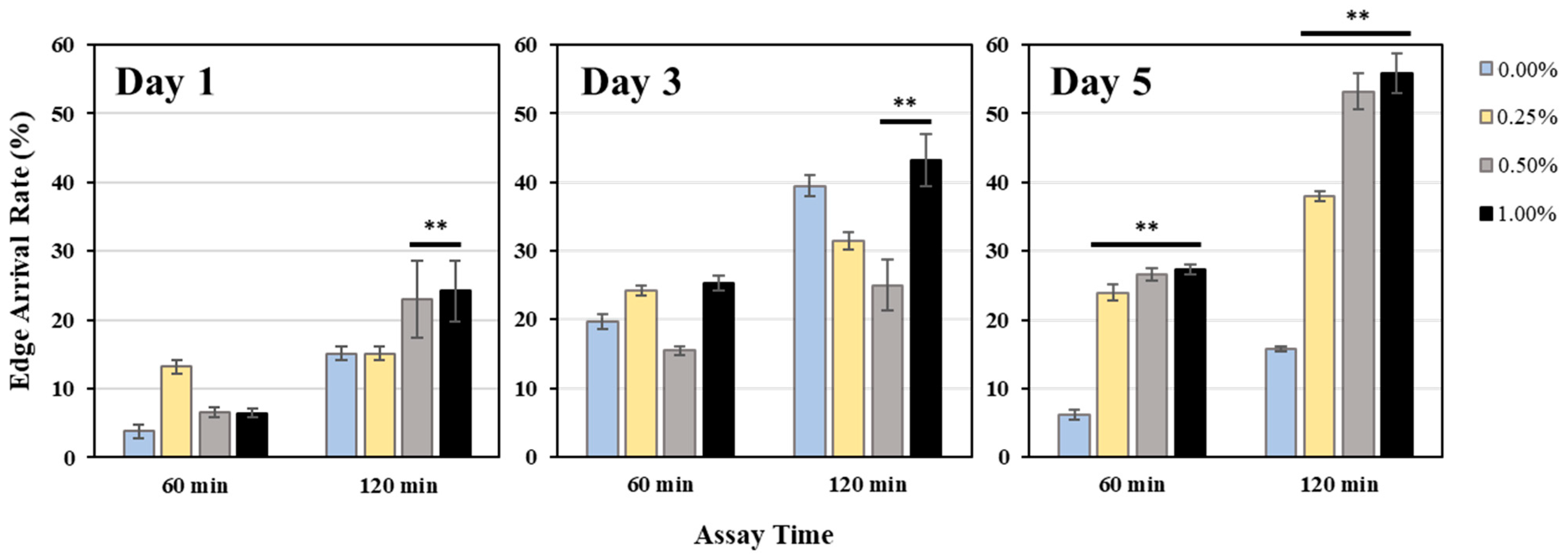
Figure 3 shows the results of edge assay measurements at 1, 3, and 5 days after the addition of WME. On the 5th day, the edge assay results showed a significant increase in the edge assay rate in proportion to the amount of WME added.

2.2.3. Body Length Measurement

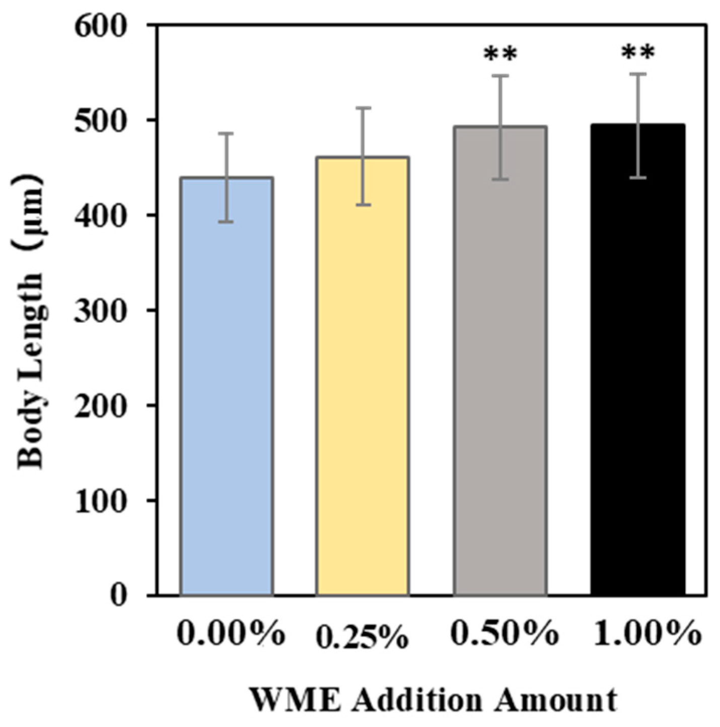
Figure 4 shows the results of body length measurements of C. elegans on the third day after the addition of WME, and it was confirmed that body length increased in proportion to the amount of WME added. The results show that the body length of C. elegans increased in proportion to the amount of WME added, especially in the 0.5% and 1.0% addition conditions.

2.3. Gene Expression Analysis

RNA was extracted from C. elegans 1, 3, and 5 days after the addition of WME, and after cDNA synthesis, RT-PCR reactions using primers in Table 1 were used to analyze the expression levels of lifespan and muscle-related genes. The results are shown in Figure 5. In terms of lifespan-related genes, sir-2.1 gene expression was significantly increased on day 1 for all 0.25, 0.5, and 1% (WME) additions. The expression of the Daf-2 gene was significantly decreased in all 0.25, 0.5, and 1% (WME) additions on days 1 and 5 (Figure 5B). For the daf-16 gene, a significant increase in expression was observed at the 0.25 and 0.5% (WME) additions on day 1 (Figure 5C). The expression of antioxidant enzymes sod-2 and sod-3 was highest in the control on day 1, but as the days passed, the expression of sod-2 and sod-3 in the control decreased (Figure 6A,B). On the other hand, with the addition of 1% WME, the expression level was lower than that of the control on day 1, but as the days progressed, the expression level increased (Figure 6A,B). The results for muscle-related genes are shown in Figure 7A–D. Overall, WME addition increased muscle-related gene expression. The myo-3, unc-22, and fhod-1 gene expression increased markedly with WME addition on days 1 and 3 (Figure 7A–C). With unc-54, the expression decreased with the passage of days in the control, while it increased with the addition of 1% WME (Figure 7D).

3. Discussion

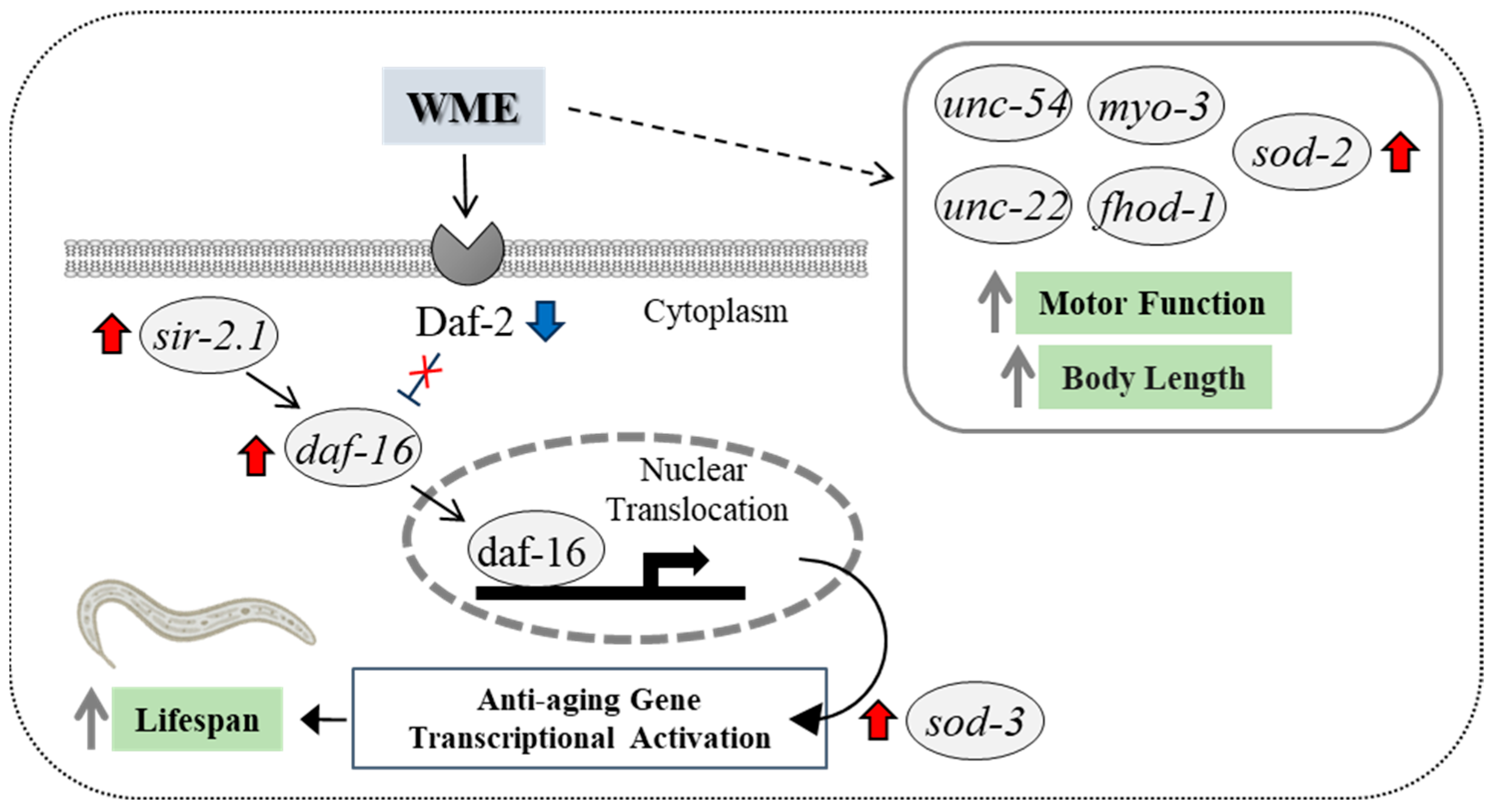
The present experiments confirm the effects of WME on lifespan extension and locomotor function in C. elegans. The daf-16 is required for lifespan extension in C. elegans, which is mediated by an increased expression of sir-2.1 [19]. Furthermore, the only receptor to which insulin/IGF1 binds is daf-2, and when insulin/IGF1 binds to daf-2 and activates the insulin/IGF1 signaling (IIS) pathway, the transcription factor DAF-16 remains in the cytosol, shortening lifespan. On the other hand, blocking the IIS pathway by inhibiting the daf-2 function enhances the nuclear translocation of DAF-16, which in turn increases the expression of the anti-aging gene cluster and prolongs lifespan [20,21,22,23,24]. The results of the present study showed that the addition of WME suppressed the expression of the daf-2 gene and increased the expression of the daf-16 gene, suggesting that the lifespan-extending effect of WME is related to its anti-aging effects via sir-2.1 and daf-16 through daf-2 suppression, as shown in Figure 8.
The daf-16 gene is known to induce the expression of a number of genes encoding oxidative stress response enzymes, such as catalase (CAT) and sod-3 (superoxide dismutase, SOD) [25,26,27,28,29]. In the present experiment, we also observed the increased expression of sod-2 and sod-3 upon the addition of WME (Figure 6). Superoxide dismutase is an enzyme involved in detoxification, and SOD-related genes are involved in protection against oxidative stress in C. elegans. The sod-3 gene encodes a daf-16-dependent SOD [30], and increased sod-3 expression via daf-16 is important for controlling oxidative stress and extending lifespan in C. elegans [25,31,32]. Increased damage during the aging process simultaneously triggers a protective superoxide response. The apparent increase in the body’s antioxidant enzymes, SOD activity, and CAT activity, which prolong lifespan, suggested that WME may have improved the antioxidant defense system in C. elegans. However, a lifespan extension was observed in strains deficient in the mitochondrial SOD gene, sod-2. This suggests that increased cytoplasmic reactive oxidative species (ROS) rather than increased mitochondrial ROS may be responsible for the short lifespan of C. elegans [33]. The sod-2 and sod-3 expression has been reported to increase lifespan [34,35,36], and mutations in sod-2 or sod-3 have been shown to suppress recovery from simulated microgravity toxicity. In addition, sod-2 deletion mutants show developmental delay, a reduced number of offspring, and a defective excretory rhythm [37], sod-2 has been implicated in C. elegans sperm pseudopodia elongation and sperm activation [38], and reduced sod-2 and sod-3 expression negatively affects locomotor behavior [39]. Considering these facts, sod-2 expression is not essential for lifespan extension, but is important for antioxidant effects (SOD) and locomotor function improvement.
Muscle mass and sarcopenia are important features of aging, and patients with sarcopenia show a decrease in myofibrils. The C. elegans rhabdomere wall muscle is a simple model for sarcomere formation, as C. elegans migration is caused by the alternating contraction and relaxation of the dorsal and ventral rhabdomeres of the body wall; thus, the monitoring of muscle function is possible. The expression of many muscle genes in this experiment was increased by WME myo-3, unc-54, unc-22, and fhod-1 (Figure 7), suggesting that these genes are involved in the improvement of motor function obtained by refractive exercise and edge assay. The myo-3 and unc-54 mRNA levels are reported to decrease with age [40,41]. In the present experiment, nc-54 expression was progressively decreased with aging in controls, but not with the addition of WME (Figure 7B). The unc-54 gene is essential for the maintenance of C. elegans’ locomotion and sarcomere structural stability, and is also used to assess the C. elegans locomotor phenotype, and myo-3 has also been suggested to be involved in body length [42]. The fhod-1 gene has been implicated in rhabdomere development and strength in animals ranging from humans to C. elegans, and in C. elegans, fhod-1 has been shown to promote sarcomere formation in the rhabdomeres and in C. elegans, and unc-22 mutations are associated with slowed or paralyzed movement and disorganized muscular architecture in adults [43] and disorganized muscle segment structure [44].

4. Materials and Methods

4.1. C. elegans Maintenance Methods

C. elegans was maintained at 20 °C on C. elegans growth agar (NGM), spaced with OP50 E. coli cultured on LB medium Miller (Nacalai Tesque, Inc., Kyoto, Japan). The methods for medium and solution preparation followed the WormBook (http://www.wormbook.org/). The N2 strain (wild-type) of C. elegans was used (Gifted by Prof Mihira, Faculty of Medicine, Tokyo Women’s Medical University, Tokyo, Japan).

4.2. C. elegans Synchronization Method

Starved L1 worms on 5- to 10-day-old NGM plates were transferred to OP50-sprayed 100 mm NGM plates and maintained at 20 °C for 65 h. Gestation stage adults were rinsed from the NGM agar plates with sterile water and collected in 50 mL centrifuge tubes. Five mL of bleach solution was added, shaken well for a few seconds, and incubated for 5 min. Once the adults were observed to be dissolved, they were immediately neutralized with 5 mL M9 Buffer and centrifuged. The pellet of released eggs was washed three times with M9 Buffer and twice with S-basal before aspirating the supernatant and adding 40 mL of S-basal. After incubation overnight at 20 °C and 180 rpm, the number of hatched L1 stage larvae in 10 μL was determined and used for testing.

4.3. Adjustment of WME

WME (Balenine B8: Kyodo Senpaku Co., Ltd., Tokyo, Japan) is sold as a food, and its standard ingredients and total content are more than 9% imidazole dipeptide, more than 8% balenine, and more than 65% total amino acids. First, 10 g of balenine B8 was mixed with distilled water to make 10 mL. The sample was sterilized by passing it through a 0.22 μm filter after dissolution by vortex mixing.

4.4. Lifespan Assay

C. elegans was adjusted with S-medium at 8–15 worms/100 μL for the number of L1 stage larvae obtained and synchronously treated by hypochlorite bleaching. The food, E. coli OP50, was suspended in S-complete, added at 6.1 mg/mL, and incubated at 20 °C, 110 rpm. Two days (~44 h) after the addition of E. coli, C. elegans was washed with sterile water using a 40 μm filter, collected, and dispensed into 96-well plates at 5–12 fish/100 μL/well. At a final concentration of 120 μM FUDR (5-fluoro-2-deoxyuridine) (Sigma, St. Louis, MO, USA), dead E. coli OP50 was heat treated at 75 °C for 30 min (E. coli OP50 was heat treated at 75 °C for 30 min to minimize the effect of WME on E. coli and confirmed dead). In addition, WME was added at 0.00% (control), 0.25%, 0.50%, and 1.00% WME; the day of WME addition was day 0, and the number of days until all C. elegans died was observed. Incubations were conducted at 20–21 °C. Sixty to one-hundred C. elegans were used for each condition, and the average lifespan was calculated. Three independent experiments were conducted to confirm the lifespan effect; n = 3, approximately 180–300 animals. In each experiment, death was determined by the lack of response to contact with the platinum wire. Unexpected deaths due to straying or wall climbing were excluded. Statistical analysis of the data was performed using the t-test.

4.5. Motor Function Assay

As in the lifespan assay, dead E. coli OP50 mixed S-medium containing WME and a final concentration of 120 μM FUDR was added to C. elegans that were washed with sterile water using 40 μm filters and collected 2 days (~44 h) after the addition of E. coli (0, 0.25, 0.50, and 1.00%). The day of WME addition was day 0, and the flexion movement assay, edge assay, and body length measurement experiments were performed on day 3.

4.5.1. Measurement of Bending Movement Frequency

Ten to fifteen C. elegans were transferred to NGM plates with 1 drop of M9 Buffer, and the number of animals that bent their bodies at least once over a 30 s period was calculated by microscopic observation (n = 12, using 3–14 animals each for a total of 80–104 animals).

4.5.2. Edge Assay

Edge assay plates were prepared by 100 mm NGM agar plates; NGM plates were air-dried on a clean bench and kept at 4 °C until use. The day before the edge assay, sterile cotton swabs were soaked with E. coli suspension and the NGM plate was rotated 360° to spread E. coli around the entire edge of the plate. The plates were incubated overnight at 20 °C and used the next day. Synchronized C. elegans were collected, C. elegans washed with M9 Buffer were placed in the center of the edge assay plate, and excess M9 Buffer was removed with a Kimwipe. The number of C. elegans that reached or did not reach the edge was counted at 60 and 120 min to determine the percent reaching the E. coli edge. Edge assays were performed 1, 3, and 5 days after the addition of WME (n = 5, calculated using 10–30 animals each for a total of 50–150 animals).

4.5.3. Measurement of C. elegans Body Length

Three days after the addition of WME, C. elegans body length was measured by microscopy and ImageJ ([https://imagej.net/ij/], accessed on 1 March 2024) image processing software (n = 3, using 10 animals each).

4.6. RNA Extraction for Gene Expression Analysis

As in the lifespan assay, dead E. coli OP50 mixed S-medium containing WME and a final concentration of 120 μM FUDR was added to C. elegans that were collected 2 days (~44 h) after the addition of E. coli and washed with sterile water using 40 μm filters. Day 0 was the day of WME addition and C. elegans were collected on days 1, 3, and 5, passed through a 40 μm mesh filter, washed with M9 Buffer, and then collected by centrifugation to pellet. C. elegans pellets were crushed by grinding in liquid nitrogen using a mortar, mixed with QIAzol Lysis Reagent (Qiagen, Hilden, Germany), and then stored at −80 °C until RNA extraction. The stored pellets were frozen and thawed three times, and total RNA was extracted according to the protocol of the RNeasy Mini Kit (Qiagen). Extracted RNA was measured for RNA concentration using a micro-spectrophotometer (DS-11, DeNovix, Wilmington, DE, USA), and the 260/280 and 260/230 ratios were confirmed to be greater than 1.8. More than 1000 animals were used for each total RNA extraction in this experiment.

4.7. RT-PCR Protocol

After synthesizing cDNA using the Affinity Script QPCR cDNA synthesis kit (Agilent, Santa Clara, CA, USA), 100 to 500 ng of RNA was subjected to PCR reactions using specific primers (Table 1) for the gene of interest using Emerald Amp PCR Master (Takara, Kusatsu, Japan). PCR reactions (initial denaturation at 97 °C for 5 min, thermal denaturation at 95 °C for 45 s, annealing at 55 °C for 45 s, extension at 72 °C for 1 min, 27 to 36 cycles, and extension at 72 °C for 10 min) were performed. After PCR reactions, the PCR products were separated on 1.5% agarose gels and visualized with ethidium bromide under UV light. The expression levels of the visualized target genes were corrected for pmp-3 gene expression, a known housekeeping gene, and graphed.

5. Conclusions

This study based on rigorous methodology and mRNA expression analysis indicates that certain aging pathways may be activated; it is difficult to formally conclude that the observed activity is responsible for the lifespan extension. Nevertheless, this is also the first experiment to confirm the lifespan-extending effect of WME with a high balenine content, and we believe that we can offer the possibility that sir-2.1, daf-2, and daf-16 genes are involved as factors influencing this lifespan-extending effect. And, the antioxidant effects via the sod-3 genes, as well as increased sod-2, myo-3, unc-54, unc-22, and fhod-1 expression by WME, are involved in improving exercise function. Saying that, we have not tested other gene candidates or performed a full ’omics-wide’ study to screen potentially new molecular factors, genes, proteins, and metabolites, which should be a target of the next research. Our results highlight the potential of WME to ameliorate age-related decline, promote longevity, and further extend a healthy lifespan. Another limitation is that the study did not look at the long-term effects of WME at 10, 20, or 30 days (aged animals). It should also be noted that in this particular experiment, the WME powder was used, which is characterized by a total imidazole dipeptide of more than 9%, of which more than 8% is balenine. However, in future experiments, we would like to consider the use of purified balenine under the possibility of obtaining it at a reasonable price (cost is a factor limiting the use of purified balenine). Finally, we would like to add that we truly believe and hope that it will be understood that this research is not aimed solely at promoting overfishing, but is fundamental research aimed at adding value to the WME as a fishery resource and utilizing it without waste.

Author Contributions

Conceptualization, formal analysis, methodology, software, writing—original draft preparation, writing—review and editing, J.S., F.T., R.R. and S.S.; validation, resources, F.T. and S.S.; investigation, J.S., A.K. and M.Y.; data curation, visualization, J.S. and A.K.; supervision, F.T. and S.S.; project administration F.T. and S.S. All authors have read and agreed to the published version of the manuscript.

Funding

This work was supported by Kyodo Senpaku Inc. Ltd. (Japan).

Institutional Review Board Statement

This study was performed under the strict guidelines of the Hoshi University of Pharmacy and Life Sciences Animal Ethics Review Board. All experimental procedures in this study involving animals (including C. elegans) were approved by the Institutional Animal Care and Use Committee of Hoshi University, and Guidelines for Proper Conduct of Animal Experiment and Related Activities in Academic Research Institutions (Ministry of Education, Culture, Sports, Science and Technology, Japan, 2006). Additionally, the researchers handling the C. elegans undertook animal experimentation training and obtained permission to use the animal center (Number: 15-A404 (M.Y.)).

Informed Consent Statement

Not applicable.

Data Availability Statement

Data are contained within the article.

Conflicts of Interest

The authors declare no conflicts of interest.

References

  1. Cocks, D.H.; Dennis, P.O.; Nelson, T.H. Isolation of 3-methyl histidine from whalemeat extract and the preparation of some derivatives. Nature 1964, 202, 184–185. [Google Scholar] [CrossRef] [PubMed]
  2. Dennis, P.O.; Lorkin, P.A. Isolation and synthesis of balenine, a dipeptide occurring in whale-meat extract. J. Chem. Soc. 1965, 4968–4972. [Google Scholar] [CrossRef] [PubMed]
  3. Suyama, M.; Suzuki, T.; Yamamoto, A. Free amino acids and related compounds in whale muscle tissue. J. Tokyo Univ. Fish. 1977, 63, 189–196. [Google Scholar]
  4. Kohen, R.; Yamamoto, Y.; Cundy, K.C.; Ames, B.N. Antioxidant activity of carnosine, homocarnosine, and anserine present in muscle and brain. Proc. Natl. Acad. Sci. USA 1988, 85, 3175–3179. [Google Scholar] [CrossRef]
  5. Boldyrev, A.A.; Aldini, G.; Derave, W. Physiology and pathophysiology of carnosine. Phys. Rev. 2013, 93, 1803–1845. [Google Scholar] [CrossRef]
  6. Sale, C.; Saunders, B.; Harris, R.C. Effect of beta-alanine supplementation on muscle carnosine concentrations and exercise performance. Amino Acids 2010, 39, 321–333. [Google Scholar] [CrossRef]
  7. Herculano, B.; Tamura, M.; Ohba, A.; Shimatani, M.; Kutsuna, N.; Hisatsune, T. β-alanyl-L-histidine rescues cognitive deficits caused by feeding a high fat diet in a transgenic mouse model of Alzheimer’s disease. J. Alzheimers Dis. 2013, 33, 983–997. [Google Scholar] [CrossRef]
  8. Hisatsune, T.; Kaneko, J.; Kurashige, H.; Cao, Y.; Satsu, H.; Totsuka, M.; Katakura, Y.; Imabayashi, E.; Matsuda, H. Effect of anserine/carnosine supplementation on verbal episodic memory in elderly people. J. Alzheimers Dis. 2016, 50, 149–159. [Google Scholar] [CrossRef]
  9. Masuoka, N.; Lei, C.; Li, H.; Hisatsune, T. Influence of Imidazole-Dipeptides on Cognitive Status and Preservation in Elders: A Narrative Review. Nutrients 2021, 13, 397. [Google Scholar] [CrossRef]
  10. Begum, G.; Cunliffe, A.; Leveritt, M. Physiological role of carnosine in contracting muscle. Int. J. Sport Nutr. Exer. Metab. 2005, 15, 493–514. [Google Scholar] [CrossRef]
  11. Van der Stede, T.; Spaas, J.; de Jager, S.; De Brandt, J.; Hansen, C.; Stautemas, J.; Vercammen, B.; De Baere, S.; Croubels, S.; Van Assche, C.-H.; et al. Extensive profiling of histidine-containing dipeptides reveals species- and tissue-specific distribution and metabolism in mice, rats, and humans. Acta Physiol. 2023, 239, e14020. [Google Scholar] [CrossRef] [PubMed]
  12. Ishihara, K.; Watanabe, R.; Kato, T.; Seko, T.; Matsuda, T.; Omura, Y.; Shigemura, Y.; Kawabata, Y.; Maegawa, T. Isolation of balenine from opah (Lampris megalopsis) muscle and comparison of antioxidant and iron-chelating activities with other major imidazole dipeptides. Food Chem. 2021, 364, 130343. [Google Scholar] [CrossRef] [PubMed]
  13. de Jager, S.; Vermeulen, A.; De Baere, S.; Van der Stede, T.; Lievens, E.; Croubels, S.; Jäger, R.; Purpura, M.; Bourgois, J.G.; Derave, W. Acute balenine supplementation in humans as a natural carnosinase-resistant alternative to carnosine. Sci. Rep. 2023, 13, 6484. [Google Scholar] [CrossRef] [PubMed]
  14. Nakajima, T. Anti fatigue effect of whale-meat extract (balenine), on swimming mouse model. Food Process. Ingred. 2006, 41, 62–64. (In Japanese) [Google Scholar]
  15. Sugino, T.; Yasunagawa, G.; Fukuda, M. Effect of whale meat extract on fatigue induced by physical load and daily activities in humans. Jpn. Pharmcol. Ther. 2013, 41, 879–893. [Google Scholar]
  16. Wada, N.; Yamanaka, S.; Shibato, J.; Rakwal, R.; Hirako, S.; Iizuka, Y.; Kim, H.; Matsumoto, A.; Kimura, A.; Takenoya, F.; et al. Behavioral and omics analyses study on potential involvement of dipeptide balenine through supplementation in diet of senescence-accelerated mouse prone 8. Genom. Data 2016, 10, 38–50. [Google Scholar] [CrossRef]
  17. Yang, M.; Sun, L.; Kawabata, Y.; Murayama, F.; Maegawa, T.; Nikawa, T.; Hirasaka, K. Balenine, Imidazole Dipeptide Promotes Skeletal Muscle Regeneration by Regulating Phagocytosis Properties of Immune Cells. Mar. Drugs 2022, 20, 313. [Google Scholar] [CrossRef]
  18. Dhondt, I.; Verschuuren, C.; Zečić, A.; Loier, T.; Braeckman, B.P.; De Vos, W.H. Prediction of biological age by morphological staging of sarcopenia in C. elegans. Dis. Model Mech. 2021, 14, dmm049169. [Google Scholar] [CrossRef]
  19. Tissenbaum, H.A.; Guarente, L. Increased dosage of a sir-2 gene extends lifespan in C. elegans. Nature 2001, 410, 227–230. [Google Scholar] [CrossRef]
  20. Kimura, K.D.; Tissenbaum, H.A.; Liu, Y.; Ruvkun, G. daf-2, an insulin receptor-like gene that regulates longevity and diapause in C. elegans. Science 1997, 277, 942–946. [Google Scholar] [CrossRef]
  21. Kenyon, C. The plasticity of aging: Insights from long-lived mutants. Cell 2005, 120, 449–460. [Google Scholar] [CrossRef]
  22. Sun, X.; Chen, W.D.; Wang, Y.D. DAF-16/FOXO transcription factor in aging and longevity. Front. Pharmacol. 2017, 8, 548. [Google Scholar] [CrossRef]
  23. Lamitina, S.T.; Strange, K. Transcriptional targets of DAF-16 insulin signaling pathway protect C. elegans from extreme hypertonic stress. Am. J. Physiol. Cell Physiol. 2005, 288, 467–474. [Google Scholar] [CrossRef]
  24. Lee, H.; Lee, S.V. Recent Progress in Regulation of Aging by Insulin/IGF-1 Signaling in C. elegans. Mol. Cells 2022, 45, 763–770. [Google Scholar] [CrossRef] [PubMed]
  25. Zhang, Z.; Li, J.; Li, F.; Wang, T.; Luo, X.; Li, B.; You, Y.; Wu, C.; Liu, X. Jujubae Fructus extract prolongs lifespan and improves stress tolerance in C. elegans dependent on DAF-16/SOD-3. Sci. Rep. 2024, 14, 13713. [Google Scholar] [CrossRef]
  26. Libina, N.; Berman, J.R.; Kenyon, C. Tissue-specific activities of C. elegans DAF-16 in the regulation of lifespan. Cell 2003, 115, 489–502. [Google Scholar] [CrossRef]
  27. McElwee, J.; Bubb, K.; Thomas, J.H. Transcriptional outputs of the C. elegans forkhead protein DAF-16. Aging Cell 2003, 2, 111–121. [Google Scholar] [CrossRef]
  28. Murphy, C.T.; McCarroll, S.A.; Bargmann, C.I.; Fraser, A.; Kamath, R.R.; Arhinger, J.; Li, H.; Kenyon, C. Genes that act downstream of DAF-16 to influence the lifespan of C. elegans. Nature 2003, 424, 277–283. [Google Scholar] [CrossRef]
  29. Ayoub, I.M.; Eldahshan, O.A.; Roxo, M.; Zhang, S.; Wink, M.; Singab, A.N.B. Stress resistance, antiaging, and neuroprotective activities of baicalein 5,6-dimethyl ether and Alnus rugosa extract in C. elegans model. Arch. Pharm. 2024, e2400464. [Google Scholar] [CrossRef]
  30. Chavez, V.; Mohri-Shiomi, A.; Maadani, A.; Vega, L.A.; Garsin, D.A. Oxidative stress enzymes are required for DAF-16-mediated immunity due to generation of reactive oxygen species by C. elegans. Genetics 2007, 176, 1567–1577. [Google Scholar] [CrossRef]
  31. Jiang, S.; Deng, N.; Zheng, B.; Li, T.; Liu, R.H. Rhodiola extract promotes longevity and stress resistance of C. elegans via DAF-16 and SKN-1. Food Funct. 2021, 12, 4471–4483. [Google Scholar] [CrossRef]
  32. Pei, H.; Lin, Z.; Yao, K.; Luo, Y.; Tong, P.; Chen, H.; Wu, Y.; Wu, Z.; Gao, J. Ovalbumin promotes innate immune response of Caenorhabditis elegans through DAF-16 and SKN-1 pathways in insulin/IGF-1 signaling. J. Physiol. Biochem. 2024, 80, 541–559. [Google Scholar] [CrossRef]
  33. Schaar, C.E.; Dues, D.J.; Spielbauer, K.K.; Machiela, E.; Cooper, J.F.; Senchuk, M.; Hekimi, S.; Van Raamsdonk, J.M. Mitochondrial and cytoplasmic ROS have opposing effects on lifespan. PLoS Genet. 2015, 11, e1004972. [Google Scholar] [CrossRef]
  34. Xu, J.; Du, P.; Liu, X.; Xu, X.; Ge, Y.; Zhang, C. Curcumin supplementation increases longevity and antioxidant capacity in C. elegans. Front. Pharmacol. 2023, 14, 1195490. [Google Scholar] [CrossRef]
  35. Ma, J.; Xu, X.; Wang, R.; Yan, H.; Yao, H.; Zhang, H.; Jiang, S.; Xu, A. Lipopolysaccharide exposure induces oxidative damage in C. elegans: Protective effects of carnosine. BMC Pharmacol. Toxicol. 2020, 21, 85. [Google Scholar] [CrossRef]
  36. Niu, Y.; Liao, J.; Zhou, H.; Wang, C.C.; Wang, L.; Fan, Y. Flavonoids from Lycium barbarum Leaves Exhibit Anti-Aging Effects through the Redox-Modulation. Molecules 2022, 27, 4952. [Google Scholar] [CrossRef]
  37. Zhao, L.; Rui, Q.; Wang, D. Molecular basis for oxidative stress induced by simulated microgravity in nematode C. elegans. Sci. Total Environ. 2017, 607–608, 1381–1390. [Google Scholar] [CrossRef]
  38. Sakamoto, T.; Imai, H. Hydrogen peroxide produced by superoxide dismutase SOD-2 activates sperm in C. elegans. J. Biol. Chem. 2017, 292, 14804–14813. [Google Scholar] [CrossRef]
  39. Li, Y.; Yu, S.; Wu, Q.; Tang, M.; Pu, Y.; Wang, D. Chronic Al2O3-nanoparticle exposure causes neurotoxic effects on locomotion behaviors by inducing severe ROS production and disruption of ROS defense mechanisms in nematode C. elegans. J. Hazard. Mater. 2012, 219–220, 221–230. [Google Scholar] [CrossRef]
  40. Matheny, C.J.; Qadota, H.; Bailey, A.O.; Valdebenito-Silva, S.; Oberhauser, A.F.; Benian, G.M. The myosin chaperone UNC-45 has an important role in maintaining the structure and function of muscle sarcomeres during adult aging. Mol. Biol. Cell 2024, 35, ar98. [Google Scholar] [CrossRef]
  41. Adamla, F.; Ignatova, Z. Somatic expression of unc-54 and vha-6 mRNAs declines but not pan-neuronal rgef-1 and unc-119 expression in aging C. elegans. Sci. Rep. 2015, 5, 10692. [Google Scholar] [CrossRef]
  42. Dahl-Halvarsson, M.; Pokrzywa, M.; Rauthan, M.; Pilon, M.; Tajsharghi, H. Myosin storage myopathy in C. elegans and human cultured muscle cells. PLoS ONE 2017, 12, e0170613. [Google Scholar] [CrossRef]
  43. Harada, S.; Hashizume, T.; Nemoto, K.; Shao, Z.; Higashitani, N.; Etheridge, T.; Szewczyk, N.J.; Fukui, K.; Higashibata, A.; Higashitani, A. Fluid dynamics alter C. elegans body length via TGF-beta/DBL-1 neuromuscular signaling. NPJ Microgravity 2016, 2, 16006. [Google Scholar] [CrossRef]
  44. Sundaramurthy, S.; Votra, S.; Laszlo, A.; Davies, T.; Pruyne, D. FHOD-1 is the only formin in C. elegans that promotes striated muscle growth and Z-line organization in a cell autonomous manner. Cytoskeleton 2020, 77, 422–441. [Google Scholar] [CrossRef]
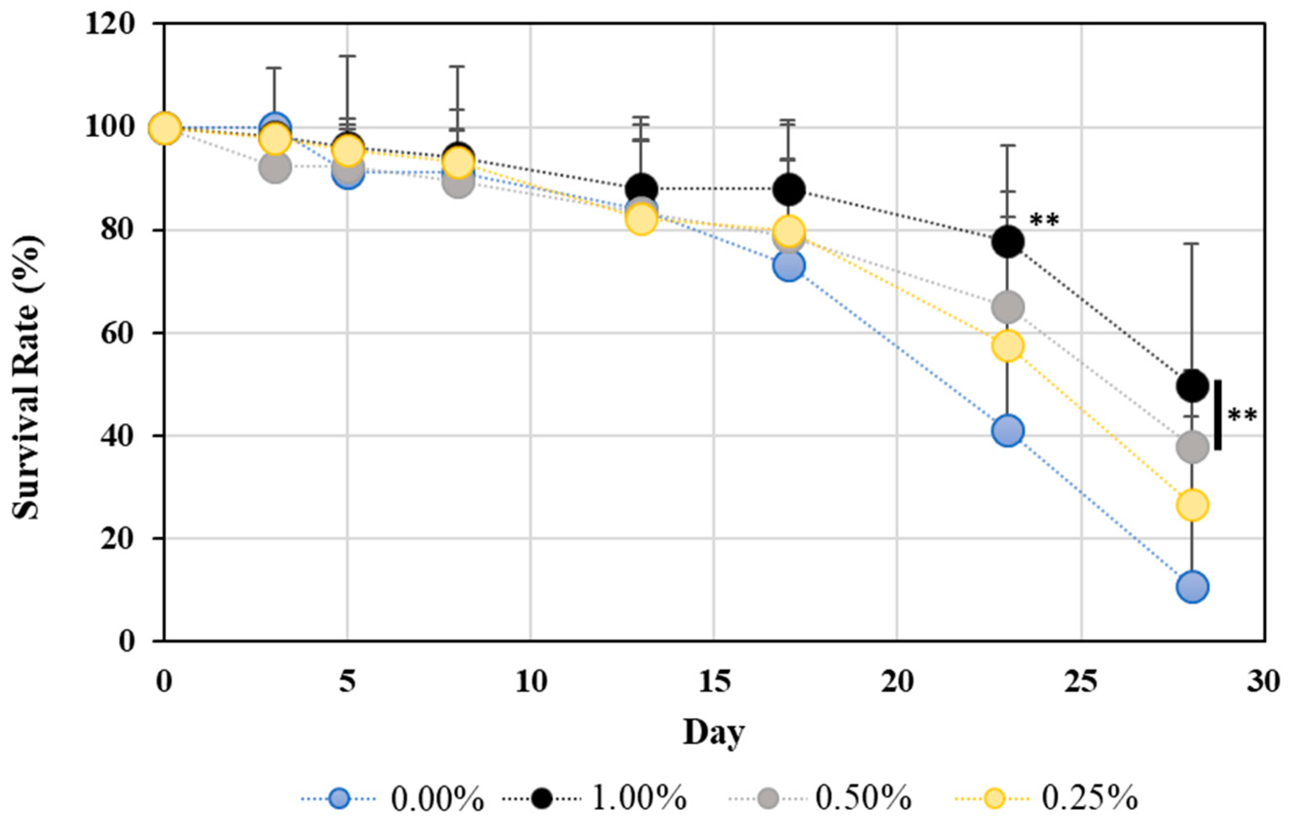
Figure 1. Lifespan extension effect of WME on C. elegans. The graph shows the survival rate, with the day of WME addition taken as 100%. Results are presented as mean ± SE ** p < 0.01 when compared to control (0.00% WME) values by Student’s t test.
Ijms 25 12833 g001
Figure 2. Refraction motility assay measurement results on day 3 after WME addition. Results are presented as mean ± SE ** p < 0.01, * p < 0.05 when compared to control (0.00% WME) values by Student’s t test.
Ijms 25 12833 g002
Figure 3. Edge assay measurement results on days 1, 3, and 5 after WME addition. The E. coli edge arrival rate after 60 and 120 min is shown. Results are presented as mean ± SE ** p < 0.01 when compared to control (0.00% WME) values by Student’s t test.
Ijms 25 12833 g003
Figure 4. Measurement of body length (μm) of C. elegans 3 days after WME addition. Results are presented as mean ± SE ** p < 0.01 when compared to control (0.00% WME) values by Student’s t test.
Ijms 25 12833 g004
Figure 5. Lifespan-related sir-2.1 (A), daf-2 (B), and daf-16 (C) gene expression results of C. elegans 1, 3, and 5 days after WME addition. Results are presented as mean ± SE ** p < 0.01, * p < 0.05 when compared to control (0.00% WME) values by Student’s t test.
Ijms 25 12833 g005
Figure 6. Antioxidant-related sod-2 (A) and sod-3 (B) gene expression results of C. elegans 1, 3, and 5 days after WME addition. Results are presented as mean ± SE ** p < 0.01, * p < 0.05 when compared to control (0.00% WME) values by Student’s t test.
Ijms 25 12833 g006
Figure 7. Muscle-related myo-3 (A), nuc-54 (B), nuc-22 (C), fhod-1 (D) gene expression results in C. elegans 1, 3, and 5 days after WME addition. Results are presented as mean ± SE ** p < 0.01, * p < 0.05 when compared with control (0.00% WME) values by Student’s t test.
Ijms 25 12833 g007
Figure 8. Scheme of WME-induced lifespan extension and motor function improvement in C. elegans. The figure depicts that under the present experimental conditions, WME improves lifespan and motor function through the upregulation (red arrows) of the sir-2.1 and daf-16 genes and the sod-3 gene, as well as the sod-2, myo-3, unc-54, unc-22, and fhod-1 genes; and, daf-2 downregulation (blue arrow). However, there are other possibilities that have not yet been tested, such as other factors that regulate lifespan and motion capacity. These cannot be excluded in the current study.
Ijms 25 12833 g008
Table 1. Primer design of genes used in RT-PCR analysis.
Forward PrimerReverse Primer
Accession (Gene)Nucleotide Sequence (5′-3′)Nucleotide Sequence (5′-3′)Gene Name
NM_001269678agttgaatgggattggatgaagctatcgaggctgtgtcaatgtcpmp-3
NM_001268555ttgaaactggttcgtgatgttcgtttttcaatgggatttggtgtsir-2.1
NM_065249atgagcatcatccacttgtctggtcagcgaacgtacaaactcagdaf-2
AF032112ttcaatcgtgtggaattgtagctgggatgaatgtgttggaattadaf-16
NM_059889ctggactaatttggcaaaggacagtagtaagcgtgctcccagacsod-2
NM_078363agaaccttcaaaggagctgatgctgcttttattgtcgagcattgsod-3
NM_073664ttaatgctcacgtgtctgctctacgtctctgttctccatccaatmyo-3
NM_061195gaatctgaattggacggagaacggagaaagagcatgtagggatgunc-54
NM_001313484agacctggtggacactgaatctgagtgagtgaggaggaaggagafhod-1
NM_069872aagctagagtgcaaggaacacccttaaaggtgtttccggtcttgunc-22
Disclaimer/Publisher’s Note: The statements, opinions and data contained in all publications are solely those of the individual author(s) and contributor(s) and not of MDPI and/or the editor(s). MDPI and/or the editor(s) disclaim responsibility for any injury to people or property resulting from any ideas, methods, instructions or products referred to in the content.