Article

The Absence of Thyroid-Stimulating Hormone Receptor Expression on Natural Killer T Cells: Implications for the Immune–Endocrine Interaction

by
Emilia Adamska-Fita
1,
Przemysław Wiktor Śliwka
1,2,
Małgorzata Karbownik-Lewińska
1,2,
Andrzej Lewiński
1 and
Magdalena Stasiak
1,*
1
Department of Endocrinology and Metabolic Diseases, Polish Mother’s Memorial Hospital—Research Institute, 93-338 Lodz, Poland
2
Department of Endocrinology and Metabolic Diseases, Medical University of Lodz, 93-338 Lodz, Poland
*
Author to whom correspondence should be addressed.
Int. J. Mol. Sci. 2024, 25(21), 11434; https://doi.org/10.3390/ijms252111434
Submission received: 1 September 2024 / Revised: 1 October 2024 / Accepted: 22 October 2024 / Published: 24 October 2024
(This article belongs to the Special Issue Hormone Signaling in Human Health and Diseases, 2nd Edition)

Abstract

:
The expression of thyroid-stimulating hormone receptor (TSHR) has been documented on various immune cells, including B lymphocytes, T lymphocytes, Natural Killer (NK) cells, monocytes, and dendritic cells (DCs). Natural Killer T (NKT) cells serve as a crucial link between innate and adaptive immunity, playing significant roles in immunological interactions and autoimmune diseases. The aim of the present study was to evaluate the presence of TSHR on NKT cells. Our research involved patients with thyroid disease, as well as healthy controls. Peripheral blood mononuclear cells (PBMCs) and, thereafter, NKT cells were isolated from 86 patients with benign nodular thyroid disease with and without autoimmune thyroid disease (AITD) (28 and 56 cases, respectively), and TSHR expression was analyzed using fluorescence-activated cell sorting (FACS). In order to confirm the results, the reverse-transcription polymerase chain reaction (RT-PCR) method was used in cells obtained from healthy individuals. Our findings obtained with application of the FACS method revealed that TSHR is not expressed on NKT cells in either AITD or non-AITD patients, though TSHR was detected in the total PBMC population (TSHR+ cells 2.77%). The absence of TSHR on NKT cells was further confirmed with RT-PCR in healthy individuals (p < 0.0001). These results questioned the previously suggested direct influence of NKT cells on AITD development.

1. Introduction

The functional interplay between the immune and endocrine systems is complex, with numerous cell types and humoral mediators involved, yet not fully understood [1]. Apart from the well-known direct effects of thyroid hormones on the immune system, alterations in the hypothalamic–pituitary–thyroid axis, particularly changes in thyroid-stimulating hormone (TSH) levels, may be of significance. While the role of thyroid-stimulating hormone receptor (TSHR) in thyroid function is well established, recent studies suggested TSHR expression in certain immune cells, potentially influencing immune responses [2,3]. In studies performed by immunoprecipitation and by flow cytometry, TSHRs were found on B and T lymphocytes, Natural Killer (NK) cells, monocytes, and—in a large amount—on dendritic cells (DCs) [2,3]. Although the impact of TSH levels on the quantity of Natural Killer T (NKT) cells in peripheral blood has been postulated [4,5], no reported studies have explored the expression of TSHR on NKT cells.
NKT cells represent a unique T cell subpopulation characterized by the presence of markers of both NK cells (NK1.1, CD56) and T cells (TCR—T cell receptor). This dual marker expression makes NKT cells essential for bridging innate and adaptive immunity. NKT cells constitute approximately 0.1% of peripheral blood lymphocytes [6]. Unlike conventional T αβ lymphocytes, NKT cells possess TCR receptors capable of recognizing glycolipid, glycosphingolipid, and lipid structures presented on non-polymorphic CD1d molecules on both professional and non-professional antigen-presenting cells. This unique feature allows NKT cells to recognize both foreign and self-lipid antigens, granting them regulatory and potentially effector roles in various immune responses, including those related to autoimmune diseases, allergies, infections, and cancers [7].
NKT cells are crucial regulators of autoimmune diseases [8,9]. Dysregulation or deficiency of NKT cells is associated with type 1 diabetes, lupus erythematosus, multiple sclerosis, myasthenia gravis, Guillain–Barré syndrome, and autoimmune thyroid diseases (AITDs) [10,11,12,13,14]. AITDs are a group of disorders characterized by an immune system attack against the thyroid gland antigens, including Graves’ disease (GD) and Hashimoto’s thyroiditis (HT). These diseases involve complex interactions between genetic predispositions and environmental factors, resulting in autoantibody production targeting thyroid-specific proteins, including TSHR [15]. TSHR is part of the G protein-coupled receptor group and is composed of a wide extracellular domain, seven transmembrane passages, and a small intracellular domain [16]. TSHR is mostly expressed in the basolateral membrane of thyrocytes where its stimulation enhances iodine uptake, the synthesis and secretion of thyroid hormones, and the proliferation of thyroid follicular cells, and regulates the expression of thyroid-specific genes like those coding thyroglobulin (Tg), thyroid peroxidase (TPO), and sodium/iodide symporter (NIS) [17,18,19,20]. Moreover, several extrathyroidal cell types with expression of TSHR have been revealed, including immune system cells [2,3,21]. Regardless of the low TSHR expression on non-thyroidal cells, the very high binding affinity for TSH contributes to activating the response despite the low density of TSHR on the cellular surface [22]. Many studies have been conducted to test molecular mechanisms involving TSHR in the pathogenesis of AITD. Cuddihy et al. in 1995 discovered the first potential single-nucleotide polymorphism (SNP) of codon 52 of TSHR gene connected with GD in a female population [23]. Recent studies, including three meta-analyses, confirmed important associations between SNPs in intron 1 of the TSHR gene and the risk of GD [24,25,26]. In a previous study performed in our center, a significant increase in the percentage of NKT cells after administration of recombinant human TSH (rhTSH) was observed [4], suggesting a direct mechanism of TSH action on NKT cells. Such a mechanism was also postulated in other studies [5]. Therefore, in order to evaluate whether the mechanism of TSH action on NKT is direct or indirect, the present study aimed to assess TSHR expression on NKT cells, analyzing NKT cells isolated from the peripheral blood of individuals with and without AITDs.

2. Results

2.1. Comparison of AITD and Non-AITD Groups

The mean age of the patients was 55.54 ± 11.4 and 61.72 ± 14.43 years in the AITD and non-AITD groups, respectively. The non-AITD group included 45 women and 13 men, while the AITD group consisted of 23 women and 5 men, with 2 cases of overt hyperthyroidism, 3 cases of subclinical hyperthyroidism, and 2 cases of subclinical hypothyroidism.
The AITD and non-AITD groups were similar in terms of age and gender. The comparison of both groups revealed statistically significant higher anti-TPO and anti-Tg levels in patients with AITD. TRAb level was higher in the AITD group but the difference did not reach statistical significance, as there was only one patient with newly diagnosed GD and a high TRAb level, while other GD patients were either on thiamazole or had received radioiodine therapy. The characteristics of the AITD and non-AITD groups are presented in Table 1.

2.2. Absence of TSHR Expression on NKT Cells

No expression of TSHR on the surface of NKT cells was detected by FACS in either the AITD or non-AITD patient group. Neither thyroid function nor thyroid-related pharmacotherapy in AITD group influenced this observation. This finding was further confirmed by an analysis performed with RT-PCR gene assays in the cells obtained from the healthy blood donors.
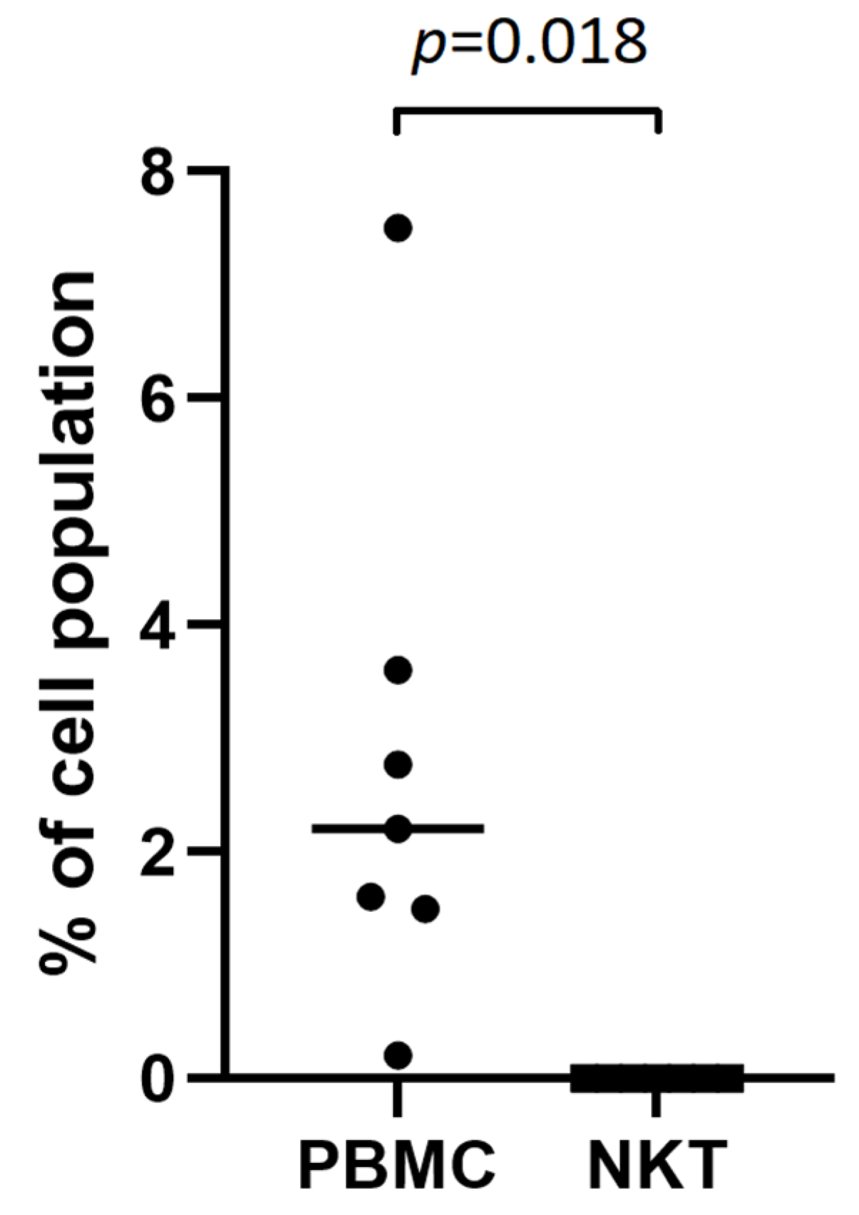
FACS analysis of NKT cells revealed no TSHR+ NKT cells in any of the patients, showing only neglectable detection of high-autofluorescence individual cells, analogous to the isotype control (Figure 1E). However, positive control staining conducted on seven patients detected TSHR+ cells (2.77%) among the total population of PBMCs (Figure 1F). The difference was statistically significant with p = 0.018 (Figure 2).
For further confirmation of the obtained results, a more sensitive method of RT-PCR was used in NKT cells isolated from healthy individuals. To achieve high RNA concentrations, NKT cells were isolated from up to 1 × 10⁸ PBMCs isolated from 10 buffy coats collected from healthy donors. The purity of the NKT cells after magnetic sorting—prior to RNA isolation—was measured by FACS and showed a high (median 94.15%) percentage of NKT cells (Figure 3). During the analysis, one result was excluded due to low amplification of the tested genes.
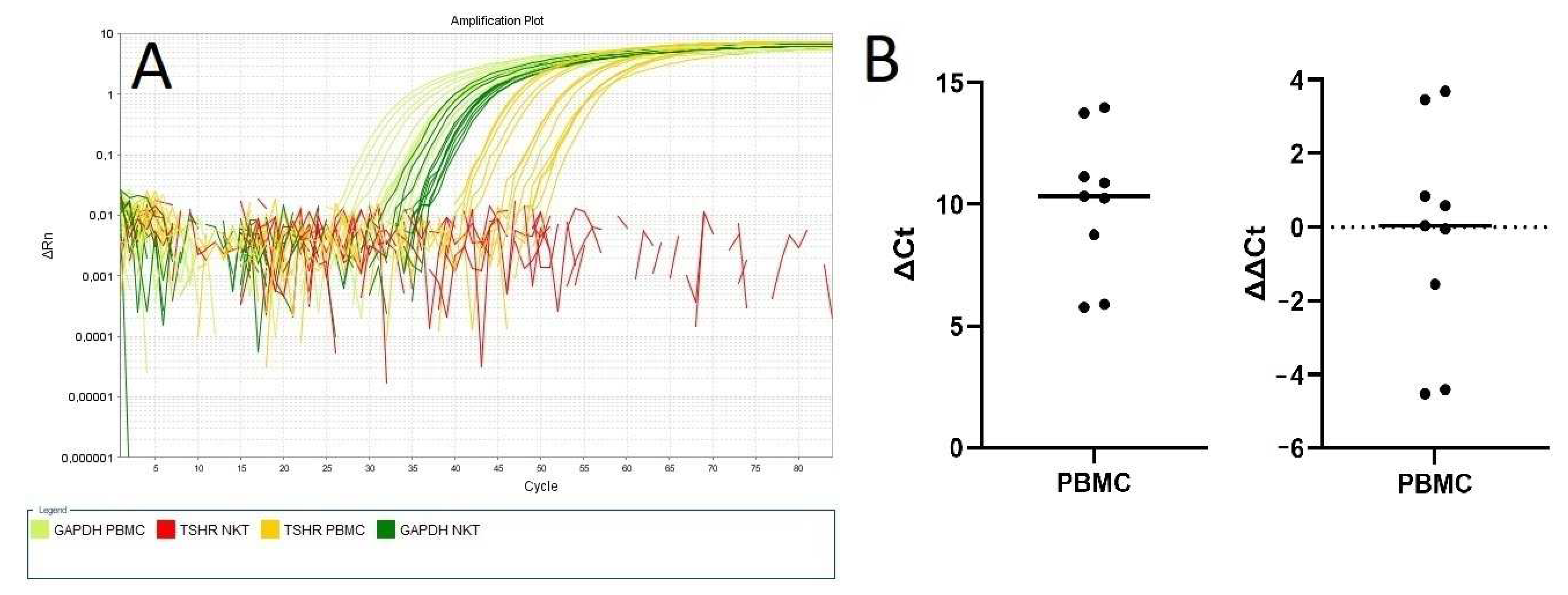
RT-PCR analysis has demonstrated no TSHR expression in NKT cells, while TSHR expression was present in the total PBMC population (Figure 4A). The results were normalized as positive (1) vs. negative (0) and significant difference was confirmed with Fisher’s exact test with p < 0.00001, and with chi-square test with p = 0.0002. The median value of ΔCt (relative TSHR gene expression to housekeeping gene expression) for the TSHR gene in PBMCs was 10.3 (Figure 4B).

3. Discussion

This study provides the first evidence of the absence of TSHR on the surface of NKT cells in the peripheral blood of both healthy individuals and patients with benign thyroid nodular disease with and without AITD. The use of both FACS and RT-PCR strengthens the validity of our findings. FACS provides a robust method for detecting surface proteins, while RT-PCR allows for the detection of mRNA expression, confirming the absence of TSHR at both the protein and gene levels in NKT cells. The absence of TSHR expression on NKT cells in both AITD and non-AITD patients suggests that NKT cells do not participate directly in the TSHR-mediated pathways involved in thyroid autoimmunity. This finding is significant as it challenges previous assumptions about the involvement of these immune cells in AITD pathophysiology [5,27,28,29]. However, this does not exclude the involvement of NKT cells in AITD development driven through indirect mechanisms involving other humoral, cellular, or hormonal factors and further modulated by already triggered pathological processes. Additionally, NKT cells are suggested to play a role in AITD development and progression at the local thyroid gland tissue level, through the switch of activating versus inhibiting regulatory roles [29].
Despite the lack of TSHR on NKT cells, the relationship between peripheral blood NKT cell quantity and TSH levels has been previously documented. Adamczewski et al. [4] showed a significant increase in NKT cells after in vivo administration of rhTSH in patients after total thyroidectomy due to differentiated thyroid cancer. Moreover, Miko et al. [5] found a remarkably elevated peripheral NKT level in euthyroid and subclinical hypothyroid women with thyroid autoimmunity experiencing reproductive failure.
The detection of TSHR expression in the total PBMC population, but not in isolated NKT cells, suggests that TSHR expression on immune cells might be restricted to specific PBMC subpopulations. This aligns with previous studies identifying a high expression of TSHR mainly on DCs and B cells but a low expression on T cells or NK cells [2,3]. As mentioned above, NKT cells may be affected by abnormal TSH levels through complex, indirect interactions with other immune cells. One potential intermediary mechanism involving NKT cells in AITD pathophysiology may be related to interactions with antigen-presenting cells (APCs). APCs can capture and transform antigens in order to present them within the context of appropriate major histocompatibility complex (MHC) molecules [30]. NKT cells have a diverse range of TCRs, allowing them to recognize various antigens, though CD1d-restricted invariant NKT (iNKT) cells specifically recognize lipid antigens presented by CD1d molecules [31]. CD1d is a non-polymorphic, MHC class I-like molecule expressed on various APCs, primarily DCs, and to a lesser extent on B cells and macrophages. Its primary function is to present lipid antigens, such as glycolipids and glycosphingolipids, to NKT cells. The interaction between CD1d and the TCR on NKT cells is crucial for their activation. Upon recognizing lipid antigens presented by CD1d, NKT cells rapidly produce a diverse array of cytokines, such as interferon gamma (IFN-γ), interleukin-4 (IL-4), and interleukin-17 (IL-17), modulating the immune response. This activation triggers both direct cytotoxic effects and the recruitment and activation of other immune cells, thereby playing a pivotal role in bridging innate and adaptive immunity. The ability of CD1d to present both self and foreign lipid antigens allows NKT cells to participate in immune surveillance and regulation, contributing to the body’s defense mechanisms against infections, tumor surveillance, and the modulation of autoimmune responses [32,33].
A role of DCs, as a population of the most potent APCs, should be considered in the TSH-mediated regulation of NKT cell function. In humans, two main DC groups are known: conventional/myeloid (cDCs) and plasmacytoid (pDCs) [34,35]. Through the expression of numerous co-stimulatory molecules and a unique secretion profile, DCs are involved in cross-interactions with other immune cells. Thus, DCs play a role in stimulating the immune response and ensuring immunological tolerance, making them key subjects in research on the pathogenesis of autoimmune diseases [36]. Interestingly, only a few studies assess DCs’ role in human AITD. Leskela et al. [37] revealed a notably elevated amount of pDCs in thyroid tissue compared to peripheral blood in patients with GD and HT. Stasiołek et al. [38] observed a significantly higher percentage of cDCs in fine-needle aspiration biopsy (FNAB) material than in peripheral blood in a group of AITD patients. Other studies also revealed an increased DC population in thyroid-infiltrating cells in patients with both GD and HT [39,40,41]. Elevated NKT [5,29] and DC [37,38,39,40,41] levels in AITD patients suggest a complex, indirect, co-stimulatory interaction between NKT cells and DCs as a possible mechanism involving NKT cells in thyroid diseases.
Another potential mechanism involving TSH in correlation with peripheral NKT cells was also postulated. Studies by Bessoles et al. [42] highlighted the complex interaction between glycolipids and environmental signals in regulating cytokine production by NKT cells, thereby modulating immune responses. To elucidate NKT cells’ regulatory mechanisms, their activation by interleukin-2 (IL-2) and associated signaling pathways were examined. IL-2 uniquely activates the Signal Transducer and Activator of Transcription 6 (STAT6) pathway in NKT cells, leading to the production of both pro-inflammatory (IFN-γ) and anti-inflammatory (IL-4) cytokines, and to cell proliferation.
Clinical studies by Komorowski et al. [43,44] indicated that patients with primary hypothyroidism and elevated TSH levels exhibited increased IL-2 concentrations in peripheral blood, suggesting that elevated TSH levels may influence IL-2 production. Hence, it can be hypothesized that TSH might modulate NKT cell function indirectly through IL-2 mediation, thereby affecting the immune response. This linkage provides a potential pathway wherein TSH could exert its effects on NKT cells via IL-2, underscoring a novel intersection between endocrine and immune regulation.
This study has potential limitations as we used only two methods for the detection of TSHR expression on NKT cells. The FACS method has been broadly accepted for the detection of TSHR on cells of non-thyroid origin including peripheral blood cells [45,46,47]. Anti-TSHR antibody (clone C-10) was already proven to sufficiently detect TSHR [48,49,50,51]. Additionally, we confirmed that Alexa Fluor-conjugated antibody was sufficient for detecting even low TSHR expression (Figure 1F). However, we are aware that the FACS method is not sufficient to prove the absence of expression of any receptor. Therefore, our findings were verified by a very sensitive method of RT-PCR, which is considered a gold standard in gene expression analysis, including TSHR expression in immune cells [52,53]. Nevertheless, we believe that our results require further confirmation in studies with the application of other methods, including those with the highest available sensitivity (e.g., next-generation sequencing, NGS).

4. Materials and Methods

4.1. Patients

This study involved 86 patients with cytologically benign thyroid nodules (68 and 18 females and males, respectively) treated in the Department of Endocrinology and Metabolic Diseases, Polish Mother’s Memorial Hospital—Research Institute, Łódź, Poland. Among the study group, 28 patients were diagnosed with AITD, including 7 cases of Hashimoto’s thyroiditis treated with L-thyroxine, 14 cases of euthyroid chronic thyroiditis, and 7 cases of Graves’ disease, with 5 patients treated with thiamazole and 2 euthyroid patients who had received radioiodine therapy. The mean age of the study participants was 59.7 ± 13.69 years.

4.2. Inclusion Criteria

AITD diagnosis was based on standard criteria including elevated thyroid peroxidase antibody (anti-TPO) level and/or elevated thyroglobulin antibody (anti-Tg) level, or elevated TSH receptor antibody (TRAb) level [54].

4.3. Biochemical Analysis

Serum levels of TSH, free triiodothyronine (FT3), free thyroxine (FT4), anti-Tg, anti-TPO, and TRAb were measured by the electrochemiluminescence immunoassay (ECLIA) with Cobas e601 analyzer (Roche Diagnostics, Indianapolis, IN, USA).

4.4. NKT Cell Isolation

Peripheral blood samples (2 × 4.9 mL) were collected from each patient via venipuncture into EDTA-containing Blood Collecting Systems (Sarstedt, Nümbrecht, Germany). Peripheral blood mononuclear cells (PBMCs) were isolated by gradient centrifugation at 400 g for 30 min using Histopaque®-1077 (Thermo Fisher Scientific, Waltham, MA, USA). NKT cells were then isolated from PBMCs using a CD3+ CD56+ NKT Cell isolation kit with a magnetic bead cell separator (Miltenyi Biotec, Bergisch Gladbach, Germany). In each patient, the cell purity was measured and the median NKT cell percentage was 93.2% (range 99.1–80.1%). NKT cells were gated as CD3+ CD56+ cells for further analysis.

4.5. Fluorescence-Activated Cell Sorting (FACS)

To analyze TSHR expression on NKT cells, fluorescence-activated cell sorting (FACS) was employed. Fluorochrome-conjugated antibodies against human CD3 (Becton Dickinson, Franklin Lakes, NJ, USA) and CD56 (Becton Dickinson, NJ, USA) were used to identify NKT cells, and an antibody against human TSHR (Santa Cruz, Santa Cruz, CA, USA) was used to determine TSHR expression on isolated NKT cells. Additional positive staining for antibody validation was conducted on whole blood samples from 7 patients (including 3 patients with AITD and 4 patients without AITD) to recognize TSHR+ cells in the PBMC population (Figure 1A–D). Appropriate isotype controls were used. FACS analyses were conducted using a BD FACSCanto II flow cytometer (Becton Dickinson, NJ, USA).

4.6. Reverse-Transcription Polymerase Chain Reaction (RT-PCR)

In order to confirm the obtained results with the application of a different method, TSHR expression was analyzed by reverse-transcription polymerase chain reaction (RT-PCR). To obtain a high number of NKT cells (0.5–1 × 10⁶), needed for sufficient RNA isolation, up to 100 mL of peripheral blood would have been needed from each patient. Therefore, for ethical reasons, we obtained a 10 buffy coats from the healthy blood donors registered in the Regional Center of Blood Donation and Treatment. PBMCs and NKT cells were isolated as described above, using a CD3+ CD56+ NKT Cell isolation kit with a magnetic bead cell separator (Miltenyi Biotec, Germany). Total ribonucleic acid (RNA) was extracted from both PBMCs and NKT cells, and TSHR expression was analyzed by RT-PCR using TaqMan assays. The GAPDH gene was used as a housekeeping gene. Analyses were performed on a 7500 Real-Time PCR System (Thermo Fisher Scientific, USA). Each measurement was conducted in triplets.

4.7. Statistical Analysis

Descriptive statistics of the collected material contained the mean and standard deviation (SD). For comparisons between the groups, Student’s t-test for normally distributed variables and the Mann–Whitney U test for the other ones were used. Positive/negative values were tested using Fisher’s exact test as well as with the chi-square test with Yates correction. The normality of data distributions was verified by the Shapiro–Wilk test. In all the tests, p-value < 0.05 was considered significant. Statistical Package for the Social Sciences (SPSS 20.0) software for Windows was used for all the calculations.

4.8. Ethics Procedures

Written informed consent was obtained from all patients for the procedures performed after their purpose and course were thoroughly explained. This study was approved by the Ethics Committee of the Polish Mother’s Memorial Hospital—Research Institute, Lodz, Poland (approval code—41/2021).

5. Conclusions

The present study demonstrated for the first time that TSHR expression was not found on NKT cells. This observation provides new insight on the potential mechanism of TSH’s impact on NKT cells, which seems to be indirect. These findings have significant implications as therapeutic strategies targeting TSHR on immune cells should consider the specific immune cell subtypes involved in the disease development mechanisms. As the expression of TSHR on NKT cells had not been studied before, our findings require further confirmation with the application of different methods.

Author Contributions

Conceptualization, E.A.-F., P.W.Ś. and M.S.; methodology, E.A.-F., P.W.Ś. and M.S.; software, E.A.-F., P.W.Ś. and M.S.; validation, E.A.-F., P.W.Ś. and A.L.; formal analysis, M.K.-L., A.L. and M.S.; investigation, E.A.-F. and P.W.Ś.; resources, E.A.-F., P.W.Ś. and M.S.; data curation, E.A.-F. and P.W.Ś.; writing—original draft preparation, E.A.-F., P.W.Ś. and M.S.; writing—review and editing, M.K.-L., A.L. and M.S.; visualization, E.A.-F. and P.W.Ś.; supervision, M.K.-L., A.L. and M.S.; project administration, M.K.-L., A.L. and M.S.; funding acquisition, E.A.-F., A.L. and M.S. All authors have read and agreed to the published version of the manuscript.

Funding

This research was funded by the Polish Mother’s Memorial Hospital—Research Institute, Lodz, Poland, grant number 8GW/2021.

Institutional Review Board Statement

The study was conducted in accordance with the Declaration of Helsinki, and approved by the Ethics Committee of the Polish Mother’s Memorial Hospital—Research Institute, Lodz, Poland (approval code—41/2021).

Informed Consent Statement

Informed consent was obtained from all subjects involved in the study.

Data Availability Statement

The source data are available on reasonable request from the corresponding author.

Conflicts of Interest

The authors declare no conflicts of interest.

Abbreviations

AITDautoimmune thyroid disease
Anti-Tgthyroid antithyroglobulin antibodies
Anti-TPOthyroid peroxidase antibodies
APCsantigen-presenting cells
CDcluster of differentiation
DCsdendritic cells
pDCsplasmocytoid dendritic cells
cDCsconventional dendritic cells
EDTAethylenediamine tetraacetic acid
FACSfluorescence-activated cell sorting
FNABfine-needle aspiration biopsy
GAPDHglyceraldehyde-3-phosphate dehydrogenase
GDGraves’s disease
HTHashimoto’s thyroiditis
IFN-yinterferon-gamma
IL-2interleukin-2
IL-4interleukin-4
IL-17interleukin-17
iNKTinvariant Natural Killer T cells
MCHmajor histocompatibility complex
NKNatural Killer cells
NKTNatural Killer T cells
NISsodium/iodide symporter
PBMCsperipheral blood mononuclear cells
mRNAmessenger ribonucleic acid
rhTSHrecombinant human thyroid stimulating hormone
RNAribonucleic acid
RT-PCRreverse-transcription polymerase chain reaction
SNPsingle-nucleotide polymorphism
STAT6Signal Transducer and Activator of Transcription 6
TCRT-cell receptor
Tgthyroglobulin
TPOthyroid peroxidase
TRAbthyroid-stimulating hormone receptor antibodies
TSHthyroid-stimulating hormone
TSHRthyroid-stimulating hormone receptor

References

  1. Jara, E.L.; Muñoz-Durango, N.; Llanos, C.; Fardella, C.; González, P.A.; Bueno, S.M.; Kalergis, A.M.; Riedel, C.A. Modulating the function of the immune system by thyroid hormones and thyrotropin. Immunol. Lett. 2017, 184, 76–83. [Google Scholar] [CrossRef] [PubMed]
  2. Berczi, I. Neuroendocrine Regulation of Natural Immunity. NeuroImmune Biol. 2005, 5, 215–262. [Google Scholar]
  3. Bağriaçik, E.U.; Klein, J.R. The thyrotropin (thyroid-stimulating hormone) receptor is expressed on murine dendritic cells and on a subset of CD45RBhigh lymph node T cells: Functional role for thyroid-stimulating hormone during immune activation. J. Immunol. 2000, 15, 6158–6165. [Google Scholar] [CrossRef] [PubMed]
  4. Adamczewski, Z.; Stasiołek, M.; Zygmunt, A.; Śliwka, P.W.; Wieczorek-Szukała, K.; Lewiński, A. Recombinant Human Thyroid-Stimulating Hormone Increases the Percentages of Natural Killer T Cells and B Lymphocytes in Human Peripheral Blood In Vivo. Front. Endocrinol. 2020, 11, 543845. [Google Scholar] [CrossRef]
  5. Miko, E.; Meggyes, M.; Doba, K.; Farkas, N.; Bogar, B.; Barakonyi, A.; Szereday, L.; Szekeres-Bartho, J.; Mezosi, E. Characteristics of peripheral blood NK and NKT-like cells in euthyroid and subclinical hypothyroid women with thyroid autoimmunity experiencing reproductive failure. J. Reprod. Immunol. 2017, 124, 62–70. [Google Scholar] [CrossRef]
  6. Kaszubowska, L.; Piotrowska, A.; Siedlecka-Kroplewska, K.; Kmiec, Z. NKT cells as a connecting element between innate and adaptive immunity. Postepy Biol. Komorki. 2013, 40, 697–724. [Google Scholar]
  7. Bojarska-Junak, A.; Tabarkiewicz, J.; Roliński, J. NKT cells: Their development, mechanisms and effects of action. Postepy Hig. Med. Dosw. 2013, 15, 65–78. [Google Scholar] [CrossRef]
  8. Hervier, B.; Beziat, V.; Haroche, J.; Mathian, A.; Lebon, P.; Ghillani-Dalbin, P.; Musset, L.; Debré, P.; Amoura, Z.; Vieillard, V. Phenotype and function of natural killer cells in systemic lupus erythematosus: Excess interferon-g production in patients with active disease. Arthritis Rheum. 2011, 63, 1698–1706. [Google Scholar] [CrossRef]
  9. Rodacki, M.; Svoren, B.; Butty, V.; Besse, W.; Laffel, L.; Benoist, C.; Mathis, D. Altered natural killer cells in type 1 diabetic patients. Diabetes 2007, 56, 177–185. [Google Scholar] [CrossRef]
  10. Novak, J.; Griseri, T.; Beaudoin, L.; Lehuen, A. Regulation of type 1 diabetes by NKT cells. Int. Rev. Immunol. 2007, 26, 49–72. [Google Scholar] [CrossRef]
  11. Zarobkiewicz, M.K.; Morawska, I.; Michalski, A.; Roliński, J.; Bojarska-Junak, A. NKT and NKT-like Cells in Autoimmune Neuroinflammatory Diseases-Multiple Sclerosis, Myasthenia Gravis and Guillain-Barre Syndrome. Int. J. Mol. Sci. 2021, 22, 9520. [Google Scholar] [CrossRef] [PubMed]
  12. Chen, J.; Wu, M.; Wang, J.; Li, X. Immunoregulation of NKT Cells in Systemic Lupus Erythematosus. J. Immunol. Res. 2015, 2015, 206731. [Google Scholar] [CrossRef] [PubMed]
  13. Watanabe, M.; Nakamura, Y.; Matsuzuka, F.; Takamura, Y.; Miyauchi, A.; Iwatani, Y. Decrease of intrathyroidal CD161Valpha24Vbeta11 NKT cells in Graves’ disease. Endocr. J. 2008, 55, 199–208. [Google Scholar] [CrossRef] [PubMed]
  14. Nagayama, Y.; Watanabe, K.; Niwa, M.; McLachlan, S.M.; Rapoport, B. Schistosoma mansoni and alpha-galactosylceramide: Prophylactic effect of Th1 Immune suppression in a mouse model of Graves’ hyperthyroidism. J. Immunol. 2004, 173, 2167–2173. [Google Scholar] [CrossRef]
  15. McLeod, D.S.; Cooper, D.S. The incidence and prevalence of thyroid autoimmunity. Endocrine 2012, 42, 252–265. [Google Scholar] [CrossRef]
  16. Rapoport, B.; McLachlan, S.M. The Thyrotropin Receptor in Graves’ Disease. Thyroid. Off. J. Am. Thyroid Assoc. 2007, 17, 911–922. [Google Scholar] [CrossRef]
  17. Vassart, G.; Dumont, J.E. The Thyrotropin Receptor and the Regulation of Thyrocyte Function and Growth. Endocr. Rev. 1992, 13, 596–611. [Google Scholar] [CrossRef]
  18. Chu, Y.-D.; Yeh, C.-T. The Molecular Function and Clinical Role of Thyroid Stimulating Hormone Receptor in Cancer Cells. Cells 2020, 9, 1730. [Google Scholar] [CrossRef]
  19. Williams, G.R. Extrathyroidal Expression of TSH Receptor. Ann. Endocrinol. 2011, 72, 68–73. [Google Scholar] [CrossRef]
  20. Postiglione, M.P.; Parlato, R.; Rodriguez-Mallon, A.; Rosica, A.; Mithbaokar, P.; Maresca, M.; Marians, R.C.; Davies, T.F.; Zannini, M.S.; De Felice, M.; et al. Role of the Thyroid-Stimulating Hormone Receptor Signaling in Development and Differentiation of the Thyroid Gland. Proc. Natl. Acad. Sci. USA 2002, 99, 15462–15467. [Google Scholar] [CrossRef]
  21. Wenzek, C.; Boelen, A.; Westendorf, A.M.; Engel, D.R.; Moeller, L.C.; Führer, D. The Interplay of Thyroid Hormones and the Immune System—Where We Stand and Why We Need to Know About It. Eur. J. Endocrinol. 2022, 186, R65–R77. [Google Scholar] [CrossRef] [PubMed]
  22. Nagayama, Y.; Takeshita, A.; Luo, W.; Ashizawa, K.; Yokoyama, N.; Nagataki, S. High affinity binding of thyrotropin (TSH) and thyroid-stimulating autoantibody for the TSH receptor extracellular domain. Thyroid 1994, 4, 155–159. [Google Scholar] [CrossRef] [PubMed]
  23. Cuddihy, R.M.; Dutton, C.M.; Bahn, R.S. A polymorphism in the extracellular domain of the thyrotropin receptor is highly associated with autoimmune thyroid disease in females. Thyroid 1995, 5, 89–95. [Google Scholar] [CrossRef] [PubMed]
  24. Qian, X.; Xu, K.; Jia, W.; Lan, L.; Zheng, X.; Yang, X.; Cui, D. Association between TSHR gene polymorphism and the risk of Graves’ disease: A meta-analysis. J. Biomed. Res. 2016, 30, 466–475. [Google Scholar] [CrossRef] [PubMed]
  25. Gong, J.; Jiang, S.J.; Wang, D.K.; Dong, H.; Chen, G.; Fang, K.; Cui, J.R.; Lu, F.E. Association of polymorphisms of rs179247 and rs12101255 in thyroid stimulating hormone receptor intron 1 with an increased risk of Graves’ disease: A meta-analysis. J. Huazhong Univ. Sci. Technol. Med. Sci. 2016, 36, 473–479. [Google Scholar] [CrossRef]
  26. Xiong, H.; Wu, M.; Yi, H.; Wang, X.; Wang, Q.; Nadirshina, S.; Zhou, X.; Liu, X. Genetic associations of the thyroid stimulating hormone receptor gene with Graves diseases and Graves ophthalmopathy: A meta-analysis. Sci. Rep. 2016, 6, 30356. [Google Scholar] [CrossRef]
  27. Burek, C.L.; Sharma, R.B.; Rose, N.R. NKT cell regulation of autoimmune thyroiditis. Autoimmunity 2003, 36, 405–408. [Google Scholar] [CrossRef]
  28. Sharma, R.B.; Fan, X.; Caturegli, P.; Rose, N.R.; Burek, C.L. Invariant NKT Cell Lines Derived from the NOD.H2 Mouse Enhance Autoimmune Thyroiditis. J. Thyroid Res. 2011, 2011, 895923. [Google Scholar]
  29. Guo, H.; Xu, B.; Yang, X.; Wang, Y.; Liu, X.; Cui, C.; Jiang, Y. A high frequency of peripheral blood NKG2D+NK and NKT cells in euthyroid patients with new onset hashimoto’s thyroiditis—A pilot study. Immunol. Investig. 2014, 43, 312–323. [Google Scholar] [CrossRef]
  30. Savina, A.; Amigorena, S. Phagocytosis and antigen presentation in dendritic cells. Immunol. Rev. 2007, 219, 143–156. [Google Scholar] [CrossRef]
  31. Bendelac, A.; Savage, P.B.; Teyton, L. The biology of NKT cells. Annu. Rev. Immunol. 2007, 25, 297–336. [Google Scholar] [CrossRef] [PubMed]
  32. Chaudhry, M.S.; Karadimitris, A. Role and regulation of CD1d in normal and pathological B cells. J. Immunol. 2014, 193, 4761–4768. [Google Scholar] [CrossRef] [PubMed]
  33. Brigl, M.; Brenner, M.B. CD1: Antigen presentation and T cell function. Annu. Rev. Immunol. 2004, 22, 817–890. [Google Scholar] [CrossRef] [PubMed]
  34. Shortman, K.; Liu, Y.J. Mouse and Human Dendritic Cell Subtypes. Nat. Rev. 2002, 21, 151–161. [Google Scholar] [CrossRef]
  35. Reizis, B. Plasmacytoid Dendritic Cells: Development, Regulation, and Function. Immunity 2019, 50, 37–50. [Google Scholar] [CrossRef]
  36. Turley, S.J. Dendritic cells: Inciting and inhibiting autoimmunity. Curr. Opin. Immunol. 2002, 14, 765–770. [Google Scholar] [CrossRef]
  37. Leskela, S.; Rodríguez-Munoz, A.; de la Fuente, H.; Figueroa-Vega, N.; Bonay, P.; Martín, P.; Serrano, A.; Sánchez-Madrid, F.; González-Amaro, R.; Marazuela, M. Plasmacytoid dendritic cells in patients with autoimmune thyroid disease. J. Clin. Endocrin. Metab. 2013, 98, 2822–2833. [Google Scholar] [CrossRef]
  38. Stasiołek, M.; . Śliwka, P.W.; Stasiak, M.; Krawczyk-Rusiecka, K.; Skowrońska-Jóźwiak, E.; Adamczewski, Z.; Lewiński, A. Differences of the Structure of Immune Regulatory Cell Populations between Cellular Material from Sonographically Detected Focal Thyroid Lesions and Peripheral Blood in Humans. Int. J. Mol. Sci. 2019, 20, 918. [Google Scholar] [CrossRef]
  39. Quadbeck, B.; Eckstein, A.K.; Tews, S.; Walz, M.; Hoermann, R.; Mann, K.; Gieseler, R. Maturation of thyroidal dendritic cells in Graves’ disease. Scand. J. Immunol. 2002, 55, 612–620. [Google Scholar] [CrossRef]
  40. Hammerstad, S.S.; Jahnsen, F.L.; Tauriainen, S.; Hyöty, H.; Paulsen, T.; Norheim, I.; Dahl-Jørgensen, K. Inflammation and increased myxovirus resistance protein A expression in thyroid tissue in the early stages of Hashimoto’s thyroiditis. Thyroid 2013, 23, 334–341. [Google Scholar] [CrossRef]
  41. Hammerstad, S.S.; Jahnsen, F.; Tauriainen, S.; Hyöty, H.; Paulsen, T.; Norheim, I.; Dahl-Jørgensen, K. Immunological Changes and Increased Expression of Myxovirus Resistance Protein A in Thyroid Tissue of Patients with Recent Onset and Untreated Graves’ Disease. Thyroid 2014, 24, 537–544. [Google Scholar] [CrossRef] [PubMed]
  42. Bessoles, S.; Fouret, F.; Dudal, S.; Besra, G.S.; Sanchez, F.; Lafont, V. IL-2 triggers specific signaling pathways in human NKT cells leading to the production of pro- and anti-inflammatory cytokines. J. Leukoc. Biol. 2008, 84, 224–233. [Google Scholar] [CrossRef] [PubMed]
  43. Komorowski, J. Increased interleukin-2 level in patients with primary hypothyroidism. Clin. Immunol. Immunopathol. 1992, 63, 200–202. [Google Scholar] [CrossRef] [PubMed]
  44. Komorowski, J.; Zylińska, K.; Pawlikowski, M.; Stepień, H. Stimulatory effect of thyrotropin (TSH) on interleukin-2 (IL-2) release from human peripheral blood lymphocytes. A dose-response study in vitro. Horm. Metab. Res. 1993, 25, 598–599. [Google Scholar] [CrossRef] [PubMed]
  45. Douglas, R.S.; Afifiyan, N.F.; Hwang, C.J.; Chong, K.; Haider, U.; Richards, P.; Gianoukakis, A.G.; Smith, T.J. Increased generation of fibrocytes in thyroid-associated ophthalmopathy. J. Clin. Endocrinol. Metab. 2010, 95, 430–438. [Google Scholar] [CrossRef]
  46. Fernando, R.; Placzek, E.; Reese, E.A.; Placzek, A.T.; Schwartz, S.; Trierweiler, A.; Niziol, L.M.; Raychaudhuri, N.; Atkins, S.; Scanlan, T.S.; et al. Elevated Serum Tetrac in Graves’ Disease: Potential Pathogenic Role in Thyroid-Associated Ophthalmopathy. J. Clin. Endocrinol. Metab. 2017, 102, 776–785. [Google Scholar] [CrossRef]
  47. Gillespie, E.F.; Papageorgiou, K.I.; Fernando, R.; Raychaudhuri, N.; Cockerham, K.P.; Charara, L.K.; Goncalves, A.C.; Zhao, S.X.; Ginter, A.; Lu, Y.; et al. Increased expression of TSH receptor by fibrocytes in thyroid-associated ophthalmopathy leads to chemokine production. J. Clin. Endocrinol. Metab. 2012, 97, E740–E746. [Google Scholar] [CrossRef]
  48. Ma, R.; Gan, L.; Guo, J.; Peng, Z.; Wu, J.; Harrison, A.R.; Qian, J. Insights into Ferroptosis: Targeting Glycolysis to Treat Graves’ Orbitopathy. J. Clin. Endocrinol. Metab. 2022, 16, 1994–2003. [Google Scholar] [CrossRef]
  49. Zulkarnain, Z.; Ulhaq, Z.S.; Sujuti, H.; Soeatmadji, D.W.; Zufry, H.; Wuragil, D.K.; Marhendra, A.P.W.; Riawan, W.; Kurniawati, S.; Oktanella, Y.; et al. Comparative performance of ELISA and dot blot assay for TSH-receptor antibody detection in Graves’ disease. J. Clin. Lab. Anal. 2022, 36, e24288. [Google Scholar] [CrossRef]
  50. Wu, Z.; Xi, Z.; Xiao, Y.; Zhao, X.; Li, J.; Feng, N.; Hu, L.; Zheng, R.; Zhang, N.; Wang, S.; et al. TSH-TSHR axis promotes tumor immune evasion. J. Immunother. Cancer 2022, 10, e004049. [Google Scholar] [CrossRef]
  51. Lee, S.I.; Kim, D.K.; Seo, E.J.; Choi, E.J.; Kwon, Y.W.; Jang, I.H.; Lee, J.C.; Kim, H.Y.; Shong, M.; Kim, J.H.; et al. Role of Krüppel-Like Factor 4 in the Maintenance of Chemoresistance of Anaplastic Thyroid Cancer. Thyroid 2017, 27, 1424–1432. [Google Scholar] [CrossRef] [PubMed]
  52. Kim, M.; Oh, S.W.; Youn, H.; Na, J.; Kang, K.W.; Park, D.J.; Park, Y.J.; Jang, J.J.; Lee, K.E.; Jung, K.C.; et al. Thyroid-Related Protein Expression in the Human Thymus. Int. J. Endocrinol. 2017, 2017, 8159892. [Google Scholar] [CrossRef]
  53. Gao, H.; Lu, X.; Huang, H.; Ji, H.; Zhang, L.; Su, Z. Thyroid-stimulating hormone level is negatively associated with fertilization rate in patients with polycystic ovary syndrome undergoing in vitro fertilization. Int. J. Gynaecol. Obstet. 2021, 155, 138–145. [Google Scholar] [CrossRef] [PubMed]
  54. Muller, I.; Moran, C.; Lecumberri, B.; Decallonne, B.; Robertson, N.; Jones, J.; Dayan, C.M. 2019 European Thyroid Association Guidelines on the Management of Thyroid Dysfunction following Immune Reconstitution Therapy. Eur. Thyroid J. 2019, 8, 173–185. [Google Scholar] [CrossRef] [PubMed]
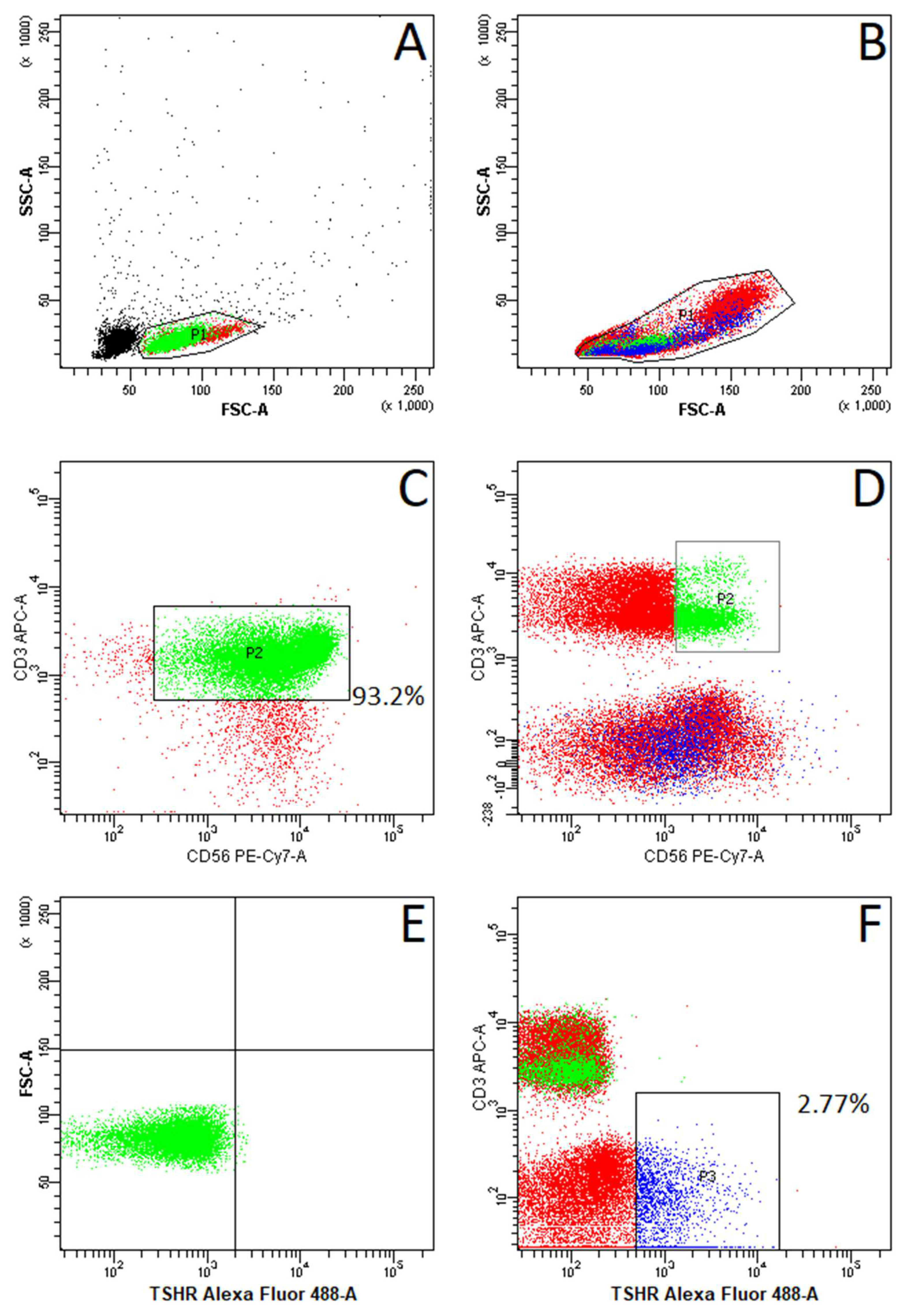
Figure 1. Exemplary plots of flow cytometry fluorescence-activated cell sorting (FACS) analysis showing the gating strategy of analyzed cells: (A): Natural Killer T (NKT) cells after magnetic sorting. (B): Peripheral blood mononuclear cells (PBMCs). (C): CD3+ CD56+ NKT cells after sorting. (D): CD3+ CD56+ NKT cells (green) among PBMCs. (E): Thyroid-stimulating hormone receptor (TSHR)-expressing NKT cells after sorting. (F): TSHR-expressing cells among PBMCs (blue). Black dots represent particles not recognized as single cells. Red dots represent negatively gated cells.
Ijms 25 11434 g001
Figure 2. Percentage of cells expressing TSHR among whole PBMC fractions and in NKT cells.
Ijms 25 11434 g002
Figure 3. Gating strategy showing the purity of NKT cells isolated from buffy coats.
Ijms 25 11434 g003
Figure 4. Reverse-transcription polymerase chain reaction (RT-PCR) results: (A): Amplification plots of target genes—(glyceraldehyde-3-phosphate dehydrogenase) GAPDH and TSHR from PBMC and NKT cells. (B): All measured ΔCt (relative TSHR gene expression to housekeeping gene expression) and ΔΔCt (ΔCt normalized to ΔCt median) values for TSHR gene in PBMC cells. Each dot identifies the mean expression calculated from triplets.
Ijms 25 11434 g004
Table 1. Clinical characteristics of the study group.
Patients with AITD
Mean ± SD
(n = 28)
Patients without AITD
Mean ± SD
(n = 58)
p Value
(AITD Versus Non-AITD Group)
Age55.54 ± 11.461.72 ± 14.43p = 0.05
Female/Male (n)23/545/13p = 0.63
fT4 (0.93–1.7 ng/dL)1.25 ± 0.271.27 ± 0.34p = 0.9
fT3 (2–4.4 pg/mL)2.83 ± 1.012.94 ± 0.69p = 0.56
TSH (0.27–4.2 uIU/mL)1.6 ± 2.391.3 ± 1.1p = 0.42
anti-TPO (<34 IU/mL)157.28 ± 149.0411.39 ± 4.9p < 0.05 *
anti-Tg (<115 IU/mL)187.37 ± 193.6515.5 ± 8.12p < 0.05 *
TRAb (0.8–1.75 IU/L)2.63 ± 5.170.99 ± 0.26p = 0.09
Abbreviations: fT4, free thyroxine; fT3, free triiodothyronine; TSH, thyroid-stimulating hormone; anti-TPO, thyroid peroxidase antibodies; anti-Tg, thyroid antithyroglobulin antibodies; TRAb, thyroid-stimulating hormone receptor antibodies. * indicates statistical significance.
Disclaimer/Publisher’s Note: The statements, opinions and data contained in all publications are solely those of the individual author(s) and contributor(s) and not of MDPI and/or the editor(s). MDPI and/or the editor(s) disclaim responsibility for any injury to people or property resulting from any ideas, methods, instructions or products referred to in the content.