Review

Evolving Strategies for Use of Phytochemicals in Prevention and Long-Term Management of Cardiovascular Diseases (CVD)

1
Advanced Biotherapeutics Ltd., 20-22, Wenlock Road, London N1 7GU, UK
2
Uppsala Inc., 67 Shady Brook Drive, Colora, MD 21917, USA
3
Department Pharmacology, Faculty of Pharmacy, University of Debrecen, Nagyerdei krt. 98, 4032 Debrecen, Hungary
4
HUN-REN-UD Pharmamodul Research Group, University of Debrecen, Nagyerdei krt. 98, 4032 Debrecen, Hungary
*
Authors to whom correspondence should be addressed.
Int. J. Mol. Sci. 2024, 25(11), 6176; https://doi.org/10.3390/ijms25116176
Submission received: 21 March 2024 / Revised: 16 May 2024 / Accepted: 22 May 2024 / Published: 4 June 2024
(This article belongs to the Special Issue Molecular Mechanism Study of Natural Products for Human Health)

Abstract

:
This report describes major pathomechanisms of disease in which the dysregulation of host inflammatory processes is a major factor, with cardiovascular disease (CVD) as a primary model, and reviews strategies for countermeasures based on synergistic interaction between various agents, including drugs and generally regarded as safe (GRAS) natural medical material (NMM), such as Ginkgo biloba, spice phytochemicals, and fruit seed flavonoids. The 15 well-defined CVD classes are explored with particular emphasis on the extent to which oxidative stressors and associated ischemia-reperfusion tissue injury contribute to major symptoms. The four major categories of pharmaceutical agents used for the prevention of and therapy for CVD: statins, beta blockers (β-blockers), blood thinners (anticoagulants), and aspirin, are presented along with their adverse effects. Analyses of major cellular and molecular features of drug- and NMM-mediated cardioprotective processes are provided in the context of their development for human clinical application. Future directions of the evolving research described here will be particularly focused on the characterization and manipulation of calcium- and calcineurin-mediated cascades of signaling from cell surface receptors on cardiovascular and immune cells to the nucleus, with the emergence of both protective and pathological epigenetic features that may be modulated by synergistically-acting combinations of drugs and phytochemicals in which phytochemicals interact with cells to promote signaling that reduces the effective dosage and thus (often) toxicity of drugs.

1. Introduction and Main Topic Review

Inflammation-associated oxidative stress and tissue injury: Dysregulated inflammation is a driving mechanism underlying the major symptoms of cardiovascular disease (CVD) and pathologenesis of many illnesses. Pathological inflammation is currently treated with small molecule pharmaceutical agents that are typically designed to act at single critical checkpoints in the inflammatory cascade. This strategy may be effective for suppressing major symptoms of many diseases. However, inflammatory processes are rarely cured by anti-inflammatory drugs presently available for human use and long-term treatments are often limited by drug cost and toxicity. Moreover, many phytochemicals widely distributed in human diet, particularly in common spices, have anti-inflammatory properties but are individually non- or marginally therapeutic. Previously, the authors of this report and others have shown that multiple phytochemicals may interact collectively to achieve efficacy similar to pharmaceutical drugs [1,2,3,4]. A major limitation of using phytochemicals in healthcare is that most of these compounds individually have very low potencies in comparison to anti-inflammatory drugs currently in use and require high doses to achieve even modest efficacy. These limitations can, however, be overcome by the use of formulations more reflective of a healthy diet, containing multiple phytochemicals that interact collectively. This phenomenon, called “phytochemical synergy” is a major concept considered in the current study. Spice-derived phytochemicals are an excellent example of this paradigm for inhibiting inflammatory tissue damage manifesting as disease symptoms. These dietary materials have been shown to constrain pro-inflammatory signaling mechanisms such as redox reactions and the nuclear factor-kappa beta (NF-kB) pathway to their appropriate physiologic venues—offering promise for the management of pathological inflammation [5,6].
Spice flavorings and nuclear-factor kappa beta Processes that involve NF-kB have emerged as very important mediators of disease- or trauma-associated inflammation, particularly as a major characteristic of syndromes with increased incidence among elderly populations such as cancer, atherosclerosis, cardiovascular disorders, diabetes, allergies, asthma, Alzheimer’s disease, osteoporosis, and many forms of autoimmunity [5,6].
The physiological and pharmacological mechanisms by which phytochemicals affect pro-inflammatory signaling pathways are an increasing focus of cutting-edge nutritional science. A case example of how this research may allow increasing use of phytochemicals in preventive medicine and therapy is provided by the characterization of SIRT1, a histone deacetylase (HDAC) enzyme upregulated by stress and also by certain compounds found in the human diet, such resveratrol, a phytochemical produced by grapes. SIRT1 directly binds to and regulates molecules of the NF-kB cascade [7,8], and may also impact the epigenomic profile in cells that express this enzyme. One specific outcome of this activity is that resveratrol can act through SIRT1 to inhibit NF-kB, resulting in the enhancement of tumor necrosis factor-alpha (TNF-A)-mediated apoptosis [7,8]. Apoptosis, a cell death program that removes damaged or dysregulated inflammatory cells, is a major mechanism for down-regulating inflammation and preventing it from becoming pathological [9,10].
Resveratrol analogs are being developed by Sirtris Pharmaceuticals partnered with GlaxoSmithKline as proprietary, orally available, small molecule drugs with the potential to treat age-associated diseases, such as type 2 diabetes, through the modulation of sirtuin activity. These efforts by Sirtris focus on modulating the expression of sirtuins. Curcumin, the major active phytochemical component of the spice turmeric, has shown some efficacy against inflammatory and neoplastic disorders—including pancreatic cancer, which is notoriously resistant to therapy [11,12]. Curcumin is also the subject of research to develop more potent analogs with increased bioavailability [5,6]. Other examples of therapeutic application of dietary phytochemicals include the use of preparations extracted from red pepper as topical analgesics and anti-inflammatories. Potent analogs of compounds found in pepper, such as capsaicin, have also been developed for medical use. Nevertheless, synthetic phytochemical analogs have the disadvantage of high cost and potential for adverse side effects that are avoided by naturally occurring bioactive compounds. A major objective in the development of phytochemicals for use in healthcare is to identify combinations of common foods and flavorings that synergize in strengthening immunoregulation.
Recent reports show the validity of phytochemical synergy as a healthcare strategy. For example, green tea concentrate and preparations of vanilloid compounds obtained from capsicum (red pepper) exhibit anti-cancer properties suggestive of synergy between polyphenols in tea called catechins and capsicum vanilloids, resulting in the inhibition of tumor-associated NADH oxidase (tNOX), an enzyme contributing to cancer proliferation [13,14]. Other studies demonstrated that combinations of curcumin and resveratrol exhibit 4-fold antioxidant synergism in heme-enhanced oxidation reactions [15,16]. Curcumin and resveratrol have also been observed to synergistically inhibit IL-8 expression by human cells in vitro exposed to the chemical weapon sulfur mustard (Cowan et al. unpublished observations). An area of special interest but limited experimentation and information is the ability of phytochemicals to augment the therapeutic potential of pharmaceutical drugs by enhancing their potency, which allows a reduction in drug dose, thus lessening toxicity [9,10,17,18,19]. An example of this effect is provided by the use of Ginkgo biloba extracts to lower the effective dosage of the immunosuppressive drug, FK506, required for cardioprotection in vivo [18,20]. Related work with ginkgolide B (GB), a component molecule of Ginkgo biloba extract, reduces the dosage of the immunosuppressant drug cyclosporin A required to inhibit inflammatory cytokine expression by human T cells in vitro [3,6]. Ginkgo leaf extract has also been shown to interact with vitamin C and the antioxidant carotenoid astaxanthin to mediate anti-inflammatory effects with the same potency as Ibuprofen, but without risk of the adverse gastrointestinal side effects caused by Ibuprofen and other NSAIDs [21,22]. These and related studies support the hypothesis that components of phytochemical-rich food and herbal products can interact to synergistically improve the efficacy of drug therapies [20].
Some of the biochemical, physiological, and pharmacological mechanisms mediated by phytochemicals have been reviewed; examples of this work include “Reasoning for Seasoning” [5], which characterized links between pro-inflammatory cellular signaling pathways and the capacity of spice-derived phytochemicals to inhibit NF-kB-mediated inflammatory processes [23].
Strategies for managing cancer using combinations of multiple phytochemicals (“Multifocal Signal Modulation Therapy”) were proposed by McCarty (2021) [24], and a comprehensive review of how the pro-apoptotic action of NF-kB-inhibitory phytochemicals may act as a pivotal defense against pathological inflammation and disease is described in Martins (2006) [10].
Previous and ongoing investigations suggest a major role for inflammation in chemical toxicity and support the potential efficacy of anti-inflammatory drugs and phytochemicals against chemical warfare agents (reviewed by Cowan et al., 2004, 2006, and unpublished observations); and: [19,25,26].
This work was the basis for Multi-Threat Medical Countermeasures (MTMC) technology which aimed to develop treatments for pathologies triggered by multiple classes of chemical agents with a single drug protocol (Cowan et al., 2004, 2006, and unpublished observations) [19,25,26,27]. MTMC technology may also find more general public heath applications because of the commonality of inflammatory mechanisms in chemical insult and disease [26,28,29]. MTMC synergistic combinations of anti-inflammatory drugs and/or phytochemicals have been confirmed in assay and animal models [29,30]. Further drugs with MTMC primary or pleotropic anti-inflammatory pharmacology are prescribed and administered to millions of patients, often in polypharmacy, with potential synergy and implications for the design of MTMC polypills [30]. MTMC can be a generic drug or supplements, thereby addressing affordability, which are practiced in any small clinic, reducing R&D costs. MTMC synergy assays and animal models for inflammation are readily available [26,31]. GLP-1 diet drugs are FDA approved for cardiovascular disease and have demonstrated efficacy for myriad other inflammatory pathologies [32]. Concerns of cost for on-patent MTMC vs. more affordable generic MTMC have been considered [28].
IL-8 is produced at the end of NF-kB-mediated cytokine cascades and its level in peripheral blood is a particularly reliable biomarker of pathological inflammation and the efficacy of anti-inflammatory therapies. IL-8 is a chemotactic, which strongly recruits neutrophils and inhibits their apoptosis, thereby impairing the resolution of inflammation and establishing conditions for chronic disease [5,6,10]. In previous studies, elevated levels of IL-8 have been associated with insult, injury, and a variety of pathologies and disease; these include chemical insult, specifically sulfur mustard exposure, and acute respiratory distress syndrome (ARDS) and associated multi-organ dysfunction syndrome (MODS) [25,26,33,34]. Sulfur mustard-increased IL-8 is a biomarker for the severity of mustard-induced inflammatory pathology and may also be used to gauge the efficacy of medical countermeasures to this chemical weapon [19,25,26].
IL-8, along with CRP and the pro-inflammatory cytokine interleukin-6 (IL-6), is an indicator of pulmonary disease severity in mustard-exposed persons [35,36,37,38].
The phytochemical capsaicin and its more potent drug analogs inhibit sulfur mustard-increased IL-8 production in cell culture and show efficacy against sulfur mustard-associated pathology in the mouse ear vesicant model [19,26,39]. Compounds that inhibit IL-8 production and reverse the anti-apoptotic and pro-inflammatory effects of this cytokine include many phytochemicals such as capsicum, curcumin, and resveratrol [5,10,25]. Such apoptotic pharmacology is also commonly observed for phytochemicals and drugs used as chemical agent countermeasures [10,25]. The proven capacity of chemical agents to trigger inflammatory processes and the role of inflammation in a wide range of pathologies makes phytochemical synergy fertile ground for research [19,40].
The capacity of numerous phytochemicals to inhibit IL-8 expression and associated inflammation makes this cytokine a logical biomarker for measuring LPS-increased inflammatory response by PBMC in the present study. The data on Aqua Spice™ food flavorings presented in this report advance the concept of phytochemical synergy. Phytochemical synergy technology is being used to design ImmuneACCORD™ dietary supplements. ImmuneACCORD Technology which encompasses the process of screening for phytochemical synergy, seeks to define mechanistic links between diet, dietary supplements, and drug discovery with the goal of more affordable and improved public health.

2. Cardiovascular Diseases (CVDs) and Ischemia-Reperfusion: A Paradigm of Oxidative Stress-Mediated Pathogenesis

Cardiovascular diseases are a category of syndromes involving acute and progressive damage to vascular and heart tissues which are classed into 15 well-defined disease processes, including venous thrombosis, heart failure, stroke, myocardial infarction (heart attack), coronary artery diseases (CADs), such as carditis, angina, and ventricular fibrillation, which are responsible for sudden cardiac death [41], hypertensive heart disease, rheumatic heart disease, peripheral arterial disease, valvular heart disease, congenital heart disease, thromboembolic disease, aortic aneurysms, and cardiomyopathy [42,43]. The phenomenon of dysregulated inflammation occurs as a prominent aspect of the 15th class of CVD on this list, ischemic/reperfusion (I/R)-associated disease [4,42,43,44,45], which is a major focus of the present report. The mitigation of these syndromes with synergistically acting phytochemicals present in functional foods is the clinical objective of the in vitro animal and human clinical strategies described here. The experiments described in the studies described in this review were conducted to assess combinations of drugs and natural products, in particular, ‘functional foods’ such as sour cherry extracts and spices for synergism, in suppressing the major pro-inflammatory mechanisms contributing to CVD and all other diseases in which these processes are contributors to pathogensis and symptom severity. CVD is here presented as a representative paradigm of disease in which hyperactivated host inflammation is a primary driver of emergence and progression of major symptoms. It is further important to emphasize that since this phenomenon is common to almost all known disorders, the outcomes of this work are broadly applicable for an enormous number of clinical venues. The findings revealed by the investigations described in this report are particularly exciting since they suggest a means of expanding and refining the known health benefits of foodstuffs that are widely available at low cost. Particularly exciting are reports showing the strong ability of commonly used spice flavorings to reduce the severity of cardiovascular ischemia/reperfusion-related symptoms, notably, turmeric [46], ginger [47,48], capsicum/capsaicin—in red pepper [49,50]—including paprika [51,52] and nutmeg [53,54,55]. Each of these spices contain bioactive compounds that interact to exhibit very potent anti-inflammatory synergy, with high potential for the long-term clinical management of CVD and many other disorders at minimal cost and with no adverse side effects.
Within the past half century, cardiovascular disorders have emerged as the primary cause of mortality worldwide, except among African populations, with the highest rates observed among residents of affluent nations, whose lifestyle-associated risk factors, notably fat-rich diets and general ‘overnutrition’, heavy tobacco and alcohol use, and lack of exercise, are typically higher than among people of developing countries [42,44]. Additionally, some strains of pathogenic streptococci responsible for strep throat may trigger host autoreactive attack on cardiovascular tissue, resulting in rheumatic heart disease following long latency periods if the primary infection is not treated with antibiotics at the time of occurrence [42,44].
The toll of these disorders is striking and has substantially increased in recent years. By 2015, 17.9 million deaths among human populations were attributable annually to cardiovascular diseases, which is an increase of 25.8% (12.3 million) from the number recorded in 1990 [43,56] and continued increases have been noted recently [4,42,57]. It is nevertheless interesting to note that, during the past decade, the personal utilization of risk reduction strategies available through public education has resulted in some abatement of mortality rates in affluent societies. This includes, prominently, an awareness of the benefits to general health of the dietary intake of polyphenols, especially in flavonoid-rich foods, and flavorings such as the spices described in the present report [5,6,46,47,48,49,50,51,52,53,54,55,56,57,58,59,60].
The data shown here and an analysis of the work by other investigators underscores this trend in public health consciousness and demonstrates how certain spice combinations may exert additive and, in some cases synergistic capacity for the prevention and long-term, side effect-free, management of CVDs, along with a diverse range of other disorders in which major symptoms result from hyperinflammatory reactions.
The underlying pathophysiological mechanisms for each of the syndromes listed above have been extensively documented. Peripheral and coronary artery disease and stroke, often progressing concomitantly with atherosclerotic changes to the vasculature, show strong correlations with sleep disorders and related psychological influences [61,62,63], lack of exercise, alcohol and tobacco use, elevated blood cholesterol, obesity with insulin resistance and resulting type 2 diabetes, obesity, and high blood pressure [42,44]. Significantly, in the context of findings reported here, approximately 53% of CVD-associated mortality is currently linked to dietary risk factors [64].
Ultrastructural alterations typically observed in the heart and vascular tissue of CVD patients that correlate with deteriorated function, typically include fibrotic changes and the accumulation of amyloid deposits in the interstitium—as shown by the immunohistochemical micrograph in Figure 1. Inflammatory heart diseases are particularly debilitating, refractory to treatment (especially long-term), and often fatal [44,65]. Disorders in this category include such conditions as endocarditis, in which the heart’s inner layer, the endocardium, becomes inflamed, with resulting damage to the heart valves [66]. Likewise, cardiomegaly, a disorder characterized by the hypertrophic enlargement of the organ, with resulting loss of function, and myocarditis, a disease process in which monocytes and lymphocytes or eosinophiles infiltrate the heart muscle, are consequences of a failure to regulate inflammatory signaling processes, with consequent tissue damage [67,68,69,70,71,72]. Significantly, the triggering etiology for these pathologies may be viral, bacterial, or toxin-induced [69,70], thus raising the risk that some infectious diseases may engender cardiovascular complications.
A major positive aspect to the current understanding of these diseases is that it is estimated that as much as 90% of CVD may be prevented by intuitively sensible lifestyle measures, especially an avoidance of alcohol and tobacco use, along with exercise and a healthy diet rich in foods containing antioxidant polyphenols [73,74,75]. It has also been observed that patient compliance with regimens for the therapy of CVD risk factors such as diabetes, high blood pressure, and elevated blood lipids, also has significant corollary effects in CVD prevention and treatment [73,74,75].
An increasingly broad and versatile selection of drugs for the prevention and treatment of CVD has come into clinical use in recent years. A detailed consideration of the full range of emerging CVD drugs is beyond the scope of the present report. Thus, in order to clearly present how phytochemical synergy may be used as a countermeasure to the onset and progression of the driving inflammatory pathology underlying CVD—and indeed, all other diseases at some level—the major advantages and drawbacks to four of the best-known representatives of CVD pharmacotherapies will be considered here, with these being statins, beta blockers, blood thinners, and aspirin.

3. Representative Pharmaceutical Agents Used for CVD

3.1. Statins

These are a class of agents widely used for the prevention and management of CVD which function through the inhibition of the enzyme 3-hydroxy-3-methyl-glutaryl-coenzyme A (HMG-CoA) reductase, which in mammalian cells, including humans, is the major NADH-dependent regulator of mevalonate metabolism and processes responsible for the biosynthesis of isoprenoids, including cholesterol. In healthy cells, the activity of this enzyme is competitively suppressed through negative feedback thus limiting the availability of cholesterol to levels of optimal use in healthy physiologic function [76,77]. Statins inhibit HMG-CoA reductase, thereby reducing cholesterol production at levels that, depending on the overall health of an individual and the presence of other risk factors, may decrease the development of dyslipidemia and the resulting illness and death [76,78].
Adverse effects of statins. The statin-mediated suppression of cholesterol production is accompanied by a concomitant reduction in the blood levels of low-density lipoprotein (LDL) cholesterol, a complex of cholesterol with a carrier protein, responsible for the transport of fats to cells. Most cholesterol in a body is bound in this form. Elevated LDL levels correlate with an increased risk of stroke, heart disease, and related problems, and influences that reduce LDL also decrease the risk of a wide range of cardiovascular problems [79,80]. The LDL-lowering properties of statin drugs have thus made this class of compounds a highly potent clinical countermeasure to CVDs; nevertheless, their use is also associated with a number of significantly adverse side effects such as aberrant liver enzyme activity in peripheral blood and an elevated risk for type 2 diabetes [81,82], along with a rare but severe occurrence of muscle impairment and pain [83]—and anecdotal but increasingly frequent reports of hepatotoxicity.

3.2. Beta Blockers (β-Blockers)

This class of drugs is a compound that competitively antagonizes the binding of the endogenous catecholamines, adrenaline (epinephrine) and noradrenaline (norepinephrine), to adrenergic beta receptors which are their cognate sites expressed by cells of the sympathetic nervous system and by major organs participating in stress responses, notably, downstream events such as fight-or-flight response [84,85]. This class of drugs is found to be particularly valuable in stabilizing cardiac arrhythmias to reduce the occurrence of secondary heart attacks following a first attack, and are also useful in the treatment of high blood pressure [85,86]. Three variants of β-adrenergic receptors are known (β1, β2, and β3), each with characteristic anatomical distribution to major organs such as the heart, lungs, uterus, smooth and skeletal muscle, kidneys, gastrointestinal tract, and vascular and adipose cells [84,85]. Some beta blockers are capable of blocking the activation of all three of these receptors, while others exhibit selectivity for one receptor type [85,87]. The stimulation of beta receptors with adrenaline on components of the sympathetic nervous system, especially within the kidneys, smooth muscle, heart muscle, arteries, and tracheobronchal tree, produce stress responses that may be deleterious to the heath of an affected individual. Consequently, beta blockers that inhibit the binding of this hormone to its complementary beta receptor may attenuate its adverse effects. Indeed, these drugs have proven very useful since the pioneering work of James Black in 1964 showing the potent capacity of the beta receptor-blocking drugs, pronethalol and propranolol, in the treatment of angina pectoris, which is considered to be one of the of the 20th century’s most significant developments in clinical pharmacology [88]. The sympatholytic β1 activity of beta blockers decrease incidence and severity of congestive heart failure—and further suppress pathological inflammatory activity through effects on the renin–angiotensin system [85,89]. The interaction of these drugs with their receptors in the kidneys reduces renin secretion, causing a principal effect of suppressed oxygen requirements of the heart through decreased extracellular fluid volume, thereby engendering concomitant increased capacity of the blood to carry oxygen [89]. The notable beneficial consequence to this on the prognosis of heart failure and associated inflammatory tissue damage arises from the inhibition of catecholamine influences on the heart, a phenomenon shown to be responsible for the elevated expression of reactive oxygen species (ROS) and other mediators of inflammation such as the cytokines TNF-A, interleukin-1beta (IL-1B), and interleukin-6 (IL-6)—in addition to the catecholamine-mediated deleterious remodeling of cardiac tissue [90,91], high oxygen demand, and decreased cardiac contractile efficiency—effects which collectively decrease the ejection fraction volume and exacerbate inflammatory damage to the heart [91]. These very potent effects have resulted in substantial benefits for the prevention and management of CVD. Indeed, by 2004, the use of beta blockers was shown to be primarily responsible for a 4.5% reduction in mortality during a 13-month representative timeframe in which clinical evaluations were conducted [92].
Adverse effects of beta blockers. The clinical advantages of beta blockers notwithstanding, these drugs are also associated with significantly serious adverse side effects. For example, this repertoire of problematic outcomes includes the results of a 2007 investigation indicating that their use when co-administered with diuretics for hypertension increase the risk for type 2 diabetes—which may be counteracted, to some extent, by combining such a regimen with angiotensin II receptor blockers [93]. Moreover, lipophilic beta blockers, including the commonly used drugs propranolol and metoprolol, which have high penetration across the blood–brain barrier, are known to result in sleep disturbances, including insomnia and nightmares [94].
Further disruption of healthy physiologic processes contributing to metabolic syndrome may occur as a result of the inhibition by these drugs of β2-adrenoceptor-mediated glycogenolysis by the liver and subsequent pancreatic glucagon release, which normally increases plasma glucose as needed, but, when inhibited, may lead to hypoglycemia [93].
Other beta blocker-associated side effects include exacerbation of Raynaud’s syndrome, cold extremities, hypotension, bradycardia, heart failure, dizziness and fatigue, bronchospasm, peripheral vasoconstriction, hair loss, insomnia, nightmares and hallucinations, insomnia, erectile dysfunction, diarrhea, dyspnea, blurred vision, and nausea [94,95]. The above-described complications and many other adverse effects make this class of drugs very much a double-edged sword for the prevention of and therapy for CVD.

3.3. Blood Thinners (Anticoagulants)

Pharmaceutical agents in this class are anticoagulants, which interfere with the normal cascade of biochemical events leading to clot formation—a drug-mediated effect that extends clotting time and decreases blood viscosity, thereby reducing the risk of thrombus formation and stroke [96,97]. These drugs may have both a direct and corollary regulatory effect on dysregulated inflammation in ischemia/reperfusion-associated pathologies, through relief of pro-ischemic influences, such as ‘Virchow’s Triad’: myocardial infarction-associated endothelial injury from blood stasis due to thrombus-forming ventricular dysfunction, and hypercoagulability induced by inflammatory mediators—in particular ROS and inflammatory cytokines (Virchow’s Triad) [98]. In addition to their clinical use for thrombic disorders, blood thinning compounds are often added to the working fluid of some kinds of medical equipment such as heart–lung machines, blood transfusion bags, and dialysis apparatus [99,100].
Adverse effects of blood thinners. The most dangerous and frequently occurring side effect associated with the use of blood thinners is the induction of both major and nonmajor bleeding—the severity and threat to health for a patient being significantly dependent on the type of drug used, pre-existing health conditions, and age [101]. For example, the annual incidence of life-threatening bleeding events for persons administered warfarin, a commonly-used anticouagulant, was 1–3% annually as of 2011 [102,103]. A principal hazard associated with many of these agents is their vitamin K antagonism properties—which increases bleeding risk and has motivated the development of new, non-vitamin K antagonist oral anticoagulants which appear less prone to causing life-threatening bleeding events versus warfarin [104,105]. A particularly useful agent in this class is apixaban, marketed as Eliquis, which has an excellent safety and efficacy profile and has emerged as a primary choice of caregivers for antithrombotic therapy [103,106]. Apixaban has nevertheless been shown to carry some risk of bleeding events as revealed by comparisons with edoxaban, reported in 2022 [107], and a 2017 study showing that this occurred in 107 people administered the drug, per 1000 in the study population, with minor bleeding in 167 per 1000 subjects [108].

3.4. Aspirin

This compound, formed by the esterification of salacylic acid to acetylsalicylic acid (ASA), is a naturally occurring component of willow bark which has extensive historical use by humans as an analgesic in the form of bark extracts and tinctures—and is currently marketed without requirement for medical prescription in purified form as a nonsteroidal anti-inflammatory drug (NSAID) used to suppress fever, pain, and other inflammation-associated aspects of a wide range of illness and trauma—with corollary use as an antithrombitic [109,110]. There is also some evidence for the value of this agent in decreasing the risk of ischemic stroke and secondary heart attack in susceptible individuals [111,112]; nevertheless, support for use of regular aspirin intake as general prophylaxis against CVD is substantially anecdotal at the time of this writing [113,114].
Adverse effects of Aspirin. Aspirin is sold in most countries as a generic drug, generally regarded as safe (GRAS) due to its long historical usage record in many societies. Nevertheless, some adverse side effects of the agent are known—and it should therefore be used with caution appropriate to the presence of risk factors for a particular patient, with awareness of the attendant drawbacks. For example, the compound is known to cause gastrointestinal irritation, with resulting stomach upset [112]. For some persons, this side effect may become severe, resulting in the development of bleeding from the stomach and, occasionally, ulcers [112,115]. Other adverse outcomes of aspirin use include increased severity of respiratory symptoms in asthma patients and exacerbation of bleeding by patients being treated with blood thinner regimens [116]. Additionally, high aspirin doses may result in tinnitus (ringing in the ears) and may increase risk of Reye syndrome in children [117].

4. Emerging Plant Phytochemical Supplements with Cardiovascular Applications and Functional Foods

4.1. Ginkgolides

Ginkgolides are phytochemical compounds present in the leaves, roots, fruit, and bark of the Ginkgo tree, the oldest living survivor of its taxonomic order: Ginkgolides, with its origins as a distinct class of plant species dating from over 290 million years ago. The plant is a native of China, where its fruit is consumed—and other parts of the plant have been used in traditional medicine through the history of Chinese societies, with widespread usage documented from the 11th century C.E [118,119]. Anecdotal but well documented descriptions of this use describe it as showing benefit in the treatment of kidney and bladder disorders, bronchitis, and asthma—with numerous reports suggesting its value in the treatment of dementia [118,119]. An analysis of these reports by the European Medicines Agency Committee on Herbal Medicinal Products in the context of modern understanding of natural medical materials (NMM) concluded that extracts of ginkgo leaf may be of mild benefit for peripheral vascular disease and dementia in elderly patients [120,121]. Nevertheless, investigations during the late 1900s and the early part of this century, failed to reveal the potent capacity of the plant extracts to prevent or remediate any serious disease. This notwithstanding, (weak) preliminary evidence exists for the ability of ginkgo to at least mildly ameliorate major symptoms of dementia and tardive dyskinesia symptoms in schizophrenia patients [121,122].
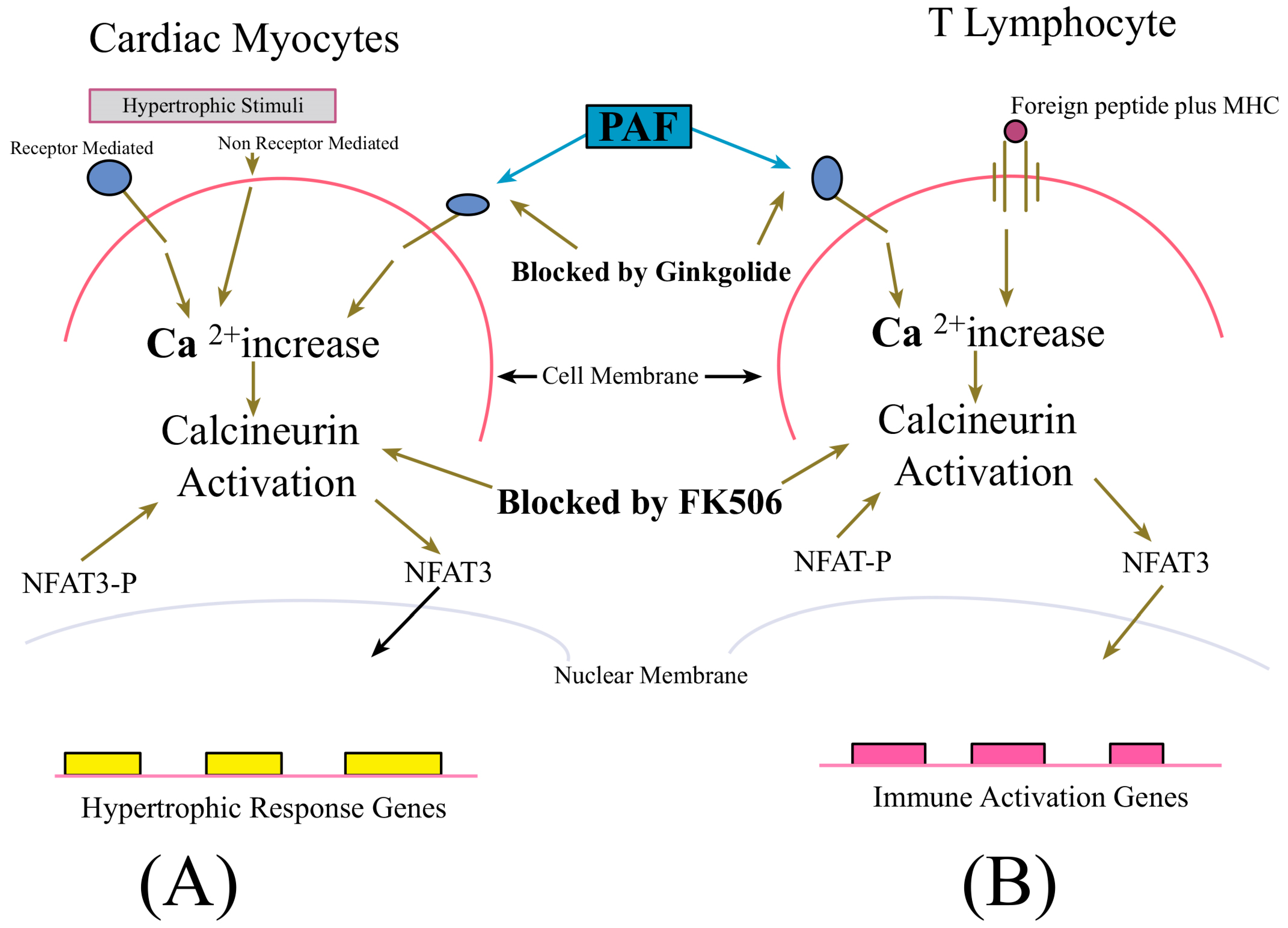
The discouraging outcomes of searches by other investigators for evidence of the clinical value of ginkgo components initially led our laboratory to exclude this NMM as a candidate for the development of novel strategies for the use of dietary and complementary medical approaches to counteract dysregulated inflammatory processes in disease—particularly CVD. Nevertheless, the biochemical nature of phytochemicals in the plant was sufficiently intriguing to lead members of our laboratory to speculate that one or more of them might exhibit potentiation of the cardioprotective effect of FK506, a macrolide immunosuppressant, which at the time of our research planning had been shown to inhibit cellular signaling processes in cardiac myocytes, known to be the underlying pathogenic mechanism of cardiac hypertrophy—as shown in Figure 2 below. Extracts of ginkgo leaves contain proanthocyanidins, bilobalides, phenolic acids, quercetin, flavonoid glycosides, such as kaempferol, isorhamnetin, myricetin, and terpene trilactones [chemical analysis and quality control] of Ginkgo biloba leaves, extracts, and phytopharmaceuticals [123,124,125]. The leaves also contain unique ginkgo polyphenols alkylphenols and biflavones [125]. This pharmacopia served as incentive to test the leaf extract (EGb761) for capacity to amplify the effects of FK506 in counteracting major consequences of cardiovascular I/R injury with a corollary effect of inhibiting the potentially pathologican pro-inflammatory activity of activated T lymphocytes—via the parallel signal modulation illustrated in Figure 2. The major clinical benefit being the potentiation of pharmacological effects and reduced dosage requirements for FK506—with resultant lowered toxicity in patients. The possibility of achieving such an effect was raised by the observation that terpene trilactone ginkgolides present at high concentrations in EGb761 exhibit dual ability as a potent antioxidant and platelet-activating factor (PAF) receptor antagonist, as shown below in Figure 2 [126].
The authors of the present report undertook the experiments previously described to test a hypothesis that combined therapy with macrolides, such as FK506 co-administered with ginkgolides, would reveal synergism in the cardioprotective capacity of these agents. Our results validate observations by other investigators demonstrating the cardioprotective properties of FK505 [127,129,130], and the discovery in our laboratory that combination therapy using FK506 plus EGb 761 synergistically augments postischemic cardiac function, while reducing the occurrence of reperfusion-induced arrhythmias [18], offering the potential for expanding the clinical utility of FK506 and allowing therapy with this drug and other macrolides—possibly including rapamycin, at lower doses than are currently useful [131].
CVD, cardiac surgery, and other heart trauma frequently result in periods of myocardial ischemia. The restoration of blood supply and reoxygenation of the myocardial tissue causes characteristic reperfusion injury, manifested by symptoms that include ventricular arrhythmias and deterioration in cardiac functions [132,133]. This effect is due substantially to myocardial membrane lipid oxidation by pro-inflammatory free radical species produced in a burst during the first minutes of reflow, resulting in impairment of the cell membranes [134,135]. Moreover, myocardial function may be improved by the administration of antioxidants following ischemic events [134,136,137]. Extending our hypothetical model at the time the above experiments were conducted, it was speculated that EGb 761 and selected component ginkgolides were expected to interact with FK506 so as to diminish reperfusion injury by two major mechanisms: one involving the quenching of reperfusion-associated free radicals and a second, dependent on their PAF-receptor antagonist properties—which limits cytoplasmic calcium increase in cardiomyocytes [138,139], thereby reducing the level of transcription factor nuclear factor activation of transcription (NF-AT)-dependent signals for the expression of pro-hypertrophic and pro-inflammatory genes in cells of the isolated hearts.

4.2. Heme Oxygenase Inducers

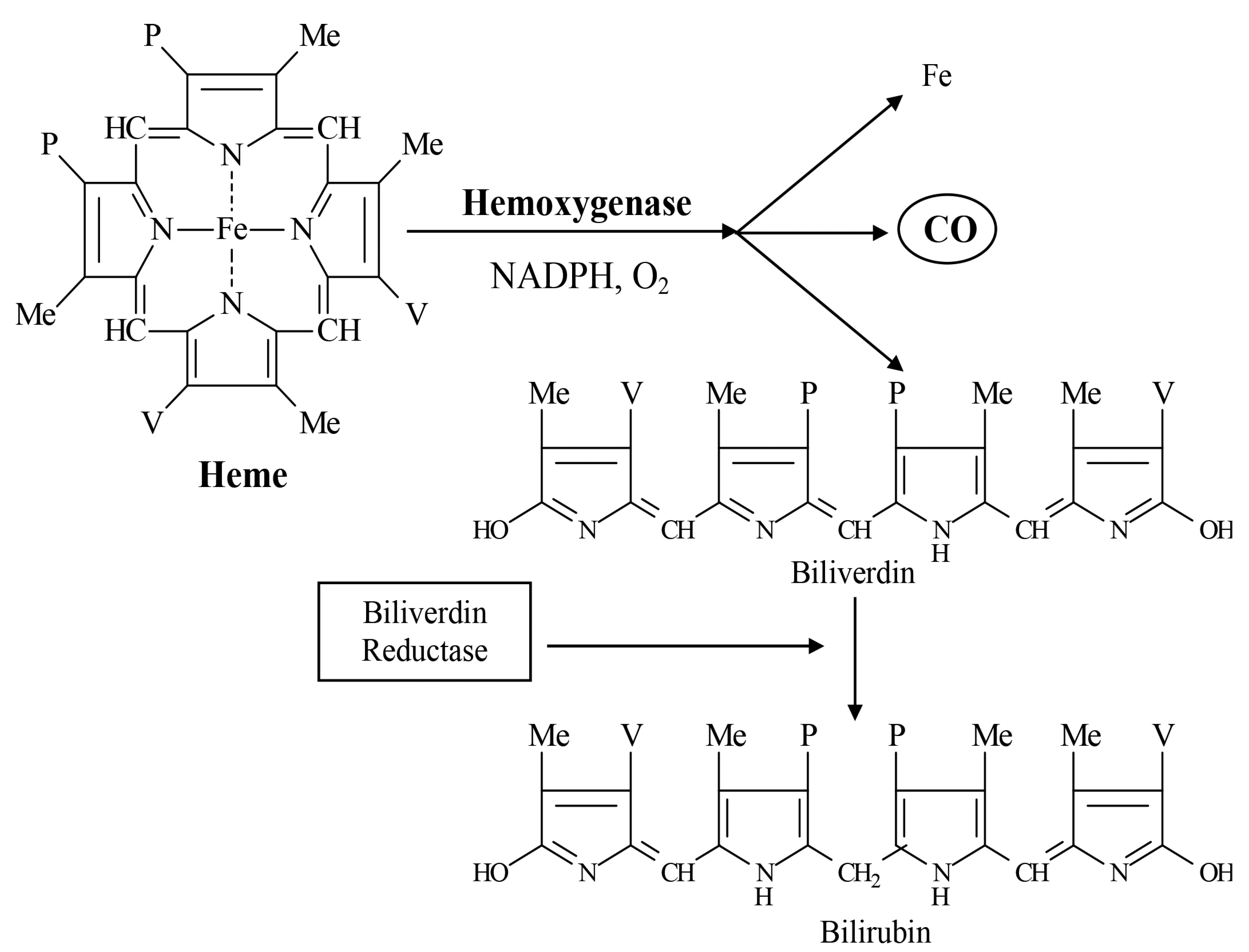
Heme oxygenases (HMOX) are heat shock protein (HSP) enzymes with a major physiological function of degrading heme released by red blood cell turnover to biliverdin and bilirubin; ferrous iron (Fe2+); and carbon monoxide (CO), as shown below in Figure 3. The potential toxicogenic effects of Fe2+ are counteracted through its sequestration to the ferritin protein, but the metal nevertheless remains capable of generating reactive oxygen species via Haber–Weiss or Fenton reactions [138,140]. Bilirubin and CO, produced as the end products of the HMOX degradation of heme, act as essential signaling molecules for a diverse array of essential physiologic processes—with bioactive properties that have become the focus of numerous efforts to prevent and treat chronic illnesses which have proven refractory to current therapeutic approaches [141,142].

5. Major Physiologic Effects of HMOX Product Metabolites

Carbon monoxide (CO): This signaling molecule stimulates cGMP-mediated vasodilation and suppresses ET-1—thus reducing the severity of hypertension—inhibits the proliferation of vascular smooth muscle cells (reducing need for stenting of cardiac patients); suppresses ventricular fibrillation and ischemia/reperfusion injury; and stabilizes Na+ and K+ distribution across membranes of cardiovascular cells [2,143].
Bilirubin: This potent endogenous antioxidant stabilizes Na+ and K+ distribution across membranes of cardiovascular cells and quenches ROS at levels that inhibit ischemia/reperfusion injury and diminish the occurrence of atherosclerosis and heart failure [2].
Three HMOX isoforms are encoded by most vertebrate genomes and expressed in response to epigenetic cues. These are as follows: Heme oxygenase 1 (commonly HO-1, aka HSP32), a stress-inducible form with immunoregulatory and cytoprotective roles which have made it the major focus of efforts to develop this enzyme class as clinical tools [144,145]. Heme oxygenase 2 (HO-2) is a constitutively expressed variant produced primarily in the brain, endothelial cells, gastrointestinal tract, and testes [144,146]. Moreover, whereas HO-1 is inducible by a wide variety of stressors and drugs, HO-2 is inducible only in response to adrenal glucocorticoids [147]. Heme oxygenase 3 (HO-3) is the third heme oxygenase variant, a 33 kDa protein expressed in kidneys, liver, and prostate [144,147] but with no clear biological role established for this isoform at the time of this writing.
In addition to its hemodynamic/housekeeping role, enzymes in this class serve as essential cellular cytoprotectants through their antioxidant capacity, protecting cells and tissues against oxidative damage by maintaining the tight regulation of inflammatory processes [2,148]. As understanding of the bioactive properties of this remarkable heat shock protein has increased—especially in the decade prior to this writing, the potential for the development of novel products and strategies based on its manipulation seemed enormous. A major advantage that HMOX-based interventions would have versus the currently available countermeasures to pathological inflammation, is a consequence of the fact that because exogenously administered antioxidants such as vitamins localize mainly to interstitial spaces, their clinical utility is limited—making them mostly unsuitable as first line countermeasures to pathologically activated inflammatory processes [128,149].
Exogenous antioxidants are typically small molecules obtained through diet, which decrease oxidative stress burden by the chemical inactivation of reactive oxygen metabolites. One well-known example is vitamin C (l-ascorbic acid or l-ascorbate), an aqueous reducing agent for oxygen radicals, which is, in turn, regenerated by glutathione-mediated reduction [128,150]. This vitamin is a major dietary antioxidant which additionally acts as a cofactor for antioxidant enzymes [128]. Vitamin E (tocopherols) are another major class of antioxidant vitamins. Tocopherols are lipid-soluble methylated phenols which reduce oxidized ascorbate thus augmenting vitamin C bioactivity [151,152]. In vertebrates, they act in concert with other small molecules such as ascorbate and the tripeptide glutathione, which includes a cysteine sulfhydryl group (-SH) that functions as a proton donor in the quenching of oxygen metabolites [153,154]. Several vertebrate enzymes degrade reactive oxygen species into molecules that are either harmless or rapidly metabolized. For example, superoxide dismutases convert superoxide into hydrogen peroxide (H2O2), oxygen, and hydrogen (McCord and Fridovich 1988)—a reaction occurring in tandem with the conversion of H2O2 into O2 and water by catalase [155,156], and peroxidase activity, which neutralizes organic hyperperoxides, such as the toxic products of lipid peroxidation, thereby supporting cell membrane integrity and the regulation of potentially pro-inflammatory and pathological cellular signaling processes [157]. Nevertheless, host antioxidant defenses in the form of endogenously produced compounds, such as glutathione, are typically overwhelmed by high levels of reactive oxygen species that occur as a major features of processes by which dysregulated inflammatory signaling leads to disease—including the critical properties of cancer and carcinogenesis [128,158]. By contrast, heme oxygenases perform their biological functions both at their sites of synthesis within cells, in which form they are termed intracellular HOs (iHOs), along with extracellular forms, (eHOs) which are distributed systemically in the circulation and other anatomical venues [128,158]. Moreover, in most reactions, HOs do not directly inactivate the cytotoxic properties of reactive oxygen molecules. Rather, their primary contribution to antioxidant defense occurs via the immunoregulatory effects of HMOX product metabolites CO and bilirubin—as previously described in Section 4 and Section 5 of this report, with a contributing discussion in Haines and Tosaki, 2018 [2,159]. Thus, HOs seemed to offer enormously valuable clinical tools which, due to their diverse venues for biological activity, avoid the limitations of exogenously administered drugs (including vitamins C, E, and others with antioxidant properties) which distribute to anatomical sites where their bioactivities are not major contributors to counteracting the underlying causes of inflammatory disease. Particularly intriguing findings have emerged from studies of heme oxygenase participation in cardiovascular physiology.

6. Heme Oxygenase in Cardiovascular Disease: Limits of Clinical Use

Ongoing studies of HO-1 biology are increasingly focused on the characterization of the enzyme’s capacity to mitigate the major symptom severity of cardiovascular disease [2]. The importance of these HOs to cardiac health is underscored by an emerging understanding of discoveries that CVD risk is increased significantly in persons with HO-1 gene promoter polymorphisms that adversely affect normal HO expression and regulation [160,161]. Moreover, since the 1980s, it is known that CO produced as a product of HO-1 heme degradation stimulates cardiovascular cells to augment vascular tone and vasodilation through guanylate cyclase stimulation and increased cGMP expression by vascular endothelial cells. These processes, which are physiologically coordinated with nitric oxide (NO) signaling, have been observed in hypertensive rodent models to result in increased coronary arterial pressure and blood flow while at the same time reducing total peripheral resistance [162,163]. These effects are reversed by the inhibition of HO-1 [163]. CO-induced guanylate cyclase activity, which increases cGMP-mediated events, has additionally been observed to inhibit the proliferation of vascular smooth muscle cells (SMC) with resulting occlusion of cardiac vasculature by neointima [163,164]. These observations offer the potential for novel strategies in the prevention of pathological vascular remodelling and for improved drug-eluting stent design. Additionally, bilirubin produced by HO-1 activity reduces platelet aggregation and leukocyte—processes that contribute to cardiovascular inflammation [163,164].
Both CO and bilirubin produced by HO-1 activity in the cells of cardiovascular tissue ameliorate a diverse range of cardiovascular pathologies due to the effect of these two degradation products on vascular tone [163,164]—along with the stabilization of K+ and Na+ ion membrane distribution [165]. Moreover, the regulatory effects of HO-1-generated bilirubin on the expression of ROS by NADPH oxidase has also been demonstrated to be cardioprotective as evidenced by the prolonged survival of mouse-to-rat cardiac xenografts, the inhibition of IR and balloon injury, and improved cardiac functions [163,166,167].
Related studies by the authors of this report and other investigators demonstrate HO-1 activity and enhanced carbon monoxide signaling induced by ischemia/reperfusion injury diminishes infarct size and enhances post-ischemic recovery in rat hearts ex vivo [168,169,170].
HO also exhibits potential for use as a countermeasure to cardiac hypertrophy, thereby suggesting that inducers of the enzyme may be useful in combination with FK506 and ginkgolides as described in the present report [18]. For example, an assessment of HO-1 as an antihypertrophic agent—published in 2009—demonstrated that an overexpression of HO-1 in cardiomyocytes in which NF-kB-dependent hypertrophy had been triggered via exposure to hydrogen peroxide caused the suppression of hypertrophy-associated cellular changes. This is a representative example of the plethora of exciting clinical applications of HO biology [171,172]. These encouraging research outcomes notwithstanding, by 2018, it had become evident that the development of novel HO-based strategies available to the public had fallen short of this apparent promise. Attempts to base workable clinical protocols on known properties of the enzyme encountered obstacles in the design of formulations capable of selectively increasing HO activities at sites optimal for preventing and treating disease [141,148]. In recognition of the above-mentioned constraints on the clinical use of HOs, the authors of this report undertook the characterization of contributions of these enzymes to cardiovascular function, and the remediation of CVD [2,4,173]. The above-described analyses of findings by other investigators was combined with in vitro, animal, and human clinical studies at the University of Debrecen and Kuwait University to evaluate clinical efficacy in phytochemical HO inducers—notably fruit seed kernel flavonoids—to counteract hyperinflammatory tissue damage in CVD and other disorders. This research initiative began in 2004, with the discovery by authors of this report of potent HO-inducing properties in the flavonoid fraction of seed kernels of sour cherry (Prunus cerasus) [165], and was extended in 2014 by a demonstration of the cardioprotective ability of sour cherry seed kernel flavonoids [174].
Thereafter, a major effort was undertaken by this laboratory to utilize HO inducers in sour cherry seed extracts (SCE) to mitigate the major symptoms of various serious chronic illnesses through the capacity of these enzymes to quench pathological ROS levels. The major focus of this research was in the realm of cardiovascular medicine. Representative studies in this area include the evaluation of endothelin-1 (ET-1) to attenuate hypertrophic signaling in H9c2 rat cardiomyoblasts in vitro, corresponding to in vivo processes underlying cardiac hypertrophy [159]; the treatment of rat hearts ex vivo, with alpha-melanocyte-stimulating hormone (alpha-MSH) to induce vasodilation and mediate cardioprotection via an HO-dependent pathway [175]; and a demonstration in rats of beta carotene (BC)-mediated reduction in infarct size, increased total antioxidant capacity of the myocardium, and other cardioprotective effects occurring at relatively low BC dose but (counterintuitively) diminished at higher doses of BC—an effect which may be due to the inefficient clearance of Fe2+ produced by HO activity [176].

7. ‘Biotherapeutic’ Use of Heme Oxygenase in Osteoarthritis Treatment: Phase 1 Human Trials

Particularly encouraging evidence that the SCE flavonoid-mediated induction of HO activity may be a breakthrough means for the medical use of HO was offered by a human clinical study published in 2014 by authors of this report, showing that a topical SCE preparation dramatically reduced pain and inflammation-associated serum and immune parameters through systemic HO activation in a cohort of Kuwaiti osteoarthritis (OA) patients [177].
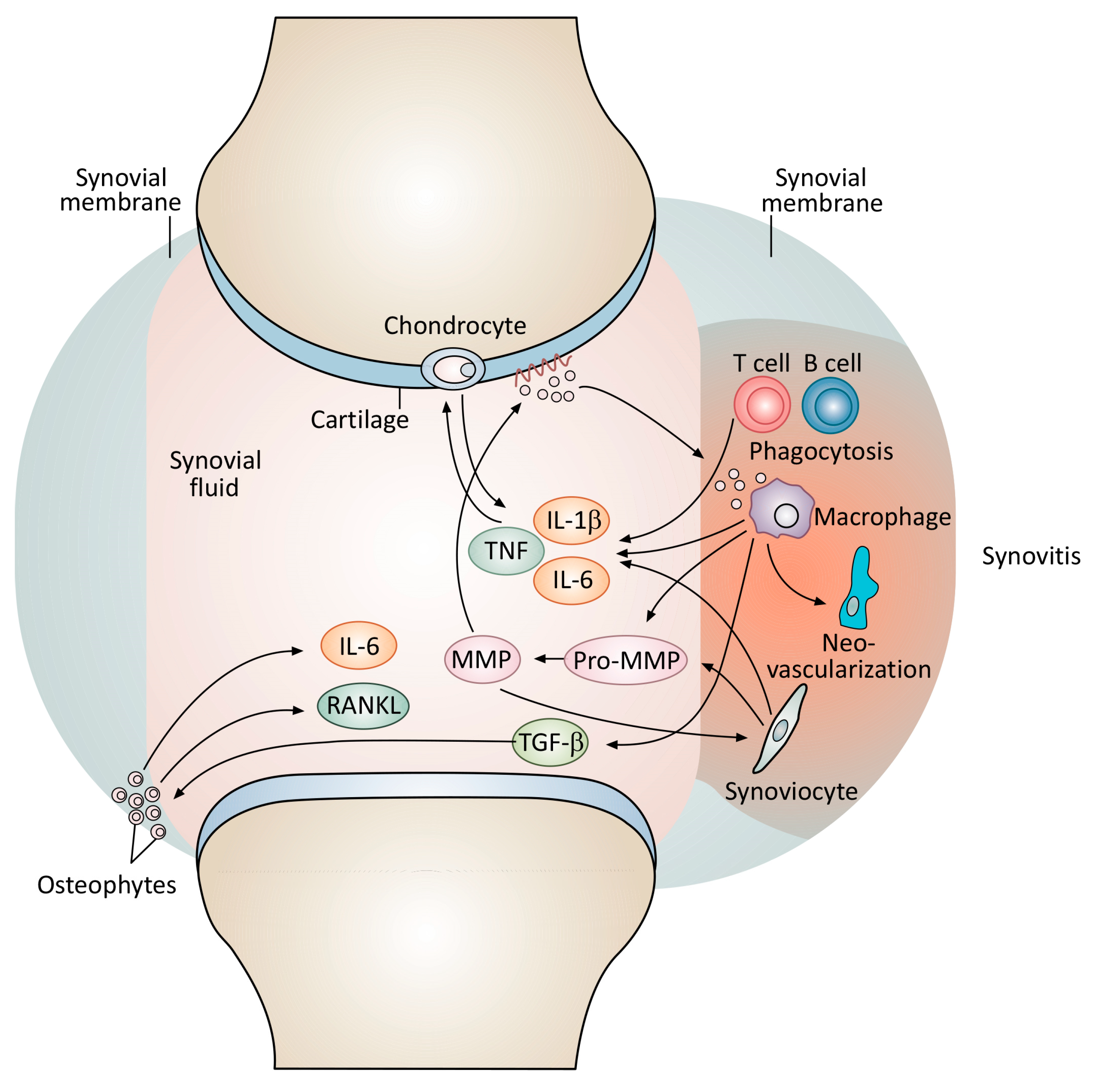
Figure 4 shows the major known pathomechanisms for OA. Potentially destructive lymphocyte activation and the resultant production of inflammatory mediators served as major therapeutic targets for the human study described in Mahmoud et al., 2014. As described by that report, the systemic SCE-mediated induction of HOs diminished pathological T cell activation, resulting in significantly decreased pain, along with a significant reduction in the serum content of C-reactive protein (CRP) [177]. The outcomes of this clinical investigation and related studies suggest that the capacity of sour cherry seed flavonoids to potently remediate pain and other symptoms of serious if dysregulated inflammatory processes—as described—finally provides a means to overcome obstacles to the wide clinical use of heme oxygenases—reviewed by Hopper et al., 2018 [141].

8. Dietary Phytochemicals/Functional Foods

Almost all foods alter an individual’s risk for and response to disease and those rich in plant phytochemicals may be particularly beneficial for CVD prevention and management. Foods selected and blended to ‘support’ endogenous countermeasures to ill health are known as “functional foods”, a term that encompasses a wide range of substances regularly consumed as components of the human diet. As discussed in the first part of Section 2 of this report, supplements, which may include phytochemical-rich edible material such as spices like turmeric and nutmeg, capable of increasing the expression of the endogenous antioxidant enzyme heme oxygenase and extract of sour cherry seeds, which also stimulates high expression of the enzyme, are classed as functional foods [55,178]. A particularly valuable outcome of the experiments on which this report is based, is the creation of an easy-to-use in vitro platform, by which functional food preparations (in the form of Kancor Aqua Spices and other foods containing “natural medical material” (NMM) may be used as aqueous phase reagents—and be assessed for synergism in their capacity to down-regulate potentially pathological signaling processes in human cells.
Dietary modifications to reduce CVD susceptibility and treat its symptoms are strategies most completely under the control of individuals at risk for developing these diseases; however, they are also among the most difficult to pursue successfully. Adherence to heart-healthy diets at levels sufficient to result in a significantly positive impact on cardiovascular health typically requires a level of commitment and sustained discipline that many people are incapable of, even with the knowledge that failure to do so puts their life in jeopardy. Moreover, highly specialized dietary regimens, with attendant involvement of medical caregivers and paid advisers, often require financial outlays that exceed the resources most people have to draw on. The investigation on which this report is based was conducted to aid in meeting this challenge. The data shown here offer an approach by which optimal combinations of selected dietary components with known benefits in stabilizing disease-associated cellular signaling pathways may be rapidly identified—leading to food and flavoring compositions with preventive and therapeutic potency in the same range as pharmaceutical drugs. First, some major heart-healthy foods should be considered.
Most people have some insight into aspects of diet and lifestyle contributing to generally good health—including the avoidance and management of CVD. This includes commonsense behavior such as regular exercise and the avoidance of smoking and alcohol. Moreover, in the latter part of the 1900s, continuing into this century, scientific understanding and public education have revealed a correlation between certain food groups and various aspects of health. For example, outcomes of a meta-analysis published in 2014 provided comprehensive descriptions of how fruits and vegetables decrease the CVD risk and mortality [179]. Related studies demonstrated vegan diets to correlate with better cardio-metabolic profiles versus omnivorous food intake [180,181]. Interestingly, the Mediterranean diet, which is high in fats, is more effective in controlling cholesterol and high blood pressure than ‘generic’ low fat diets [182,183].
Some foods contain phytochemicals that are particularly effective at restoring tight physiologic regulation to pro-inflammatory cellular signaling that becomes hyperactivated in all diseases at some level, in particular cardiovascular ischemia/reperfusion pathologies. As described above in this report, four spice flavorings are rich in these compounds and were selected as representative foods to demonstrate the capacity of the in vitro model used here to identify combinations of each of the four choices that exhibit synergism in the suppression of inflammatory signaling predictive of potential for the prevention and management of a wide range of inflammatory tissue injury, including those forms characteristic of ischemic disease.

9. Future Directions

A major emergent theme in the ongoing efforts to characterize the underlying molecular biological contributors to CVD and develop low cost strategies for preventive medicine and therapy is the increasing recognition of synergism as a major tool for augmenting the clinical efficacy and reducing the toxicity of drugs currently in use for both human and veterinary medicine. A paradigm of this broad strategy is provided by observations that terpene trilactone components of EGB761, an extract of Ginkgo biloba leaves, augmented the cardioprotective capacity of FK506, a macrolide immunosuppressant, and lowered the dosage of the drug needed to inhibit potentially pathological levels of inflammatory cytokine expression by ischemic/reperfused heart tissue in vivo [18,20]. In in vitro studies using a human T cell model, EGB761 was further shown to synergize with cyclosporine—another widely used but toxic immunosuppressant, in reducing the production of inflammatory cytokines [3,6]. The molecular mechanism by which ginkgo terpenoids interacted with both FK506 and cyclosporine to lower the drug dosage required for outcomes predictive of clinical value was due to the modulation of calcium signaling resulting from the engagement of PAF receptors on the surface of cardiac cells and T lymphocytes to affect calcineurin-mediated transcription factor translocation into the nucleus with improved efficiency and at drug lower dosages [3,6,18,21]. Consequently, ongoing investigations have focused on this molecular cascade. Specifically, candidate GRAS compounds—with priority given to components of the human diet—are being evaluated for their capacity to modulate calcium–calcineurin signaling with greater efficiency than ginkgolides. The preliminary results of this work have revealed highly intriguing evidence of the synergistic suppression of inflammatory cytokine expression by human cells commonly used spice flavorings. Experiments to fully characterize this effect are ongoing. Results at the time of this writing are particularly encouraging since the robust suppression of inflammatory tissue damage has been demonstrated for turmeric [46], ginger [47,48], capsicum/capsaicin—in red pepper [49,50], paprika [51,52], and nutmeg [53,54,55] as described in Section 2 of this report. In summary, the research described here describes the current understanding of cardiovascular diseases and approaches to its management, and explores novel strategies for the use of GRAS natural medical materials to potentiate clinical efficacy and reduce the toxicity of currently used therapies for CVDs.

10. Conclusions

Cardiovascular diseases have been primarily characterized by the dysfunction of the myocardium as the primary and major cause of morbidity and mortality and have shown an increased rate across the globe. Both experimental and clinical studies show that the activation of inflammatory responses, myocardial necrotic, apoptotic, and autophagic processes substantially aggravate cardiac dysfunction, leading to the development of heart failure, ventricular arrhythmias, and sudden cardiac death.
Many studies described that phytochemical components originated from vegetables, seeds, fruits, and different parts of trees, e.g., leaves and roots, are having several therapeutic applications for various diseases based on their action mechanisms in both animal and human subjects focusing on cardiovascular diseases [184]. We also discuss in the present review how the natural compounds modulate their molecular targets and signal transduction mechanisms in cardiovascular diseases including inflammation ischemia/reperfusion-induced injury as oxidative mediated pathogenesis, providing indications for their use in further clinical trials.
The use of phytochemical ingredients based on mostly classic and traditional Chinese medicines for the treatment and prevention of cardiovascular diseases are gradually being introduced nowadays.
Finally, it is of interest to note that phytochemicals, although it was not the theme of the current review, are being found to have substantial epigenetic potential in modifying gene expressions, resulting in the inhibition of the spreading of various tumors [185]. The epigenetic effects of phytochemicals need further and controlled studies in tumor therapies under both experimental and clinical conditions.

Author Contributions

D.D.H., F.M.C., and A.T. each participated in the conceptual development and experimental design of the laboratory and clinical research described in the present report—along with co-authorship of the supporting publications cited here. D.D.H., F.M.C., and A.T. each participated in the organization and production of the present report, with D.D.H. performing the majority of writing required for its publication. All authors have read and agreed to the published version of the manuscript.

Funding

This review and the publication fees are funded and supported by HUN-REN-UD (Hungarian Research Nertwork-University of Debrecen).

Acknowledgments

This research was funded by the HUN-REN-UD, Pharmamodul Research Group and the HUN-REN Hungarian Research Network. The authors acknowledge the contribution of the flavoring formulator team at Mane Kancor Ingredients Ltd., Kochi, Kerala 683104, India, in particular Sashi Sasheendaran and Geemon Korah of Kancor Ltd. Kancor’s AquaSpiceTM products were configured to allow for the use of spices such as turmeric with lipid-soluble bioactive components to be used reproducibly as reagents in aqueous-soluble cell culture reagents—a technical feature that enabled assessments to be made for the phytochemical synergy reported here. The contributions of Iloki Assanga Simon Bernard, Department of Biological Sciences, Universidad de Sonora, Blvd. Luis Encinas y Rosales, Hermosillo, Sonora, Mexico is also here acknowledged. Bernard’s experience and insight into the extent to which reagents used to extract and purify natural products used in the present investigation influenced the bioactivity of their components allowed for the optimization of their use to regulate potentially pathological inflammation.

Conflicts of Interest

Donald David Haines and Fred M. Cowan are employed at Advanced Biotherapeutics LTD and Uppsala Inc. located at 67 Shady Brook Drive in Colora. The authors declare that this research was conducted in the absence of any commercial or financial relationships that could be construed as a potential conflict of interest. The funders had no role in the design of the study; in the collection, analyses, or interpretation of data; in the writing of the manuscript, or in the decision to publish the results. Fred M. Cowan President Uppsala Inc. has a pending patent on MTMC technology. Compositions and methods for prevention and treatment of immune complex disease. Published as U.S. Patent Application Publication No. US 2021/0046183 (Cowan); Application Serial No. 17/064,669 https://ppubs.uspto.gov/dirsearch-public/print/downloadPdf/20230390385 (accessed on 16 May 2024). The MTMC claims for generics and supplements are not directly enforceable or profitable. The goal is to promote MTMC technology in any small clinic or practice outside IP, bearing malpractice regulations.

References

  1. Ryter, S.W. Heme Oxygenase-1: An Anti-Inflammatory Effector in Cardiovascular, Lung, and Related Metabolic Disorders. Antioxidants 2022, 11, 555. [Google Scholar] [CrossRef]
  2. Haines, D.; Tosaki, A. Emerging Clinical Applications of Heme Oxygenase. Curr. Pharm. Des. 2018, 24, 2227–2228. [Google Scholar] [CrossRef]
  3. Mahmoud, F.F.; Haines, D.D.; Abul, H.T.; Abal, A.T.; Onadeko, B.O.; Wise, J.A. In Vitro Effects of Astaxanthin Combined with Ginkgolide B on T Lymphocyte Activation in Peripheral Blood Mononuclear Cells from Asthmatic Subjects. J. Pharmacol. Sci. 2004, 94, 129–136. [Google Scholar] [CrossRef]
  4. Haines, D.D.; Tosaki, A. Heme Degradation in Pathophysiology of and Countermeasures to Inflammation-Associated Disease. Int. J. Mol. Sci. 2020, 21, 9698. [Google Scholar] [CrossRef]
  5. Aggarwal, B.B.; Shishodia, S. Suppression of the Nuclear Factor-kappaB Activation Pathway by Spice-Derived Phytochemicals: Reasoning for Seasoning. Ann. N. Y. Acad. Sci. 2004, 1030, 434–441. [Google Scholar] [CrossRef]
  6. Angelopoulou, E.; Paudel, Y.N.; Papageorgiou, S.G.; Piperi, C. Elucidating the Beneficial Effects of Ginger (Zingiber Officinale Roscoe) in Parkinson’s Disease. ACS Pharmacol. Transl. Sci. 2022, 5, 838–848. [Google Scholar] [CrossRef]
  7. Yeung, F.; Hoberg, J.E.; Ramsey, C.S.; Keller, M.D.; Jones, D.R.; Frye, R.A.; Mayo, M.W. Modulation of NF-kappaB-Dependent Transcription and Cell Survival by the SIRT1 Deacetylase. EMBO J. 2004, 23, 2369–2380. [Google Scholar] [CrossRef]
  8. Yan, P.; Li, Z.; Xiong, J.; Geng, Z.; Wei, W.; Zhang, Y.; Wu, G.; Zhuang, T.; Tian, X.; Liu, Z.; et al. LARP7 Ameliorates Cellular Senescence and Aging by Allosterically Enhancing SIRT1 Deacetylase Activity. Cell Rep. 2021, 37, 110038. [Google Scholar] [CrossRef]
  9. Xu, Z.; Qin, Y.; Lv, B.; Tian, Z.; Zhang, B. Intermittent Fasting Improves High-Fat Diet-Induced Obesity Cardiomyopathy via Alleviating Lipid Deposition and Apoptosis and Decreasing m6A Methylation in the Heart. Nutrients 2022, 14, 251. [Google Scholar] [CrossRef]
  10. Martin, K.R. Targeting Apoptosis with Dietary Bioactive Agents. Exp. Biol. Med. 2006, 231, 117–129. [Google Scholar] [CrossRef]
  11. Karaboga Arslan, A.K.; Uzunhisarcıklı, E.; Yerer, M.B.; Bishayee, A. The Golden Spice Curcumin in Cancer: A Perspective on Finalized Clinical Trials during the Last 10 Years. J. Cancer Res. Ther. 2022, 18, 19–26. [Google Scholar] [CrossRef]
  12. Dhillon, N.; Aggarwal, B.B.; Newman, R.A.; Wolff, R.A.; Kunnumakkara, A.B.; Abbruzzese, J.L.; Ng, C.S.; Badmaev, V.; Kurzrock, R. Phase II Trial of Curcumin in Patients with Advanced Pancreatic Cancer. Clin. Cancer Res. 2008, 14, 4491–4499. [Google Scholar] [CrossRef]
  13. Bal, S.; Sharangi, A.B.; Upadhyay, T.K.; Khan, F.; Pandey, P.; Siddiqui, S.; Saeed, M.; Lee, H.-J.; Yadav, D.K. Biomedical and Antioxidant Potentialities in Chilli: Perspectives and Way Forward. Molecules 2022, 27, 6380. [Google Scholar] [CrossRef]
  14. Morré, D.J.; Morré, D.M. Synergistic Capsicum-Tea Mixtures with Anticancer Activity. J. Pharm. Pharmacol. 2003, 55, 987–994. [Google Scholar] [CrossRef]
  15. Abadi, A.J.; Mirzaei, S.; Mahabady, M.K.; Hashemi, F.; Zabolian, A.; Hashemi, F.; Raee, P.; Aghamiri, S.; Ashrafizadeh, M.; Aref, A.R.; et al. Curcumin and Its Derivatives in Cancer Therapy: Potentiating Antitumor Activity of Cisplatin and Reducing Side Effects. Phytother. Res. 2022, 36, 189–213. [Google Scholar] [CrossRef]
  16. Aftab, N.; Vieira, A. Antioxidant Activities of Curcumin and Combinations of This Curcuminoid with Other Phytochemicals. Phytother. Res. 2010, 24, 500–502. [Google Scholar] [CrossRef]
  17. Mishra, N.; Agarwal, R. Research Models of Sulfur Mustard- and Nitrogen Mustard-Induced Ocular Injuries and Potential Therapeutics. Exp. Eye Res. 2022, 223, 109209. [Google Scholar] [CrossRef]
  18. Haines, D.D.; Bak, I.; Ferdinandy, P.; Mahmoud, F.F.; Al-Harbi, S.A.; Blasig, I.E.; Tosaki, A. Cardioprotective Effects of the Calcineurin Inhibitor FK506 and the PAF Receptor Antagonist and Free Radical Scavenger, EGb 761, in Isolated Ischemic/Reperfused Rat Hearts. J. Cardiovasc. Pharmacol. 2000, 35, 37–44. [Google Scholar] [CrossRef]
  19. Cowan, F.M.; Broomfield, C.A.; Nealley, E.W.; Petrali, J.P. Anti-Inflammatory and Morphocytotoxic Actions of 4-Methyl-2-Mercaptopyridine-1-Oxide in Sulfur Mustard-Exposed Skin and Lung Epithelial Cell Cultures: Implications for Broad Spectrum Countermeasures; U.S. Army Medical Research and Materiel Command Medical Defense Bioscience Review: Baltimore, MD, USA, 2006. [Google Scholar]
  20. Zhang, L.; Li, G.; Tao, S.; Xia, P.; Chaudhry, N.; Kaura, S.; Stone, S.S.; Liu, M. Ginkgo Biloba Extract Reduces Cardiac and Brain Inflammation in Rats Fed a HFD and Exposed to Chronic Mental Stress through NF-κB Inhibition. Mediators Inflamm. 2022, 2022, 2408598. [Google Scholar] [CrossRef]
  21. Wang, Y.; Xu, Y.; Xu, X.; Wang, H.; Wang, D.; Yan, W.; Zhu, J.; Hao, H.; Wang, G.; Cao, L.; et al. Ginkgo Biloba Extract Ameliorates Atherosclerosis via Rebalancing Gut Flora and Microbial Metabolism. Phytother. Res. 2022, 36, 2463–2480. [Google Scholar] [CrossRef]
  22. Haines, D.D.; Varga, B.; Bak, I.; Juhasz, B.; Mahmoud, F.F.; Kalantari, H.; Gesztelyi, R.; Lekli, I.; Czompa, A.; Tosaki, A. Summative Interaction between Astaxanthin, Ginkgo Biloba Extract (EGb761) and Vitamin C in Suppression of Respiratory Inflammation: A Comparison with Ibuprofen. Phytother. Res. 2011, 25, 128–136. [Google Scholar] [CrossRef]
  23. Coutinho-Wolino, K.S.; Almeida, P.P.; Mafra, D.; Stockler-Pinto, M.B. Bioactive Compounds Modulating Toll-like 4 Receptor (TLR4)-Mediated Inflammation: Pathways Involved and Future Perspectives. Nutr. Res. 2022, 107, 96–116. [Google Scholar] [CrossRef]
  24. McCarty, M.F. Nutraceutical, Dietary, and Lifestyle Options for Prevention and Treatment of Ventricular Hypertrophy and Heart Failure. Int. J. Mol. Sci. 2021, 22, 3321. [Google Scholar] [CrossRef]
  25. Rafati-Rahimzadeh, M.; Rafati-Rahimzadeh, M.; Kazemi, S.; Jafarian Amiri, S.R.; Soleymani, A.; Moghadamnia, A.A. Ophthalmological Aspects of Mustard Gas Poisoning (Focus on Management). Caspian J. Intern. Med. 2022, 13, 458–468. [Google Scholar] [CrossRef]
  26. Cowan, F.M.; Broomfield, C.A.; Stojiljkovic, M.P.; Smith, W.J. A Review of Multi-Threat Medical Countermeasures against Chemical Warfare and Terrorism. Mil. Med. 2004, 169, 850–855. [Google Scholar] [CrossRef]
  27. Wang, X.; Sun, Y.; Wang, Q.; Liu, F.; Yang, W.; Sui, X.; Yang, J.; Zhang, M.; Wang, S.; Xiao, Z.; et al. Potential Common Mechanisms of Cytotoxicity Induced by Amide Herbicides via TRPA1 Channel Activation. Int. J. Environ. Res. Public. Health 2022, 19, 7985. [Google Scholar] [CrossRef]
  28. Cowan, F. Revisiting Medical Implications of the Multi-Threat Medical Countermeasure Hypothesis; ResearchGate GmbH: Berlin, Germany, 2023. [Google Scholar]
  29. Cowan, F.M. Phytochemical Combinations That Regulate Pathological Immunity. U.S. Patent US20110305779A1, 1 December 2011. [Google Scholar]
  30. Dasuni Wasana, P.W.; Hasriadi; Muangnoi, C.; Vajragupta, O.; Rojsitthisak, P.; Rojsitthisak, P.; Towiwat, P. Curcumin and Metformin Synergistically Modulate Peripheral and Central Immune Mechanisms of Pain. Sci. Rep. 2022, 12, 9713. [Google Scholar] [CrossRef]
  31. Lehár, J.; Krueger, A.S.; Avery, W.; Heilbut, A.M.; Johansen, L.M.; Price, E.R.; Rickles, R.J.; Short, G.F.; Staunton, J.E.; Jin, X.; et al. Synergistic Drug Combinations Tend to Improve Therapeutically Relevant Selectivity. Nat. Biotechnol. 2009, 27, 659–666. [Google Scholar] [CrossRef]
  32. Effects of Once-Weekly Semaglutide 2.4 Mg on C-Reactive Protein in Adults with Overweight or Obesity (STEP 1, 2, and 3): Exploratory Analyses of Three Randomised, Double-Blind, Placebo-Controlled, Phase 3 Trials—PubMed. Available online: https://pubmed.ncbi.nlm.nih.gov/36467859/ (accessed on 10 May 2024).
  33. Surbatovic, M.; Filipovic, N.; Radakovic, S.; Stankovic, N.; Slavkovic, Z. Immune Cytokine Response in Combat Casualties: Blast or Explosive Trauma with or without Secondary Sepsis. Mil. Med. 2007, 172, 190–195. [Google Scholar] [CrossRef]
  34. Alharbi, S.H. Anti-Inflammatory Role of Glucagon-like Peptide 1 Receptor Agonists and Its Clinical Implications. Ther. Adv. Endocrinol. Metab. 2024, 15, 20420188231222367. [Google Scholar] [CrossRef]
  35. Shahriary, A.; Panahi, Y.; Shirali, S.; Rahmani, H. Relationship of Serum Levels of Interleukin 6, Interleukin 8, and C-Reactive Protein with Forced Expiratory Volume in First Second in Patients with Mustard Lung and Chronic Obstructive Pulmonary Diseases: Systematic Review and Meta-Analysis. Postepy Dermatol. Alergol. 2017, 34, 192–198. [Google Scholar] [CrossRef]
  36. Attaran, D.; Lari, S.M.; Khajehdaluee, M.; Ayatollahi, H.; Towhidi, M.; Asnaashari, A.; Marallu, H.G.; Mazloomi, M.; Mood, M.B. Highly Sensitive C-Reactive Protein Levels in Iranian Patients with Pulmonary Complication of Sulfur Mustard Poisoning and Its Correlation with Severity of Airway Diseases. Hum. Exp. Toxicol. 2009, 28, 739–745. [Google Scholar] [CrossRef]
  37. Attaran, D.; Lari, S.M.; Towhidi, M.; Marallu, H.G.; Ayatollahi, H.; Khajehdaluee, M.; Ghanei, M.; Basiri, R. Interleukin-6 and Airflow Limitation in Chemical Warfare Patients with Chronic Obstructive Pulmonary Disease. Int. J. Chron. Obstruct Pulmon Dis. 2010, 5, 335–340. [Google Scholar] [CrossRef]
  38. Pourfarzam, S.; Ghazanfari, T.; Yaraee, R.; Ghasemi, H.; Hassan, Z.M.; Faghihzadeh, S.; Ardestani, S.K.; Kariminia, A.; Fallahi, F.; Soroush, M.R.; et al. Serum Levels of IL-8 and IL-6 in the Long Term Pulmonary Complications Induced by Sulfur Mustard: Sardasht-Iran Cohort Study. Int. Immunopharmacol. 2009, 9, 1482–1488. [Google Scholar] [CrossRef]
  39. Etemad, L.; Moshiri, M.; Balali-Mood, M. Delayed Complications and Long-Term Management of Sulfur Mustard Poisoning: A Narrative Review of Recent Advances by Iranian Researchers Part ІІ: Clinical Management and Therapy. Iran. J. Med. Sci. 2018, 43, 235–247. [Google Scholar]
  40. Ramos, E.; Gil-Martín, E.; De Los Ríos, C.; Egea, J.; López-Muñoz, F.; Pita, R.; Juberías, A.; Torrado, J.J.; Serrano, D.R.; Reiter, R.J.; et al. Melatonin as Modulator for Sulfur and Nitrogen Mustard-Induced Inflammation, Oxidative Stress and DNA Damage: Molecular Therapeutics. Antioxidants 2023, 12, 397. [Google Scholar] [CrossRef]
  41. Czegledi, A.; Tosaki, A.; Gyongyosi, A.; Zilinyi, R.; Tosaki, A.; Lekli, I. Electrically-Induced Ventricular Fibrillation Alters Cardiovascular Function and Expression of Apoptotic and Autophagic Proteins in Rat Hearts. Int. J. Mol. Sci. 2019, 20, 1628. [Google Scholar] [CrossRef]
  42. GBD 2019 Stroke Collaborators. Global, Regional, and National Burden of Stroke and Its Risk Factors, 1990–2019: A Systematic Analysis for the Global Burden of Disease Study 2019. Lancet Neurol. 2021, 20, 795–820. [Google Scholar] [CrossRef]
  43. GBD 2013 Mortality and Causes of Death Collaborators. Global, Regional, and National Age-Sex Specific All-Cause and Cause-Specific Mortality for 240 Causes of Death, 1990–2013: A Systematic Analysis for the Global Burden of Disease Study 2013. Lancet 2015, 385, 117–171. [Google Scholar] [CrossRef]
  44. Mendis, S.; Puska, P.; Norrving, B.; World Health Organization; World Heart Federation; World Stroke Organization. Global Atlas on Cardiovascular Disease Prevention and Control; World Health Organization: Geneva, Switzerland, 2011. [Google Scholar]
  45. Koltai, M.; Tosaki, A.; Hosford, D.; Braquet, P. Ginkgolide B Protects Isolated Hearts against Arrhythmias Induced by Ischemia but Not Reperfusion. Eur. J. Pharmacol. 1989, 164, 293–302. [Google Scholar] [CrossRef]
  46. Chen, C.; Yu, L.-T.; Cheng, B.-R.; Xu, J.-L.; Cai, Y.; Jin, J.-L.; Feng, R.-L.; Xie, L.; Qu, X.-Y.; Li, D.; et al. Promising Therapeutic Candidate for Myocardial Ischemia/Reperfusion Injury: What Are the Possible Mechanisms and Roles of Phytochemicals? Front. Cardiovasc. Med. 2021, 8, 792592. [Google Scholar] [CrossRef]
  47. Xue, Y.; Zhang, M.; Zheng, B.; Zhang, Y.; Chu, X.; Liu, Y.; Li, Z.; Han, X.; Chu, L. [8]-Gingerol Exerts Anti-myocardial Ischemic Effects in Rats via Modulation of the MAPK Signaling Pathway and L-type Ca2+ Channels. Pharmacol. Res. Perspect. 2021, 9, e00852. [Google Scholar] [CrossRef]
  48. Cheng, X.; Hu, J.; Liu, X.; Tibenda, J.J.; Wang, X.; Zhao, Q. Therapeutic Targets by Traditional Chinese Medicine for Ischemia-Reperfusion Injury Induced Apoptosis on Cardiovascular and Cerebrovascular Diseases. Front. Pharmacol. 2022, 13, 934256. [Google Scholar] [CrossRef]
  49. Mandal, D.; Sarkar, T.; Chakraborty, R. Critical Review on Nutritional, Bioactive, and Medicinal Potential of Spices and Herbs and Their Application in Food Fortification and Nanotechnology. Appl. Biochem. Biotechnol. 2023, 195, 1319–1513. [Google Scholar] [CrossRef]
  50. Villa-Rivera, M.G.; Ochoa-Alejo, N. Chili Pepper Carotenoids: Nutraceutical Properties and Mechanisms of Action. Molecules 2020, 25, 5573. [Google Scholar] [CrossRef]
  51. Munjuluri, S.; Wilkerson, D.A.; Sooch, G.; Chen, X.; White, F.A.; Obukhov, A.G. Capsaicin and TRPV1 Channels in the Cardiovascular System: The Role of Inflammation. Cells 2021, 11, 18. [Google Scholar] [CrossRef]
  52. Castrejón-Téllez, V.; del Valle-Mondragón, L.; Pérez-Torres, I.; Guarner-Lans, V.; Pastelín-Hernández, G.; Ruiz-Ramírez, A.; Díaz-Juárez, J.A.; Varela-López, E.; Oidor-Chan, V.H.; Vargas-González, A.; et al. TRPV1 Contributes to Modulate the Nitric Oxide Pathway and Oxidative Stress in the Isolated and Perfused Rat Heart during Ischemia and Reperfusion. Molecules 2022, 27, 1031. [Google Scholar] [CrossRef]
  53. Yan, T.; Zhu, X.; Zhang, X.; Jia, X.; Liu, J.; Wang, X.; Xiao, Y.; Xiao, Z.; Liu, T.; Dong, Y. The Application of Proteomics and Metabolomics to Reveal the Molecular Mechanism of Nutmeg-5 in Ameliorating Cardiac Fibrosis Following Myocardial Infarction. Phytomedicine 2022, 105, 154382. [Google Scholar] [CrossRef]
  54. Ayer, A.; Zarjou, A.; Agarwal, A.; Stocker, R. Heme Oxygenases in Cardiovascular Health and Disease. Physiol. Rev. 2016, 96, 1449–1508. [Google Scholar] [CrossRef]
  55. Dkhil, M.A.; Abdel Moneim, A.E.; Hafez, T.A.; Mubaraki, M.A.; Mohamed, W.F.; Thagfan, F.A.; Al-Quraishy, S. Myristica Fragrans Kernels Prevent Paracetamol-Induced Hepatotoxicity by Inducing Anti-Apoptotic Genes and Nrf2/HO-1 Pathway. Int. J. Mol. Sci. 2019, 20, 993. [Google Scholar] [CrossRef]
  56. Wang, H.; Naghavi, M.; Allen, C.; Barber, R.M.; Bhutta, Z.A.; Carter, A.; Casey, D.C.; Charlson, F.J.; Chen, A.Z.; Coates, M.M.; et al. Global, Regional, and National Life Expectancy, All-Cause Mortality, and Cause-Specific Mortality for 249 Causes of Death, 1980–2015: A Systematic Analysis for the Global Burden of Disease Study 2015. Lancet 2016, 388, 1459–1544. [Google Scholar] [CrossRef] [PubMed]
  57. GBD 2019 Mental Disorders Collaborators. Global, Regional, and National Burden of 12 Mental Disorders in 204 Countries and Territories, 1990-2019: A Systematic Analysis for the Global Burden of Disease Study 2019. Lancet Psychiatry 2022, 9, 137–150. [Google Scholar] [CrossRef] [PubMed]
  58. Chen, Y.; Zhang, Y.; Zhang, M.; Yang, H.; Wang, Y. Consumption of Coffee and Tea with All-Cause and Cause-Specific Mortality: A Prospective Cohort Study. BMC Med. 2022, 20, 449. [Google Scholar] [CrossRef]
  59. Institute of Medicine (US) Committee on Preventing the Global Epidemic of Cardiovascular Disease: Meeting the Challenges in Developing Countries; Fuster, V.; Kelly, B.B. Epidemiology of Cardiovascular Disease. In Promoting Cardiovascular Health in the Developing World: A Critical Challenge to Achieve Global Health; National Academies Press (US): Washington, DC, USA, 2010. [Google Scholar]
  60. Moran, A.E.; Forouzanfar, M.H.; Roth, G.A.; Mensah, G.A.; Ezzati, M.; Murray, C.J.L.; Naghavi, M. Temporal Trends in Ischemic Heart Disease Mortality in 21 World Regions, 1980 to 2010: The Global Burden of Disease 2010 Study. Circulation 2014, 129, 1483–1492. [Google Scholar] [CrossRef]
  61. Powell-Wiley, T.M.; Poirier, P.; Burke, L.E.; Després, J.-P.; Gordon-Larsen, P.; Lavie, C.J.; Lear, S.A.; Ndumele, C.E.; Neeland, I.J.; Sanders, P.; et al. Obesity and Cardiovascular Disease: A Scientific Statement From the American Heart Association. Circulation 2021, 143, e984–e1010. [Google Scholar] [CrossRef] [PubMed]
  62. Jackson, C.L.; Redline, S.; Emmons, K.M. Sleep as a Potential Fundamental Contributor to Disparities in Cardiovascular Health. Annu. Rev. Public. Health 2015, 36, 417–440. [Google Scholar] [CrossRef] [PubMed]
  63. Wang, R.; Dong, Y.; Weng, J.; Kontos, E.Z.; Chervin, R.D.; Rosen, C.L.; Marcus, C.L.; Redline, S. Associations among Neighborhood, Race, and Sleep Apnea Severity in Children. A Six-City Analysis. Ann. Am. Thorac. Soc. 2017, 14, 76–84. [Google Scholar] [CrossRef] [PubMed]
  64. Petersen, K.S.; Kris-Etherton, P.M. Diet Quality Assessment and the Relationship between Diet Quality and Cardiovascular Disease Risk. Nutrients 2021, 13, 4305. [Google Scholar] [CrossRef] [PubMed]
  65. Ssekubugu, R.; Makumbi, F.; Enriquez, R.; Lagerström, S.R.; Yeh, P.T.; Kennedy, C.E.; Gray, R.H.; Negesa, L.; Serwadda, D.M.; Kigozi, G.; et al. Cardiovascular (Framingham) and Type II Diabetes (Finnish Diabetes) Risk Scores: A Qualitative Study of Local Knowledge of Diet, Physical Activity and Body Measurements in Rural Rakai, Uganda. BMC Public. Health 2022, 22, 2214. [Google Scholar] [CrossRef]
  66. Roldan, C.A.; Sibbitt, W.L.; Greene, E.R.; Qualls, C.R.; Jung, R.E. Libman-Sacks Endocarditis and Associated Cerebrovascular Disease: The Role of Medical Therapy. PLoS ONE 2021, 16, e0247052. [Google Scholar] [CrossRef]
  67. Heymans, S.; Lakdawala, N.K.; Tschöpe, C.; Klingel, K. Dilated Cardiomyopathy: Causes, Mechanisms, and Current and Future Treatment Approaches. Lancet 2023, 402, 998–1011. [Google Scholar] [CrossRef]
  68. Japp, A.G.; Gulati, A.; Cook, S.A.; Cowie, M.R.; Prasad, S.K. The Diagnosis and Evaluation of Dilated Cardiomyopathy. J. Am. Coll. Cardiol. 2016, 67, 2996–3010. [Google Scholar] [CrossRef] [PubMed]
  69. Lampejo, T.; Durkin, S.M.; Bhatt, N.; Guttmann, O. Acute Myocarditis: Aetiology, Diagnosis and Management. Clin. Med. 2021, 21, e505–e510. [Google Scholar] [CrossRef]
  70. Cihakova, D.; Rose, N.R. Pathogenesis of Myocarditis and Dilated Cardiomyopathy. Adv. Immunol. 2008, 99, 95–114. [Google Scholar] [CrossRef]
  71. Pagura, L.; Imazio, M.; Merlo, M.; Sinagra, G. Ten questions about eosinophilic myocarditis. G Ital. Cardiol. 2022, 23, 259–267. [Google Scholar] [CrossRef]
  72. Brambatti, M.; Matassini, M.V.; Adler, E.D.; Klingel, K.; Camici, P.G.; Ammirati, E. Eosinophilic Myocarditis: Characteristics, Treatment, and Outcomes. J. Am. Coll. Cardiol. 2017, 70, 2363–2375. [Google Scholar] [CrossRef]
  73. Khurshid, S.; Al-Alusi, M.A.; Churchill, T.W.; Guseh, J.S.; Ellinor, P.T. Accelerometer-Derived “Weekend Warrior” Physical Activity and Incident Cardiovascular Disease. JAMA 2023, 330, 247–252. [Google Scholar] [CrossRef]
  74. McGill, H.C.; McMahan, C.A.; Gidding, S.S. Preventing Heart Disease in the 21st Century: Implications of the Pathobiological Determinants of Atherosclerosis in Youth (PDAY) Study. Circulation 2008, 117, 1216–1227. [Google Scholar] [CrossRef] [PubMed]
  75. O’Donnell, M.J.; Chin, S.L.; Rangarajan, S.; Xavier, D.; Liu, L.; Zhang, H.; Rao-Melacini, P.; Zhang, X.; Pais, P.; Agapay, S.; et al. Global and Regional Effects of Potentially Modifiable Risk Factors Associated with Acute Stroke in 32 Countries (INTERSTROKE): A Case-Control Study. Lancet 2016, 388, 761–775. [Google Scholar] [CrossRef] [PubMed]
  76. Heusch, G.; Schulz, R. Pleiotropic Effects of Dronedarone on Ischemia/Reperfusion Injury in Heart and Brain. Cardiovasc. Drugs Ther. 2012, 26, 257–263. [Google Scholar] [CrossRef]
  77. Costa, C.H.S.; Oliveira, A.R.S.; Dos Santos, A.M.; da Costa, K.S.; Lima, A.H.L.E.; Alves, C.N.; Lameira, J. Computational Study of Conformational Changes in Human 3-Hydroxy-3-Methylglutaryl Coenzyme Reductase Induced by Substrate Binding. J. Biomol. Struct. Dyn. 2019, 37, 4374–4383. [Google Scholar] [CrossRef] [PubMed]
  78. Farmer, J.A. Aggressive Lipid Therapy in the Statin Era. Prog. Cardiovasc. Dis. 1998, 41, 71–94. [Google Scholar] [CrossRef]
  79. Brandts, J.; Ray, K.K. Novel and Future Lipid-Modulating Therapies for the Prevention of Cardiovascular Disease. Nat. Rev. Cardiol. 2023, 20, 600–616. [Google Scholar] [CrossRef] [PubMed]
  80. Carson, J.A.S.; Lichtenstein, A.H.; Anderson, C.A.M.; Appel, L.J.; Kris-Etherton, P.M.; Meyer, K.A.; Petersen, K.; Polonsky, T.; Van Horn, L.; American Heart Association Nutrition Committee of the Council on Lifestyle and Cardiometabolic Health; et al. Dietary Cholesterol and Cardiovascular Risk: A Science Advisory From the American Heart Association. Circulation 2020, 141, e39–e53. [Google Scholar] [CrossRef] [PubMed]
  81. Naci, H.; Brugts, J.; Ades, T. Comparative Tolerability and Harms of Individual Statins: A Study-Level Network Meta-Analysis of 246 955 Participants from 135 Randomized, Controlled Trials. Circ. Cardiovasc. Qual. Outcomes 2013, 6, 390–399. [Google Scholar] [CrossRef] [PubMed]
  82. Ruscica, M.; Sirtori, C.R.; Carugo, S.; Banach, M.; Corsini, A. Bempedoic Acid: For Whom and When. Curr. Atheroscler. Rep. 2022, 24, 791–801. [Google Scholar] [CrossRef] [PubMed]
  83. Abd, T.T.; Jacobson, T.A. Statin-Induced Myopathy: A Review and Update. Expert. Opin. Drug Saf. 2011, 10, 373–387. [Google Scholar] [CrossRef] [PubMed]
  84. Arcangelo, V.P.; Peterson, A.M. Pharmacotherapeutics for Advanced Practice: A Practical Approach; Lippincott Williams & Wilkins: Philadelphia, PA, USA, 2006; ISBN 978-0-7817-5784-3. [Google Scholar]
  85. Arnold, S.V. Beta-Blockers: The Constantly Swinging Pendulum. J. Am. Coll. Cardiol. 2023, 81, 2312–2314. [Google Scholar] [CrossRef] [PubMed]
  86. Freemantle, N.; Cleland, J.; Young, P.; Mason, J.; Harrison, J. Beta Blockade after Myocardial Infarction: Systematic Review and Meta Regression Analysis. BMJ 1999, 318, 1730–1737. [Google Scholar] [CrossRef]
  87. Frishman, W.H.; Cheng-Lai, A.; Nawarskas, J. Current Cardiovascular Drugs, 4th ed.; Current Medicine Group: London, UK, 2005; ISBN 978-1-57340-221-7. [Google Scholar]
  88. Stapleton, M.P. Sir James Black and Propranolol. The Role of the Basic Sciences in the History of Cardiovascular Pharmacology. Tex. Heart Inst. J. 1997, 24, 336–342. [Google Scholar]
  89. Schneier, F.R. Clinical Practice. Social Anxiety Disorder. N. Engl. J. Med. 2006, 355, 1029–1036. [Google Scholar] [CrossRef] [PubMed]
  90. Wang, P.; Xu, S.; Xu, J.; Xin, Y.; Lu, Y.; Zhang, H.; Zhou, B.; Xu, H.; Sheu, S.-S.; Tian, R.; et al. Elevated MCU Expression by CaMKIIδB Limits Pathological Cardiac Remodeling. Circulation 2022, 145, 1067–1083. [Google Scholar] [CrossRef] [PubMed]
  91. Tyrer, P. Anxiolytics Not Acting at the Benzodiazepine Receptor: Beta Blockers. Prog. Neuropsychopharmacol. Biol. Psychiatry 1992, 16, 17–26. [Google Scholar] [CrossRef] [PubMed]
  92. Tindall, B. Better Playing through Chemistry; The New York Times: New York, NY, USA, 2004. [Google Scholar]
  93. Elliott, W.J.; Meyer, P.M. Incident Diabetes in Clinical Trials of Antihypertensive Drugs: A Network Meta-Analysis. Lancet 2007, 369, 201–207. [Google Scholar] [CrossRef] [PubMed]
  94. Pope, E. Commentary: Beta-Blockers and Sleep Problems. Pediatr. Dermatol. 2021, 38, 378–379. [Google Scholar] [CrossRef] [PubMed]
  95. Cojocariu, S.A.; Maștaleru, A.; Sascău, R.A.; Stătescu, C.; Mitu, F.; Leon-Constantin, M.M. Neuropsychiatric Consequences of Lipophilic Beta-Blockers. Medicina 2021, 57, 155. [Google Scholar] [CrossRef] [PubMed]
  96. Pickrell, B.B.; Daly, M.C.; Freniere, B.; Higgins, J.P.; Safa, B.; Eberlin, K.R. Leech Therapy Following Digital Replantation and Revascularization. J. Hand Surg. Am. 2020, 45, 638–643. [Google Scholar] [CrossRef] [PubMed]
  97. Azzopardi, E.A.; Whitaker, I.S.; Rozen, W.M.; Naderi, N.; Kon, M. Chemical and Mechanical Alternatives to Leech Therapy: A Systematic Review and Critical Appraisal. J. Reconstr. Microsurg. 2011, 27, 481–486. [Google Scholar] [CrossRef]
  98. Camaj, A.; Fuster, V.; Giustino, G.; Bienstock, S.W.; Sternheim, D.; Mehran, R.; Dangas, G.D.; Kini, A.; Sharma, S.K.; Halperin, J.; et al. Left Ventricular Thrombus Following Acute Myocardial Infarction: JACC State-of-the-Art Review. J. Am. Coll. Cardiol. 2022, 79, 1010–1022. [Google Scholar] [CrossRef]
  99. Drop, J.G.F.; Wildschut, E.D.; Gunput, S.T.G.; de Hoog, M.; van Ommen, C.H. Challenges in Maintaining the Hemostatic Balance in Children Undergoing Extracorporeal Membrane Oxygenation: A Systematic Literature Review. Front. Pediatr. 2020, 8, 612467. [Google Scholar] [CrossRef]
  100. Dobrovolskaia, M.A.; McNeil, S.E. Safe Anticoagulation When Heart and Lungs Are “on Vacation”. Ann. Transl. Med. 2015, 3, S11. [Google Scholar] [CrossRef] [PubMed]
  101. Yee, J.; Kaide, C.G. Emergency Reversal of Anticoagulation. West. J. Emerg. Med. 2019, 20, 770–783. [Google Scholar] [CrossRef] [PubMed]
  102. Zareh, M.; Davis, A.; Henderson, S. Reversal of Warfarin-Induced Hemorrhage in the Emergency Department. West. J. Emerg. Med. 2011, 12, 386–392. [Google Scholar] [CrossRef] [PubMed]
  103. Bahit, M.C.; Vora, A.N.; Li, Z.; Wojdyla, D.M.; Thomas, L.; Goodman, S.G.; Aronson, R.; Jordan, J.D.; Kolls, B.J.; Dombrowski, K.E.; et al. Apixaban or Warfarin and Aspirin or Placebo After Acute Coronary Syndrome or Percutaneous Coronary Intervention in Patients With Atrial Fibrillation and Prior Stroke: A Post Hoc Analysis From the AUGUSTUS Trial. JAMA Cardiol. 2022, 7, 682–689. [Google Scholar] [CrossRef] [PubMed]
  104. Ageno, W.; Donadini, M. Breadth of Complications of Long-Term Oral Anticoagulant Care. Hematology Am. Soc. Hematol. Educ. Program. 2018, 2018, 432–438. [Google Scholar] [CrossRef] [PubMed]
  105. Schrag, D.; Uno, H.; Rosovsky, R.; Rutherford, C.; Sanfilippo, K.; Villano, J.L.; Drescher, M.; Jayaram, N.; Holmes, C.; Feldman, L.; et al. Direct Oral Anticoagulants vs Low-Molecular-Weight Heparin and Recurrent VTE in Patients With Cancer: A Randomized Clinical Trial. JAMA 2023, 329, 1924–1933. [Google Scholar] [CrossRef] [PubMed]
  106. Chan, N.; Sobieraj-Teague, M.; Eikelboom, J.W. Direct Oral Anticoagulants: Evidence and Unresolved Issues. Lancet 2020, 396, 1767–1776. [Google Scholar] [CrossRef] [PubMed]
  107. Marston, X.L.; Wang, R.; Yeh, Y.-C.; Zimmermann, L.; Ye, X.; Gao, X.; Brüggenjürgen, B.; Unverdorben, M. Comparison of Clinical Outcomes of Edoxaban versus Apixaban, Dabigatran, Rivaroxaban, and Vitamin K Antagonists in Patients with Atrial Fibrillation in Germany: A Real-World Cohort Study. Int. J. Cardiol. 2022, 346, 93–99. [Google Scholar] [CrossRef]
  108. Kahale, L.A.; Hakoum, M.B.; Tsolakian, I.G.; Matar, C.F.; Barba, M.; Yosuico, V.E.D.; Terrenato, I.; Sperati, F.; Schünemann, H.; Akl, E.A. Oral Anticoagulation in People with Cancer Who Have No Therapeutic or Prophylactic Indication for Anticoagulation. Cochrane Database Syst. Rev. 2017, 12, CD006466. [Google Scholar] [CrossRef]
  109. Jeffreys, D. Aspirin: The Remarkable Story of a Wonder Drug; Bloomsbury: London, UK, 2005; ISBN 978-0-7475-7083-7. [Google Scholar]
  110. Elwood, P.; Protty, M.; Morgan, G.; Pickering, J.; Delon, C.; Watkins, J. Aspirin and Cancer: Biological Mechanisms and Clinical Outcomes. Open Biol. 2016, 12, 220124. [Google Scholar] [CrossRef]
  111. Mendelson, S.J.; Prabhakaran, S. Diagnosis and Management of Transient Ischemic Attack and Acute Ischemic Stroke: A Review. JAMA 2021, 325, 1088–1098. [Google Scholar] [CrossRef] [PubMed]
  112. Aspirin Monograph for Professionals. Available online: https://www.drugs.com/monograph/aspirin.html (accessed on 14 August 2023).
  113. Tasoudis, P.T.; Kyriakoulis, I.G.; Sagris, D.; Diener, H.C.; Ntaios, G. Clopidogrel Monotherapy versus Aspirin Monotherapy in Patients with Established Cardiovascular Disease: Systematic Review and Meta-Analysis. Thromb. Haemost. 2022, 122, 1879–1887. [Google Scholar] [CrossRef] [PubMed]
  114. Soodi, D.; VanWormer, J.J.; Rezkalla, S.H. Aspirin in Primary Prevention of Cardiovascular Events. Clin. Med. Res. 2020, 18, 89–94. [Google Scholar] [CrossRef]
  115. Yeomans, N.D. Editorial: Risk of Gastric and Duodenal Ulcers among New Users of Low-Dose Aspirin. Aliment. Pharmacol. Ther. 2022, 56, 334–335. [Google Scholar] [CrossRef]
  116. Stevens, W.W.; Jerschow, E.; Baptist, A.P.; Borish, L.; Bosso, J.V.; Buchheit, K.M.; Cahill, K.N.; Campo, P.; Cho, S.H.; Keswani, A.; et al. The Role of Aspirin Desensitization Followed by Oral Aspirin Therapy in Managing Patients with Aspirin-Exacerbated Respiratory Disease: A Work Group Report from the Rhinitis, Rhinosinusitis and Ocular Allergy Committee of the American Academy of Allergy, Asthma & Immunology. J. Allergy Clin. Immunol. 2021, 147, 827–844. [Google Scholar] [CrossRef]
  117. Singh, R.; Tiwari, M.K.; Gangopadhyay, D.; Mishra, P.C.; Mishra, H.; Srivastava, A.; Singh, R.K. Detection and Monitoring of in Vitro Formation of Salicylic Acid from Aspirin Using Fluorescence Spectroscopic Technique and DFT Calculations. J. Photochem. Photobiol. B 2018, 189, 292–297. [Google Scholar] [CrossRef] [PubMed]
  118. Crane, P. Ginkgo: The Tree That Time Forgot; Yale University Press: London, UK, 2013; ISBN 978-0-300-18751-9. [Google Scholar]
  119. Chen, Y.; Fu, C.; Wu, Z.; Xu, H.; Liu, H.; Schneider, H.; Lin, J. Ginkgo Biloba . Trends Genet. 2021, 37, 488–489. [Google Scholar] [CrossRef]
  120. Varga, E.; Bodi, A.; Ferdinandy, P.; Droy-Lefaix, M.-T.; Blasig, I.E.; Tosaki, A. The Protective Effect of EGb 761 in Isolated Ischemic/Reperfused Rat Hearts: A Link Between Cardiac Function and Nitric Oxide Production J Cardiovasc Pharmacol. 1999, 34, 711–717. 34. [CrossRef]
  121. Montes, P.; Ruiz-Sanchez, E.; Rojas, C.; Rojas, P. Ginkgo Biloba Extract 761: A Review of Basic Studies and Potential Clinical Use in Psychiatric Disorders. CNS Neurol. Disord. Drug Targets 2015, 14, 132–149. [Google Scholar] [CrossRef] [PubMed]
  122. Zheng, W.; Xiang, Y.-Q.; Ng, C.H.; Ungvari, G.S.; Chiu, H.F.K.; Xiang, Y.-T. Extract of Ginkgo Biloba for Tardive Dyskinesia: Meta-Analysis of Randomized Controlled Trials. Pharmacopsychiatry 2016, 49, 107–111. [Google Scholar] [CrossRef]
  123. Satoh, H.; Nishida, S. Electropharmacological Actions of Ginkgo Biloba Extract on Vascular Smooth and Heart Muscles. Clin. Chim. Acta 2004, 342, 13–22. [Google Scholar] [CrossRef]
  124. Koleva, I.I.; van Beek, T.A.; Linssen, J.P.H.; de Groot, A.; Evstatieva, L.N. Screening of Plant Extracts for Antioxidant Activity: A Comparative Study on Three Testing Methods. Phytochem. Anal. 2002, 13, 8–17. [Google Scholar] [CrossRef] [PubMed]
  125. van Beek, T.A.; Montoro, P. Chemical Analysis and Quality Control of Ginkgo Biloba Leaves, Extracts, and Phytopharmaceuticals. J. Chromatogr. A 2009, 1216, 2002–2032. [Google Scholar] [CrossRef] [PubMed]
  126. Haines, D.; Tosaki, A.; Mahmoud, F.F. Immunosuppressive Compositions Comprising an Immunophilin-Binding Compound and a Gingkolide Compound. Application PCT/US2001/014718 (WO2001085206A2 WIPO (PCT), 8 May 2001. [Google Scholar]
  127. Molkentin, J.D.; Lu, J.R.; Antos, C.L.; Markham, B.; Richardson, J.; Robbins, J.; Grant, S.R.; Olson, E.N. A Calcineurin-Dependent Transcriptional Pathway for Cardiac Hypertrophy. Cell 1998, 93, 215–228. [Google Scholar] [CrossRef] [PubMed]
  128. Haines, D.D.; Lekli, I.; Teissier, P.; Bak, I.; Tosaki, A. Role of Haeme Oxygenase-1 in Resolution of Oxidative Stress-Related Pathologies: Focus on Cardiovascular, Lung, Neurological and Kidney Disorders. Acta Physiol. 2012, 204, 487–501. [Google Scholar] [CrossRef]
  129. Ulengin-Talkish, I.; Cyert, M.S. A Cellular Atlas of Calcineurin Signaling. Biochim. Biophys. Acta Mol. Cell Res. 2023, 1870, 119366. [Google Scholar] [CrossRef] [PubMed]
  130. Deng, C.; Chen, Y.; Zhang, L.; Wu, Y.; Li, H.; Wu, Y.; Wang, B.; Sun, Z.; Li, Y.; Lv, Q.; et al. Delivery of FK506-Loaded PLGA Nanoparticles Prolongs Cardiac Allograft Survival. Int. J. Pharm. 2020, 575, 118951. [Google Scholar] [CrossRef] [PubMed]
  131. Jahr, H.; van der Windt, A.E.; Timur, U.T.; Baart, E.B.; Lian, W.-S.; Rolauffs, B.; Wang, F.-S.; Pufe, T. Physosmotic Induction of Chondrogenic Maturation Is TGF-β Dependent and Enhanced by Calcineurin Inhibitor FK506. Int. J. Mol. Sci. 2022, 23, 5110. [Google Scholar] [CrossRef] [PubMed]
  132. Tosaki, A.; Droy-Lefaix, M.T.; Pali, T.; Das, D.K. Effects of SOD, Catalase, and a Novel Antiarrhythmic Drug, EGB 761, on Reperfusion-Induced Arrhythmias in Isolated Rat Hearts. Free Radic. Biol. Med. 1993, 14, 361–370. [Google Scholar] [CrossRef] [PubMed]
  133. Tosaki, A. ArrhythmoGenoPharmacoTherapy. Front. Pharmacol. 2020, 11, 616. [Google Scholar] [CrossRef]
  134. Yao, L.; He, F.; Zhao, Q.; Li, D.; Fu, S.; Zhang, M.; Zhang, X.; Zhou, B.; Wang, L. Spatial Multiplexed Protein Profiling of Cardiac Ischemia-Reperfusion Injury. Circ. Res. 2023, 133, 86–103. [Google Scholar] [CrossRef]
  135. Tosaki, A.; Engelman, D.T.; Engelman, R.M.; Das, D.K. The Evolution of Diabetic Response to Ischemia/Reperfusion and Preconditioning in Isolated Working Rat Hearts. Cardiovasc. Res. 1996, 31, 526–536. [Google Scholar] [CrossRef] [PubMed]
  136. Murohara, Y.; Yui, Y.; Hattori, R.; Kawai, C. Effects of Superoxide Dismutase on Reperfusion Arrhythmias and Left Ventricular Function in Patients Undergoing Thrombolysis for Anterior Wall Acute Myocardial Infarction. Am. J. Cardiol. 1991, 67, 765–767. [Google Scholar] [CrossRef] [PubMed]
  137. Ambrosio, G.; Zweier, J.L.; Becker, L.C. Apoptosis Is Prevented by Administration of Superoxide Dismutase in Dogs with Reperfused Myocardial Infarction. Basic. Res. Cardiol. 1998, 93, 94–96. [Google Scholar] [CrossRef] [PubMed]
  138. Nguyen, P.D.; Gooijers, I.; Campostrini, G.; Verkerk, A.O.; Honkoop, H.; Bouwman, M.; de Bakker, D.E.M.; Koopmans, T.; Vink, A.; Lamers, G.E.M.; et al. Interplay between Calcium and Sarcomeres Directs Cardiomyocyte Maturation during Regeneration. Science 2023, 380, 758–764. [Google Scholar] [CrossRef] [PubMed]
  139. Bagnis, C.; Deray, G.; Dubois, M.; Pirotzky, E.; Jacquiaud, C.; Baghos, W.; Aupetit, B.; Braquet, P.; Jacobs, C. Prevention of Cyclosporin Nephrotoxicity with a Platelet-Activating Factor (PAF) Antagonist. Nephrol. Dial. Transplant. 1996, 11, 507–513. [Google Scholar] [CrossRef] [PubMed]
  140. NaveenKumar, S.K.; SharathBabu, B.N.; Hemshekhar, M.; Kemparaju, K.; Girish, K.S.; Mugesh, G. The Role of Reactive Oxygen Species and Ferroptosis in Heme-Mediated Activation of Human Platelets. ACS Chem. Biol. 2018, 13, 1996–2002. [Google Scholar] [CrossRef]
  141. Hopper, C.P.; Meinel, L.; Steiger, C.; Otterbein, L.E. Where Is the Clinical Breakthrough of Heme Oxygenase-1 / Carbon Monoxide Therapeutics? Curr. Pharm. Des. 2018, 24, 2264–2282. [Google Scholar] [CrossRef] [PubMed]
  142. Costa Silva, R.C.M.; Correa, L.H.T. Heme Oxygenase 1 in Vertebrates: Friend and Foe. Cell Biochem. Biophys. 2022, 80, 97–113. [Google Scholar] [CrossRef]
  143. Alharbi, K.S.; Almalki, W.H.; Albratty, M.; Meraya, A.M.; Najmi, A.; Vyas, G.; Singh, S.K.; Dua, K.; Gupta, G. The Therapeutic Role of Nutraceuticals Targeting the Nrf2/HO-1 Signaling Pathway in Liver Cancer. J. Food Biochem. 2022, 46, e14357. [Google Scholar] [CrossRef]
  144. Dai, Y.; Fleischhacker, A.S.; Liu, L.; Fayad, S.; Gunawan, A.L.; Stuehr, D.J.; Ragsdale, S.W. Heme Delivery to Heme Oxygenase-2 Involves Glyceraldehyde-3-Phosphate Dehydrogenase. Biol. Chem. 2022, 403, 1043–1053. [Google Scholar] [CrossRef]
  145. Hopper, C.P.; De La Cruz, L.K.; Lyles, K.V.; Wareham, L.K.; Gilbert, J.A.; Eichenbaum, Z.; Magierowski, M.; Poole, R.K.; Wollborn, J.; Wang, B. Role of Carbon Monoxide in Host-Gut Microbiome Communication. Chem. Rev. 2020, 120, 13273–13311. [Google Scholar] [CrossRef] [PubMed]
  146. Otterbein, L.E.; Choi, A.M. Heme Oxygenase: Colors of Defense against Cellular Stress. Am. J. Physiol. Lung Cell Mol. Physiol. 2000, 279, L1029–L1037. [Google Scholar] [CrossRef] [PubMed]
  147. Hopper, C.P.; Zambrana, P.N.; Goebel, U.; Wollborn, J. A Brief History of Carbon Monoxide and Its Therapeutic Origins. Nitric Oxide 2021, 111–112, 45–63. [Google Scholar] [CrossRef] [PubMed]
  148. McClung, J.A.; Levy, L.; Garcia, V.; Stec, D.E.; Peterson, S.J.; Abraham, N.G. Heme-Oxygenase and Lipid Mediators in Obesity and Associated Cardiometabolic Diseases: Therapeutic Implications. Pharmacol. Ther. 2022, 231, 107975. [Google Scholar] [CrossRef] [PubMed]
  149. Bjørklund, G.; Shanaida, M.; Lysiuk, R.; Butnariu, M.; Peana, M.; Sarac, I.; Strus, O.; Smetanina, K.; Chirumbolo, S. Natural Compounds and Products from an Anti-Aging Perspective. Molecules 2022, 27, 7084. [Google Scholar] [CrossRef] [PubMed]
  150. Gęgotek, A.; Skrzydlewska, E. Ascorbic Acid as Antioxidant. Vitam. Horm. 2023, 121, 247–270. [Google Scholar] [CrossRef] [PubMed]
  151. Zaaboul, F.; Liu, Y. Vitamin E in Foodstuff: Nutritional, Analytical, and Food Technology Aspects. Compr. Rev. Food Sci. Food Saf. 2022, 21, 964–998. [Google Scholar] [CrossRef] [PubMed]
  152. Wang, X.; Quinn, P.J. Vitamin E and Its Function in Membranes. Prog. Lipid Res. 1999, 38, 309–336. [Google Scholar] [CrossRef] [PubMed]
  153. Niitsu, Y.; Sato, Y.; Takayama, T. Implications of Glutathione-S Transferase P1 in MAPK Signaling as a CRAF Chaperone: In Memory of Dr. Irving Listowsky. Proc. Jpn. Acad. Ser. B Phys. Biol. Sci. 2022, 98, 72–86. [Google Scholar] [CrossRef]
  154. Pompella, A.; Visvikis, A.; Paolicchi, A.; De Tata, V.; Casini, A.F. The Changing Faces of Glutathione, a Cellular Protagonist. Biochem. Pharmacol. 2003, 66, 1499–1503. [Google Scholar] [CrossRef]
  155. Malik, A.; Alshehri, M.A.; Alamery, S.F.; Khan, J.M. Impact of Metal Nanoparticles on the Structure and Function of Metabolic Enzymes. Int. J. Biol. Macromol. 2021, 188, 576–585. [Google Scholar] [CrossRef] [PubMed]
  156. Chelikani, P.; Fita, I.; Loewen, P.C. Diversity of Structures and Properties among Catalases. Cell Mol. Life Sci. 2004, 61, 192–208. [Google Scholar] [CrossRef] [PubMed]
  157. Kryston, T.B.; Georgiev, A.B.; Pissis, P.; Georgakilas, A.G. Role of Oxidative Stress and DNA Damage in Human Carcinogenesis. Mutat. Res. 2011, 711, 193–201. [Google Scholar] [CrossRef] [PubMed]
  158. Chibaya, L.; Karim, B.; Zhang, H.; Jones, S.N. Mdm2 Phosphorylation by Akt Regulates the P53 Response to Oxidative Stress to Promote Cell Proliferation and Tumorigenesis. Proc. Natl. Acad. Sci. USA 2021, 118, e2003193118. [Google Scholar] [CrossRef]
  159. Barta, T.; Tosaki, A.; Haines, D.; Balla, G.; Lekli, I.; Tosaki, A. Endothelin-1-Induced Hypertrophic Alterations and Heme Oxygenase-1 Expression in Cardiomyoblasts Are Counteracted by Beta Estradiol: In Vitro and in Vivo Studies. Naunyn Schmiedebergs Arch. Pharmacol. 2018, 391, 371–383. [Google Scholar] [CrossRef] [PubMed]
  160. Vitek, L.; Hinds, T.D.; Stec, D.E.; Tiribelli, C. The Physiology of Bilirubin: Health and Disease Equilibrium. Trends Mol. Med. 2023, 29, 315–328. [Google Scholar] [CrossRef] [PubMed]
  161. Wu, M.-L.; Ho, Y.-C.; Yet, S.-F. A Central Role of Heme Oxygenase-1 in Cardiovascular Protection. Antioxid. Redox Signal 2011, 15, 1835–1846. [Google Scholar] [CrossRef] [PubMed]
  162. Alhashim, A.; Abdelbary, M.; Sullivan, J.C.; Naeini, S.E.; Elmarakby, A.A. Sexual Dimorphism in Renal Heme Oxygenase-1 and Arachidonic Acid Metabolizing Enzymes in Spontaneously Hypertensive Rats versus Normotensive Wistar Kyoto Rats. Prostagland. Other Lipid Mediat. 2022, 161, 106650. [Google Scholar] [CrossRef] [PubMed]
  163. Morse, D.; Choi, A.M.K. Heme Oxygenase-1: The “Emerging Molecule” Has Arrived. Am. J. Respir. Cell Mol. Biol. 2002, 27, 8–16. [Google Scholar] [CrossRef]
  164. Satoh, T.; Wang, L.; Espinosa-Diez, C.; Wang, B.; Hahn, S.A.; Noda, K.; Rochon, E.R.; Dent, M.R.; Levine, A.R.; Baust, J.J.; et al. Metabolic Syndrome Mediates ROS-miR-193b-NFYA-Dependent Downregulation of Soluble Guanylate Cyclase and Contributes to Exercise-Induced Pulmonary Hypertension in Heart Failure With Preserved Ejection Fraction. Circulation 2021, 144, 615–637. [Google Scholar] [CrossRef]
  165. Szabo, M.E.; Gallyas, E.; Bak, I.; Rakotovao, A.; Boucher, F.; de Leiris, J.; Nagy, N.; Varga, E.; Tosaki, A. Heme Oxygenase-1-Related Carbon Monoxide and Flavonoids in Ischemic/Reperfused Rat Retina. Investig. Ophthalmol. Vis. Sci. 2004, 45, 3727–3732. [Google Scholar] [CrossRef] [PubMed]
  166. Bertolatus, J.A.; Klinzman, D.; Bronsema, D.A.; Ridnour, L.; Oberley, L.W. Evaluation of the Role of Reactive Oxygen Species in Doxorubicin Hydrochloride Nephrosis. J. Lab. Clin. Med. 1991, 118, 435–445. [Google Scholar] [PubMed]
  167. Somarathna, M.; Isayeva-Waldrop, T.; Al-Balas, A.; Guo, L.; Lee, T. A Novel Model of Balloon Angioplasty Injury in Rat Arteriovenous Fistula. J. Vasc. Res. 2020, 57, 223–235. [Google Scholar] [CrossRef] [PubMed]
  168. Varadi, J.; Lekli, I.; Juhasz, B.; Bacskay, I.; Szabo, G.; Gesztelyi, R.; Szendrei, L.; Varga, E.; Bak, I.; Foresti, R.; et al. Beneficial Effects of Carbon Monoxide-Releasing Molecules on Post-Ischemic Myocardial Recovery. Life Sci. 2007, 80, 1619–1626. [Google Scholar] [CrossRef] [PubMed]
  169. Clark, J.E.; Foresti, R.; Sarathchandra, P.; Kaur, H.; Green, C.J.; Motterlini, R. Heme Oxygenase-1-Derived Bilirubin Ameliorates Postischemic Myocardial Dysfunction. Am. J. Physiol. Heart Circ. Physiol. 2000, 278, H643–H651. [Google Scholar] [CrossRef] [PubMed]
  170. Kwong, A.M.; Luke, P.P.W.; Bhattacharjee, R.N. Carbon Monoxide Mechanism of Protection against Renal Ischemia and Reperfusion Injury. Biochem. Pharmacol. 2022, 202, 115156. [Google Scholar] [CrossRef] [PubMed]
  171. ALTamimi, J.Z.; AlFaris, N.A.; Alshammari, G.M.; Alagal, R.I.; Aljabryn, D.H.; Yahya, M.A. Esculeoside A Decreases Diabetic Cardiomyopathy in Streptozotocin-Treated Rats by Attenuating Oxidative Stress, Inflammation, Fibrosis, and Apoptosis: Impressive Role of Nrf2. Medicina 2023, 59, 1830. [Google Scholar] [CrossRef] [PubMed]
  172. Brunt, K.R.; Tsuji, M.R.; Lai, J.H.; Kinobe, R.T.; Durante, W.; Claycomb, W.C.; Ward, C.A.; Melo, L.G. Heme Oxygenase-1 Inhibits Pro-Oxidant Induced Hypertrophy in HL-1 Cardiomyocytes. Exp. Biol. Med. 2009, 234, 582–594. [Google Scholar] [CrossRef] [PubMed]
  173. Haines, D.D.; Tosaki, A. Role of Heme Oxygenases in Cardiovascular Syndromes and Co-Morbidities. Curr. Pharm. Des. 2018, 24, 2322–2325. [Google Scholar] [CrossRef]
  174. Czompa, A.; Gyongyosi, A.; Czegledi, A.; Csepanyi, E.; Bak, I.; Haines, D.D.; Tosaki, A.; Lekli, I. Cardioprotection Afforded by Sour Cherry Seed Kernel: The Role of Heme Oxygenase-1. J. Cardiovasc. Pharmacol. 2014, 64, 412–419. [Google Scholar] [CrossRef]
  175. Vecsernyes, M.; Szokol, M.; Bombicz, M.; Priksz, D.; Gesztelyi, R.; Fulop, G.A.; Varga, B.; Juhasz, B.; Haines, D.; Tosaki, A. Alpha-Melanocyte-Stimulating Hormone Induces Vasodilation and Exerts Cardioprotection through the Heme-Oxygenase Pathway in Rat Hearts. J. Cardiovasc. Pharmacol. 2017, 69, 286–297. [Google Scholar] [CrossRef] [PubMed]
  176. Csepanyi, E.; Czompa, A.; Haines, D.; Lekli, I.; Bakondi, E.; Balla, G.; Tosaki, A.; Bak, I. Cardiovascular Effects of Low versus High-Dose Beta-Carotene in a Rat Model. Pharmacol. Res. 2015, 100, 148–156. [Google Scholar] [CrossRef] [PubMed]
  177. Mahmoud, F.F.; Al-Awadhi, A.M.; Haines, D.D. Amelioration of Human Osteoarthritis Symptoms with Topical ‘Biotherapeutics’: A Phase I Human Trial. Cell Stress. Chaperones 2015, 20, 267–276. [Google Scholar] [CrossRef] [PubMed]
  178. Bak, I.; Lekli, I.; Juhasz, B.; Varga, E.; Varga, B.; Gesztelyi, R.; Szendrei, L.; Tosaki, A. Isolation and Analysis of Bioactive Constituents of Sour Cherry (Prunus Cerasus) Seed Kernel: An Emerging Functional Food. J. Med. Food 2010, 13, 905–910. [Google Scholar] [CrossRef] [PubMed]
  179. Wang, X.; Ouyang, Y.; Liu, J.; Zhu, M.; Zhao, G.; Bao, W.; Hu, F.B. Fruit and Vegetable Consumption and Mortality from All Causes, Cardiovascular Disease, and Cancer: Systematic Review and Dose-Response Meta-Analysis of Prospective Cohort Studies. BMJ 2014, 349, g4490. [Google Scholar] [CrossRef]
  180. Benatar, J.R.; Stewart, R.A.H. Cardiometabolic Risk Factors in Vegans; A Meta-Analysis of Observational Studies. PLoS ONE 2018, 13, e0209086. [Google Scholar] [CrossRef] [PubMed]
  181. Dybvik, J.S.; Svendsen, M.; Aune, D. Vegetarian and Vegan Diets and the Risk of Cardiovascular Disease, Ischemic Heart Disease and Stroke: A Systematic Review and Meta-Analysis of Prospective Cohort Studies. Eur. J. Nutr. 2023, 62, 51–69. [Google Scholar] [CrossRef] [PubMed]
  182. Glenn, A.J.; Li, J.; Lo, K.; Jenkins, D.J.A.; Boucher, B.A.; Hanley, A.J.; Kendall, C.W.C.; Shadyab, A.H.; Tinker, L.F.; Chessler, S.D.; et al. The Portfolio Diet and Incident Type 2 Diabetes: Findings From the Women’s Health Initiative Prospective Cohort Study. Diabetes Care 2023, 46, 28–37. [Google Scholar] [CrossRef] [PubMed]
  183. Nordmann, A.J.; Suter-Zimmermann, K.; Bucher, H.C.; Shai, I.; Tuttle, K.R.; Estruch, R.; Briel, M. Meta-Analysis Comparing Mediterranean to Low-Fat Diets for Modification of Cardiovascular Risk Factors. Am. J. Med. 2011, 124, 841–851.e2. [Google Scholar] [CrossRef]
  184. Guo, B.; Yu, Y.; Wang, M.; Li, R.; He, X.; Tang, S.; Liu, Q.; Mao, Y. Targeting the JAK2/STAT3 Signaling Pathway with Natural Plants and Phytochemical Ingredients: A Novel Therapeutic Method for Combatting Cardiovascular Diseases. Biomed. Pharmacother. 2024, 172, 116313. [Google Scholar] [CrossRef]
  185. Rajendran, P.; Abdelsalam, S.A.; Renu, K.; Veeraraghavan, V.; Ben Ammar, R.; Ahmed, E.A. Polyphenols as Potent Epigenetics Agents for Cancer. Int. J. Mol. Sci. 2022, 23, 11712. [Google Scholar] [CrossRef] [PubMed]
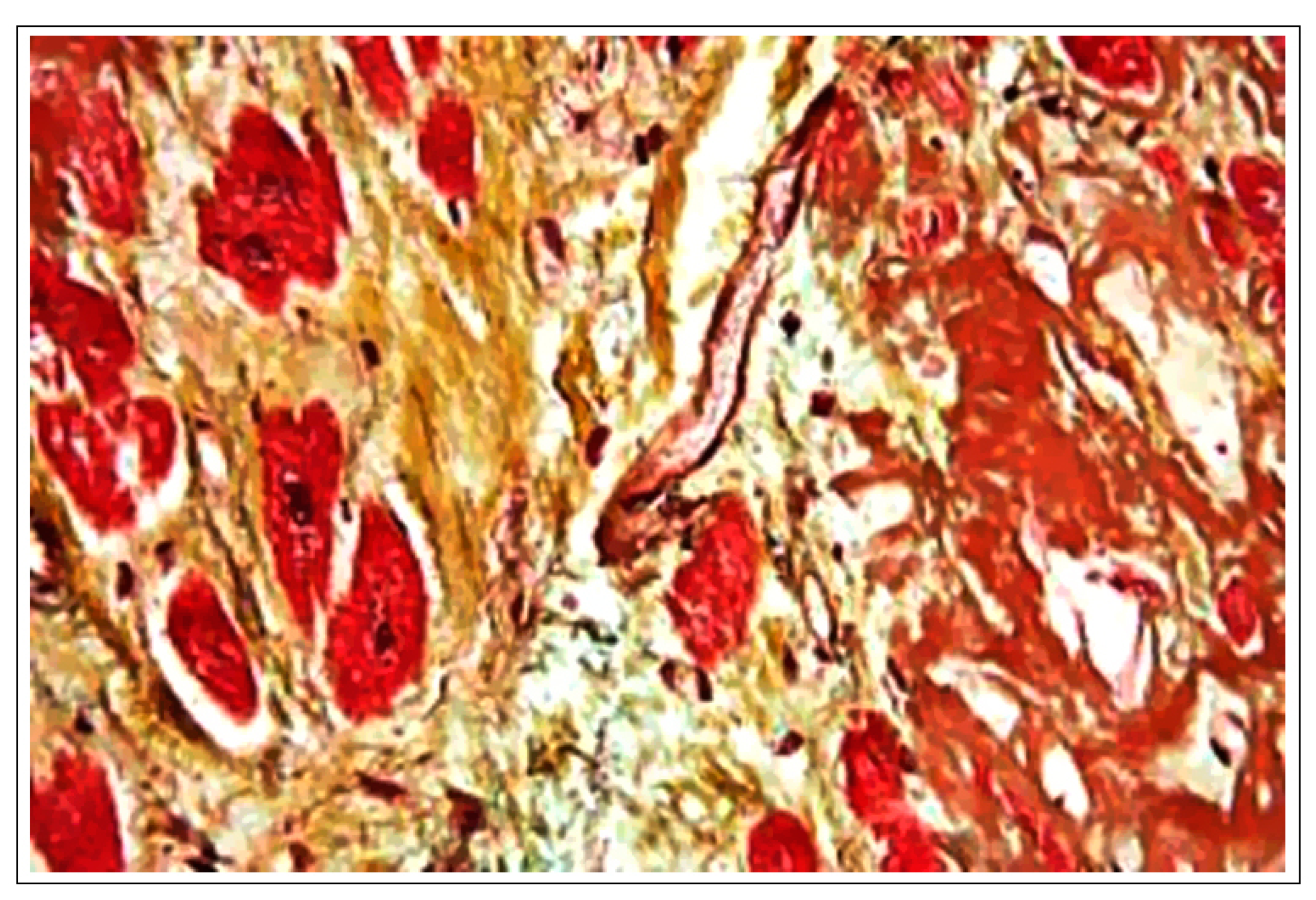
Figure 1. High magnification micrograph of senile cardiac amyloidosis. Movat stain (black = nuclei, elastic fibres; yellow = collagen, reticular fibers; blue = ground substance, mucin; bright red = fibrin; red = muscle). Human autopsy specimen. The micrograph shows amyloid (extracellular muddy brown material—right of image), abundant lipofuscin (dark red granular material) and myocardial fibrosis (yellow—left of image). Related images Intermed. mag. (H&E). High mag. (H&E). Very high mag. (H&E). Intermed. mag. (congo red). High mag. (congo red). Very high mag. (congo red). Very high mag. (Movat’s stain). SOURCE: Wikimedia Commons, Copyright © 2011 Michael Bonert (https://en.wikipedia.org/wiki/Cardiovascular_disease#/media/File:Cardiac_amyloidosis_very_high_mag_movat.jpg (accessed on 16 May 2024)).
Ijms 25 06176 g001
Figure 2. Parallel regulation of intracellular calcium-mediated hypertrophic/arrhythmogenic stimulus in cardiac myocytes and immune activation in T lymphocytes using FK506, and PAF-receptor-inhibitory ginkgolides from leaf extract (EGb761). In cardiac myocytes, the mechanical stimulation of stretch receptors and macromolecular stimulation of cell membrane-embedded Ca2+ channels increase cytoplasmic Ca2+ levels, causing calcineurin phosphatase-mediated linearization of NFAT transcription factors with resulting expression of genes contributing to hypertrophy and arrhythmogenesis [127]. An analogous process results in the activation of T lymphocytes, engendering pro-inflammatory reactions that may become pathological [128].
Ijms 25 06176 g002
Figure 3. Heme oxygenase activity: reaction stoichiometry and physiological effects of product metabolites. Heme oxygenases degrade heme by cleavage of the heme ring at its alpha-methene bridge, producing carbon monoxide (CO), ferrous ion (Fe2+), and biliverdin, which is further reacted by biliverdin reductase to bilirubin (a major endogenous antioxidant).
Ijms 25 06176 g003
Figure 4. Major pathomechanisms contributing to OA-associated articular tissue damage. Physical trauma and reactive oxygen species produced as an adaptive response by host cells increase T cell, macrophage, and synoviocyte production of pro-inflammatory cytokines, including interleukin-6 (IL-6), TNF-A, and interleukin1-beta (IL1-B). From Mahmoud FF, Al-Awadhi AM, Haines DD. Amelioration of human osteoarthritis symptoms with topical ‘biotherapeutics’: a phase I human trial. Adapted with permission from Ref. [177].
Ijms 25 06176 g004
Disclaimer/Publisher’s Note: The statements, opinions and data contained in all publications are solely those of the individual author(s) and contributor(s) and not of MDPI and/or the editor(s). MDPI and/or the editor(s) disclaim responsibility for any injury to people or property resulting from any ideas, methods, instructions or products referred to in the content.