Review

Long Noncoding RNAs in Diet-Induced Metabolic Diseases

1
Molecular Nutritional Science, Department of Nutritional Sciences, University of Vienna, 1090 Vienna, Austria
2
Clinical Pharmacy Group, Department of Pharmaceutical Sciences, University of Vienna, 1090 Vienna, Austria
*
Author to whom correspondence should be addressed.
Int. J. Mol. Sci. 2024, 25(11), 5678; https://doi.org/10.3390/ijms25115678
Submission received: 15 April 2024 / Revised: 17 May 2024 / Accepted: 21 May 2024 / Published: 23 May 2024
(This article belongs to the Section Biochemistry)

Abstract

:
The prevalence of metabolic diseases, including type 2 diabetes and metabolic dysfunction-associated steatotic liver disease (MASLD), is steadily increasing. Although many risk factors, such as obesity, insulin resistance, or hyperlipidemia, as well as several metabolic gene programs that contribute to the development of metabolic diseases are known, the underlying molecular mechanisms of these processes are still not fully understood. In recent years, it has become evident that not only protein-coding genes, but also noncoding genes, including a class of noncoding transcripts referred to as long noncoding RNAs (lncRNAs), play key roles in diet-induced metabolic disorders. Here, we provide an overview of selected lncRNA genes whose direct involvement in the development of diet-induced metabolic dysfunctions has been experimentally demonstrated in suitable in vivo mouse models. We further summarize and discuss the associated molecular modes of action for each lncRNA in the respective metabolic disease context. This overview provides examples of lncRNAs with well-established functions in diet-induced metabolic diseases, highlighting the need for appropriate in vivo models and rigorous molecular analyses to assign clear biological functions to lncRNAs.

1. Introduction

Based on current data from the Global Burden of Disease Study 2019, the worldwide prevalence of metabolic diseases, referring to metabolic dysregulated processes such as type 2 diabetes (T2D) or metabolic dysfunction-associated steatotic liver disease (MASLD), is still growing [1]. While the estimated worldwide prevalence of MASLD is ~32.4% [2], ~529 million people worldwide suffered from T2D in 2021 [3], contributing to a high economic burden [4,5]. The main risk factors and metabolic comorbidities of MASLD or T2D include obesity, insulin resistance, hyperlipidemia, or high blood pressure [6,7]. Indeed, data from population-based studies suggest that ~878 million people suffered from obesity in 2022 [8]. Besides genetic factors and a low physical activity, general overnutrition as well as the intake of certain macronutrients is associated with obesity, MASLD, or T2D [9,10]. Indeed, a Westernized diet, rich in monosaccharides like fructose and fats that are rich in short-chain fatty acids (SFAs), is associated with the development of MASLD [9] and T2D [11]. However, the molecular mechanisms contributing to diet-induced metabolic dysregulated processes are, so far, not fully understood.
In recent years, noncoding RNAs have been increasingly implicated in the response to different nutritional conditions and in diet-induced metabolic diseases [12,13]. While the molecular functions of microRNAs, small ~22 nucleotide (nt)-long regulatory RNAs that are known to post-transcriptionally repress target mRNAs to which they bind [14], are well established (also in the context of metabolic diseases [15]), relatively little is known about the precise molecular roles of another class of noncoding RNAs termed long noncoding RNAs (lncRNAs). lncRNAs are generally defined by a sequence length of more than 200 nt and by the lack of a protein-coding open reading frame [16]. To better distinguish lncRNA genes from medium-sized ones—mostly RNA polymerase III-transcribed RNAs—a recently published consensus statement recommends a different, slightly bigger cut-off size of 500 nt for lncRNA genes [17]. However, irrespective of the precise definition of their length, lncRNAs comprise a very heterogenous group of noncoding transcripts not only in terms of their genomic location and biogenesis but also in terms of their biological and molecular functions. lncRNAs are mostly transcribed by polymerase II, often spliced and polyadenylated. Some of them exist as circularized transcripts that are produced by the back-splicing of certain coding as well as noncoding RNAs [18]. They can originate from intergenic regions or overlap with other coding or noncoding genes in either the sense or the antisense orientation. In addition, active DNA elements are often transcribed, resulting in the production of enhancer- or promoter-derived lncRNA transcripts [16,17].
While ~75% of the human genome is transcribed into RNAs, with most of them representing noncoding RNAs with tens of thousands being annotated as lncRNAs [19,20], the physiological and molecular roles of the vast majority of these noncoding transcripts are still unknown [21]. Nevertheless, lncRNA functions can be generally categorized into two modes: (1) the transcriptional regulation of neighboring gene loci in the nucleus, and (2) the modulation of numerous biological processes at various cellular locations and compartments that are distant to the lncRNA transcription site. On a molecular level, lncRNAs have been shown to interact with other RNA, DNA, or protein molecules to control their stability, activity, or subcellular localization in distinct cellular compartments. Some of them have been also demonstrated to encode microproteins or peptides that can exert important cellular functions [22,23]. Of note, each of these modes of action needs to be experimentally determined on a case-by-case basis for each lncRNA candidate gene to imply meaningful biological functions for the encoded transcript [21]. So far, several critical roles have been described for lncRNAs in normal physiology, including, for example, development and stem cell function [24,25], as well as in pathophysiological disease conditions, such as neurological disorders [26], cancer [27] or cardiac disease [28].
While lncRNAs have also been shown to be dysregulated in models of metabolic liver disease, such as MASLD [29] (and a few lncRNAs have been suggested as playing a role in nutritional response and metabolic pathways [13]), clear proof of their functional relevance and molecular modes of action in their respective diet and disease contexts remains missing for many, if not most, of the lncRNA genes. Here, we aim to provide a detailed overview of selected lncRNAs with established functions in diet-induced metabolic disorders that have been proven and validated in suitable in vivo models, i.e., in genetic mouse models with lncRNA loss and gain of function. This review, therefore, does not represent a summary of all lncRNAs that have been published in the context of diet-related metabolic diseases but rather a selection of lncRNA studies that have used appropriate lncRNA knockout and transgenic mouse models to demonstrate a clear functional relationship between an lncRNA and a diet-induced phenotype in mice.

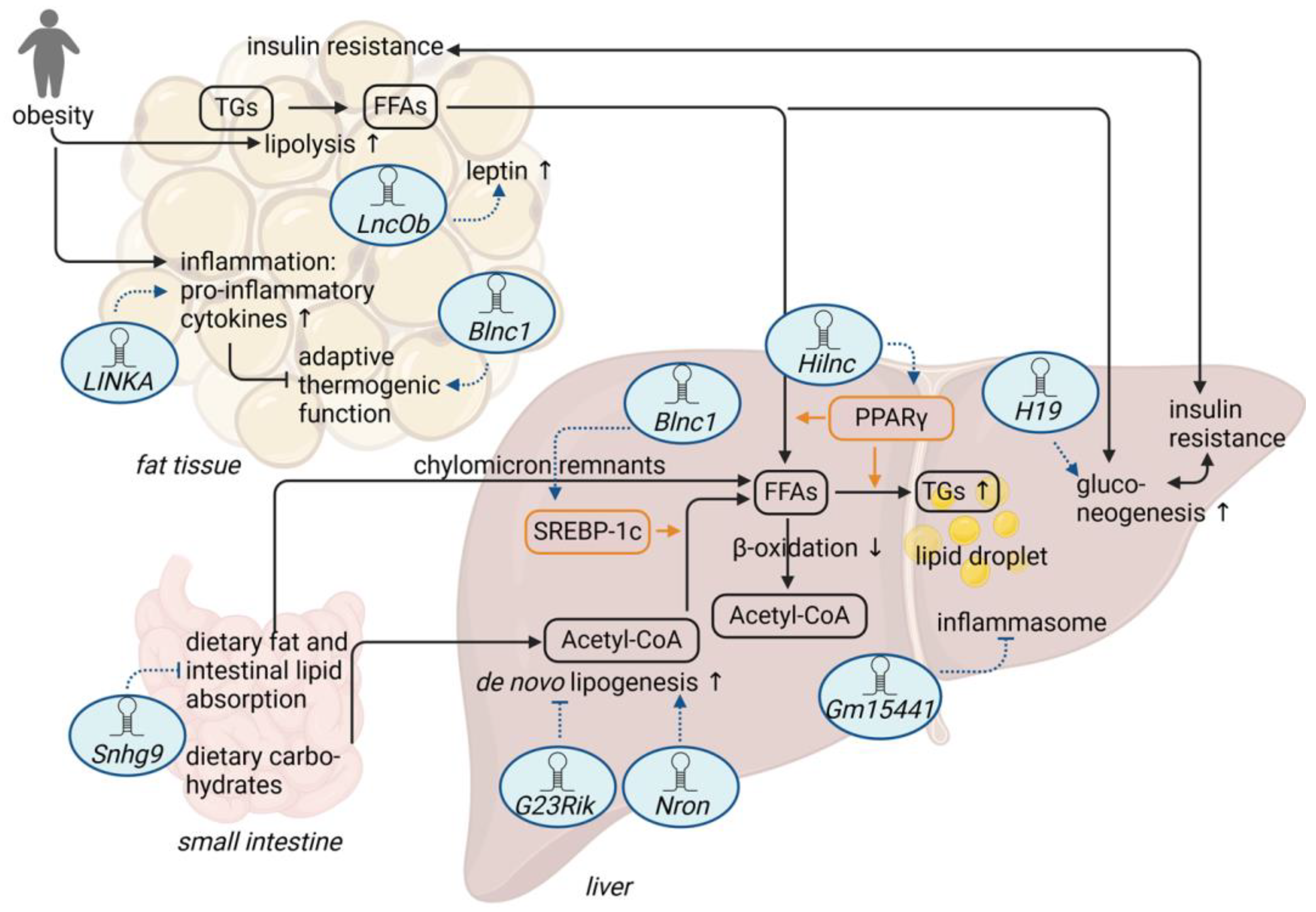
2. Adipose Tissue-Related lncRNAs and Diet-Induced Obesity

Weight gain in obesity mainly results from energy intake that exceeds energy expenditure [30]. This positive energy balance is supported by excess dietary energy intake and reduced physical activity [30]. A Western dietary pattern especially contributes to adipose tissue dysfunction and is associated with obesity and related metabolic dysfunction [31]. While white adipose tissue (WAT) is able to store excess energy, brown fat thermogenesis in brown adipose tissue (BAT) contributes to energy expenditure in form of thermogenesis [32], and studies suggest that the browning of WAT might protect against obesity and associated metabolic diseases [33].
Several studies suggest that lncRNAs are associated with white adipocyte differentiation [34,35,36,37]. Long intergenic non-coding RNA for kinase activation (LINKA, also known as LINK-A or LINC01139), an approximately 1.5 kilobase (kb)-long and predominantly cytoplasmic intergenic lncRNA, has been initially described as a prognostic lncRNA that positively regulates hypoxia-inducible factor 1 alpha (HIF1α) signaling in triple negative breast cancer [38]. Mechanistically, LINKA facilitates the heparin-binding (HB)-EGF-mediated recruitment of two kinases, breast tumor kinase (BRK) and leucin-rich repeat kinase 2 (LRRK2), to the EGFR-GPNMB (epidermal growth factor receptor–glycoprotein NMB) dimer, which results in the phosphorylation of HIF1α at two sites, leading to HIF1α stabilization and activation under normoxic conditions [38]. Recently, LINKA has also been shown to be a human-specific lncRNA that is induced in the breast tissue, subcutaneous WAT, and visceral adipose tissue of overweight patients and whose expression correlates with increasing body mass indices [39]. In a mouse model, in which human LINKA had been knocked-in into the mouse genome, LINKA overexpression promoted high-fat-diet (HFD)-induced obesity and insulin resistance [39]. Moreover, mice with LINKA overexpression suffer from inflammatory alterations in adipose tissue as well as mammary glands. LINKA promotes the production of pro-inflammatory factors in mouse mammary glands via stabilization of the transcription factor HIF1α, which, in turn, transcriptionally activates inflammatory genes, such as interleukin 1 beta (Il1b) and C-X-C motif chemokine ligand 16 (Cxcl16). This is associated with suppressed energy expenditure and a reduced adaptive thermogenic function in mice, as demonstrated by a significant reduction in uncoupling protein 1 (Ucp1), which facilitates heat generation in the mitochondria of BAT and related thermogenesis genes in several adipose tissues after cold exposure [39]. On a molecular level, LINKA cooperates with HB-EGF to stabilize HIF1α (Figure 1a), potentially via a similar mechanism previously described in human breast cancer cells [38].
Blnc1 (brown fat lncRNA 1, also known as AK038898) was originally discovered as an approximately 1 kb long conserved murine intergenic lncRNA that is critical for brown adipogenesis [40]. Blnc1 is induced during brown and beige adipocyte differentiation and is required for the induction of thermogenic gene expression, including the Ucp1 gene (Figure 1b). Blnc1, a primarily nuclear lncRNA, has been proposed to form a ribonucleoprotein complex with the EBF transcription factor 2 (EBF2), an important regulator of BAT, to drive the thermogenic phenotype [40]. Interestingly, Blnc1 itself is a target of EBF2, and both Blnc1 and EBF2 are required for efficient brown adipogenesis, suggesting that Blnc1–EBF2 cooperatively regulate thermogenesis in brown fat. In a later study [41], Blnc1 was found to be conserved in both humans and mice, also sharing its function in brown adipogenesis. Moreover, a conserved RNA domain was identified in human and mouse Blnc1 that is critical for brown adipocyte differentiation and thermogenesis. This RNA domain has been reported to bind to heterogeneous nuclear ribonucleoprotein U (HNRNPU), a ubiquitously expressed RNA binding protein, which enables the formation of the Blnc1–EBF2 complex and, hence, the activation of the thermogenic program [41]. In addition, HNRNPU has been reported to recruit Blnc1 into another ribonucleoprotein complex containing the transcription factor ZBTB7B (zinc finger and BTB domain-containing protein 7), a potent driver of brown adipogenesis and thermogenesis [42]. ZBTB7B has been shown to be required for the adaptation to the cold and the induction of a thermogenic gene program. Interestingly, in both the genetic and HFD-induced mouse models of obesity, Blnc1 expression was significantly increased in epididymal WAT (eWAT) and BAT, positively correlated with body weight, and induced in cold-induced thermogenesis [43]. Moreover, Zhao et al. demonstrated that transgenic mice with adipocyte-specific Blnc1 inactivation exacerbated HFD-induced insulin resistance and hepatic steatosis, while body weight gain was not significantly different between control and knockout mice [43]. However, knockout mice showed increased brown fat whitening and adipose tissue inflammation during HFD feeding, while Ucp1 mRNA expression and further markers of thermogenesis were reduced in these mice compared to HFD-fed controls [43]. On the contrary, a fat-specific transgenic expression of Blnc1 protected mice from HFD-induced obesity, adipose tissue dysfunction, and metabolic dysfunction like insulin resistance and hepatic steatosis [43,44].
The peptide hormone leptin is able to regulate food intake and body mass and seems to be upregulated in obese patients, which suggests that these patients suffer from leptin resistance [45]. Moreover, serum leptin concentrations correlate with the amount of body fat [46]. While leptin is also produced by other tissues such as the stomach [47], two leptin enhancer sequences up- and downstream of the leptin gene have been identified as necessary for fat-specific leptin expression in mice. Moreover, it has been verified that the proximal leptin promoter sequence is regulated by the two leptin enhancers and is required for robust leptin expression [48]. In addition to the identification of two transcription factors (EBF transcription factor 1/EBF1 and nuclear factor 1/NF1) that bind to the leptin enhancers and promoter, several RNA binding proteins have been also detected to be enriched at the leptin promoter, suggesting that RNAs may be involved in the regulation of leptin expression as well. Indeed, one 9 kb long lncRNA is co-transcribed with leptin from a region further upstream of the upstream leptin enhancer. This lncRNA, also called long noncoding RNA upstream of leptin (Lnclep or LncOb), is spliced to an ~850 nt long mature lncRNA that shows considerable conservation in humans. LncOb is only expressed in adipose tissue, is co-regulated with leptin, and does not bind to leptin enhancers (Figure 1c). It rather binds to the leptin proximal promoter and interacts with RNA binding proteins identified to be enriched at the leptin promoter [48]. LncOb is highly correlated with leptin mRNA in HFD-induced obese mice and exclusively expressed in inguinal WAT and eWAT [48]. Like in mice, RNA corresponding to LncOb in humans is expressed in adipose tissue and is correlated with leptin mRNA, while it is not expressed in liver or muscle tissue. Remarkably, knockout of LncOb in mice was associated with an accelerated body weight gain and dysregulated leptin expression under HFD as compared to wild-type control mice [48]. This phenotype could be reverted by an infusion of the leptin hormone into diet-induced obese mice. Of note, a single-nucleotide polymorphism has been identified in the human LncOb gene that correlates with decreased leptin expression and obesity in humans [48]. Although these findings clearly point to a leptin-dependent function of LncOb in fat metabolism and adipose tissue homeostasis, the precise mechanism of how this lncRNA confers transcriptional regulation of the leptin locus remains an interesting open question.

3. LncRNAs and Diet-Induced Hepatic Steatosis

Adipose tissue dysfunction results in an impaired ability to store fat and has therefore been linked to the pathogenesis of hepatic steatosis [49]. The hallmark of hepatic steatosis is the excessive accumulation of triglycerides in hepatocytes resulting from esterification of free fatty acids (FFAs) in the liver. These FFAs can originate from various sources, such as (1) release of FFAs from increased lipolysis in adipose tissue that reach the liver via the blood, (2) FFAs arising from de novo lipogenesis (DNL) from carbohydrates, and (3) from the intake of dietary fats that reach the liver via FFAs in the blood or via chylomicron remnants [50,51]. In principle, triglycerides can be excreted from the liver as very-low-density lipoprotein (VLDL), stored in lipid droplets in the liver or metabolized as FFAs via β-oxidation [51,52,53].

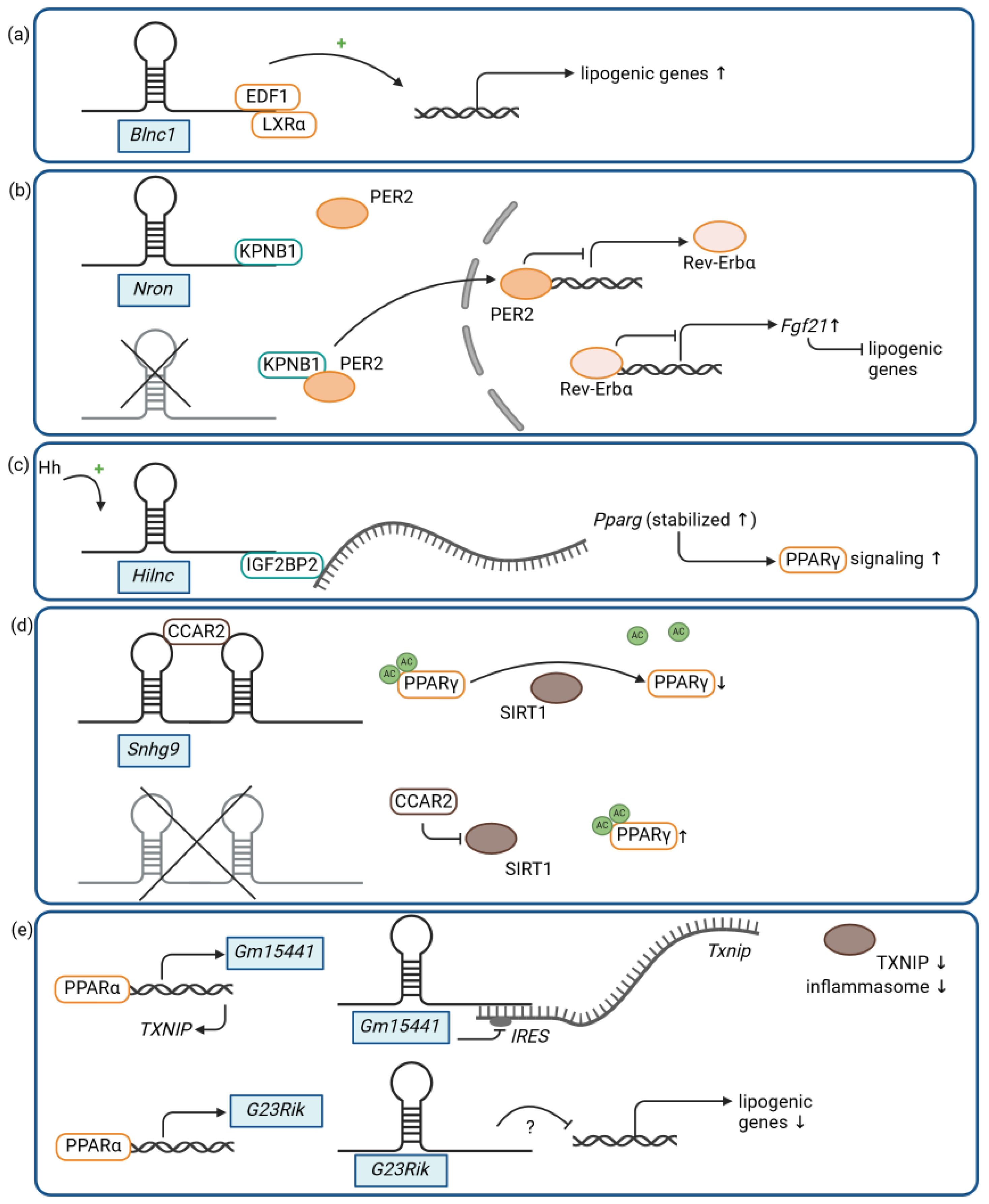
3.1. LncRNAs and Lipogenesis

Recent data suggest that several lncRNAs are dysregulated in MASLD patients (for an overview, see [54,55]), and studies report that lncRNAs are involved in lipogenesis and fatty acid metabolism (for an overview, see [56]). Indeed, Blnc1 function is not restricted to the adipose tissue, as it has been shown to regulate lipogenesis in the liver as well [57]. As demonstrated by Zhao et al., Blnc1 and Srebp1c (sterol regulatory element-binding transcription factor 1c), a gene known to regulate the expression of genes involved in DNL and triglyceride synthesis, are similarly induced in the livers of HFD-fed mice, and Blnc1 expression correlates with body weight and liver triglyceride content [57]. A liver-specific Blnc1 knockout model attenuates HFD-induced weight gain, hepatic steatosis, and insulin resistance in mice, resulting in protection from MASLD. As in its adipose-specific functions, Blnc1 appears to facilitate the formation of a distinct ribonucleoprotein transcriptional complex in the liver, consisting of Blnc1, endothelial differentiation-related factor 1 (EDF1), and liver X receptor alpha (LXRα), which drives lipogenic gene expression, including the expression of Srebp1c (Figure 2a) [57]. These findings demonstrate that a conserved lncRNA can modulate metabolic functions in various tissues and cell types by a similar yet distinct molecular mechanism, which is the formation of different ribonucleoprotein transcriptional complexes for the control of metabolism-associated gene programs.
Another lncRNA, which is not only relevant in adipose tissue but also in the liver, is the lncRNA noncoding repressor of NFAT (Nron) [58], a roughly 3 kb long antisense transcript derived from an intron of the protein-coding gene Mvb12b (multivesicular body subunit 12B). Nron is expressed in various tissues, such as liver, adipose tissue, or muscle tissue. It was initially described as a negative regulator of nuclear transport and, hence, the activity of NFAT (nuclear factor of activated T cells), a class of transcription factors that are responsive to calcium signaling and modulate the immune response in T lymphocytes [59]. Nron interacts with nuclear transport factors like KPNB1 (karyopherin subunit beta 1), scaffold proteins like IQGAP1 (IQ motif containing GTPase-activating protein 1), and known NFAT kinases like CK1 (casein kinase 1) to form an RNA-protein scaffold complex that regulates NFAT phosphorylation and subcellular localization [59,60]. Disruption of this complex by either silencing Nron or IQGAP1 results in dephosphorylation, nuclear import, and the activation of NFAT [60]. Recently, it has been demonstrated that Nron levels decrease in the mouse liver after HFD feeding [58]. In line with these findings, patients with MASLD suffer from decreased NRON expression in the liver [58]. Unexpectedly, Liu et al. demonstrated that Nron knockout mice were protected from HFD-induced weight gain, obesity, and insulin resistance, and that Nron depletion promoted adipocyte lipolysis and lipid turnover [58]. This was associated with an amelioration of HFD-induced hepatic steatosis, while HFD-induced hepatic inflammation and apoptosis were not affected. By contrast, Nron overexpression leads to an intensification of the HFD-induced hepatic metabolic changes [58]. Interestingly, the effects observed in Nron-deficient animals could not be attributed to an activation of NFAT signaling. In the liver and in isolated hepatocytes, Nron loss-of-function mutation promotes the binding of the circadian clock regulator PER2 (period circadian clock 2) to KPNB1, a nucleocytoplasmic transport protein that also interacts with Nron [59], resulting in the nuclear translocation of PER2. Nuclear PER2 represses the expression of another circadian clock gene, Rev-Erbα, also known as nuclear receptor subfamily 1 group D member 1 (Nr1d1), which in turn activates the expression of fibroblast growth factor 21 (Fgf21), a key regulator of energy metabolism in the liver (Figure 2b). Increased FGF21 levels inhibit lipogenesis and induce fatty acid oxidation via an activation of AMPK (AMP-activated protein kinase) signaling, eventually improving hepatic steatosis under HFD. On the contrary, diet-induced effects on the adipose tissue in Nron knockout mice, such as attenuated obesity, appear to be independent of the Nron–KPNB1–PER2– Rev-Erbα–FGF21 axis [58].
In a screen for lncRNAs regulated by Hedgehog (Hh) signaling, Gm16364 has been identified as an approximately 800 kb long murine Hh-induced lncRNA, also termed Hilnc (Hedgehog signaling-induced long noncoding RNA) [61]. Hh signaling is a deeply conserved signaling pathway regulating development and tissue homeostasis, in which Hh ligands activate their respective receptors and eventually activate the Gli family of transcription factors [62,63]. Hh signaling has also been implicated in metabolic processes in the adipose tissue, as well as in the liver and in metabolic liver diseases [64,65]. While Hilnc is directly induced by Hh signaling via Gli transcription factors in various tissues, it was significantly upregulated in liver tissue in a diet-induced mouse model of hepatic steatosis [61]. Remarkably, the depletion of Hilnc, either through promoter mutation or the deletion of exons 2 and 3, reduced DNL and protected mice from HFD-induced obesity, impaired glucose metabolism, and hepatic steatosis. On the other hand, adenovirus-mediated overexpression of Hilnc in the livers of Hilnc knockout mice promoted lipid accumulation under an HFD. On a molecular level, Hilnc has been shown to bind via a distinct RNA region to the RNA binding protein IGF2BP2 (insulin-like growth factor 2 mRNA binding protein 2) in the liver and to positively modulate PPARγ (peroxisome proliferator-activated receptor gamma) signaling [61], a central regulator of lipid metabolism [66]. IGF2BP2 regulates the stability and translation of mRNAs to which it binds [67,68], and Hilnc is required for the proper function of IGF2BP2 as a post-transcriptional regulator of interacting mRNAs, such as the Pparg mRNA (Figure 2c). In the livers of HFD-fed wild-type mice, Pparg levels, as well as PPARγ signaling, were induced in a Hilnc- and IGF2BP2-dependent manner, affecting lipid metabolism and promoting hepatic steatosis [61]. Of note, while there appears to be no syntenic lncRNA or sequence-based homolog of Hilnc in the human genome, a potential functional human homolog, referred to as h-Hilnc, has been identified, which is also responsive to Hh signaling, regulates PPARG via IGF2BP2, and is involved in lipid metabolism and lipid accumulation in human liver cells [61].

3.2. LncRNA and Intestinal Lipid Absorption

Alterations of intestinal microbiota composition and an impaired intestinal barrier function in humans are associated with several metabolic diseases, including diabetes and MASLD [69,70]. Indeed, studies in germ-free mice further indicate that microbiota promotes monosaccharide absorption from intestinal lumen, which contributes to hepatic DNL [71]. Moreover, intestinal microbiota also seems to affect intestinal lipid absorption [72], and lncRNAs might be able to affect microbiome-host-interaction [73]. In line with these findings, small nucleolar RNA host gene 9 (Snhg9) has been shown to be significantly lower expressed in small intestines from conventional mice as compared to germ-free or antibiotic-treated mice, suggesting that Snhg9 expression is decreased in the presence of microbiota [74]. Snhg9, an approximately 200 nt lncRNA encoding the small nucleolar RNA 78 (Snora78), has been further shown to bind via distinct RNA structures to the cell cycle and apoptosis protein 2 (CCAR2), an endogenous inhibitor of sirtuin 1 (SIRT1), a known repressor of PPARγ activity as well as expression. Accordingly, Snhg9 sequesters CCAR2, resulting in the release of SIRT1 from CCAR2-mediated inhibition and, consequently, in the SIRT1-mediated repression of PPARγ (Figure 2d) [74]. In line with these findings, Snhg9 knockout in 3T3-L1 cells increases PPARγ expression and activates PPARγ signaling, thereby suppressing the lipid metabolism, which can be reversed by Snhg9 overexpression [74]. Likewise, a small intestinal epithelial cell-specific overexpression of Snhg9 impairs PPARγ expression and the genes involved in fatty acid absorption, transport, and synthesis in mice, resembling the effects seen in germ-free or antibiotic-treated animals. Snhg9 transgenic mice showed reduced intestinal lipid absorption after being fed an HFD and were therefore also protected from HFD-induced obesity, insulin resistance, and hepatic steatosis when compared to wild-type controls [74]. The physiological suppression of Snhg9 by microbiota is accomplished via a Toll-like receptor-mediated activation of myeloid cells and subsequent cytokine-triggered signaling to the intestinal epithelial cells, resulting in the intestinal repression of Snhg9 [74]. Although the benefits of microbiota-mediated regulation of the lipid metabolism for the host organism, as well as the precise components of the microbiota that are necessary for this regulation, are still unknown, these results demonstrate how microbiota can positively influence lipid absorption and fat metabolism by downregulating a noncoding RNA in host intestinal epithelial cells. An intriguing open question is also whether these functions are evolutionarily conserved and whether human SNHG9 displays the same functions in the regulation of lipid absorption and fat metabolism.

3.3. LncRNAs and Metabolic Stress

The PPARα-induced murine lncRNA Gm15441 is an approximately 1.7 kb long transcript antisense to a protein-coding gene—thioredoxin-interacting protein (Txnip)—that consists of four exons, two of which overlap the Txnip gene [75]. Upon PPARα activation by either a pharmacological compound or fasting, Gm15441 is dramatically and specifically increased in the mouse liver. Gm15441 and its antisense transcript, Txnip, are both direct PPARα targets, whereas their regulation upon PPARα activation is inverse. Upon metabolic stress, such as fasting, Gm15441 is induced in a PPARα-dependent manner in the liver and inhibits Txnip translation likely via the interaction of its exon 3 with the overlapping antisense sequence of the 5′ untranslated region of Txnip, which harbors internal ribosome entry site (IRES) sequences critical for efficient Txnip translation (Figure 2e) [75]. The reduced TXNIP levels result in decreased activation of the NLR family pyrin domain-containing-3-(NLRP3)-mediated inflammasome accompanied by a reduced cleavage of caspase 1 (CASP1) and impaired maturation of IL1B. In a Gm15441 mouse knockout model, in which the expression of Gm15441 is specifically impaired while Txnip expression is unaffected, Gm15441 deficiency promoted the activation of the NLRP3-mediated inflammasome as well as lipid accumulation in the liver upon PPARα activation, either through pharmacological intervention or acute fasting [75]. Interestingly, there is another murine lncRNA, called 3930402G23Rik (or G23Rik), that has been shown to also be induced by PPARα signaling specifically in the liver and to modulate hepatic liver metabolism [76]. Acute fasting in G23Rik-deficent mice potentiated hepatic lipid accumulation accompanied by an increase in cholesterol and triglyceride levels [76]. Although the molecular mechanism of how G23Rik contributes to the regulation of the PPARα-mediated lipid metabolism is unknown, the two examples, Gm15441 and G23Rik, demonstrate how lncRNAs can respond to metabolic stress and modulate critical metabolic pathways, such as PPAR signaling.

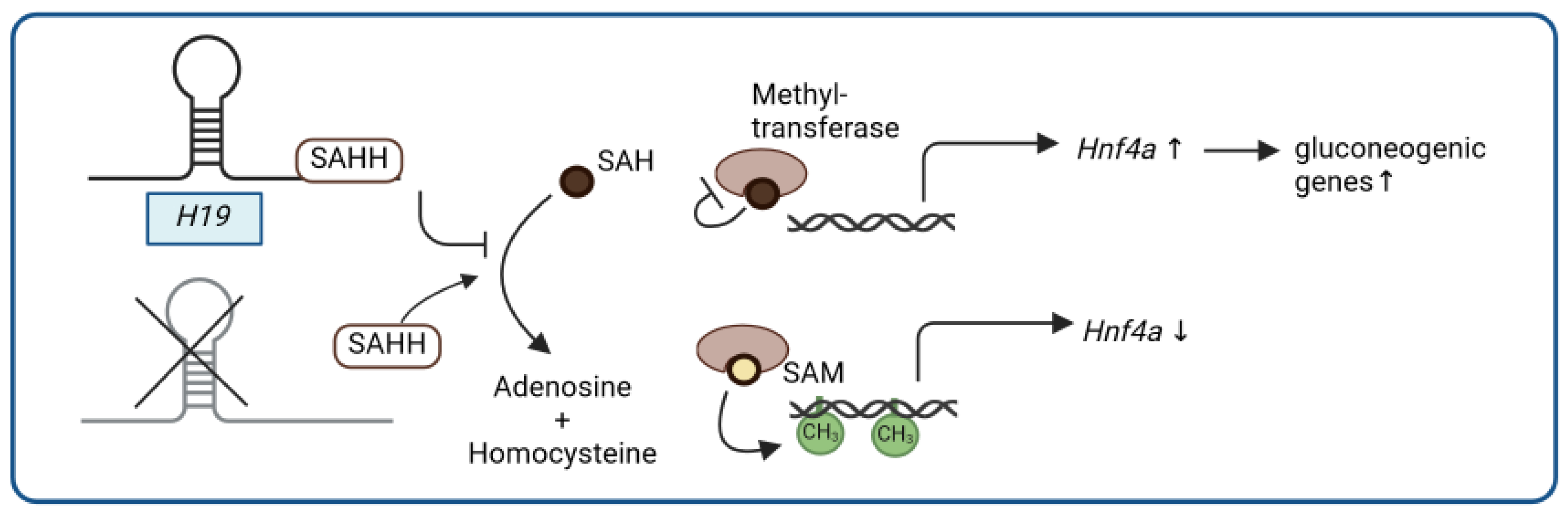
4. LncRNA and Glucose Metabolism

Insulin resistance is often a common characteristic of obesity and diet-induced metabolic diseases, such as T2D or MASLD [77]. While a dysregulated lncRNA profile seems to be involved in the pathogenesis of diet-induced metabolic diseases such as MASLD [54,55], recent evidence suggests that lncRNAs also contribute to the regulation of insulin signaling and glucose metabolism in several tissues and to the pathogenesis of insulin resistance (reviewed in detail by others [78,79]). By controlling pathways of glucose metabolism, the liver has a major role in the control of glucose homeostasis [80], and insulin resistance seems to be associated with an increase in hepatic glucose output, lipogenesis, and VLDL secretion, leading to a combination of hyperglycemia and hypertriglyceridemia [81].
Noncoding RNAs, such as the lncRNA H19, have been described as performing key roles in liver biology and disease [82]. H19 is known as a ~2.3 kb long paternally imprinted lncRNA originating from the insulin growth factor 2 (IGF2/Igf2) locus in humans and mice [83]. On the maternal allele, H19 is expressed while Igf2 expression is prevented; this reciprocal regulation of H19 and Igf2 is important during fetal development. After birth, H19 is downregulated in most tissues, except in the skeletal muscle and heart [83]. H19 expression has been furthermore shown to be increased in the livers of T2D patients [84], and, consistent with H19 expression in T2D patients, H19 levels are also increased in livers of mice with diet-induced impaired glucose metabolism [85]. In line with these findings, the liver-specific overexpression of H19 results in impaired glucose homeostasis with hyperglycemia and insulin resistance, whereas a whole-body knockout of H19 in mice leads to improved insulin sensitivity and reduced insulin-dependent endogenous glucose production in the liver, associated with the reduced expression of gluconeogenic genes [85]. On a molecular level, H19 negatively regulates the promoter methylation and, hence, activates the expression of the hepatocyte nuclear factor 4α (Hnf4a), a transcription factor important for gluconeogenesis. As previously reported for other tissues [86], H19 binds to S-adenosylhomocysteine hydrolase (SAHH) in mouse liver as well [85]. SAHH hydrolyzes S-adenosylhomocysteine (SAH), an inhibitor of S-adenosylmethionine (SAM)-dependent methyltransferases, which results in the activation of DNA methyltransferases and, consequently, results in increased DNA promoter methylation. It has been suggested that H19 may cause Hnf4a promoter hypomethylation via the H19/SAHH pathway through binding to SAHH, as well as the subsequent accumulation of SAH and inhibition of DNA methyltransferases (Figure 3) [85]. In line with these findings, temporally increased H19 levels upon fasting or chronically elevated H19 levels reduce Hnf4a promoter methylation and induce a gluconeogenic gene expression program. It is noteworthy that previously established modes of action for H19, including H19-encoded miR-675 or Igf2 expression, appear to be dispensable for this metabolic function [85]. Moreover, H19 has been implicated as contributing to a range of metabolic diseases, such as diet-induced hepatic steatosis, although the underlying molecular mechanisms are less well understood and require more investigation [87,88,89,90,91].

5. Conclusions

As summarized in this review, several studies suggest that lncRNAs are important regulatory factors in the pathogenesis of obesity and diet-induced metabolic diseases, such as MASLD or T2D (Figure 4, Table 1). Indeed, data from patients, as well as animal models, indicate tissue-specific regulation of lncRNAs in diet-associated metabolic diseases. Still, the functional roles of the discussed lncRNAs and their relationship with obesity, MASLD, or T2D were mainly assessed in preclinical studies and need to be translated to human pathophysiology. The lncRNAs are involved in different metabolic pathways, including adipocyte differentiation, lipogenesis, or gluconeogenesis. While some lncRNAs are related to metabolic stress during fasting, others respond to dietary changes. Importantly, some lncRNAs like Blnc1 have functional relevance in several tissues, such as adipose tissue and the liver, acting on several metabolic pathways. Additionally, these lncRNAs might also have the potential to serve as biomarkers for the progression and management of diet-induced metabolic diseases [55,92]. Indeed, H19 or NRON expression is altered in patients with T2D or MASLD [58,84].
On a cellular level, lncRNAs have been shown to act via diverse molecular modes of action to respond to nutritional conditions and to modulate diet-induced metabolic diseases (Figure 4). lncRNAs can thereby regulate transcription of key metabolic genes, either directly as part of a larger transcriptional ribonucleoprotein complex (Blnc1) or indirectly via the regulation of the stability (LINKA) or activity and expression (Snhg9) of metabolism-related transcription factors, such as HIF1α or PPARγ, respectively. lncRNAs like H19 can also cause promoter hypomethylation of gluconeogenic genes in the liver via binding to and inhibition of the SAH-converting enzyme SAHH in liver cells, leading to an inhibition of SAM-dependent DNA methyltransferases. Another possible control mechanism of key metabolic pathways, such as PPARγ, is the lncRNA-mediated and IGF2BP2-dependent stabilization of Pparg mRNA, resulting in increased PPARγ signaling, as demonstrated for Hilnc. In addition, lncRNAs can affect the translation of target proteins as antisense transcripts that block, for example, IRES sequences of a target mRNA, such as the lncRNA Gm15441 and the Txnip mRNA, which results in the inhibition of the inflammasome in the liver. Besides protein stability and expression, lncRNAs may also affect the subcellular localization of target proteins critical for metabolic diseases, as exemplified by the Nron–KPNB1 interaction, which prevents the nuclear translocation of the circadian clock protein PER2 and subsequently promotes the expression of Fgf21 and lipogenic genes.
In summary, these selected examples of lncRNAs with distinct roles in diet-induced metabolic diseases demonstrate the necessity of applying appropriate genetic and transgenic mouse models to study the physiological function of new candidate lncRNA genes in diet-dependent disease contexts. Moreover, rigorous testing of the molecular modes of action of selected lncRNA genes further bolsters the functional relevance of the respective lncRNA transcripts in response to nutritional challenges and metabolic diseases. Our selection of lncRNAs described in this review is not supposed to be a comprehensive summary of all diet- and nutrition-associated lncRNAs. Rather, we chose a set of lncRNAs that underwent rigorous testing of their physiological in vivo functions, as well as their molecular modes of action. We foresee that there will be more lncRNAs with important functions in nutrition, metabolism, and metabolic disease that can be uncovered by future studies and that can broaden our understanding of the role of the noncoding transcriptome in metabolic pathways and disease.

Author Contributions

Conceptualization, A.B. and F.K.; writing—original draft preparation, A.B. and F.K.; writing—review and editing, A.B. and F.K.; visualization, A.B. and F.K. All authors have read and agreed to the published version of the manuscript.

Funding

Open Access Funding by the University of Vienna.

Acknowledgments

Figures created with BioRender.com.

Conflicts of Interest

The authors declare no conflicts of interest.

Abbreviations

Ac—Acetylation, AMPK—AMP-activated protein kinase, BAT—brown adipose tissue, Blnc1—brown fat lncRNA 1, BRK—breast tumor kinase, CASP1—caspase 1, CCAR2—cell cycle and apoptosis protein 2, CK1—casein kinase 1, Cxcl16—C-X-C motif chemokine ligand 16, DNL—de novo lipogenesis, EBF2—EBF transcription factor 2, EDF1—endothelial differentiation-related factor 1, EGFR-GPNMB—epidermal growth factor receptor–glycoprotein NMB, eWAT—epididymal WAT, FFAs—free fatty acids, Fgf21—fibroblast growth factor 21, G23Rik—3930402G23Rik, HFD—high-fat diet, Hh—Hedgehog, HIF1α—hypoxia-inducible factor 1 alpha, Hilnc—Hedgehog signaling-induced long noncoding RNA, Hnf4a—hepatocyte nuclear factor 4α, HNRNPU—heterogeneous nuclear ribonucleoprotein U, IGF2—insulin growth factor 2, IGF2BP2—insulin-like growth factor 2 mRNA binding protein 2, IL1B—interleukin 1 beta, IRES—internal ribosome entry site, IQGAP1—IQ motif containing GTPase-activating protein 1, kb—kilobases, KPNB1—karyopherin subunit beta 1, LINKA—long intergenic noncoding RNA for kinase activation, lncRNA—long noncoding RNA, LRRK2—leucin-rich repeat kinase 2, HB—heparin-binding, LXRα—liver X receptor alpha, MASLD—metabolic dysfunction-associated steatotic liver disease, Mvb12b—multivesicular body subunit 12B, NAFLD—nonalcoholic fatty liver disease, NFAT—nuclear factor of activated T cells, NLRP3—NLR family pyrin domain-containing 3, Nron—noncoding repressor of NFAT, Nr1d1—nuclear receptor subfamily 1 group D member 1, nt—nucleotides, PER2—period circadian clock 2, PPARγ—peroxisome proliferator-activated receptor gamma, SAH—S-adenosylhomocysteine, SAHH—S-adenosylhomocysteine hydrolase, SAM—S-adenosylmethionine, SIRT1—sirtuin 1, Snhg9—small nucleolar RNA host gene 9, Snora78—small nucleolar RNA 78, Srebp1c—sterol regulatory element-binding transcription factor 1c, T2D—type 2 diabetes, TGs – triglycerides, Txnip—thioredoxin-interacting protein, Ucp1—uncoupling protein 1, VLDL—very-low-density lipoprotein, WAT—white adipose tissue, ZBTB7B—zinc finger and BTB domain-containing protein 7.

References

  1. Chew, N.W.S.; Ng, C.H.; Tan, D.J.H.; Kong, G.; Lin, C.; Chin, Y.H.; Lim, W.H.; Huang, D.Q.; Quek, J.; Fu, C.E.; et al. The global burden of metabolic disease: Data from 2000 to 2019. Cell Metab. 2023, 35, 414–428.e413. [Google Scholar] [CrossRef] [PubMed]
  2. Riazi, K.; Azhari, H.; Charette, J.H.; Underwood, F.E.; King, J.A.; Afshar, E.E.; Swain, M.G.; Congly, S.E.; Kaplan, G.G.; Shaheen, A.A. The prevalence and incidence of NAFLD worldwide: A systematic review and meta-analysis. Lancet Gastroenterol. Hepatol. 2022, 7, 851–861. [Google Scholar] [CrossRef] [PubMed]
  3. Ong, K.L.; Stafford, L.K.; McLaughlin, S.A.; Boyko, E.J.; Vollset, S.E.; Smith, A.E.; Dalton, B.E.; Duprey, J.; Cruz, J.A.; Hagins, H. Global, regional, and national burden of diabetes from 1990 to 2021, with projections of prevalence to 2050: A systematic analysis for the Global Burden of Disease Study 2021. Lancet 2023, 402, 203–234. [Google Scholar] [CrossRef] [PubMed]
  4. Alzaid, A.; Ladrón de Guevara, P.; Beillat, M.; Lehner Martin, V.; Atanasov, P. Burden of disease and costs associated with type 2 diabetes in emerging and established markets: Systematic review analyses. Expert. Rev. Pharmacoecon. Outcomes Res. 2021, 21, 785–798. [Google Scholar] [CrossRef] [PubMed]
  5. Younossi, Z.M.; Blissett, D.; Blissett, R.; Henry, L.; Stepanova, M.; Younossi, Y.; Racila, A.; Hunt, S.; Beckerman, R. The economic and clinical burden of nonalcoholic fatty liver disease in the United States and Europe. Hepatology 2016, 64, 1577–1586. [Google Scholar] [CrossRef] [PubMed]
  6. Huh, Y.; Cho, Y.J.; Nam, G.E. Recent Epidemiology and Risk Factors of Nonalcoholic Fatty Liver Disease. J. Obes. Metab. Syndr. 2022, 31, 17–27. [Google Scholar] [CrossRef]
  7. Mokdad, A.H.; Ford, E.S.; Bowman, B.A.; Dietz, W.H.; Vinicor, F.; Bales, V.S.; Marks, J.S. Prevalence of Obesity, Diabetes, and Obesity-Related Health Risk Factors, 2001. JAMA 2003, 289, 76–79. [Google Scholar] [CrossRef]
  8. Phelps, N.H.; Singleton, R.K.; Zhou, B.; Heap, R.A.; Mishra, A.; Bennett, J.E.; Paciorek, C.J.; Lhoste, V.P.; Carrillo-Larco, R.M.; Stevens, G.A. Worldwide trends in underweight and obesity from 1990 to 2022: A pooled analysis of 3663 population-representative studies with 222 million children, adolescents, and adults. Lancet 2024, 403, 1027–1050. [Google Scholar] [CrossRef] [PubMed]
  9. Berná, G.; Romero-Gomez, M. The role of nutrition in non-alcoholic fatty liver disease: Pathophysiology and management. Liver Int. 2020, 40 (Suppl. 1), 102–108. [Google Scholar] [CrossRef]
  10. Petroni, M.L.; Brodosi, L.; Marchignoli, F.; Sasdelli, A.S.; Caraceni, P.; Marchesini, G.; Ravaioli, F. Nutrition in Patients with Type 2 Diabetes: Present Knowledge and Remaining Challenges. Nutrients 2021, 13, 2748. [Google Scholar] [CrossRef]
  11. Adeva-Andany, M.M.; Rañal-Muíño, E.; Vila-Altesor, M.; Fernández-Fernández, C.; Funcasta-Calderón, R.; Castro-Quintela, E. Dietary habits contribute to define the risk of type 2 diabetes in humans. Clin. Nutr. ESPEN 2019, 34, 8–17. [Google Scholar] [CrossRef] [PubMed]
  12. Deiuliis, J.A. MicroRNAs as regulators of metabolic disease: Pathophysiologic significance and emerging role as biomarkers and therapeutics. Int. J. Obes. 2016, 40, 88–101. [Google Scholar] [CrossRef]
  13. Lovell, C.D.; Anguera, M.C. Long Noncoding RNAs That Function in Nutrition: Lnc-ing Nutritional Cues to Metabolic Pathways. Annu. Rev. Nutr. 2022, 42, 251–274. [Google Scholar] [CrossRef] [PubMed]
  14. Bartel, D.P. Metazoan MicroRNAs. Cell 2018, 173, 20–51. [Google Scholar] [CrossRef] [PubMed]
  15. Rottiers, V.; Näär, A.M. MicroRNAs in metabolism and metabolic disorders. Nat. Rev. Mol. Cell Biol. 2012, 13, 239–250. [Google Scholar] [CrossRef]
  16. Ulitsky, I.; Bartel, D.P. lincRNAs: Genomics, evolution, and mechanisms. Cell 2013, 154, 26–46. [Google Scholar] [CrossRef]
  17. Mattick, J.S.; Amaral, P.P.; Carninci, P.; Carpenter, S.; Chang, H.Y.; Chen, L.L.; Chen, R.; Dean, C.; Dinger, M.E.; Fitzgerald, K.A.; et al. Long non-coding RNAs: Definitions, functions, challenges and recommendations. Nat. Rev. Mol. Cell Biol. 2023, 24, 430–447. [Google Scholar] [CrossRef]
  18. Patop, I.L.; Wüst, S.; Kadener, S. Past, present, and future of circRNAs. Embo J. 2019, 38, e100836. [Google Scholar] [CrossRef]
  19. Djebali, S.; Davis, C.A.; Merkel, A.; Dobin, A.; Lassmann, T.; Mortazavi, A.; Tanzer, A.; Lagarde, J.; Lin, W.; Schlesinger, F.; et al. Landscape of transcription in human cells. Nature 2012, 489, 101–108. [Google Scholar] [CrossRef]
  20. Volders, P.J.; Verheggen, K.; Menschaert, G.; Vandepoele, K.; Martens, L.; Vandesompele, J.; Mestdagh, P. An update on LNCipedia: A database for annotated human lncRNA sequences. Nucleic Acids Res. 2015, 43, D174–D180. [Google Scholar] [CrossRef]
  21. Kopp, F.; Mendell, J.T. Functional Classification and Experimental Dissection of Long Noncoding RNAs. Cell 2018, 172, 393–407. [Google Scholar] [CrossRef]
  22. Makarewich, C.A.; Olson, E.N. Mining for Micropeptides. Trends Cell Biol. 2017, 27, 685–696. [Google Scholar] [CrossRef]
  23. Wright, B.W.; Yi, Z.; Weissman, J.S.; Chen, J. The dark proteome: Translation from noncanonical open reading frames. Trends Cell Biol. 2022, 32, 243–258. [Google Scholar] [CrossRef]
  24. Perry, R.B.; Ulitsky, I. The functions of long noncoding RNAs in development and stem cells. Development 2016, 143, 3882–3894. [Google Scholar] [CrossRef] [PubMed]
  25. Batista, P.J.; Chang, H.Y. Long noncoding RNAs: Cellular address codes in development and disease. Cell 2013, 152, 1298–1307. [Google Scholar] [CrossRef]
  26. Ang, C.E.; Trevino, A.E.; Chang, H.Y. Diverse lncRNA mechanisms in brain development and disease. Curr. Opin. Genet. Dev. 2020, 65, 42–46. [Google Scholar] [CrossRef]
  27. Schmitt, A.M.; Chang, H.Y. Long Noncoding RNAs in Cancer Pathways. Cancer Cell 2016, 29, 452–463. [Google Scholar] [CrossRef]
  28. Kreutzer, F.P.; Fiedler, J.; Thum, T. Non-coding RNAs: Key players in cardiac disease. J. Physiol. 2020, 598, 2995–3003. [Google Scholar] [CrossRef]
  29. Karri, K.; Waxman, D.J. Dysregulation of murine long noncoding single-cell transcriptome in nonalcoholic steatohepatitis and liver fibrosis. Rna 2023, 29, 977–1006. [Google Scholar] [CrossRef]
  30. Romieu, I.; Dossus, L.; Barquera, S.; Blottière, H.M.; Franks, P.W.; Gunter, M.; Hwalla, N.; Hursting, S.D.; Leitzmann, M.; Margetts, B.; et al. Energy balance and obesity: What are the main drivers? Cancer Causes Control 2017, 28, 247–258. [Google Scholar] [CrossRef]
  31. Longo, M.; Zatterale, F.; Naderi, J.; Parrillo, L.; Formisano, P.; Raciti, G.A.; Beguinot, F.; Miele, C. Adipose Tissue Dysfunction as Determinant of Obesity-Associated Metabolic Complications. Int. J. Mol. Sci. 2019, 20, 2358. [Google Scholar] [CrossRef]
  32. Saely, C.H.; Geiger, K.; Drexel, H. Brown versus white adipose tissue: A mini-review. Gerontology 2012, 58, 15–23. [Google Scholar] [CrossRef]
  33. Liu, X.; Zhang, Z.; Song, Y.; Xie, H.; Dong, M. An update on brown adipose tissue and obesity intervention: Function, regulation and therapeutic implications. Front. Endocrinol. 2022, 13, 1065263. [Google Scholar] [CrossRef]
  34. Sun, L.; Goff, L.A.; Trapnell, C.; Alexander, R.; Lo, K.A.; Hacisuleyman, E.; Sauvageau, M.; Tazon-Vega, B.; Kelley, D.R.; Hendrickson, D.G.; et al. Long noncoding RNAs regulate adipogenesis. Proc. Natl. Acad. Sci. USA 2013, 110, 3387–3392. [Google Scholar] [CrossRef]
  35. Squillaro, T.; Peluso, G.; Galderisi, U.; Di Bernardo, G. Long non-coding RNAs in regulation of adipogenesis and adipose tissue function. eLife 2020, 9, e59053. [Google Scholar] [CrossRef]
  36. Liu, S.; Sheng, L.; Miao, H.; Saunders, T.L.; MacDougald, O.A.; Koenig, R.J.; Xu, B. SRA gene knockout protects against diet-induced obesity and improves glucose tolerance. J. Biol. Chem. 2014, 289, 13000–13009. [Google Scholar] [CrossRef]
  37. Chen, G.; Yu, D.; Nian, X.; Liu, J.; Koenig, R.J.; Xu, B.; Sheng, L. LncRNA SRA promotes hepatic steatosis through repressing the expression of adipose triglyceride lipase (ATGL). Sci. Rep. 2016, 6, 35531. [Google Scholar] [CrossRef] [PubMed]
  38. Lin, A.; Li, C.; Xing, Z.; Hu, Q.; Liang, K.; Han, L.; Wang, C.; Hawke, D.H.; Wang, S.; Zhang, Y.; et al. The LINK-A lncRNA activates normoxic HIF1α signalling in triple-negative breast cancer. Nat. Cell Biol. 2016, 18, 213–224. [Google Scholar] [CrossRef]
  39. Chen, Y.; Chen, H.; Wang, Y.; Liu, F.; Fan, X.; Shi, C.; Su, X.; Tan, M.; Yang, Y.; Lin, B.; et al. LncRNA LINK-A Remodels Tissue Inflammatory Microenvironments to Promote Obesity. Adv. Sci. 2024, 11, e2303341. [Google Scholar] [CrossRef]
  40. Zhao, X.Y.; Li, S.; Wang, G.X.; Yu, Q.; Lin, J.D. A long noncoding RNA transcriptional regulatory circuit drives thermogenic adipocyte differentiation. Mol. Cell 2014, 55, 372–382. [Google Scholar] [CrossRef]
  41. Mi, L.; Zhao, X.Y.; Li, S.; Yang, G.; Lin, J.D. Conserved function of the long noncoding RNA Blnc1 in brown adipocyte differentiation. Mol. Metab. 2017, 6, 101–110. [Google Scholar] [CrossRef] [PubMed]
  42. Li, S.; Mi, L.; Yu, L.; Yu, Q.; Liu, T.; Wang, G.X.; Zhao, X.Y.; Wu, J.; Lin, J.D. Zbtb7b engages the long noncoding RNA Blnc1 to drive brown and beige fat development and thermogenesis. Proc. Natl. Acad. Sci. USA 2017, 114, E7111–E7120. [Google Scholar] [CrossRef]
  43. Zhao, X.Y.; Li, S.; DelProposto, J.L.; Liu, T.; Mi, L.; Porsche, C.; Peng, X.; Lumeng, C.N.; Lin, J.D. The long noncoding RNA Blnc1 orchestrates homeostatic adipose tissue remodeling to preserve metabolic health. Mol. Metab. 2018, 14, 60–70. [Google Scholar] [CrossRef]
  44. Tang, S.; Zhu, W.; Zheng, F.; Gui, W.; Zhang, W.; Lin, X.; Li, H. The Long Noncoding RNA Blnc1 Protects Against Diet-Induced Obesity by Promoting Mitochondrial Function in White Fat. Diabetes Metab. Syndr. Obes. 2020, 13, 1189–1201. [Google Scholar] [CrossRef] [PubMed]
  45. Klok, M.D.; Jakobsdottir, S.; Drent, M.L. The role of leptin and ghrelin in the regulation of food intake and body weight in humans: A review. Obes. Rev. 2007, 8, 21–34. [Google Scholar] [CrossRef]
  46. Considine, R.V.; Sinha, M.K.; Heiman, M.L.; Kriauciunas, A.; Stephens, T.W.; Nyce, M.R.; Ohannesian, J.P.; Marco, C.C.; McKee, L.J.; Bauer, T.L.; et al. Serum immunoreactive-leptin concentrations in normal-weight and obese humans. N. Engl. J. Med. 1996, 334, 292–295. [Google Scholar] [CrossRef]
  47. Kim, M.H.; Kim, H. Role of Leptin in the Digestive System. Front. Pharmacol. 2021, 12, 660040. [Google Scholar] [CrossRef]
  48. Dallner, O.S.; Marinis, J.M.; Lu, Y.H.; Birsoy, K.; Werner, E.; Fayzikhodjaeva, G.; Dill, B.D.; Molina, H.; Moscati, A.; Kutalik, Z.; et al. Dysregulation of a long noncoding RNA reduces leptin leading to a leptin-responsive form of obesity. Nat. Med. 2019, 25, 507–516. [Google Scholar] [CrossRef]
  49. Parker, R. The role of adipose tissue in fatty liver diseases. Liver Res. 2018, 2, 35–42. [Google Scholar] [CrossRef]
  50. Donnelly, K.L.; Smith, C.I.; Schwarzenberg, S.J.; Jessurun, J.; Boldt, M.D.; Parks, E.J. Sources of fatty acids stored in liver and secreted via lipoproteins in patients with nonalcoholic fatty liver disease. J. Clin. Investig. 2005, 115, 1343–1351. [Google Scholar] [CrossRef]
  51. Parthasarathy, G.; Revelo, X.; Malhi, H. Pathogenesis of Nonalcoholic Steatohepatitis: An Overview. Hepatol. Commun. 2020, 4, 478–492. [Google Scholar] [CrossRef]
  52. Friedman, S.L.; Neuschwander-Tetri, B.A.; Rinella, M.; Sanyal, A.J. Mechanisms of NAFLD development and therapeutic strategies. Nat. Med. 2018, 24, 908–922. [Google Scholar] [CrossRef]
  53. Postic, C.; Girard, J. Contribution of de novo fatty acid synthesis to hepatic steatosis and insulin resistance: Lessons from genetically engineered mice. J. Clin. Investig. 2008, 118, 829–838. [Google Scholar] [CrossRef]
  54. Shi, N.; Sun, K.; Tang, H.; Mao, J. The impact and role of identified long noncoding RNAs in nonalcoholic fatty liver disease: A narrative review. J. Clin. Lab. Anal. 2023, 37, e24943. [Google Scholar] [CrossRef]
  55. Zeng, Q.; Liu, C.H.; Wu, D.; Jiang, W.; Zhang, N.; Tang, H. LncRNA and circRNA in Patients with Non-Alcoholic Fatty Liver Disease: A Systematic Review. Biomolecules 2023, 13, 560. [Google Scholar] [CrossRef]
  56. Shabgah, A.G.; Norouzi, F.; Hedayati-Moghadam, M.; Soleimani, D.; Pahlavani, N.; Navashenaq, J.G. A comprehensive review of long non-coding RNAs in the pathogenesis and development of non-alcoholic fatty liver disease. Nutr. Metab. 2021, 18, 22. [Google Scholar] [CrossRef]
  57. Zhao, X.Y.; Xiong, X.; Liu, T.; Mi, L.; Peng, X.; Rui, C.; Guo, L.; Li, S.; Li, X.; Lin, J.D. Long noncoding RNA licensing of obesity-linked hepatic lipogenesis and NAFLD pathogenesis. Nat. Commun. 2018, 9, 2986. [Google Scholar] [CrossRef]
  58. Liu, B.; Zhong, Y.; Huang, D.; Yang, L.; Wang, P.; Yang, L.; Zhang, F.; Li, X.; Liang, M.; Huang, K.; et al. LncRNA Nron deficiency protects mice from diet-induced adiposity and hepatic steatosis. Metabolism 2023, 148, 155609. [Google Scholar] [CrossRef]
  59. Willingham, A.T.; Orth, A.P.; Batalov, S.; Peters, E.C.; Wen, B.G.; Aza-Blanc, P.; Hogenesch, J.B.; Schultz, P.G. A strategy for probing the function of noncoding RNAs finds a repressor of NFAT. Science 2005, 309, 1570–1573. [Google Scholar] [CrossRef]
  60. Sharma, S.; Findlay, G.M.; Bandukwala, H.S.; Oberdoerffer, S.; Baust, B.; Li, Z.; Schmidt, V.; Hogan, P.G.; Sacks, D.B.; Rao, A. Dephosphorylation of the nuclear factor of activated T cells (NFAT) transcription factor is regulated by an RNA-protein scaffold complex. Proc. Natl. Acad. Sci. USA 2011, 108, 11381–11386. [Google Scholar] [CrossRef]
  61. Jiang, Y.; Peng, J.; Song, J.; He, J.; Jiang, M.; Wang, J.; Ma, L.; Wang, Y.; Lin, M.; Wu, H.; et al. Loss of Hilnc prevents diet-induced hepatic steatosis through binding of IGF2BP2. Nat. Metab. 2021, 3, 1569–1584. [Google Scholar] [CrossRef]
  62. Rubin, L.L.; de Sauvage, F.J. Targeting the Hedgehog pathway in cancer. Nat. Rev. Drug Discov. 2006, 5, 1026–1033. [Google Scholar] [CrossRef] [PubMed]
  63. Ingham, P.W.; McMahon, A.P. Hedgehog signaling in animal development: Paradigms and principles. Genes. Dev. 2001, 15, 3059–3087. [Google Scholar] [CrossRef]
  64. Teperino, R.; Aberger, F.; Esterbauer, H.; Riobo, N.; Pospisilik, J.A. Canonical and non-canonical Hedgehog signalling and the control of metabolism. Semin. Cell Dev. Biol. 2014, 33, 81–92. [Google Scholar] [CrossRef] [PubMed]
  65. Schwabe, R.F.; Tabas, I.; Pajvani, U.B. Mechanisms of Fibrosis Development in Nonalcoholic Steatohepatitis. Gastroenterology 2020, 158, 1913–1928. [Google Scholar] [CrossRef] [PubMed]
  66. Walczak, R.; Tontonoz, P. PPARadigms and PPARadoxes: Expanding roles for PPARγ in the control of lipid metabolism. J. Lipid Res. 2002, 43, 177–186. [Google Scholar] [CrossRef]
  67. Huang, H.; Weng, H.; Sun, W.; Qin, X.; Shi, H.; Wu, H.; Zhao, B.S.; Mesquita, A.; Liu, C.; Yuan, C.L.; et al. Recognition of RNA N(6)-methyladenosine by IGF2BP proteins enhances mRNA stability and translation. Nat. Cell Biol. 2018, 20, 285–295. [Google Scholar] [CrossRef]
  68. Nielsen, J.; Christiansen, J.; Lykke-Andersen, J.; Johnsen, A.H.; Wewer, U.M.; Nielsen, F.C. A family of insulin-like growth factor II mRNA-binding proteins represses translation in late development. Mol. Cell Biol. 1999, 19, 1262–1270. [Google Scholar] [CrossRef]
  69. Cui, Y.; Wang, Q.; Chang, R.; Zhou, X.; Xu, C. Intestinal Barrier Function-Non-alcoholic Fatty Liver Disease Interactions and Possible Role of Gut Microbiota. J. Agric. Food Chem. 2019, 67, 2754–2762. [Google Scholar] [CrossRef]
  70. Yang, G.; Wei, J.; Liu, P.; Zhang, Q.; Tian, Y.; Hou, G.; Meng, L.; Xin, Y.; Jiang, X. Role of the gut microbiota in type 2 diabetes and related diseases. Metabolism 2021, 117, 154712. [Google Scholar] [CrossRef]
  71. Bäckhed, F.; Ding, H.; Wang, T.; Hooper, L.V.; Koh, G.Y.; Nagy, A.; Semenkovich, C.F.; Gordon, J.I. The gut microbiota as an environmental factor that regulates fat storage. Proc. Natl. Acad. Sci. USA 2004, 101, 15718–15723. [Google Scholar] [CrossRef]
  72. Martinez-Guryn, K.; Hubert, N.; Frazier, K.; Urlass, S.; Musch, M.W.; Ojeda, P.; Pierre, J.F.; Miyoshi, J.; Sontag, T.J.; Cham, C.M.; et al. Small Intestine Microbiota Regulate Host Digestive and Absorptive Adaptive Responses to Dietary Lipids. Cell Host Microbe 2018, 23, 458–469.e455. [Google Scholar] [CrossRef] [PubMed]
  73. Fardi, F.; Khasraghi, L.B.; Shahbakhti, N.; Salami Naseriyan, A.; Najafi, S.; Sanaaee, S.; Alipourfard, I.; Zamany, M.; Karamipour, S.; Jahani, M.; et al. An interplay between non-coding RNAs and gut microbiota in human health. Diabetes Res. Clin. Pract. 2023, 201, 110739. [Google Scholar] [CrossRef] [PubMed]
  74. Wang, Y.; Wang, M.; Chen, J.; Li, Y.; Kuang, Z.; Dende, C.; Raj, P.; Quinn, G.; Hu, Z.; Srinivasan, T.; et al. The gut microbiota reprograms intestinal lipid metabolism through long noncoding RNA Snhg9. Science 2023, 381, 851–857. [Google Scholar] [CrossRef]
  75. Brocker, C.N.; Kim, D.; Melia, T.; Karri, K.; Velenosi, T.J.; Takahashi, S.; Aibara, D.; Bonzo, J.A.; Levi, M.; Waxman, D.J.; et al. Long non-coding RNA Gm15441 attenuates hepatic inflammasome activation in response to PPARA agonism and fasting. Nat. Commun. 2020, 11, 5847. [Google Scholar] [CrossRef]
  76. Kim, D.; Kim, B.; Brocker, C.N.; Karri, K.; Waxman, D.J.; Gonzalez, F.J. Long non-coding RNA G23Rik attenuates fasting-induced lipid accumulation in mouse liver. Mol. Cell Endocrinol. 2022, 557, 111722. [Google Scholar] [CrossRef] [PubMed]
  77. Mastrototaro, L.; Roden, M. Insulin resistance and insulin sensitizing agents. Metabolism 2021, 125, 154892. [Google Scholar] [CrossRef]
  78. Yang, W.; Lyu, Y.; Xiang, R.; Yang, J. Long Noncoding RNAs in the Pathogenesis of Insulin Resistance. Int. J. Mol. Sci. 2022, 23, 6054. [Google Scholar] [CrossRef]
  79. Tello-Flores, V.A.; Beltrán-Anaya, F.O.; Ramírez-Vargas, M.A.; Esteban-Casales, B.E.; Navarro-Tito, N.; Alarcón-Romero, L.D.C.; Luciano-Villa, C.A.; Ramírez, M.; Del Moral-Hernández, Ó.; Flores-Alfaro, E. Role of Long Non-Coding RNAs and the Molecular Mechanisms Involved in Insulin Resistance. Int. J. Mol. Sci. 2021, 22, 7256. [Google Scholar] [CrossRef]
  80. Han, H.S.; Kang, G.; Kim, J.S.; Choi, B.H.; Koo, S.H. Regulation of glucose metabolism from a liver-centric perspective. Exp. Mol. Med. 2016, 48, e218. [Google Scholar] [CrossRef]
  81. Qian, G.; Morral, N. Role of non-coding RNAs on liver metabolism and NAFLD pathogenesis. Hum. Mol. Genet. 2022, 31, R4–R21. [Google Scholar] [CrossRef] [PubMed]
  82. Wang, Y.; Hylemon, P.B.; Zhou, H. Long Noncoding RNA H19: A Key Player in Liver Diseases. Hepatology 2021, 74, 1652–1659. [Google Scholar] [CrossRef] [PubMed]
  83. Gabory, A.; Jammes, H.; Dandolo, L. The H19 locus: Role of an imprinted non-coding RNA in growth and development. Bioessays 2010, 32, 473–480. [Google Scholar] [CrossRef] [PubMed]
  84. Nilsson, E.; Matte, A.; Perfilyev, A.; de Mello, V.D.; Käkelä, P.; Pihlajamäki, J.; Ling, C. Epigenetic Alterations in Human Liver From Subjects With Type 2 Diabetes in Parallel With Reduced Folate Levels. J. Clin. Endocrinol. Metab. 2015, 100, E1491–E1501. [Google Scholar] [CrossRef] [PubMed]
  85. Zhang, N.; Geng, T.; Wang, Z.; Zhang, R.; Cao, T.; Camporez, J.P.; Cai, S.Y.; Liu, Y.; Dandolo, L.; Shulman, G.I.; et al. Elevated hepatic expression of H19 long noncoding RNA contributes to diabetic hyperglycemia. JCI Insight 2018, 3, 304. [Google Scholar] [CrossRef]
  86. Zhou, J.; Yang, L.; Zhong, T.; Mueller, M.; Men, Y.; Zhang, N.; Xie, J.; Giang, K.; Chung, H.; Sun, X.; et al. H19 lncRNA alters DNA methylation genome wide by regulating S-adenosylhomocysteine hydrolase. Nat. Commun. 2015, 6, 10221. [Google Scholar] [CrossRef]
  87. Geng, T.; Liu, Y.; Xu, Y.; Jiang, Y.; Zhang, N.; Wang, Z.; Carmichael, G.G.; Taylor, H.S.; Li, D.; Huang, Y. H19 lncRNA Promotes Skeletal Muscle Insulin Sensitivity in Part by Targeting AMPK. Diabetes 2018, 67, 2183–2198. [Google Scholar] [CrossRef] [PubMed]
  88. Li, Y.; Zhang, Y.; Hu, Q.; Egranov, S.D.; Xing, Z.; Zhang, Z.; Liang, K.; Ye, Y.; Pan, Y.; Chatterjee, S.S.; et al. Functional significance of gain-of-function H19 lncRNA in skeletal muscle differentiation and anti-obesity effects. Genome Med. 2021, 13, 137. [Google Scholar] [CrossRef] [PubMed]
  89. Wang, Y.; Tai, Y.L.; Way, G.; Zeng, J.; Zhao, D.; Su, L.; Jiang, X.; Jackson, K.G.; Wang, X.; Gurley, E.C.; et al. RNA binding protein HuR protects against NAFLD by suppressing long noncoding RNA H19 expression. Cell Biosci. 2022, 12, 172. [Google Scholar] [CrossRef]
  90. Gao, Y.; Wu, F.; Zhou, J.; Yan, L.; Jurczak, M.J.; Lee, H.Y.; Yang, L.; Mueller, M.; Zhou, X.B.; Dandolo, L.; et al. The H19/let-7 double-negative feedback loop contributes to glucose metabolism in muscle cells. Nucleic Acids Res. 2014, 42, 13799–13811. [Google Scholar] [CrossRef]
  91. Wang, H.; Cao, Y.; Shu, L.; Zhu, Y.; Peng, Q.; Ran, L.; Wu, J.; Luo, Y.; Zuo, G.; Luo, J.; et al. Long non-coding RNA (lncRNA) H19 induces hepatic steatosis through activating MLXIPL and mTORC1 networks in hepatocytes. J. Cell Mol. Med. 2020, 24, 1399–1412. [Google Scholar] [CrossRef] [PubMed]
  92. Chi, T.; Lin, J.; Wang, M.; Zhao, Y.; Liao, Z.; Wei, P. Non-Coding RNA as Biomarkers for Type 2 Diabetes Development and Clinical Management. Front. Endocrinol. 2021, 12, 630032. [Google Scholar] [CrossRef] [PubMed]
Figure 1. Adipose-tissue-related lncRNAs and diet-induced obesity: (a) LINKA, (b) Blnc1, and (c) LncOb (P = phosphorylation, ↑ = increase, ↓ = decrease, ? = unclear mechanism). Figure created with BioRender.com.
Ijms 25 05678 g001
Figure 2. LncRNAs and diet-induced hepatic steatosis: (a) Blnc1, (b) Nron, (c) Hilnc, (d) Snhg9, and (e) Gm15441 and G23Rik (Ac = acetylation, ↑ = increase, ↓ = decrease, ? = unclear mechanism.). Figure created with BioRender.com.
Ijms 25 05678 g002
Figure 3. LncRNA and glucose metabolism: H19 (CH3 = methylation, ↑ = increase, ↓ = decrease). Figure created with BioRender.com.
Ijms 25 05678 g003
Figure 4. Summary of discussed lncRNAs and their roles in diet-induced obesity, hepatic steatosis, and insulin resistance. (FFAs = free fatty acids, TGs = triglycerides, ↑ = increase, ↓ = decrease) Figure created with BioRender.com.
Ijms 25 05678 g004
Table 1. Summary of discussed lncRNAs.
LncRNARegulatory MechanismRole in Diet-Associated
Metabolic Disorders
LINKAHB-EGF-mediated stabilization of the transcription factor HIF1α and induction of pro-inflammatory genesLINKA overexpression + HFD:
↑ obesity; ↑ insulin resistance [39]
Blnc1formation of a ribonucleoprotein transcriptional complex with EBF2 and ZBTB7B via HNRNPU and induction of thermogenic genesadipocyte-specific Blnc1 knockout + HFD:
↑ insulin resistance; ↑ hepatic steatosis
fat-specific transgenic expression of Blnc1 + HFD:
↓ adipose tissue dysfunction; ↓ insulin resistance; ↓ hepatic steatosis [43]
Blnc1formation of a ribonucleoprotein transcriptional complex with EDF1 and LXRα and induction of lipogenic genesliver-specific Blnc1 knockout + HFD:
↓ weight gain; ↓ hepatic steatosis; ↓ insulin resistance [57]
LncObbinding to the leptin promoter and co-regulation of leptin expressionLncOb knockout + HFD:
↑ body weight gain [48]
Nronbinding to KPNB1 and negative regulation of the nuclear translocation of the transcriptional repressor PER2, indirectly resulting in the repression of Fgf21 expression and increased lipogenesisNron knockout + HFD:
↓ obesity; ↓ insulin resistance; ↓ hepatic steatosis [58]
Hilncbinding to IGF2BP2 and positive modulation of PPARγ signalingHilnc knockout + HFD:
↓ obesity; ↓ impaired glucose metabolism; ↓ hepatic steatosis [61]
Snhg9binding to CCAR2, resulting in SIRT1-mediated repression of PPARγ signalingintestinal epithelial cell-specific transgenic expression of Snhg9 + HFD:
↓ obesity; ↓ insulin resistance;
↓ hepatic steatosis [74]
Gm15441antisense transcript to the protein-coding gene Txnip, resulting in the inhibition of Txnip translation and the TXNIP-mediated inflammasomeGm15441 knockout + acute fasting:
↑ hepatic steatosis [75]
G23Rikinhibition of lipogenic genes via an unknown mechanismG23Rik knockout + acute fasting:
↑ hepatic steatosis [76]
H19binding to SAHH leading to promoter hypomethylation and activation of a gluconeogenetic gene programH19 knockout:
↑ insulin-dependent suppression of hepatic glucose production
liver-specific overexpression of H19:
↑ hyperglycemia; ↑ insulin resistance [85]
↑ = increase, ↓ = decrease.
Disclaimer/Publisher’s Note: The statements, opinions and data contained in all publications are solely those of the individual author(s) and contributor(s) and not of MDPI and/or the editor(s). MDPI and/or the editor(s) disclaim responsibility for any injury to people or property resulting from any ideas, methods, instructions or products referred to in the content.

