
Recent Advances in Collagen Antimicrobial Biomaterials for Tissue Engineering Applications: A Review

 ,
,  ,
,  and
and 
Abstract
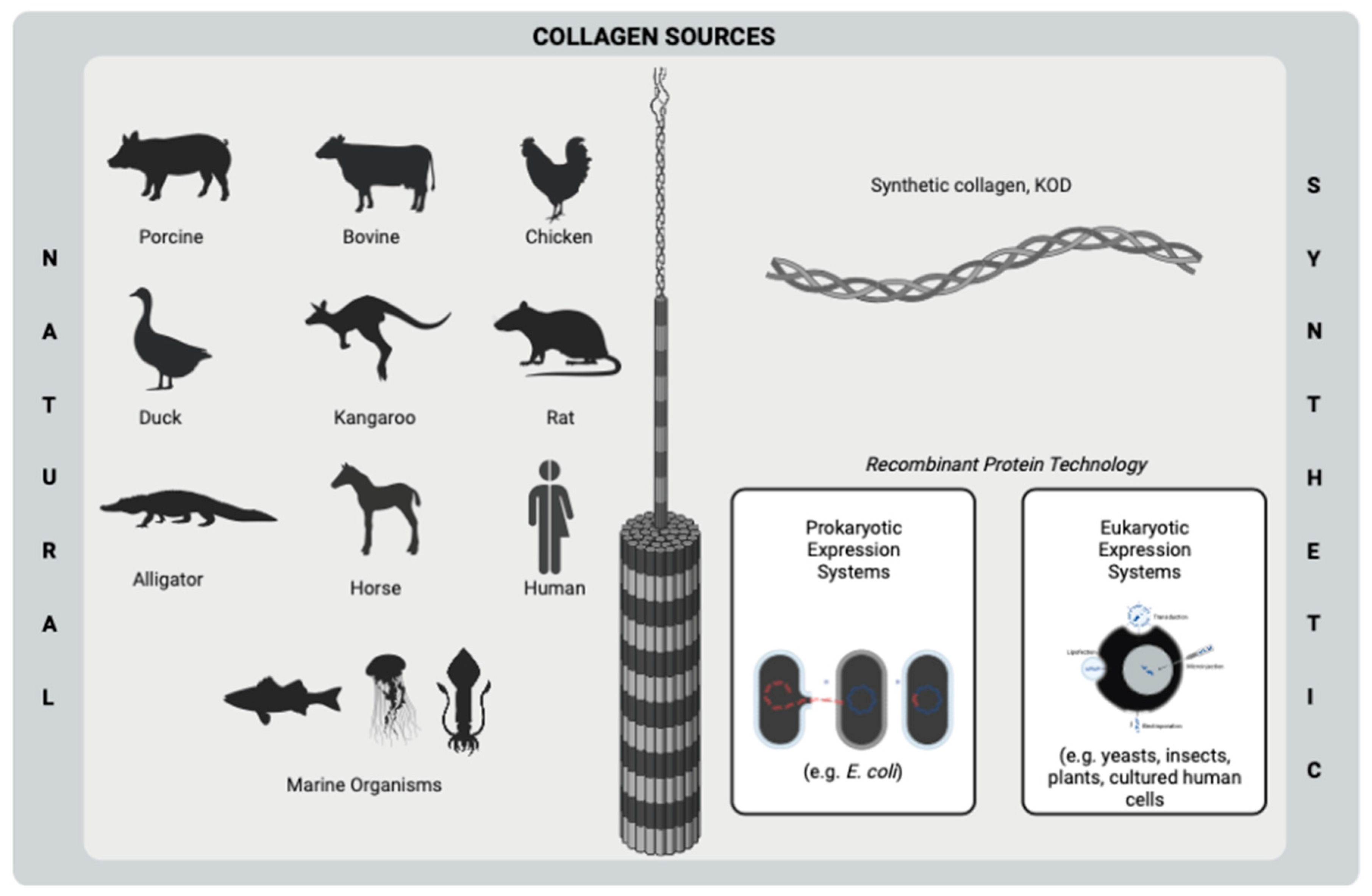
1. Introduction
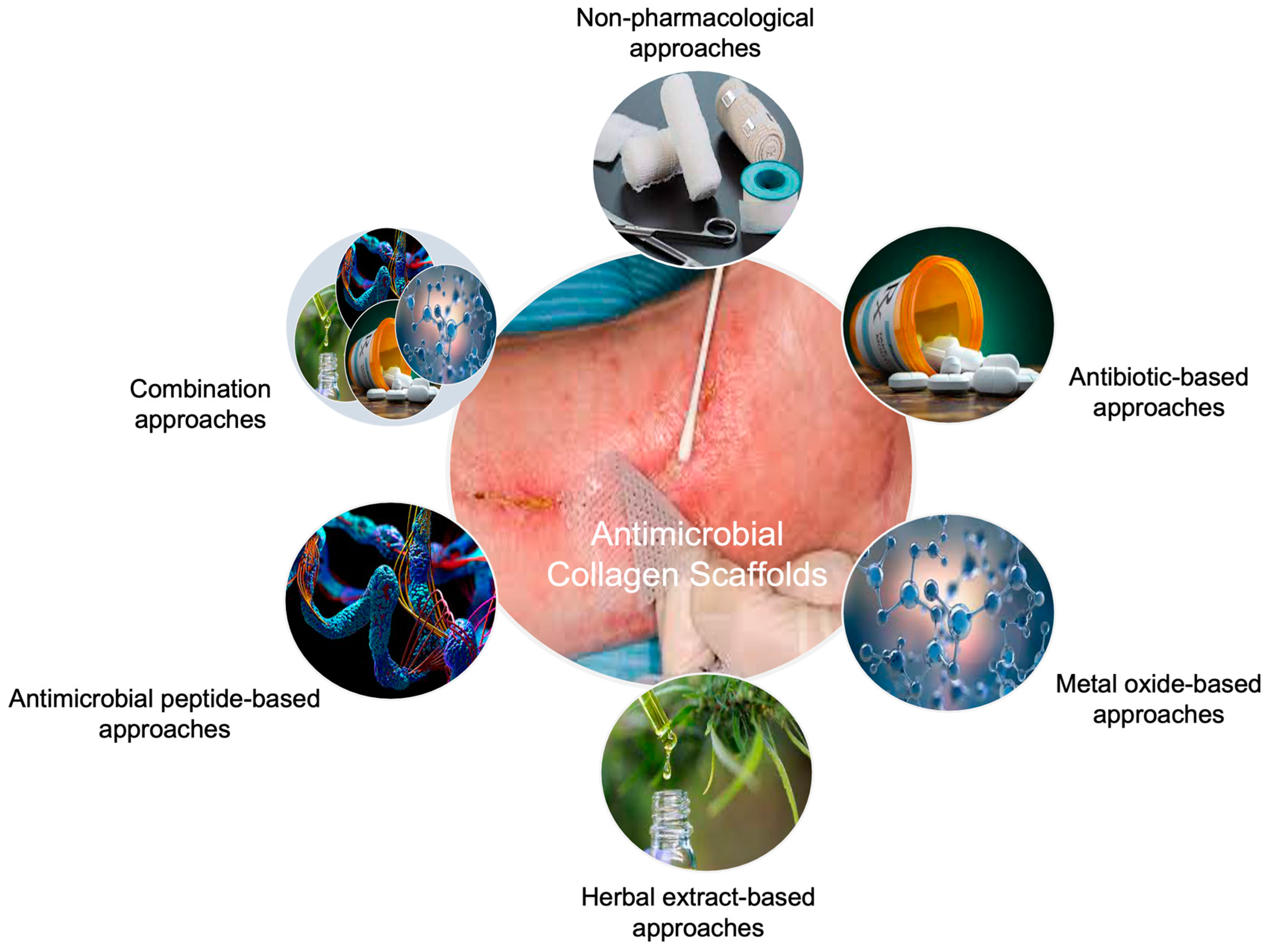
2. Non-Pharmacological Approaches
3. Pharmacological Approaches
3.1. Antibiotic-Based Approaches

| Composition | Collagen Source | Scaffold Form | Crosslinking | Therapeutic Agent | Release Profile | AST | Bacterial Strain/ Cell Line | Antibacterial Activity | Hypothetic Material | Refs. |
|---|---|---|---|---|---|---|---|---|---|---|
| Col (5% w/v) | Porcine dermis | Sponge | HMDI (0.625–10% w/v) | Cef, Ran (0–500 µg/mL) | 10 µg/mL of 90% Cef and 95% Ran released by day 7. | Disc diffusion | E. coli, S. epidermidis Adult HDFs | 100 µg/mL of Cef showed activity on tested strains, while Ran did not. | Localized drug delivery vehicle | [74] |
| Col (1% w/w) Chi (1% w/w) HA (1% w/w) | Rat tail tendon | Thin film | Not available | Gentamicin sulfate (0.4 mg/cm2 film) | Not studied | Disc diffusion | S. aureus, E. coli, P. aeruginosa No in vitro cell culture | Drug-loaded scaffolds showed approximately 25–30 mm of inhibition zone. | Antibacterial film | [61] |
| Col (8 w%) Hap (0–15 w%) | Type I (not specified) | Micro/nanostructured layers | EDC/NHS (4:1 w ratio) | Vancomycin hydrochloride (10 w% of Col) | The max released concentration of vancomycin exceeded the MIC by up to 60–75 times for 4 weeks. | Disc diffusion | MRSA, S. epidermidis, E. faecalis SAOS-2 osteosarcoma cells | Inhibition zone diameters did not differ from standard antibiotic discs significantly. | Local drug carrier | [62] |
| Col (3 mg/mL) | Bovine tendon | Sponge | Not available | Mupirocin (2 mg/mL) (caged into silica microspheres) | Almost 90% of mupirocin was released within 3 days from sponges. | Broth dilution | B. subtilis, S. aureus, E. coli, P. aeruginosa 3T3-L1 fibroblasts | Drug-loaded wound dressings did not show sufficient antibacterial activity on B. subtilis and E. coli. | Wound dressing | [70] |
| Col Chi (2% w/v) | Mouse tail tendon | Asymmetric membrane | TPP (0.2% w/v) | Minocycline (15 µg/mL) (caged into Chi NPs) | Minocycline had sustained release until the 7th day. | Live/dead bacterial double staining | P. gingivalis, F. nucleatum MC3T3-E1 osteoblasts, L929 fibroblasts | Membranes showed 95.3%, and 92.1% of bacteriostatic activity against P. gingivalis and F. nucleatum, respectively. | Scaffold for the prevention of infection and guide bone regeneration | [65] |
| Col hydrolysate (5 mg/mL) PLA (5 mg/mL) cHap (10 mg) | Type I (not specified) | 3D-printed porous scaffold | Alkali hydrolysis (1:1 NaOH:EtOH and 0.5% w/v citric acid) | Minocycline hydrochloride (0.5 mg/mL) | A burst release of minocycline was observed within the first hour. | Disc diffusion, biofilm inhibition assay | S. aureus hBM-MSCs | Drug-loaded scaffolds showed smaller inhibition zone than standard antibiotic discs. | Antimicrobial and osteogenic scaffold | [66] |
| Col (10 w%) PLA EC (7:3, 8:2, 9:1 EC/PLA w ratio) | Fish collagen | Nanofibrous mat | Not available | Silver sulfadiazine (0.25, 0.5, 0.75 w%) | 28 ppm of silver ions were released from 0.75 w% drug-loaded mats within 96 h. | Disc diffusion | Bacillus, E. coli NIH 3T3 fibroblasts | Only 0.75% of drugs including scaffolds showed antibacterial activity against tested strains. | Wound dressing | [75] |
| Col (10, 20, 40% w/w) PVP (30% w/v) PLA PEO (Shell–Col/PVP, Core: 80:20 PLA/PEO w/w) | Bovine tendon | Nanofibrous mat | Not available | Cefazolin sodium | 44.15%, 40.80%, and 37.76% of cefazolin were released for samples containing 10%, 20%, and 30% (w/w) collagen after 6 days. | Disc diffusion | MRSA, E. coli, P. aeruginosa No in vitro cell culture | Fabricated mats showed slightly higher antibacterial activity against P. aeruginosa. | Antibacterial patch for wound healing | [71] |
| Col (10 mg/mL) | Fish collagen | Hydrogel | Alginate dialdehyde (2–10 mg/mL) | Tetracycline hydrochloride (0.01–0.2 mg/mL) | Almost 20% of antibiotics with a concentration equal to or higher than 0.1 mg/mL were released during 600 min. | Zone inhibition | S. aureus 3T3 fibroblasts | Dressings did not show high inhibition rates of S. aureus. | Wound dressing | [67] |
| Col BG (0.5 mg/mL) | Bovine | Membrane (Commercial product) | Not available | Tetracycline hydrochloride (0.05, 0.2, 0.35 mg/mL) | More than 50% of tetracycline releases within the first 6 h, and significant release was observed in 24 h. | Zone inhibition Plate counting | S. aureus (different strains), S. epidermidis MG-63 osteosarcoma cells | Developed scaffolds could significantly inhibit S. aureus growth. | Scaffold for the prevention of biomaterial-related infections | [68] |
| Col (1.5% w/v) Chi (1.5% w/v) CPO (1–4 w%) | Bovine | Sponge | EDC/NHS (3 w%, 2:1 EDC:NHS w ratio) | Ciprofloxacin hydrochloride (1 mg/mL) | Almost 80% of CFH was released from scaffolds including 4% CPO within 200 h. | Zone inhibition | E. coli, S. aureus HDFs | Scaffolds displayed good inhibition zones against both strains. | Skin tissue engineering scaffold | [73] |
| Col (5% w/v) Hap (10 w%) | Rat tail | Sponge | EDC (0.1 mM) | Doxycycline containing Hap NPs (10 w%) | A sustained release of doxycycline (about 70%) was achieved over 14 days. | Time-kill assay | S. aureus, P. aeruginosa BM-MSCs | Antibiotic addition significantly reduced the number of colonies within 24 h. | Bone tissue engineering scaffold | [69] |
| Col (1 w%) CNC (5 w%) | Bovine tendon | Sponge | GTA (0.25%) | Gentamicin sulfate (25 mg/mL) impregnated gelatin microspheres | Gentamicin was completely released after 144 h of the incubation period. | Disc diffusion | E. coli, S. aureus NIH-3T3 fibroblasts | Composite scaffolds showed higher antibacterial activity against E. coli than S. aureus. | Antibacterial skin scaffold | [63] |
| Col Chi (4, 8, 16% total polymer, various Col/Chi w ratio) | Fish | Sponge | Not available | Norfloxacin (1 w%) | An almost complete release of the drug was observed within 20 h. | Not studied | No in vitro cell culture | Not studied | Scaffold for skin regeneration | [76] |
| Col (6.5 mg/mL) | Bovine tendon | Film | EDC/NHS (1:1:6 w ratio EDC/NHS/Col-Tobramycin) | Tobramycin (15 mg/mL) | The burst release of tobramycin (40%) was observed within the first 4 h. | Plate counting | S. aureus Human corneal epithelial cells | Tobramycin-loaded films showed significantly higher inhibition than pristine films. | Scaffold for corneal repair | [64] |
| Col Na-Alginate Hap | Cowhide | Sponge | Genipin CaCl2 (10 w%) | Amoxicillin (0.5, 1, 2 mg/mL) | The long-term drug release effect was investigated. | Zone inhibition | E. coli rASCs | Scaffolds could effectively inhibit E. coli growth. | Composite scaffold for infected bone defects | [77] |
3.2. Non-Antibiotic-Based Approaches
3.2.1. Metal Oxide-Based Approaches
3.2.2. Antimicrobial Peptide-Based Approaches
3.2.3. Herbal Extract-Based Approaches
| Composition | Collagen Source | Scaffold Form | Crosslinking | Therapeutic Agent | Release Profile | AST | Bacterial Strain/ Cell Line | Antibacterial Activity | Hypothetic Material | Refs. |
|---|---|---|---|---|---|---|---|---|---|---|
| Metal oxide-based approaches | ||||||||||
| Col Chi (Various Col/Chi w% ratio) | Goat tendon | Thin film | EDC/NHS (2:1 M ratio) | Silver NPs (0.5 w%) | Not studied | Growth inhibition | E. coli, S. aureus MG-63 osteosarcoma cells | Up to 37% and 27% of growth inhibition was observed against E. coli and S. aureus, respectively. | Composite bone tissue engineering scaffold | [113] |
| Col FN CS (10:1:3 × 10⁻⁵ g/g Col/CS/FN) | Bovine tendon | Sponge | GTA (2.5% v/v) | Silver NPs (1 × 10⁻⁴ g/g polymers) | Not studied | Disc diffusion | F.nucleatum, P. gingivalis Gingival fibroblasts | Hybrid sponges showed slightly higher antimicrobial activity against F. nucleatum. | Oral cavity lesion dressing | [114] |
| Col (8% w/v) | Fish collagen | Nanofibrous mat | GTA (50% w/v) | Silver NPs (0.2% w/v) | Cumulatively, almost 100% of silver ions were released within 25 h. | Microdilution, disc diffusion | S. aureus, P. aeruginosa No in vitro cell culture | Approximately 3.2 and 2.3 cm of inhibition zone diameter was observed after 48 h against S. aureus and P. aeruginosa, respectively. | Wound dressing | [82] |
| Col (10% w/w) | Porcine | Hydrogel | BDDGE | Silver NPs (0.2 µM) | Steady silver concentration was reached within 0.5 h of incubation | Growth inhibition and Time-kill assays | S. aureus, S. epidermidis, E. coli, P. aeruginosa Human epidermal keratinocytes, and dermal fibroblasts | Hybrid hydrogels could inhibit the growth of all tested bacteria. | Implantable anti-infective hybrid biomaterial | [83] |
| Col (5% w/w) His (0, 1, 2% w/w) | Porcine | Membrane | EDC/NHS (3.55 and 2.13 mg/g) | Silver NPs | Not studied | Disc diffusion, bacterial suspension | P. aeruginosa, S. aureus L929 fibroblasts | Developed membranes did not show sufficient antimicrobial activity against both tested strains. | Dressing for full-thickness burn wounds | [85] |
| Col Chi (9:1 Col/Chi w ratio) | Bovine tendon | Hydrogel | EDC/NHS | Silver NPs (0, 2, 5, 10, 20 ppm) | Not studied | Disc diffusion | E. coli, S. aureus Mouse embryo fibroblasts, HaCaTs | Developed wound dressings showed higher inhibition of S. aureus growth. | Wound dressing | [84] |
| Col Hap (various w ratio) | Fish scale | Membrane | Genipin (0.003 g) | Silver NPs (0.05 w%) | Not studied | Disc diffusion | E. coli, S. aureus MG-63 osteosarcoma cells | Scaffolds presented less inhibition zone compared to standard ampicillin discs. | Bone filler | [115] |
| Col (0.5% w/w) | Bovine tendon | Sponge | Dialdehyde xanthan gum (10 mg/mL) | Silver NPs (10 mg/mL) | Not studied | Disc diffusion, bacterial infiltration | E. coli, S. aureus, P. aeruginosa L929 fibroblasts | An increase in silver NP concentration resulted in an increased inhibition rate against tested strains. | Antibacterial wound dressing | [86] |
| Col Sago starch (1, 2, 3 µM) | Fish scale | Sponge | Not available | Sago starch capped silver NPs (1:1 w ratio to Col) | Not studied | Broth dilution | S. aureus, E. coli NIH-3T3 fibroblasts | A lower minimum inhibitory concentration was examined against E. coli. | Scaffold for tissue regeneration applications | [116] |
| Col (1% w/w) Dextran | Calf hide | Hydrogel | GTA (0.25% v/v) | Zinc oxide NPs | Not studied | Not studied | No in vitro cell culture | Not studied | Wound dressing | [117] |
| Col (0.7 w%) | Bovine | Hydrogel | GTA (1% v/v) | Zinc oxide NPs (2, 3, 5 w%) | Not studied | Disc diffusion | S. aureus, E. coli No in vitro cell culture | The inhibition zone diameter decreased with increasing zinc oxide concentration against S. aureus. | Wound dressing | [118] |
| Col PCL (1:2, 1:1, 2:1, 3:1 Col/PCL w ratio) | Type I (not specified) | Nanofibrous mat | Not available | Zinc oxide quantum dots (0–0.75% w/v) | Not studied | Plate counting | E. coli, S. aureus L929 fibroblasts, 3T3 fibroblasts | The number of living bacteria was significantly reduced by the addition of 0.75% of NPs. | Antibacterial wound dressing | [87] |
| Col (1% w/w) | Calf hide | Sponge | GTA (0.5 w%) | Zinc titanate | Not studied | Disc diffusion | S. epidermidis, B. cereus, E. coli, S. enterica, P. putida MG-63 osteosarcoma cells, 3T3 fibroblasts HaCaTs | The porous nanocomposites exerted higher antimicrobial activity against S. epidermidis. | Anti-infection biomaterial | [119] |
| Col (5 mg/mL) Chi (5 mg/mL) | Pig skin | Sponge | GTA (2.5% w/w) | TiO2 NPs (1–7%) | Not studied | Bacterial culture, SEM imaging | S. aureus Mouse fibroblasts, red blood cells | Increased TiO2 amount led to reduced S. aureus colonies on the surface of the scaffold. | Wound dressing | [120] |
| Col (3.47 w%) Chi (3 w%) | Bovine | Nanofibrous mat | GTA (1 w%) | Zinc oxide NPs (1:1:1 w ratio Col:Chi:Zinc oxide) | Not studied | Disc diffusion | S. aureus Hep-2 cells | Membranes showed 4–8 mm of inhibition zone diameter against S. aureus. | Scaffold for skin tissue regeneration | [121] |
| Col Chi (1:9 Col/Chi w ratio) | Not specified | Sponge | Dehydrothermal crosslink at 105 °C for 24 h | Zinc oxide NPs (1, 3, 5 w%) | Not studied | Disc diffusion | E. coli, S. aureus No in vitro cell culture | S. aureus was found more sensitive to developed scaffolds than E. coli. | Antibacterial product | [122] |
| Antimicrobial peptide-based approaches | ||||||||||
| Col (3 mg/mL) | Bovine | Hydrogel | EDC/NHS (50 mM EDC, 25 mM NHS) | AMP GL13K (1 mM) | Burst release was observed from 21 to 28 days. | ATP bioluminescence, live/dead assays | S. gordonii, E. coli hBM-MSCs | AMP GL13K coating significantly demonstrated less effects on the membrane integrity of S. gordonii. | Scaffold for bone/dental tissue growth and infection prevention | [96] |
| Col (0.6% w/v) HA (0.5% w/v) Alginate (1.2% w/v) | Type I (not specified | Sponge | EDC/NHS (0.6 mg/mL EDC, 0.3 mg/mL NHS) | AMP Tet213 (500 µg/mL) | Sustained release (68.4 ± 10.2%) was observed after 14 days. | Zone inhibition, colony counting | E. coli, MRSA, S. aureus NIH-3T3 fibroblasts | The addition of AMP Tet213 into hybrid scaffolds gave rise to almost full inhibition of E. coli and S. aureus. | Mixed-bacteria-infected wound dressing | [95] |
| Col (2.5–3 mg/mL) HA (1.5 mg/mL) | Rat tail tendon | Polyelectrolyte multilayers | GTA (8% w/v) | AMP LL37 (2, 8, 16 µM) | Sustained release of the AMP killed planktonic bacteria. | Broth dilution, bacterial adhesion test, live/dead assay | E. coli Primary rat hepatocytes | The incorporation of 16 µM of AMP LL37 showed almost 3% of live bacteria on the scaffold surface. | Antimicrobial coating | [97] |
| Col PLC (14% w/v) | Not specified | Membrane (ready-to-use product) | Not available | AMP LL37 (10–40 µM) | Membranes containing different LL-37 concentrations released LL-37 in the same quantity. | Not studied | L929 fibroblasts | Not studied | Collagen membrane for guided bone regeneration | [98] |
| Herbal extract-based approaches | ||||||||||
| Col (2% w/v) | Bovine skin | Membrane | Not available | Propolis NPs (200 µg/mL) | Not studied | Not studied | HDFs | Not studied | Dermal patch | [123] |
| Col CNC (7 w%) | Bovine tendon | Sponge | Not available | Curcumin (5 mg/mL) | 99.3% of curcumin was released within the first 24 h. | Disc diffusion | E. coli, S. aureus, P. aeruginosa No in vitro cell culture | Curcumin significantly enhanced the antimicrobial activity of pristine porous scaffolds. | Full-thickness burn dressing | [110] |
| Col (1% w/w) Col/Gel microparticles (50, 125, 250 mg) | Bovine | Sponge | GTA (0.02% v/v) | Calendula officinalis extract (1% v/v) | Incomplete release of the extract was observed within 14 days at pH 5.5 and 7.4. | Not studied | L929 fibroblasts | Not studied | Dermal substitute | [124] |
| Col | Goat tendon | Aerogel | Wheatgrass (1, 2, 3% w/v) | Wheatgrass (1, 2, 3% w/v) | Not studied | Agar diffusion | E. coli, B. subtilis Swiss 3T6 fibroblasts, HaCaTs | Hybrid aerogels showed smaller inhibition zones than commercial ampicillin discs against B. subtilis. | Wound dressing | [111] |
| Col (10 mg/mL) GSP (25–100 w% to Col) | Cowhide trimming waste | Sponge | Chloroform extract of cinnamon bark (14.28% v/v) | Cinnamon bark powder (2 g) | Not studied | Broth dilution | B. subtilis, S. aureus, E. coli No in vitro cell culture | The addition of cinnamon bark powder led to great inhibition of all tested strains. | Antimicrobial wound dressing | [107] |
| Col (9 mg/mL) | Type I (Not specified) | Sponge | Not available | Berberine-oleanolic acid (1–5%) | All samples released about 70% of the drug within 1 h. | Filter paper diffusion | S. aureus, E. coli MG-63 osteosarcoma cells | Gram-positive bacteria were found more sensitive to developed scaffolds than Gram-negative bacteria. | Scaffold for postoperative bacterial bone infection | [125] |
| Col (1% w/v) Chi (1% w/v) Hap (5% w/v) PCL (20–80 mg/mL) PVA (0.5–3% w/v) | Bovine tendon | Sponge | GTA (0.1% v/v) | Cissus quadrangularis caged PCL nanoparticles | Cumulatively more than 80% of the extract was released within 21 days. | Not studied | MC3T3-E1 osteoblasts | Not studied | Bone tissue engineering scaffold | [108] |
| Col (1% w/v) | Rat tail tendon | Film | Not available | Thymol (0.25–4 mg/cm2) | Not studied | Dehydrogenase activity assay, ATP bioluminescence, microbial penetration assay | S. aureus, E. coli, P. aeruginosa Red blood cells | 4 mg/cm2 of thymol including films indicated almost full inhibition of all tested strains. | Antibacterial film for wound care applications | [109] |
| Col (11 w% middle layers; 10 w% inner layers) PCL (10 w% outer layers; 11 w% middle layer) | Rat tail | Nanofibrous mat | Not available | Melilotus officinalis (2, 4, 8% w/w) | Not studied | Not studied | L929 fibroblasts | Not studied | Diabetic foot ulcer dressing | [112] |
| Col Lipid NPs (10:1 w ratio Col/Lipid NPs) | Bovine tendon | Sponge | Not available | Curcumin into lipid NPs | The complete release of curcumin-loaded NPs was observed within 25 days. | Not studied | NIH 3T3 fibroblasts, HaCaTs | Not studied | Composite cryostructurate for wound healing | [126] |
| Col (10 mg/mL) Annona polysaccharide (7.5 mg/mL) | Bovine Achilles tendon | Sponge | Chloroform extract of cinnamon bark | Tetrahydrocurcumin microspheres | 28.95 ± 1.7% of the drug was released within 12 h from the composite scaffold. | Disc diffusion | B. subtilis, P. aeruginosa, S. aureus NIH 3T3 fibroblasts | Approximately 20 mm and 10 mm inhibition zone diameters were evaluated against S. aureus around the positive control and composite scaffold, respectively. | Antimicrobial wound dressing | [127] |
| Col (60% v/v in shell) PVA (50% v/v in core) | Type I (Not specified) | Nanofibrous core–shell mat | Not available | Licorice roots (50% v/v in core, and 40% v/v in shell) | Not studied | Disc diffusion | S. aureus, P. aeruginosa No in vitro cell culture | Bio-nano scaffolds did not show any activity on the inhibition of P. aeruginosa growth. | Hybrid bio-nano wound dressing | [128] |
4. Combination Approaches
| Composition | Collagen Source | Scaffold Form | Crosslinking | Therapeutic Agent | Release Profile | AST | Bacterial Strain/ Cell Line | Antibacterial Activity | Hypothetic Material | Refs. |
|---|---|---|---|---|---|---|---|---|---|---|
| Col hydrolysate (2.66% w/v) Chi (1.5% w/v) | Bovine tendon Rabbit skin | Nanofibrous mat | Not available | Lemon balm and Dill EOs (60 mg/mL each, 1:1 ratio) | Not studied | Disc diffusion | S. aureus, E. coli, E. faecalis, S. typhimurium No in vitro cell culture | While EOs only did not show efficient antimicrobial activity, EO-including membranes, showed significantly higher activity against tested strains. | Medical wound dressing | [103] |
| Col (5% w/v) | Not specified | Sponge | GTA (2.5% v/v) | AMPs Pac-525 and KSL-W (1.5 mg/mL) into PLGA microspheres | Burst release of AMPs occurred within 2 days in both microspheres and scaffolds. | Oxford cup disc diffusion | S. aureus, E. coli MC-3T3 fibroblasts | Lower doses of AMPs could not lead to inhibition of S. aureus and E. coli growth. | Scaffold for infective bone defect repair | [134] |
| Col (3.5% w/v) Chi (1.5% w/v) | Hydrolyzed peptide | Bilayer sponge | GTA (0.025% v/v) | Silymarin (0.5, 1, 2% w/w), and silver NPs (3% w/w) | A sustained release of antioxidants was observed over 120 h. | Not studied | Cos-7 fibroblasts | Not studied | Antioxidant and antibacterial wound dressing | [129] |
| Col | Rat tail tendon | Hydrogel | Incubation of Col solution in saturated NH3 chamber | Silver NPs (67, 6.7, 0.67 mg/g), and Cannabis sativa oil (0.15 mL) | Only 1.5 g of the silver content is released after 24 h. | Disc diffusion, broth dilution | S. aureus, P. aeruginosa MDCK epithelial cells | Inhibition zone diameter of 67 mg/g silver-NP-including hydrogels increased from 1.45 to 1.75 cm with the addition of EO. | Wound dressing | [135] |
| Col (1% w/v) | Fish scale | Sponge | EDC/NHS (1:2:2 GO:EDC:NHS molar ratio) | Curcumin (0.5 w%), and GO NPs (2 mg/mL) | 82.5% of loaded curcumin was released within 96 h. | Disc diffusion | P. aeruginosa, S. aureus NIH-3T3 fibroblasts | The inhibition zone diameters around hybrid scaffolds were evaluated as approximately 16 and 15 mm against S. aureus, and P. aeruginosa, respectively. | Wound dressing | [130] |
| Col | Rat tail tendon | Membrane | Curcumin caged silver NPs (10, 20 µM) | Curcumin (20–100 µM) caged silver NPs | Not studied | Broth dilution | E. coli, B. subtilis HaCaTs | 20 µM curcumin-caged silver NPs showed 95% growth inhibition of E. coli. | Scaffold for biomedical engineering | [136] |
| Col (3 mg/mL) | Rat tail tendon | Sponge | Plumbagin (1–5 µM) | Plumbagin (1–5 µM) caged silver NPs | Not studied | Disc diffusion, broth microdilution | E. coli B. subtilis No in vitro cell culture | Hybrid scaffolds presented better antimicrobial activity against B. subtilis. | Wound dressing | [131] |
| Col (8 w%) Hap (0, 5, 15 w%) | Type I (Not specified) | Nanofibrous mat | EDC/NHS (4:1 w ratio EDC:NHS) | Vancomycin hydrochloride, gentamicin sulfate (10 w% total, 1:1 w ratio) | High concentrations of vancomycin and gentamicin were released for 21 days. | Disc diffusion | MRSA, S. epidermidis, E. faecalis SAOS-2 osteosarcoma cells | The synergetic effect of two antibiotics yielded increased inhibition zone diameters on MRSA. | Scaffold for the treatment of prosthetic joint infection | [137] |
| Col (2, 3, 4 mg/mL) Fibrinogen (1.25 mg/mL) Thrombin (0.156 IU/mL) | Bovine | Hydrogel | Not available | Collagen mimetic peptide tethered vancomycin (1.25 mg/gel) into liposomes (30 µg/gel) | Complete vancomycin release was achieved within 12 h. | Broth dilution | S. aureus, MRSA NIH-3T3 fibroblasts | Hybrid hydrogels presented higher antimicrobial activity than pristine hydrogels with less than 104 CFU/wound up to the 9th day. | Scaffold for the MRSA-associated treatment | [132] |
| Col (1% w/v) | Fish scale | Sponge | GTA (0.25% v/v) | Mupirocin (1:1 w ratio) and Macrotyloma uniflorum extract (10% v/v) | 94% of mupirocin was released within 72 h. | Disc diffusion | B. subtilis, S. aureus, P. vulgaris, E. coli NIH-3T3 fibroblasts, HaCaTs | The highest antimicrobial activity of composite dressings was observed on S. aureus. | Burn wound dressing | [138] |
| Col (20 w%) PCL Zein (15 w% PCL/Zein with various ratios) | Fish | Nanofibrous mat | Not available | Zinc oxide NPs (1 w%) and Aloe vera (5, 8 w%) | Approximately 70% of zinc oxide NPs released within 30 days. | Disc diffusion | S. aureus, E. coli Human gingival fibroblasts | The combination of zinc oxide NPs with Aloe vera increased the growth inhibition rate of both bacteria. | Wound dressing | [139] |
| Col (0.5% w/v) | Rat tail | Bilayer sponge | GTA (25% v/v) | Fibrinogen and silver NPs | 50% of the included fibrinogen was released within 5 days. | Zone inhibition | E. coli No in vitro cell culture | The one-fold increase in silver NPs concentration did not enhance the antimicrobial activity of scaffolds significantly. | Skin tissue engineering scaffold | [140] |
| Col (6 mg/mL) Elastin-like peptide (18 mg/mL) (1:3 Col/ELP) | Rat tail tendon | Hydrogel | EDC/NHS | rhBMP-2 (0.005% w/v) doxycycline hyclate (0.5% w/w) | Bi-phasic release of doxycycline was observed with an initial burst release followed by a sustained release. | Zone inhibition | E. coli, P. aeruginosa, S. sanguinis hASCs | The developed hydrogels could not exert effective activity against E. coli. | Bone regenerative hydrogel | [141] |
| Col (2 w%) PCL (15 w%) Chi (2 w%) PEO (5 w%) | Type I (not specified) | 3-layered nanofibrous mat | Not available | Silver sulfadiazine (3 mg/mL), EGF, and bFGF (25 µg/mL each) | Between days 5 and 20, the sustained release was achieved with a cumulative release of about 80%. | Antibiotic tube dilution | P. aeruginosa, S. aureus HDFs | Minimum inhibitory concentration was evaluated as 15 and 30 µg/mL against P. aeruginosa and S. aureus, respectively. | Wound dressing | [133] |
| Col (1% w/v) | Bovine skin | Sponge | GTA (0–1% w/v) | OTC (1 g/L) DXC (1 g/L) | About 70% of OTC was released from 0.5% of GTA crosslinked scaffolds within 600 min. | Broth dilution | E. coli, E. faecalis, S. aureus Dermal fibroblasts of mouse cell line | Oxytetracycline led to more inhibition growth of tested bacteria. | Dressing for prevention and treatment of infections at the application site | [142] |
| Col PVA (1:3 w/w PVA/Col) | Bovine tendon | Membrane | Not available | Ciprofloxacin and tobramycin (0.3% w/v for soaking method, 5% w/w for mixing method) | CP showed more sustained and controlled release. 95% of CP was released after 48 h. | Microdilution, time-kill assay | S. aureus, E. coli No in vitro cell culture | The efficacy of membranes to kill the tested bacteria was found independent of their release profile. | Ulcerative keratitis dressing | [143] |
| Col (4 mg/mL) | Not specified | Sponge | Triphenyl phosphate (10% v/v) | Mupirocin (50 mg) in 5% w/v Chi microspheres and Piper betle extract (5% v/v) | More than 50% of both drugs are released at the end of 12 h. | Agar disc diffusion | E. coli, S. aureus No in vitro cell culture | The combination of two antimicrobials slightly increased the antimicrobial activity against both strains. | Wound dressing | [144] |
| Col (1.06 mg/mL) | Rapana venosa | Sponge | Not available | Salvia officinalis extract loaded mesoporous silica NPs (10, 20 mg/mL) | Not studied | Broth microdilution | P. aeruginosa, S. aureus HaCaTs, Human Mel-Juso skin carcinoma cells | The hybrid scaffolds showed at least a two-fold higher minimum inhibitory concentration for P. aeruginosa. | Wound dressing | [145] |
5. Conclusions
Author Contributions
Funding
Institutional Review Board Statement
Informed Consent Statement
Data Availability Statement
Conflicts of Interest
References
- Vera, D.M.A.; Haynes, M.H.; Ball, A.R.; Dai, T.; Astrakas, C.; Kelso, M.J.; Hamblin, M.R.; Tegos, G.P. Strategies to potentiate antimicrobial photoinactivation by overcoming resistant phenotypes. Photochem. Photobiol. 2012, 88, 499–511. [Google Scholar] [CrossRef]
- Cai, J.; Liu, R. Introduction to Antibacterial Biomaterials. Biomater. Sci. 2020, 8, 6812–6813. [Google Scholar] [CrossRef] [PubMed]
- Voidarou, C.; Bezirtzoglou, E.; Alexopoulos, A.; Plessas, S.; Stefanis, C.; Papadopoulos, I.; Vavias, S.; Stavropoulou, E.; Fotou, K.; Tzora, A. Occurrence of Clostridium perfringens from different cultivated soils. Anaerobe 2011, 17, 320–324. [Google Scholar] [CrossRef]
- Tan, S.Y.; Tatsumura, Y. Alexander Fleming (1881–1955): Discoverer of penicillin. Singap. Med. J. 2015, 56, 366. [Google Scholar] [CrossRef] [PubMed]
- Nelli, A.; Voidarou, C.; Venardou, B.; Fotou, K.; Tsinas, A.; Bonos, E.; Fthenakis, G.C.; Skoufos, I.; Tzora, A. Antimicrobial and Methicillin Resistance Pattern of Potential Mastitis-Inducing Staphylococcus aureus and Coagulase-Negative Staphylococci Isolates from the Mammary Secretion of Dairy Goats. Biology 2022, 11, 1591. [Google Scholar] [CrossRef] [PubMed]
- Organisation for Economic Cooperation and Development; European Union. Health at a Glance: Europe 2018: State of Health in the EU Cycle; OECD Publishing, Paris/European Union: Brussels, Belgium, 2018. [Google Scholar]
- Suetens, C.; Latour, K.; Kärki, T.; Ricchizzi, E.; Kinross, P.; Moro, M.L.; Jans, B.; Hopkins, S.; Hansen, S.; Lyytikäinen, O. Prevalence of healthcare-associated infections, estimated incidence and composite antimicrobial resistance index in acute care hospitals and long-term care facilities: Results from two European point prevalence surveys, 2016 to 2017. Eurosurveillance 2018, 23, 1800516. [Google Scholar] [CrossRef]
- Cassini, A.; Plachouras, D.; Eckmanns, T.; Abu Sin, M.; Blank, H.-P.; Ducomble, T.; Haller, S.; Harder, T.; Klingeberg, A.; Sixtensson, M. Burden of six healthcare-associated infections on European population health: Estimating incidence-based disability-adjusted life years through a population prevalence-based modelling study. PLoS Med. 2016, 13, e1002150. [Google Scholar] [CrossRef]
- U.S. Centers for Disease Control and Prevention. Antibiotic Resistance Threats in the United State; Department of Health and Human Services, CDC: Atlanta, GA, USA, 2019. [Google Scholar]
- Organisation for Economic Cooperation and Development; European Union. Tackling Wasteful Spending on Health; OECD Publishing: Paris, France, 2017. [Google Scholar]
- Prestinaci, F.; Pezzotti, P.; Pantosti, A. Antimicrobial resistance: A global multifaceted phenomenon. Pathog. Glob. Health 2015, 109, 309–318. [Google Scholar] [CrossRef]
- Dadgostar, P. Antimicrobial resistance: Implications and costs. Infect. Drug Resist. 2019, 12, 3903. [Google Scholar] [CrossRef]
- Murugaiyan, J.; Kumar, P.A.; Rao, G.S.; Iskandar, K.; Hawser, S.; Hays, J.P.; Mohsen, Y.; Adukkadukkam, S.; Awuah, W.A.; Jose, R.A.M. Progress in alternative strategies to combat antimicrobial resistance: Focus on antibiotics. Antibiotics 2022, 11, 200. [Google Scholar] [CrossRef]
- Majumder, M.A.A.; Rahman, S.; Cohall, D.; Bharatha, A.; Singh, K.; Haque, M.; Gittens-St Hilaire, M. Antimicrobial stewardship: Fighting antimicrobial resistance and protecting global public health. Infect. Drug Resist. 2020, 13, 4713–4738. [Google Scholar] [CrossRef] [PubMed]
- Tarín-Pelló, A.; Suay-García, B.; Pérez-Gracia, M.-T. Antibiotic resistant bacteria: Current situation and treatment options to accelerate the development of a new antimicrobial arsenal. Expert Rev. Anti-Infect. Ther. 2022, 20, 1095–1108. [Google Scholar] [CrossRef]
- Ricard-Blum, S. The collagen family. Cold Spring Harb. Perspect. Biol. 2011, 3, a004978. [Google Scholar] [CrossRef] [PubMed]
- Ferreira, A.M.; Gentile, P.; Chiono, V.; Ciardelli, G. Collagen for bone tissue regeneration. Acta Biomater. 2012, 8, 3191–3200. [Google Scholar] [CrossRef]
- Stark, Y.; Suck, K.; Kasper, C.; Wieland, M.; van Griensven, M.; Scheper, T. Application of collagen matrices for cartilage tissue engineering. Exp. Toxicol. Pathol. 2006, 57, 305–311. [Google Scholar] [CrossRef] [PubMed]
- Zhang, G.; Young, B.; Ezura, Y.; Favata, M.; Soslowsky, L.; Chakravarti, S.; Birk, D.E. Development of tendon structure and function: Regulation of collagen fibrillogenesis. J. Musculoskelet Neuronal Interact. 2005, 5, 5–21. [Google Scholar]
- Priya, S.G.; Jungvid, H.; Kumar, A. Skin tissue engineering for tissue repair and regeneration. Tissue Eng. Part B Rev. 2008, 14, 105–118. [Google Scholar] [CrossRef] [PubMed]
- Kalic, T.; Kamath, S.D.; Ruethers, T.; Taki, A.C.; Nugraha, R.; Le, T.T.; Humeniuk, P.; Williamson, N.A.; Hira, D.; Rolland, J.M. Collagen—An important fish allergen for improved diagnosis. J. Allergy Clin. Immunol. Pract. 2020, 8, 3084–3092.e3010. [Google Scholar] [CrossRef] [PubMed]
- Sorushanova, A.; Delgado, L.M.; Wu, Z.; Shologu, N.; Kshirsagar, A.; Raghunath, R.; Mullen, A.M.; Bayon, Y.; Pandit, A.; Raghunath, M.; et al. The collagen suprafamily: From biosynthesis to advanced biomaterial development. Adv. Mater. 2019, 31, 1801651. [Google Scholar] [CrossRef]
- Rezvani Ghomi, E.; Nourbakhsh, N.; Akbari Kenari, M.; Zare, M.; Ramakrishna, S. Collagen-based biomaterials for biomedical applications. J. Biomed. Mater. Res. 2021, 109, 1986–1999. [Google Scholar] [CrossRef]
- David, G. Collagen-based 3D structures—Versatile, efficient materials for biomedical applications. In Biopolymer-Based Formulations, 1st ed.; Elsevier: Amsterdam, The Netharlands, 2020; pp. 881–906. [Google Scholar]
- Lee, C.H.; Singla, A.; Lee, Y. Biomedical applications of collagen. Int. J. Pharm. 2001, 221, 1–22. [Google Scholar] [CrossRef]
- Sundar, G.; Joseph, J.; John, A.; Abraham, A. Natural collagen bioscaffolds for skin tissue engineering strategies in burns: A critical review. Int. J. Polym. Mater. 2021, 70, 593–604. [Google Scholar] [CrossRef]
- Singh, O.; Gupta, S.S.; Soni, M.; Moses, S.; Shukla, S.; Mathur, R.K. Collagen dressing versus conventional dressings in burn and chronic wounds: A retrospective study. J. Cutan. Aesthetic Surg. 2011, 4, 12. [Google Scholar] [CrossRef] [PubMed]
- Avila Rodríguez, M.I.; Rodríguez Barroso, L.G.; Sánchez, M.L. Collagen: A review on its sources and potential cosmetic applications. J. Cosmet. Dermatol. 2018, 17, 20–26. [Google Scholar] [CrossRef] [PubMed]
- Ge, X. Antimicrobial biomaterials with non-antibiotic strategy. Biosurf. Biotribol. 2019, 5, 71–82. [Google Scholar] [CrossRef]
- Li, S.; Dong, S.; Xu, W.; Tu, S.; Yan, L.; Zhao, C.; Ding, J.; Chen, X. Antibacterial hydrogels. Adv. Sci. 2018, 5, 1700527. [Google Scholar] [CrossRef]
- 3M™ Promogran Prisma™ Matrix. Available online: https://www.3m.com/3M/en_US/p/d/b5005265080/ (accessed on 13 February 2023).
- ColActive® Plus Powder Ag. Available online: https://www.woundsource.com/product/colactive-plus-powder-ag (accessed on 13 February 2023).
- Septocoll® E—Biomet. Available online: https://www.yumpu.com/en/document/view/42479805/septocollar-e-biomet (accessed on 13 February 2023).
- DermaCol™ Collagen Matrix Dressing. Available online: https://dermarite.com/product/dermacol/ (accessed on 13 February 2023).
- DermaCol/Ag™ Collagen Matrix Dressing with Silver. Available online: https://dermarite.com/product/dermacolag/ (accessed on 13 February 2023).
- SilvaKollagen® Gel Silver Collagen Wound Gel. Available online: https://dermarite.com/product/silvakollagen-gel/ (accessed on 13 February 2023).
- Puracol Plus AG+ Collagen Wound Dressings with Silver. Available online: https://punchout.medline.com/product/Puracol-Plus-AG-Collagen-Wound-Dressings-with-Silver/Collagen-Dressings/Z05-PF00137?question=&index=P1&indexCount=1 (accessed on 13 February 2023).
- SEESKIN P—Collagen Particle Dressing. Available online: https://www.synerheal.com/product-page/seeskin-p-collagen-particle-dressing (accessed on 13 February 2023).
- CollaSorb. Available online: https://www.vitalitymedical.com/collasorb.html (accessed on 13 February 2023).
- GENTA-COLL ® Resorb Collagen Gentamicin Sponge. Available online: https://resorba.com/region/row/product/biosurgicals/genta-coll-resorb/ (accessed on 13 February 2023).
- COLLAMYCIN (Gentamicin Collagen Gel). Available online: https://www.synerheal.com/product-page/collamycin-gentamicin-collagen-gel (accessed on 13 February 2023).
- Gencoll Gel. Available online: https://www.cologenesis.net/medicated-gencoll-gel.htm (accessed on 13 February 2023).
- Colloskin-M. Available online: https://www.cologenesis.net/colloskin-m.htm (accessed on 13 February 2023).
- Collofiber-MM Medicated. Available online: https://www.cologenesis.net/collofiber-mm-medicated.htm (accessed on 13 February 2023).
- Coloplug. Available online: https://www.cologenesis.net/coloplug-collagen-sponge.htm (accessed on 13 February 2023).
- Diacoll-S. Available online: https://www.cologenesis.net/diacoll-s-sterile-collagen-sheet.htm (accessed on 13 February 2023).
- Irastorza, A.; Zarandona, I.; Andonegi, M.; Guerrero, P.; de la Caba, K. The versatility of collagen and chitosan: From food to biomedical applications. Food Hydrocoll. 2021, 116, 106633. [Google Scholar] [CrossRef]
- Yuan, H.; Chen, L.; Hong, F.F. A biodegradable antibacterial nanocomposite based on oxidized bacterial nanocellulose for rapid hemostasis and wound healing. ACS Appl. Mater. Interfaces 2019, 12, 3382–3392. [Google Scholar] [CrossRef]
- Ramadass, S.K.; Perumal, S.; Gopinath, A.; Nisal, A.; Subramanian, S.; Madhan, B. Sol–gel assisted fabrication of collagen hydrolysate composite scaffold: A novel therapeutic alternative to the traditional collagen scaffold. ACS Appl. Mater. Interfaces 2014, 6, 15015–15025. [Google Scholar] [CrossRef] [PubMed]
- Mahmoudifard, M. Graphene family in cancer therapy: Recent progress in Cancer Gene/Drug delivery applications. J. Mater. Chem. B 2023, 11, 2568–2613. [Google Scholar]
- Oladele, I.; Agbabiaka, O.; Olasunkanmi, O.; Balogun, A.; Popoola, M. Non-synthetic sources for the development of hydroxyapatite. J. Appl. Biotechnol. Bioeng. 2018, 5, 88–95. [Google Scholar]
- Zhou, T.; Sui, B.; Mo, X.; Sun, J. Multifunctional and biomimetic fish collagen/bioactive glass nanofibers: Fabrication, antibacterial activity and inducing skin regeneration in vitro and in vivo. Int. J. Nanomed. 2017, 12, 3495. [Google Scholar] [CrossRef] [PubMed]
- Dolete, G.; Tihăuan, B.M.; Tutunaru, O.; Mocanu, I.-C.; Balaş, C.; Lavinia, I.; Ardelean, D.S.D.; Kamerzan, C.M.; Maier, S.S. Development and sequential analysis of a collagen-chitosan wound management biomaterial. Rom. Biotechnol. Lett. 2019, 24, 108–117. [Google Scholar] [CrossRef]
- ISO 20743:2007; Textiles—Determination of Antibacterial Activity of Antibacterial Finished Products. International Organization for Standardization: Geneva, Switzerland, 2007.
- Gilarska, A.; Lewandowska-Łańcucka, J.; Guzdek-Zając, K.; Karewicz, A.; Horak, W.; Lach, R.; Wójcik, K.; Nowakowska, M. Bioactive yet antimicrobial structurally stable collagen/chitosan/lysine functionalized hyaluronic acid–based injectable hydrogels for potential bone tissue engineering applications. Int. J. Biol. Macromol. 2020, 155, 938–950. [Google Scholar] [CrossRef] [PubMed]
- Valenzuela-Rojo, R.D.; López-Cervantes, J.; Sánchez-Machado, D.I.; Escárcega-Galaz, A.A.; del Rosario Martínez-Macias, M. Antibacterial, mechanical and physical properties of collagen-chitosan sponges from aquatic source. Sustain. Chem. Pharm. 2020, 15, 100218. [Google Scholar] [CrossRef]
- Rajasree, S.R.; Gobalakrishnan, M.; Aranganathan, L.; Karthih, M. Fabrication and characterization of chitosan based collagen/gelatin composite scaffolds from big eye snapper Priacanthus hamrur skin for antimicrobial and anti oxidant applications. Mater. Sci. Eng. C 2020, 107, 110270. [Google Scholar] [CrossRef] [PubMed]
- Bian, T.; Pang, N.; Xing, H. Preparation and antibacterial evaluation of a beta-tricalcium phosphate/collagen nanofiber biomimetic composite scaffold. Mater. Chem. Phys. 2021, 273, 125059. [Google Scholar] [CrossRef]
- Gao, Y.; Kang, Y.; Wang, T.; Li, C.; Shen, S.; Qu, C.; Gong, S.; Liu, P.; Yang, L.; Liu, J. Alginate microspheres-collagen hydrogel, as a novel 3D culture system, enhanced skin wound healing of hUCMSCs in rats model. Colloids Surf. B 2022, 219, 112799. [Google Scholar] [CrossRef]
- Dantas, G.; Sommer, M.O.; Oluwasegun, R.D.; Church, G.M. Bacteria subsisting on antibiotics. Science 2008, 320, 100–103. [Google Scholar] [CrossRef]
- Michalska-Sionkowska, M.; Kaczmarek, B.; Walczak, M.; Sionkowska, A. Antimicrobial activity of new materials based on the blends of collagen/chitosan/hyaluronic acid with gentamicin sulfate addition. Mater. Sci. Eng. C 2018, 86, 103–108. [Google Scholar] [CrossRef]
- Suchý, T.; Šupová, M.; Klapková, E.; Adamková, V.; Závora, J.; Žaloudková, M.; Rýglová, Š.; Ballay, R.; Denk, F.; Pokorný, M. The release kinetics, antimicrobial activity and cytocompatibility of differently prepared collagen/hydroxyapatite/vancomycin layers: Microstructure vs. nanostructure. Eur. J. Pharm. Sci. 2017, 100, 219–229. [Google Scholar] [CrossRef]
- Zhu, Q.; Teng, J.; Liu, X.; Lan, Y.; Guo, R. Preparation and characterization of gentamycin sulfate-impregnated gelatin microspheres/collagen–cellulose/nanocrystal scaffolds. Polym. Bull. 2018, 75, 77–91. [Google Scholar] [CrossRef]
- Liu, Y.; Ren, L.; Long, K.; Wang, L.; Wang, Y. Preparation and characterization of a novel tobramycin-containing antibacterial collagen film for corneal tissue engineering. Acta Biomater. 2014, 10, 289–299. [Google Scholar] [CrossRef] [PubMed]
- Ma, S.; Adayi, A.; Liu, Z.; Li, M.; Wu, M.; Xiao, L.; Sun, Y.; Cai, Q.; Yang, X.; Zhang, X. Asymmetric collagen/chitosan membrane containing minocycline-loaded chitosan nanoparticles for guided bone regeneration. Sci. Rep. 2016, 6, 31822. [Google Scholar] [CrossRef] [PubMed]
- Martin, V.; Ribeiro, I.A.; Alves, M.M.; Gonçalves, L.; Claudio, R.A.; Grenho, L.; Fernandes, M.H.; Gomes, P.; Santos, C.F.; Bettencourt, A.F. Engineering a multifunctional 3D-printed PLA-collagen-minocycline-nanoHydroxyapatite scaffold with combined antimicrobial and osteogenic effects for bone regeneration. Mater. Sci. Eng. C 2019, 101, 15–26. [Google Scholar] [CrossRef]
- Yu, X.; Yuan, Q.; Yang, M.; Liu, R.; Zhu, S.; Li, J.; Zhang, W.; You, J.; Xiong, S.; Hu, Y. Development of biocompatible and antibacterial collagen hydrogels via dialdehyde polysaccharide modification and tetracycline hydrochloride loading. Macromol. Mater. Eng. 2019, 304, 1800755. [Google Scholar] [CrossRef]
- Rivadeneira, J.; Di Virgilio, A.; Audisio, M.; Boccaccini, A.; Gorustovich, A. Evaluation of antibacterial and cytotoxic effects of nano-sized bioactive glass/collagen composites releasing tetracycline hydrochloride. J. Appl. Microbiol. 2014, 116, 1438–1446. [Google Scholar] [CrossRef] [PubMed]
- Semyari, H.; Salehi, M.; Taleghani, F.; Ehterami, A.; Bastami, F.; Jalayer, T.; Semyari, H.; Hamed Nabavi, M.; Semyari, H. Fabrication and characterization of collagen–hydroxyapatite-based composite scaffolds containing doxycycline via freeze-casting method for bone tissue engineering. J. Biomater. Appl. 2018, 33, 501–513. [Google Scholar] [CrossRef]
- Perumal, S.; kumar Ramadass, S.; Madhan, B. Sol–gel processed mupirocin silica microspheres loaded collagen scaffold: A synergistic bio-composite for wound healing. Eur. J. Pharm. Sci. 2014, 52, 26–33. [Google Scholar] [CrossRef]
- Hajikhani, M.; Emam-Djomeh, Z.; Askari, G. Fabrication and characterization of mucoadhesive bioplastic patch via coaxial polylactic acid (PLA) based electrospun nanofibers with antimicrobial and wound healing application. Int. J. Biol. Macromol. 2021, 172, 143–153. [Google Scholar] [CrossRef]
- Yilmaz Atay, H. Antibacterial activity of chitosan-based systems. In Functional Chitosan: Drug Delivery and Biomedical Applications; Springer Nature: Berlin/Heidelberg, Germany, 2019; pp. 457–489. [Google Scholar]
- Tripathi, S.; Singh, B.N.; Divakar, S.; Kumar, G.; Mallick, S.P.; Srivastava, P. Design and evaluation of ciprofloxacin loaded collagen chitosan oxygenating scaffold for skin tissue engineering. Biomed. Mater. 2021, 16, 025021. [Google Scholar] [CrossRef]
- Tsekoura, E.; Helling, A.; Wall, J.; Bayon, Y.; Zeugolis, D. Battling bacterial infection with hexamethylene diisocyanate cross-linked and Cefaclor-loaded collagen scaffolds. Biomed. Mater. 2017, 12, 035013. [Google Scholar] [CrossRef]
- Ahmadian, S.; Ghorbani, M.; Mahmoodzadeh, F. Silver sulfadiazine-loaded electrospun ethyl cellulose/polylactic acid/collagen nanofibrous mats with antibacterial properties for wound healing. Int. J. Biol. Macromol. 2020, 162, 1555–1565. [Google Scholar] [CrossRef]
- Mahmoud, A.A.; Salama, A.H. Norfloxacin-loaded collagen/chitosan scaffolds for skin reconstruction: Preparation, evaluation and in-vivo wound healing assessment. Eur. J. Pharm. Sci. 2016, 83, 155–165. [Google Scholar] [CrossRef]
- Song, Y.; Hu, Q.; Liu, Q.; Liu, S.; Wang, Y.; Zhang, H. Design and fabrication of drug-loaded alginate/hydroxyapatite/collagen composite scaffolds for repairing infected bone defects. J. Mater. Sci. 2023, 58, 911–926. [Google Scholar] [CrossRef]
- Feris, K.; Otto, C.; Tinker, J.; Wingett, D.; Punnoose, A.; Thurber, A.; Kongara, M.; Sabetian, M.; Quinn, B.; Hanna, C. Electrostatic interactions affect nanoparticle-mediated toxicity to gram-negative bacterium Pseudomonas aeruginosa PAO1. Langmuir 2010, 26, 4429–4436. [Google Scholar] [CrossRef]
- Luan, B.; Huynh, T.; Zhou, R. Complete wetting of graphene by biological lipids. Nanoscale 2016, 8, 5750–5754. [Google Scholar] [CrossRef] [PubMed]
- Armentano, I.; Arciola, C.R.; Fortunati, E.; Ferrari, D.; Mattioli, S.; Amoroso, C.F.; Rizzo, J.; Kenny, J.M.; Imbriani, M.; Visai, L. The interaction of bacteria with engineered nanostructured polymeric materials: A review. Sci. World J. 2014, 2014, 1–18. [Google Scholar] [CrossRef]
- Gao, W.; Thamphiwatana, S.; Angsantikul, P.; Zhang, L. Nanoparticle approaches against bacterial infections. Wiley Interdiscip. Rev. Nanomed. Nanobiotechnol. 2014, 6, 532–547. [Google Scholar] [CrossRef]
- Rath, G.; Hussain, T.; Chauhan, G.; Garg, T.; Goyal, A.K. Collagen nanofiber containing silver nanoparticles for improved wound-healing applications. J. Drug Target. 2016, 24, 520–529. [Google Scholar] [CrossRef] [PubMed]
- Alarcon, E.I.; Udekwu, K.I.; Noel, C.W.; Gagnon, L.B.-P.; Taylor, P.K.; Vulesevic, B.; Simpson, M.J.; Gkotzis, S.; Islam, M.M.; Lee, C.-J. Safety and efficacy of composite collagen–silver nanoparticle hydrogels as tissue engineering scaffolds. Nanoscale 2015, 7, 18789–18798. [Google Scholar] [CrossRef] [PubMed]
- You, C.; Li, Q.; Wang, X.; Wu, P.; Ho, J.K.; Jin, R.; Zhang, L.; Shao, H.; Han, C. Silver nanoparticle loaded collagen/chitosan scaffolds promote wound healing via regulating fibroblast migration and macrophage activation. Sci. Rep. 2017, 7, 10489. [Google Scholar] [CrossRef]
- Song, J.; Zhang, P.; Cheng, L.; Liao, Y.; Xu, B.; Bao, R.; Wang, W.; Liu, W. Nano-silver in situ hybridized collagen scaffolds for regeneration of infected full-thickness burn skin. J. Mater. Chem. B 2015, 3, 4231–4241. [Google Scholar] [CrossRef]
- Ge, L.G.; Xu, Y.X.; Li, X.; Yuan, L.; Tan, H.; Li, D.; Mu, C. Fabrication of Antibacterial Collagen-Based Composite Wound Dressing. ACS Sustain. Chem. Eng. 2018, 6, 9153–9166. [Google Scholar] [CrossRef]
- Li, P.; Ruan, L.; Wang, R.; Liu, T.; Song, G.; Gao, X.; Jiang, G.; Liu, X. Electrospun Scaffold of Collagen and Polycaprolactone Containing ZnO Quantum Dots for Skin Wound Regeneration. J. Bionic Eng. 2021, 18, 1378–1390. [Google Scholar] [CrossRef] [PubMed]
- Lei, J.; Sun, L.; Huang, S.; Zhu, C.; Li, P.; He, J.; Mackey, V.; Coy, D.H.; He, Q. The antimicrobial peptides and their potential clinical applications. Am. J. Transl. Res. 2019, 11, 3919. [Google Scholar] [PubMed]
- Lazzaro, B.P.; Zasloff, M.; Rolff, J. Antimicrobial peptides: Application informed by evolution. Science 2020, 368, eaau5480. [Google Scholar] [CrossRef]
- Kalelkar, P.P.; Riddick, M.; Garcia, A.J. Biomaterial-based antimicrobial therapies for the treatment of bacterial infections. Nat. Rev. Mater. 2021, 7, 39–54. [Google Scholar] [CrossRef]
- Kumar, P.; Kizhakkedathu, J.N.; Straus, S.K. Antimicrobial peptides: Diversity, mechanism of action and strategies to improve the activity and biocompatibility in vivo. Biomolecules 2018, 8, 4. [Google Scholar] [CrossRef]
- Ghalei, S.; Handa, H. A review on antibacterial silk fibroin-based biomaterials: Current state and prospects. Mater. Today Chem. 2022, 23, 100673. [Google Scholar] [CrossRef]
- Strempel, N.; Strehmel, J.; Overhage, J. Potential application of antimicrobial peptides in the treatment of bacterial biofilm infections. Curr. Pharm. Des. 2015, 21, 67–84. [Google Scholar] [CrossRef] [PubMed]
- Bacalum, M.; Radu, M. Cationic antimicrobial peptides cytotoxicity on mammalian cells: An analysis using therapeutic index integrative concept. Int. J. Pept. Res. Ther. 2015, 21, 47–55. [Google Scholar] [CrossRef]
- Lin, Z.; Wu, T.; Wang, W.; Li, B.; Wang, M.; Chen, L.; Xia, H.; Zhang, T. Biofunctions of antimicrobial peptide-conjugated alginate/hyaluronic acid/collagen wound dressings promote wound healing of a mixed-bacteria-infected wound. Int. J. Biol. Macromol. 2019, 140, 330–342. [Google Scholar] [CrossRef] [PubMed]
- Ye, Z.; Zhu, X.; Mutreja, I.; Boda, S.K.; Fischer, N.G.; Zhang, A.; Lui, C.; Qi, Y.; Aparicio, C. Biomimetic mineralized hybrid scaffolds with antimicrobial peptides. Bioact. Mater. 2021, 6, 2250–2260. [Google Scholar] [CrossRef] [PubMed]
- Cassin, M.E.; Ford, A.J.; Orbach, S.M.; Saverot, S.E.; Rajagopalan, P. The design of antimicrobial LL37-modified collagen-hyaluronic acid detachable multilayers. Acta Biomater. 2016, 40, 119–129. [Google Scholar] [CrossRef]
- Prakobkarn, J.; Makeudom, A.; Jenvoraphot, T.; Supanchart, C.; Krisanaprakornkit, S.; Punyodom, W.; Daranarong, D. Biphasic nanofibrous scaffolds based on collagen and PLC for controlled release LL-37 in guided bone regeneration. J. Appl. Polym. Sci. 2022, 139, 51629. [Google Scholar] [CrossRef]
- Hsu, S. Green tea and the skin. J. Am. Acad. Dermatol. 2005, 52, 1049–1059. [Google Scholar] [CrossRef] [PubMed]
- Thangapazham, R.L.; Sharad, S.; Maheshwari, R.K. Phytochemicals in wound healing. Adv. Wound Care 2016, 5, 230–241. [Google Scholar] [CrossRef]
- Agarwal, T.; Tan, S.-A.; Onesto, V.; Law, J.X.; Agrawal, G.; Pal, S.; Lim, W.L.; Sharifi, E.; Moghaddam, F.D.; Maiti, T.K. Engineered herbal scaffolds for tissue repair and regeneration: Recent trends and technologies. Biomed. Eng. Adv. 2021, 2, 100015. [Google Scholar] [CrossRef]
- Fathi, M.; Ahmadi, N.; Forouhar, A.; Hamzeh Atani, S. Natural Hydrogels, the Interesting Carriers for Herbal Extracts. Food Rev. Int. 2021, 38, 713–737. [Google Scholar] [CrossRef]
- Râpă, M.; Gaidau, C.; Mititelu-Tartau, L.; Berechet, M.-D.; Berbecaru, A.C.; Rosca, I.; Chiriac, A.P.; Matei, E.; Predescu, A.-M.; Predescu, C. Bioactive Collagen Hydrolysate-Chitosan/Essential Oil Electrospun Nanofibers Designed for Medical Wound Dressings. Pharmaceutics 2021, 13, 1939. [Google Scholar] [CrossRef]
- De Luca, I.; Pedram, P.; Moeini, A.; Cerruti, P.; Peluso, G.; Di Salle, A.; Germann, N. Nanotechnology Development for Formulating Essential Oils in Wound Dressing Materials to Promote the Wound-Healing Process: A Review. Appl. Sci. 2021, 11, 1713. [Google Scholar] [CrossRef]
- Ersanli, C.; Tzora, A.; Skoufos, I.; Fotou, K.; Maloupa, E.; Gridoriadou, K.; Voidarou, C.; Zeugolis, D.I. The Assessment of Antimicrobial and Anti-Biofilm Activity of Essential Oils against Staphylococcus aureus Strains. Antibiotics 2023, 12, 384. [Google Scholar] [CrossRef]
- Vaou, N.; Stavropoulou, E.; Voidarou, C.; Tsigalou, C.; Bezirtzoglou, E. Towards advances in medicinal plant antimicrobial activity: A review study on challenges and future perspectives. Microorganisms 2021, 9, 2041. [Google Scholar] [CrossRef] [PubMed]
- Cheirmadurai, K.; Thanikaivelan, P.; Murali, R. Highly biocompatible collagen–Delonix regia seed polysaccharide hybrid scaffolds for antimicrobial wound dressing. Carbohydr. Polym. 2016, 137, 584–593. [Google Scholar] [CrossRef]
- Thongtham, N.; Chai-in, P.; Unger, O.; Boonrungsiman, S.; Suwantong, O. Fabrication of chitosan/collagen/hydroxyapatite scaffolds with encapsulated Cissus quadrangularis extract. Polym. Adv. Technol. 2020, 31, 1496–1507. [Google Scholar] [CrossRef]
- Walczak, M.; Michalska-Sionkowska, M.; Kaczmarek, B.; Sionkowska, A. Surface and antibacterial properties of thin films based on collagen and thymol. Mater. Today Commun. 2020, 22, 100949. [Google Scholar] [CrossRef]
- Guo, R.; Lan, Y.; Xue, W.; Cheng, B.; Zhang, Y.; Wang, C.; Ramakrishna, S. Collagen-cellulose nanocrystal scaffolds containing curcumin-loaded microspheres on infected full-thickness burns repair. J. Tissue Eng. Regen. Med. 2017, 11, 3544–3555. [Google Scholar] [CrossRef]
- Govindarajan, D.; Duraipandy, N.; Srivatsan, K.V.; Lakra, R.; Korapatti, P.S.; Jayavel, R.; Kiran, M.S. Fabrication of hybrid collagen aerogels reinforced with wheat grass bioactives as instructive scaffolds for collagen turnover and angiogenesis for wound healing applications. ACS Appl. Mater. Interfaces 2017, 9, 16939–16950. [Google Scholar] [CrossRef]
- Derakhshan, M.A.; Nazeri, N.; Khoshnevisan, K.; Heshmat, R.; Omidfar, K. Three-layered PCL-collagen nanofibers containing melilotus officinalis extract for diabetic ulcer healing in a rat model. J. Diabetes Metab. Disord. 2022, 21, 313–321. [Google Scholar] [CrossRef]
- Socrates, R.; Prymak, O.; Loza, K.; Sakthivel, N.; Rajaram, A.; Epple, M.; Kalkura, S.N. Biomimetic fabrication of mineralized composite films of nanosilver loaded native fibrillar collagen and chitosan. Mater. Sci. Eng. C 2019, 99, 357–366. [Google Scholar] [CrossRef]
- Craciunescu, O.; Seciu, A.-M.; Zarnescu, O. In vitro and in vivo evaluation of a biomimetic scaffold embedding silver nanoparticles for improved treatment of oral lesions. Mater. Sci. Eng. C 2021, 123, 112015. [Google Scholar] [CrossRef]
- Mudhafar, M.; Zainol, I.; Alsailawi, H.; Aiza Jaafar, C. Synthesis and characterization of fish scales of hydroxyapatite/collagen–silver nanoparticles composites for the applications of bone filler. JKCerS 2021, 59, 229–239. [Google Scholar] [CrossRef]
- Mandal, A.; Sekar, S.; Meera, K.M.S.; Mukherjee, A.; Sastry, T.P.; Mandal, A.B. Fabrication of collagen scaffolds impregnated with sago starch capped silver nanoparticles suitable for biomedical applications and their physicochemical studies. Phys. Chem. Chem. Phys. 2014, 16, 20175–20183. [Google Scholar] [CrossRef] [PubMed]
- Păunica-Panea, G.; Ficai, A.; Marin, M.M.; Marin, Ș.; Albu, M.G.; Constantin, V.D.; Dinu-Pîrvu, C.; Vuluga, Z.; Corobea, M.C.; Ghica, M.V. New collagen-dextran-zinc oxide composites for wound dressing. J. Nanomater. 2016, 2016, 34. [Google Scholar] [CrossRef]
- Neacsu, I.-A.; Melente, A.E.; Holban, A.-M.; Ficai, A.; Ditu, L.-M.; Kamerzan, C.-M.; TIHĂUAN, B.M.; Nicoara, A.I.; Bezirtzoglou, E.; CHIFIRIUC, C. Novel hydrogels based on collagen and ZnO nanoparticles with antibacterial activity for improved wound dressings. Rom. Biotechnol. Lett. 2019, 24, 317–323. [Google Scholar] [CrossRef]
- Albu, M.G.; Vladkova, T.G.; Ivanova, I.A.; Shalaby, A.S.; Moskova-Doumanova, V.S.; Staneva, A.D.; Dimitriev, Y.B.; Kostadinova, A.S.; Topouzova-Hristova, T.I. Preparation and biological activity of new collagen composites, part I: Collagen/zinc titanate nanocomposites. Appl. Biochem. Biotechnol. 2016, 180, 177–193. [Google Scholar] [CrossRef] [PubMed]
- Fan, X.; Chen, K.; He, X.; Li, N.; Huang, J.; Tang, K.; Li, Y.; Wang, F. Nano-TiO2/collagen-chitosan porous scaffold for wound repairing. Int. J. Biol. Macromol. 2016, 91, 15–22. [Google Scholar] [CrossRef] [PubMed]
- Tiplea, R.E.; Lemnaru, G.-M.; Trușcă, R.D.; Holban, A.; Kaya, M.G.A.; Dragu, L.D.; Ficai, D.; Ficai, A.; Bleotu, C. Antimicrobial films based on chitosan, collagen, and zno for skin tissue regeneration. Biointerface Res. Appl. Chem 2021, 11, 11985–11995. [Google Scholar]
- Alfata, R.; Ramahdita, G.; Yuwono, A.H. The Effect of Additional Zinc Oxide to Antibacterial Property of Chitosan/Collagen-Based Scaffold. Mater. Sci. Forum 2020, 1000, 107–114. [Google Scholar] [CrossRef]
- Pasca, P.; Cavalu, S. The influence of propolis nanoparticles on dermal fibroblasts migration: Premises for development of propolis-based collagen dermal patches. Dig. J. Nanomater. Biostructures. 2021, 16, 929–938. [Google Scholar] [CrossRef]
- Jiménez, R.A.; Millán, D.; Suesca, E.; Sosnik, A.; Fontanilla, M.R. Controlled release of an extract of Calendula officinalis flowers from a system based on the incorporation of gelatin-collagen microparticles into collagen I scaffolds: Design and in vitro performance. Drug Deliv. Transl. Res. 2015, 5, 209–218. [Google Scholar] [CrossRef]
- Chen, Z.; Li, Y.; Dai, Y.; Zhou, Z.; Hu, Y.; Liu, H. Fabrication and Characterization of a Novel Berberine-oleanolic Acid Delivery Collagen I scaffold. J. Phys. Conf. Ser. 2020, 1637, 012105. [Google Scholar] [CrossRef]
- Laghezza Masci, V.; Taddei, A.R.; Courant, T.; Tezgel, O.; Navarro, F.; Giorgi, F.; Mariolle, D.; Fausto, A.M.; Texier, I. Characterization of collagen/lipid nanoparticle–curcumin cryostructurates for wound healing applications. Macromol. Biosci. 2019, 19, 1800446. [Google Scholar] [CrossRef] [PubMed]
- Senthilkumar, C.; Kannan, P.R.; Balashanmugam, P.; Raghunandhakumar, S.; Sathiamurthi, P.; Sivakumar, S.; Arockiarajan, A.; Mary, S.A.; Madhan, B. Collagen-Annona polysaccharide scaffolds with tetrahydrocurcumin loaded microspheres for antimicrobial wound dressing. Carbohydr. Polym. Technol. Appl. 2022, 3, 100204. [Google Scholar] [CrossRef]
- Hasan, M.M.; Shahid, M.A. PVA, Licorice, and Collagen (PLC) Based Hybrid Bio-nano Scaffold for Wound Healing Application. J. Biomater. Sci. Polym. Ed. 2022, 1–15. [Google Scholar] [CrossRef]
- Shaik, M.M.; Dapkekar, A.; Rajwade, J.M.; Jadhav, S.H.; Kowshik, M. Antioxidant-antibacterial containing bi-layer scaffolds as potential candidates for management of oxidative stress and infections in wound healing. J. Mater. Sci. Mater. Med. 2019, 30, 1–13. [Google Scholar] [CrossRef]
- Mitra, T.; Manna, P.J.; Raja, S.; Gnanamani, A.; Kundu, P. Curcumin loaded nano graphene oxide reinforced fish scale collagen–a 3D scaffold biomaterial for wound healing applications. RSC Adv. 2015, 5, 98653–98665. [Google Scholar] [CrossRef]
- Duraipandy, N.; Lakra, R.; Srivatsan, K.V.; Ramamoorthy, U.; Korrapati, P.S.; Kiran, M.S. Plumbagin caged silver nanoparticle stabilized collagen scaffold for wound dressing. J. Mater. Chem. B 2015, 3, 1415–1425. [Google Scholar] [CrossRef]
- Thapa, R.K.; Kiick, K.L.; Sullivan, M.O. Encapsulation of collagen mimetic peptide-tethered vancomycin liposomes in collagen-based scaffolds for infection control in wounds. Acta Biomater. 2020, 103, 115–128. [Google Scholar] [CrossRef]
- Nejaddehbashi, F.; Hashemitabar, M.; Bayati, V.; Abbaspour, M.; Moghimipour, E.; Orazizadeh, M. Application of polycaprolactone, chitosan, and collagen composite as a nanofibrous mat loaded with silver sulfadiazine and growth factors for wound dressing. Artif. Organs 2019, 43, 413–423. [Google Scholar] [CrossRef] [PubMed]
- He, Y.; Jin, Y.; Ying, X.; Wu, Q.; Yao, S.; Li, Y.; Liu, H.; Ma, G.; Wang, X. Development of an antimicrobial peptide-loaded mineralized collagen bone scaffold for infective bone defect repair. Regen. Biomater. 2020, 7, 515–525. [Google Scholar] [CrossRef] [PubMed]
- Antezana, P.E.; Municoy, S.; Pérez, C.J.; Desimone, M.F. Collagen Hydrogels Loaded with Silver Nanoparticles and Cannabis Sativa Oil. Antibiotics 2021, 10, 1420. [Google Scholar] [CrossRef] [PubMed]
- Srivatsan, K.V.; Duraipandy, N.; Begum, S.; Lakra, R.; Ramamurthy, U.; Korrapati, P.S.; Kiran, M.S. Effect of curcumin caged silver nanoparticle on collagen stabilization for biomedical applications. Int. J. Biol. Macromol. 2015, 75, 306–315. [Google Scholar] [CrossRef]
- Suchý, T.; Šupová, M.; Sauerová, P.; Kalbáčová, M.H.; Klapková, E.; Pokorný, M.; Horný, L.; Závora, J.; Ballay, R.; Denk, F. Evaluation of collagen/hydroxyapatite electrospun layers loaded with vancomycin, gentamicin and their combination: Comparison of release kinetics, antimicrobial activity and cytocompatibility. Eur. J. Pharm. Biopharm. 2019, 140, 50–59. [Google Scholar] [CrossRef] [PubMed]
- Muthukumar, T.; Prabu, P.; Ghosh, K.; Sastry, T.P. Fish scale collagen sponge incorporated with Macrotyloma uniflorum plant extract as a possible wound/burn dressing material. Colloids Surf. B 2014, 113, 207–212. [Google Scholar] [CrossRef]
- Ghorbani, M.; Nezhad-Mokhtari, P.; Ramazani, S. Aloe vera-loaded nanofibrous scaffold based on Zein/Polycaprolactone/Collagen for wound healing. Int. J. Biol. Macromol. 2020, 153, 921–930. [Google Scholar] [CrossRef]
- Uzunalan, G.; Ozturk, M.T.; Dincer, S.; Tuzlakoglu, K. A newly designed collagen-based bilayered scaffold for skin tissue regeneration. J. Compos. Biodegrad. Polym. 2013, 1, 8–15. [Google Scholar] [CrossRef]
- Pal, P.; Nguyen, Q.C.; Benton, A.H.; Marquart, M.E.; Janorkar, A.V. Drug-Loaded Elastin-Like Polypeptide–Collagen Hydrogels with High Modulus for Bone Tissue Engineering. Macromol. Biosci. 2019, 19, 1900142. [Google Scholar] [CrossRef]
- Tihan, G.T.; Rău, I.; Zgârian, R.G.; Ungureanu, C.; Barbaresso, R.C.; Kaya, M.G.A.; Dinu-Pîrvu, C.; Ghica, M.V. Oxytetracycline versus doxycycline collagen sponges designed as potential carrier supports in biomedical applications. Pharmaceutics 2019, 11, 363. [Google Scholar] [CrossRef]
- Daza, J.H.U.; Righetto, G.M.; Chaud, M.V.; da Conceição Amaro Martins, V.; Lopes Baratella da Cunha Camargo, I.; Maria de Guzzi Plepis, A. PVA/anionic collagen membranes as drug carriers of ciprofloxacin hydrochloride with sustained antibacterial activity and potential use in the treatment of ulcerative keratitis. J. Biomater. Appl. 2020, 35, 301–312. [Google Scholar] [CrossRef] [PubMed]
- Budhiraja, M.; Zafar, S.; Akhter, S.; Alrobaian, M.; Rashid, M.A.; Barkat, M.A.; Beg, S.; Ahmad, F.J. Mupirocin-loaded chitosan microspheres embedded in Piper betle extract containing collagen scaffold accelerate wound healing activity. AAPS PharmSciTech 2022, 23, 77. [Google Scholar] [CrossRef] [PubMed]
- Deaconu, M.; Prelipcean, A.-M.; Brezoiu, A.-M.; Mitran, R.-A.; Isopencu, G.; Matei, C.; Berger, D. Novel Collagen-Polyphenols-Loaded Silica Composites for Topical Application. Pharmaceutics 2023, 15, 312. [Google Scholar] [CrossRef] [PubMed]


| Brand Name | Company | Composition | Collagen Content (w%) | Product Form | Refs. |
|---|---|---|---|---|---|
| Promogran Prisma™ | 3M (Saint Paul, MN, USA) | Collagen, ORC, silver-ORC | 55 | Pad | [31] |
| ColActive® Plus Powder Ag | Covalon Technologies (Mississauga, Canada) | Collagen, sodium alginate, CMC, EDTA, AgCl | Not available | Pad | [32] |
| SeptocollA® E | Biomet (Warsaw, IN, USA) | Collagen fleece, gentamicin salts | Not available | Pad | [33] |
| DermaCol™ | DermaRite (North Bergen, NJ, USA) | Collagen, sodium alginate, CMC, EDTA | Not available | Pad | [34] |
| DermaCol/Ag™ | DermaRite | Collagen, sodium alginate, CMC, EDTA, AgCl | Not available | Pad | [35] |
| SilvaKollagen® | DermaRite | Hydrolyzed collagen, Ag2O | Not available | Gel | [36] |
| Puracol® Plus Ag+ | Medline (Northfield, IL, USA) | Denatured collagen, CMC, sodium alginate, silver, EDTA | Not available | Pad | [37] |
| Seeskin® P | Synerheal Pharmaceuticals (Chennai, India) | Collagen | Not available | Powder | [38] |
| CollaSorb® | Hartmann (Heidenheim an der Brenz, Germany) | Collagen, sodium alginate | 90 | Pad | [39] |
| Genta-Coll® resorb | Resorba (Nürnberg, Germany) | Collagen, gentamicin sulfate | 58.3 | Sponge | [40] |
| Collamycin | Synerheal Pharmaceuticals | Collagen, gentamicin sulfate | Not available | Gel | [41] |
| GenColl | ColoGenesis (Salem, India) | Collagen, gentamicin sulfate | Not available | Gel | [42] |
| Colloskin®M | ColoGenesis | Collagen | Not available | Pad | [43] |
| Collofiber-MM | ColoGenesis | Collagen, mupirocin, metronidazole | Not available | Powder | [44] |
| ColoPlug | ColoGenesis | Collagen | Not available | Sponge | [45] |
| Diacoll-S™ | ColoGenesis | Collagen, gentamicin sulfate | Not available | Sponge | [46] |
| Composition | Collagen Source | Scaffold Form | Crosslinking | AST | Bacterial Strain/ Cell Line | Antibacterial Activity | Hypothetic Material | Refs. |
|---|---|---|---|---|---|---|---|---|
| Col Chi (2% w/v) | Bovine tendon | Bilayer sponge/nanofibers | Not available | Disc diffusion | S. aureus, E. coli No in vitro cell culture | The recovery efficiency of E. coli and S. aureus from composite matrix was evaluated at 52%, and 36%, respectively. | Chronic wound dressing | [53] |
| Col (8% w/v) BG (10:1 Col/BG) | Tilapia skin | Nanofibrous mat | GTA vapor for 24 h | Antibacterial activity assay | S. aureus HaCaTs, HDFs, HUVECs | Col/BG dressings led to a significant reduction in S. aureus colonies. | Wound dressing | [52] |
| Col OBC Chi (1:0.9:0.25 w ratio of OBC/Col/Chi) | Fish skin | Sponge | Not available | ISO20743-2007 [54] | E. coli, S. aureus, K. xylinus L929 fibroblasts | Developed scaffolds could not completely inhibit the growth of tested bacteria. | Antibacterial hemostatic dressing for internal bleeding control | [48] |
| Col Chi HA (Various w ratios) | Rat tail | Hydrogel | Genipin (2, 10, 20 mM) | Well diffusion | S. aureus, E. coli MG-63 osteosarcoma cells | Developed hydrogels provided more antibacterial activity against E. coli. | Bone tissue engineering scaffold | [55] |
| Col Chi Alginate (Various w%) | Tilapia skin | Sponge | Not available | Agar diffusion | S. aureus No in vitro cell culture | Composite sponges did not show an effective inhibitory effect on S. aureus. | Cutaneous wound dressing | [56] |
| Col Chi Gelatin (40:40:20 Col/Chi/Gelatin w%) | Priacanthus hamrur skin | Sponge | Not available | Disc diffusion | S. aureus, E. coli No in vitro cell culture | The addition of chitosan slightly increased the S. aureus inhibition. | Antibacterial and antioxidant bio-scaffold | [57] |
| Collagen hydrolysate Chitosan TEOS (0.5–2% w/v) | Bovine tendon | Sponge | Not available | Disc diffusion | B. subtilis, S. aureus, E. coli, P. aeruginosa NIH 3T3 fibroblasts | Developed sponges did not show any antimicrobial activity against P. aeruginosa. | Modern collagen wound dressing against traditional collagen dressings | [49] |
| Collagen β-TCP (9:1 Col/β-TCP w ratio) | Type I (Not specified) | Nanofibrous mat | GTA (25, 50 v%) | Turbidimetric method | E. coli, S. aureus BMSCs | Composite mats displayed a more than two-fold higher inhibition rate against E. coli. | Bioactive bone scaffold | [58] |
| Collagen (2.5, 5, 10 mg/mL) Na-Alginate microspheres (3% w/v) | Type I (Not specified) | Hydrogel | Not available | Not studied | hUCMSCs | Not studied | Wound dressing | [59] |
Disclaimer/Publisher’s Note: The statements, opinions and data contained in all publications are solely those of the individual author(s) and contributor(s) and not of MDPI and/or the editor(s). MDPI and/or the editor(s) disclaim responsibility for any injury to people or property resulting from any ideas, methods, instructions or products referred to in the content. |
© 2023 by the authors. Licensee MDPI, Basel, Switzerland. This article is an open access article distributed under the terms and conditions of the Creative Commons Attribution (CC BY) license (https://creativecommons.org/licenses/by/4.0/).
Share and Cite
Ersanli, C.; Tzora, A.; Skoufos, I.; Voidarou, C.; Zeugolis, D.I. Recent Advances in Collagen Antimicrobial Biomaterials for Tissue Engineering Applications: A Review. Int. J. Mol. Sci. 2023, 24, 7808. https://doi.org/10.3390/ijms24097808
Ersanli C, Tzora A, Skoufos I, Voidarou C, Zeugolis DI. Recent Advances in Collagen Antimicrobial Biomaterials for Tissue Engineering Applications: A Review. International Journal of Molecular Sciences. 2023; 24(9):7808. https://doi.org/10.3390/ijms24097808
Chicago/Turabian StyleErsanli, Caglar, Athina Tzora, Ioannis Skoufos, Chrysoula (Chrysa) Voidarou, and Dimitrios I. Zeugolis. 2023. "Recent Advances in Collagen Antimicrobial Biomaterials for Tissue Engineering Applications: A Review" International Journal of Molecular Sciences 24, no. 9: 7808. https://doi.org/10.3390/ijms24097808
APA StyleErsanli, C., Tzora, A., Skoufos, I., Voidarou, C., & Zeugolis, D. I. (2023). Recent Advances in Collagen Antimicrobial Biomaterials for Tissue Engineering Applications: A Review. International Journal of Molecular Sciences, 24(9), 7808. https://doi.org/10.3390/ijms24097808