Article

Shotgun Proteomics Analysis, Functional Networks, and Peptide Biomarkers for Seafood-Originating Biogenic-Amine-Producing Bacteria

1
Department of Food Technology, Spanish National Research Council (CSIC), Institute of Marine Research (IIM-CSIC), 36208 Vigo, Spain
2
Department of Microbiology and Parasitology, Faculty of Pharmacy, University of Santiago de Compostela, 15898 Santiago de Compostela, Spain
3
Department of Analytical Chemistry, Nutrition and Food Science, Food Technology Division, School of Veterinary Sciences, University of Santiago de Compostela, Campus Lugo, 27002 Lugo, Spain
*
Authors to whom correspondence should be addressed.
Int. J. Mol. Sci. 2023, 24(9), 7704; https://doi.org/10.3390/ijms24097704
Submission received: 3 March 2023 / Revised: 12 April 2023 / Accepted: 20 April 2023 / Published: 22 April 2023
(This article belongs to the Special Issue Food Safety - Transcriptomics and Proteomics)

Abstract

:
Biogenic amine-producing bacteria are responsible for the production of basic nitrogenous compounds (histamine, cadaverine, tyramine, and putrescine) following the spoilage of food due to microorganisms. In this study, we adopted a shotgun proteomics strategy to characterize 15 foodborne strains of biogenic-amine-producing bacteria. A total of 10,673 peptide spectrum matches belonging to 4081 peptides and corresponding to 1811 proteins were identified. Relevant functional pathways were determined, and strains were differentiated into hierarchical clusters. An expected protein-protein interaction network was created (260 nodes/1973 interactions). Most of the determined proteins were associated with networks/pathways of energy, putrescine metabolism, and host-virus interaction. Additionally, 556 peptides were identified as virulence factors. Moreover, 77 species-specific peptide biomarkers corresponding to 64 different proteins were proposed to identify 10 bacterial species. This represents a major proteomic dataset of biogenic-amine-producing strains. These results may also be suitable for new treatments for food intoxication and for tracking microbial sources in foodstuffs.

1. Introduction

Biogenic amines (BAs) are low-molecular-weight nitrogenous compounds that are principally generated by the decarboxylation of free amino acids or by the deamination/amination or transamination of aldehydes and ketones [1]. In food, BAs are created in the process of microbial, animal, and vegetable metabolism since the primary source of BAs is the decarboxylation of amino acids by fermentation, putrefaction, or decomposition [2]. Fish, cheese, soy sauce, meat, wine, and beer are some products that often generate BAs [3]. Histamine, cadaverine, tyramine, putrescine, spermidine, and spermine are the main BAs used as indicators for food spoilage [4] (Figure 1).
Histamine is the most common BA responsible for food poisoning. Histamine was originally described by Dale in 1910 [5] as a small molecule produced by the decarboxylation of histidine [5]. Histamine induces a variety of biological processes, including the regulation of physiological functions in the gut, the stimulation of the nasal mucous membrane, and the release of gastric acids; additionally, the more serious processes it induces involve vasodilation and inflammation for triggering anaphylactic responses, which are similar to allergic responses and can be life-threatening [6]. These BAs can be degraded by two enzymes, namely, diamine oxidase or histaminase and histamine-N-methyltransferase; some point mutations in the genes encoding these enzymes are associated with several disorders, such as ulcerative colitis and even autism. This suggests that rapid histamine removal is important to prevent harmful pathological events such as a bronchospasm, a dangerous symptom occurring in anaphylactic reactions.
Although putrescine and cadaverine are also common BAs present in foods, these compounds were believed to only be toxic in large concentrations. However, in 2019, del Rio et al. [7] carried out an in vitro study demonstrating that these two BAs display cytotoxic action (causing cell necrosis) at concentrations found in some foodstuffs (such as fish and fermented food).
In humans, in addition to endogenously produced histamine and trace amines derived from commensal bacteria in the gut, BAs can be internalized through the ingestion of food. There are a variety of bacteria that synthetize and secrete histamine and other BAs as metabolic products, thus generating significant amounts of these compounds that can accumulate in foodstuffs (as a result of improper storage). In 1999, Ben-Gigirey et al. [8,9] reported the isolation of both cadaverine- and histamine-producing bacteria from frozen or fresh albacore (Thunnus alalunga). BAs can accumulate in food via the metabolic processes of microorganisms that produce decarboxylases; these enzymes can exert their action on amino acid precursors, which is an absent process in the ‘normal’ metabolism of animals or plants. If a bioactive amine is produced in large quantities, the foodstuffs involved are prime candidates for food poisoning and could constitute a major threat to public health due to severe symptoms of intoxication. On the other hand, even low BA levels can lead to food intolerance among susceptible people, particularly those afflicted with low levels of diamine oxidase activity, which could be exacerbated by the intake of histamine-containing foods. An example is the so-called ‘scombroid food poisoning’, one of the main forms of seafood poisoning; this poisoning results from eating fish containing histamine (scombrotoxin), which is produced by contaminating bacteria. The symptoms appear soon after fish consumption and include headaches, flushed skin, itchy skin, or abdominal cramps and can last for 2 to 3 days. Depending on the geographical zone, different types of fish can be responsible for food poisoning, including bluefish, tuna, sardines, anchovies, and turbot [10]; these fish contain high levels of histidine, which is rapidly transformed into histamine by bacteria during storage [11,12]. Fermented foods can also contain high levels of BAs, which are undoubtedly produced by contaminating microorganisms during fermentation that is improperly controlled [13]. Hence, it is essential to identify the critical step in fermentation that results in bacterial contamination; this is particularly important in the dairy industry, as a variety of products are produced by microbial fermentation, such as cheeses ripened with bacterial or yeast starters, including lactic acid bacteria. It is very concerning that high amounts of BAs were not only detected in yogurt but also in both raw and processed milk, including pasteurized, UHT, and reconstituted powered milks [1,14].
Contaminative biogenic-amine-producing bacteria usually belong to the group of ‘normal’ microbiota that inhabit animals or plants from which food originates, and these microorganisms include members of the family Enterobacteriaceae (i.e., Escherichia coli, Klebsiella spp., Hafnia alvei, Proteus spp., Salmonella spp., and Serratia spp.), the family Vibrionaceae (i.e., Vibrio alginoliticus), and Pseudomonas or Pseudomonas-like species. Considering that these bacteria are usually present in the starting material and that most microorganisms can grow extremely fast, it is advisable to promptly commence the food preservation process and quickly and unambiguously identify the relevant microbial organisms present in foodstuffs. Takahashi et al. (2003) [15] established a PCR-based strategy for the quick determination of histamine-producing Gram-negative bacteria, while Coton and Coton (2005) [16] applied a similar method (multiplex PCR) for the discovery of bacterial histidine decarboxylase (hdc) genes present in Gram-positive bacteria (Lactococcus, Enterococcus, and Streptococcus), which have been described as more significant producers of BAs in fermented food [17,18,19]. Real-time PCR was also utilized for the quantification of histamine in wine [20], cheese products [21], and fish [3]. More recently, new techniques involving LC-ESI-MS/MS-based proteomics have provided a rapid approach to identifying the bacterial species comprising and the bacteriophages present in pathogenic bacteria [22,23,24,25]. This approach is also valid for studying the different antibiotic resistance mechanisms displayed by bacteria, such as the strategies used by pathogenic streptococcal species [26] and Listeria monocytogenes [27]. Another advantage of this novel method is that its corresponding analyses can be directly obtained from foodstuffs, as they do not require bacterial enrichment; hence, the microorganisms being studied do not have to be cultivated in a laboratory. There are currently a variety of techniques that can be applied to quantitate the levels of biogenic amines secreted by actively growing BA-producing bacteria, including HPLC-based methods [28] or classic microbiological procedures such as the approach taken by Tao et al. (2009) [29], which involves bacterial growth in differential agar media.
In this manuscript, the most relevant BAs in seafoods (fish) are addressed, and the relevance of these molecules for food quality and safety are reported. Fish is an extremely perishable food product and contains a vulnerable matrix that can include high levels of BAs [1]. In this work, we used a shotgun proteomic technique to quickly and easily characterize 15 different foodborne strains of biogenic-amine-producing bacteria for the first time. The proteome repository was then subjected to some functional bioinformatics examinations, such as (i) functional pathway, gene ontology (GO), and hierarchical clustering analyses; (ii) protein network analysis; (iii) the identification of virulence factors; and (iv) the selection of putative species-specific peptide biomarkers for the distinction of foodborne biogenic-amine-producing bacteria.

2. Results and Discussion

2.1. Shotgun Proteomics Data Repository

Fifteen different seafood-based biogenic-amine-producing bacteria were analyzed in this study (Table 1). Bacterial peptides were obtained via the trypsin digestion of protein mixtures and a subsequent analysis using LC-ESI-MS/MS, as presented previously [22,23,24,30]. A total of 10,673 peptide spectrum matches (PSMs) belonging to 4081 nonredundant peptides were determined, which belonged to 1811 annotated proteins from the Proteobacteria UniProt/TrEMBL database (August 2022) (Supplementary Data S1). The MS/MS proteomics data were deposited in the ProteomeXchange Consortium via the PRIDE [31] storage website with the dataset identifier PXD039320.
To the best of our knowledge, the current data constitute the largest dataset of proteins and peptides of seafood-based biogenic-amine-producing bacteria identified to date. This valuable protein repository will add novel and important content to public protein databases and will hopefully be useful for novel research.

2.2. Label-Free Quantification (LFQ) of Biogenic-Amine-Producing Bacteria and Hierarchical Clustering

The relative label-free quantification of each type of bacteria was also executed to define the level of protein abundance in each sample. Supplementary Data S2 contains these results.
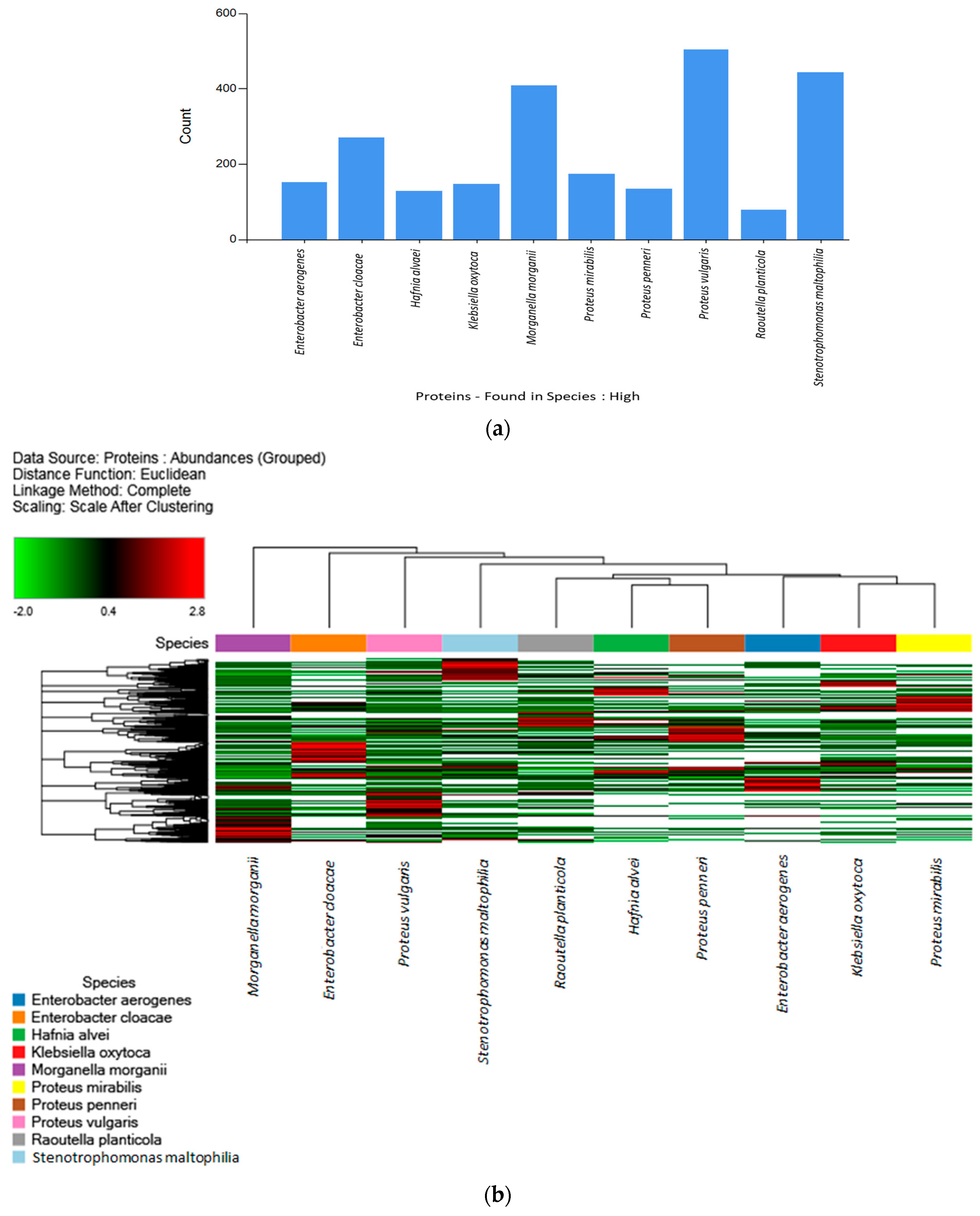
Comparisons of the high-abundance proteins of each species and strain were performed. Figure 2a displays the distribution of the high-abundance proteins determined for each of the 15 strains. Among them, Proteus vulgaris, Stenotrophomonas maltophilia, and Morganella morganii were the three main species with the most high-abundance proteins. The distribution of the high-abundance proteins for all samples analyzed via LFQ is illustrated in a heatmap diagram in Figure 2b. Euclidean hierarchical distance was used to differentiate three main clusters. Cluster A (strains H6, H2, H9, and H14: Morganella morganii, Enterobacter cloacae, Proteus vulgaris, and Stenotrophomonas maltophilia), Cluster B (strains H12, H3, and H8: Raoutella planticola, Hafnia alvei, and Proteus penneri), and Cluster C (strains H1, H4, and H7: Enterobacter aerogenes, Klebsiella oxytoca, and Proteus mirabilis). As in Figure 2a, the clusters of Figure 2b were divided according to the number of proteins that were more upregulated (Red) (as determined via LFQ) versus those proteins that were more downregulated (Green) for the different strains.
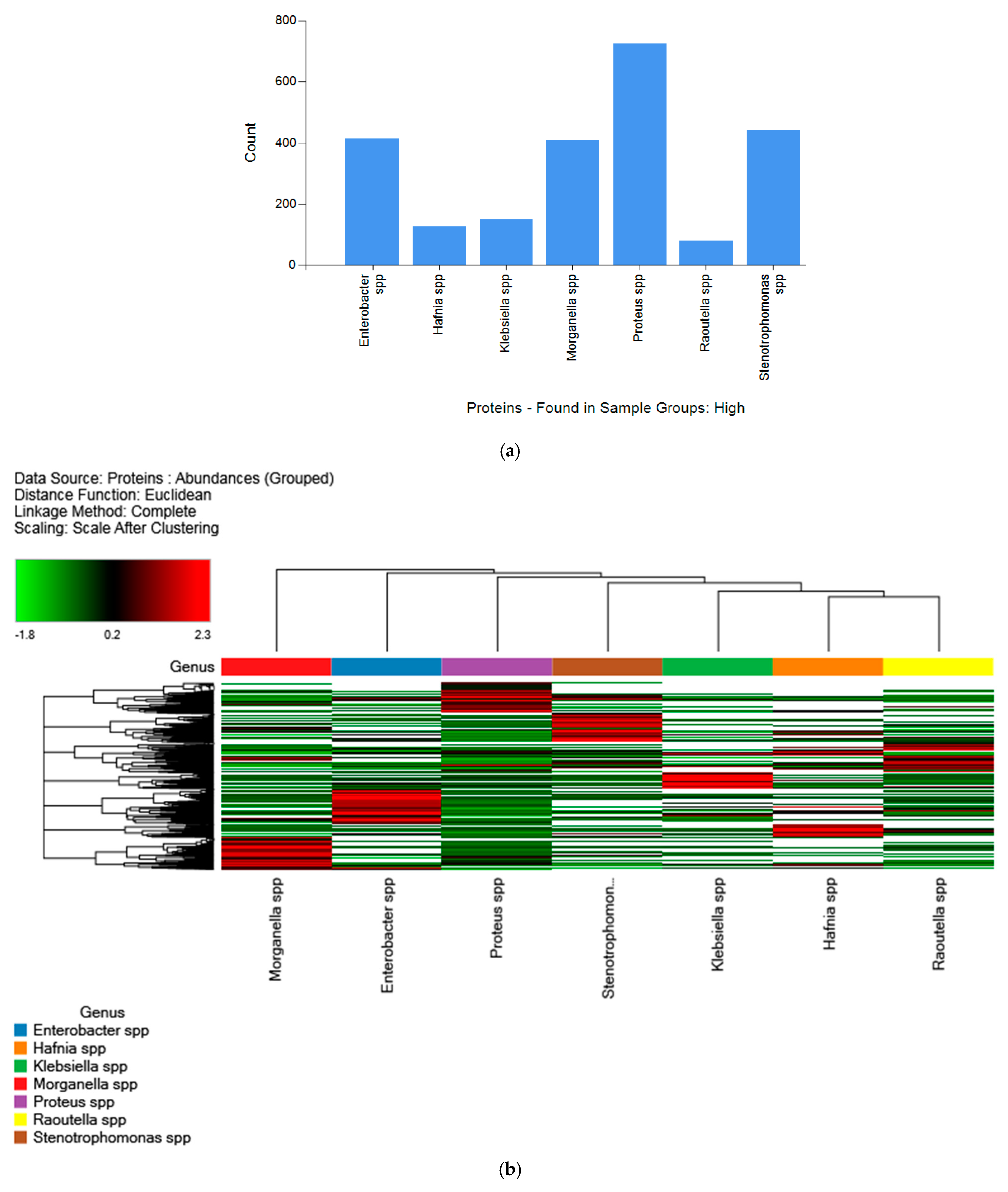
Regarding the different genera, Figure 3a shows the high-abundance proteins for each genus (Enterobacter spp., Hafnia spp., Klebsiella spp., Morganella spp., Proteus spp., Raoultella spp., and Stenotrophomonas spp.). Among them, Proteus spp. was the most represented genus with the most high-abundance proteins. The distribution of the high-abundance proteins for all samples grouped by genus and analyzed via LFQ is illustrated in a heatmap diagram in Figure 3b. Finally, all strains were arranged according to Euclidean hierarchical distance. Seven principal clusters were differentiated, which corresponded to the different genus types.
To obtain further insights regarding functional interpretation, the present repository was investigated using several functional in silico analyses, comprising (i) functional pathways, GO enrichment and hierarchical clustering, (ii) functional network analysis, (iii) the discovery of virulence factors, and (iv) the selection of potential species-specific peptide biomarkers.

2.3. Functional Pathways and Gene Ontology (GO)

The global protein repository of foodborne strains of biogenic-amine-producing bacteria was individually examined using functional bioinformatics tools, such as functional pathway analysis and GO term enrichment.
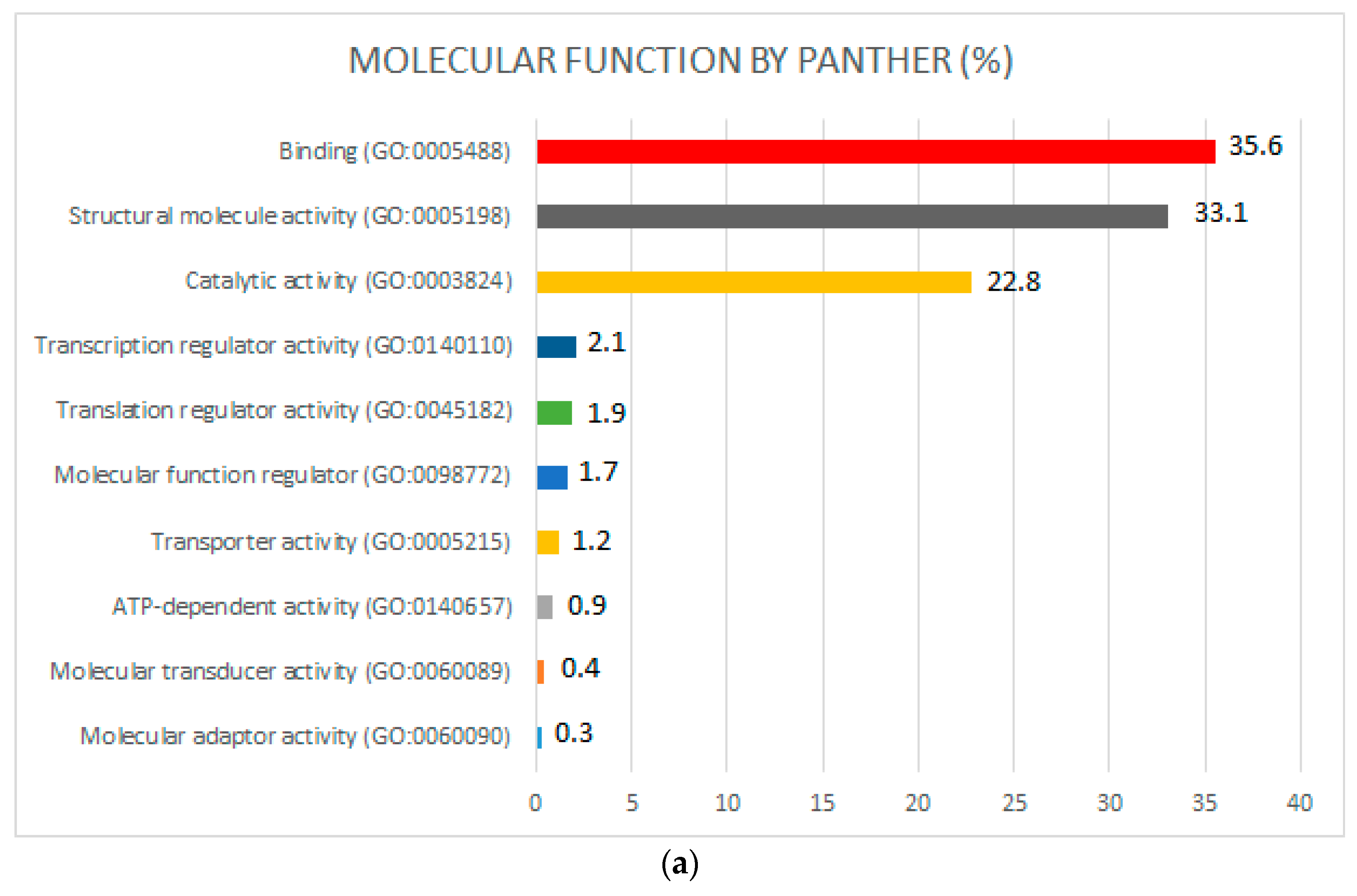
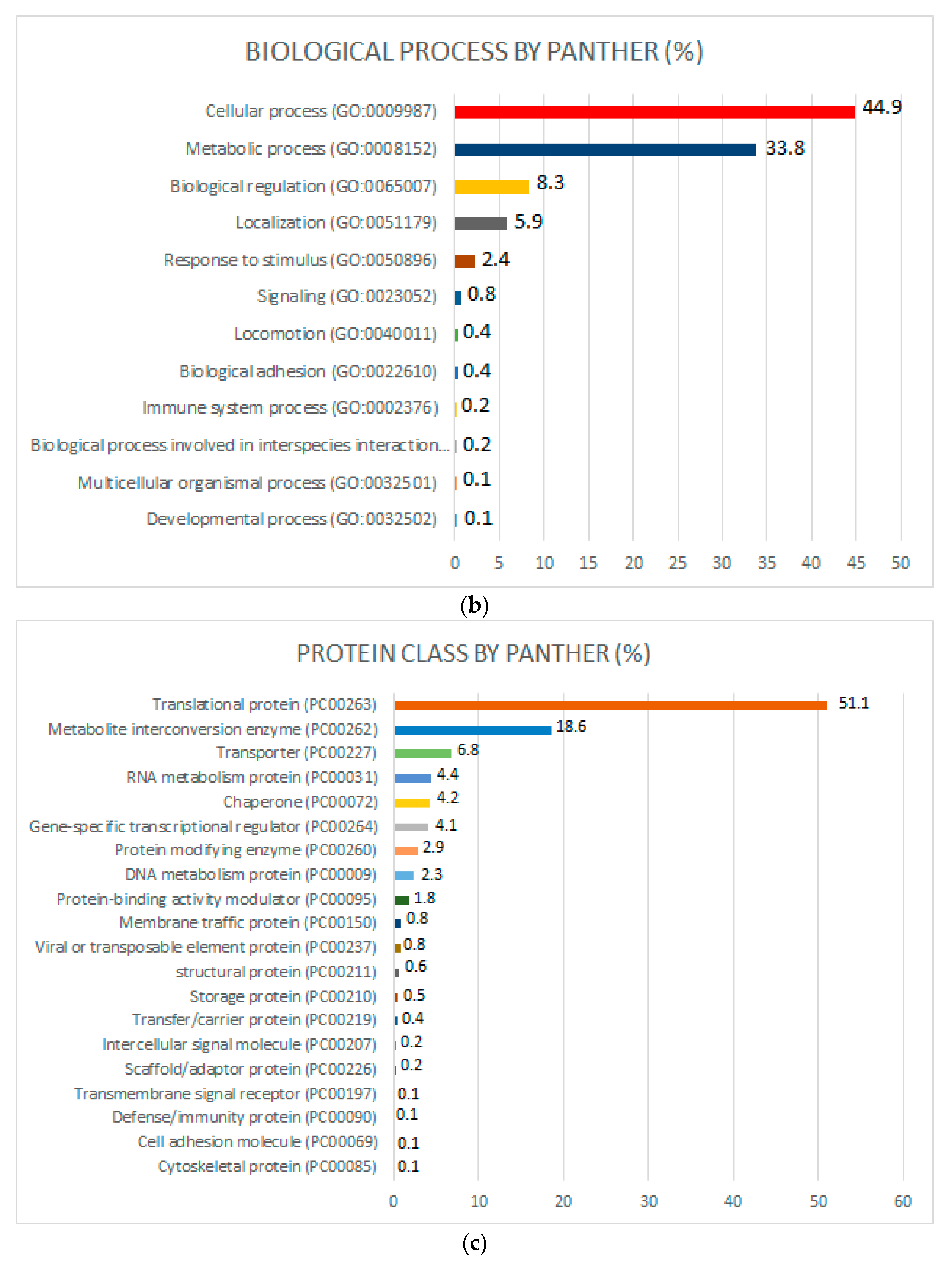
PANTHER analysis was performed using gene names (considering all nonredundant proteins), revealing the presence of 10 different molecular functions (Figure 4a), 12 different biological processes (Figure 4b), and 20 different protein classes (Figure 4c) in the complete global proteome repository.
According to the molecular function classification procedure (Figure 4a), the most important molecular functions were binding (35.6%), structural molecule activity (33.1%), and catalytic activity (22.8%). Within the binding function group, ribosomal proteins, oxidorreductases, chaperones, DNA metabolism proteins, deaminases, isomerases, transferases, translation elongation factor proteins, mutases, and protein kinases were found. In the structural molecule activity group, ribosomal proteins and tubulins were detected. Regarding catalytic activity, decarboxylases, nucleotide kinases, oxidases, kinases, pyrophosphatases, isomerases, transferases, deaminases, proteases, dehydrogenases, and mutases were observed.
According to the classification of biological processes (Figure 4b), the most remarkable categories were cellular processes (44.9%), metabolic processes (33.8%), biological regulation (8.3%), localization (5.9%), response to stimulus (2.4%), and signaling (0.8%). Regarding cellular processes, ribosomal proteins, decarboxylases, pyrophosphatases, vesicle coat proteins, isomerases, transferases, and translocation initiation factors were found. Concerning the metabolic process group, ribosomal proteins, decarboxylases, pyrophosphatases, translation release factor, chaperone, isomerases, transferases, and metalloproteases were detected. In the biological regulation group, ribosomal protein, chaperone, membrane traffic protein, primary active transporter, and storage proteins were observed.
According to the classification of protein classes (Figure 4c), the most prominent classes were translational proteins (51.1%), metabolite interconversion enzymes (18.6%), and transporters (6.8%). Within the translational protein group, ribosomal protein and translation initiation/elongation/release factors were observed. In the metabolite interconversion enzyme group, different enzyme groups were observed, including dehydrogenases, carbohydrate kinases, aldolases, isomerases, hydrolases, glycosidases, transferases, oxidases, glucosidases, peroxidases, mutases, dehydratases, phospholiases, isomerases, and deaminases. Within the transporter category, ATP synthase, ATP-binding cassettes, amino acid transporters, and ion channels were detected.
The existence of high concentrations of decarboxylases in these functional classifications influences the formation of biogenic amines by the bacteria. During the deterioration of fish, the occurrence of bacterial strains with high proteolytic enzyme activity increases the breakdown of proteins as well as the accessibility of small peptides and specific free amino acids that are decarboxylated in particular biogenic amines [32]. In fish, the principal studied biogenic amines include histamine (derived from histidine), putrescine (derived from arginine, glutamine, methionine, and ornithine), cadaverine (derived from lysine), tyramine (derived from tyrosine), spermidine (derived from agmatine, methionine, putrescine, and spermine), and spermine (derived from agmatine, methionine, putrescine, and spermidine) [33].

2.4. Biogenic Amine-Related Proteins and Peptides Detected via LC-ESI-MS/MS

Table 2 summarizes the list of biogenic amine-related proteins and peptides detected via LC-ESI-MS/MS for the corresponding strains.
Agmatine and cadaverine are aliphatic polyamine biogenic amines derived from the amino acids arginine and lysine, respectively [33]. Two different related proteins (arginine ABC transporter substrate-binding protein and lysine–arginine–ornithine-binding periplasmic protein) and three different peptides (IDAVFGDTAVVTEWLK, C*TWVGSDFDSLIPSLK, and IGTDATYAPFSSK) were detected via shotgun proteomics in the K. oxytoca strain. The metabolism of agmatine and cadaverine requires the initial presence and transport of arginine or lysine, respectively, in the periplasm of the cells. In Gram-negative bacteria, solute-binding proteins are localized in the periplasm and involved in nitrogen compound transport (GO:0071705) and amine transport (GO:0015837).
Histamine is a heterocyclic biogenic amine derived from the amino acid histidine [2]. Histamine is present in most foods but is more abundant in fish and fishery products. This biogenic amine is the major agent behind “scombroid poisoning” or “histamine poisoning” [11,34]. A total of five different related proteins (histidine kinase, histidine phosphatase, histidine-binding periplasmic protein, histidine triad nucleotide-binding protein, and histidine ammonia-lyase) were detected via shotgun proteomics (Table 2). Nine peptides of histidine kinase (GO:0004673) were identified via LC-ESI-MS/MS analysis in different strains (IDSEDLPHVRASVAR (present in M. morganii), LAM*NLRTRLFLSISALITVALLGLLLGLVSVM*QM*AGSQ-EILIR (S. maltophilia), M*IAEAANADSKQAQR (K. oxytoca and R. planticola), TIDQINQQKIQLEQEIADRK (P. penneri and P. vulgaris), GEADATLDSEVSAWRAVAR (P. vulgaris), LSSELWNC*KIDPTQAEM*AM*INILANAR (P. mirabilis), SEASENTVDLIVEDEGSGIPK (P. mirabilis), NEEARDNLISELTAR (P. vulgaris), and RYAYSEQLGDLLQR (S. maltophilia)). In addition, a peptide from histidine phosphatase (GO:0101006) was detected via LC-MS/MS (HAQASEYGSALFVAVGQAKQVK) in the H. alvei strain. It is well known that histidine kinase/phosphatase regulates histamine synthesis and signal transduction by activating histidine decarboxylase through phosphorylation/dephosphorylation [35]. Moreover, a peptide of histidine-binding periplasmic protein (IGVLQGTTQETYGNEHWAPK) was detected in the K. oxytoca strain, and two peptides of histidine triad nucleotide-binding protein (EIPSDIVYQDELVTAFR, IAEQEGIAEDGYR) were detected in the E. cloacae strain. These proteins are involved in nitrogen compound transport and amine transport (GO:0071705, GO:0015837). Finally, a peptide (LAAM*QQALGAQIAAVEEDR) of histidine ammonia-lyase was identified via LC-ESI-MS/MS in the M. morganii strain. This cytosolic enzyme catalyzes the first reaction in histidine catabolism: the nonoxidative deamination of histidine to trans-urocanic acid (GO:0004397).
Putrescine is an aliphatic biogenic amine derived from the amino acids arginine or ornithine in one step or two steps after glutamine or methionine is transformed into ornithine and then putrescine. The ingestion of food containing high amounts of putrescine can lead to grave toxicological consequences. In fact, putrescine can react with nitrite to form N-nitrosamines, which are carcinogenic agents [36]. Additionally, putrescine induces significant effects that enhance the toxicological effects of other BAs, particularly histamine and tyramine [37]. In seafood such as fish, squid, and octopus, putrescines are also dominant biogenic amines [38]. Lysine–arginine–ornithine-binding periplasmic protein and two peptides (C*TWVGSDFDSLIPSLK; IGTDATYAPFSSK) were also identified via LC-ESI-MS/MS analysis in the K. oxytoca strain. The metabolism of putrescine also requires the initial presence and transport of arginine or ornithine in the periplasm of the cells (GO:0071705 and GO:0015837). In addition, three proteins responsible for glutamine and methionine transport and amino/amido transferase were identified via shotgun proteomics in K. oxytoca and H. alvei strains. These corresponded to glutamine ABC transporter periplasmic protein (peptide: AVGDSIEAQQYGIAFPK) present in K. oxytoca, glutamine-fructose-6-phosphate aminotransferase (IDAAQEAELIKALFEAPR) present in K. oxytoca, N-acetylglutaminylglutamine amidotransferase (SGANAAVDKALRLDSTVM*LVDDPVK) present in H. alvei, type 1 glutamine amidotransferase domain-containing protein (IFRTLALM*LLVTSATAFAASK) present in P. mirabilis, and L-glutamine-binding protein (ADAVIHDTPNILYFIK, AVGDSLEAQQYGIAFPK) present in K. oxytoca and H. alvei strains. Finally, the S-adenosylmethionine decarboxylase proenzyme (AdoMetDC) (ALSFNIYDVC*YAR) was detected in the S. maltophilia strain. It is involved in the synthesis of biogenic amines in several species that use aminopropyltransferases for this pathway. AdoMetDC is involved in the production of S-adenosyl-1-(methylthio)-3-propylamine (decarboxylated S-adenosylmethionine) [39]. In contrast to many amino acid decarboxylases that use pyridoxal 5′-phosphate as a cofactor, AdoMetDC uses a covalently bound pyruvate residue. This decarboxylase is involved in the polyamine biosynthetic pathway, as it generates the n-propylamine residue needed for the synthesis of spermidine and spermine from putrescine [40,41].
Spermidine is an aliphatic polyamine derived from putrescine, agmantine, methionine, or spermine [42]. It is a precursor to other polyamines, such as spermine and its structural isomer thermospermine. Spermidine in fish tissue can potentiate the toxic effect of histamine by inhibiting intestinal histamine-catabolic enzymes [43]. Two spermidine-related proteins were identified via LC-MS/MS (spermidine/putrescine import ATP-binding protein and S-adenosylmethionine decarboxylase proenzyme). One peptide (VDEVHDNAEAEGLIGYIR) of spermidine/putrescine import ATP-binding protein was detected via LC-ESI-MS/MS in the P. vulgaris strain. This protein is part of the ABC transporter complex PotABCD that is involved in spermidine/putrescine import [44]. In addition, one peptide (ALSFNIYDVC*YAR) of the S-adenosylmethionine decarboxylase proenzyme was detected in the S. maltophilia strain. This enzyme is necessary for the biosynthesis of polyamines such as spermine and spermidine from the diamine putrescine [39].
Spermine is an aliphatic polyamine derived from agmatine, methionine, or spermidine [2]. One spermine-related protein was identified via LC-ESI-MS/MS (S-adenosylmethionine decarboxylase proenzyme). A peptide (ALSFNIYDVC*YAR) of the S-adenosylmethionine decarboxylase proenzyme was detected in the S. maltophilia strain. In addition, spermine has been reported to modify the connections between polyamines and DNA. In fact, spermine has been reported to function as a free radical scavenger protecting DNA from oxidative stress [45]. More precisely, the higher the cationic charge, the higher the degree of DNA-protein binding enhancement; thus, spermine has been characterized as more potent than spermidine and putrescine.
Finally, further decarboxylases (e.g., phosphatidylserine decarboxylase and 4-carboxymuconolactone decarboxylase) and deaminases (e.g., 2-iminobutanoate/2-iminoopropanoate deaminase and glucosamine-6-phosphate deaminase) were identified via shotgun proteomics (Supplementary Data S1), but according to the literature, they are involved in other metabolic pathways, which was also demonstrated previously via PANTHER analysis.

2.5. Network Analysis

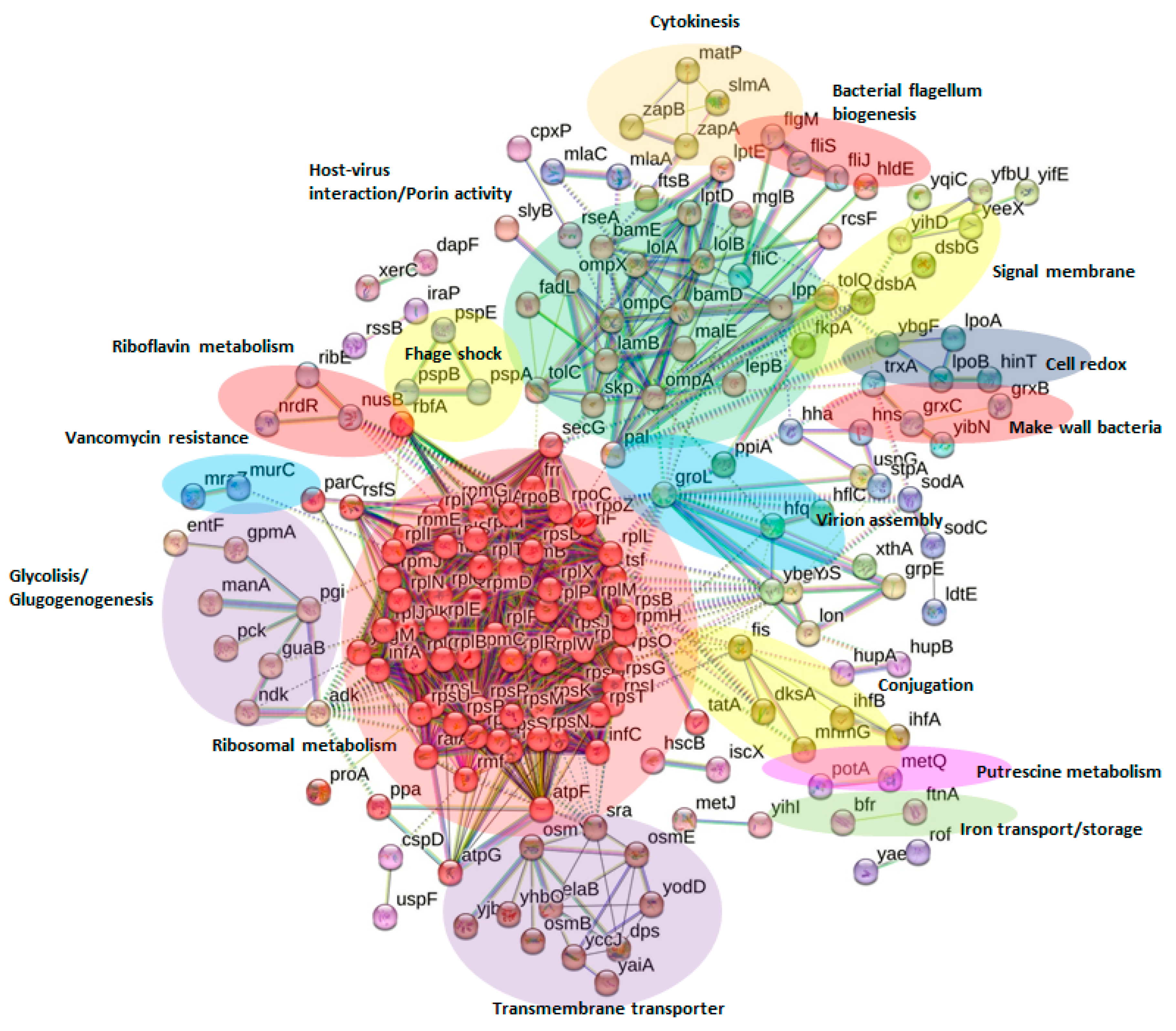
Network analysis was executed using STRING v.11.5 software (https://string-db.org/, accessed on 6 December 2022) [46], wherein all the proteins identified in this study were investigated and compared with the genome of the model organism E. coli K12 MG1655, which was the genetically closest group available in the portal (Figure 5). Every protein—protein interaction was assigned to the network in accordance with its confidence score. To reduce the occurrence of false positives and false negatives, all expected interactions were tagged as “high-confidence” (≥0.7) in the STRING program were selected for this work.
Thus, the final network for the global protein repository consisted of 260 nodes (proteins) and 1973 edges (interactions) (Figure 5). All proteins used in the network were discovered during the proteomic experiments (see the codes of the gene column in Supplementary Data S1). This protein network is the first inclusive interactomics map for relevant seafood-based, biogenic-amine foodborne strains.
Cluster networks were generated using an MCL (inflation clustering) algorithm from the STRING website, and a default value of 2 was selected for all analyses. From the cluster analysis, 42 significant clusters of interactions between nodes were obtained. Figure 5 highlights the most relevant clusters (n = 15) according to the abundance of nodes involved or their biological relevance. Supplementary Data S3 includes information about the 42 clusters, protein names, and descriptions of the corresponding name codes.
The most relevant subnetworks in terms of their number of nodes are involved in ribosomal metabolism (in red; 63 nodes), host-virus interaction/porin activity (in green; 22 nodes), transmembrane transport (in violet; 12 nodes), and glycolysis (in dark violet; 8 nodes).
Other subnetworks that contain fewer nodes but have great biological importance are related to bacterial flagellum biogenesis (in red; four nodes), vancomycin (an antibiotic) resistance (in blue; three nodes), and putrescine metabolism (in pink; three nodes). Further study of the aforementioned subnetworks and protein-protein interactions will be very beneficial for the development of new therapeutic treatments for bacterial dispersion, antibiotic resistance, and food intoxication via biogenic-amine-produced putrescine.

2.6. Virulence Factors

Many seafood-originating biogenic-amine-producing bacteria are pathogens with well-known virulence. It has been reported that Enterobacter bacteria are increasingly exhibiting a multidrug resistance phenotype [47]. Moreover, K. oxytoca can acquire antimicrobial resistance and carry multiple virulence genes, such as capsular polysaccharides and fimbriae [48]. The virulence of other species analyzed in this study, such as H. alvei [49], M. morganii [50], P. vulgaris [51], S. maltophilia [52], and R. planticola [53], has been previously reported.
A total of 556 peptides belonging to virulence factors (nonredundant peptides) were identified in this study. They included toxins, polypeptides involved in antibiotic resistance, and proteins related to cell colonization and immune evasion. The 556 virulent peptides (Supplementary Data S4) are displayed in groups in accordance with the principal roles in which they are involved (e.g., toxin generation/transport, colonization and immune evasion factors, antimicrobial compounds, other tolerance proteins that play a role in resistance to toxic substances, etc.). In addition, the main proteins of the identified virulence factors are displayed in Table 3.
In this study, several peptides involved in antimicrobial resistance or the production/transport of toxic substances were identified (Supplementary Data S4). Ten of the proteins characterized were associated with antibiotic resistance, and 91 peptides were related to other tolerances. Four peptides were identified as penicillin-binding proteins. Peptides associated with acriflavine and methicillin resistance and belonging to the TetR family of regulators (TFRs) were determined. TetR proteins regulate antibiotic and quorum-sensing processes as well as antibiotic resistance. In addition, two peptides of the GCN-2-related N-acetyl transferase (GNAT) family of acetyltransferases, which provide antibiotic resistance [54], were also identified. Peptides of proteins involved in other bacterial tolerances (e.g., thermotolerance and osmotolerance) were also identified. Accordingly, this work has identified many peptides that belong to groups of peptides of bacterial general stress response proteins, heat shock proteins, and cold-shock-like proteins (CSPs), among others [55,56].
A total of 20 peptides corresponding to proteins that are involved in bacterial toxicity were identified. These peptides include ecotin, lipoprotein toxin enterocidin B, antitoxin ParD, and addition module toxin GnSA/GnsB. As an example of some of the roles these peptides play, ecotin is an inhibitor of multiple complement-dependent processes found in bacteria [57].
In this study, 349 peptides involved in colonization and immune evasion were identified. Bacterial internalization into the host is facilitated by these proteins, resulting in subsequent infection and propagation. Transcriptional regulators involved in the control of virulence factors were also found for the analyzed strains, including two peptides identified as LysR and SlyA [58,59]. LysR regulates virulence factors, such as extracellular polysaccharides, toxins, and bacteriocins. Fimbria are located on the surfaces of bacteria; they are involved in adherence to target cells and biofilm formation [60]. Lysis proteins belonging to the LysM domain were identified; this domain was identified in enzymes involved in bacterial cell wall degradation [61]. Additionally, several peptides of peptidases and proteases were identified. This includes members of the Lon protease family and subtilisin, among others. Different peptides of the Superoxide dismutase enzyme (SOD) were identified. SOD is a metalloenzyme that defends against reactive oxygen species produced by neutrophils and macrophages [62]. The presence of open channels facilitates passive penetration though the outer membrane. We have identified several porins or outer-membrane proteins (OMPs), such as the porins OmpA, OmpX, and OmpC; substrate-specific porins, such as maltoporin, which is also called LamB; and TonB-dependent receptors, such as FhuA [47]. In addition, many peptides were determined to be other virulence factors, such as VacJ family lipoprotein VacJ (virulence-associated chromosome locus J); the chaperone protein Skp, which assists in the folding and insertion of many OMPs [63]; and the Osmy chaperone.
In this study, four peptides of antibacterial proteins were identified, including one peptide that belongs to a bacteriocin and the remaining three to a colicin-like protein. Colicins are antimicrobial proteins typically produced by E. coli that degrade internal cellular elements [64].
ABC transporters, like many other bacterial transporters, are involved in resistance or tolerance and bacterial propagation during infection [65]. We identified different ABC transporters related to virulence (Table 3).
Furthermore, sixteen peptides of alternative virulent factors were identified, such as proteins related to mobile genetic elements’ transposases, recombinases, plasmids, and viral DNA fragments, which are considered the major mechanism for acquiring antibiotic resistance. Moreover, pilus conforms to a typical method of horizontal transfer between bacteria, which is another mechanism of obtaining virulence determinants [66]. We identified 43 peptides of phage proteins, such as bacteriophage CI repressor and capsid scaffolding protein, but mainly phage shock proteins. Finally, we identified bacterial proteins determined in the UniProt database in different phage strains (Klebsiella phage vB_KpM_FBKp24, Klebsiella phage vB_KppS-Storm, Stenotrophomonas phage BUCT608, and Stenotrophomonas phage).

2.7. Potential Species-Specific Peptide Biomarkers

To select potential peptide biomarkers for the 15 different biogenic-amine-producing bacterial strains, we implemented a massive comparison of the proteomics data with respect to the proteins and peptides included in databases. The suitable peptides that were identified via LC-ESI-MS/MS in only one specific species were verified in terms of their specificity and sequence homology using the BLASTp algorithm [67] (Supplementary Data S5). Table 4 summarizes the analysis of the 77 species-specific tryptic peptide biomarkers belonging to 64 different proteins that were suggested for the unequivocal identification of the different seafood-originating biogenic-amine-producing bacteria of 10 different species (E. aerogenes, E. cloacae, H. alvei, K. oxytoca, M. morganii, P. mirabilis, P. penneri, P. vulgaris, R. planticola, and S. maltophilia).
All the peptides included herein have been proposed as potential biomarkers for the first time and will be very convenient for further studies using targeted proteomics approaches to identify the different seafood-originating biogenic-amine-producing bacteria in foodstuffs.

3. Materials and Methods

3.1. Bacterial Strains

A total of 15 different seafood-originating biogenic-amine-producing bacteria were included in this work (Table 1). Strains were previously studied via MALDI-TOF-MS and 16S rRNA sequencing [10,68]. All bacterial strains were activated in brain–heart infusion (BHI) and incubated in vials at 31 °C for 24 h. Then, strain cultures were expanded on plate count agar (PCA) at 31 °C for 24 h. Samples were prepared in triplicate.

3.2. Protein Extraction

Protein extracts were obtained as described by Carrera et al. (2017) [24,69,70]. Concisely, the biomass of bacterial cells was mixed with a solution of 1% trifluoracetic acid/50% acetonitrile. After several extractions with glass beads conducted for 10 min at 4 °C, the supernatants were centrifuged for 10 min at 40,000× g (J221-M centrifuge, Beckman, Brea, CA, USA). The supernatant was then solubilized with lysis buffer containing 60 mM Tris-HCl pH 7.5, 1% lauryl maltoside, 5 mM phenylmethanesulfonylfluoride (PMSF), and 1% dithiothreitol (DTT). The solution was transferred to a new vial, and the quantity of protein was revealed via the bicinchoninic acid method (Sigma Chemical Co., St. Louis, MO, USA). This method was chosen because a similar procedure has been applied previously for protein extraction via MALDI-TOF MS analysis [68].

3.3. Peptide Sample Preparation

Proteins were digested with trypsin, as described previously [71]. A total of 100 μg of protein extracts was dried under vacuum and solubilized in 25 μL of 8 M urea in 25 mM of ammonium bicarbonate at pH 8.0. After 5 min of sonication, DTT was added at a final concentration of 10 mM and incubated at 37 °C for 1 h. Then, iodoacetamide was supplemented at a final concentration of 50 mM and incubated at room temperature in darkness for 1 h. Next, the sample was diluted four times to a final concentration of 2 M urea with 25 mM ammonium bicarbonate (pH 8.0) and subjected to digestion with trypsin (ratio 1:100) (Promega, Wisconsin, WI, USA) at 37 °C overnight.

3.4. Shotgun LC-ESI-MS/MS Analysis in a LTQ-Orbitrap Instrument

Peptides were acidified with 5% formic acid (FA) until attaining pH 2, cleaned on a C18 MicroSpinTM column (The Nest Group, Southborough, MA, USA), and analyzed via LC-MS/MS using a Proxeon EASY-nLC II LC machine (Thermo Scientific, San Jose, CA, USA) coupled with an LTQ-Orbitrap XL (Thermo Fisher Scientific). Separation of peptides (2 µg) was implemented on an RP column (EASY-Spray column, 50 cm × 75 µm ID, PepMap C18, 2 µm particles, 100 Å pore size, Thermo Fisher Scientific) with a 10 mm precolumn (Accucore XL C18, Thermo Scientific) containing 0.1% FA in Milli-Q water and 98% ACN and 0.1% FA as mobile phases A and B, respectively. A 240 min linear gradient from 5 to 35% B at a flow rate of 300 nL/min was used. A capillary temperature of 230 °C and spray voltage of 1.95 kV were used for ionization. Peptides were analyzed from 400 to 1600 amu (1 µscan) in positive mode, followed by 10 data-dependent CID MS/MS scans (1 µscans) using an isolation width of 3 amu and a normalized collision energy of 35%. Fragmented masses were set in dynamic exclusion for 30 s after the second fragmentation event. Unassigned charged ions were omitted from MS/MS analysis.

3.5. LC-ESI-MS/MS Data Processing

MS/MS spectra were identified using SEQUEST-HT (Proteome Discoverer 2.4 package, Thermo Fisher Scientific) and compared to the Proteobacteria UniProt/TrEMBL database (with 2,627,375 protein sequence entries dating from August 2022). MS/MS spectra were analyzed using fully tryptic cleavage constraints, and up to two missed cleavage sites were permissible. Windows for tolerance were set at 10 ppm for precursor ions and 0.06 Da for MS/MS fragment ions. The variable modifications permitted were methionine oxidation (Mox), carbamidomethylation of Cys (C*), and acetylation of the N-terminus of a protein (N-Acyl).
The results were subjected to statistical analysis to determine the false discovery rate (FDR) regarding peptides using a decoy database and the Percolator algorithm included in the Proteome Discoverer 2.4 program [72]. The FDR was kept below 1% for further analysis. The MS/MS proteomics data have been deposited to the ProteomeXchange Consortium via PRIDE with the dataset identifier PXD039320.
To determine relative protein abundance for each strain, a label-free quantification (LFQ) method was used by applying the Minora Feature Detector node and the ANOVA (individual proteins) method included in the Proteome Discover 2.4 software (Thermo Fisher Scientific). Peak areas of ion features from the same peptide for different charge forms were combined into one value.

3.6. Euclidean Hierarchical Clustering

The function heatmap.2 of the statistical package R (version (v) 4.1.1) (http://www.r-project.org, accessed on 25 January 2023) was used to achieve the Euclidean hierarchical clustering of the data. The Ggplots v.4.1.1 package, the Euclidean distance metric, and the complete linkage for the agglomeration method were used as constraints.

3.7. Functional Analysis: Gene Ontology (GO) and Pathways Analysis

The nonredundant protein IDs (column “Gene name” in Supplemental Data S1) were submitted to the PANTHER software (http://www.pantherdb.org/, accessed on 30 November 2022) for grouping established based on the following main types of interpretations: molecular function, biological process, and protein class. The statistical significance was also provided as a percentage. For this procedure, all orthologous gene ID entries were included as a reference set. The pathway analysis data were clustered, thus providing an approximation of the statistical significance of over- or underrepresentation according to the GO descriptors of the proteins in the proteome.

3.8. Network Analysis

Protein network was developed by incorporating the orthologous gene IDs into the STRING program (v.11.5) (http://string-db.org/, accessed on 6 December 2022) [46]. STRING is an enormous database of known and predicted protein interactions. Proteins are denoted with nodes, and interactions are represented as continuous lines. All edges were reinforced by at least one reference from the literature or from canonical information deposited in the STRING dataset. The confidence score was set at ≥0.7 (high confidence). MCL algorithm included on the STRING website was used to generate cluster networks, and a default value of 2 was assigned for all analyses.

3.9. Virulence Factors

Virulence Factor of Pathogenic Bacteria Database (VFDB) (http://www.mgc.ac.cn/VFs/, accessed on 13 December 2022) was used to characterize virulence factors. Additionally, we prolonged the analysis to include virulence factors that are contained in several scientific publications [47,48,49,50,51,52,53].

3.10. Selection of Potential Peptide Biomarkers

BLASTp algorithm applied to each identified peptide by LC-MS/MS was used to determine homologies and exclusiveness with respect to protein sequences recorded in the NCBI database [67].

4. Conclusions

This article presents the first shotgun proteomics study of 15 different foodborne strains of biogenic-amine-producing bacteria. By means of a rapid and easy procedure for preparing proteins, the results were used to differentiate several protein datasets, which, in turn, were used to determine relevant functional pathways and differentiate strains into different Euclidean hierarchical clusters. Additionally, a predicted protein-protein interaction network for foodborne biogenic-amine-producing bacteria was created. Most proteins were classified under pathways and networks related to energy, putrescine metabolism, and host-virus interactions. Additionally, 556 different virulence factors were identified. Most of these factors corresponded to functions/roles such as toxins, antimicrobial compound production, antimicrobial resistance, additional resistances and tolerances, host colonization and immune evasion, ABC transporters, phage proteins, and alternative virulence factors and proteins involved in horizontal transfer. Finally, 77 prospective species-specific peptide biomarkers corresponding to 64 different proteins were screened to identify unique potential peptide biomarkers for 10 biogenic-amine-producing bacterial species. To date, these results constitute the largest dataset of peptides and proteins from foodborne biogenic-amine-producing bacterial species strains. This repository provides data that can be used in further studies to develop new therapeutic treatments for biogenic-amine-producing bacterial species with respect to food intoxication and for the tracking of microbial sources in foodstuffs.

Supplementary Materials

The following supporting information can be downloaded at: https://www.mdpi.com/article/10.3390/ijms24097704/s1.

Author Contributions

M.C. and K.B. performed the analysis. M.C., A.G.A. and T.G.V. wrote the manuscript; M.C., A.G.A., T.G.V., P.C.-M. and M.P. conceptualized, revised, and corrected the paper. P.C.-M. and M.C. co-supervised the work. M.C., P.C.-M., J.B.-V. and M.P. obtained the funding. All authors have read and agreed to the published version of the manuscript.

Funding

A.G.A. thanks the USC for the “Convocatoria de Recualificación do Sistema Universitario Español-Margarita Salas” postdoc grant under the “Plan de Recuperación Transformación” program funded by the Spanish Ministry of Universities through the European Union’s NextGeneration EU funds. This work has received financial support from the Xunta de Galicia and the European Union (European Social Fund2013ESF), the Spanish Ministry of Economy and Competitivity Project AGL 2.013-48.244-R, and the European Regional Development Fund (ERDF) (2007–2013). This study was also financed by the GAIN-Xunta de Galicia Project (IN607D 2017/01) and the Spanish AEI/EU-FEDER PID2019-103845RB-C21 project. It was also supported by the Plan Complementario en Ciencias Marinas (PCCM) funded by the Ministry of Science and Innovation (Activity 3.6.B. NANOSEAOMICS).

Institutional Review Board Statement

Not applicable.

Informed Consent Statement

Not applicable.

Data Availability Statement

The MS/MS proteomics data have been deposited to the ProteomeXchange Consortium via PRIDE with the dataset identifier PXD039320.

Conflicts of Interest

The authors declare no conflict of interest.

References

  1. Spano, G.; Russo, P.; Lonvaud-Funel, A.; Lucas, P.; Alexandre, H.; Grandvalet, C.; Coton, E.; Coton, M.; Barnavon, L.; Bach, B.; et al. Biogenic amines in fermented foods. Eur. J. Clin. Nutr. 2010, 64, S95–S100. [Google Scholar] [CrossRef]
  2. Silla Santos, M.H. Biogenic amines: Their importance in foods. Int. J. Food Microbiol. 1996, 29, 213–231. [Google Scholar] [CrossRef]
  3. Feddern, V.; Mazzuco, H.; Fonseca, F.N.; De Lima, G.J.M.M. A review on biogenic amines in food and feed: Toxicological aspects, impact on health and control measures. Anim. Prod. Sci. 2019, 59, 608–618. [Google Scholar] [CrossRef]
  4. Trinki, M.; Herrero, A.M.; Jiménez-Colmenero, F.; Ruiz-Capillas, C. Quality assessment of fresh meat from several species based on free amino acid and biogenic amine contents during chilling storage. Foods 2018, 7, 132. [Google Scholar] [CrossRef] [PubMed]
  5. Dale, H.H.; Laidlaw, P.P. The physiological action of β-iminazolylethylamine. J. Physiol. 1910, 41, 318–344. [Google Scholar] [CrossRef]
  6. Maintz, L.; Novak, N. Histamine and histamine intolerance. Am. J. Clin. Nutr. 2007, 85, 1185–1196. [Google Scholar] [CrossRef]
  7. del Rio, B.; Redruello, B.; Linares, D.M.; Ladero, V.; Ruas-Madiedo, P.; Fernandez, M.; Martin, M.C.; Alvarez, M.A. The biogenic amines putrescine and cadaverine show in vitro cytotoxicity at concentrations that can be found in foods. Sci. Rep. 2019, 9, 120. [Google Scholar] [CrossRef] [PubMed]
  8. Ben-Gigirey, B.; Vieites Baptista De Sousa, J.M.; Villa, T.G.; Barros-Velazquez, J. Changes in Biogenic Amines and Microbiological Analysis in Albacore (Thunnus alalunga) Muscle during Frozen Storage. J. Food Prot. 1998, 61, 608–615. [Google Scholar] [CrossRef]
  9. Ben-Gigirey, B.; Vieites Baptista De Sousa, J.M.; Villa, T.G.; Barros-Velazquez, J. Histamine and Cadaverine Production by Bacteria Isolated from Fresh and Frozen Albacore (Thunnus alalunga). J. Food Prot. 1999, 62, 933–939. [Google Scholar] [CrossRef] [PubMed]
  10. Fernández-No, I.C.; Böhme, K.; Calo-Mata, P.; Barros-Velázquez, J. Characterisation of histamine-producing bacteria from farmed blackspot seabream (Pagellus bogaraveo) and turbot (Psetta maxima). Int. J. Food Microbiol. 2011, 151, 182–189. [Google Scholar] [CrossRef]
  11. Ridolo, E.; Martignago, I.; Senna, G.; Ricci, G. Scombroid syndrome: It seems to be fish allergy but. it isn’t. Curr. Opin. Allergy Clin. Immunol. 2016, 16, 516–521. [Google Scholar] [CrossRef]
  12. Gardini, F.; Özogul, Y.; Suzzi, G.; Tabanelli, G.; Özogul, F. Technological factors affecting biogenic amine content in foods: A review. Front. Microbiol. 2016, 7, 1218. [Google Scholar] [CrossRef]
  13. Bodmer, S.; Imark, C.; Kneubühl, M. Biogenic amines in foods: Histamine and food processing. Inflamm. Res. 1999, 48, 296–300. [Google Scholar] [CrossRef] [PubMed]
  14. Linares, D.M.; Martĺn, M.C.; Ladero, V.; Alvarez, M.A.; Fernández, M. Biogenic Amines in Dairy Products. Crit. Rev. Food Sci. Nutr. 2011, 51, 691–703. [Google Scholar] [CrossRef] [PubMed]
  15. Takahashi, H.; Kimura, B.; Yoshikawa, M.; Fujii, T. Cloning and sequencing of the histidine decarboxylase genes of gram-negative, histamine-producing bacteria and their application in detection and identification of these organisms in fish. Appl. Environ. Microbiol. 2003, 69, 2568–2579. [Google Scholar] [CrossRef] [PubMed]
  16. Coton, E.; Coton, M. Multiplex PCR for colony direct detection of Gram-positive histamine- and tyramine-producing bacteria. J. Microbiol. Methods 2005, 63, 296–304. [Google Scholar] [CrossRef] [PubMed]
  17. Ladero, V.; Fernández, M.; Calles-Enríquez, M.; Sánchez-Llana, E.; Cañedo, E.; Martín, M.C.; Alvarez, M.A. Is the production of the biogenic amines tyramine and putrescine a species-level trait in enterococci? Food Microbiol. 2012, 30, 132–138. [Google Scholar] [CrossRef]
  18. Linares, D.M.; Del Río, B.; Ladero, V.; Martínez, N.; Fernández, M.; Martín, M.C.; Álvarez, M.A. Factors influencing biogenic amines accumulation in dairy products. Front. Microbiol. 2012, 3, 180. [Google Scholar] [CrossRef]
  19. Costa, M.P.; Balthazar, C.F.; Rodrigues, B.L.; Lazaro, C.A.; Silva, A.C.O.; Cruz, A.G.; Conte Junior, C.A. Determination of biogenic amines by high-performance liquid chromatography (HPLC-DAD) in probiotic cow’s and goat’s fermented milks and acceptance. Food Sci. Nutr. 2015, 3, 172–178. [Google Scholar] [CrossRef]
  20. Coton, E.; Rollan, G.; Bertrand, A.; Lonvaud-Funel, A. Histamine-Producing Lactic Acid Bacteria in Wines: Early Detection, Frequency, and Distribution. Am. J. Enol. Vitic. 1998, 49, 199–204. [Google Scholar] [CrossRef]
  21. Fernández, M.; Del Río, B.; Linares, D.M.; Martín, M.C.; Alvarez, M.A. Real-time polymerase chain reaction for quantitative detection of histamine-producing bacteria: Use in cheese production. J. Dairy Sci. 2006, 89, 3763–3769. [Google Scholar] [CrossRef] [PubMed]
  22. Abril, A.G.; Carrera, M.; Böhme, K.; Barros-Velázquez, J.; Cañas, B.; Rama, J.L.R.; Villa, T.G.; Calo-Mata, P. Characterization of Bacteriophage Peptides of Pathogenic Streptococcus by LC-ESI-MS/MS: Bacteriophage Phylogenomics and Their Relationship to Their Host. Front. Microbiol. 2020, 11, 1241. [Google Scholar] [CrossRef]
  23. Abril, A.G.; Carrera, M.; Böhme, K.; Barros-Velázquez, J.; Cañas, B.; Rama, J.L.R.; Villa, T.G.; Calo-Mata, P. Proteomic Characterization of Bacteriophage Peptides from the Mastitis Producer Staphylococcus aureus by LC-ESI-MS/MS and the Bacteriophage Phylogenomic Analysis. Foods 2021, 10, 799. [Google Scholar] [CrossRef] [PubMed]
  24. Carrera, M.; Böhme, K.; Gallardo, J.M.; Barros-Velázquez, J.; Cañas, B.; Calo-Mata, P. Characterization of foodborne strains of Staphylococcus aureus by shotgun proteomics: Functional networks, virulence factors and species-specific peptide biomarkers. Front. Microbiol. 2017, 8, 2458. [Google Scholar] [CrossRef]
  25. Calo-Mata, P.; Abril, A.G.; Velázquez, J.B.; Villa, T.G. Omics for the Investigation of Bacteriophages in Foodborne Bacteria; Foodomics: Omics strategies and applications in food science; Chapter 6; The Royal Society of Chemistry: London, UK, 2021. [Google Scholar]
  26. Abril, A.G.; Carrera, M.; Böhme, K.; Barros-Velázquez, J.; Rama, J.L.R.; Calo-Mata, P.; Sánchez-Pérez, A.; Villa, T.G. Proteomic Characterization of Antibiotic Resistance, and Production of Antimicrobial and Virulence Factors in Streptococcus Species Associated with Bovine Mastitis. Could Enzybiotics Represent Novel Therapeutic Agents Against These Pathogens? Antibiotics 2020, 9, 302. [Google Scholar] [CrossRef] [PubMed]
  27. Abril, A.G.; Carrera, M.; Böhme, K.; Barros-Velázquez, J.; Calo-Mata, P.; Sánchez-Pérez, A.; Villa, T.G. Proteomic characterization of antibiotic resistance in Listeria and production of antimicrobial and virulence factors. Int. J. Mol. Sci. 2021, 22, 8141. [Google Scholar] [CrossRef]
  28. Landete, J.M.; Ferrer, S.; Pardo, I. Improved enzymatic method for the rapid determination of histamine in wine. Food Addit. Contam. 2004, 21, 1149–1154. [Google Scholar] [CrossRef] [PubMed]
  29. Tao, Z.; Sato, M.; Abe, N.; Yamaguchi, T.; Nakano, T. Simple and rapid detection of histamine-forming bacteria by differential agar medium. Food Control 2009, 20, 903–906. [Google Scholar] [CrossRef]
  30. Abril, A.G.; Quintela-Baluja, M.; Villa, T.G.; Calo-Mata, P.; Barros-Velázquez, J.; Carrera, M. Proteomic Characterization of Virulence Factors and Related Proteins in Enterococcus Strains from Dairy and Fermented Food Products. Int. J. Mol. Sci. 2022, 23, 10971. [Google Scholar] [CrossRef]
  31. Perez-Riverol, Y.; Bai, J.; Bandla, C.; García-Seisdedos, D.; Hewapathirana, S.; Kamatchinathan, S.; Kundu, D.J.; Prakash, A.; Frericks-Zipper, A.; Eisenacher, M.; et al. The PRIDE database resources in 2022: A hub for mass spectrometry-based proteomics evidences. Nucleic Acids Res. 2022, 50, D543–D552. [Google Scholar] [CrossRef] [PubMed]
  32. Karovičová, J.; Kohajdová, Z. Biogenic Amines in Food. Chem. Pap. 2005, 59, 70–79. [Google Scholar]
  33. Prester, L. Biogenic amines in fish, fish products and shellfish: A review. Food Addit. Contam.—Part A 2011, 28, 1157–1560. [Google Scholar] [CrossRef]
  34. Hungerford, J.M. Histamine and Scombrotoxins. Toxicon 2021, 201, 115–126. [Google Scholar] [CrossRef]
  35. Kyriakidis, D.A.; Theodorou, M.C.; Tiligada, E. Histamine in two component system-mediated bacterial signaling. Front. Biosci. 2012, 17, 1108–1119. [Google Scholar]
  36. ten Brink, B.; Damink, C.; Joosten, H.M.L.J.; Huis in ′t Veld, J.H.J. Occurrence and formation of biologically active amines in foods. Int. J. Food Microbiol. 1990, 11, 73–84. [Google Scholar] [CrossRef]
  37. Taylor, S.L.; World Health Organization. Histamine Poisoning Associated with Fish, Cheese, and Other Foods/by Steve L. Taylor; World Health Organization: Geneva, Switzerland, 1985. [Google Scholar]
  38. Hu, Y.; Huang, Z.; Li, J.; Yang, H. Concentrations of biogenic amines in fish, squid and octopus and their changes during storage. Food Chem. 2012, 135, 2604–2611. [Google Scholar] [CrossRef]
  39. Pegg, A.E. S-Adenosylmethionine decarboxylase. Essays Biochem. 2009, 46, 25–46. [Google Scholar] [PubMed]
  40. Van Poelje, P.D.; Snell, E.E. Pyruvoyl-dependent enzymes. Annu. Rev. Biochem. 1990, 59, 29–59. [Google Scholar] [CrossRef] [PubMed]
  41. Pegg, A.E.; Xiong, H.; Feith, D.J.; Shantz, L.M. S-adenosylmethionine decarboxylase: Structure, function and regulation by polyamines. Biochem. Soc. Trans. 1998, 26, 580–586. [Google Scholar] [CrossRef] [PubMed]
  42. Zarei, M.; Najafzadeh, H.; Enayati, A.; Pashmforoush, M. Biogenic Amines Content of Canned Tuna Fish Marketed in Iran. J. Toxicol. Sci. 2011, 3, 190–193. [Google Scholar]
  43. Hungerford, J.M.; Arefyev, A.A. Flow-injection assay of enzyme inhibition in fish using immobilized diamine oxidase. Anal. Chim. Acta 1992, 261, 351–359. [Google Scholar] [CrossRef]
  44. Kurihara, S.; Suzuki, H.; Oshida, M.; Benno, Y. A novel putrescine importer required for type 1 pili-driven surface motility induced by extracellular putrescine in Escherichia coli K-12. J. Biol. Chem. 2011, 286, 10185–10192. [Google Scholar] [CrossRef] [PubMed]
  45. Ha, H.C.; Sirisoma, N.S.; Kuppusamy, P.; Zweier, J.L.; Woster, P.M.; Casero, R.A. The natural polyamine spermine functions directly as a free radical scavenger. Proc. Natl. Acad. Sci. USA 1998, 95, 11140–11145. [Google Scholar] [CrossRef] [PubMed]
  46. Szklarczyk, D.; Franceschini, A.; Wyder, S.; Forslund, K.; Heller, D.; Huerta-Cepas, J.; Simonovic, M.; Roth, A.; Santos, A.; Tsafou, K.P.; et al. STRING v10: Protein-protein interaction networks, integrated over the tree of life. Nucleic Acids Res. 2015, 43, D447–D452. [Google Scholar] [CrossRef] [PubMed]
  47. Mishra, M.; Panda, S.; Barik, S.; Sarkar, A.; Singh, D.V.; Mohapatra, H. Antibiotic Resistance Profile, Outer Membrane Proteins, Virulence Factors and Genome Sequence Analysis Reveal Clinical Isolates of Enterobacter Are Potential Pathogens Compared to Environmental Isolates. Front. Cell. Infect. Microbiol. 2020, 10, 54. [Google Scholar] [CrossRef] [PubMed]
  48. Yang, J.; Long, H.; Hu, Y.; Feng, Y.; McNally, A.; Zong, Z. Klebsiella oxytoca Complex: Update on Taxonomy, Antimicrobial Resistance, and Virulence. Clin. Microbiol. Rev. 2022, 35, e00006-21. [Google Scholar] [CrossRef]
  49. Li, T.; Mei, Y.; He, B.; Sun, X.; Li, J. Reducing quorum sensing-mediated virulence factor expression and biofilm formation in Hafnia alvei by using the potential quorum sensing inhibitor L-carvone. Front. Microbiol. 2019, 10, 3324. [Google Scholar] [CrossRef]
  50. Liu, H.; Zhu, J.; Hu, Q.; Rao, X. Morganella morganii, a non-negligent opportunistic pathogen. Int. J. Infect. Dis. 2016, 50, 10–17. [Google Scholar] [CrossRef] [PubMed]
  51. Ahmed, S.M.; Elkamel, A.A. Proteus vulgaris, an emergin fish pathogen in Egypt. Assiut Vet. Med. J. 2006, 52, 36–50. [Google Scholar]
  52. Yang, Q.; Li, W.; Du, C.; Liu, Y.; Ma, S.; Yu, X.; Yao, W.; Wu, Z. Emerging pathogens caused disease and mortality in freshwater mussels, Hyriopsis cumingii, in China. Aquac. Res. 2020, 51, 5096–5105. [Google Scholar] [CrossRef]
  53. Hajjar, R.; Ambaraghassi, G.; Sebajang, H.; Schwenter, F.; Su, S.H. Raoultella ornithinolytica: Emergence and resistance. Infect. Drug Resist. 2020, 13, 1091–1104. [Google Scholar] [CrossRef]
  54. Vetting, M.W.; Luiz, L.P.; Yu, M.; Hegde, S.S.; Magnet, S.; Roderick, S.L.; Blanchard, J.S. Structure and functions of the GNAT superfamily of acetyltransferases. Arch. Biochem. Biophys. 2005, 433, 212–226. [Google Scholar] [CrossRef]
  55. Ermolenko, D.N.; Makhatadze, G.I. Bacterial cold-shock proteins. Cell. Mol. Life Sci. C 2002, 59, 1902–1913. [Google Scholar] [CrossRef] [PubMed]
  56. Barcarolo, M.V.; Gottig, N.; Ottado, J.; Garavaglia, B.S. Participation of two general stress response proteins from Xanthomonas citri subsp. citri in environmental stress adaptation and virulence. FEMS Microbiol. Ecol. 2020, 96, fiaa138. [Google Scholar] [CrossRef]
  57. Nagy, Z.A.; Szakács, D.; Boros, E.; Héja, D.; Vígh, E.; Sándor, N.; Józsi, M.; Oroszlán, G.; Dobó, J.; Gál, P.; et al. Ecotin, a microbial inhibitor of serine proteases, blocks multiple complement dependent and independent microbicidal activities of human serum. PLoS Pathog. 2019, 15, e1008232. [Google Scholar] [CrossRef] [PubMed]
  58. Abdelhamed, H.; Ramachandran, R.; Narayanan, L.; Ozdemir, O.; Cooper, A.; Olivier, A.K.; Karsi, A.; Lawrence, M.L. Contributions of a LysR transcriptional regulator to Listeria monocytogenes virulence and identification of its regulons. J. Bacteriol. 2020, 202, 300087-20. [Google Scholar] [CrossRef]
  59. Michaux, C.; Sanguinetti, M.; Reffuveille, F.; Auffray, Y.; Posteraro, B.; Gilmore, M.S.; Hartke, A.; Giard, J.C. SlyA is a transcriptional regulator involved in the virulence of Enterococcus faecalis. Infect. Immun. 2011, 79, 2638–2645. [Google Scholar] [CrossRef]
  60. Epler Barbercheck, C.R.; Bullitt, E.; Andersson, M. Bacterial adhesion pili. Subcell. Biochem. 2018, 87, 1–18. [Google Scholar]
  61. Bateman, A.; Bycroft, M. The structure of a LysM domain from E. coli membrane-bound lytic murein transglycosylase D (MltD). J. Mol. Biol. 2000, 299, 1113–1119. [Google Scholar] [CrossRef] [PubMed]
  62. Broxton, C.N.; Culotta, V.C. SOD Enzymes and Microbial Pathogens: Surviving the Oxidative Storm of Infection. PLoS Pathog. 2016, 12, e1005295. [Google Scholar] [CrossRef] [PubMed]
  63. Walton, T.A.; Sandoval, C.M.; Fowler, C.A.; Pardi, A.; Sousa, M.C. The cavity-chaperone Skp protects its substrate from aggregation but allows independent folding of substrate domains. Proc. Natl. Acad. Sci. USA 2009, 106, 1772–1777. [Google Scholar] [CrossRef]
  64. Jin, X.; Kightlinger, W.; Kwon, Y.C.; Hong, S.H. Rapid production and characterization of antimicrobial colicins using Escherichia coli-based cell-free protein synthesis. Synth. Biol. 2018, 3, ysy004. [Google Scholar] [CrossRef]
  65. Tanaka, K.J.; Song, S.; Mason, K.; Pinkett, H.W. Selective substrate uptake: The role of ATP-binding cassette (ABC) importers in pathogenesis. BBA—Biomembr. 2018, 1860, 868–877. [Google Scholar] [CrossRef]
  66. Palmer, K.L.; Kos, V.N.; Gilmore, M.S. Horizontal gene transfer and the genomics of enterococcal antibiotic resistance. Curr. Opin. Microbiol. 2010, 13, 632–639. [Google Scholar] [CrossRef]
  67. Altschul, S.F.; Gish, W.; Miller, W.; Myers, E.W.; Lipman, D.J. Basic local alignment search tool. J. Mol. Biol. 1990, 215, 403–410. [Google Scholar] [CrossRef]
  68. Fernández-No, I.C.; Böhme, K.; Gallardo, J.M.; Barros-Velázquez, J.; Cañas, B.; Calo-Mata, P. Differential characterization of biogenic amine-producing bacteria involved in food poisoning using MALDI-TOF mass fingerprinting. Electrophoresis 2010, 31, 1116–1127. [Google Scholar]
  69. Böhme, K.; Fernández-No, I.C.; Barros-Velázquez, J.; Gallardo, J.M.; Calo-Mata, P.; Cañas, B. Species differentiation of seafood spoilage and pathogenic gram-negative bacteria by MALDI-TOF mass fingerprinting. J. Proteome Res. 2010, 9, 3169–3183. [Google Scholar] [CrossRef] [PubMed]
  70. Böhme, K.; Fernández-No, I.C.; Barros-Velázquez, J.; Gallardo, J.M.; Cañas, B.; Calo-Mata, P. Comparative analysis of protein extraction methods for the identification of seafood-borne pathogenic and spoilage bacteria by MALDI-TOF mass spectrometry. Anal. Methods 2010, 2, 1941–1947. [Google Scholar] [CrossRef]
  71. Carrera, M.; Cañas, B.; Gallardo, J.M. The sarcoplasmic fish proteome: Pathways, metabolic networks and potential bioactive peptides for nutritional inferences. J. Proteom. 2013, 78, 211–220. [Google Scholar] [CrossRef]
  72. Käll, L.; Canterbury, J.D.; Weston, J.; Noble, W.S.; MacCoss, M.J. Semi-supervised learning for peptide identification from shotgun proteomics datasets. Nat. Methods 2007, 4, 923–925. [Google Scholar] [CrossRef] [PubMed]
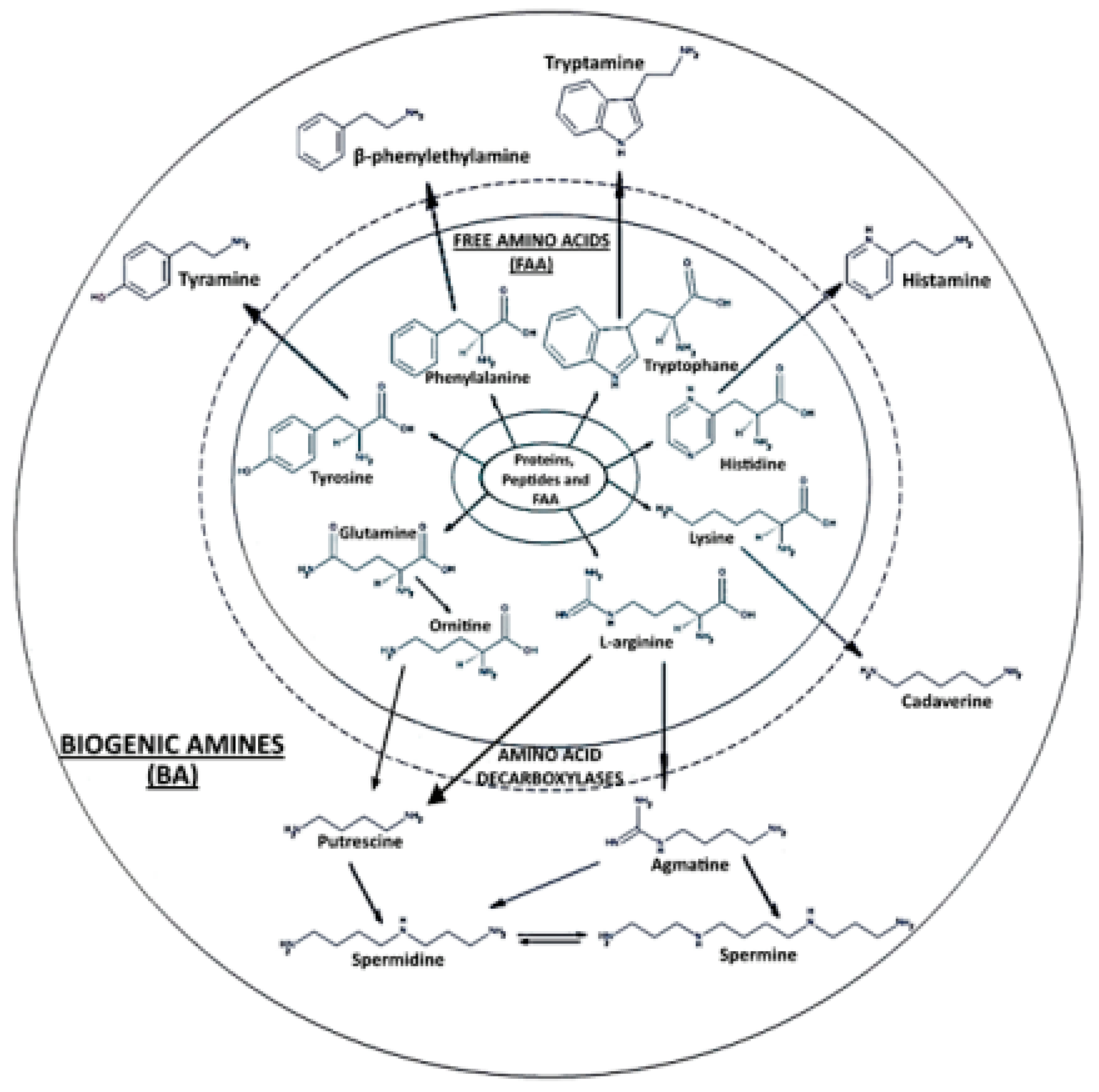
Figure 1. Biogenic amines. (Modified from [4]; common license.)
Ijms 24 07704 g001
Figure 2. (a) Distribution of the high-abundance proteins for each biogenic-amine-producing strain determined via LFQ; y-axis (count) is the number of identified proteins. (b) Heatmap from the shotgun proteomics analysis of 15 different biogenic amine foodborne strains. Every bar corresponds to the presence or absence of a particular protein. Red = upregulated proteins; green = downregulated proteins. Euclidean hierarchical distances were sorted for all strains. Three principal clusters were differentiated.
Ijms 24 07704 g002
Figure 3. (a) Distribution of the high-abundance proteins for each genus determined using LFQ; (b) Heatmap from the shotgun proteomics analysis of 15 different biogenic amine foodborne strains according to different genera. The y-axis (count) represents the number of identified proteins. Every bar corresponds to the presence or absence of a particular protein. Red = upregulated proteins; green = down regulated proteins. The Euclidean hierarchical distances were sorted according to different genera (Enterobacter spp., Hafnia spp., Klebsiella spp., Morganella spp., Proteus spp., Raoultella spp., and Stenotrophomonas spp. Seven principal clusters were differentiated, which corresponded to the different genus types.
Ijms 24 07704 g003
Figure 4. (a) Molecular functions of the biogenic-amine-producing bacterial proteome identified using shotgun proteomics and categorized via PANTHER using the gene names as inputs for the software; (b) biological processes of biogenic-amine-producing bacterial proteome identified via shotgun proteomics and categorized via PANTHER; (c) protein classes of biogenic-amine-producing bacterial proteome identified via shotgun proteomics and categorized via PANTHER.
Ijms 24 07704 g004aIjms 24 07704 g004b
Figure 5. Protein interactome network for the global protein repository of biogenic-amine-producing bacterial foodborne strains using STRING v.11.5 software. High-confidence interactions (≥0.7) in STRING software were selected for this study. The final network for the global protein repository consists of 260 nodes (proteins) and 1973 edges (interactions). Nodes represent the proteins, and the interactions between proteins are represented by continuous lines when referring to direct interactions (physical) or with dotted lines when referring to indirect interactions (functional).
Ijms 24 07704 g005
Table 1. The biogenic amine-producing bacterial strains considered in this study.
SampleBacterial StrainCodeSourceGenBank
H1Enterobacter aerogenesEbAe1ATCC 13048FJ971882
H2Enterobacter cloacaeEbCl1ATCC 13047FJ971883
H3Hafnia alveiHaAl2ATCC 9760FJ971884
H4Klebsiella oxytocaKlOx1ATCC 13182FJ971867
H5Morganella morganiiMoMo1BM 65FJ971858
H6Morganella morganiiMoMo2ATCC 8076FJ971868
H7Proteus mirabilisPrMi1ATCC 14153FJ971887
H8Proteus penneriPrPe1ATCC 33519FJ971869
H9Proteus vulgarisPrVu1ATCC 9484FJ971888
H10Proteus vulgarisSard1SardineJN630885
H11Proteus vulgarisSard2SardineJN630886
H12Raoultella planticolaRaPl2ATCC 33531FJ971885
H13Stenotrophomonas maltophilia25MC6Albacore tunaFJ971861
H14Stenotrophomonas maltophilia5PC6Albacore tunaFJ971863
H15Stenotrophomonas maltophiliaStMa215MFFJ971862
Table 2. Biogenic amine-related proteins and peptides detected via LC-ESI-MS/MS for the corresponding strains.
Biogenic AminePrecursorProteins Identified by LC-ESI-MS/MSPeptidesSampleBacterial Strain
AgmatineArginineArginine ABC transporter substrate-binding proteinIDAVFGDTAVVTEWLKH4K. oxytoca
Lysine-arginine-ornithine-binding periplasmic proteinC*TWVGSDFDSLIPSLKH4K. oxytoca
IGTDATYAPFSSKH4K. oxytoca
CadaverineLysineLysine-arginine-ornithine-binding periplasmic proteinC*TWVGSDFDSLIPSLKH4K. oxytoca
IGTDATYAPFSSKH4K. oxytoca
HistamineHistidineHistidine kinaseIDSEDLPHVRASVARH6M. morganii
LAM*NLRTRLFLSISALITVALLGLLLGLVSVM*QM*AGSQEILIRH13S. maltophilia
M*IAEAANADSKQAQRH4, H12K. oxytoca, R. planticola
TIDQINQQKIQLEQEIADRKH8, H9 P. penneri, P. vulgaris
GEADATLDSEVSAWRAVARH11P. vulgaris
LSSELWNC*KIDPTQAEM*AM*INILANARH7P. mirabilis
SEASENTVDLIVEDEGSGIPKH7P. mirabilis
NEEARDNLKLISELTARH11P. vulgaris
RYAYSEQLGDLLQRH15S. maltophilia
Histidine phosphataseHAQASEYGSALFVAVGQAKQVKH3H. alvei
Histidine-binding periplasmic proteinIGVLQGTTQETYGNEHWAPKH4K. oxytoca
Histidine triad nucleotide-binding proteinEIPSDIVYQDELVTAFRH4K. oxytoca
IAEQEGIAEDGYRH2E. cloacae
Histidine ammonia-lyaseLAAM*QQALGAQIAAVEEDRH2E. cloacae
H6M. morganii
PutrescineArginineLysine-arginine-ornithine-binding periplasmic proteinC*TWVGSDFDSLIPSLKH4K. oxytoca
IGTDATYAPFSSKH4K. oxytoca
GlutamineGlutamine ABC transporter periplasmic proteinAVGDSIEAQQYGIAFPKH4K. oxytoca
Glutamine-fructose-6-phosphate aminotransferaseIDAAQEAELIKALFEAPRH4K. oxytoca
N-acetylglutaminylglutamine amidotransferaseSGANAAVDKALRLDSTVM*LVDDPVKH3H. alvei
MethionineType 1 glutamine amidotransferase domain-containing proteinIFRTLALM*LLVTSATAFAASKH7P. mirabilis
L-glutamine-binding proteinADAVIHDTPNILYFIKH4K. oxytoca
OrnithineAVGDSLEAQQYGIAFPKH3H. alvei
S-adenosylmethionine decarboxylase proenzymeALSFNIYDVC*YARH15S. maltophilia
SpermidineAgmatineSpermidine/putrescine import ATP-binding proteinVDEVHDNAEAEGLIGYIRH9P. vulgaris
MethionineS-adenosylmethionine decarboxylase proenzymeALSFNIYDVC*YARH15S. maltophilia
Putrescine
Spermine
SpermineAgmatine
MethionineS-adenosylmethionine decarboxylase proenzymeALSFNIYDVC*YARH15S. maltophilia
Spermidine
C* (carbamidomethyl cysteine), M*(methionine oxidation).
Table 3. Proteins corresponding to bacterial resistance to antibiotics, antimicrobial-related proteins, and other virulence factors identified in the strains analyzed.
FuntionProteinSample
ToxinsEntericidin A/B family lipoproteinH1, H2, H3, H4
Addiction module toxin, GnsA/GnsB familyH12
Antitoxin ParDH15, H13
EcotinH1, H3, H4
Antimicrobial compounds productionBacteriocin immunity proteinH9
Colicin immunity protein/pyocin immunity proteinH1
Antimicrobial resistancePenicillin-binding protein activator LpoBH4
TetR family transcriptional regulatorH5, H15
Acriflavine resistance protein BH61
Methicillin resistance proteinH15
GNAT family N-acetyltransferaseH1, H5
Additional resistances and tolerancesAcid stress chaperone HdeBH5, H6
Cold shock protein CspAH4, H14
Cold shock protein CspCH4
Cold shock protein CspDH14, H6, H8
Cold shock protein CspEH4
Copper resistance proteinH5, H6
Envelope stress response membrane protein PspBH1, H3, H5, H8, H9
General stress proteinH4, H13, H15
Heat shock survival AAA family ATPase ClpKH2, H4
Stress response protein ElaBH13
Stress response translation initiation inhibitor YciHH5
Stress-induced bacterial acidophilic repeat motifH2
Universal stress proteinH2, H3, H4, H12
YdeI family stress tolerance OB fold proteinH2
CsbD family proteinH2, H3, H9, H15
Peroxide/acid resistance protein YodDH2
Putative ‘Cold-shock’ DNA-binding domain proteinH15
L,D-transpeptidase YnhGH4
Protein sufAH5
Spy/CpxP family protein refolding chaperoneH4
Host colonization and immune evasionBeta-aspartyl-peptidaseH9
Chaperone protein SkpH1, H2, H3, H4, H5, H7, H8, H9, H12
Chemotaxis protein CheYH13
Cytosol nonspecific dipeptidaseH2
Filamentous hemagglutininH2
Fimbrial protein FimVH15
Type 1 fimbrial proteinH8
Flagellar biosynthesis protein FliCH14
Flagellar hook protein FlgEH13
Flagellar secretion chaperone FliSH15
FlagellinH8, H10, H11, H13, H14, H15
HemagluttininH1
Hemolysin expression modulator HhaH3, H7
Inhibitor of vertebrate lysozymeH3
Isoaspartyl peptidase/L-asparaginaseH7, H11, H13, H15
Lipoprotein involved in copper homeostasis and adhesionH4
Lon proteaseH1
LysMH2, H4, H13, H15
LysR family transcriptional regulatorH12
MaltoporinH4
Ferrichrome porin FhuAH4
MipA/OmpV family proteinH4
Molecular chaperone OsmYH2, H3, H4
OmpAH1, H2, H3, H4, H8, H13
OmpCH4, H10
OmpDH4
OmpFH4
OmpK36H4
Omptin family outer membrane proteaseH4
OmpXH4
Phosphate-selective porin OprO/OprPH15
OsmC family peroxiredoxinH4
Outer membrane lipoprotein RcsFH4
Peptidase S74H15
Peptidase S8 and S53 subtilisin kexin sedolisinH13
Periplasmic serine endoprotease DegP-likeH15
IAP aminopeptidase MEROPS family M28CH1
Membrane-bound metallopeptidaseH9
MetalloproteaseH4
Alkaline serine proteaseH14
Signal peptidase IH12
Superoxide dismutaseH4, H7, H9, H15, H13, H2
Tautomerase PptAH2
Tautomerase ydcEH12
TonBH4, H14, H15
Transcriptional regulator SlyAH1, H2, H3, H4, H12
Twitching motility protein PilHH14
Type I restriction enzyme endonucleaseH4
Type VI secretion proteinH15
VacJ family lipoproteinH15
ABC transportersAmino acid ABC transporterH2
Arginine ABC transporter substrate-binding proteinH4
Glutamine ABC transporter periplasmic proteinH3, H4
Manganese ABC transporterH4
Iron ABC transporterH4
Oligopeptide ABC transporterH5, H6
Putative ABC-type sugar transport systemH1
Ribose ABC transporter substrate-binding protein RbsBH4
Xylose ABC transporter, periplasmic xylose-binding protein XylFH2
Phage proteinsPresumed capsid scaffolding protein (GpO)H10
Bacteriophage CI repressorH1
Beta_helix domain-containing protein (Klebsiella phage vB_KpM_FBKp24)H7
Phage portal protein, HK97 familyH5
Phage shock protein PspAH1, H2, H3, H4, H5, H6, H7, H9, H11, H12
Uncharacterized protein (Stenotrophomonas phage BUCT608)H12
Terminase (Klebsiella phage vB_KppS-Storm)H11
Uncharacterized protein (Stenotrophomonas phage Marzo)H11
Alternative virulence factors and proteins involved in horizontal transferPilus assembly protein, pilin FimAH5
IS3 family transposaseH5
Major type 1 subunit fimbrin (Pilin)H12
Plasmid stability proteinH3
Plasmid-related proteinH7
Rop family plasmid primer RNA-binding proteinH5
TransposaseH15
Tyrosine recombinase XerCH5
MobC family plasmid mobilization relaxosome proteinH5
Table 4. Potential species-specific tryptic peptide biomarkers of seafood-originating biogenic-amine-producing bacteria. Specificity was determined after similarity search using BLASTp.
ProteinPeptideSampleSpecific by Blastp
DUF883 domain-containing proteinNLADTLEEVLNSSTDKSKEELGKH1E. aerogenes
Uncharacterized proteinLLQLALAAIDSADEAGVSHEDIDNQHTEEEASPLTPKH1E. aerogenes
Hemagluttinin domain-containing proteinSVAQNAAAITDTRH1E. aerogenes
Anti-adapter protein IraPLIQDIETAMEQVKPGPLVDDRDTQLLQQYIKH1E. aerogenes
Cell division protein ZapBNTTLAQEVQSAQHGREELERENSQLRH1E. aerogenes
Der GTPase-activating protein YihIKPIPLGVTESTPAVKH1E. aerogenes
DUF1471 domain-containing proteinAREEGAKGFVVNSAGGDNHMYGTATIYKH2E. cloacae
DUF2511 domain-containing proteinSSGQPISVIQIDDPSSPGQKH2E. cloacae
Uncharacterized proteinESGFEGELTDLSDDILIYHLKH2E. cloacae
Universal stress protein UspFM*FNSILVPVDISESRH2E. cloacae
DNA-binding proteinIKDNNAEYVEPLDMLAELC*EDNKLLAAELRH3Hafnia alvei
ATP synthase subunit bKAQIIDEAKVEAEQERH3Hafnia alvei
DUF883_C domain-containing proteinGVANEAAGQVEESYGEATNSHQHRLEGQARH3Hafnia alvei
Exodeoxyribonuclease 7 small subunitAPAAPSFEQALSELEQIVTHLESGELPLEDALNEFERH3Hafnia alvei
ATP synthase subunit bAQIIDEAKVEAEQERNKH3Hafnia alvei
Cell division protein ZapBESLVRENEQLKEEQTAWQERH3Hafnia alvei
50S ribosomal protein L10IVEGTPFEC*LKDTFVGPTLVAFSMEHPGAAARH3Hafnia alvei
Uncharacterized proteinRKLSPAEELALGKH3Hafnia alvei
Major outer membrane lipoprotein LppVDQLSNDVNAM*RADVQTAKDDAARH3Hafnia alvei
DUF1471 domain-containing proteinIGDVSAEVRDGTM*DDIVKH3Hafnia alvei
Uncharacterized proteinESDAEREEKTFTWKPSAVRH3Hafnia alvei
Cell division protein ZapBENEQLKEEQTAWQERH3Hafnia alvei
Membrane proteinNGVPESGFTLDVVPNDQADASGGQVVGHC*ENDTQKH3Hafnia alvei
Chaperone protein SkpATELQGQERDLQSKH3Hafnia alvei
Outer membrane lipoprotein SlyBAVQIQGGDESNAIGAIGGAVLGGFLGNTIGGGTGRH4K. oxytoca
Putative porinNYVEANGGISWTPLTPLTIKH4K. oxytoca
TolC-like proteinQAGIQDVTYQTDQQTLILNTATAYFKH4K. oxytoca
MaltoporinSSESGGSGTFADRDQFGNRH4K. oxytoca
Glutamate/aspartate ABC transporter substrate-binding proteinLIPVTSQNRIPLLQNGTFDFEC*GSTTNNLARH6M. morganii
Integration host factor subunit alphaAEM*SENLSEKLDLSKRH5M. morganii
FAD assembly factor SdhEGRPDDEALYQIIRH5M. morganii
Cell division protein ZapBVQQALDTITLLQM*EIEELKEKNDALNQEVQGARH7P. mirabilis
Outer membrane protein assembly factor BamEVRQETLTLTFDNNGILTKH7P. mirabilis
DUF2594 family proteinAISEVADEQQAETFRNTLNQIKH7P. mirabilis
DNA-binding proteinIIADFLGVAPSEIWPSRYFHPETGELLERH7P. mirabilis
Inner membrane protein YhcBSSNELMPDMPEQENPFNYRH8P. penneri
DUF1043 family proteinSSNELM*PDM*PAQDNPFNYRH9P. vulgaris
LipoproteinQQAQETENQALDKADQLTDQAKH9P. vulgaris
Arsenate reductaseSLELADPQLSEDALIQAIVDNPKH12R. planticola
Uncharacterized proteinSEHAAQGKSDSVGSQVSEGAQKTWNKH12R. planticola
Uncharacterized proteinEAEQLDNDKNFYYQEAKH12R. planticola
Exported proteinVGTISSTGQTAPGDARAELLKH12R. planticola
Uncharacterized proteinTPLSDTDFANKILASQANQEYVRH12R. planticola
DUF1311 domain-containing proteinSRDGELDTALYDDSQPGNLQGELNDVMRH15S. maltophilia
Type VI secretion proteinNAPAADTQNFYNAPAPRH15S. maltophilia
Uncharacterized proteinVLAAGGTAAQALAASQAAARH15S. maltophilia
Uncharacterized proteinGSTTVAGQDISLNQDFKH15S. maltophilia
Uncharacterized proteinDQKDLPHPDAEAQRPDPVSPLQAKH15S. maltophilia
DNA-binding proteinSLIAQAEKQQSKH15S. maltophilia
Uncharacterized proteinAQSTQDLGLHTSC*RH15S. maltophilia
Uncharacterized proteinTYFDYSEEQPFIRH15S. maltophilia
30S ribosomal protein S16VGFYNPVAQGGEKH15S. maltophilia
Cu(I)-responsive transcriptional regulatorLGEDDQTPVPVDTAKH15S. maltophilia
Antitoxin ParDQLIAEGLASGPSAPLAPDHFDKLRH15S. maltophilia
DUF3606 domain-containing proteinAAVAEVGPTAAAVRH15S. maltophilia
TonB-dependent receptorNASSGPGAVVLSPTHPDNPIPGQASRH15S. maltophilia
RidA family proteinAFDNLKAVAEAAGGSLDQVVRH13S. maltophilia
LysM peptidoglycan-binding domain-containing proteinKADFSGVSGSVDSTAEQVPKH13S. maltophilia
Uncharacterized proteinSANAAATAAQEAADAAAAKH13S. maltophilia
DUF4124 domain-containing proteinANLALLDGGGQVMQDTDGDGKADTPLAPEQRH13S. maltophilia
LysM peptidoglycan-binding domain-containing proteinADFSGVSGSVDSTAEQVPKH13S. maltophilia
DUF4124 domain-containing proteinSPQAAASAETPAAPVPEQC*STARH13S. maltophilia
FlagellinFTSTIANLNTNSENLSAARH13S. maltophilia
Uncharacterized proteinDAADKTAAASEQAAADTQQALDKAADATANAADQAKH13S. maltophilia
Attachment proteinLLGDIAKDLTNAPLEDIQKH13S. maltophilia
Antitoxin ParDQLIAEGLASGPAVPVTAATFERH13S. maltophilia
Uncharacterized proteinTQADAILADSAANEYDKSLAAQLASQAAYQTDDTPAAVAYLKH13S. maltophilia
Excinuclease ATPase subunitVEQSLQELISSQAAKH13S. maltophilia
Uncharacterized conserved protein, DUF2147SIVEISQAANGTLTGKH13S. maltophilia
50S ribosomal protein L31 type BSTM*GTKETIQWEDGNEYPLVKH13S. maltophilia
LysM peptidoglycan-binding domain-containing proteinADFSGVSASVDSTADVVSGGTYTVQKGDSLSKH13S. maltophilia
DUF3613 domain-containing proteinSFEHEIPDFFEADVAKH13S. maltophilia
Poly(Hydroxyalkanoate) granule-associated proteinLHVPTADEVTALEARIDALQARH13S. maltophilia
Heme exporter protein DAVKQDAAAPLSTELERH13S. maltophilia
RNA polymerase-binding transcription factor DksATDEATGRPILPTGYKPGSEEEYM*SPLQQEYFRH13S. maltophilia
Transcriptional regulatorLEALDALLPSDSPNPIDLLERH13S. maltophilia
CsbD-like proteinIQKGVGEVQSDVGKARH13S. maltophilia
C* (carbamidomethyl cysteine); M* (methionine oxidation).
Disclaimer/Publisher’s Note: The statements, opinions and data contained in all publications are solely those of the individual author(s) and contributor(s) and not of MDPI and/or the editor(s). MDPI and/or the editor(s) disclaim responsibility for any injury to people or property resulting from any ideas, methods, instructions or products referred to in the content.