Article

SELENOP rs3877899 Variant Affects the Risk of Developing Advanced Stages of Retinopathy of Prematurity (ROP)

by
Ewa Strauss
1,*,
Danuta Januszkiewicz-Lewandowska
2,3,
Alicja Sobaniec
4 and
Anna Gotz-Więckowska
5
1
Institute of Human Genetics, Polish Academy of Sciences, Strzeszynska 32, 60-479 Poznan, Poland
2
Department of Pediatric Oncology, Hematology and Transplantology, Poznan University of Medical Sciences, Szpitalna 27/33, 60-572 Poznan, Poland
3
Department of Medical Diagnostics, Dobra Street 38a, 60-595 Poznan, Poland
4
Department of Neonatology, Poznan University of Medical Sciences, Polna 33, 60-535 Poznan, Poland
5
Department of Ophthalmology, Poznan University of Medical Sciences, Szamarzewskiego 84, 60-569 Poznan, Poland
*
Author to whom correspondence should be addressed.
Int. J. Mol. Sci. 2023, 24(8), 7570; https://doi.org/10.3390/ijms24087570
Submission received: 12 March 2023 / Revised: 6 April 2023 / Accepted: 17 April 2023 / Published: 20 April 2023
(This article belongs to the Section Molecular Pathology, Diagnostics, and Therapeutics)

Abstract

:
The significance of selenoproteins for the incidence of prematurity and oxidative-damage-related diseases in premature newborns is poorly understood. The latter are at risk for ROP as well as BPD, IVH, PDA, RDS, and NEC, which is particularly high for newborns with extremely low gestational age (ELGA) and extremely low birth weight (ELBW). This study evaluates the hypothesis that variation in the selenoprotein-encoding genes SELENOP, SELENOS, and GPX4 affects the risk of ROP and other comorbidities. The study included infants born ≤ 32 GA, matched for onset and progression of ROP into three groups: no ROP, spontaneously remitting ROP, and ROP requiring treatment. SNPs were determined with predesigned TaqMan SNP genotyping assays. We found the association of the SELENOP rs3877899A allele with ELGA (defined as <28 GA), ROP requiring treatment, and ROP not responsive to treatment. The number of RBC transfusions, ELGA, surfactant treatment, and coexistence of the rs3877899A allele with ELGA were independent predictors of ROP onset and progression, accounting for 43.1% of the risk variation. In conclusion, the SELENOP rs3877899A allele associated with reduced selenium bioavailability may contribute to the risk of ROP and visual impairment in extremely preterm infants.

1. Introduction

Prematurity is one of the major health-threatening problems in neonates [1]. Preterm babies are highly susceptible to oxidative stress due to an imbalance between the oxidant and antioxidant systems. The generation of free radicals induces oxidative damage to multiple body organs and systems and results in the development of typical premature infant diseases, including bronchopulmonary dysplasia (BPD), intraventricular hemorrhage (IVH), patent ductus arteriosus (PDA), respiratory distress syndrome (RDS), necrotizing enterocolitis (NEC), diffuse white matter injury (DWMI), or retinopathy of prematurity (ROP). The risk is particularly high for newborns with extremely low gestational age (ELGA) and extremely low birth weight (ELBW) [2,3].
Selenium (Se) and selenoproteins perform an essential function in fetal development and health by regulating antioxidant and inflammatory processes [4,5,6,7]. Se’s nutritional status of the fetus and newborn depends on the mother’s antioxidant status. Uptake of this micronutrient in the fetus occurs via placental transfer, mainly during the 3rd trimester of pregnancy. Se is obtained through diet, and its plasma concentrations vary by country depending on its content in soil and Se-containing supplement intake [8]. Concentrations of Se observed in European adults and infants are relatively low, which may contribute to the relatively high impact of Se deficiency on the occurrence of oxidative stress-related diseases in European populations.
There is evidence that cord blood Se levels positively correlate with gestational age (GA), birth weight (BW), and Apgar score at 5 min, confirming the protective role of Se against the occurrence of preterm delivery [9]. Consequently, lower Se levels in preterm infants than in term-born ones are observed. This deficiency can be further exacerbated in the postnatal period due to prolonged parenteral nutrition and poor intestinal absorption. Considering that Se concentrations increase in healthy breastfed infants after birth, the Se deficiency found in preterm infants must be clinically important [10]. The bioavailability of Se in the body and redox homeostasis are partly determined by selenoprotein activity, which is under genetic control [5,11]. The specific role of selenoproteins in development is indicated by observations of a phenotype of severe gene mutations in the selenoprotein biosynthesis pathway that result in global developmental delay [12].
The relationship between variants in genes encoding selenoproteins and the risk of developing ROP and other complications of prematurity has not yet been investigated. In this study, we evaluated the association of four SNPs in the genes encoding selenoprotein P (SelP, SELENOP gene), selenoprotein S (SelS, SELENOS gene), and glutathione peroxidase 4 (GPX4, GPX4 gene) with the development of ROP and its clinical course. The associations and interactions between these variants and the presence of other comorbidities (BPD, IVH, PDA, RDS, and NEC) and extremely low gestational age and birth weight were also investigated.

2. Results

2.1. Clinical Risk Factors of ROP Development and Unsuccessful Treatment

Population characteristics are shown in Figure 1, while risk factors for developing ROP and treatment failure are shown in Table 1. In the study group of 190 children, as many as 173 (91.0%) completed the entire neonatal and ocular follow-up (Figure 1). The remaining 17 children (9.0%) were excluded from the study. Among them, 7 (3.7%) died, and another 10 (5.3%) were lost in follow-up.
Ultimately, 121 children with ROP were included in the group. Among them, 55 (31.8%) were diagnosed with ROP that regressed spontaneously, and 66 (38.2% of the group) had ROP requiring treatment. In the latter group, LP was performed as the first line of treatment in 46 infants (69.7%); 19 infants (28.8%) received IVR injections; and 1 infant (1.5%) was treated simultaneously with LP and IVR. Treatment failure was observed in 19 cases of ROP (11.0%). In the studied group, ROP was diagnosed on average at 52.9 ± 12.4 days of life (7.6 weeks), at a mean post-menstrual age (PMA) of 34.4 ± 2.0 weeks.
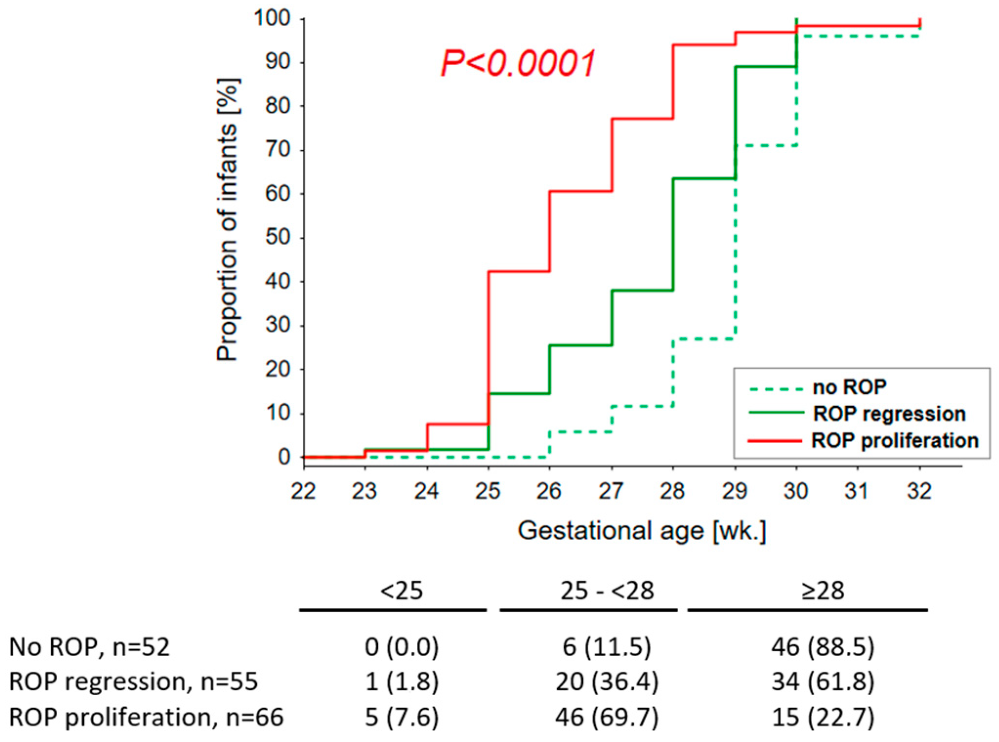
The presence of ROP and its progression to advanced stages was negatively correlated with GA (β = 0.591; p < 0.0001), BW (β = 0.509; p < 0.0001), and Apgar scores at 1 and 5 min (β = 0.323; p < 0.0001 and β = 0.267; p < 0.001), while being positively correlated with the number of blood transfusions (β = 0.554; p < 0.0001), duration of mechanical ventilation (β = 0.548; p < 0.0001), the occurrence of other factors related to respiratory failure (treatment with surfactant and resuscitation), and the presence of comorbidities: BPD (β = 0.477; p < 0.0001), IVH (β = 0.364; p = 0.0001), sepsis (β = 0.296; p < 0.001), RDS (β = 0.262; p = 0.001), and NEC (β = 0.234, p = 0.002, Table 1). There were no significant correlations between risk factors present at birth (i.e., fetal bladder rupture and delivery by cesarean section) and the development and progression of ROP. Among the neonates with ROP requiring treatment, the only factor significantly associated with failure of laser therapy was multiple blood transfusions (8.0 ± 3.6 vs. 4.5 ± 2.0 p = 0.026); other factors showing an association at the level of a statistical trend were low BW (p = 0.191) and the coexistence of IVH (p = 0.142). All these children required resuscitation and mechanical ventilation. Details of the relationship between age and the occurrence of ROP are shown in Figure 2.

2.2. Polymorphisms in Selenoprotein Genes and the Incidence, Severity, and Treatment of ROP

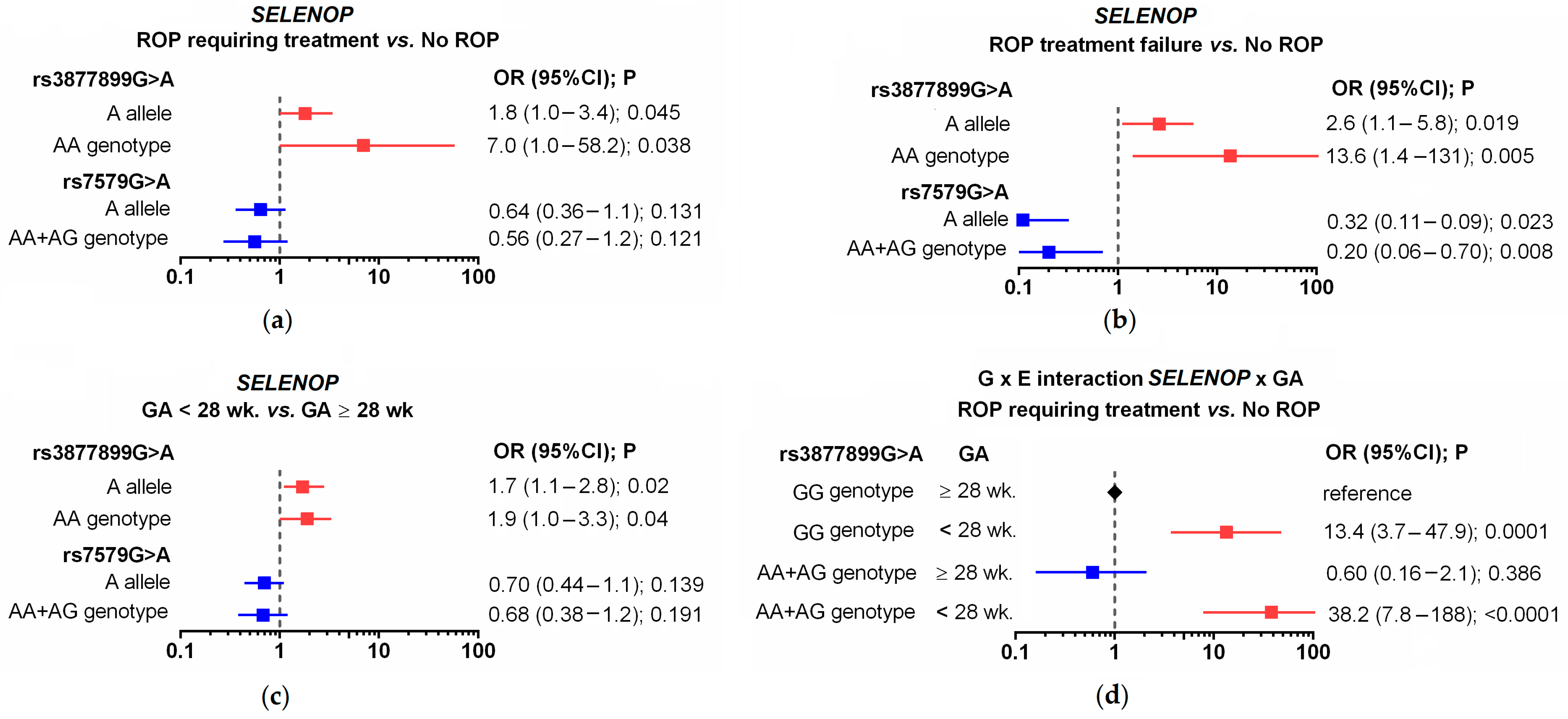
The genotype distributions of the studied SNPs showed no deviations from the Hardy–Weinberg equilibrium. The studied SELENOP SNPs were found to be in significant linkage disequilibrium (r = 0.348; p < 0.0001). An association was found between the SELENOP SNPs and the occurrence of advanced ROP and/or treatment failure (Figure 3a and Supplemental Table S1). The frequency of the SELENOP rs3877899A allele in premature infants without ROP was 0.202; in those with spontaneous regression of ROP, it was 0.255; in those with proliferative ROP, it was 0.318; and in cases with unsuccessful treatment, it was as high as 0.395. The corresponding frequencies of rs3877899AA homozygotes were: 1.9%, 9.1%, 12.1%, and 21.1%. Among the ROP cases requiring treatment, 50% of rs3877899AA homozygotes experienced treatment failure.
Statistically significant associations were observed for the rs3877899A allele and the rs3877899AA homozygotes and the occurrence of ROP requiring treatment (OR = 1.8, p = 0.045; OR = 7.0, p = 0.038, respectively) or ROP with treatment failure (OR = 2.6, p = 0.019; OR = 13.6, p = 0.005, respectively, in relation to the clinical course without ROP), Figure 3b. On the other hand, the SELENOP rs3877899A allele was associated with a lower risk of ROP treatment failure. However, this effect may be partly attributed to linkage disequilibrium between the studied SNPs. The frequency of the SELENOP rs7579A allele was 0.324 in preterm infants without ROP and 0.132 in infants with unsuccessful ROP treatment (OR = 0.32; p = 0.023). A protective effect was observed in the dominant model (OR = 0.20, p = 0.008). The other studied SNPs in the SELENOS and GPX4 genes were not associated with ROP occurrence, progression, or treatment effectiveness.

2.3. Polymorphisms in Selenoprotein Genes and the Incidence of Other Complications of Prematurity

RDS, IVH, BPD, PDA, NEC, LBW, ELGA, and ELBW were studied. An association between SELENOP rs3877899 and ELGA was observed (Figure 3c; Supplemental Table S2). More than 52% of infants born before 28 weeks of gestation carried the rs3877899A allele, compared with 38% of infants born after at least 28 weeks of gestation (p = 0.02). Carriers of this allele had a 1.9-fold increased risk of being born with ELGA (p = 0.04). No other associations were found between the selenoprotein genotype and the occurrence of other complications of prematurity.

2.4. Gene-Environment Interaction between the SELENOP Genotype and ELGA

A gene-environment interaction between the SELENOP rs3877899 genotype and ELGA affected the risk of developing ROP and its progression to advanced stages. The co-occurrence of the rs3877899GA+AA genotype with ELGA was observed in 3.8% of cases without ROP, in 16.4% of cases with spontaneously regressive ROP, and in 45.5% of cases with ROP requiring treatment (Table 2; Figure 3d). Compared to children with the rs3877899GG genotype who were born ≥28 GA, children with both risk factors (the rs3877899GA+AA genotype and ELGA) had a 35-fold increased risk of proliferative ROP (p < 0.0001). The Rothman synergy index in both cases indicated a multiplicative interaction. The co-occurrence of the studied risk factors was associated with a 1.6-fold and 3.1-fold greater impact with respect to the sum of their separate influences. There was no effect of the coexistence of these risk factors on ROP treatment. Both risk factors were present in a similar proportion of successfully treated children (n = 21; 45%) and in the subgroup with treatment failure (n = 9; 47.4%).
Similar effects were observed for the co-occurrence of the SELENOP rs3877899GA+AA genotype with ELBW, but they were less significant, indicating a secondary effect in relation to age. The coexistence of the rs3877899GA+AA genotype and ELBW was observed in 5.7% of children without ROP, 16.4% of children with spontaneously regressive ROP, and 36.4% of children with ROP requiring treatment. These frequencies correspond to a 5-fold (95% CI: 1.2–22.2; p = 0.024) and 20-fold (95% CI: 5.1–81.6); p < 0.0001) increase in the risk of developing spontaneously regressive ROP or ROP requiring treatment. Only for ROP requiring treatment did the observed relationships indicate a multiplicative interaction (S = 1.5).

2.5. Independent Predictors of the Occurrence and Progression of ROP

The results of the multivariate analysis for the occurrence and progression of ROP are presented in Table 3.
Factors showing significant association in univariate analysis were selected for further evaluation; they included ELGA, ELBW, Apgar score at 1 min, number of RCB transfusions, surfactant treatment, resuscitation, mechanical ventilation, duration of mechanical ventilation, presence of NEC, BPD, IVH, RDS, sepsis, or the SELENOS rs3877899 genotype (A allele dose), and co-occurrence of the SELENOS rs3877899GA+AA genotype with ELGA. All factors examined were included in model 2, whereas model 1 did not include the co-occurrence of the rs3877899GA+AA genotype with ELGA. Regression analysis demonstrated that the most important risk factors for the onset and progression of ROP were the number of RCB transfusions, ELGA, and surfactant treatment, which jointly accounted for 41.3% of the risk variation (30.0%, 8.5%, and 2.1%, respectively). When considering the combined effect of genotype and age, this increased risk variability by 1.8%.

3. Discussion

ROP is a disorder resulting from abnormal retinal vascularization and is a leading cause of visual impairment and blindness in children [13]. The overall incidence of any stage of ROP ranges from 15% to 68%, depending on the studied population. It is diagnosed on average at 8 wk. of life (34–35 PMA). Lower GA and BW are major risk factors for the development of the disorder. Among ELBW infants, the observed percentage of any stage ROP reaches 80%, and the percentage of ROP cases requiring treatment reaches 24%; in infants with GA ≤ 25 weeks, these rates amount to 100% and 67%, respectively [14,15]. The frequency of blindness in children attributable to ROP ranges from 10% to 37.4% worldwide [16].
The mechanism through which retinal lesions develop in premature infants is well documented. It is linked to a lack of effective regulation of the retinal and choroidal circulation, which promotes retinal vasoconstriction, endothelial cell degeneration, inflammation, and uncontrolled neovascularization. The pathogenesis of these changes is multifactorial and includes damage induced by oxidative stress and resulting from neonatal immaturity and related factors. Among them, the presence of RDS, prolonged oxygen therapy, and multiple blood transfusions are the most important [15]. The results of this study are consistent with previous clinical observations: low GA was the strongest predictor of ROP presence and progression to advanced stages, followed by a high number of blood transfusions and prolonged mechanical ventilation. Numerous blood transfusions were also a risk factor for ROP treatment failure. Among the comorbidities, the strongest correlation with ROP was shown by BPD and IVH, which suggests a common etiopathogenesis. The number of RBC transfusions, ELGA, and surfactant treatment proved to be independent predictors of ROP onset and progression, accounting for 41.3% of the risk variability in the studied population.
The role of Se and genetic variation in enzymes involved in maintaining optimal function of the antioxidant system [6] and susceptibility to complications induced by oxidative stress [17,18,19] in premature infants was previously demonstrated, but the impact of selenoproteins has yet to be investigated. Therefore, this study focused on the role of genetic variants for selenoproteins that act as antioxidants in plasma (SeP) and in the intracellular space, including the endoplasmic reticulum (SelS), cytoplasm (GPX4), and mitochondria (GPX4). The studied variants affect protein function or concentration in the body and have been previously linked to cancer and cardiovascular disease [20,21,22,23,24,25]. Among them, only SNPs in SELENOP were found to be associated with ROP. SeP is a multifunctional protein that not only acts as an antioxidant but also stores and transports Se. Unlike other selenoproteins, which contain only a single selenocysteine incorporated into the N-terminal domain, it possesses multiple selenocysteine residues (up to 9) in the C-terminal domain [26].
A summary of the univariate analysis results is presented in Figure 2. The rarer allele of the rs3877899 SNP (variant A) was found to be a risk factor for proliferative, more advanced ROP. In contrast, the rarer allele of the rs7579 SNP (variant A) played a protective role; however, this effect was probably a consequence of linkage disequilibrium between the studied SNPs. The rs3877899A allele was also associated with ELGA in the studied children, and both factors (age and the rs3877899 genotype) interacted to further increase the risk of ROP. In univariate analysis, the coexistence of the rs3877899GA+AA and ELGA genotypes further amplified the risk of spontaneously regressive ROP and ROP requiring treatment (by factors of 1.6 and 3.1, respectively). After adjusting for the influence of other risk factors, the combined effect of the risk genotype and ELGA explained 1.8% of the variation in ROP incidence and progression. This impact was similar to the influence of surfactant use, which amounted to 2.1%. It can be assumed that rs3877899A coexisting with ELGA is also a significant risk factor for ROP treatment failure. Treatment was required primarily for children of low birth age, and the children whose treatment proved unsuccessful were characterized by an extremely high frequency of the rs3877899A allele (0.395 vs. 0.287 in cases treated successfully) and its homozygotes (21.1% vs. 8.5% in patients treated successfully; significance level for both p = 0.2). The frequency of the rs3877899A allele in the subgroup of children with ROP treatment failure was significantly higher than in the subgroup without ROP (0.395 vs. 0.202; p = 0.019), which featured only a small percentage of children with ELGA.
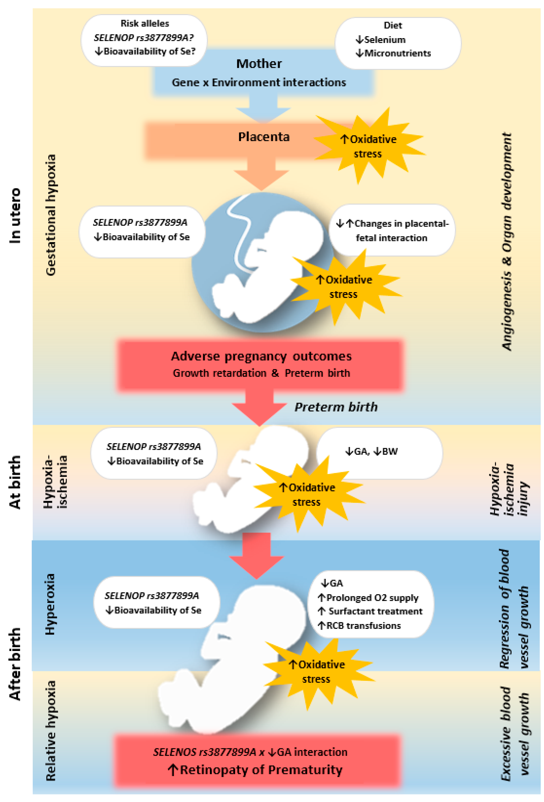
The observed effects of SNPs were consistent with expectations based on previously described allelic functions. The studied SNPs were found to alter the synthesis ratio of the two isoforms of SeP, which differ in size and Se content. The rs3877899A and rs7579G alleles favor the production of a 60-kDa Se-rich isoform with 10 Sec residues, while the rs3877899A and rs7579G alleles favor the production of a 50-kDa Se-poor isoform containing only one Sec residue [27]. The rs3877899 variant is also involved in the regulation of protein stability and cellular protein uptake. These SNPs were found to affect selenium’s bioavailability for the synthesis of all other selenoproteins by influencing the body’s Se status and transport, as well as the effectiveness of supplementation [28,29,30]. Our findings regarding the association of the rs3877899A allele with ELGA are also in line with previous clinical studies in the Italian population, which demonstrated the association of SNPs in antioxidant enzymes with low GA and/or BW (for superoxide dismutase 2) as well as complications of prematurity, including ROP (for superoxide dismutase 1, 3, and catalase) [7,31]. Both the latter results and our own findings suggest that the development of the antioxidant system during fetal life is significantly involved in the maintenance of pregnancy through uteroplacental-fetal interactions. After birth, this system can be crucial for proper retinal function and development. Possible mechanisms are shown in Figure 4.
The importance of selenoproteins for development is illustrated by the embryonic lethal phenotype of knockout mice for the genes of the selenoprotein biosynthesis pathway. In humans, carriers of mutations in these genes (including SECISBP2, TRU-TCA1-1, and SEPSECS) display the severe multisystem phenotypes of global developmental delay with features of myopathy and cerebellar and optic atrophy [12].
The examined SELENOS and GPX4 variants were not associated with complications of prematurity, ELGA, or ELBW. The effects of these SNPs are likely too weak to be demonstrated in a small group. On the other hand, the adverse effect of the rs3877899A allele is associated with decreased production of other selenoproteins and may exacerbate the effect of carrying other unfavorable genetic variants. It may contribute to increased production of pro-inflammatory cytokines [4,21,31], lipid peroxides [32], and antioxidant barrier depression [20,33,34], related to the genetic deficiency in SeP and GPX4.
The specific role of Se and selenoproteins in the development of diseases associated with prematurity is still poorly understood. The literature data indicate that Se is an important contributor to ROP [10], pregnancy outcomes [35], the risk of intrauterine growth retardation and preterm birth [36], as well as the functioning of the immune and antioxidant systems in preterm infants [37]. Recent data confirm the significant role of oxidative stress in both the incidence and severity of ROP [38].
Given the lack of guidelines for standardized Se monitoring methods and the absence of Se availability for supplementation, it is reasonable to conclude that Se deficiency in extremely preterm infants is probably common and underdiagnosed [6]. ELGA and SELENOP genotypes may be important factors in identifying preterm infants at high risk of developing advanced ROP. Whether Se supplementation will prevent advanced ROP as well as avoid morbidity and mortality in preterm infants remains an open question [35]. Randomized clinical trials have shown that postnatal Se supplementation in very low birth weight infants can reduce one or more episodes of sepsis. However, such supplementation has not improved neonatal outcomes in terms of ROP, survival, or lung diseases [39]. These outcomes may be due to insufficient dosages of supplementation, a lack of maternal supplementation during pregnancy, or the modifying effects of the SELENOP genotype. The influence of genotype was previously demonstrated in the UK population, where pregnant women who carry the rs3877899A allele could better maintain their Se status during pregnancy and were more responsive to Se supplementation than carriers of the rs3877899G allele [29]. The impact of the rs3877899A allele on Se supplementation was also observed in women from the general population [28]. These results are promising for the potential use of protective supplementation during pregnancy. However, developing a personalized therapy approach presents a challenge as it would require clinical trials involving several thousand pregnant women. The supplementation of another micronutrient, zinc, in preterm infants was significantly associated with reduced mortality but not comorbidities such as BPD, ROP, bacterial sepsis, or NEC [40]. A limitation of the aforementioned studies in the infant cohorts was the low prevalence of the diseases studied. For example, in one study on ROP involving 193 premature infants, the incidence of ROP was 3.2% in the non-supplemented zinc group and 0% in the supplemented group. The incidence of ROP in the Se-supplemented groups was higher, but it did not exceed 33%. In addition to the effects of antioxidant micronutrients, SeP has been shown to inhibit vascular endothelial growth factor-stimulated cell proliferation, tubule formation, and migration in human umbilical vein endothelial cells [41], indicating that SeP may directly affect the induction of neoangiogenesis in ROP and represent a therapeutic target.
Two potential limitations should be kept in mind when analyzing the results of our study. Firstly, our study did not take into account the influence of the genotypes of the studied genes in mothers on the predisposition to the analyzed neonatal diseases. However, the SELENOP rs3877899 genotype in the mother can be speculated to influence uteroplacental-fetal interactions, pregnancy complications, and preterm delivery since mothers of children with the rs3877899AA homozygote genotype are also carriers of the rs3877899A allele. Secondly, the presence of polymorphisms in other genes that may influence the occurrence of preterm birth, particularly those involved in the inflammatory process, cannot be excluded. The SELENOS rs34713741 SNP may be associated with circulating levels of proinflammatory cytokines, but the regulation of inflammation is complex, so further studies that include more genes involved in the inflammatory process are warranted to fully elucidate the etiology of preterm birth and the related complications. The strong point of the study was the high statistical power of the genetic analysis for ROP. A post hoc analysis of the study revealed a statistical power of 96.3%, 97.6%, and 45% for the association between the SELENOP rs3877899AA genotype and the incidence of ROP requiring treatment, ROP treatment failure, and ELGA, respectively. In the GxE interaction study, the estimated statistical power was 96.1% for SELENOP rs3877899GA+AA genotypes and 99.9% for ELGA in the analysis of the proliferative type of ROP (requiring treatment). For the analysis of ROP undergoing spontaneous remission, the estimated statistical power was <80% and 94.4%, respectively. The statistical power of the tested interaction effect in both analyses was lower than 80%. These findings confirm the significance of the SELENOP rs3877899A allele and ELGA in the pathogenesis of severe forms of ROP. Nonetheless, validation of the presence of GxE interactions between these risk factors requires replication of studies on a larger population of prematurely born children.
In conclusion, this association study supports the importance of selenoproteins and potential Se deficiency in the pathogenesis of ROP. Analysis of SELENOP SNPs may be helpful in identifying those preterm infants with ELGA who are at particularly high risk for the progression of ROP to advanced stages and who may benefit from preventive Se supplementation. For a better understanding of the role of selenoproteins in the pathogenesis of ROP, it could be important to investigate the relationship between SeP levels, changes in newborns’ condition after birth, and the timing of ROP occurrence.

4. Materials and Methods

4.1. Study Population

We carried out a prospective study in which premature infants born between 22 and 32 weeks of gestation were screened for the presence and advancement of ROP. Based on ophthalmic criteria, a population of 190 children was selected, with similar proportions of subjects with no ROP, spontaneously regressing ROP, and ROP requiring treatment. All the infants were hospitalized at the Department of Neonatology in the Gynecology and Obstetrics Clinical Hospital (Level III Hospital) of the Poznan University of Medical Sciences between 2009 and 2017. Homogenic consistency was maintained as all infants were of Caucasian origin. The study design excluded multiple pregnancy births, infants with chromosomal abnormalities or TORCH infections (rubella, herpes, cytomegalovirus, toxoplasmosis, etc.), and infants without antenatal steroid therapy.

4.2. Clinical Features and Outcomes

The following clinical factors were analyzed: gestational age (GA, weeks), birth weight (BW, grams), sex, time of preterm rupture of the fetal bladder (days), mode of delivery (vaginal delivery vs. cesarean section), Apgar scores at 1 and 5 min, blood transfusions, surfactant and oxygen therapy, and duration of mechanical ventilation (days). In addition to ROP, the evaluated comorbidities of prematurity included sepsis, jaundice, RDS, IVH, BPD, NEC, and PVL. The employed criteria for diagnosing RDS, NEC, IVH, and BPD have been described in detail previously [42]. The diagnosis of PVL was based on abnormal US or MRI results. Sepsis and neonatal jaundice were diagnosed on the basis of clinical symptoms and standardized laboratory tests. The diagnosis of intrauterine hypotrophy was based on the World Health Organization criteria, namely, fetal weight below the 10th percentile according to percentile grids for the appropriate gestational age. Subgroups of newborns with ELGA and ELBW were distinguished based on a birth age lower than 28 weeks and a body weight of less than 1000 g.
The methods of ROP diagnosis, classification, and treatment have been described in detail by Chmielarz-Czarnocinska et al. [43]. In short, the infants were screened with indirect ophthalmoscopy by a team of three trained pediatric ophthalmologists specializing in ROP. The examination was performed under topical anesthesia with proxymetacaine after pupil dilation with tropicamide 1% and phenylephrine 2.5% drops applied three times. The initial funduscopic examination was performed four weeks after birth, and follow-up examinations were performed every 7–10 days during the hospital stay or every 1–3 weeks, depending on the severity of the lesions, after the patient’s discharge from the hospital (until full retinal vascularization, lesion stabilization, or the need for treatment). Based on the Early Treatment for Retinopathy of Prematurity guidelines, treatment was provided within 72 h of detection of severe ROP using laser photocoagulation (LP) and/or Lucentis therapy (intravitreal ranibizumab injections, IVR). Peripheral retinal ablations were carried out using a 810-nm diode laser with confluent burns (Iris Medical OcuLight SL, IRIDEX Corporation, CA, USA) under general anesthesia. The patients were examined on the day following the procedure and every 7–10 days until total ROP regression was ascertained, changes were stabilized, or retreatment was necessary. All treated infants were monitored by a team of participating physicians.

4.3. Genotyping

The biological material for genetic research was obtained from buccal swabs taken from the inner surface of the child’s cheek. DNA isolation was performed using the innuPREP Swab DNA Kit (Analytik Jena, Jena, Germany). The presence of the studied SNPs—SELENOP rs3877899 (c.700G>A, p.Ala234Ser), SELENOP rs7579 (c.*14G>A, 3’UTR variant), SELENOS rs34713741 (2 kb C>T 5’UTR variant), and GPX4 rs713041 (c.660T>A; p.Leu220=)—was determined with predesigned TaqMan SNP genotyping assays containing probes for allelic discrimination (respectively: C2841533_10, C8806056_10, C3091980_10, and C2561693_20), using the ABI 7900HT Fast Real-Time PCR System (Life Technologies, Carlsbad, CA, USA). The SELENOS rs34713741 SNP was selected for this research instead of the most studied variant of the promoter region (−105G>A, rs28665122), as the commercially available predesigned assay gave inconclusive results. Both SNPs are in complete linkage disequilibrium in the Central European population; hence, the tested SNP can be considered a linked marker. The genotyping success rate was 99%. Samples with failed genotype calls at more than 1 SNP were excluded from the analysis.

4.4. Statistical Analysis

Genotype frequencies were tested for the Hardy–Weinberg equilibrium (HWE) using a χ2 test available at https://ihg.helmholtz-muenchen.de/cgi-bin/hw/hwa1.pl (accessed on 12 December 2022). The individual effects of the studied factors on the onset and progression of ROP were evaluated using linear regression analysis to determine the significance of the trend. The regression beta coefficient (β) was determined to compare the strength of the influence of the studied factors. In this analysis, the groups were assigned the following designations: 0—no ROP; 1—ROP requiring treatment; and 2—ROP not requiring treatment. Univariate analyses assessing the effect of the studied factors on the treatment outcome used the χ2 or Fisher’s test for qualitative variables and the t-test or Mann–Whitney U test for quantitative variables. Kaplan–Meier curves were used to visualize the relationship between age at birth and the onset and course of ROP. The values of odds ratios (ORs) and 95% confidence intervals (95%CIs) were also calculated for the genotypes. Gene-environment interactions were assessed using the method described by Botto and Khoury [44], which is based on the use of a multivariate table with a two-by-four layout. The assessment was performed using univariate analyses (χ2 or Fisher’s test). The Rothman synergy index (S), which indicates a deviation from the additive model of interactions, was also determined. The interpretation of the coefficient values is as follows: S = 1 indicates no interaction; S < 1 indicates a relative decrease; and S > 1 indicates an increase in the strength of the interactions between the two factors. A multivariable stepwise regression analysis was performed to evaluate the potential independent effect of the studied variables on the occurrence and progression of ROP. Quanto software (developed by The Division of Biostatistics, Keck School of Medicine of the University of Southern California, Los Angeles, CA, USA) was used for the post hoc statistical power analysis. All other analyses were performed using STATISTICA version 13.0 (Dell, Tulsa, OK, USA) and GraphPad Prism version 6.04 (GraphPad Software, Boston, MA, USA) software. The observed differences were considered significant at p < 0.05.

Supplementary Materials

The supporting information can be downloaded at: https://www.mdpi.com/article/10.3390/ijms24087570/s1.

Author Contributions

Conceptualization, E.S., A.G.-W., D.J.-L. and A.S.; methodology, E.S. and A.G.-W.; investigation, E.S., A.G.-W. and A.S.; data curation, E.S., A.G.-W. and A.S.; writing—original draft preparation E.S. and D.J.-L.; writing—review and editing A.G.-W. and A.S.; visualization, E.S.;. supervision, E.S. and A.G.-W.; funding acquisition, E.S., A.G.-W. and D.J.-L. All authors have read and agreed to the published version of the manuscript.

Funding

The work has been supported by the Scientific Research Committee in Poland under grant no. 2 P05E 098 30 (AGW, ES) and the Poznan University of Medical Sciences research grant no. 502-14-11121500-41229. The APC was funded by the Poznan University of Medical Sciences and the Institute of Human Genetics.

Institutional Review Board Statement

All procedures performed in studies involving human participants were in accordance with the ethical standards of the institutional and/or national research committee and with the 1964 Helsinki declaration and its later amendments or comparable ethical standards. The study was approved by the Bioethics Committee of the Poznan University of Medical Sciences (no. 1140/05 and 1117/18).

Informed Consent Statement

Informed consent was obtained from all parents.

Data Availability Statement

All data generated or analyzed during this study are included in this published article (and its Supplementary Materials File).

Acknowledgments

We would like to thank the students Aleksandra Landowska, Malgorzata Czarnecka, and Weronika Starzak for their significant contribution to DNA isolation, preparation of DNA sample banks, and/or genotyping (under the supervision of ES).

Conflicts of Interest

The authors declare no competing interests.

References

  1. Kassabian, S.; Fewer, S.; Yamey, G.; Brindis, C.D. Building a global policy agenda to prioritize preterm birth: A qualitative analysis on factors shaping global health policymaking. Gates Open. Res. 2020, 4, 65. [Google Scholar] [CrossRef]
  2. Ozsurekci, Y.; Aykac, K. Oxidative Stress Related Diseases in Newborns. Oxid. Med. Cell. Longev. 2016, 2016, 2768365. [Google Scholar] [CrossRef]
  3. Lembo, C.; Buonocore, G.; Perrone, S. Oxidative Stress in Preterm Newborns. Antioxidants 2021, 10, 1672. [Google Scholar] [CrossRef]
  4. Curran, J.E.; Jowett, J.B.; Elliott, K.S.; Gao, Y.; Gluschenko, K.; Wang, J.; Abel Azim, D.M.; Cai, G.; Mahaney, M.C.; Comuzzie, A.G.; et al. Genetic variation in selenoprotein S influences inflammatory response. Nat. Genet. 2005, 37, 1234–1241. [Google Scholar] [CrossRef]
  5. Donadio, J.L.S.; Duarte, G.B.S.; Borel, P.; Cozzolino, S.M.F.; Rogero, M.M. The influence of nutrigenetics on biomarkers of selenium nutritional status. Nutr. Rev. 2021, 79, 1259–1273. [Google Scholar] [CrossRef]
  6. Tindell, R.; Tipple, T. Selenium: Implications for outcomes in extremely preterm infants. J. Perinatol. 2018, 38, 197–202. [Google Scholar] [CrossRef] [PubMed]
  7. Poggi, C.; Giusti, B.; Vestri, A.; Pasquini, E.; Abbate, R.; Dani, C. Genetic polymorphisms of antioxidant enzymes in preterm infants. J. Matern. Fetal Neonatal Med. 2012, 25 (Suppl. 4), 131–134. [Google Scholar] [CrossRef] [PubMed]
  8. Thurnham, D.I.; Northrop-Clewes, C.A. Inflammation and biomarkers of micronutrient status. Curr. Opin. Clin. Nutr. Metab. Care 2016, 19, 458–463. [Google Scholar] [CrossRef]
  9. Makhoul, I.R.; Sammour, R.N.; Diamond, E.; Shohat, I.; Tamir, A.; Shamir, R. Selenium concentrations in maternal and umbilical cord blood at 24–42 weeks of gestation: Basis for optimization of selenium supplementation to premature infants. Clin. Nutr. 2004, 23, 373–381. [Google Scholar] [CrossRef]
  10. Yang, H.; Ding, Y.; Chen, L. Effect of trace elements on retinopathy of prematurity. J. Huazhong Univ. Sci. Technol. Med. Sci. 2007, 27, 590–592. [Google Scholar] [CrossRef] [PubMed]
  11. Tarhonska, K.; Raimondi, S.; Specchia, C.; Wieczorek, E.; Reszka, E.; Krol, M.B.; Gromadzinska, J.; Wasowicz, W.; Socha, K.; Borawska, M.H.; et al. Association of allelic combinations in selenoprotein and redox related genes with markers of lipid metabolism and oxidative stress—Multimarkers analysis in a cross-sectional study. J. Trace Elem. Med. Biol. 2022, 69, 126873. [Google Scholar] [CrossRef]
  12. Schoenmakers, E.; Chatterjee, K. Human Genetic Disorders Resulting in Systemic Selenoprotein Deficiency. Int. J. Mol. Sci. 2021, 22, 12927. [Google Scholar] [CrossRef]
  13. Kim, S.J.; Port, A.D.; Swan, R.; Campbell, J.P.; Chan, R.V.P.; Chiang, M.F. Retinopathy of prematurity: A review of risk factors and their clinical significance. Surv. Ophthalmol. 2018, 63, 618–637. [Google Scholar] [CrossRef]
  14. Demir, S.; Yücel, Ö.E.; Niyaz, L.; Karakuş, G.; Arıtürk, N. Incidence of retinopathy of prematurity in the middle Black Sea region of Turkey over a 10-year period. J. Aapos 2015, 19, 12–15. [Google Scholar] [CrossRef]
  15. Yucel, O.E.; Eraydin, B.; Niyaz, L.; Terzi, O. Incidence and risk factors for retinopathy of prematurity in premature, extremely low birth weight and extremely low gestational age infants. BMC Ophthalmol. 2022, 22, 367. [Google Scholar] [CrossRef]
  16. Hong, E.H.; Shin, Y.U.; Bae, G.H.; Choi, Y.J.; Ahn, S.J.; Sobrin, L.; Hong, R.; Kim, I.; Cho, H. Nationwide incidence and treatment pattern of retinopathy of prematurity in South Korea using the 2007–2018 national health insurance claims data. Sci. Rep. 2021, 11, 1451. [Google Scholar] [CrossRef] [PubMed]
  17. Swan, R.; Kim, S.J.; Campbell, J.P.; Paul Chan, R.V.; Sonmez, K.; Taylor, K.D.; Li, X.; Chen, Y.I.; Rotter, J.I.; Simmons, C.; et al. The genetics of retinopathy of prematurity: A model for neovascular retinal disease. Ophthalmol. Retina 2018, 2, 949–962. [Google Scholar] [CrossRef] [PubMed]
  18. Kim, S.J.; Sonmez, K.; Swan, R.; Campbell, J.P.; Ostmo, S.; Chan, R.V.P.; Nagiel, A.; Drenser, K.A.; Berrocal, A.M.; Horowitz, J.D.; et al. Identification of candidate genes and pathways in retinopathy of prematurity by whole exome sequencing of preterm infants enriched in phenotypic extremes. Sci. Rep. 2021, 11, 4966. [Google Scholar] [CrossRef] [PubMed]
  19. Tao, T.; Meng, X.; Xu, N.; Li, J.; Cheng, Y.; Chen, Y.; Huang, L. Ocular phenotype and genetical analysis in patients with retinopathy of prematurity. BMC Ophthalmol. 2022, 22, 22. [Google Scholar] [CrossRef]
  20. Bermano, G.; Pagmantidis, V.; Holloway, N.; Kadri, S.; Mowat, N.A.; Shiel, R.S.; Arthur, J.R.; Mathers, J.C.; Daly, A.K.; Broom, J.; et al. Evidence that a polymorphism within the 3’UTR of glutathione peroxidase 4 is functional and is associated with susceptibility to colorectal cancer. Genes. Nutr. 2007, 2, 225–232. [Google Scholar] [CrossRef]
  21. Du, X.H.; Dai, X.X.; Xia Song, R.; Zou, X.Z.; Yan Sun, W.; Mo, X.Y.; Lu Bai, G.; Xiong, Y.M. SNP and mRNA expression for glutathione peroxidase 4 in Kashin-Beck disease. Br. J. Nutr. 2012, 107, 164–169. [Google Scholar] [CrossRef]
  22. Li, J.; Zhu, Y.; Zhou, Y.; Jiang, H.; Chen, Z.; Lu, B.; Shen, X. The SELS rs34713741 Polymorphism Is Associated with Susceptibility to Colorectal Cancer and Gastric Cancer: A Meta-Analysis. Genet. Test. Mol. Biomark. 2020, 24, 835–844. [Google Scholar] [CrossRef] [PubMed]
  23. Meplan, C.; Hughes, D.J.; Pardini, B.; Naccarati, A.; Soucek, P.; Vodickova, L.; Hlavata, I.; Vrana, D.; Vodicka, P.; Hesketh, J.E. Genetic variants in selenoprotein genes increase risk of colorectal cancer. Carcinogenesis 2010, 31, 1074–1079. [Google Scholar] [CrossRef] [PubMed]
  24. Strauss, E.; Oszkinis, G.; Staniszewski, R. SEPP1 gene variants and abdominal aortic aneurysm: Gene association in relation to metabolic risk factors and peripheral arterial disease coexistence. Sci. Rep. 2014, 4, 7061. [Google Scholar] [CrossRef] [PubMed]
  25. Sutherland, A.; Kim, D.H.; Relton, C.; Ahn, Y.O.; Hesketh, J. Polymorphisms in the selenoprotein S and 15-kDa selenoprotein genes are associated with altered susceptibility to colorectal cancer. Genes. Nutr. 2010, 5, 215–223. [Google Scholar] [CrossRef] [PubMed]
  26. Mostert, V.; Lombeck, I.; Abel, J. A novel method for the purification of selenoprotein P from human plasma. Arch. Biochem. Biophys. 1998, 357, 326–330. [Google Scholar] [CrossRef]
  27. Meplan, C.; Nicol, F.; Burtle, B.T.; Crosley, L.K.; Arthur, J.R.; Mathers, J.C.; Hesketh, J.E. Relative abundance of selenoprotein P isoforms in human plasma depends on genotype, se intake, and cancer status. Antioxid. Redox Signal. 2009, 11, 2631–2640. [Google Scholar] [CrossRef]
  28. Méplan, C.; Crosley, L.K.; Nicol, F.; Beckett, G.J.; Howie, A.F.; Hill, K.E.; Horgan, G.; Mathers, J.C.; Arthur, J.R.; Hesketh, J.E. Genetic polymorphisms in the human selenoprotein P gene determine the response of selenoprotein markers to selenium supplementation in a gender-specific manner (the SELGEN study). FASEB J. 2007, 21, 3063–3074. [Google Scholar] [CrossRef]
  29. Mao, J.; Vanderlelie, J.J.; Perkins, A.V.; Redman, C.W.; Ahmadi, K.R.; Rayman, M.P. Genetic polymorphisms that affect selenium status and response to selenium supplementation in United Kingdom pregnant women. Am. J. Clin. Nutr. 2016, 103, 100–106. [Google Scholar] [CrossRef]
  30. Kurokawa, S.; Bellinger, F.P.; Hill, K.E.; Burk, R.F.; Berry, M.J. Isoform-specific binding of selenoprotein P to the β-propeller domain of apolipoprotein E receptor 2 mediates selenium supply. J. Biol. Chem. 2014, 289, 9195–9207. [Google Scholar] [CrossRef]
  31. Giusti, B.; Vestrini, A.; Poggi, C.; Magi, A.; Pasquini, E.; Abbate, R.; Dani, C. Genetic polymorphisms of antioxidant enzymes as risk factors for oxidative stress-associated complications in preterm infants. Free. Radic. Res. 2012, 46, 1130–1139. [Google Scholar] [CrossRef]
  32. Villette, S.; Kyle, J.A.; Brown, K.M.; Pickard, K.; Milne, J.S.; Nicol, F.; Arthur, J.R.; Hesketh, J.E. A novel single nucleotide polymorphism in the 3’ untranslated region of human glutathione peroxidase 4 influences lipoxygenase metabolism. Blood Cells Mol. Dis. 2002, 29, 174–178. [Google Scholar] [CrossRef]
  33. Foster, C.B.; Aswath, K.; Chanock, S.J.; McKay, H.F.; Peters, U. Polymorphism analysis of six selenoprotein genes: Support for a selective sweep at the glutathione peroxidase 1 locus (3p21) in Asian populations. BMC Genet. 2006, 7, 56. [Google Scholar] [CrossRef] [PubMed]
  34. Beharier, O.; Tyurin, V.A.; Goff, J.P.; Guerrero-Santoro, J.; Kajiwara, K.; Chu, T.; Tyurina, Y.Y.; St Croix, C.M.; Wallace, C.T.; Parry, S.; et al. PLA2G6 guards placental trophoblasts against ferroptotic injury. Proc. Natl. Acad. Sci. USA 2020, 117, 27319–27328. [Google Scholar] [CrossRef]
  35. Duntas, L.H. Selenium and at-risk pregnancy: Challenges and controversies. Thyroid. Res. 2020, 13, 16. [Google Scholar] [CrossRef]
  36. Lewandowska, M.; Więckowska, B.; Sajdak, S.; Lubiński, J. First Trimester Microelements and their Relationships with Pregnancy Outcomes and Complications. Nutrients 2020, 12, 1108. [Google Scholar] [CrossRef] [PubMed]
  37. Humberg, A.; Fortmann, I.; Siller, B.; Kopp, M.V.; Herting, E.; Göpel, W.; Härtel, C. Preterm birth and sustained inflammation: Consequences for the neonate. Semin. Immunopathol. 2020, 42, 451–468. [Google Scholar] [CrossRef]
  38. Boskabadi, H.; Marefat, M.; Maamouri, G.; Abrishami, M.; Abrishami, M.; Shoeibi, N.; Sanjari, M.S.; Mobarhan, M.G.; Shojaei, S.R.H.; Tavallaei, S.; et al. Evaluation of pro-oxidant antioxidant balance in retinopathy of prematurity. Eye 2022, 36, 148–152. [Google Scholar] [CrossRef] [PubMed]
  39. Darlow, B.A.; Austin, N.C. Selenium supplementation to prevent short-term morbidity in preterm neonates. Cochrane Database Syst. Rev. 2003, 2003, Cd003312. [Google Scholar] [CrossRef] [PubMed]
  40. Terrin, G.; Berni Canani, R.; Passariello, A.; Messina, F.; Conti, M.G.; Caoci, S.; Smaldore, A.; Bertino, E.; De Curtis, M. Zinc supplementation reduces morbidity and mortality in very-low-birth-weight preterm neonates: A hospital-based randomized, placebo-controlled trial in an industrialized country. Am. J. Clin. Nutr. 2013, 98, 1468–1474. [Google Scholar] [CrossRef]
  41. Ishikura, K.; Misu, H.; Kumazaki, M.; Takayama, H.; Matsuzawa-Nagata, N.; Tajima, N.; Chikamoto, K.; Lan, F.; Ando, H.; Ota, T.; et al. Selenoprotein P as a diabetes-associated hepatokine that impairs angiogenesis by inducing VEGF resistance in vascular endothelial cells. Diabetologia 2014, 57, 1968–1976. [Google Scholar] [CrossRef] [PubMed]
  42. Kosik, K.; Szpecht, D.; Al-Saad, S.R.; Karbowski, L.M.; Kurzawińska, G.; Szymankiewicz, M.; Drews, K.; Wolski, H.; Seremak-Mrozikiewicz, A. Single nucleotide vitamin D receptor polymorphisms (FokI, BsmI, ApaI, and TaqI) in the pathogenesis of prematurity complications. Sci. Rep. 2020, 10, 21098. [Google Scholar] [CrossRef] [PubMed]
  43. Chmielarz-Czarnocińska, A.; Pawlak, M.; Szpecht, D.; Choręziak, A.; Szymankiewicz-Bręborowicz, M.; Gotz-Więckowska, A. Management of retinopathy of prematurity (ROP) in a Polish cohort of infants. Sci. Rep. 2021, 11, 4522. [Google Scholar] [CrossRef] [PubMed]
  44. Botto, L.D.; Khoury, M.J. Commentary: Facing the challenge of gene-environment interaction: The two-by-four table and beyond. Am. J. Epidemiol. 2001, 153, 1016–1020. [Google Scholar] [CrossRef] [PubMed]
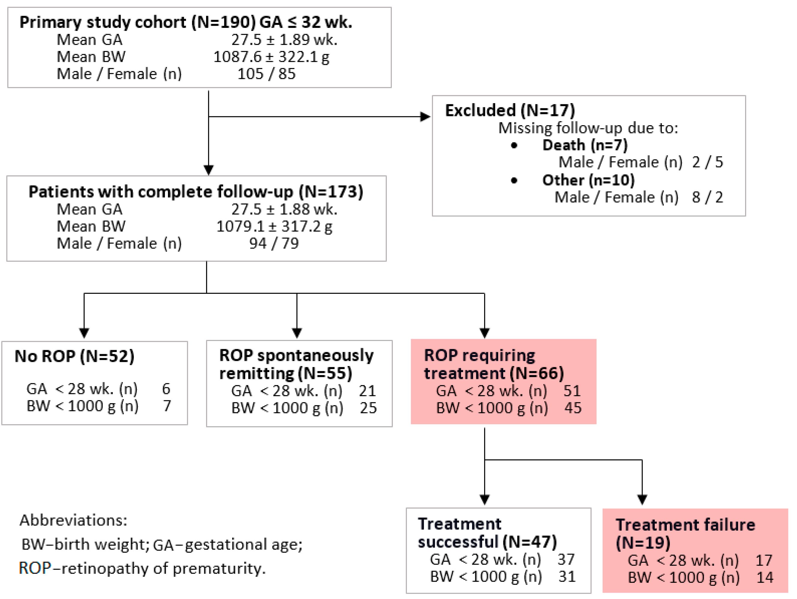
Figure 1. Consolidated scheme of the study’s inclusion criteria and population characteristics.
Ijms 24 07570 g001
Figure 2. The relationship between birth age and the occurrence and course of ROP.
Ijms 24 07570 g002
Figure 3. Summarized results of univariate analyses of associations and gene-environment interactions (G × E) between selenoprotein-encoding genes, gestational age below 28 weeks (GA < 28 wk.), and ROP. Association of the SELENOP rs3877899 and rs7579 SNPs with the occurrence of ROP requiring treatment (a), ROP treatment failure (b), and GA < 28 wk. (c). Multiplicative G × E interaction between the SELENOP rs3877899 genotype and GA < 28 wk. affects the incidence of ROP requiring treatment (d).
Ijms 24 07570 g003
Figure 4. Overview of the postulated mechanism through which a child’s SELENOP genotype influences in utero and postnatal homeostatic disorders leading to ROP occurrence. The placenta facilitates important in utero functions, including nutrient transfer, metabolism, gas exchange, neuroendocrine signaling, growth hormone production, and immune control. Insufficient dietary selenium supply, as observed in European countries, may contribute to inadequate protection of the placenta from oxidative stress during pregnancy, affecting fetal development. This may be exacerbated by genetic risk factors present in the mother and child, resulting in reduced Se bioavailability. Among them are variants in the gene encoding selenoprotein P, the body’s main Se store and transporter. Abnormalities in fetal development can lead to changes in placental-fetal interactions and result in premature birth. During and after delivery, the immature newborn is exposed to oxidative stress associated with changes in environmental oxygen concentration. The use of oxygen therapy after birth inhibits angiogenesis in the eye. After the termination of this treatment, relative hypoxia occurs, which is a strong stimulus for vascular growth and the development of ROP. Extreme prematurity and carrying the SELENOP rs3877899 A allele is associated with depression of the antioxidant barrier and the occurrence of oxidative damage. Oxidative stress is strongly related to ROP pathogenesis because of the susceptibility of the phospholipid-rich retina to reactive oxygen species that can be generated in high or low oxygen. Other mechanisms of the protective effects of selenoprotein P may include the inhibition of VEGF-stimulated neoangiogenesis.
Ijms 24 07570 g004
Table 1. Demographic and clinical parameters associated with ROP and failure of its treatment.
Parameter Incidence and Outcome of ROPROP Requiring Treatment
I
No ROP
N = 52
II
ROP Not Requiring Treatment
N = 55
III
ROP
Requiring TreatmentN = 66
β, pfor trend
I/II/III
IIIa
ROP Treated Successfully
N = 47
IIIb
ROP Treated Unsuccessfully
N = 19
p
IIIa vs. IIIb
Gestational age [wk.]
Mean (SD)28.9 (1.2)27.7 (1.7)26.2 (1.6)−0.591, <0.000126.4 (1.8)25.9 (1.6)0.375
Range26–3223–3023–3423–3224–29
Body mass [g]
Mean (SD)1317.7 (326.6)1044.3 (266.9)920.2 (226.1)−0.509, <0.0001943.4 (242.9)862.6 (170.1)0.191
Range640–2080490–1600500–1700500–1700570–1160
Intrauterine hypotrophy; n (%)3 (7.7)7 (12.7)2 (3.1)−0.051, 0.5042 (4.4)0 (0.0)0.358
Male sex; n (%)26 (50.0)31 (56.4)37 (57.8)0.063, 0.41228 (62.2)9 (47.4)0.279
Apgar 1; Median (Q1; Q3)6 (4; 8)4 (2; 6)3 (3; 6)−0.323, <0.00013 (1; 6)3.5 (1; 6)0.769
Apgar 5; Median (Q1; Q3)7 (7; 8)7 (6; 8)7 (6; 7)−0.267, <0.0017 (6; 7)7 (5; 7)0.960
No. of RBC transfusions; Mean (SD)1.4 (1.12)2.8 (2.2)5.5 (2.8)0.554, <0.00014.5 (2.0)8.0 (3.6)0.026
Risk factors at birth
  Ruptured fetal bladder; n (%)19 (36.5)18 (32.7)22 (34.9)−0.012, 0.87314 (31.8)8 (42.1)0.440
  Ruptured fetal bladder period [d]; Mean (SD)3.6 (6.4)4.6 (10.9)5.1 (14.0)0.058, 0.4496.7 (16.4)1.6 (2.6)0.192
  Delivery by caesarean section; n (%)29 (55.8)27 (49.1)29 (45.3)−0.085, 0.26922 (48.9)7 (36.8)0.384
Parameters related to respiratory failure
  Surfactant treatment; n (%)17 (23.7)27 (41.8)46 (71.9)0.327, 0.000533 (73.3)13 (68.4)0.695
  Resuscitation; n (%)34 (65.4)49 (89.1)62 (96.9)0.354, 0.000143 (95.6)19 (100.0)0.358
  Mechanical ventilation; n (%)32 (61.5)43 (78.2)65 (98.5)0.388, <0.000146 (97.9)19 (100.0)0.529
  Mechanical ventilation period [d]; Mean (SD)6.1 (9.0)18.8 (19.2)33.7 (20.9)0.548, <0.000133.9 (21.5)33.3 (19.7)0.908
Complications of prematurity; n (%)
  NEC6 (11.5)17 (31.5)24 (37.5)0.234, 0.00216 (35.6)8 (42.1)0.628
  BPD7 (13.5)27 (49.1)46 (71.9)0.477, <0.000134 (75.6)12 (63.2)0.321
  PDA8 (15.4)21 (38.2)21 (32.8)0.149, 0.05215 (33.3)6 (31.6)0.893
  IVH22 (42.3)40 (72.7)54 (84.4)0.364, 0.000136 (80.0)18 (94.7)0.142
  RDS23 (44.2)26 (47.3)48 (75.0)0.262, 0.00134 (75.6)14 (73.7)0.877
  DWMI1 (9.9)7 (12.7)8 (12.5)0.144, 0.0617 (15.6)1 (5.3)0.262
  Sepsis6 (11.5)14 (25.9)28 (43.7)0.296, <0.00120 (44.4)8 (42.1)0.866
  Neonatal jaundice49 (94.2)47 (85.5)57 (89.1)−0.064, 0.33239 (86.7)18 (94.7)0.353
Abbreviations and symbols: BPD—bronchopulmonary dysplasia; DWMI—diffuse white matter injury; IVH—intraventricular hemorrhage; NEC—necrotizing enterocolitis; PDA—patent ductus arteriosus; RDS—respiratory distress syndrome; ROP—retinopathy of prematurity.
Table 2. The effect of gene-environment interaction between the SELENOP genotype and extremely low gestational age (ELGA) on the incidence and severity of retinopathy of prematurity (ROP).
Risk FactorsIncidence and Outcome of ROPStatistical Analysis OR (95%CI); p
rs3877899ELGAI
No ROP
II
ROP not requiring treatmen
III
ROP requiring treatment
II vs. IIII vs. I
GG≥28 wk.28 (52.8)20 (36.4)11 (16.7)referencereference
GG<28 wk.4 (7.5)12 (21.8)21 (31.8)4.2 (1.2–14.9); 0.02713.4 (3.7–47.9); 0.0001
GA+AA≥28 wk.18 (34.0)14 (25.5)4 (6.1)1.1 (0.44–2.7); 0.8540.6 (0.16–2.1); 0.386
GA+AA<28 wk.2 (3.8)9 (16.4)30 (45.5)6.3 (1.2–32.4); 0.02838.2 (7.8–187.7); < 0.0001
OR expected from additive model (G+E) a, Synergy index b 4.3; 1.6 ↑12.9; 3.1 ↑↑
a-Expected OR = OR1 Observed (GG and <28 wk.) + OR2 Observed (GA+AA and ≥28 wk.) − 1; b-The Rothman synergy index (S).
Table 3. Multivariable analysis—independent predictors of the occurrence and progression of ROP.
Risk FactorsImpact on the Occurrence and Progression of ROP
Model 1Model 2
βInfluence [%]pβInfluence [%]p
Number of RCB transfusions0.31530.7<0.000010.32330.7<0.00001
ELGA0.3468.5<0.000010.2328.5<0.00001
Surfactant treatment0.1522.10.0170.1692.10.017
rs3877899GA+AA and ELGANA0.1721.80.024
Regression model summary
Influence [%]41.343.1
SignificanceF(3.2) = 38.7; p < 0.00001F(4.164) = 31.1; p < 0.00001
Disclaimer/Publisher’s Note: The statements, opinions and data contained in all publications are solely those of the individual author(s) and contributor(s) and not of MDPI and/or the editor(s). MDPI and/or the editor(s) disclaim responsibility for any injury to people or property resulting from any ideas, methods, instructions or products referred to in the content.