Article

Identification of Optimal Reference Genes for qRT-PCR Normalization for Physical Activity Intervention and Omega-3 Fatty Acids Supplementation in Humans

by
Agata Grzybkowska
1,
Katarzyna Anczykowska
1,
Jędrzej Antosiewicz
2,
Szczepan Olszewski
2,
Magdalena Dzitkowska-Zabielska
1,3 and
Maja Tomczyk
1,*
1
Faculty of Physical Education, Gdansk University of Physical Education and Sport, 80-336 Gdansk, Poland
2
Department of Bioenergetics and Physiology of Exercise, Medical University of Gdansk, 80-211 Gdansk, Poland
3
Center of Translational Medicine, Medical University of Gdansk, 80-952 Gdansk, Poland
*
Author to whom correspondence should be addressed.
Int. J. Mol. Sci. 2023, 24(7), 6734; https://doi.org/10.3390/ijms24076734
Submission received: 1 March 2023 / Revised: 29 March 2023 / Accepted: 30 March 2023 / Published: 4 April 2023
(This article belongs to the Special Issue Novel Insights into Biochemical and Molecular Nutrition)

Abstract

:
The quantitative polymerase chain reaction (qRT-PCR) technique gives promising opportunities to detect and quantify RNA targets and is commonly used in many research fields. This study aimed to identify suitable reference genes for physical exercise and omega-3 fatty acids supplementation intervention. Forty healthy, physically active men were exposed to a 12-week eicosapentaenoic acid (EPA) and docosahexaenoic acid (DHA) supplementation and standardized endurance training protocol. Blood samples were collected before and after the intervention and mRNA levels of six potential reference genes were tested in the leukocytes of 18 eligible participants using the qRT-PCR method: GAPDH (Glyceraldehyde-3-phosphate dehydrogenase), ACTB (Beta actin), TUBB (Tubulin Beta Class I), RPS18 (Ribosomal Protein S18), UBE2D2 (Ubiquitin-conjugating enzyme E2 D2), and HPRT1 (Hypoxanthine Phosphoribosyltransferase 1). The raw quantification cycle (Cq) values were then analyzed using RefFinder, an online tool that incorporates four different algorithms: NormFinder, geNorm, BestKeeper, and the comparative delta-Ct method. Delta-Ct, NormFinder, BestKeeper, and RefFinder comprehensive ranking have found GAPDH to be the most stably expressed gene. geNorm has identified TUBB and HPRT as the most stable genes. All algorithms have found ACTB to be the least stably expressed gene. A combination of the three most stably expressed genes, namely GAPDH, TUBB, and HPRT, is suggested for obtaining the most reliable results.

1. Introduction

Quantitative polymerase chain reaction (qRT-PCR) is a versatile technique that is widely used in many research fields to determine the relative change in mRNA levels of tested genes. Gene expression analysis has become more affordable and accessible over the last 15 years [1]. Evaluating gene expression in human leukocytes has been previously used in, e.g., cancer [2], multiple sclerosis research in human cells [3], as well as in other species [4]. However, its application in studies involving physical training and supplementation is limited. A salient feature of qRT-PCR is the determination of relative gene expression results represented as a quantification cycle (Cq) value. The Cq value indicates the PCR cycle number at which the fluorescent signal generated by the amplification of the target gene surpasses the background fluorescence level. Obtaining relative results means that data need to be normalized with at least two or three stably expressed genes, called reference genes. However, the Cq value is not the only result obtained from qRT-PCR and should be taken into consideration together with other key measurements, such as amplification efficiency and melting curve analysis.
The identification of stably expressed genes in human cells, that could be used as reference genes in research, is fundamental for obtaining reliable and reproducible results, yet the use of a single reference gene without proof of validation is common in literature [5].
It has also been demonstrated that reference genes must vary for different types of cells and interventions, as no single gene could be used as a reference [6]. Hence, the use of multiple (usually two or three) reference genes should be adopted as a gold standard, as it significantly reduces the risk of producing artefactual data [7,8]. MIQE (Minimum Information for Publication of Quantitative Real-Time PCR Experiments) guidelines state that reference genes should be chosen and validated according to the specific experimental design [9]. Using several mathematic algorithms is in line with this recommendation. Physical activity can influence leukocytes, as they are powerful mediators that produce chemokines, cytokines, and growth factors, which are crucial for recovery and adaptive processes [10]. Similarly, omega-3 polyunsaturated fatty acids (n-3 PUFAs) are proven to influence DNA (Deoxyribonucleic acid) methylation changes in leukocytes, which is one way of regulating gene expression [11]. These two factors are considered to be promising targets for improving human health and could influence a great number of mRNA levels in leukocytes. There is growing evidence that increasing the amount of omega-3 fatty acids in the diet, particularly EPA and DHA acids, can promote a number of health benefits, including but not limited to the regulation of vascular and immune functions or inflammation [12]. However, there are few long-term studies in physically active individuals evaluating the effects of omega-3 fatty acids supplementation along with endurance training, on physical performance indicators [13], which was the aim of our previous work [14]. The pleiotropic effect of omega-3 fatty acids in the human body has been confirmed by published papers [15]. It has also been proven on a molecular level. In this regard, Bouwens et al., found that high doses of EPA and DHA supplementation altered the expression of 1040 genes [16]. This indicates the complexity and importance of finding genes that are not affected by omega-3 ingestion and thus, present stable expression in qRT-PCR research.
The purpose of the present study was to identify stably expressed genes in leukocytes obtained from healthy, physically active men to be used as solid reference genes. Genes were analyzed before and after 12 weeks of endurance training, combined with omega-3 fatty acids supplementation, to evaluate their effects on physical performance [14].
In silico analysis was performed using RefFinder, a web multi-tool that incorporates five methods: NormFinder, BestKeeper, geNorm, comparative ΔCq value, and RefFinder comprehensive ranking. Each method uses a different algorithm. NormFinder results are presented with a stability value (S), whereby a lower stability value represents relatively more stable expression [17]. The BestKeeper algorithm is based on the standard deviation (SD) and coefficient of variance and results with lower (SD) values are thought to provide better reference genes [18]. The GeNorm method is based on the exclusion of the least stable reference gene and results are presented as expression stability (M value), where lower M values indicate better stability [7]. Comparative ΔCq ranking calculates the average of standard deviation (Average STDEV), and again, lower Average STDEV values translate into more stable expression of the tested gene [19]. The most recent tool is RefFinder, which incorporates all of the aforementioned algorithms and enables a comprehensive analysis based on the geomean of ranking values (GM). A lower GM indicates greater stability [20]. We chose six potential reference genes for further analysis, based on published literature: GAPDH, ACTB, TUBB, RPS18, UBE2D2, and HPRT1. We aimed to compare commonly used reference genes with alternative genes identified in recent literature. GAPDH, ACTB, and RPS18, which are widely used in research due to their historical use in Northern Blots, were also assessed, even though their stability has been questioned by some [21]. Our results suggest that GAPDH, TUBB, and HPRT1 are the most stably expressed genes after an endurance training intervention and n-3 PUFAs supplementation period. ACTB consistently proved to be the least stably expressed. These results could enable other researchers to choose their reference genes more accurately in further studies, with regards to both physical activity and/or supplementation trials, which would make published data more accurate and easier to reproduce.

2. Results

2.1. mRNA Levels of the Candidate Reference Genes

The Cq value was obtained for six potential reference genes using the qRT-PCR method in both the omega-3 supplemented and placebo groups. The range of expression levels in the supplemented and control groups are presented in Figure 1 for all tested genes.
The observed Cq values of all tested genes ranged from 14.4 (ACTB) to 33.7 (UBE2D2). Lower Cq values suggest a notable abundance of a tested gene within the analyzed samples. GAPDH showed the lowest Cq values with a mean value of 20.07, while the HPRT gene showed the highest mean value of 25.15. The biggest range within the gene was identified for RPS18 (range = 15.14), which can be a preliminary indicator of stability, as mentioned by Giri A. and Sundar I.K [22]. No statistically significant differences between tested groups were found.

2.2. Evaluation of Candidate Reference Genes’ Expression after 12-Week Intervention

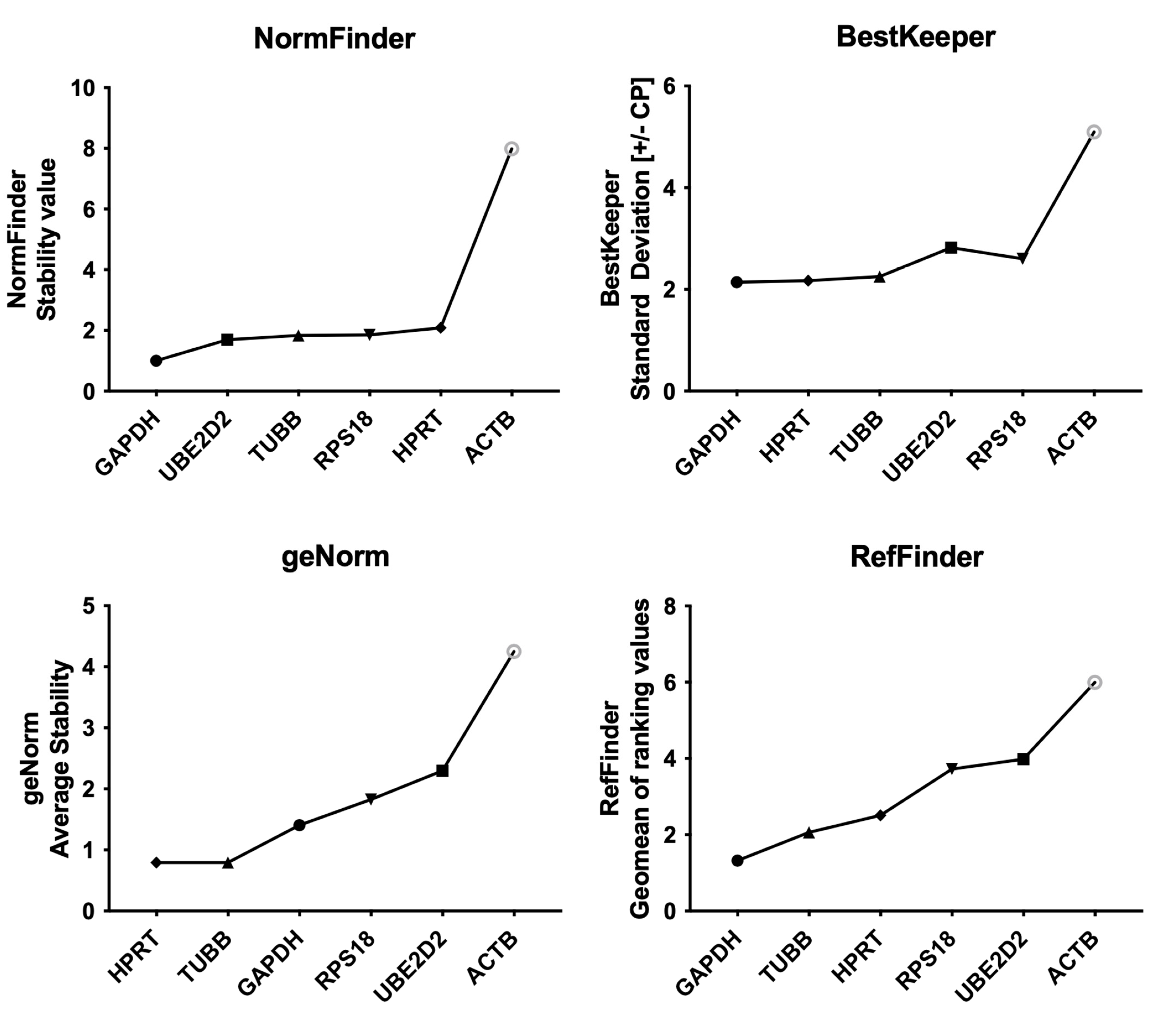
Since no statistically significant differences in Cq values were found between the placebo and omega-3-supplemented groups, all sample data were used for RefFinder in silico analysis. According to NormFinder, the most stable gene is GAPDH with a stability value (S) of 0.73. Other genes showed higher stability values: TUBB (S = 1.62), RPS18 (S = 1.86), UBE2D2 (S = 2.05), HPRT (S = 2.14), and the least stable ACTB (S = 5.48).
BestKeeper showed similar results when considering the SD [± crossing point values] with values presented in descending order, as follows: GAPDH (SD = 1.79), TUBB (SD = 1.84), HPRT (SD = 1.88), RPS18 (SD = 2.32), UBE2D2 (SD = 2.68), and ACTB (SD = 3.85).
The geNorm stability value (M) presented different results, identifying TUBB and HPRT as the most stably expressed genes with the same stability value of M = 0.74. GAPDH was ranked as being less stable with a result of M = 1.42, followed by RPS18 (M = 1.86), and UBE2D2 (M = 2.32). Still, geNorm also listed ACTB (M = 3.46) as the least stably expressed gene. As described above, ACTB consistently proved to be the least stably expressed gene by all integrated methods. GAPDH was the most stable according to all methods except for the geNorm algorithm.
Comparative delta-Ct ranking presented as the Average of standard deviation (Average STDEV) places GAPDH first, with the most stable result of Average STDEV = 2.64. The second most stably expressed gene was TUBB with a result of 2.83, followed by RPS18 (Average STDEV = 3.05), HPRT (Average STDEV = 3.09) and UBE2D2 (Average STDEV = 3.40). ACTB was listed at the bottom of the ranking with an Average STDEV number of 5.74. Nevertheless, RefFinder comprehensive ranking, which calculates the geomean of ranking values (GM), rated GAPDH as the best reference gene with a GM value of 1.32. The next suitable gene was TUBB (GM = 1.68), then HPRT (GM = 2.78), RPS18 (GM = 3.46), UBE2D2 (GM = 4.73), and ACTB (GM = 6.00). The results are presented in Figure 2.

3. Discussion

The effects of omega-3 supplementation and physical activity on human health and performance have been extensively researched over the last 10 years. Both of these factors have proven to be beneficial for human health, especially in regard to the prevention and management of civilization diseases, such as obesity, cardiovascular diseases, and mental health disorders [23,24].
Yet, few of these studies are based on long-term (>7–12 weeks), high-dose supplementation, even though both of these factors seem to be crucial to promote notable changes.
It has been previously demonstrated by Browning et al., 2012 that the amount of time needed for EPA and DHA incorporation in platelets varies between 4 and8 weeks, in the erythrocyte membrane after a minimum of 8 weeks, and in blood mononuclear cells after 6–9 months [25]. This study underscores the importance of an extended supplementation period. We have obtained similar results regarding fatty acid composition in erythrocytes. Both EPA and DHA as % of fatty acids in erythrocytes increased after a 12-week omega-3 supplementation period to a level that is considered within a target range (specific data has been shown and discussed by Tomczyk et al., 2023 [14]). This proves a physiological change and confirms the efficacy of the used dosage amount and duration in the participants of our study.
The effect of omega-3 fatty acids consumption, specifically on gene expression, is also well described in the literature; however, long-term and high-dose studies are scarce. For instance, Myhrstad et al., 2014, conducted a study in which 36 subjects ingested 8 g of either fish oil, including 1.6 g of DHA + EPA (n = 17) or sunflower oil (n = 19) for 7 days [26]. Microarray analysis was used to investigate the effect of fish oil supplementation on the transcriptome profile in PBMCs, before and after the 1-week experimental period. According to the authors, subjects were also tested after 3 weeks of supplementation. Interestingly, the authors claim that their results varied more between groups after 1 week of supplementation than after 3 weeks, which stands in opposition to data demonstrated by Browning et al., 2012 [25].
A long-term study was performed by Schmidt et al., who used qRT-PCR and microarrays to test whole-genome gene expression profiles after a 12-week exposure to high doses of n-3 PUFAs (1.14 g DHA and 1.56 g EPA) in normo- and dyslipidemic men [27]. In this study, identification of the composition of fatty acids in red blood cell membranes showed no statistically significant differences. This finding differs from the results found in the subjects of our study [14]. For qRT-PCR, Schmidt et al., [27] chose GAPDH and ribosomal protein S2 (RPS2) as reference genes based on the geNorm algorithm. The authors found increased expression of genes encoding antioxidative enzymes and a decrease in the expression of genes encoding prooxidative enzymes.
More extensive and detailed research was conducted by Bouwens et al., 2009, which involved a 26-week intervention. They tested the influence of EPA and DHA as well as high-oleic acid sunflower oil on gene expression. PBMCs from a total of 111 subjects were tested at two different doses of omega-3 fatty acids: 1.8 g EPA + DHA/d (n = 36), 0.4 g EPA + DHA/d (n = 37) and a placebo group: 4.0 g high-oleic acid sunflower oil (HOSF)/d (n = 38) [16]. A high intake of EPA + DHA was effective in altering 1040 genes, while a lower intake of fish oil influenced the expression of 298 genes. The affected genes highlight the possible anti-inflammatory and antiatherogenic properties of fish oil consumption, which is commensurate with the studies mentioned above. Moreover, this research clearly demonstrated the importance of finding stably expressed genes, since ingesting n-3 PUFAs affects the expression of a great number of genes. Normalization of the mRNA levels by suitable reference genes is a crucial step and one that affects the final reported results, as shown in many published reports [22,28,29].
In this study, we tested the stability of six potential reference genes in leukocytes obtained from healthy men, who were exposed to 12 weeks of endurance training, coupled with a high dose of omega-3 fatty acids supplementation (2.234 g EPA and 0.916 g DHA). As mentioned before, despite the abundance of literature, it is impossible to identify a single reference gene adequate for different interventions. However, we believe that the identification of possible candidate genes can, across replicate studies, allow researchers to better target reference genes in their own experimental work and may also guide interventional strategies based on the genes identified.
To the best of our knowledge, this is the first in vivo study to identify optimal reference genes in human leukocytes after a standardized endurance training and omega-3 supplementation protocol. To do this, we employed an online tool (RefFinder) that incorporates four different algorithms in order to compare the Cq values of GAPDH, ACTB, TUBB, RPS18, UBE2D2, and HPRT1.
One of the most widely used reference genes is the GAPDH gene, which encodes glyceraldehyde 3-phosphate dehydrogenase. This enzyme is involved in the process of glycolysis and in several non-metabolic processes, such as activation of transcription, initiation of apoptosis [30], or rapid axonal or axoplasmic transport [31]. Likewise, the β-ACTIN gene (ACTB) is characterized by stable expression, as it encodes a highly conserved protein that is involved in cell mobility, structure, and integrity [32]. HGPRTase, encoded by the HPRT1 gene, plays a crucial role in recovering purines from degraded DNA to reintroduce them into purine synthesis pathways [33]. The RPS18 gene carries information about the ribosomal protein, a component of the 40S subunit, and is involved in the binding of fMet-tRNA and thus, initiation of the translation process [34]. Another candidate gene is TUBB. It encodes beta tubulin protein, which is implicated in maintaining the structure of microtubules [35]. The Ubiquitin-conjugating enzyme E2 D2 is a protein that in humans is encoded by the UBE2D2 gene. Protein ubiquitination regulates the degradation of misfolded, damaged, or short-lived proteins and is mediated by a cascade of enzymes that includes E2 (ubiquitin coupling) enzyme. UBE2D2 is claimed to be one of the most stable reference genes [36].
Apart from geNorm, all of the algorithms included in the RefFinder tool showed that the single most stably expressed gene in this study was GAPDH. The MIQE guidelines suggest using more than one reference gene for more reliable results [9]. The need for using several reference genes was also discussed by Leal et al., 2015 [28].
The comprehensive data from all four software algorithms showed that GAPDH, TUBB, and HPRT are the most stable genes and using all three could be beneficial to obtain the most valid results. Moreover, GAPDH, HPRT, and TUBB are genes from different functional classes, which minimizes the risk of co-regulation. According to Vandesompele et al. [7], this diversity adds to the study’s robustness. The credibility of a single software package for choosing optimal reference genes is inconclusive and thus, RefFinder was chosen as it offers the benefits of applying and comparing multiple algorithms simultaneously. Some authors reported identical results for NormFinder, BestKeeper, and those algorithms used by RefFinder, while some show substantially different outputs for all three algorithms in and outside of RefFinder [37,38,39]. However, it should be noted that RefFinder does not take qRT-PCR efficiency data into account and De Spiegelaere et al., 2015 found that the results are similar to those that assume 100% efficiency of input data. This could possibly hinder the current findings and must be taken into consideration. Even though ACTB has been thoroughly tested as a reference gene, published data regarding its stability is inconclusive [40,41]. Our results consistently identified ACTB as the most unstable gene for the n3-PUFAs supplementation and endurance training intervention. For all that, our suggestion is that the contradictions found in the data regarding ACTB might be caused largely by the problem with the primer design, which was discussed in detail by Sun et al., 2012 [42].
As mentioned above, published data regarding the selection of appropriate reference genes is inconclusive. This might be due to the nature, specificity, and vulnerability of the PCR method [43]; hence, it is highly recommended to use more than one reference gene to obtain the most reliable results, preferably two or three. Other authors have emphasized the importance of choosing the right reference genes immediately prior to experimentation, with necessary adjustments to the cells, tissue, and methods being used [37,44]. It must also be stressed that there is no universally stable reference gene. In addition, results from our study cannot be directly applied to other studies, but they can help other researchers find the most suitable reference genes to normalize their data.

4. Materials and Methods

4.1. Ethics

The study was approved by the Bioethical Committee of Regional Medical Society in Gdansk (NKBBN/628/2019). The protocol was constructed according to the Declaration of Helsinki. All study participants were given an oral and written explanation of the study aims and written consent was obtained from each participant prior to the experiment.

4.2. Study Setting and Subjects

This study is part of a larger research project with details presented elsewhere [14]. Briefly, the effect of 12 weeks of endurance training with simultaneous omega-3 fatty acids supplementation was studied in healthy men. Participants received either omega-3 fatty acids or medium chain triglycerides (MCTs) in a daily dose of 2234 mg of eicosapentaenoic acid (EPA) + 916 mg of docosahexaenoic acid (DHA) (OMEGA-3 group) or 4000 mg of MCTs (PLACEBO group). Before and after intervention, blood samples were collected for omega-3 index (O3I) assessment, a sum of EPA and DHA expressed as a percent of total fatty acids in erythrocytes, which is a valid biomarker of omega-3 PUFA status. Moreover, a graded exercise test to exhaustion with assessment of VO2peak, running economy (RE), and a 1500-m run trial, was conducted. Out of 40 eligible participants, 26 male runners (37 ± 4 years old; 77 ± 10 kg body weight; VO2peak 54.2 ± 6 mL·kg−1·min−1) completed the protocol. Blood samples for gene expression analysis were collected in a fasted state, before and after the 12-week experimentation period. In the final gene analyses, 18 individuals were included (n = 10 in the OMEGA-3 group and n = 8 in the PLACEBO group). Detailed data on the inclusion and exclusion criteria are described by Tomczyk et al., 2023 [14]. The research protocol and the exclusion criteria for the gene analyses are presented in Figure 3.

4.3. Blood Collection, RNA Extraction and Reverse Transcription

A modification of previously described protocols for blood collection and RNA extraction were employed [45,46], with different reagents and lab equipment used in the current study. To obtain leukocytes, 2 mL of venous blood was collected from each participant into vacutainers spray-coated with K3EDTA. Within 15 min of collection, the blood was mixed with Red Blood Cell Lysis Buffer (RBCL) (A&A Biotechnology, Gdynia, Poland), incubated for 15 min, and centrifuged at 3000× g at 4 °C for 10 min. The obtained platelet was washed and later lysed using Fenozol (A&A Biotechnology, Gdynia, Poland). Finally, samples were stored at −20 °C for up to 3 months. Further RNA isolation was carried out using the modified Chomczynski and Sacchi method [47]. A total of 200 µL of chloroform (POCH, Gliwice, Poland) was used, samples were centrifuged, and the aqueous phase was mixed with 500 µL of isopropanol (POCH, Gliwice, Poland) and spun again. The obtained platelet was washed with ethanol, dried, and resuspended with PCR-grade water. Gel electrophoresis was performed to check for the quality and integrity of selected RNA. Nucleic Acid purity and concentration were determined. At UV 260/280, a ratio of 1.75–2.2 was accepted and at UV 260/230, a ratio >1.8 was accepted as pure RNA suitable for further analysis, based on available data [40,48]. At this stage, 4 samples from the supplemented group and 4 samples from the placebo group were excluded from further analysis due to the unacceptable UV 260/280 ratio or insufficient amount of obtained material.
Reverse transcription was performed using the AffinityScript qPCR cDNA synthesis kit (Agilent Technologies, Warszawa, Poland) and applied according to the manufacturer’s protocol with 1000 ng of RNA. The obtained cDNA was immediately frozen at −20 °C and stored for up to 1 month without repeated freeze-thaw cycles. cDNA was later diluted 1:10 with PCR-grade water immediately before the qRT-PCR step.

4.4. Selection of Potential Reference Genes and Primer Design

Potential reference genes (RGs) were chosen according to the data presented in published papers [3,40,41,49,50,51,52,53,54]. The genes tested in this study are listed in Table 1.
Candidate reference genes primers were either obtained from published literature or the real-time PCR primer database (PrimerBank https://pga.mgh.harvard.edu/primerbank/ (accessed on 15 November 2022)). Primer sequence, product length, and source are listed in Table 2. An efficiency of 100% was assumed for all used primers.
The specificity of potential reference genes and the obtained material was randomly checked through 2.0% agarose gel electrophoresis and later (for all samples) using a melt curve analysis.

4.5. Quantitative Real-Time Polymerase Chain Reaction

The AriaMx Real-Time PCR System (Agilent Technologies, Warszawa, Poland) and Brilliant III Ultra-Fast QPCR Master Mix—Agilent (Agilent Technologies, Warszawa, Poland) were used to perform qRT-PCR analyses. A total of 10 samples from the omega-3 supplemented group and 8 samples from the placebo group at 2 time points were analyzed at this stage. For the analysis, 2 µL of diluted cDNA of each sample was loaded in triplicates into 96-well PCR plates previously filled with 8 µL of MasterMix each. The thermal cycling conditions comprised an activation step: 95 °C for 10 min followed by 40 cycles of annealing, and an extension step: 95 °C for 15 s and 60 °C for 1 min. Additionally, the melt curve analysis was performed for each reaction to confirm the specific amplification of the target genes. At this point, 2 PRE-intervention samples from the supplemented group were excluded due to probable contamination (seen as Cq values > 35 and odd Tm values). On each plate, negative controls were included to verify the absence of contamination.

4.6. Evaluation of Stable Reference Genes for leukocytes

To compare the Cq value between the placebo and experimental groups, the D’Agostino–Pearson Normality Test was applied using GraphPad Prism version 9 for Windows, GraphPad Software, San Diego, CA, USA, www.graphpad.com (accessed on 21 January 2023). Subsequently, an unpaired t test was performed to check for statistical differences. Since no statistically significant differences were found, all Cq values were included in further analysis. In accordance with the Real-time PCR Data Markup Language (RDML), we have used the abbreviation for quantification cycle value (Cq) instead of the cycle threshold value (Ct) [58].
To evaluate the most stable reference genes, the RefFinder tool was used [20]. The RefFinder is an online software tool that integrates algorithms from the NormFinder, BestKeeper, and geNorm programs, as well as the comparative delta-Ct method.

5. Conclusions

Our results show that GAPDH, TUBB, and HPRT could be suitable reference genes for studies involving physical exercise and omega-3 supplementation in humans. We do not recommend using ACTB as a reference gene, based on current findings, as well as data presented in the literature. We believe there is a strong need for long-term (>7–12 weeks) molecular studies on this topic to accommodate the expected time course of adaptation. The information gained would enable a better understanding of the interplay between n-3 PUFAs supplementation and endurance training, and how these factors co-regulate changes in mRNA levels that, ultimately, mediate functional aspects of human health and performance.

Author Contributions

Conceptualization, M.T. and A.G.; methodology, A.G. and K.A.; software, A.G. and S.O.; validation, A.G., K.A. and S.O.; formal analysis, A.G.; investigation, M.T., A.G., K.A. and S.O.; resources, M.T. and J.A.; data curation, A.G.; writing—original draft preparation, A.G.; writing—review and editing, A.G., M.D.-Z., M.T. and J.A.; visualization, A.G.; supervision, M.T. and J.A.; project administration, M.T. and J.A.; funding acquisition, M.T. and J.A. All authors have read and agreed to the published version of the manuscript.

Funding

This research was funded by the National Science Center (Poland), grant number 2018/31/N/NZ7/02962.

Institutional Review Board Statement

This study was conducted in accordance with the Declaration of Helsinki and approved by the Bioethical Committee of Regional Medical Society in Gdansk (NKBBN/628/2019, approval date 3 December 2019).

Informed Consent Statement

Informed consent was obtained from all subjects involved in the study.

Data Availability Statement

The data presented in this study are available upon request from the corresponding author.

Acknowledgments

Authors would like to thank Maciej Chroboczek for his helpful advice regarding data curation.

Conflicts of Interest

The authors declare no conflict of interest.

Abbreviations

ACTBbeta-actin
Average STDEVAverage of standard deviation
Cqquantification cycle
Ctcycle threshold
DHAdocosahexaenoic acid
DNADeoxyribonucleic acid
EPAeicosapentaenoic acid
GAPDHGlyceraldehyde-3-phosphate Dehydrogenase
HPRT1Hypoxanthine Phosphoribosyltransferase 1
MCTsMedium Chain Triglycerides
MIQEMinimum Information for Publication of Quantitative Real-Time PCR Experiments
n-3 PUFAsomega-3 polyunsaturated fatty acids
PBMCPeripheral Blood Mononuclear Cell
qPCRQuantitative Polymerase Chain Reaction
qRT-PCRQuantitative Real-time Polymerase Chain Reaction
RBCLRed Blood Cell Lysis Buffer
RDMLReal-time PCR Data Markup Language
RGsreference genes
RPS18Ribosomal Protein S18
SDstandard deviation
TUBBTubulin Beta Class I
UBE2D2Ubiquitin-conjugating enzyme E2 D2
VO2peakpeak oxygen uptake

References

  1. Singh, K.P.; Miaskowski, C.; Dhruva, A.A.; Flowers, E.; Kober, K.M. Mechanisms and Measurement of Changes in Gene Expression. Biol. Res. Nurs. 2018, 20, 369–382. [Google Scholar] [CrossRef]
  2. Baine, M.J.; Mallya, K.; Batra, S.K. Quantitative Real-Time PCR Expression Analysis of Peripheral Blood Mononuclear Cells in Pancreatic Cancer Patients. In Pancreatic Cancer: Methods and Protocols; Methods in Molecular Biology; Su, G.H., Ed.; Humana Press: Totowa, NJ, USA, 2013; pp. 157–173. ISBN 978-1-62703-287-2. [Google Scholar]
  3. Oturai, D.B.; Søndergaard, H.B.; Börnsen, L.; Sellebjerg, F.; Romme Christensen, J. Identification of Suitable Reference Genes for Peripheral Blood Mononuclear Cell Subset Studies in Multiple Sclerosis. Scand. J. Immunol. 2016, 83, 72–80. [Google Scholar] [CrossRef] [PubMed] [Green Version]
  4. Chen, I.-H.; Chou, L.-S.; Chou, S.-J.; Wang, J.-H.; Stott, J.; Blanchard, M.; Jen, I.-F.; Yang, W.-C. Selection of Suitable Reference Genes for Normalization of Quantitative RT-PCR in Peripheral Blood Samples of Bottlenose Dolphins (Tursiops truncatus). Sci. Rep. 2015, 5, 15425. [Google Scholar] [CrossRef] [PubMed] [Green Version]
  5. Bustin, S.A.; Benes, V.; Garson, J.; Hellemans, J.; Huggett, J.; Kubista, M.; Mueller, R.; Nolan, T.; Pfaffl, M.W.; Shipley, G.; et al. The Need for Transparency and Good Practices in the QPCR Literature. Nat. Methods 2013, 10, 1063–1067. [Google Scholar] [CrossRef]
  6. Huggett, J.; Dheda, K.; Bustin, S.; Zumla, A. Real-Time RT-PCR Normalisation; Strategies and Considerations. Genes Immun. 2005, 6, 279–284. [Google Scholar] [CrossRef] [Green Version]
  7. Vandesompele, J.; De Preter, K.; Pattyn, F.; Poppe, B.; Van Roy, N.; De Paepe, A.; Speleman, F. Accurate Normalization of Real-Time Quantitative RT-PCR Data by Geometric Averaging of Multiple Internal Control Genes. Genome Biol. 2002, 3, research0034.1. [Google Scholar] [CrossRef] [PubMed] [Green Version]
  8. Riedel, G.; Rüdrich, U.; Fekete-Drimusz, N.; Manns, M.P.; Vondran, F.W.R.; Bock, M. An Extended ΔCT-Method Facilitating Normalisation with Multiple Reference Genes Suited for Quantitative RT-PCR Analyses of Human Hepatocyte-Like Cells. PLoS ONE 2014, 9, e93031. [Google Scholar] [CrossRef]
  9. Bustin, S.A.; Benes, V.; Garson, J.A.; Hellemans, J.; Huggett, J.; Kubista, M.; Mueller, R.; Nolan, T.; Pfaffl, M.W.; Shipley, G.L.; et al. The MIQE Guidelines: Minimum Information for Publication of Quantitative Real-Time PCR Experiments. Clin. Chem. 2009, 55, 611–622. [Google Scholar] [CrossRef] [Green Version]
  10. Connolly, P.H.; Caiozzo, V.J.; Zaldivar, F.; Nemet, D.; Larson, J.; Hung, S.; Heck, J.D.; Hatfield, G.W.; Cooper, D.M. Effects of Exercise on Gene Expression in Human Peripheral Blood Mononuclear Cells. J. Appl. Physiol. 2004, 97, 1461–1469. [Google Scholar] [CrossRef]
  11. Tremblay, B.L.; Guénard, F.; Rudkowska, I.; Lemieux, S.; Couture, P.; Vohl, M.-C. Epigenetic Changes in Blood Leukocytes Following an Omega-3 Fatty Acid Supplementation. Clin. Epigenetics 2017, 9, 43. [Google Scholar] [CrossRef] [Green Version]
  12. Calder, P.C. Very Long Chain Omega-3 (n-3) Fatty Acids and Human Health. Eur. J. Lipid Sci. Technol. 2014, 116, 1280–1300. [Google Scholar] [CrossRef]
  13. Lewis, N.A.; Daniels, D.; Calder, P.C.; Castell, L.M.; Pedlar, C.R. Are There Benefits from the Use of Fish Oil Supplements in Athletes? A Systematic Review. Adv. Nutr. Bethesda Md. 2020, 11, 1300–1314. [Google Scholar] [CrossRef]
  14. Tomczyk, M.; Jost, Z.; Chroboczek, M.; Urbański, R.; Calder, P.C.; Fisk, H.L.; Sprengel, M.; Antosiewicz, J. Effects of 12 Wk of Omega-3 Fatty Acid Supplementation in Long-Distance Runners. Med. Sci. Sports Exerc. 2023, 55, 216. [Google Scholar] [CrossRef] [PubMed]
  15. Ruscica, M.; Sirtori, C.R.; Carugo, S.; Calder, P.C.; Corsini, A. Omega-3 and Cardiovascular Prevention—Is This Still a Choice? Pharmacol. Res. 2022, 182, 106342. [Google Scholar] [CrossRef]
  16. Bouwens, M.; van de Rest, O.; Dellschaft, N.; Bromhaar, M.G.; de Groot, L.C.; Geleijnse, J.M.; Müller, M.; Afman, L.A. Fish-Oil Supplementation Induces Antiinflammatory Gene Expression Profiles in Human Blood Mononuclear Cells. Am. J. Clin. Nutr. 2009, 90, 415–424. [Google Scholar] [CrossRef] [PubMed] [Green Version]
  17. Andersen, C.L.; Jensen, J.L.; Ørntoft, T.F. Normalization of Real-Time Quantitative Reverse Transcription-PCR Data: A Model-Based Variance Estimation Approach to Identify Genes Suited for Normalization, Applied to Bladder and Colon Cancer Data Sets. Cancer Res. 2004, 64, 5245–5250. [Google Scholar] [CrossRef] [Green Version]
  18. Pfaffl, M.W.; Tichopad, A.; Prgomet, C.; Neuvians, T.P. Determination of Stable Housekeeping Genes, Differentially Regulated Target Genes and Sample Integrity: BestKeeper--Excel-Based Tool Using Pair-Wise Correlations. Biotechnol. Lett. 2004, 26, 509–515. [Google Scholar] [CrossRef]
  19. Silver, N.; Best, S.; Jiang, J.; Thein, S.L. Selection of Housekeeping Genes for Gene Expression Studies in Human Reticulocytes Using Real-Time PCR. BMC Mol. Biol. 2006, 7, 33. [Google Scholar] [CrossRef] [PubMed] [Green Version]
  20. Xie, F.; Xiao, P.; Chen, D.; Xu, L.; Zhang, B. MiRDeepFinder: A MiRNA Analysis Tool for Deep Sequencing of Plant Small RNAs. Plant Mol. Biol. 2012, 80, 75–84. [Google Scholar] [CrossRef]
  21. Cheng, W.-C.; Chang, C.-W.; Chen, C.-R.; Tsai, M.-L.; Shu, W.-Y.; Li, C.-Y.; Hsu, I.C. Identification of Reference Genes across Physiological States for QRT-PCR through Microarray Meta-Analysis. PLoS ONE 2011, 6, e17347. [Google Scholar] [CrossRef] [Green Version]
  22. Giri, A.; Sundar, I.K. Evaluation of Stable Reference Genes for QPCR Normalization in Circadian Studies Related to Lung Inflammation and Injury in Mouse Model. Sci. Rep. 2022, 12, 1764. [Google Scholar] [CrossRef]
  23. Elagizi, A.; Kachur, S.; Carbone, S.; Lavie, C.J.; Blair, S.N. A Review of Obesity, Physical Activity, and Cardiovascular Disease. Curr. Obes. Rep. 2020, 9, 571–581. [Google Scholar] [CrossRef] [PubMed]
  24. Djuricic, I.; Calder, P.C. Beneficial Outcomes of Omega-6 and Omega-3 Polyunsaturated Fatty Acids on Human Health: An Update for 2021. Nutrients 2021, 13, 2421. [Google Scholar] [CrossRef] [PubMed]
  25. Browning, L.M.; Walker, C.G.; Mander, A.P.; West, A.L.; Madden, J.; Gambell, J.M.; Young, S.; Wang, L.; Jebb, S.A.; Calder, P.C. Incorporation of Eicosapentaenoic and Docosahexaenoic Acids into Lipid Pools When given as Supplements Providing Doses Equivalent to Typical Intakes of Oily Fish. Am. J. Clin. Nutr. 2012, 96, 748–758. [Google Scholar] [CrossRef] [Green Version]
  26. Myhrstad, M.C.W.; Ulven, S.M.; Günther, C.-C.; Ottestad, I.; Holden, M.; Ryeng, E.; Borge, G.I.; Kohler, A.; Brønner, K.W.; Thoresen, M.; et al. Fish Oil Supplementation Induces Expression of Genes Related to Cell Cycle, Endoplasmic Reticulum Stress and Apoptosis in Peripheral Blood Mononuclear Cells: A Transcriptomic Approach. J. Intern. Med. 2014, 276, 498–511. [Google Scholar] [CrossRef] [Green Version]
  27. Schmidt, S.; Stahl, F.; Mutz, K.-O.; Scheper, T.; Hahn, A.; Schuchardt, J.P. Transcriptome-Based Identification of Antioxidative Gene Expression after Fish Oil Supplementation in Normo- and Dyslipidemic Men. Nutr. Metab. 2012, 9, 45. [Google Scholar] [CrossRef] [Green Version]
  28. Leal, M.F.; Astur, D.C.; Debieux, P.; Arliani, G.G.; Franciozi, C.E.S.; Loyola, L.C.; Andreoli, C.V.; Smith, M.C.; Pochini, A. de C.; Ejnisman, B.; et al. Identification of Suitable Reference Genes for Investigating Gene Expression in Anterior Cruciate Ligament Injury by Using Reverse Transcription-Quantitative PCR. PLoS ONE 2015, 10, e0133323. [Google Scholar] [CrossRef] [Green Version]
  29. Ren, G.; Juhl, M.; Peng, Q.; Fink, T.; Porsborg, S.R. Selection and Validation of Reference Genes for QPCR Analysis of Differentiation and Maturation of THP-1 Cells into M1 Macrophage-like Cells. Immunol. Cell Biol. 2022, 100, 822–829. [Google Scholar] [CrossRef]
  30. Tarze, A.; Deniaud, A.; Le Bras, M.; Maillier, E.; Molle, D.; Larochette, N.; Zamzami, N.; Jan, G.; Kroemer, G.; Brenner, C. GAPDH, a Novel Regulator of the pro-Apoptotic Mitochondrial Membrane Permeabilization. Oncogene 2007, 26, 2606–2620. [Google Scholar] [CrossRef] [Green Version]
  31. Zala, D.; Hinckelmann, M.-V.; Yu, H.; Lyra da Cunha, M.M.; Liot, G.; Cordelières, F.P.; Marco, S.; Saudou, F. Vesicular Glycolysis Provides On-Board Energy for Fast Axonal Transport. Cell 2013, 152, 479–491. [Google Scholar] [CrossRef] [PubMed] [Green Version]
  32. Solomon, T.; Rajendran, M.; Rostovtseva, T.; Hool, L. How Cytoskeletal Proteins Regulate Mitochondrial Energetics in Cell Physiology and Diseases. Philos. Trans. R. Soc. B Biol. Sci. 2022, 377, 20210324. [Google Scholar] [CrossRef]
  33. Hirose, E.; Yokoya, A.; Kawamura, K.; Suzuki, K. Analysis of Differentially Expressed Genes on Human X Chromosome Harboring Large Deletion Induced by X-Rays. J. Radiat. Res. 2023, 64, 300–303. [Google Scholar] [CrossRef]
  34. Ilin, A.A.; Malygin, A.A.; Karpova, G.G. Ribosomal Protein S18e as a Putative Molecular Staple for the 18S RRNA 3′-Major Domain Core. Biochim. Biophys. Acta 2011, 1814, 505–512. [Google Scholar] [CrossRef] [PubMed]
  35. Sferra, A.; Petrini, S.; Bellacchio, E.; Nicita, F.; Scibelli, F.; Dentici, M.L.; Alfieri, P.; Cestra, G.; Bertini, E.S.; Zanni, G. TUBB Variants Underlying Different Phenotypes Result in Altered Vesicle Trafficking and Microtubule Dynamics. Int. J. Mol. Sci. 2020, 21, 1385. [Google Scholar] [CrossRef] [PubMed] [Green Version]
  36. Lee, S.O.; Lee, C.K.; Ryu, K.S.; Chi, S.W. The RING Domain of Mitochondrial E3 Ubiquitin Ligase 1 and Its Complex with Ube2D2: Crystallization and X-Ray Diffraction. Acta Crystallogr. Sect. F Struct. Biol. Commun. 2020, F76, 1–7. [Google Scholar] [CrossRef] [PubMed]
  37. Spiegelaere, W.D.; Dern-Wieloch, J.; Weigel, R.; Schumacher, V.; Schorle, H.; Nettersheim, D.; Bergmann, M.; Brehm, R.; Kliesch, S.; Vandekerckhove, L.; et al. Reference Gene Validation for RT-QPCR, a Note on Different Available Software Packages. PLoS ONE 2015, 10, e0122515. [Google Scholar] [CrossRef] [Green Version]
  38. Ledderose, C.; Heyn, J.; Limbeck, E.; Kreth, S. Selection of Reliable Reference Genes for Quantitative Real-Time PCR in Human T Cells and Neutrophils. BMC Res. Notes 2011, 4, 427. [Google Scholar] [CrossRef] [Green Version]
  39. Xue, W.; Wang, L.; Li, X.; Tang, M.; Li, J.; Ding, B.; Kawabata, S.; Li, Y.; Zhang, Y. Evaluation of Reference Genes for Quantitative PCR in Eustoma Grandiflorum under Different Experimental Conditions. Horticulturae 2022, 8, 164. [Google Scholar] [CrossRef]
  40. Roy, J.G.; McElhaney, J.E.; Verschoor, C.P. Reliable Reference Genes for the Quantification of MRNA in Human T-Cells and PBMCs Stimulated with Live Influenza Virus. BMC Immunol. 2020, 21, 4. [Google Scholar] [CrossRef]
  41. Jeon, R.-H.; Lee, W.-J.; Son, Y.-B.; Bharti, D.; Shivakumar, S.B.; Lee, S.-L.; Rho, G.-J. PPIA, HPRT1, and YWHAZ Genes Are Suitable for Normalization of MRNA Expression in Long-Term Expanded Human Mesenchymal Stem Cells. BioMed Res. Int. 2019, 2019, e3093545. [Google Scholar] [CrossRef] [Green Version]
  42. Sun, Y.; Li, Y.; Luo, D.; Liao, D.J. Pseudogenes as Weaknesses of ACTB (Actb) and GAPDH (Gapdh) Used as Reference Genes in Reverse Transcription and Polymerase Chain Reactions. PLoS ONE 2012, 7, e41659. [Google Scholar] [CrossRef]
  43. Taylor, S.C.; Nadeau, K.; Abbasi, M.; Lachance, C.; Nguyen, M.; Fenrich, J. The Ultimate QPCR Experiment: Producing Publication Quality, Reproducible Data the First Time. Trends Biotechnol. 2019, 37, 761–774. [Google Scholar] [CrossRef] [PubMed] [Green Version]
  44. Chapman, J.R.; Waldenström, J. With Reference to Reference Genes: A Systematic Review of Endogenous Controls in Gene Expression Studies. PLoS ONE 2015, 10, e0141853. [Google Scholar] [CrossRef] [Green Version]
  45. Grzybkowska, A.; Anczykowska, K.; Ratkowski, W.; Aschenbrenner, P.; Antosiewicz, J.; Bonisławska, I.; Żychowska, M. Changes in Serum Iron and Leukocyte MRNA Levels of Genes Involved in Iron Metabolism in Amateur Marathon Runners—Effect of the Running Pace. Genes 2019, 10, 460. [Google Scholar] [CrossRef] [PubMed] [Green Version]
  46. Żychowska, M.; Grzybkowska, A.; Wiech, M.; Urbański, R.; Pilch, W.; Piotrowska, A.; Czerwińska-Ledwig, O.; Antosiewicz, J. Exercise Training and Vitamin C Supplementation Affects Ferritin MRNA in Leukocytes without Affecting Prooxidative/Antioxidative Balance in Elderly Women. Int. J. Mol. Sci. 2020, 21, 6469. [Google Scholar] [CrossRef]
  47. Chomczynski, P.; Sacchi, N. Single-Step Method of RNA Isolation by Acid Guanidinium Thiocyanate-Phenol-Chloroform Extraction. Anal. Biochem. 1987, 162, 156–159. [Google Scholar] [CrossRef]
  48. Desjardins, P.; Conklin, D. NanoDrop Microvolume Quantitation of Nucleic Acids. J. Vis. Exp. JoVE 2010, 45, e2565. [Google Scholar] [CrossRef] [Green Version]
  49. Ceriani, C.; Streeter, G.S.; Lemu, K.J.; James, K.S.; Ghofrani, S.; Allard, B.; Shook-Sa, B.E.; Margolis, D.M.; Archin, N.M. Defining Stable Reference Genes in HIV Latency Reversal Experiments. J. Virol. 2021, 95, e02305-20. [Google Scholar] [CrossRef]
  50. Geigges, M.; Gubser, P.M.; Unterstab, G.; Lecoultre, Y.; Paro, R.; Hess, C. Reference Genes for Expression Studies in Human CD8+ Naïve and Effector Memory T Cells under Resting and Activating Conditions. Sci. Rep. 2020, 10, 9411. [Google Scholar] [CrossRef]
  51. Usarek, E.; Barańczyk-Kuźma, A.; Kaźmierczak, B.; Gajewska, B.; Kuźma-Kozakiewicz, M. Validation of QPCR Reference Genes in Lymphocytes from Patients with Amyotrophic Lateral Sclerosis. PLoS ONE 2017, 12, e0174317. [Google Scholar] [CrossRef] [Green Version]
  52. Kaszubowska, L.; Wierzbicki, P.M.; Karsznia, S.; Damska, M.; Ślebioda, T.J.; Foerster, J.; Kmieć, Z. Optimal Reference Genes for QPCR in Resting and Activated Human NK Cells—Flow Cytometric Data Correspond to QPCR Gene Expression Analysis. J. Immunol. Methods 2015, 422, 125–129. [Google Scholar] [CrossRef] [PubMed]
  53. Aggarwal, A.; Jamwal, M.; Viswanathan, G.K.; Sharma, P.; Sachdeva, M.S.; Bansal, D.; Malhotra, P.; Das, R. Optimal Reference Gene Selection for Expression Studies in Human Reticulocytes. J. Mol. Diagn. 2018, 20, 326–333. [Google Scholar] [CrossRef] [PubMed] [Green Version]
  54. Kozmus, C.E.P.; Potočnik, U. Reference Genes for Real-Time QPCR in Leukocytes from Asthmatic Patients before and after Anti-Asthma Treatment. Gene 2015, 570, 71–77. [Google Scholar] [CrossRef] [PubMed]
  55. Chen, G.; Zuo, S.; Tang, J.; Zuo, C.; Jia, D.; Liu, Q.; Liu, G.; Zhu, Q.; Wang, Y.; Zhang, J.; et al. Inhibition of CRTH2-Mediated Th2 Activation Attenuates Pulmonary Hypertension in Mice. J. Exp. Med. 2018, 215, 2175–2195. [Google Scholar] [CrossRef] [PubMed] [Green Version]
  56. Spandidos, A.; Wang, X.; Wang, H.; Seed, B. PrimerBank: A Resource of Human and Mouse PCR Primer Pairs for Gene Expression Detection and Quantification. Nucleic Acids Res. 2010, 38, D792–D799. [Google Scholar] [CrossRef] [PubMed] [Green Version]
  57. Żychowska, M.; Grzybkowska, A.; Zasada, M.; Piotrowska, A.; Dworakowska, D.; Czerwińska-Ledwig, O.; Pilch, W.; Antosiewicz, J. Effect of Six Weeks 1000 Mg/Day Vitamin C Supplementation and Healthy Training in Elderly Women on Genes Expression Associated with the Immune Response—A Randomized Controlled Trial. J. Int. Soc. Sports Nutr. 2021, 18, 19. [Google Scholar] [CrossRef]
  58. Lefever, S.; Hellemans, J.; Pattyn, F.; Przybylski, D.R.; Taylor, C.; Geurts, R.; Untergasser, A.; Vandesompele, J.; on behalf of the RDML consortium. RDML: Structured Language and Reporting Guidelines for Real-Time Quantitative PCR Data. Nucleic Acids Res. 2009, 37, 2065–2069. [Google Scholar] [CrossRef] [PubMed] [Green Version]
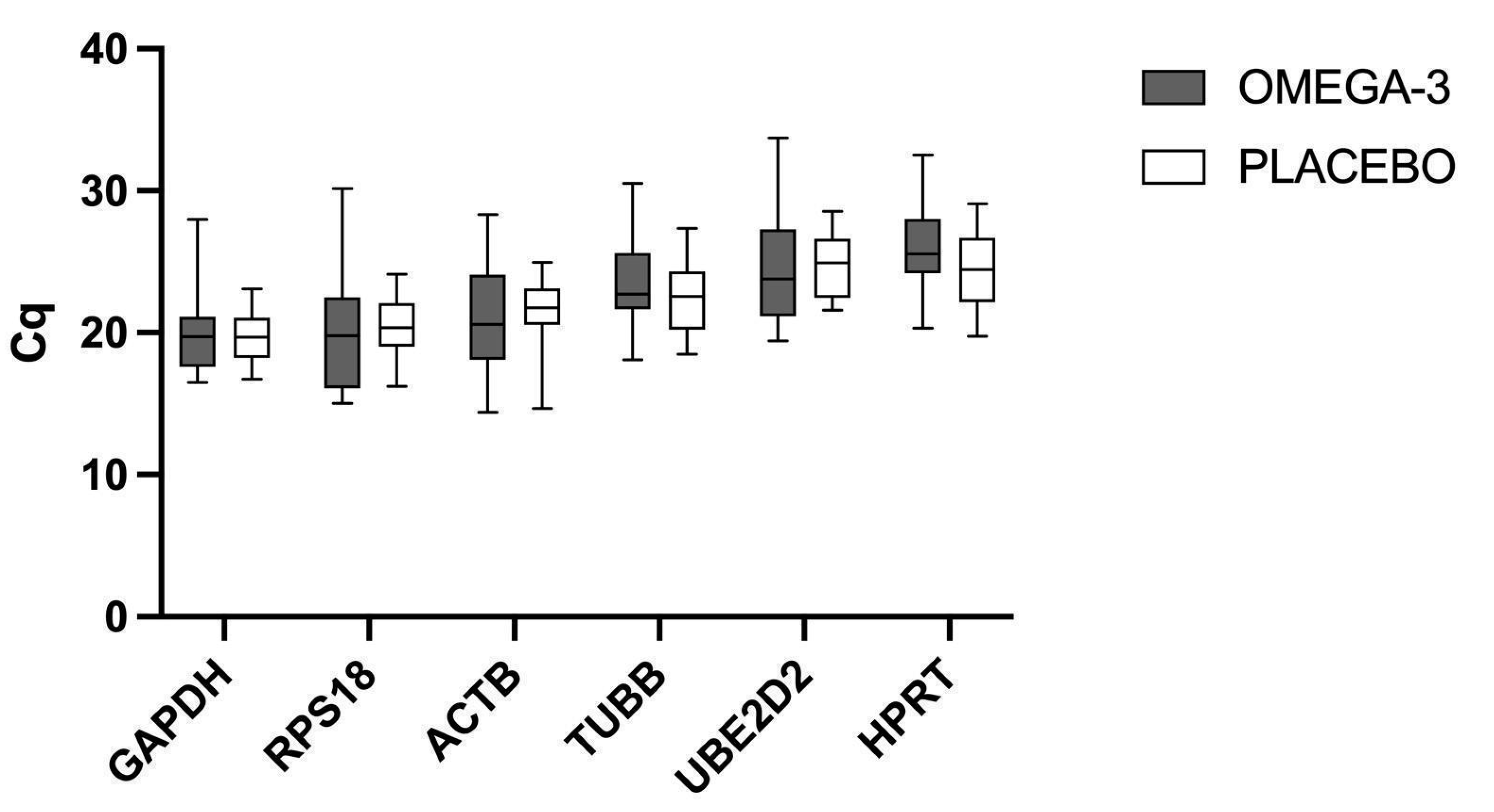
Figure 1. Comparison of Cq values for omega-3 supplemented vs. placebo group. The box plot for each reference gene represents the median, interquartile range, and the upper and lower range of raw Cq values for each experimental group. No statistically significant differences were found between groups.
Ijms 24 06734 g001
Figure 2. A comparison of results obtained via the RefFinder tool. The most stably expressed genes are represented by lower values obtained by three algorithms: NormFinder, BestKeeper, geNorm, and a comprehensive ranking of all algorithms together with the comparative Delta-Ct method using RefFinder.
Ijms 24 06734 g002
Figure 3. Flow diagram of the study process and group sizes.
Ijms 24 06734 g003
Table 1. List of candidate reference genes evaluated in this study. Function information based on data published at the human genome database https://www.genecards.org/ (accessed on 15 November 2022).
Gene SymbolGene Accession NumberNameFunction
ACTBNM_001101Beta actinCytoskeletal protein
GAPDHNM_002046Glyceraldehyde-3-phosphate dehydrogenaseOxidoreductase in glycolysis and gluconeogenesis
RPS18NM_022551.3Ribosomal Protein S18Encodes a ribosomal protein that is a component of the 40S subunit
TUBBNM_001293212.2Tubulin Beta Class IForms a dimer with alpha-tubulin and acts as a structural component of microtubules
UBE2D2NM_003339.3Ubiquitin-conjugating enzyme E2 D2Degradates misfolded, damaged, or short-lived proteins in eukaryotes
HPRT1NM_000194.3Hypoxanthine Phosphoribosyltransferase 1Plays a central role in the generation of purine nucleotides through the purine salvage pathway
Table 2. Primer sequences, amplicon size and source for the sequences for each of the tested genes.
SymbolPrimer SequenceAmplicon SizeSource
ACTBF: GAGAAAATCTGGCACCACACC177Chen et al., 2018 [55]
R: GGATAGCACAGCCTGGATAGCAA
GAPDHF: TCTCCTCTGACTTCAACAGCGAC126Andersen et al., 2004 [17]
R: CCCTGTTGCTGTAGCCAAATTC
RPS18F: GCGGCGGAAAATAGCCTTTG139Spandidos et al., 2010 [56]
R: GATCACACGTTCCACCTCATC
TUBBF: CTAGAACCTGGGACCATGGA191Żychowska et al., 2021 [57]
R: TGCAGGCAGTCACAGCTCT
UBE2D2F: GTACTCTTGTCCATCTGTTCTCTG120Roy et al., 2020 [40]
R: CCATTCCCGAGCTATTCTGTT
HPRT1F: CGAGATGTGATGAAGGAGATGG97Jeon et al., 2019 [41]
R: TGATGTAATCCAGCAGGTCAGC
Disclaimer/Publisher’s Note: The statements, opinions and data contained in all publications are solely those of the individual author(s) and contributor(s) and not of MDPI and/or the editor(s). MDPI and/or the editor(s) disclaim responsibility for any injury to people or property resulting from any ideas, methods, instructions or products referred to in the content.