Communication

MTHFR c.665C>T and c.1298A>C Polymorphisms in Tailoring Personalized Anti-TNF-α Therapy for Rheumatoid Arthritis

1
Medical Genetics Laboratory, Department of Neuroscience and Rehabilitation, University of Ferrara, 44121 Ferrara, Italy
2
Laboratory of Immunorheumatology and Tissue Regeneration, IRCCS Istituto Ortopedico Rizzoli, 40136 Bologna, Italy
3
Medicine and Rheumatology Unit, IRCCS Istituto Ortopedico Rizzoli, 40136 Bologna, Italy
4
Division of Rheumatology, Azienda USL-IRCCS di Reggio Emilia, 42122 Reggio Emilia, Italy
5
University-Hospital of Modena, University of Modena and Reggio Emilia, 41124 Modena, Italy
6
Section of Hematology and Rheumatology, Department of Medical Sciences, University of Ferrara, 44121 Ferrara, Italy
7
Rheumatology Unit, Sant’Anna University Hospital, 44124 Ferrara, Italy
8
University Center for Studies on Gender Medicine, University of Ferrara, 44121 Ferrara, Italy
*
Author to whom correspondence should be addressed.
Int. J. Mol. Sci. 2023, 24(4), 4110; https://doi.org/10.3390/ijms24044110
Submission received: 2 February 2023 / Revised: 14 February 2023 / Accepted: 16 February 2023 / Published: 18 February 2023
(This article belongs to the Section Molecular Genetics and Genomics)

Abstract

:
Rheumatoid arthritis (RA) is an inflammatory autoimmune disease with a prevalence of 1%. Currently, RA treatment aims to achieve low disease activity or remission. Failure to achieve this goal causes disease progression with a poor prognosis. When treatment with first-line drugs fails, treatment with tumor necrosis factor-α (TNF-α) inhibitors may be prescribed to which many patients do not respond adequately, making the identification of response markers urgent. This study investigated the association of two RA-related genetic polymorphisms, c.665C>T (historically referred to as C677T) and c.1298A>C, in the MTHFR gene as response markers to an anti-TNF-α therapy. A total of 81 patients were enrolled, 60% of whom responded to the therapy. Analyses showed that both polymorphisms were associated with a response to therapy in an allele dose-dependent manner. The association for c.665C>T was significant for a rare genotype (p = 0.01). However, the observed opposite trend of association for c.1298A>C was not significant. An analysis revealed that c.1298A>C, unlike c.665C>T, was also significantly associated with the drug type (p = 0.032). Our preliminary results showed that the genetic polymorphisms in the MTHFR gene were associated with a response to anti-TNF-α therapy, with a potential significance for the anti-TNF-α drug type. This evidence suggests a role for one-carbon metabolism in anti-TNF-α drug efficacy and contributes to further personalized RA interventions.

1. Introduction

Rheumatoid arthritis (RA) is an autoimmune and chronic inflammatory disease that symmetrically affects the polyarthritis of small and large joints, and causes joint and periarticular structural damage [1]. RA has a prevalence of 0.4% to 1.3% across the world population [2] and induces significant morbidity with a decreased quality of life and an increased mortality rate [3]. The symptoms of RA vary between early and advanced RA. Early RA—characterized by symptoms such as a flu-like feeling, fatigue, morning stiffness, and swollen and tender joints, which are accompanied by increased levels of C-reactive protein (CRP) and an increased erythrocyte sedimentation rate (ESR) [4]—differs from advanced and insufficiently treated RA, which is characterized by severe manifestations such as pleural effusions, lung disease, vasculitis, lymphomas, atherosclerosis, hematologic abnormalities, keratoconjunctivitis, rheumatic nodules, joint malalignments, motion limitations, and bone and cartilage destruction (reviewed in detail in [5,6,7]).
According to the American College of Rheumatology (ACR) and the European Alliance of Associations for Rheumatology (EULAR), the current therapeutic goal for RA treatment is to achieve remission or a low disease activity [8,9] by following a structured algorithm of add-on and switch-off disease-modifying anti-rheumatic drug (DMARD) therapies [10]. Generally, the treatment is initiated by one of the conventional synthetic DMARDs such as methotrexate, sulfasalazine, hydroxychloroquine, or leflunomide as a monotherapy or a combination therapy [11]. DMARD efficacy varies significantly among RA patients. For example, methotrexate, which is usually prescribed as the first-line drug, is not effective for 30% to 50% of patients [12,13,14,15,16]. If the response to the first-line therapy is not adequate or fails within 3 to 6 months of the initiation, tumor necrosis factor-α (TNF-α) inhibitors, as biologic DMARDs, should be added to the therapy. Moreover, according to the ACR, if early RA patients experience a high disease activity followed by poor prognosis factors, the use of TNF-α inhibitors as an immediate first-line therapy is recommended [17].
Currently, there are five TNF-α inhibitor drugs, which present similar efficacy and safety profiles. These drugs include adalimumab, golimumab, infliximab, certolizumab, and etanercept [17]. Although these TNF-α inhibitor drugs have significantly improved RA treatment, approximately 40% to 44% of patients do not respond to them adequately [18] and might present with complications such as the development of serious adverse effects, including severe infections, malignancies, congestive heart failure, demyelinating disorders, skin reactions, and drug-induced lupus [19] or a reduced efficacy of the therapy due to the immunogenicity of the drug [20]. Therefore, the identification of the response markers for these drugs would provide optimal treatments for both responder and non-responder patients. Efficient pharmacogenetic markers, with the capability of promptly identifying patients with a lower chance to respond to therapy, could facilitate the provision of individually tailored therapies and ultimately prevent the progression of the disease. So far, several single nucleotide polymorphisms (SNPs) in different known loci such as NUBPL (rs2378945), NCTN5 (rs1813443), PLA2G4A (rs12142623 and rs4651370) [21], LINC02549 (rs7767069), LARRC55 (rs717117G) [22], MED15 (rs113878252), MAFB (rs6065221) [23], CD84 (rs6427528 and rs1503860) [24], TNF (rs1800629), EYA4 (rs17301249) [25], PDZD2 (rs1532269) [26], and CCL21 (rs2812378) [27] genes or in unknown loci, including rs4411591, rs7767069, rs1447722, and rs1568885 [21], have been identified, which are associated with a response to anti-TNF treatments in RA. The evidence supports an association between SNPs in the MTHFR genes c.665C>T (rs1801133, historically referred to as c.677C>T or C677T) and c.1298A>C (rs1801131) and the occurrence risk of RA [28,29,30] or the expression of inflammation markers [31,32], conditions during which inflammatory cytokines such as TNF-α, which is the direct target of TNF-α inhibitor drugs, play a fundamental role [33]. However, further studies addressing the association of these polymorphisms with the response to TNF-α inhibitor drugs in RA patients have not been performed. Therefore, in this study, we aimed to investigate the association of these two SNPs with the response to TNF-α inhibitor drugs in RA patients.

2. Results

2.1. Characteristics of Study Subjects and Response to Therapy

After the follow-up period, 81 patients who received anti-TNF-α therapy as a monotherapy or in combination with other phase I drugs (including hydroxychloroquine, corticosteroids, MTX, sulfasalazine, and leflunomide) were selected and included in the further analyses. Patients who responded successfully to the first-line therapy, or who received non-TNF-α-targeting biologic DMARDs, or who did not complete the follow-up period, or with recorded data not passing the quality control were excluded. The average age of these patients was 56.24 ± 12.96 years and 67.9% of them were female. Among the patients, 4 did not respond to the first anti-TNF-α drug choice and received a second anti-TNF-α drug, which increased the record size to 85. Overall, 60% of the patients responded to therapy with an anti-TNF-α drug, and there was no significant difference between the monotherapy or combination therapy groups either with the overall response (GR+MR vs. NR) to the treatment (p = 0.30) or with the degree of response (GR vs. MR, p = 0.46; GR vs. NR, p = 0.98; MR vs. NR, p = 0.12). The characteristics of the patients are summarized in Table 1.
Among the studied parameters, no associations were detected between the response to the anti-TNF-α therapy and parameters such as sex (p = 0.79), age (p = 0.21), exposure to tobacco smoking (p = 0.78), or the presence of RF in serum (p = 0.52) or ACPA (p = 0.70).

2.2. SNP Association Analysis

The genotype distribution of both the c.665C>T and c.1298A>C SNPs were in a Hardy–Weinberg equilibrium; their frequencies are presented in Table 2.
The analyses showed that the c.665C>T genotypes were associated with the response to the anti-TNF-α therapy in an allele dose-dependent manner. The association was significant for the TT homozygous patients, presenting a 7-fold increased chance of responding to anti-TNF-α therapy compared with CC homozygotes (OR = 7.00, 95% CI 1.60–30.80, p = 0.010). The RA cases bearing a CT heterozygous genotype showed a moderate trend towards an association with the response to therapy (OR = 2.14, 95% CI 0.78–5.91, p = 0.14). Regarding the MTHFR c.1298A>C polymorphism, a similar but reversed allele dose-dependent trend of an association with a response to the anti-TNF-α therapy was observed. Among the responders, the AC heterozygous patients showed a trend towards a reverse association with a response to the therapy with TNF-α inhibitors (OR = 0.52, 95% CI 0.21–1.32, p = 0.17) whereas the CC homozygous cases showed a 12-fold reduced chance of responding to therapy compared with AA homozygotes (OR = 0.082, 95% CI 0.009–0.78, p = 0.029) (Figure 1).
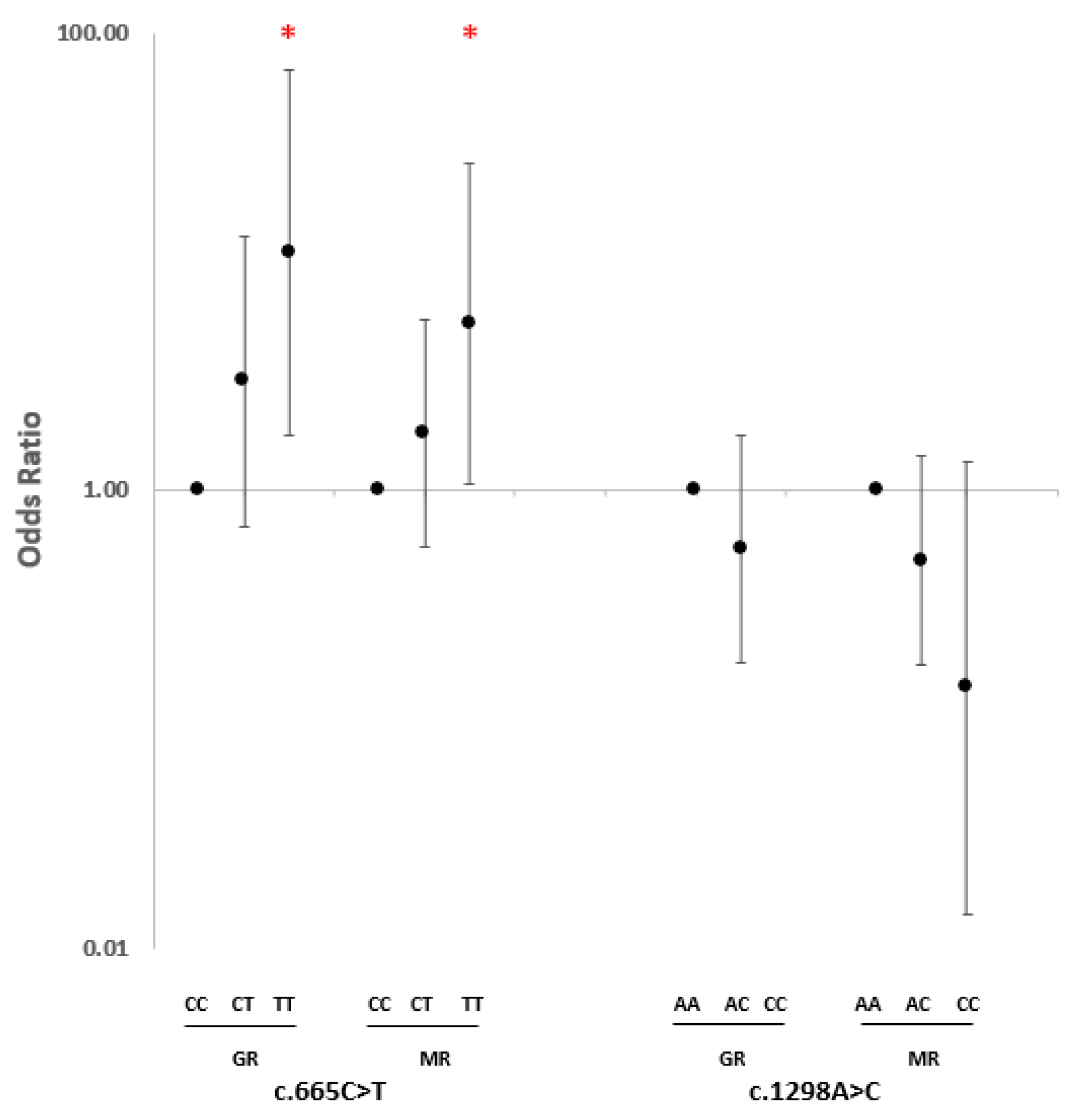
Further analyses among the responders revealed that the association with c.665C>T was stronger in the GR group (CT heterozygous: OR = 3.02, 95% CI 0.70–13.00, p = 0.14; TT homozygous: OR = 10.89, 95% CI 1.73–68.54, p = 0.01) compared with the MR group (CT heterozygous: OR = 1.77, 95% CI 0.56–5.53, p = 0.34; TT homozygous: OR = 5.33, 95% CI 1.07–26.61, p = 0.04). Similarly, the level of response showed a very comparable association with c.1298A>C among the heterozygous subjects (GR: OR = 0.56, 95% CI 0.18–1.73, p = 0.32; MR: OR = 0.50, 95% CI 0.17–1.42, p = 0.19). However, due to the small sample size, it was not possible to compare the level of response in the CC homozygous subjects (Figure 2).
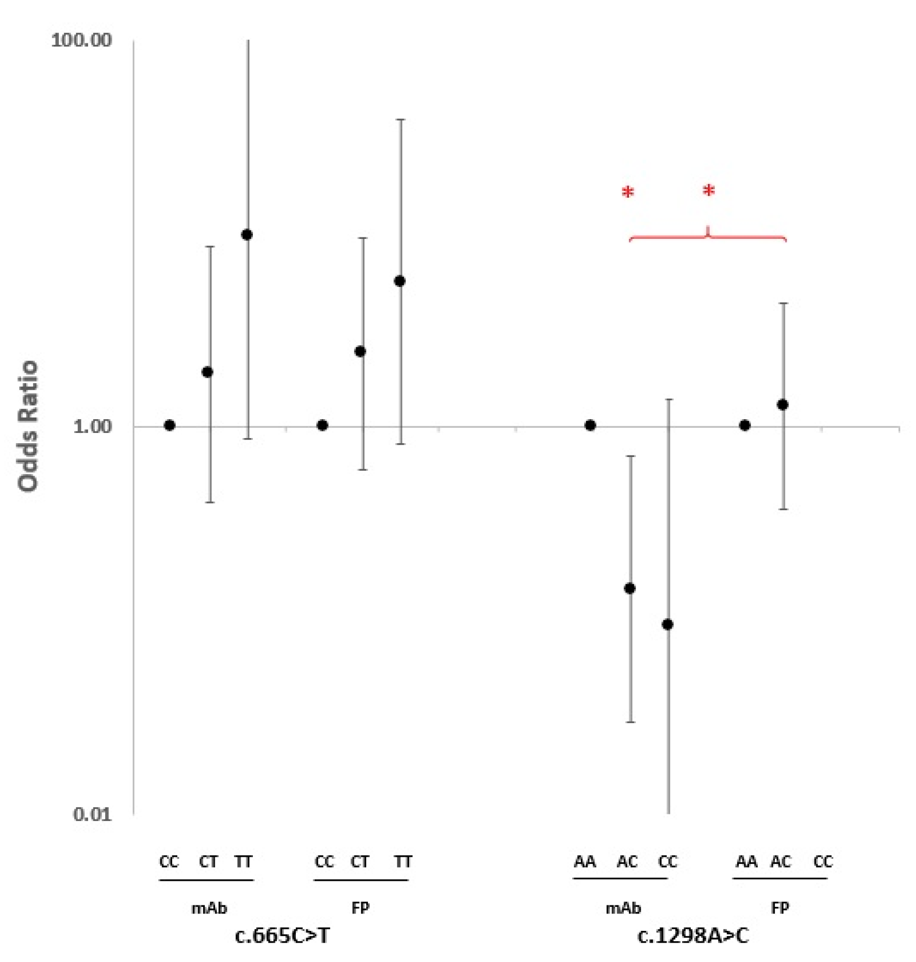
As both MTHFR polymorphisms presented an association with a response to the therapy with anti-TNF-α drugs, we wondered whether the association was influenced by the specific type of inhibitor. As shown in Figure 3, a similar allele dose-dependent trend towards an association of c.665C>T genotypes with the therapy response was observed among the RA cases treated with anti-TNF-α monoclonal antibody (mAb) drugs and in cases treated with an anti-TNF-α fusion protein (FP). However, the stratification of cases according to the type of TNF-α inhibitors provided evidence that among patients with the c.1298AC genotype, the trend towards a reduced response to therapy was restricted only to cases that had been treated with anti-TNF-α mAbs (OR = 0.15, 95% CI 0.03–0.71, p = 0.017). No AC genotype effect was observed in those treated with TNF-α FP (OR = 1.29, 95% CI 0.38–4.39, p = 0.69) (Figure 3). Due to the lack of CC homozygous cases treated with anti-TNF-α FP, it was not possible to determine the influence of the drug type among the CC homozygotes.

3. Discussion

In the present study, we have provided evidence that in patients with RA, the response to anti-TNF-α therapy is influenced by common polymorphisms of the MTHFR gene in an allele dose-dependent manner. Both c.665C>T and c.1298A>C variants have been reported to be associated with RA occurrence [29,30] but, to our knowledge, this is the first evidence of an association between the MTHFR variants and a response to anti-TNF-α drugs.
Notably, the minor allele of the c.665C>T variant was associated with a therapeutic response whereas the c.1298A>C variant was inversely associated. The finding of an opposite effect of the two studied variants was not surprising because of their close physical proximity and the consequent high linkage disequilibrium between them as well as the fact that their minor alleles are very rarely detected on the same chromosome [34].
The MTHFR enzyme catalyzes the synthesis of 5-methyltetrahydrofolate and contributes to the removal of homocysteine by its remethylation to methionine [35]. The C>T substitution at nt 665 determines an alanine-to-valine change that increases the thermolability of the enzyme and impairs the binding of flavin adenine dinucleotide (FAD) and, therefore, causes a reduced catalytic activity [36]. Individuals carrying the c.665TT genotype have an increased need for folate intake in order to maintain adequate concentrations of folate in the serum and red blood cells as well as to avoid increasing the level of total homocysteine (tHcy) in their plasma [37].
The A-to-C transition at nt 1298 results in a glutamate-to-alanine substitution in the regulatory-binding domain of the enzyme, causing the reduced binding of S-adenosyl-methionine (SAM) and leading to a decreased enzyme activity [38], but to a lesser extent than the c.665C>T variant [39].
Both variants are associated with a higher expression of inflammation markers [31,32] and the risk of occurrence of RA [28,29,30]. Although the exact mechanism of the association of these two SNPs with systemic inflammation is not known, possible explanations point to an increased level of tHcy, which is correlated with serum C4, CRP, and the IgM level [40] as well as increased oxidative stress [41,42] and DNA hypomethylation [43]. These explanations mainly refer to c.665TT homozygotes and c.665CT/c.1298AC double heterozygotes [39].
TNF, as the first released cytokine during injuries or stress [44], is secreted by immune cells and plays multiple roles in immunity, inflammation, homeostasis, cell proliferation, and programmed cell death [45]. MTHFR enzyme polymorphisms may unbalance one-carbon metabolism and affect homocysteine levels, DNA methylation, and the cellular redox state; this could have effects on the RA inflammatory scenario in which TNF plays a key role. Therefore, observing the allele dose-dependent response to anti-TNF-α drugs in the c.665C>T polymorphism could be explained by the level of the MTHFR enzyme function and the degree of the consequences of its dysfunctionality in contributing to inflammation, which could be proportional to the level of TNF. We also observed an allele dose-dependent response, although in an opposite direction, to anti-TNF-α drugs in the c.1298A>C polymorphism, which could be explained by the fact that these two polymorphisms have a strong linkage disequilibrium [46]; the T-C haplotype defined by their minor alleles is very rare [34]. In our study, all c.1298CC homozygotes were also c.665CC wild-type homozygotes. Therefore, considering the influence of the c.1298A>C SNP only on the regulatory domain of the enzyme, patients with the CC homozygous genotype for c.1298A>C had a more functional enzyme compared with the patients with TT homozygous for the c.665C>T variant and were probably less affected by an MTHFR dysfunctionality-related inflammatory process.
TNF-α is initially produced in a precursor transmembrane (tmTNF) form on the cell surface; after cleavage by metalloproteinases, it is then released in a mature and soluble (sTNF) form, which mediates its biological activities through TNF-α receptors type 1 and type 2 (TNF-R1 and TNF-R2) [33]. The five common anti-TNF-α drugs are not structurally identical. Adalimumab and golimumab are full human IgG1 mAbs. Infliximab is a chimeric IgG1 mAb. Certolizumab is a PEGylated Fab fragment of IgG1 mAb without an Fc portion. Etanercept is a dimeric fusion protein of TNF-R2 with IgG1 Fc [33]. All of these anti-TNF-α drugs mainly target and neutralize sTNF; however, activities against tmTNF and Fc receptor-expressing cells have been reported [47,48]. In our study, we observed that after stratifying the cases according to the type of anti-TNF-α drug used in the therapy (i.e., mAbs vs. the fusion protein of the TNF receptor), the trend of the response for the c.1298A>C variant was dissimilar to the c.665C>T SNP, with a significant difference between the two groups (p = 0.032). It is known that the distribution, pharmacological half-life, and degradation of anti-TNF-α drugs have several differences [49,50,51]. It has been found that etanercept, as an FP, weakly binds to tmTNF compared with mAbs counterparts [48,52]; unlike mAbs agents, only one etanercept can bind to each molecule of tmTNF, with less efficacy in blocking the downstream activities of tmTNF [53,54]. Considering the observation of an opposite trend of the response to mAb and FP anti-TNF-α drugs for the c.1298AC heterozygotes, it is worth noting that, regardless of the differences in the drug design, it seemed that the efficacy of etanercept (as an FP drug) in comparison with the mAbs agents was being influenced by the c.1298A>C polymorphism, unlike c.665C>T, in the MTHFR gene. If different polymorphisms and genotypes of MTHFR change the enzyme activity differently, it is possible to expect dissimilar consequences such as different contributions to an inflammatory status, which could be inferred by how slightly different drugs can act differently.
This study should be considered to be preliminary, and the results should be carefully interpreted as it was based on a small cohort of patients. In order to have an established concept of the effects of different polymorphisms on the MTHFR gene and their association with drug ADME (absorption, distribution, metabolism, and excretion) as well as the response to different anti-TNF-α therapies in RA patients, a replication study with a larger sample size should be carried out. In addition, considering the strong linkage disequilibrium of these two SNPs, a diplotype analysis is of utmost importance; however, due to the limited number of cases, it was not possible to perform such an analysis.
In conclusion, considering the failure of a phase I therapy in RA patients who have to initiate an anti-TNF-α regime as a phase II therapy, having efficient response-predictive markers such as pharmacogenetic markers could help patients know ahead of time whether the drug is likely to benefit and be safe for them without facing the complications associated with anti-TNF-α drugs. Our study showed that the c.665C>T and c.1298A>C polymorphisms in the MTHFR gene influenced the response to the anti-TNF-α therapy in RA patients, although in opposite directions and with a few differences depending on the drug type. Our results suggest a role of one-carbon metabolism in the efficacy of anti-TNF-α drugs, and may pave the way for more personalized interventions in the treatment of RA. In perspective, the development of an accurate prediction model based on a wide range of pharmacogenetic, metabolic [55], clinical [56], circulating microRNA [57,58], and serological [59] markers—which may influence the treatment response with an additive score significance—could tailor better personalized anti-TNF-α treatments for RA patients.

4. Materials and Methods

4.1. Subjects

A total of 528 RA patients included in the regional RA registry of the Emilia-Romagna region were enrolled from the Rheumatology units of five Italian hospitals, including the Sant’Anna University Hospital, Ferrara; the Rizzoli Orthopaedic Institute, Bologna; the S. Maria Nuova Hospital, Reggio Emilia; the Sant’Orsola-Malpighi University Hospital, Bologna; and the University Hospital, Modena. RA was diagnosed according to the ACR and EULAR criteria of 2010 [60]. A peripheral blood sample was collected from each case before initiating the therapy. For each patient, the anthropometric data (including age, sex, exposure data to tobacco smoking, and serological data for the presence of rheumatoid factor (RF) and anti-citrullinated protein antibodies (APCA)) were collected. Patients were followed-up for 12 months, and the response to the therapy was assessed according to the EULAR response criteria [61,62] and categorized as no response (NR), moderate response (MR), and good response (GR) (Table 3). Written informed consent was obtained from each patient, and the study was reviewed and approved by the ethical board of the Province of Ferrara (16 October 2014).

4.2. DNA Extraction and Genotyping

Genomic DNA was extracted from peripheral blood samples using Nucleon BACC1 (GE Healthcare, Chalfont Saint Giles, UK) or QIAamp DNA Blood Mini (Qiagen, Hilden, Germany) kits, according to the manufacturer’s instructions. The DNA was titrated by a Qubit 2.0 fluorometer (Invitrogen, Singapore) using a Qubit dsDNA BR assay kit (Life Technologies, Carlsbad, CA, USA). The genotyping of the two SNPs in the MTHFR gene (including c.665C>T (rs1801133) and c.1298A>C (rs1801131)) was performed using pre-designed TaqMan 5′ exonuclease assays (Applied Biosystems, Foster City, CA, USA); subsequently, the fluorescence signals of the probes were detected by an ABI 7300 Real-Time PCR System (Applied Biosystems), according to the supplier’s protocol.

4.3. Statistical Analysis

The data were assessed for allele and genotype frequencies and the Hardy–Weinberg equilibrium. The odds ratio (OR) with a 95% confidence interval (CI) was calculated for four different comparisons, including the overall response vs. no response (GR+MR vs. NR), good response vs. no response (GR vs. NR), moderate response vs. no response (MR vs. NR), and good response vs. moderate response (GR vs. MR), assuming a co-dominant genetic model. The analyses were further assessed under dominant and recessive models. A p-value < 0.05 was considered to be statistically significant. All analyses were performed using Microsoft Excel (2016).

Author Contributions

Conceptualization, M.R.; methodology, A.R. and M.R.; formal analysis, A.R. and M.R.; investigation, A.R., L.P., E.A., J.C., R.M., M.G., C.S., and M.R.; resources, M.R. and R.M.; data curation, L.P., A.R., and M.R.; writing—original draft preparation, A.R. and M.R.; supervision, M.R.; funding acquisition, M.R. and R.M. All authors have read and agreed to the published version of the manuscript.

Funding

This research was supported by the University of Ferrara, grant FAR-2021, and by the Italian Ministry of Health 5x1000 fund “anno 2019, redditi 2018” (IRCCS Istituto Ortopedico Rizzoli, Bologna).

Institutional Review Board Statement

The study was conducted according to the guidelines of the Declaration of Helsinki and approved by the Ethics Committee of the Province of Ferrara on 16 October 2014.

Informed Consent Statement

Informed consent was obtained from all subjects involved in the study.

Data Availability Statement

The datasets generated during the study are available from the corresponding author upon reasonable request.

Acknowledgments

This study would not have been possible without the collaboration of all RA patients as well as the medical and nursing staff who took part in the project.

Conflicts of Interest

The authors declare no conflict of interest.

Abbreviations

RA: rheumatoid arthritis; TNF-α: tumor necrosis factor-α; CRP: C-reactive protein; ESR: erythrocyte sedimentation rate; ACR: American College of Rheumatology; EULAR: European Alliance of Associations for Rheumatology; DMARDs: disease-modifying anti-rheumatic drugs; SNPs: single nucleotide polymorphisms; RF: rheumatoid factor; APCA: anti-citrullinated protein antibodies; NR: no response; MR: moderate response; GR: good response; OR: odds ratio; CI: confidence interval; CV: coefficient of variation; SD: standard deviation; mAb: monoclonal antibody; FP: fusion protein; FAD: flavin adenine dinucleotide; tHcy: total homocysteine; SAM: S-adenosyl-methionine; tmTNF: transmembrane TNF; sTNF: soluble TNF; TNF-R1: TNF-α receptor type 1; TNF-R2: TNF-α receptor type 2.

References

  1. Cush, J.J. Rheumatoid Arthritis: Early Diagnosis and Treatment. Med. Clin. N. Am. 2021, 105, 355–365. [Google Scholar] [CrossRef] [PubMed]
  2. Lin, Y.-J.; Anzaghe, M.; Schülke, S. Update on the Pathomechanism, Diagnosis, and Treatment Options for Rheumatoid Arthritis. Cells 2020, 9, 880. [Google Scholar] [CrossRef] [PubMed] [Green Version]
  3. Smolen, J.S.; Aletaha, D.; Barton, A.; Burmester, G.R.; Emery, P.; Firestein, G.S.; Kavanaugh, A.; McInnes, I.B.; Solomon, D.H.; Strand, V.; et al. Rheumatoid Arthritis. Nat. Rev. Dis. Prim. 2018, 4, 1–23. [Google Scholar] [CrossRef]
  4. Brzustewicz, E.; Henc, I.; Daca, A.; Szarecka, M.; Sochocka-Bykowska, M.; Witkowski, J.; Bryl, E. Autoantibodies, C-Reactive Protein, Erythrocyte Sedimentation Rate and Serum Cytokine Profiling in Monitoring of Early Treatment. Cent. Eur. J. Immunol. 2017, 42, 259–268. [Google Scholar] [CrossRef] [PubMed]
  5. Smolen, J.S.; Aletaha, D.; McInnes, I.B. Rheumatoid Arthritis. Lancet 2016, 388, 2023–2038. [Google Scholar] [CrossRef]
  6. Littlejohn, E.A.; Monrad, S.U. Early Diagnosis and Treatment of Rheumatoid Arthritis. Prim. Care 2018, 45, 237–255. [Google Scholar] [CrossRef]
  7. Aletaha, D.; Smolen, J.S. Diagnosis and Management of Rheumatoid Arthritis: A Review. JAMA 2018, 320, 1360–1372. [Google Scholar] [CrossRef]
  8. Smolen, J.S.; Aletaha, D.; Bijlsma, J.W.J.; Breedveld, F.C.; Boumpas, D.; Burmester, G.; Combe, B.; Cutolo, M.; de Wit, M.; Dougados, M.; et al. Treating Rheumatoid Arthritis to Target: Recommendations of an International Task Force. Ann. Rheum. Dis. 2010, 69, 631–637. [Google Scholar] [CrossRef]
  9. Felson, D.T.; Smolen, J.S.; Wells, G.; Zhang, B.; van Tuyl, L.H.D.; Funovits, J.; Aletaha, D.; Allaart, C.F.; Bathon, J.; Bombardieri, S.; et al. American College of Rheumatology/European League against Rheumatism Provisional Definition of Remission in Rheumatoid Arthritis for Clinical Trials. Ann. Rheum. Dis. 2011, 70, 404–413. [Google Scholar] [CrossRef] [Green Version]
  10. Smolen, J.S.; Landewé, R.; Breedveld, F.C.; Buch, M.; Burmester, G.; Dougados, M.; Emery, P.; Gaujoux-Viala, C.; Gossec, L.; Nam, J.; et al. EULAR Recommendations for the Management of Rheumatoid Arthritis with Synthetic and Biological Disease-Modifying Antirheumatic Drugs: 2013 Update. Ann. Rheum. Dis. 2014, 73, 492–509. [Google Scholar] [CrossRef]
  11. Inoue, K.; Yuasa, H. Molecular Basis for Pharmacokinetics and Pharmacodynamics of Methotrexate in Rheumatoid Arthritis Therapy. Drug Metab. Pharmacokinet. 2014, 29, 12–19. [Google Scholar] [CrossRef] [PubMed] [Green Version]
  12. Braun, J.; Kästner, P.; Flaxenberg, P.; Währisch, J.; Hanke, P.; Demary, W.; von Hinüber, U.; Rockwitz, K.; Heitz, W.; Pichlmeier, U.; et al. Comparison of the Clinical Efficacy and Safety of Subcutaneous versus Oral Administration of Methotrexate in Patients with Active Rheumatoid Arthritis: Results of a Six-Month, Multicenter, Randomized, Double-Blind, Controlled, Phase IV Trial. Arthritis Rheum. 2008, 58, 73–81. [Google Scholar] [CrossRef] [PubMed]
  13. Klareskog, L.; van der Heijde, D.; de Jager, J.P.; Gough, A.; Kalden, J.; Malaise, M.; Martín Mola, E.; Pavelka, K.; Sany, J.; Settas, L.; et al. Therapeutic Effect of the Combination of Etanercept and Methotrexate Compared with Each Treatment Alone in Patients with Rheumatoid Arthritis: Double-Blind Randomised Controlled Trial. Lancet 2004, 363, 675–681. [Google Scholar] [CrossRef]
  14. Lima, A.; Bernardes, M.; Azevedo, R.; Medeiros, R.; Seabra, V. Pharmacogenomics of Methotrexate Membrane Transport Pathway: Can Clinical Response to Methotrexate in Rheumatoid Arthritis Be Predicted? Int. J. Mol. Sci. 2015, 16, 13760–13780. [Google Scholar] [CrossRef] [Green Version]
  15. Mori, S.; Hirose, J.; Yonemura, K. Contribution of HLA-DRB1*04 Alleles and Anti-Cyclic Citrullinated Antibodies to Development of Resistance to Disease-Modifying Antirheumatic Drugs in Early Rheumatoid Arthritis. Clin. Rheumatol. 2010, 29, 1357–1366. [Google Scholar] [CrossRef] [PubMed]
  16. Weinblatt, M.E.; Kaplan, H.; Germain, B.F.; Block, S.; Solomon, S.D.; Merriman, R.C.; Wolfe, F.; Wall, B.; Anderson, L.; Gall, E. Methotrexate in Rheumatoid Arthritis. A Five-Year Prospective Multicenter Study. Arthritis Rheum. 1994, 37, 1492–1498. [Google Scholar] [CrossRef]
  17. Radner, H.; Aletaha, D. Anti-TNF in Rheumatoid Arthritis: An Overview. Wien. Med. Wochenschr. 2015, 165, 3–9. [Google Scholar] [CrossRef] [PubMed]
  18. Feldmann, M.; Maini, R.N. Lasker Clinical Medical Research Award. TNF Defined as a Therapeutic Target for Rheumatoid Arthritis and Other Autoimmune Diseases. Nat. Med. 2003, 9, 1245–1250. [Google Scholar] [CrossRef]
  19. Gerriets, V.; Goyal, A.; Khaddour, K. Tumor Necrosis Factor Inhibitors; StatPearls Publishing: Treasure Island, FL, USA, 2022. [Google Scholar]
  20. van Schouwenburg, P.A.; Rispens, T.; Wolbink, G.J. Immunogenicity of Anti-TNF Biologic Therapies for Rheumatoid Arthritis. Nat. Rev. Rheumatol. 2013, 9, 164–172. [Google Scholar] [CrossRef]
  21. Umiċeviċ Mirkov, M.; Cui, J.; Vermeulen, S.H.; Stahl, E.A.; Toonen, E.J.M.; Makkinje, R.R.; Lee, A.T.; Huizinga, T.W.J.; Allaart, R.; Barton, A.; et al. Genome-Wide Association Analysis of Anti-TNF Drug Response in Patients with Rheumatoid Arthritis. Ann. Rheum. Dis. 2013, 72, 1375–1381. [Google Scholar] [CrossRef]
  22. Sánchez-Maldonado, J.M.; Cáliz, R.; López-Nevot, M.Á.; Cabrera-Serrano, A.J.; Moñiz-Díez, A.; Canhão, H.; Ter Horst, R.; Quartuccio, L.; Sorensen, S.B.; Glintborg, B.; et al. Validation of GWAS-Identified Variants for Anti-TNF Drug Response in Rheumatoid Arthritis: A Meta-Analysis of Two Large Cohorts. Front. Immunol. 2021, 12, 672255. [Google Scholar] [CrossRef]
  23. Julià, A.; Marsal, S. Pharmacogenomics of Anti-TNF Response in Psoriasis, Where Are We? Pharmacogenomics 2016, 17, 323–326. [Google Scholar] [CrossRef] [Green Version]
  24. Cui, J.; Stahl, E.A.; Saevarsdottir, S.; Miceli, C.; Diogo, D.; Trynka, G.; Raj, T.; Mirkov, M.U.; Canhao, H.; Ikari, K.; et al. Genome-Wide Association Study and Gene Expression Analysis Identifies CD84 as a Predictor of Response to Etanercept Therapy in Rheumatoid Arthritis. PLoS Genet. 2013, 9, e1003394. [Google Scholar] [CrossRef] [PubMed] [Green Version]
  25. O’Rielly, D.D.; Roslin, N.M.; Beyene, J.; Pope, A.; Rahman, P. TNF-Alpha-308 G/A Polymorphism and Responsiveness to TNF-Alpha Blockade Therapy in Moderate to Severe Rheumatoid Arthritis: A Systematic Review and Meta-Analysis. Pharm. J. 2009, 9, 161–167. [Google Scholar] [CrossRef]
  26. Plant, D.; Bowes, J.; Potter, C.; Hyrich, K.L.; Morgan, A.W.; Wilson, A.G.; Isaacs, J.D.; Wellcome Trust Case Control Consortium; British Society for Rheumatology Biologics Register; Barton, A. Genome-Wide Association Study of Genetic Predictors of Anti-Tumor Necrosis Factor Treatment Efficacy in Rheumatoid Arthritis Identifies Associations with Polymorphisms at Seven Loci. Arthritis Rheum. 2011, 63, 645–653. [Google Scholar] [CrossRef] [Green Version]
  27. Farragher, T.M.; Plant, D.; Flynn, E.; Eyre, S.; Bunn, D.; Thomson, W.; Symmons, D.; Barton, A. Association of a Rheumatoid Arthritis Susceptibility Variant at the CCL21 Locus with Premature Mortality in Inflammatory Polyarthritis Patients. Arthritis Care Res. 2010, 62, 676–682. [Google Scholar] [CrossRef]
  28. Shaker, O.G.; Alnoury, A.M.; Hegazy, G.A.; El Haddad, H.E.; Sayed, S.; Hamdy, A. Methylene Tetrahydrofolate Reductase, Transforming Growth Factor-Β1 and Lymphotoxin-α Genes Polymorphisms and Susceptibility to Rheumatoid Arthritis. Rev. Bras. Reum. Engl. Ed. 2016, 56, 414–420. [Google Scholar] [CrossRef] [Green Version]
  29. Yuan, Y.; Shao, W.; Li, Y. Associations between C677T and A1298C Polymorphisms of MTHFR and Susceptibility to Rheumatoid Arthritis: A Systematic Review and Meta-Analysis. Rheumatol. Int. 2017, 37, 557–569. [Google Scholar] [CrossRef]
  30. Cen, H.; Huang, H.; Zhang, L.-N.; Liu, L.-Y.; Zhou, L.; Xin, X.-F.; Zhuo, R.-J. Associations of Methylenetetrahydrofolate Reductase (MTHFR) C677T and A1298C Polymorphisms with Genetic Susceptibility to Rheumatoid Arthritis: A Meta-Analysis. Clin. Rheumatol. 2017, 36, 287–297. [Google Scholar] [CrossRef] [PubMed]
  31. Khalighi, K.; Cheng, G.; Mirabbasi, S.; Khalighi, B.; Wu, Y.; Fan, W. Opposite Impact of Methylene Tetrahydrofolate Reductase C677T and Methylene Tetrahydrofolate Reductase A1298C Gene Polymorphisms on Systemic Inflammation. J. Clin. Lab. Anal. 2018, 32, e22401. [Google Scholar] [CrossRef] [PubMed] [Green Version]
  32. Vetchinkina, E.A.; Mikhaylenko, D.S.; Kuznetsova, E.B.; Deryagina, T.A.; Alekseeva, E.A.; Bure, I.V.; Zamyatnin, A.A.; Nemtsova, M.V. Genetic Factors of Predisposition and Clinical Characteristics of Rheumatoid Arthritis in Russian Patients. J. Pers. Med. 2021, 11, 469. [Google Scholar] [CrossRef]
  33. Mitoma, H.; Horiuchi, T.; Tsukamoto, H.; Ueda, N. Molecular Mechanisms of Action of Anti-TNF-α Agents—Comparison among Therapeutic TNF-α Antagonists. Cytokine 2018, 101, 56–63. [Google Scholar] [CrossRef]
  34. Ogino, S.; Wilson, R.B. Genotype and Haplotype Distributions of MTHFR677C>T and 1298A>C Single Nucleotide Polymorphisms: A Meta-Analysis. J. Hum. Genet. 2003, 48, 1–7. [Google Scholar] [CrossRef] [Green Version]
  35. Steed, M.M.; Tyagi, S.C. Mechanisms of Cardiovascular Remodeling in Hyperhomocysteinemia. Antioxid. Redox Signal. 2011, 15, 1927–1943. [Google Scholar] [CrossRef] [Green Version]
  36. Fryar-Williams, S. Fundamental Role of Methylenetetrahydrofolate Reductase 677 C → T Genotype and Flavin Compounds in Biochemical Phenotypes for Schizophrenia and Schizoaffective Psychosis. Front. Psychiatry 2016, 7, 172. [Google Scholar] [CrossRef] [Green Version]
  37. Cortese, C.; Motti, C. MTHFR Gene Polymorphism, Homocysteine and Cardiovascular Disease. Public Health Nutr. 2001, 4, 493–497. [Google Scholar] [CrossRef] [Green Version]
  38. Friedman, G.; Goldschmidt, N.; Friedlander, Y.; Ben-Yehuda, A.; Selhub, J.; Babaey, S.; Mendel, M.; Kidron, M.; Bar-On, H. A Common Mutation A1298C in Human Methylenetetrahydrofolate Reductase Gene: Association with Plasma Total Homocysteine and Folate Concentrations. J. Nutr. 1999, 129, 1656–1661. [Google Scholar] [CrossRef] [Green Version]
  39. Weisberg, I.S.; Jacques, P.F.; Selhub, J.; Bostom, A.G.; Chen, Z.; Curtis Ellison, R.; Eckfeldt, J.H.; Rozen, R. The 1298A-->C Polymorphism in Methylenetetrahydrofolate Reductase (MTHFR): In Vitro Expression and Association with Homocysteine. Atherosclerosis 2001, 156, 409–415. [Google Scholar] [CrossRef] [PubMed]
  40. Li, T.; Chen, Y.; Li, J.; Yang, X.; Zhang, H.; Qin, X.; Hu, Y.; Mo, Z. Serum Homocysteine Concentration Is Significantly Associated with Inflammatory/Immune Factors. PLoS ONE 2015, 10, e0138099. [Google Scholar] [CrossRef] [PubMed] [Green Version]
  41. Mititelu, R.R.; Pădureanu, R.; Băcănoiu, M.; Pădureanu, V.; Docea, A.O.; Calina, D.; Barbulescu, A.L.; Buga, A.M. Inflammatory and Oxidative Stress Markers-Mirror Tools in Rheumatoid Arthritis. Biomedicines 2020, 8, 125. [Google Scholar] [CrossRef] [PubMed]
  42. Abd-Elmawla, M.A.; Rizk, S.M.; Youssry, I.; Shaheen, A.A. Impact of Genetic Polymorphism of Methylenetetrahydrofolate Reductase C677T on Development of Hyperhomocysteinemia and Related Oxidative Changes in Egyptian β-Thalassemia Major Patients. PLoS ONE 2016, 11, e0155070. [Google Scholar] [CrossRef] [Green Version]
  43. Bayarsaihan, D. Epigenetic Mechanisms in Inflammation. J Dent Res 2011, 90, 9–17. [Google Scholar] [CrossRef] [PubMed] [Green Version]
  44. Linton, M.F.; Yancey, P.G.; Davies, S.S.; Jerome, W.G.; Linton, E.F.; Song, W.L.; Doran, A.C.; Vickers, K.C. The Role of Lipids and Lipoproteins in Atherosclerosis; MDText.com, Inc.: South Dartmouth, MA, USA, 2019. [Google Scholar]
  45. Popa, C.; Netea, M.G.; van Riel, P.L.C.M.; van der Meer, J.W.M.; Stalenhoef, A.F.H. The Role of TNF-Alpha in Chronic Inflammatory Conditions, Intermediary Metabolism, and Cardiovascular Risk. J. Lipid. Res. 2007, 48, 751–762. [Google Scholar] [CrossRef] [Green Version]
  46. Fan, S.; Yang, B.; Zhi, X.; Wang, Y.; Zheng, Q.; Sun, G. Combined Genotype and Haplotype Distributions of MTHFR C677T and A1298C Polymorphisms: A Cross-Sectional Descriptive Study of 13,473 Chinese Adult Women. Medicine 2016, 95, e5355. [Google Scholar] [CrossRef]
  47. Kaymakcalan, Z.; Sakorafas, P.; Bose, S.; Scesney, S.; Xiong, L.; Hanzatian, D.K.; Salfeld, J.; Sasso, E.H. Comparisons of Affinities, Avidities, and Complement Activation of Adalimumab, Infliximab, and Etanercept in Binding to Soluble and Membrane Tumor Necrosis Factor. Clin. Immunol. 2009, 131, 308–316. [Google Scholar] [CrossRef]
  48. Vos, A.C.W.; Wildenberg, M.E.; Duijvestein, M.; Verhaar, A.P.; van den Brink, G.R.; Hommes, D.W. Anti-Tumor Necrosis Factor-α Antibodies Induce Regulatory Macrophages in an Fc Region-Dependent Manner. Gastroenterology 2011, 140, 221–230. [Google Scholar] [CrossRef] [PubMed]
  49. Suzuki, T.; Ishii-Watabe, A.; Tada, M.; Kobayashi, T.; Kanayasu-Toyoda, T.; Kawanishi, T.; Yamaguchi, T. Importance of Neonatal FcR in Regulating the Serum Half-Life of Therapeutic Proteins Containing the Fc Domain of Human IgG1: A Comparative Study of the Affinity of Monoclonal Antibodies and Fc-Fusion Proteins to Human Neonatal FcR. J. Immunol. 2010, 184, 1968–1976. [Google Scholar] [CrossRef] [Green Version]
  50. Biancheri, P.; Brezski, R.J.; Di Sabatino, A.; Greenplate, A.R.; Soring, K.L.; Corazza, G.R.; Kok, K.B.; Rovedatti, L.; Vossenkämper, A.; Ahmad, N.; et al. Proteolytic Cleavage and Loss of Function of Biologic Agents That Neutralize Tumor Necrosis Factor in the Mucosa of Patients with Inflammatory Bowel Disease. Gastroenterology 2015, 149, 1564–1574.e3. [Google Scholar] [CrossRef]
  51. Palframan, R.; Airey, M.; Moore, A.; Vugler, A.; Nesbitt, A. Use of Biofluorescence Imaging to Compare the Distribution of Certolizumab Pegol, Adalimumab, and Infliximab in the Inflamed Paws of Mice with Collagen-Induced Arthritis. J. Immunol. Methods 2009, 348, 36–41. [Google Scholar] [CrossRef] [PubMed]
  52. Van den Brande, J.M.H.; Braat, H.; van den Brink, G.R.; Versteeg, H.H.; Bauer, C.A.; Hoedemaeker, I.; van Montfrans, C.; Hommes, D.W.; Peppelenbosch, M.P.; van Deventer, S.J.H. Infliximab but Not Etanercept Induces Apoptosis in Lamina Propria T-Lymphocytes from Patients with Crohn’s Disease. Gastroenterology 2003, 124, 1774–1785. [Google Scholar] [CrossRef] [PubMed]
  53. Nesbitt, A.; Fossati, G.; Bergin, M.; Stephens, P.; Stephens, S.; Foulkes, R.; Brown, D.; Robinson, M.; Bourne, T. Mechanism of Action of Certolizumab Pegol (CDP870): In Vitro Comparison with Other Anti-Tumor Necrosis Factor Alpha Agents. Inflamm. Bowel Dis. 2007, 13, 1323–1332. [Google Scholar] [CrossRef] [PubMed]
  54. Scallon, B.; Cai, A.; Solowski, N.; Rosenberg, A.; Song, X.-Y.; Shealy, D.; Wagner, C. Binding and Functional Comparisons of Two Types of Tumor Necrosis Factor Antagonists. J. Pharm. Exp. 2002, 301, 418–426. [Google Scholar] [CrossRef] [PubMed] [Green Version]
  55. Cuppen, B.V.J.; Fu, J.; van Wietmarschen, H.A.; Harms, A.C.; Koval, S.; Marijnissen, A.C.A.; Peeters, J.J.W.; Bijlsma, J.W.J.; Tekstra, J.; van Laar, J.M.; et al. Exploring the Inflammatory Metabolomic Profile to Predict Response to TNF-α Inhibitors in Rheumatoid Arthritis. PLoS ONE 2016, 11, e0163087. [Google Scholar] [CrossRef] [PubMed] [Green Version]
  56. Min, H.K.; Kim, S.H.; Lee, S.-H.; Kim, H.-R. Baseline Bony Erosions and Time-Averaged DAS28 Predict Discontinuation of TNF Inhibitors in Rheumatoid Arthritis. Sci. Rep. 2022, 12, 19951. [Google Scholar] [CrossRef]
  57. Krintel, S.B.; Dehlendorff, C.; Hetland, M.L.; Hørslev-Petersen, K.; Andersen, K.K.; Junker, P.; Pødenphant, J.; Ellingsen, T.; Ahlquist, P.; Lindegaard, H.M.; et al. Prediction of Treatment Response to Adalimumab: A Double-Blind Placebo-Controlled Study of Circulating MicroRNA in Patients with Early Rheumatoid Arthritis. Pharm. J. 2016, 16, 141–146. [Google Scholar] [CrossRef]
  58. Castro-Villegas, C.; Pérez-Sánchez, C.; Escudero, A.; Filipescu, I.; Verdu, M.; Ruiz-Limón, P.; Aguirre, M.A.; Jiménez-Gomez, Y.; Font, P.; Rodriguez-Ariza, A.; et al. Circulating MiRNAs as Potential Biomarkers of Therapy Effectiveness in Rheumatoid Arthritis Patients Treated with Anti-TNFα. Arthritis Res. 2015, 17, 49. [Google Scholar] [CrossRef] [Green Version]
  59. Millier, M.J.; Fanning, N.C.; Frampton, C.; Stamp, L.K.; Hessian, P.A. Plasma Interleukin-23 and Circulating IL-17A+IFNγ+ Ex-Th17 Cells Predict Opposing Outcomes of Anti-TNF Therapy in Rheumatoid Arthritis. Arthritis Res. 2022, 24, 57. [Google Scholar] [CrossRef]
  60. Aletaha, D.; Neogi, T.; Silman, A.J.; Funovits, J.; Felson, D.T.; Bingham, C.O.; Birnbaum, N.S.; Burmester, G.R.; Bykerk, V.P.; Cohen, M.D.; et al. 2010 Rheumatoid Arthritis Classification Criteria: An American College of Rheumatology/European League Against Rheumatism Collaborative Initiative. Ann. Rheum. Dis. 2010, 69, 1580–1588. [Google Scholar] [CrossRef]
  61. van Gestel, A.M.; Prevoo, M.L.; van ’t Hof, M.A.; van Rijswijk, M.H.; van de Putte, L.B.; van Riel, P.L. Development and Validation of the European League Against Rheumatism Response Criteria for Rheumatoid Arthritis. Comparison with the Preliminary American College of Rheumatology and the World Health Organization/International League against Rheumatism Criteria. Arthritis Rheum. 1996, 39, 34–40. [Google Scholar] [CrossRef]
  62. van Gestel, A.M.; Haagsma, C.J.; van Riel, P.L. Validation of Rheumatoid Arthritis Improvement Criteria That Include Simplified Joint Counts. Arthritis Rheum. 1998, 41, 1845–1850. [Google Scholar] [CrossRef]
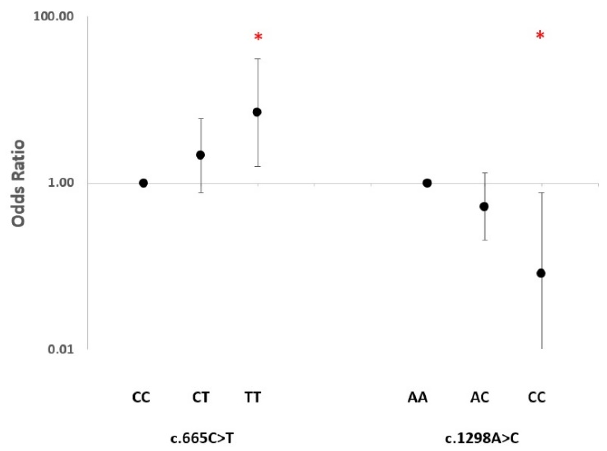
Figure 1. Odds ratio of association of MTHFR c.665C>T and c.1298A>C genotypes with response to anti-TNF-α therapy (GR+MR vs. NR) under a co-dominant model and considering wild-type homozygotes as a reference. * Significant nominal p-value.
Ijms 24 04110 g001
Figure 2. Odds ratio of association of MTHFR c.665C>T and c.1298A>C polymorphisms with the level of response to anti-TNF-α therapy considering wild-type homozygotes as a reference. * Significant nominal p-value. GR: good response; MR: moderate response.
Ijms 24 04110 g002
Figure 3. Odds ratio of association of MTHFR c.665C>T and c.1298A>C genotypes with response to anti-TNF-α drug subtypes considering wild-type homozygotes as a reference. * Significant nominal p-value. mAb: monoclonal antibody; FP: fusion protein.
Ijms 24 04110 g003
Table 1. Characteristics of the patients included in the study.
Variable (n)Stratum(%)
Age (81) *>60 years40.74
Sex (81)Female67.90
Tobacco smoking (81)Current smokers24.69
RF (61)Positive78.69
ACPA (59)Positive74.58
Response (85)
GR (21) 24.71
MR (30) 35.29
NR (34) 40
* Mean = 56.24 years; SD = 12.96 years; CV = 23%.
Table 2. MTHFR c.665C>T and c.1298A>C genotype frequencies of the patients included in the study.
SNP (n)GenotypeFrequency (%)AlleleFrequency (%)
c.665C>T (81)CC
CT
TT
28
51
21
C
T
54
46
c.1298A>C (81)AA
AC
CC
46
48
6
A
C
70
30
Table 3. EULAR therapy response criteria using DAS28.
Present DAS28DAS28 Improvement
>1.2>0.6 and ≤1.2≤0.6
≤3.2Good responseModerate responseNo response
>3.2 and ≤5.1Moderate responseModerate responseNo response
>5.1Moderate responseNo responseNo response
Disclaimer/Publisher’s Note: The statements, opinions and data contained in all publications are solely those of the individual author(s) and contributor(s) and not of MDPI and/or the editor(s). MDPI and/or the editor(s) disclaim responsibility for any injury to people or property resulting from any ideas, methods, instructions or products referred to in the content.