
Acceleration of Electrospun PLA Degradation by Addition of Gelatin

 , , , ,
Abstract
1. Introduction
2. Results
2.1. Morphology
2.2. FTIR Analysis
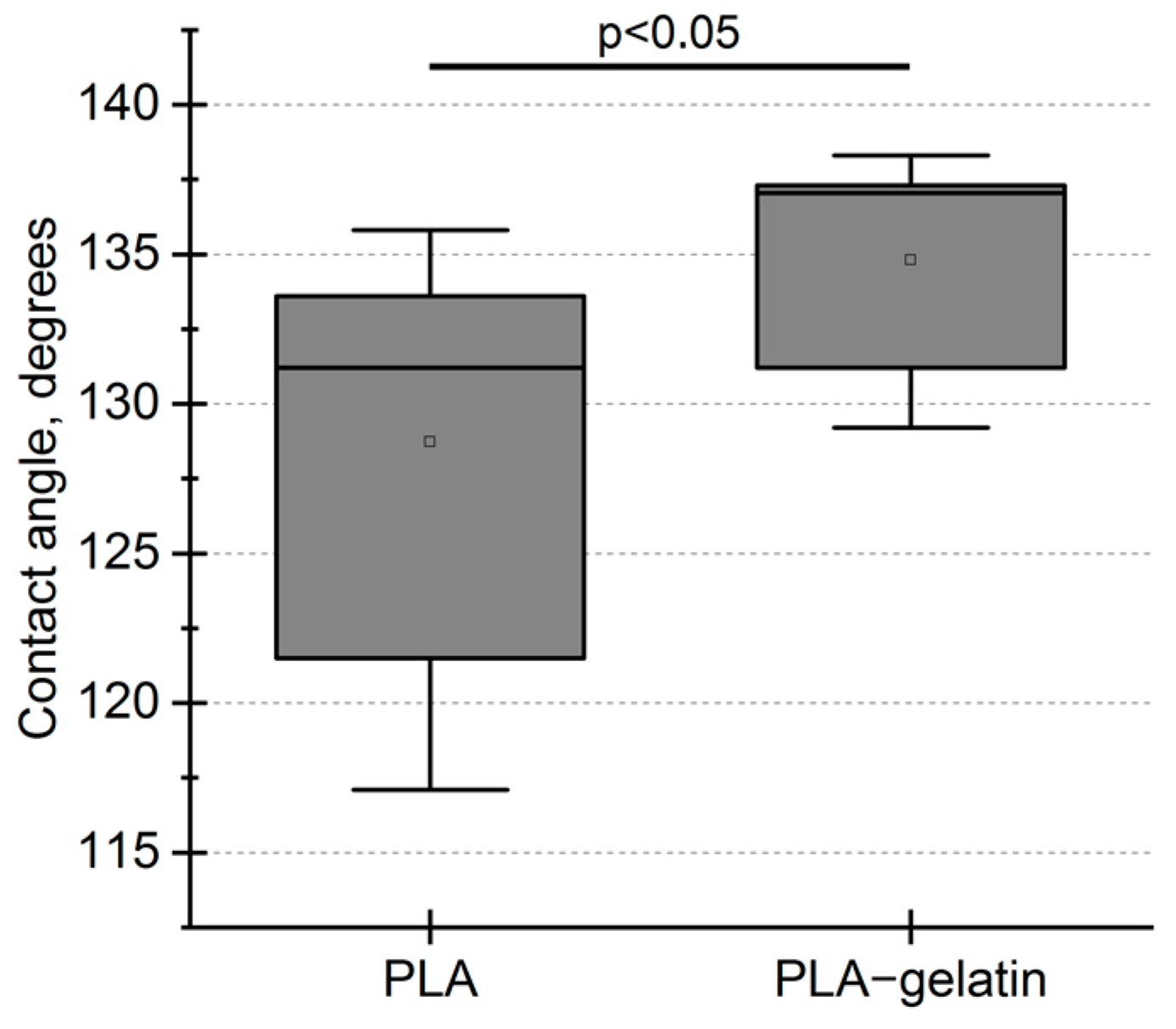
2.3. Wettability
2.4. In Vitro Degradation Analysis
2.5. Mechanical Properties
2.6. Viability of Mammalian Cells Cultured on Electrospun Mats
2.7. In Vivo Biodegradation
3. Discussion
4. Materials and Methods
4.1. Materials
4.2. Electrospinning
4.3. Fourier Transform Infrared (FT-IR) Spectroscopy
4.4. Imaging of Mats Using SEM
4.5. Contact Angle Measurements
4.6. Hydrolytic and Oxidative Degradation
4.7. Mechanical Testing
4.8. Cell Culture
4.9. Analysis of DNA Profiles and Cell Viability
4.10. In Vivo Biodegradation of Electrospun Mats
5. Conclusions
Supplementary Materials
Author Contributions
Funding
Institutional Review Board Statement
Informed Consent Statement
Data Availability Statement
Conflicts of Interest
References
- Ren, K.; Wang, Y.; Sun, T.; Yue, W.; Zhang, H. Electrospun PCL/Gelatin Composite Nanofiber Structures for Effective Guided Bone Regeneration Membranes. Mater. Sci. Eng. C 2017, 78, 324–332. [Google Scholar] [CrossRef] [PubMed]
- Liu, J.; Zhao, B.; Zhang, Y.; Lin, Y.; Hu, P.; Ye, C. PHBV and Predifferentiated Human Adipose-Derived Stem Cells for Cartilage Tissue Engineering. J. Biomed. Mater. Res. A 2010, 94, 603–610. [Google Scholar] [CrossRef] [PubMed]
- Karimi Tar, A.; Karbasi, S.; Naghashzargar, E.; Salehi, H. Biodegradation and Cellular Evaluation of Aligned and Random Poly (3-Hydroxybutyrate)/Chitosan Electrospun Scaffold for Nerve Tissue Engineering Applications. Mater. Technol. 2020, 35, 92–101. [Google Scholar] [CrossRef]
- Li, Y.; Dong, T.; Li, Z.; Ni, S.; Zhou, F.; Alimi, O.A.; Chen, S.; Duan, B.; Kuss, M.; Wu, S. Review of Advances in Electrospinning-Based Strategies for Spinal Cord Regeneration. Mater. Today Chem. 2022, 24, 100944. [Google Scholar] [CrossRef]
- Jimenez-Vergara, A.C.; Guiza-Arguello, V.; Becerra-Bayona, S.; Munoz-Pinto, D.J.; McMahon, R.E.; Morales, A.; Cubero-Ponce, L.; Hahn, M.S. Approach for Fabricating Tissue Engineered Vascular Grafts with Stable Endothelialization. Ann. Biomed. Eng. 2010, 38, 2885–2895. [Google Scholar] [CrossRef]
- Xu, H.; Li, H.; Ke, Q.; Chang, J. An Anisotropically and Heterogeneously Aligned Patterned Electrospun Scaffold with Tailored Mechanical Property and Improved Bioactivity for Vascular Tissue Engineering. ACS Appl. Mater. Interfaces 2015, 7, 8706–8718. [Google Scholar] [CrossRef]
- Hakimi, O.; Mouthuy, P.-A.; Yapp, C.; Wali, A.; Baboldashti, N.Z.; Carr, A. Evaluation of Polydioxanone Scaffolds of Varying Architecture for Tendon Repair. Orthop. Proc. 2014, 96-B, 248. [Google Scholar] [CrossRef]
- Azimi, B.; Maleki, H.; Zavagna, L.; de la Ossa, J.G.; Linari, S.; Lazzeri, A.; Danti, S. Bio-Based Electrospun Fibers for Wound Healing. J. Funct. Biomater. 2020, 11, 67. [Google Scholar] [CrossRef]
- Nedjari, S.; Eap, S.; Hébraud, A.; Wittmer, C.R.; Benkirane-Jessel, N.; Schlatter, G. Electrospun Honeycomb as Nests for Controlled Osteoblast Spatial Organization. Macromol. Biosci. 2014, 14, 1580–1589. [Google Scholar] [CrossRef]
- Maleki, H.; Azimi, B.; Ismaeilimoghadam, S.; Danti, S. Poly(Lactic Acid)-Based Electrospun Fibrous Structures for Biomedical Applications. Appl. Sci. 2022, 12, 3192. [Google Scholar] [CrossRef]
- Teixeira, S.; Eblagon, K.M.; Miranda, F.; R. Pereira, M.F.; Figueiredo, J.L. Towards Controlled Degradation of Poly(Lactic) Acid in Technical Applications. J. Carbon Res. 2021, 7, 42. [Google Scholar] [CrossRef]
- Manavitehrani, I.; Fathi, A.; Badr, H.; Daly, S.; Negahi Shirazi, A.; Dehghani, F. Biomedical Applications of Biodegradable Polyesters. Polymers 2016, 8, 20. [Google Scholar] [CrossRef]
- Ulery, B.D.; Nair, L.S.; Laurencin, C.T. Biomedical Applications of Biodegradable Polymers. J. Polym. Sci. B Polym. Phys. 2011, 49, 832. [Google Scholar] [CrossRef]
- Khorshidi, S.; Solouk, A.; Mirzadeh, H.; Mazinani, S.; Lagaron, J.M.; Sharifi, S.; Ramakrishna, S. A Review of Key Challenges of Electrospun Scaffolds for Tissue-Engineering Applications. J. Tissue Eng. Regen. Med. 2016, 10, 715–738. [Google Scholar] [CrossRef]
- Jun, I.; Han, H.-S.; Edwards, J.R.; Jeon, H. Electrospun Fibrous Scaffolds for Tissue Engineering: Viewpoints on Architecture and Fabrication. Int. J. Mol. Sci. 2018, 19, 745. [Google Scholar] [CrossRef]
- Rim, N.G.; Shin, C.S.; Shin, H. Current Approaches to Electrospun Nanofibers for Tissue Engineering. Biomed. Mater. 2013, 8, 014102. [Google Scholar] [CrossRef]
- Schiffman, J.D.; Schauer, C.L. A Review: Electrospinning of Biopolymer Nanofibers and Their Applications. Polym. Rev. 2008, 48, 317–352. [Google Scholar] [CrossRef]
- Aldana, A.A.; Abraham, G.A. Current Advances in Electrospun Gelatin-Based Scaffolds for Tissue Engineering Applications. Int. J. Pharm. 2017, 523, 441–453. [Google Scholar] [CrossRef]
- Ramírez-Rodríguez, L.C.; Quintanilla-Carvajal, M.X.; Mendoza-Castillo, D.I.; Bonilla-Petriciolet, A.; Jiménez-Junca, C. Preparation and Characterization of an Electrospun Whey Protein/Polycaprolactone Nanofiber Membrane for Chromium Removal from Water. Nanomaterials 2022, 12, 2744. [Google Scholar] [CrossRef]
- Xu, X.; Jiang, L.; Zhou, Z.; Wu, X.; Wang, Y. Preparation and Properties of Electrospun Soy Protein Isolate/Polyethylene Oxide Nanofiber Membranes. ACS Appl. Mater. Interfaces 2012, 4, 4331–4337. [Google Scholar] [CrossRef]
- Khadka, D.B.; Haynie, D.T. Protein- and Peptide-Based Electrospun Nanofibers in Medical Biomaterials. Nanomedicine 2012, 8, 1242–1262. [Google Scholar] [CrossRef] [PubMed]
- Gajić, I.; Stojanović, S.; Ristić, I.; Ilić-Stojanović, S.; Pilić, B.; Nešić, A.; Najman, S.; Dinić, A.; Stanojević, L.; Urošević, M.; et al. Electrospun Poly(Lactide) Fibers as Carriers for Controlled Release of Biochanin A. Pharmaceutics 2022, 14, 528. [Google Scholar] [CrossRef] [PubMed]
- Azizi, M.; Azimzadeh, M.; Afzali, M.; Alafzadeh, M.; Mirhosseini, H.S. Characterization and Optimization of Using Calendula Officinalis Extract in The Fabrication of Polycaprolactone/Gelatin Electrospun Nanofibers for Wound Dressing Applications. J. Adv. Mater. Process. 2018, 6, 34–46. [Google Scholar]
- Bazmandeh, A.Z.; Mirzaei, E.; Fadaie, M.; Shirian, S.; Ghasemi, Y. Dual Spinneret Electrospun Nanofibrous/Gel Structure of Chitosan-Gelatin/Chitosan-Hyaluronic Acid as a Wound Dressing: In-Vitro and in-Vivo Studies. Int. J. Biol. Macromol. 2020, 162, 359–373. [Google Scholar] [CrossRef]
- Casalini, T.; Rossi, F.; Castrovinci, A.; Perale, G. A Perspective on Polylactic Acid-Based Polymers Use for Nanoparticles Synthesis and Applications. Front. Bioeng. Biotechnol. 2019, 7, 259. [Google Scholar] [CrossRef]
- Elmowafy, E.M.; Tiboni, M.; Soliman, M.E. Biocompatibility, Biodegradation and Biomedical Applications of Poly(Lactic Acid)/Poly(Lactic-Co-Glycolic Acid) Micro and Nanoparticles. J. Pharm. Investig. 2019, 49, 347–380. [Google Scholar] [CrossRef]
- Ghasemi-Mobarakeh, L.; Prabhakaran, M.P.; Morshed, M.; Nasr-Esfahani, M.-H.; Ramakrishna, S. Electrospun Poly(ε-Caprolactone)/Gelatin Nanofibrous Scaffolds for Nerve Tissue Engineering. Biomaterials 2008, 29, 4532–4539. [Google Scholar] [CrossRef]
- Gautam, S.; Dinda, A.K.; Mishra, N.C. Fabrication and Characterization of PCL/Gelatin Composite Nanofibrous Scaffold for Tissue Engineering Applications by Electrospinning Method. Mater. Sci. Eng. C 2013, 33, 1228–1235. [Google Scholar] [CrossRef]
- Stachewicz, U.; Bailey, R.J.; Zhang, H.; Stone, C.A.; Willis, C.R.; Barber, A.H. Wetting Hierarchy in Oleophobic 3D Electrospun Nanofiber Networks. ACS Appl. Mater. Interfaces 2015, 7, 16645–16652. [Google Scholar] [CrossRef]
- Ma, M.; Mao, Y.; Gupta, M.; Gleason, K.K.; Rutledge, G.C. Superhydrophobic Fabrics Produced by Electrospinning and Chemical Vapor Deposition. Macromolecules 2005, 38, 9742–9748. [Google Scholar] [CrossRef]
- Szewczyk, P.K.; Ura, D.P.; Metwally, S.; Knapczyk-Korczak, J.; Gajek, M.; Marzec, M.M.; Bernasik, A.; Stachewicz, U. Roughness and Fiber Fraction Dominated Wetting of Electrospun Fiber-Based Porous Meshes. Polymers 2018, 11, 34. [Google Scholar] [CrossRef]
- Bagrov, D.; Perunova, S.; Pavlova, E.; Klinov, D. Wetting of Electrospun Nylon-11 Fibers and Mats. RSC Adv. 2021, 11, 11373–11379. [Google Scholar] [CrossRef]
- Pavlova, E.; Nikishin, I.; Bogdanova, A.; Klinov, D.; Bagrov, D. The Miscibility and Spatial Distribution of the Components in Electrospun Polymer-Protein Mats. RSC Adv. 2020, 10, 4672–4680. [Google Scholar] [CrossRef]
- Bagrov, D.V.; Nikishin, I.I.; Pavlova, E.R.; Klinov, D.V. Distribution of Polylactide and Gelatin in Single Electrospun Nanofibers Studied by Raman Spectroscopy; AIP Publishing LLC: Melville, NY, USA, 2019; Volume 2064, p. 040001. [Google Scholar]
- Yao, R.; He, J.; Meng, G.; Jiang, B.; Wu, F. Electrospun PCL/Gelatin Composite Fibrous Scaffolds: Mechanical Properties and Cellular Responses. J. Biomater. Sci. Polym. Ed. 2016, 27, 824–838. [Google Scholar] [CrossRef]
- Mohammadzadehmoghadam, S.; Dong, Y. Fabrication and Characterization of Electrospun Silk Fibroin/Gelatin Scaffolds Crosslinked with Glutaraldehyde Vapor. Front. Mater. 2019, 6, 91. [Google Scholar] [CrossRef]
- Perez-Puyana, V.; Wieringa, P.; Yuste, Y.; de la Portilla, F.; Guererro, A.; Romero, A.; Moroni, L. Fabrication of Hybrid Scaffolds Obtained from Combinations of PCL with Gelatin or Collagen via Electrospinning for Skeletal Muscle Tissue Engineering. J. Biomed. Mater. Res. Part A 2021, 109, 1600–1612. [Google Scholar] [CrossRef]
- Shamsah, A.; Cartmell, S.; Richardson, S.; Bosworth, L. Material Characterization of PCL:PLLA Electrospun Fibers Following Six Months Degradation In Vitro. Polymers 2020, 12, 700. [Google Scholar] [CrossRef]
- Azevedo, H.S.; Reis, R.L. Understanding the Enzymatic Degradation of Biodegradable Polymers and Strategies to Control Their Degradation Rate; CRC Press: Boca Raton, FL, USA, 2005. [Google Scholar]
- Elsawy, M.A.; Kim, K.-H.; Park, J.-W.; Deep, A. Hydrolytic Degradation of Polylactic Acid (PLA) and Its Composites. Renew. Sustain. Energy Rev. 2017, 79, 1346–1352. [Google Scholar] [CrossRef]
- Lee, S.H.; Song, W.S. Enzymatic Hydrolysis of Polylactic Acid Fiber. Appl. Biochem. Biotechnol. 2011, 164, 89–102. [Google Scholar] [CrossRef]
- Lee, S.H.; Kim, I.Y.; Song, W.S. Biodegradation of Polylactic Acid (PLA) Fibers Using Different Enzymes. Macromol. Res. 2014, 22, 657–663. [Google Scholar] [CrossRef]
- Ratnikov, B.; Deryugina, E.; Leng, J.; Marchenko, G.; Dembrow, D.; Strongin, A. Determination of Matrix Metalloproteinase Activity Using Biotinylated Gelatin. Anal. Biochem. 2000, 286, 149–155. [Google Scholar] [CrossRef] [PubMed]
- Schindelin, J.; Arganda-Carreras, I.; Frise, E.; Kaynig, V.; Longair, M.; Pietzsch, T.; Preibisch, S.; Rueden, C.; Saalfeld, S.; Schmid, B.; et al. Fiji: An Open-Source Platform for Biological-Image Analysis. Nat. Methods 2012, 9, 676–682. [Google Scholar] [CrossRef] [PubMed]


| Parameter | State | PLA | PLA–Gelatin (9:1) |
|---|---|---|---|
| Elongation at break, % | Dry | 56 ± 16 | 22 ± 10 |
| Wet | 44 ± 12 | 45 ± 7 | |
| Maximum stress, MPa | Dry | 3.5 ± 0.4 | 4.4 ± 0.4 |
| Wet | 3.4 ± 0.3 | 3.9 ± 0.4 |
Disclaimer/Publisher’s Note: The statements, opinions and data contained in all publications are solely those of the individual author(s) and contributor(s) and not of MDPI and/or the editor(s). MDPI and/or the editor(s) disclaim responsibility for any injury to people or property resulting from any ideas, methods, instructions or products referred to in the content. |
© 2023 by the authors. Licensee MDPI, Basel, Switzerland. This article is an open access article distributed under the terms and conditions of the Creative Commons Attribution (CC BY) license (https://creativecommons.org/licenses/by/4.0/).
Share and Cite
Bogdanova, A.; Pavlova, E.; Polyanskaya, A.; Volkova, M.; Biryukova, E.; Filkov, G.; Trofimenko, A.; Durymanov, M.; Klinov, D.; Bagrov, D. Acceleration of Electrospun PLA Degradation by Addition of Gelatin. Int. J. Mol. Sci. 2023, 24, 3535. https://doi.org/10.3390/ijms24043535
Bogdanova A, Pavlova E, Polyanskaya A, Volkova M, Biryukova E, Filkov G, Trofimenko A, Durymanov M, Klinov D, Bagrov D. Acceleration of Electrospun PLA Degradation by Addition of Gelatin. International Journal of Molecular Sciences. 2023; 24(4):3535. https://doi.org/10.3390/ijms24043535
Chicago/Turabian StyleBogdanova, Alexandra, Elizaveta Pavlova, Anna Polyanskaya, Marina Volkova, Elena Biryukova, Gleb Filkov, Alexander Trofimenko, Mikhail Durymanov, Dmitry Klinov, and Dmitry Bagrov. 2023. "Acceleration of Electrospun PLA Degradation by Addition of Gelatin" International Journal of Molecular Sciences 24, no. 4: 3535. https://doi.org/10.3390/ijms24043535
APA StyleBogdanova, A., Pavlova, E., Polyanskaya, A., Volkova, M., Biryukova, E., Filkov, G., Trofimenko, A., Durymanov, M., Klinov, D., & Bagrov, D. (2023). Acceleration of Electrospun PLA Degradation by Addition of Gelatin. International Journal of Molecular Sciences, 24(4), 3535. https://doi.org/10.3390/ijms24043535