Article

The Expression Pattern of Insulin-Like Growth Factor Subtype 3 (igf3) in the Orange-Spotted Grouper Epinephelus coioides and Its Function on Ovary Maturation

1
College of Marine Sciences, South China Agricultural University, Guangzhou 510642, China
2
Zhongshan Innovation Center, South China Agricultural University, Zhongshan 528400, China
*
Authors to whom correspondence should be addressed.
These authors contributed equally to this work.
Int. J. Mol. Sci. 2023, 24(3), 2868; https://doi.org/10.3390/ijms24032868
Submission received: 12 November 2022 / Revised: 13 January 2023 / Accepted: 18 January 2023 / Published: 2 February 2023
(This article belongs to the Section Biochemistry)

Abstract

:
A new insulin-like growth factor (Igf) subtype 3 (igf3) has recently been found in the bony fish orange-spotted grouper (Epinephelus coioides). However, the role of igf3 in the maturation of the ovary and sex differentiation in E. coioides is currently unknown. We examined the ovarian localization and receptor binding of the novel ortholog Igf3 using qRT-PCR, and Western blotting, combined with in situ hybridization and immunohistochemistry methods. Results demonstrated the presence of igf3 mRNA and protein in mature oocytes. Furthermore, Igf3 protein expression was not detected in testis, brain, kidney and liver homogenates. The calculated molecular weight of Igf3 was 22 kDa, which was consistent with the deduced amino acid sequence from the full-length open reading frame. The immunoreactivity showed that Igf3 was strongly present in the follicle staining fully-grown stage. The igf3 mRNA expression level was significantly positively correlated with ovarian follicular maturation. Meanwhile, Igf3 increased germinal-vesicle breakdown in a time- and dose-dependent manner. In vitro, treatment of primary ovarian cells with Igf3 up-regulated significantly the mRNA expression level of genes related to sex determination and reproduction such as forkhead boxl2 (foxl2), dosage-sensitive sex reversal adrenal hypoplasia critical region on chromosome x gene 1 (dax1), cytochrome P450 family 19 subfamily member 1 a (cyp19a1a), cytochrome P450 family 11 subfamily a member 1 a (cyp11a1a) and luteinizing hormone receptor 1 (lhr1). Overall, our results demonstrated that igf3 promotes the maturation of the ovary and plays an important role in sex differentiation in E. coioides.

1. Introduction

The insulin-like growth factor (Igf) system is an evolutionary-conserved complex that regulates cell proliferation in a variety of contexts, including growth, proliferation, survival, migration, and differentiation [1,2,3,4,5,6]. Previous studies on the functions of the Igf system in fish mainly focused on Igf1 and Igf2 [7,8,9,10,11,12,13]. Igf1 and Igf2 have been identified in fish ovaries. Indeed, igf1 expression has been reported in granulosa cells, yolk, and mature ovarian membrane cells during the initial stage of gonadal development. The igf2 mRNA and protein are localized to the granulosa cell layer of the late follicular [14,15,16]. However, igf1 and igf2 mRNA are also found in other organs, from unfertilized eggs to adult fish. The functional analysis of Igf1 and Igf2 revealed that the two genes promote growth and maturation in killifish (Fundulus grandis), short-finned eel (Anguilla australis), zebrafish (Danio rerio) and striped bass (Lateolabrax japonicas) [17,18,19,20]. Therefore, Igf1 and Igf2 are important enzymes in the reproduction and growth of fish.
Besides Igf1 and Igf2, fish also contain igf3 as an important enzyme in reproduction and growth. Furthermore, igf3 has been found in tilapia (Oreochromis niloticus), D. rerio, E. coioides and common carp (Cyprinus carpio) [1,8,21,22]. In D. rerio, the igf3 mRNA level was relatively low in early follicular but higher in mature follicular [21]. Functional analyses revealed that igf3 promotes the development, reproduction and migration of germ cells [21,23,24]. Accordingly, incubating oocytes using Igf3 promoted germinal vesicle breakdown (GVBD) and ovarian blastocyst rupture [21]. Moreover, in an in vitro study, the synthesis and expression of Igf3 were up-regulated when human chorionic gonadotropin (hCG) was added to the follicle [17,21]. Furthermore, Igf3 mRNA level increased in D. rerio both in oocytes and follicular, suggesting its potential role in the regulation of gonadal differentiation and ovulation cycle [21]. These studies demonstrate that Igf3 plays an important role in fish reproduction.
Igf3 regulates several genes involved in ovary maturation. Accordingly, treating ovarian and testicular primary cells with Igf3 up-regulated the expression of forkhead boxl2 (foxl2), dosage-sensitive sex reversal adrenal hypoplasia critical region on chromosome x gene 1 (dax1), splicing-transcription factor 1 (sf1), cytochrome P450 family 19 subfamily member 1 a (cyp19a1a), cytochrome P450 family 11 subfamily a member 1 a (cyp11a1) as well as cytochrome P450 family 11 subfamily b member 2 (cyp11b2) in a time- and dose-dependent manner in O. niloticus [2]. However, studies describing the precise role of Igf3 in the regulation of gonadal differentiation are currently limited [21]. Consequently, little direct evidence exists on the role of Igf3 in regulating ovary maturation. The scarcity of information limits our understanding of the role of Igf3 in various fish species.
Therefore, we examined the expression pattern of igf3 mRNA and protein orange-spotted grouper E. coioides by using five stages of follicular. The effect of Igf3 on ovarian maturation was studied by in vitro incubation of Igf3 protein and its regulatory role on genes related to sex determination and reproduction such as foxl2, dax1, cyp19a1a, cyp11a1 and cyp11b2. Furthermore, we examined the role of igf3 on reproductive development and sex determination in E. coioides. The obtained results provide a reference for understanding the role of igf3 in maturation regulation in fish.

2. Results

2.1. Distribution of igf3 mRNA in the Developing Ovary

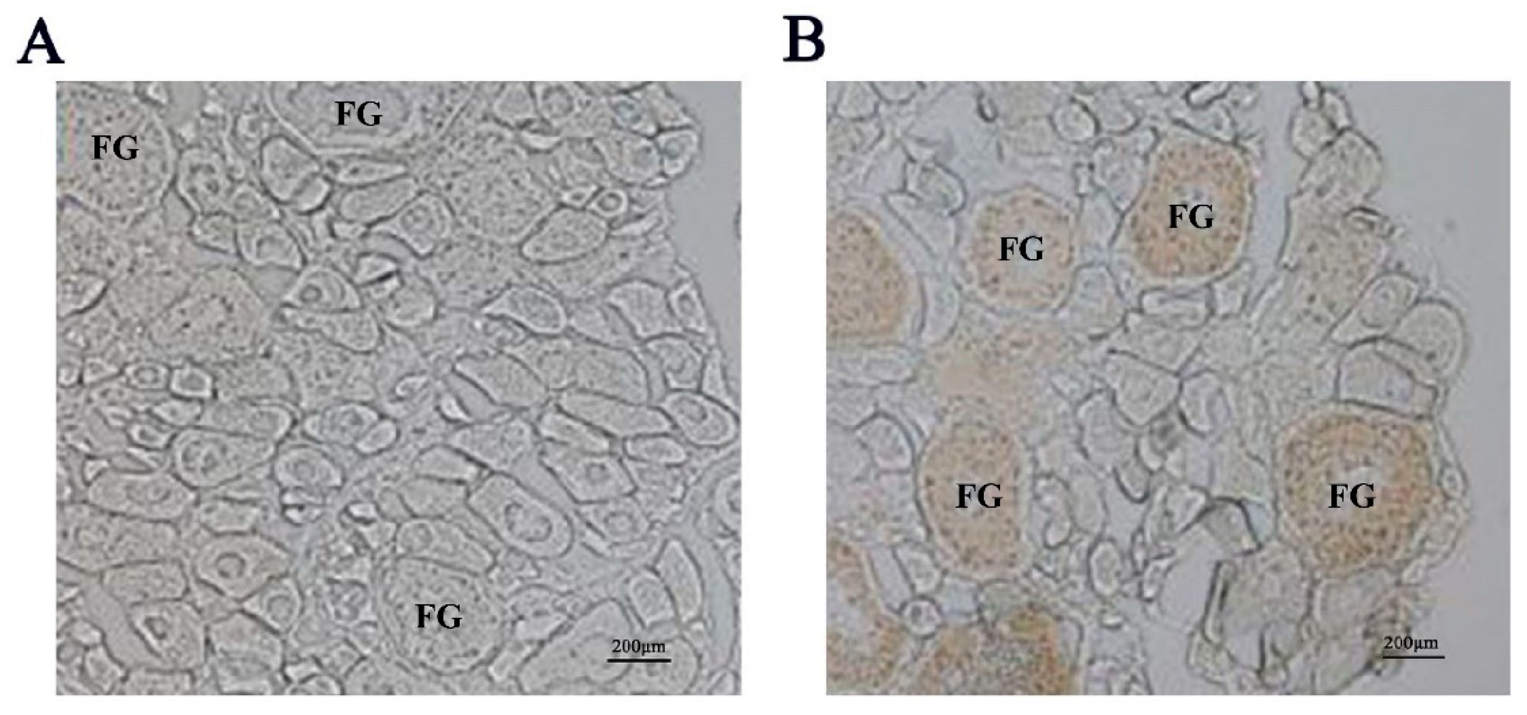
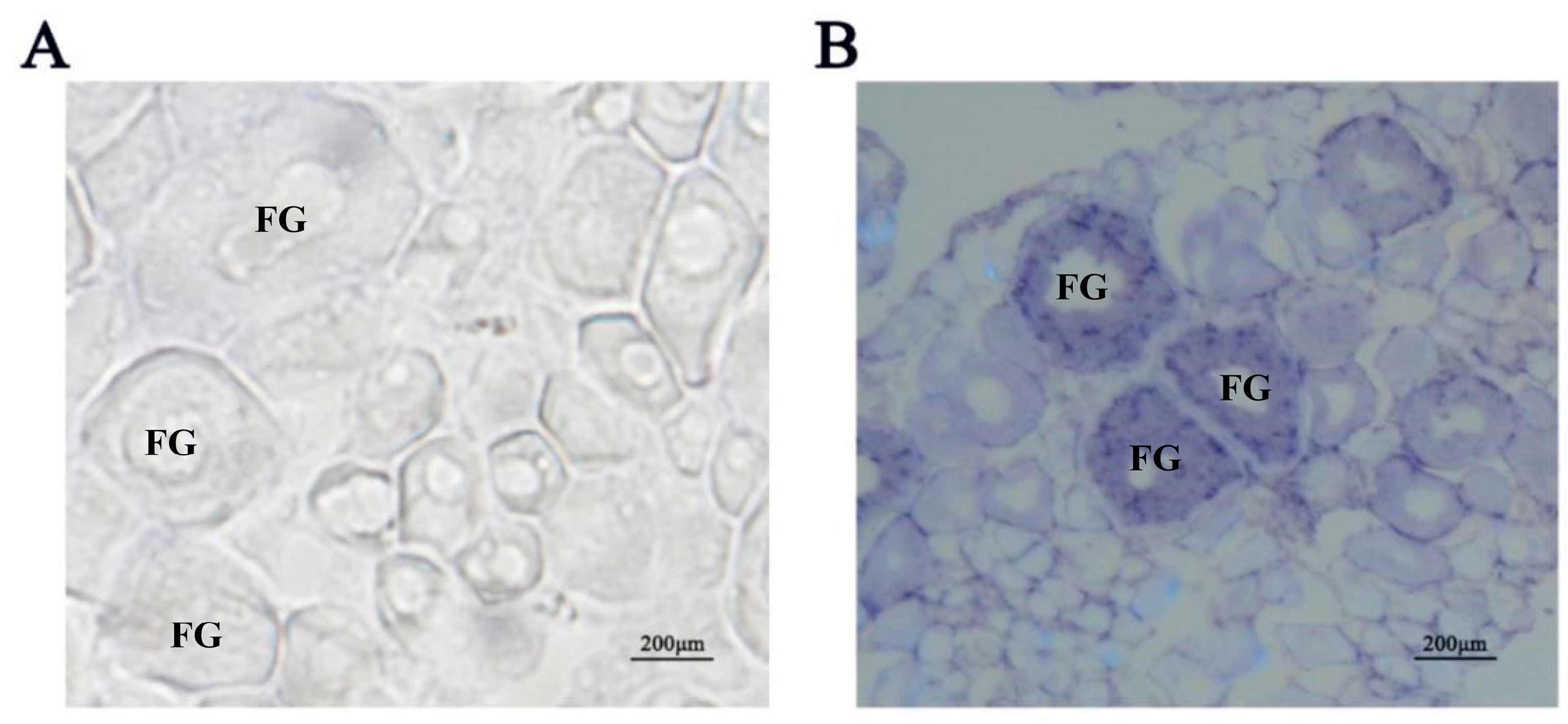
We initially detected expression of the Igf3 protein in ovary tissue homogenates by Western blot analysis. In contrast, we could not detect the presence of Igf3 in testis, brain, kidney and liver homogenates (Figure 1 and Figure S1). The calculated molecular weight of Igf3 was 22 kDa, which was consistent with the deduced amino acid sequence from the full-length ORF of Igf3. This antibody was further used for the immunohistochemistry experiment. We observed signals only in the presence of the Igf3-specific antibody. The strong immunoreactivity showed that the follicle staining in the FG stage was the strongest (Figure 2). We also examined igf3 mRNA expression in frozen sections of E. coioides ovaries. Tissues treated with sense RNA probes lacked staining, indicating that our probes were specific for igf3 mRNA (Figure 3A). We detected positive signals in follicular FG and MV stages, with FG stage follicular staining strong, but early stages had weak signals (Figure 3B).

2.2. Expression of igf3 mRNA in Different Stages of Ovarian Follicular

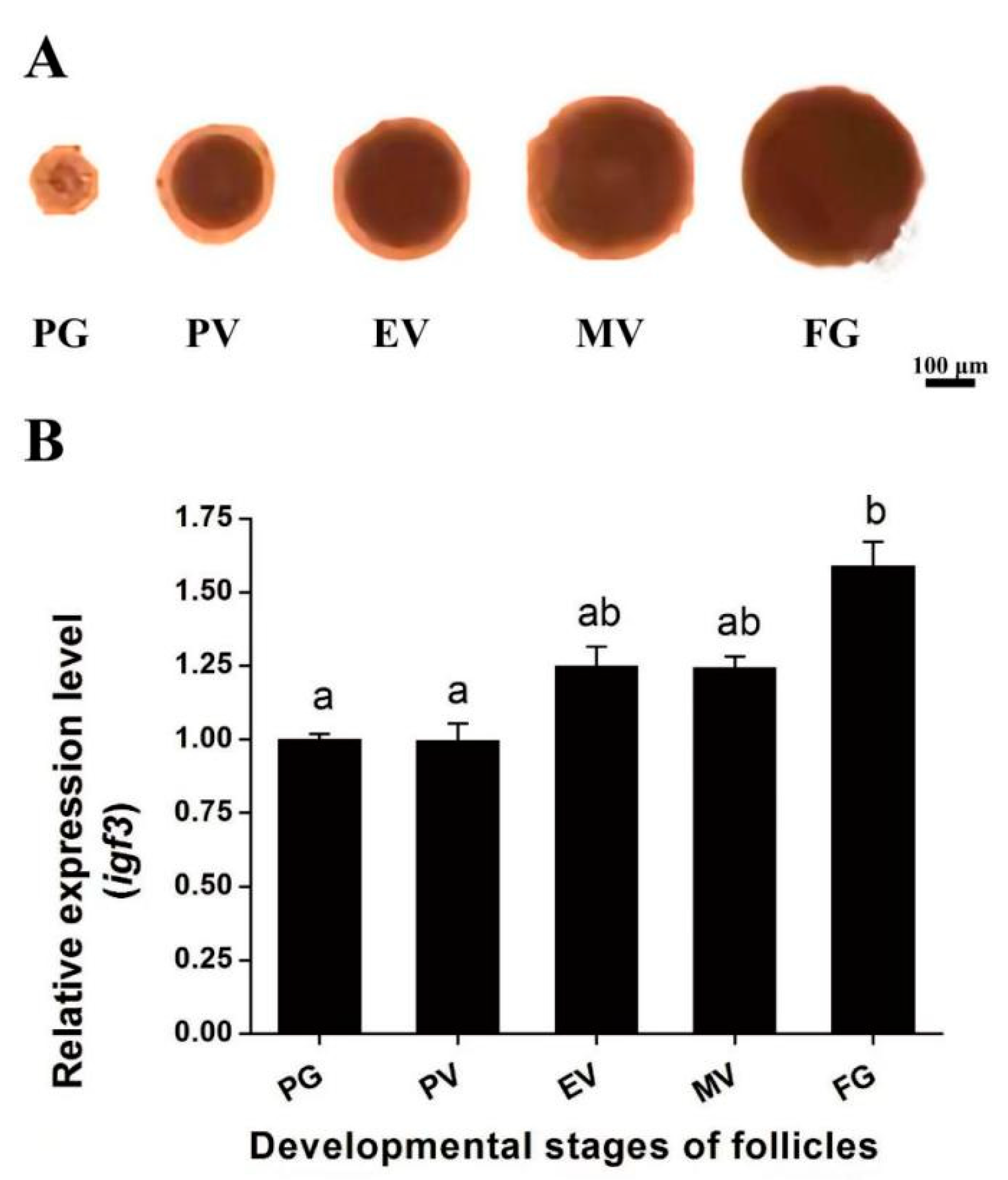
Ovarian follicular were examined at different stages of development, and we followed the accumulation of igf3 mRNA as development proceeded through the PG, PV, EV, MV and FG stages. The igf3 steady-state mRNA level correlated well with this progression (Figure 4).

2.3. Igf3 Promotes the Development and Maturation of Follicular

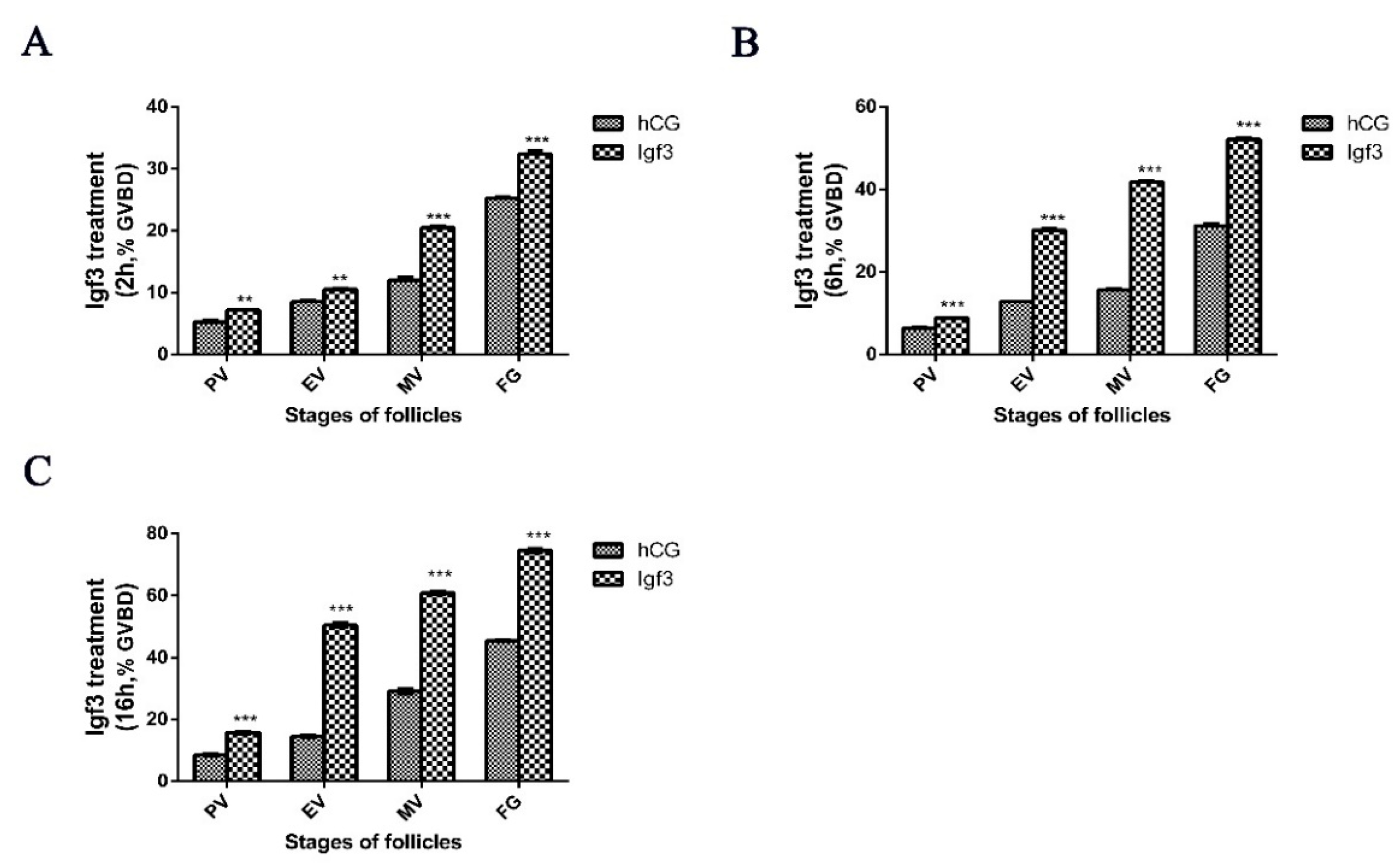
We utilized recombinant Igf3 to determine whether this could alter ovarian follicle development when added externally to follicular at the MV and FG phases of development. At a low (2 ng/mL) Igf3 level, the GVBD of follicular in neither MV nor FG significantly changed over time. At higher (20 ng/mL) concentration, the GVBD of MV phase cells was significantly up-regulated after 16 h, and for the FG phase, this occurred after 6 and 16 h. Under high level (200 ng/mL) Igf3, GVBD of follicular in MV and FG phases reached levels of significance at all three treatment times (Figure 5). Therefore, we extended the experiment and treated and examined the level of GVBD of follicular in the PV, EV, MV and FG phases by using 200 ng/mL Igf3. In all the developmental phases, Igf3 induced GVBD. However, the effects were more pronounced in the more mature follicular and as the exposure time increased (Figure 6).

2.4. Effects of Igf3 on the Expression of Genes Related to Sex Determination and Reproduction

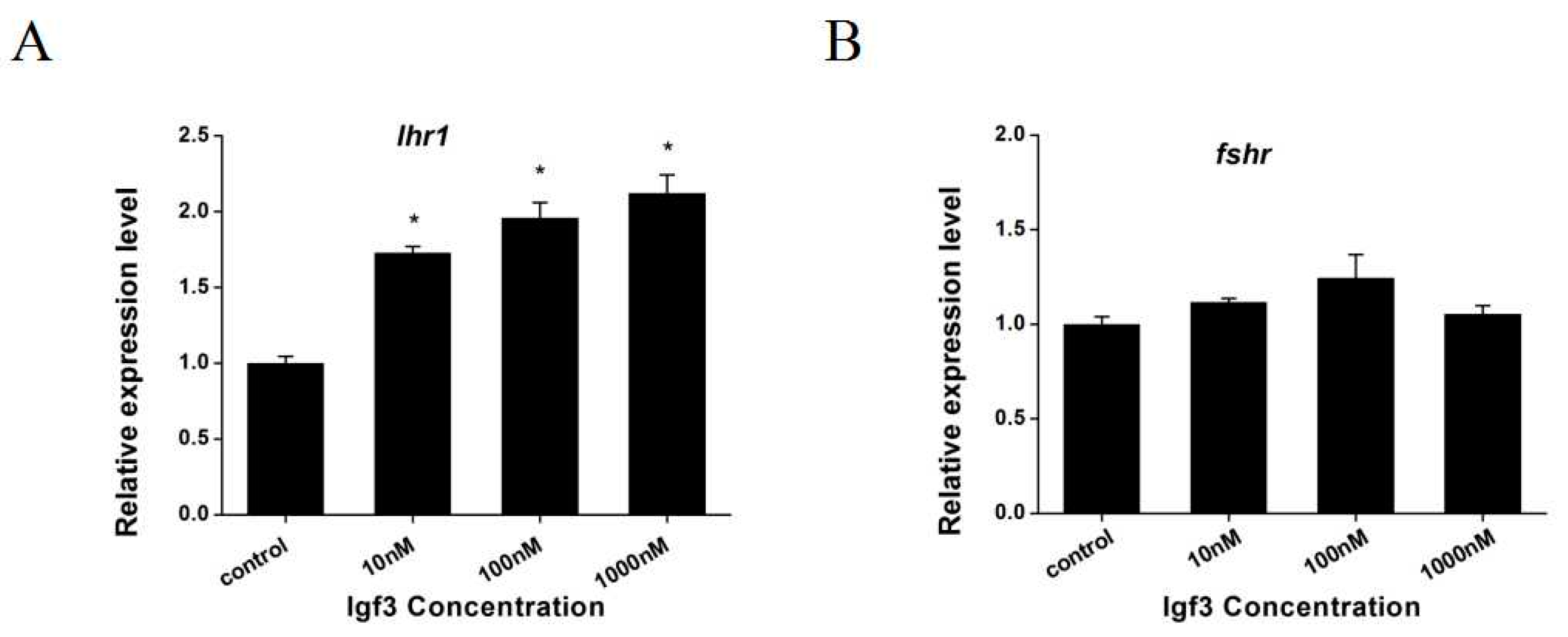
We examined whether Igf3 added to primary ovary cells altered the expression of sf1 and related genes. We measured steady-state mRNA levels in the primary ovary and found that foxl2, dax1, cyp19a1a, cyp19a1b and cyp11a1 were up-regulated when Igf3 was added to the cell culture. However, expression levels were dose-dependent such that foxl2 and cyp19a1a genes increased significantly only with 100 and 1000 nM Igf3 (Figure 7A,C). The expression of dax1 and cyp19a1b genes increased significantly under all levels of Igf3 (Figure 7B,D). The expression of the cyp11a1 gene increased significantly only with Igf3 at 1000 nM (Figure 7E). In contrast, Igf3 addition increased the mRNA expression level for luteinizing hormone receptor 1 (lhr1) in a dose-independent manner. However, the mRNA levels for follicle-stimulating hormone receptor (fshr) remained unchanged at basal levels with Igf3 addition (Figure 8).

3. Discussion

A novel Igf3 has been described in the ovaries of O. niloticus and D. rerio [1,2,14]. In this study, the Igf3 protein was expressed only in the ovary. We confirmed that Igf3 was not present in the testis, brain, kidney and liver tissues in E. coioides, similar to previous results in D. rerio [21]. This indicated that the polyclonal antibody could recognize the endogenous Igf3 protein with high specificity. Furthermore, these results confirmed that the Igf3 mature peptide was ovary-specific in E. coioides.
We found both igf3 mRNA and Igf3 in FG and MV stage follicular by in situ hybridization and immunohistochemical. Previous studies detected igf3 mRNA in the follicular cell layer of O. niloticus and D. rerio ovaries [1,21]. In these studies, igf3 mRNA was relatively low in the early follicular but increased significantly after the MV stage follicular and was high in the FG follicular [1,21]. In addition, the Igf3 protein from C. carpio was also located in ovarian granulosa cells [8]. These suggest that igf3 plays a regulatory role in E. coioides ovary maturation. Together, these results indicated that Igf3 is an ovarian-specific growth factor that is active in the late stages of follicular development. Furthermore, these studies show that igf3 mainly exists in more mature ovarian follicular and has a certain relationship with the maturation of the ovary.
To explore the functional roles of igf3 in the ovary maturation of E. coioides, we separated follicular at different stages and analyzed its temporal expression pattern in follicular of different stages by using qRT-PCR. We found that igf3 mRNA level during E. coioides follicular development increased from the early stages and was maximal at FG, similar to the results in D. rerio [21,25]. Interestingly, the igf3 expression pattern was similar to that of lhr [21,26]. This demonstrated that igf3 mRNA expression was linked to and dependent on the developmental stage. This implies that igf3 may be involved in ovarian maturation. The addition of Igf3 also induced GVBD in ovarian follicular of E. coioides in a time- and dose-dependent manner. This further indicates that Igf3 is involved in the regulation of E. coioides ovarian maturation. Igf1 induced GVBD in ovarian follicular of red seabream (Pagrus major), sea perch (Lateolabrax japonicus), white bass (Morone saxatilis), C. carpio, D. rerio and southern flounder (Paralichthys lethostigma) [12,20,27,28,29,30]. Moreover, Igf2 induced GVBD in the ovarian follicular of C. carpio, D. rerio and P. lethostigma [20,29,30]. We found similar effects for Igf3. These results show that Igf3 plays an important role in the maturation of E. coioides oocytes.
Foxl2 and dax1 are factors related to female sex differentiation, which cause animal gonads to differentiate towards ovaries [31,32,33]. Cyp19a1a and cyp19a1b are aromatase genes that induce estrogen production [34,35]. Cyp11a1 is a kind of steroidase gene that catalyzes the conversion of cholesterol to pregnenolone [36]. In addition, estrogen is involved in the sex differentiation mechanism of bony fish [32,37]. In this study, we found that Igf3 protein addition to primary ovary cells increased foxl2, dax1, cyp19a1a, cyp19a1b and cyp11a1 mRNA expression levels. This result indicates that Igf3 regulates the expression of sex-related genes and steroidase-related genes in the ovaries of E. coioides. Therefore, it is speculated that Igf3 participates in the sex determination of E. coioides and regulates its sex differentiation. In O. niloticus, Igf3 up-regulated the expression of foxl2, doublesex and mab-3-related transcription factor 1(dmrt1), cyp11a1 and cyp19a1a in a dose-dependent manner [2]. It further proves that Igf3 plays an important role in fish sex differentiation. In D. rerio, Igf3 was found to be a downstream factor of estrogen signaling in sex differentiation [38]. In addition, studies have shown that the expression level of igf3 positively correlated with that of cyp19a1a and foxl2, which have been shown to play a key role in the regulation of cyp19a1a expression [32,38,39,40]. The somatic cell signaling pathway mediated by foxl2 is a female differentiation pathway, and foxl2 can regulate the expression of cyp19a1. Therefore, it is speculated that Igf3 acts on the upstream position of foxl2 through a somatic signaling pathway mediated by foxl2. In the study, the expression of foxl2 and cyp19a1a showed a dose-dependent increase and then declined. Therefore, it is preliminary guessed that there may be feedback regulation between Igf3 and the two. Taken together, we demonstrated that Igf3 is essential for ovary differentiation in E. coioides.
Both luteinizing hormone (LH) and follicle-stimulating hormone (FSH) are involved in reproductive processes and play key roles in germ cell growth and development [41]. Ovary cells treated with Igf3 enhanced their growth and differentiation, increased related hormone levels and accelerated ovarian maturation [24,42]. In this study, Igf3 promoted lhr1 expression in a concentration-dependent manner but had no effect on fshr expression. This indicated that Igf3 regulated a gene related to reproduction in the ovary. Therefore, Igf3 was most likely involved in regulating the reproductive processes of E. coioides and promoted ovarian development and maturity. In a previous study, oogonia development into oocytes was inhibited by the Kit ligand A (kitlga), and when the igf system was inhibited in somatic cells, increased LH production hindered kitlga expression in D. rerio [43]. Our results indicated that Igf3 stimulates an increase in lhr1 expression resulting in increased LH bound to its cognate receptor. This possibly led to the down-regulation of kitlga and accelerated oocyte maturation.

4. Materials and Methods

4.1. Fish, Cells Line and Igf3 Mature Peptide

The E. coioides (the female fish with a body length of 56.5 ± 5.5 cm and a body weight of 3.6 ± 0.6 kg) used in this study were collected from the Guangdong Marine Fisheries Test Center (N22°705093′, E114°541433′) Guangdong China. The fish were kept in a recirculating seawater system that was maintained at 25 °C for two weeks before the start of experiments. Tissue samples were collected from fish and stored at −80 °C immediately. Voucher specimens were deposited at the Department of Aquatic Animal Medicine, College of Marine Sciences, South China Agricultural University, Guangzhou, China (Accession number 201805021). Igf3 mature peptide was expressed in Escherichia coli and purified in the laboratory of College of Marine Sciences, South China Agricultural University, as described previously [44].

4.2. RNA Extraction, cDNA Synthesis and Real-Time Polymerase Chain Reaction (qRT-PCR)

Total RNA from liver, kidney, brain, testis and ovarian tissues was extracted using Trizol reagent (Invitrogen, Waltham, MA, USA). The total RNA from cells was extracted using a Takara MiniBEST Universal RNA Extraction Kit (Takara, Shiga, Otsu, Japan). Reverse transcription reactions were performed using an RNase-free DNase I kit (Fermentas, Waltham, MA, USA) using the protocol provided by the manufacturer. The β-actin was used as a reference gene for qRT-PCR gene targets. The primers used in this study are given in Table 1. Each sample was run in triplicate, and reactions without templates were used as negative controls. The qRT-PCR reaction was run on a Roche Light Cycler® 480 System, and the relative quantification calculated using the ΔΔCT method [45].

4.3. Ovarian Follicle Experiment

Mature female fish were anesthetized with 1:10,000 eugenol (purity 99%, Shanghai Reagent, China) to avoid stress before harvesting. The abdomens were incised, and a small part of ovarian tissue was sampled and placed into L15 medium (Thermo Scientific, Waltham, MA, USA) and dispersed in the culture dish. The ovarian follicular development stages were classified as described previously [21]. There are five stages of follicular development that can be readily assessed by observing follicular diameters as described previously [46,47]. The primary growth (PG), previtellogenic (PV), early vitellogenic (EV), midvitellogenic (MV) and fully-grown (FG) stages are represented by follicular with diameters of <100, 200, 250, 400 and 500 μm, respectively. PG, PV, EV, MV and FG phase cells were identified and divided into parallel stages. Igf3 was added to the follicular development stages at 2, 20 and 200 ng/mL and 20 IU/mL hCG was used as a control. Germinal vesicle breakdown (GVBD) was studied by using light microscopy after 2, 6 and 16 h of incubation.

4.4. In Situ Hybridization

Intact ovaries from adult female E. coioides were dissected and fixed in 4% buffered paraformaldehyde overnight at 4 °C. The ovaries were then rinsed twice with cold phosphate-buffered saline (PBS), transferred into 30% sucrose solution for 48 h and stored at 4 °C for drying. The dried samples were frozen using dry ice and embedded in optimal cutting temperature (OCT) compound (Sakura, Torrance, CA, USA). The tissue was sliced at −21 °C, and sections were attached to cationic anti-off slides (Thermo Scientific, Waltham, MA, USA) and then covered and incubated at 42 °C for 1 to 2 d. A cDNA fragment of igf3 was amplified by PCR (Table 1) and cloned into the pGEM-T easy transcription vector (Promega, USA). Sense and antisense igf3 riboprobes were synthesized from the pGEM-T easy transcription vector construct containing igf3 after linearization with either SpeI or AatII. The RNA probes were labeled using the DIG RNA Labeling Kit (Roche, Germany). Tissue sections were first prehybridized for 30 min, and 250 μL of hybridization buffer containing 150 ng of DIG-labeled sense or antisense igf3 riboprobe was added to each slide and incubated in a humidified box at 42 °C for 16 h. After hybridization, sections were sequentially washed twice in 2 × saline-sodium citrate (SSC) (1 × SSC = 0.15 M NaCl, 15 mM Na citrate) at room temperature for 15 min and then in 1 × SSC and 0.1 × SSC at 55 °C for 1 h. The hybridization signals were detected using anti-DIG conjugated with alkaline phosphatase and visualized with the nitroblue tetrazolium chloride/5-bromo-4chloro-3-indolyl phosphate substrate solution (Dongsheng, China). The sections were dehydrated through a graded ethanol and xylene series and photographed. In this process, T7 and SP6 promoters were introduced into the upstream primers of gene-specific oligonucleotides to enable direct in vitro transcription of purified PCR fragments [48]. Among them, the sections should be treated with alcohol before observation.

4.5. Western Blotting

Western blot analysis was performed using homogenized extracts of liver, kidney, brain, testis and ovarian tissue samples using RIPA buffer (Thermo Scientific, Waltham, MA, USA). Tissue proteins were separated by using 12% sodium dodecyl sulfate–polyacrylamide gel electrophoresis (SDS-PAGE) and electrotransferred to a polyvinylidene fluoride (PVDF) membrane (Thermo Scientific, Waltham, MA, USA). Primary anti-Igf and β-actin (Invitrogen, USA) antibodies were incubated with the blots at 1:1000 dilutions for 45 min. Secondary antibodies were incubated with homogenates from testis, brain, kidney, liver and ovarian tissues before adding to the primary antibody blots. Electrochemiluminescence (ECL) detection was performed to observe the results (Applygen, Haidian, Beijing, China).

4.6. Immunohistochemistry

Ovarian tissue samples from E. coioides were embedded in paraffin and sectioned. They were then subjected to an antigen retrieval step before proceeding as described previously [49]. The sections were then blocked with 5% bovine serum albumin (BSA) for 30 min and incubated with the primary antibody at 4 °C overnight. The sections were then allowed to stand at room temperature for 30 min, washed with PBS and the secondary antibody was added dropwise and incubated at 37 °C for 1 h following the recommendations of the manufacturer (Santa Cruz Biotechnology, Dallas, TX, USA). The sections were washed with PBS and developed using 3, 3′-diaminobenzidine and counterstained with hematoxylin. The sections were then dehydrated by using graded ethanol and xylene series and observed using light microscopy.

4.7. Statistical Analysis

Data were analyzed using one-way analysis of variance (ANOVA). Before that, all data were tested for normality using the Kolmogorov–Smirnov test and homogeneity of variances using Levene’s test. Results with p-values less than 0.05 were considered statistically significant. The results are presented as mean ± S.E.M. for four replicates for each fish. All analyses were performed using Statistical Package for Social Science (SPSS) for Windows (SPSS version 20, IBM, Armonk, NY, USA).

5. Conclusions

In the study, igf3 mRNA and Igf3 protein were abundantly expressed in the more mature follicular of E. coioides. Western blot analysis demonstrated that Igf3 was an ovary-specific protein. We found that the igf3 mRNA level increased over time as ovarian follicular matured, indicating that Igf3 is a regulator of ovarian maturation in E. coioides. Igf3 promoted the expression of the sex differentiation-related genes such as foxl2, dax1, cyp19a1a, cyp19a1b, and cyp11a1 and also promoted the expression of the reproduction-related gene, lhr1. This demonstrated that igf3 plays a key role in sex differentiation and reproduction of E. coioides.

Supplementary Materials

The following supporting information can be downloaded at: https://www.mdpi.com/article/10.3390/ijms24032868/s1.

Author Contributions

Conceptualization, H.Y. and Q.W.; methodology, H.X., Y.Y. and Y.W.; software, H.Z.; data curation, B.F.; writing-original draft preparation, F.J.; writing-review and editing, F.J. and B.F.; visualization, H.X. and Y.W.; supervision, H.Y.; project administration, Q.W.; funding acquisition, H.Y. All authors have read and agreed to the published version of the manuscript.

Funding

The research was funded by [the Special Fund Project for Marine Economic Development (Six Marine Industries) of Guangdong Province] grant number [GDNRC (2022) 50]; [Guangdong Forestry Science and Technology Innovation Project] grant number [2021KJCX012, 2022KJCX019]; [Provincial Science and Technology Special Fund Project for Zhongshan City (major special project + Task list management mode)] grant number [2021sdr003]; [Key-Area Research and Development Program of Guangdong Province] grant number [2021B0202030002]; [Rural Science and Technology Commissioner Service Team project of New Rural Development Institute, South China Agricultural University] grant number [No. 2021XNYNYKJHZGJ022]; [Youth Fund Project of National Natural Science Foundation of China] grant number [No. 31502144]. And the APC was funded by the [Special Fund Project for Marine Economic Development (Six Marine Industries) of Guangdong Province] grant number [GDNRC (2022) 50].

Institutional Review Board Statement

The animal study protocol was approved by the Institutional Review Board (or Ethics Committee) of Animal Research and Ethics Committees of South China Agricultural University (protocol code: 2021B016 and date of approval: 20 March 2021).

Informed Consent Statement

Not applicable.

Data Availability Statement

All relevant data are within the paper and its Supporting Information files.

Acknowledgments

We are grateful to relevant laboratory members of State Key Laboratory of Biocontrol, Institute of Aquatic Economic Animals and the Guangdong Province Key Laboratory for Aquatic Economic Animals and Daya Bay Marine Fisheries Experiment Center Base for technical advice and valuable help during the feeding trial and sample collection. Special thanks for the technical support by Qian He from College of Veterinary Medicine, South China Agricultural University.

Conflicts of Interest

The authors report no conflicts of interest. The authors themselves are responsible for the content and writing of the paper.

References

  1. Wang, D.S.; Jiao, B.; Hu, C.; Huang, X.; Liu, Z.; Cheng, C.H. Discovery of a gonad-specific IGF subtype in teleost. Biochem. Biophys. Res. Commun. 2008, 367, 336–341. [Google Scholar] [CrossRef] [PubMed]
  2. Li, M.; Wu, F.; Gu, Y.; Wang, T.; Wang, H.; Yang, S.; Sun, Y.; Zhou, L.; Huang, X.; Jiao, B.; et al. Insulin-like growth factor 3 regulates expression of genes encoding steroidogenic enzymes and key transcription factors in the Nile tilapia gonad. Biol. Reprod. 2012, 86, 163. [Google Scholar] [CrossRef] [PubMed]
  3. Elabd, H.; Soror, E.; El-Asely, A.; El-Gawad, E.A.; Abbass, A. Dietary supplementation of Moringa leaf meal for Nile tilapia Oreochromis niloticus: Effect on growth and stress indices. Egypt. J. Aquat. Res. 2019, 45, 265–271. [Google Scholar] [CrossRef]
  4. Sparkman, A.M.; Schwartz, T.S.; Madden, J.A.; Boyken, S.E.; Ford, N.B.; Serb, J.M.; Bronikowski, A.M. Rates of molecular evolution vary in vertebrates for insulin-like growth factor-1 (IGF-1), a pleiotropic locus that regulates life history traits. Gen. Comp. Endocrinol. 2012, 178, 164–173. [Google Scholar] [CrossRef]
  5. Ozcan Gokcek, E.; Isik, R. Associations between genetic variants of the insulin-like growth factor I (IGF-I) gene and growth traits in European sea bass (Dicentrarchus labrax, L.). Fish Physiol. Biochem. 2020, 46, 1131–1138. [Google Scholar] [CrossRef]
  6. Ipsa, E.; Cruzat, V.F.; Kagize, J.N.; Yovich, J.L.; Keane, K.N. Growth hormone and insulin-like growth factor action in reproductive tissues. Front. Endocrinol. (Lausanne) 2019, 10, 777. [Google Scholar] [CrossRef]
  7. Wang, B.; Xu, Y.; Liu, X.; Liu, Q.; Liu, Y.; Zhang, Y.; Shi, B. Molecular characterization and expression profiles of insulin-like growth factors in yellowtail kingfish (Seriola lalandi) during embryonic development. Fish Physiol. Biochem. 2019, 45, 375–390. [Google Scholar] [CrossRef]
  8. Song, F.; Wang, L.; Zhu, W.; Fu, J.; Dong, J.; Dong, Z. A novel igf3 gene in common carp (Cyprinus carpio): Evidence for its role in regulating gonadal development. PLoS ONE 2016, 11, e0168874. [Google Scholar] [CrossRef]
  9. Prado, P.S.; Pinheiro, A.P.B.; Weber, A.A.; Bazzoli, N.; Rizzo, E. Expression patterns and immunolocalisation of IGF-I and IGF-II in male and female gonads of the Neotropical characid fish Astyanax fasciatus. Fish Physiol. Biochem. 2019, 45, 167–176. [Google Scholar] [CrossRef]
  10. Reinecke, M.; Björnsson, B.T.; Dickhoff, W.W.; McCormick, S.D.; Navarro, I.; Power, D.M.; Gutiérrez, J. Growth hormone and insulin-like growth factors in fish: Where we are and where to go. Gen. Comp. Endocrinol. 2005, 142, 20–24. [Google Scholar] [CrossRef] [PubMed]
  11. Mohammed-Geba, K.; Martos-Sitcha, J.A.; Galal-Khallaf, A.; Mancera, J.M.; Martinez-Rodriguez, G. Insulin-like growth factor 1 (IGF-1) regulates prolactin, growth hormone, and IGF-1 receptor expression in the pituitary gland of the gilthead sea bream Sparus aurata. Fish Physiol. Biochem. 2016, 42, 365–377. [Google Scholar] [CrossRef]
  12. Picha, M.E.; Shi, B.; Thomas, P. Dual role of IGF-II in oocyte maturation in southern flounder Paralichthys lethostigma: Up-regulation of mPRα and resumption of meiosis. Gen Comp. Endocrinol. 2012, 177, 220–230. [Google Scholar] [CrossRef]
  13. Higuchi, K.; Gen, K.; Izumida, D.; Kazeto, Y.; Hotta, T.; Takashi, T.; Aono, H.; Soyano, K. Changes in gene expression and cellular localization of insulin-like growth factors 1 and 2 in the ovaries during ovary development of the yellowtail, Seriola quinqueradiata. Gen. Comp. Endocrinol. 2016, 232, 86–95. [Google Scholar] [CrossRef] [PubMed]
  14. Berishvili, G.; D’Cotta, H.; Baroiller, J.F.; Segner, H.; Reinecke, M. Differential expression of IGF-I mRNA and peptide in the male and female gonad during early development of a bony fish, the tilapia Oreochromis niloticus. Gen. Comp. Endocrinol. 2006, 146, 204–210. [Google Scholar] [CrossRef] [PubMed]
  15. Wuertz, S.; Gessner, J.; Kirschbaum, F.; Kloas, W. Expression of IGF-I and IGF-I receptor in male and female sterlet, Acipenser ruthenus—Evidence for an important role in gonad maturation. Comp. Biochem. Phys. A Mol. Integr. Physiol. 2007, 147, 223–230. [Google Scholar] [CrossRef]
  16. Schmid, A.; Rhyn-Näf, E.; Kloas, W.; Reinecke, M. Insulin-like growth factor-I and -II in the ovary of a bony fish, Oreochromis mossambicus, the tilapia: In situ hybridisation, immunohistochemical localisation, Northern blot and cDNA sequences. Mol. Cell. Endocrinol. 1999, 156, 141–149. [Google Scholar] [CrossRef]
  17. Nelson, S.N.; Van Der Kraak, G. Characterization and regulation of the insulin-like growth factor (IGF) system in the zebrafish (Danio rerio) ovary. Gen. Comp. Endocrinol. 2010, 168, 111–120. [Google Scholar] [CrossRef] [PubMed]
  18. Srivastava, R.K.; Kraak, G.V.D. Regulation of DNA synthesis in goldfish vitellogenic ovarian-follicles by hormones and growth factors. J. Exp. Zool. A Ecol. Genet. Physiol. 1994, 270, 263–272. [Google Scholar] [CrossRef]
  19. Lokman, P.M.; George, K.A.; Divers, S.L.; Algie, M.; Young, G. 11-Ketotestosterone and IGF-I increase the size of previtellogenic oocytes from shortfinned eel, Anguilla australis, in vitro. Reproduction 2007, 133, 955–967. [Google Scholar] [CrossRef]
  20. Weber, G.M.; Sullivan, C.V. Insulin-like growth factor-I induces oocyte maturational competence but not meiotic resumption in white bass (Morone chrysops) follicles in vitro: Evidence for rapid evolution of insulin-like growth factor action. Biol. Reprod. 2005, 72, 1177–1186. [Google Scholar] [CrossRef] [Green Version]
  21. Li, J.; Liu, Z.; Wang, D.; Cheng, C.H. Insulin-like growth factor 3 is involved in oocyte maturation in zebrafish. Biol. Reprod. 2011, 84, 476–486. [Google Scholar] [CrossRef] [PubMed]
  22. Yang, H.; Chen, H.; Zhao, H.; Liu, L.; Xie, Z.; Xiao, L.; Li, S.; Zhang, Y.; Lin, H. Molecular cloning of the insulin-like growth factor 3 and difference in the expression of igf genes in orange-spotted grouper (Epinephelus coioides). Comp. Biochem. Physiol. B Biochem. Mol. Biol. 2015, 186, 68–75. [Google Scholar] [CrossRef] [PubMed]
  23. Kokai, L.E.; Marra, K.; Rubin, J.P. Adipose stem cells: Biology and clinical applications for tissue repair and regeneration. Transl. Res. 2014, 163, 399–408. [Google Scholar] [CrossRef]
  24. Maitra, S.; Das, D.; Ghosh, P.; Hajra, S.; Roy, S.S.; Bhattacharya, S. High cAMP attenuation of insulin-stimulated meiotic G2-M1 transition in zebrafish oocytes: Interaction between the cAMP-dependent protein kinase (PKA) and the MAPK3/1 pathways. Mol. Cell. Endocrinol. 2014, 393, 109–119. [Google Scholar] [CrossRef]
  25. Berishvili, G.; Baroiller, J.F.; Eppler, E.; Reinecke, M. Insulin-like growth factor-3 (IGF-3) in male and female gonads of the tilapia: Development and regulation of gene expression by growth hormone (GH) and 17alpha-ethinylestradiol (EE2). Gen. Comp. Endocrinol. 2010, 167, 128–134. [Google Scholar] [CrossRef] [PubMed]
  26. Kwok, H.-F.; So, W.-K.; Wang, Y.; Ge, W. Zebrafish gonadotropins and their receptors: I. cloning and characterization of zebrafish follicle-stimulating hormone and luteinizing hormone receptors-evidence for their distinct functions in follicle development 1. Biol. Reprod. 2005, 72, 1370–1381. [Google Scholar] [CrossRef]
  27. Weber, G.M.; Sullivan, C.V. Effects of insulin-like growth factor-I on in vitro final oocyte maturation and ovarian steroidogenesis in striped bass, Morone saxatilis. Biol. Reprod. 2000, 63, 1049–1057. [Google Scholar] [CrossRef]
  28. Kagawa, H.; Kobayashi, M.; Hasegawa, Y.; Aida, K. Insulin and insulin-like growth factors I and II induce final maturation of oocytes of red seabream, Pagrus major, in vitro. Gen. Comp. Endocrinol. 1994, 95, 293–300. [Google Scholar] [CrossRef]
  29. Mukherjee, D.; Mukherjee, D.; Sen, U.; Paul, S.; Bhattacharyya, S.P. In vitro effects of insulin-like growth factors and insulin on oocyte maturation and maturation-inducing steroid production in ovarian follicles of common carp, Cyprinus carpio. Comp. Biochem. Physiol. A Mol. Integr. Physiol. 2006, 144, 63–77. [Google Scholar] [CrossRef]
  30. Nelson, S.N.; Van Der Kraak, G. The role of the insulin-like growth factor (IGF) system in zebrafish (Danio rerio) ovarian development. Gen. Comp. Endocrinol. 2010, 168, 103–110. [Google Scholar] [CrossRef]
  31. Ottolenghi, C.; Omari, S.; Garcia-Ortiz, J.E.; Uda, M.; Crisponi, L.; Forabosco, A.; Pilia, G.; Schlessinger, D. Foxl2 is required for commitment to ovary differentiation. Hum. Mol. Genet. 2005, 14, 2053–2062. [Google Scholar] [CrossRef]
  32. Wang, D.S.; Kobayashi, T.; Zhou, L.Y.; Paul-Prasanth, B.; Ijiri, S.; Sakai, F.; Okubo, K.; Morohashi, K.; Nagahama, Y. Foxl2 up-regulates aromatase gene transcription in a female-specific manner by binding to the promoter as well as interacting with ad4 binding protein/steroidogenic factor 1. Mol. Endocrinol. 2007, 21, 712–725. [Google Scholar] [CrossRef] [PubMed]
  33. Garcia-Ortiz, J.E.; Pelosi, E.; Omari, S.; Nedorezov, T.; Piao, Y.; Karmazin, J.; Uda, M.; Cao, A.; Cole, S.W.; Forabosco, A.; et al. Foxl2 functions in sex determination and histogenesis throughout mouse ovary development. BMC Dev. Biol. 2009, 9, 36. [Google Scholar] [CrossRef] [PubMed]
  34. Duffy, T.A.; Picha, M.E.; Won, E.T.; Borski, R.J.; McElroy, A.E.; Conover, D.O. Ontogenesis of gonadal aromatase gene expression in atlantic silverside (Menidia menidia) populations with genetic and temperature-dependent sex determination. J. Exp. Zool. A Ecol. Genet. Physiol. 2010, 313, 421–431. [Google Scholar] [CrossRef]
  35. Conley, A.; Hinshelwood, M. Mammalian aromatases. Reproduction 2001, 121, 685–695. [Google Scholar] [CrossRef]
  36. Guo, I.-C.; Shih, M.-C.; Lan, H.-C.; Hsu, N.-C.; Hu, M.-C.; Chung, B.-C. Transcriptional regulation of human CYP11A1 in gonads and adrenals. J. Biomed. Sci. 2007, 14, 509–515. [Google Scholar] [CrossRef] [PubMed]
  37. Guiguen, Y.; Fostier, A.; Piferrer, F.; Chang, C.F. Ovarian aromatase and estrogens: A pivotal role for gonadal sex differentiation and sex change in fish. Gen. Comp. Endocrinol. 2010, 165, 352–366. [Google Scholar] [CrossRef]
  38. Xie, Y.; Huang, D.; Chu, L.; Liu, Y.; Sun, X.; Li, J.; Cheng, C.H.K. Igf3 is essential for ovary differentiation in zebrafishdagger. Biol. Reprod. 2021, 104, 589–601. [Google Scholar] [CrossRef]
  39. Parker, K.L.; Schimmer, B.P. Steroidogenic factor 1: A key determinant of endocrine development and function. Endocr. Rev. 1997, 18, 361–377. [Google Scholar] [CrossRef]
  40. Zhang, B.; Shozu, M.; Okada, M.; Ishikawa, H.; Kasai, T.; Murakami, K.; Nomura, K.; Harada, N.; Inoue, M. Insulin-like growth factor I enhances the expression of aromatase P450 by inhibiting autophagy. Endocrinology 2010, 151, 4949–4958. [Google Scholar] [CrossRef]
  41. van der Kraak, G. Chapter 3 the GnRH system and the neuroendocrine regulation of reproduction. Fish Physiol. 2009, 28, 115–149. [Google Scholar]
  42. Yerramalla, U.L.; Nadimpalli, S.K.; Schu, P.; Figura, K.V.; Hille-Rehfeld, A. Conserved cassette structure of vertebrate M(r) 300 kDa mannose 6-phosphate receptors: Partial cDNA sequence of fish MPR 300. Comp. Biochem. Physiol. B Biochem. Mol. Biol. 2001, 127, 433–441. [Google Scholar] [CrossRef] [PubMed]
  43. Yao, K.; Ge, W. Spatial distribution and receptor specificity of zebrafish Kit system-evidence for a Kit-mediated bi-directional communication system in the preovulatory ovarian follicle. PLoS ONE 2013, 8, e56192. [Google Scholar] [CrossRef] [PubMed]
  44. Saez, N.J.; Vincentelli, R. High-throughput expression screening and purification of recombinant proteins in E. coli. Methods Mol. Biol. 2014, 1091, 33–53. [Google Scholar]
  45. Livak, K.J.; Schmittgen, T.D. Analysis of relative gene expression data using realtime quantitative PCR and the 2(-ΔΔC(T)) method. Methods 2012, 25, 402–408. [Google Scholar] [CrossRef] [PubMed]
  46. David, R.; Wedlich, D. PCR-based RNA probes: A quick and sensitive method to improve whole mount embryo in situ hybridizations. Biotechniques 2001, 30, 769–772. [Google Scholar] [CrossRef]
  47. Shi, S.R.; Cote, R.J.; Taylor, C.R. Antigen retrieval immunohistochemistry: Past, present, and future. J. Histochem. Cytochem. 1997, 45, 327–343. [Google Scholar] [CrossRef] [PubMed]
  48. Wang, Y.; Ge, W. Developmental profiles of activin βA, βB, and follistatin expression in the zebrafish ovary: Evidence for their differential roles during sexual maturation and ovulatory cycle1. Biol. Reprod. 2004, 71, 2056–2064. [Google Scholar] [CrossRef]
  49. Pang, Y.; Thomas, P. Role of G protein-coupled estrogen receptor 1, GPER, in inhibition of oocyte maturation by endogenous estrogens in zebrafish. Dev. Biol. 2010, 342, 194–206. [Google Scholar] [CrossRef] [PubMed] [Green Version]
Figure 1. Western blot verification of Igf3.
Ijms 24 02868 g001
Figure 2. Immunohistochemical detection of Igf3 in E. coioides ovarian tissues. (A) negative control lacking primary antibody. (B) Igf3-specific polyclonal primary antibody. Note: FG indicates full grown stages of follicular.
Ijms 24 02868 g002
Figure 3. Distribution of igf3 mRNA in E. coioides ovaries from frozen tissue sections. (A) sense probe. (B) antisense probe. Note: FG indicates full grown stages of follicular.
Ijms 24 02868 g003
Figure 4. Stage-dependent expression of igf3 in developing E. coioides follicles. (A) Morphology of E. coioides follicular at different stages of maturation. PG—primary growth; PV—previtellogenic; EV—early vitellogenic; MV—midvitellogenic; FG—full-grown. (B) Expression of igf3 mRNA during different stages of ovarian follicle development. Different letters (a and b) indicate significant differences.
Ijms 24 02868 g004
Figure 5. Germinal vesicle breakdown (GVBD) alterations in midvitellogenic (MV) and fully-grown (FG) stages of follicular. Igf3 was added at the (A) MV and (B) FG stages. (n = 4). ***, p < 0.001.
Ijms 24 02868 g005
Figure 6. Effects of Igf3 on follicular Germinal vesicle breakdown (GVBD) in the previtellogenic (PV), early vitellogenic (EV), midvitellogenic (MV) and fully-grown (FG) stages of development. Cells were treated for (A) 2, (B) 6 and (C) 16 h. (n = 4). **, p < 0.01, ***, p < 0.001.
Ijms 24 02868 g006
Figure 7. Igf3 added to primary ovarian cells increases expression of sex differentiation-related genes. (A) forkhead boxl2—foxl2; (B) dosage-sensitive sex reversal adrenal hypoplasia critical region on chromosome x gene 1—dax1; (C) cytochrome P450 family 19 subfamily member 1 a—cyp19a1a; (D) cytochrome P450 family 19 subfamily a1 member b—cyp19a1b; and (E) cytochrome P450 family 11 subfamily b member 2—cyp11a1. (n = 4). *, p < 0.05, **, p < 0.01, ***, p < 0.001.
Ijms 24 02868 g007
Figure 8. Effects of Igf3 treatment on primary ovarian cells on the expression of reproduction-related genes. (A) ihr1 and (B) fshr at the indicated dosages. (n = 4). *, p < 0.05.
Ijms 24 02868 g008
Table 1. Primers used in this study.
Gene SymbolGenBank Accession NumberFull Gene NamePrimer Sequence(5′-3′)Amplification Efficiency (%)
β-actinAY510710Beta-actinForward: TTCACCACCACAGCCGAGA
Reverse: TGGTCTCGTGGATTCCGCAG
100
igf3KU743244.1Insulin-like growth factor subtype 3Forward: GTGTGTATGTTCTACTCCAC
Reverse: TCCTTCTGTAAGCTTCCCTC
100
foxl2KP688066.1Forkhead box L2Forward: CAGGTAGCCATAGCCGTCTTCT
Reverse: CCTCCACCGACGCACTTC
99
dax1HQ455522.1Dosage-sensitive sex reversal adrenal hypoplasia critical region on chromosome x gene 1Forward: CTGCCTCCACTACATCCAGT
Reverse: TGCTGACGGTCCCTATAACG
98
cyp19a1aEU239953.1Cytochrome P450 family 19 subfamily member 1 aForward: GGAGACATTGTGAGAGTCTGGATC
Reverse: TGACAGGTACATCCAGGAAGAGTC
99
cyp19a1bJF420889.1Cytochrome P450 family 19 subfamily member 1 bForward: TGACACCTGGCAAACAGTTC
Reverse: GATGGTGTCGTCTTCCAGAG
97
cyp11a1FJ807733.1Cytochrome P450 family 11 subfamily a member 1 aForward: CTCCCTCCTGCTCTGTTGA
Reverse: GCTGGCCTTGATGTCTTC
96
lhr1HQ650770.1Luteinizing hormone receptor 1Forward: GACCTTCCCATTAATCCTGCATGG
Reverse: CACGTTTCCAAAGCCTTCATCATCCTC
99
fshrHQ650769.1Follicle-stimulating hormone receptorForward: CGAGGCTGACCCTTACTTCC
Reverse: GATCCAGATGAGGACCCGTA
98
Disclaimer/Publisher’s Note: The statements, opinions and data contained in all publications are solely those of the individual author(s) and contributor(s) and not of MDPI and/or the editor(s). MDPI and/or the editor(s) disclaim responsibility for any injury to people or property resulting from any ideas, methods, instructions or products referred to in the content.