Review

Prevention of Metabolic Syndrome by Phytochemicals and Vitamin D

1
Department of Biotechnology, Tokyo College of Biotechnology, Tokyo 114-0032, Japan
2
Vino Science Japan Inc., Kanagawa 210-0855, Japan
3
Department of Biochemistry and Systems Biomedicine, Juntendo University Graduate School of Medicine, Tokyo 113-8421, Japan
4
Faculty of Medical Science, Juntendo University, Chiba 279-0013, Japan
*
Author to whom correspondence should be addressed.
Int. J. Mol. Sci. 2023, 24(3), 2627; https://doi.org/10.3390/ijms24032627
Submission received: 21 December 2022 / Revised: 24 January 2023 / Accepted: 24 January 2023 / Published: 30 January 2023
(This article belongs to the Special Issue Bioactive Compounds in Metabolic Syndrome)

Abstract

:
In recent years, attention has focused on the roles of phytochemicals in fruits and vegetables in maintaining and improving the intestinal environment and preventing metabolic syndrome. A high-fat and high-sugar diet, lack of exercise, and excess energy accumulation in the body can cause metabolic syndrome and induce obesity, diabetes, and disorders of the circulatory system and liver. Therefore, the prevention of metabolic syndrome is important. The current review shows that the simultaneous intake of phytochemicals contained in citruses and grapes together with vitamin D improves the state of gut microbiota and immunity, preventing metabolic syndrome and related diseases. Phytochemicals contained in citruses include polyphenols such as hesperidin, rutin, and naringin; those in grapes include quercetin, procyanidin, and oleanolic acid. The intake of these phytochemicals and vitamin D, along with prebiotics and probiotics, nurture good gut microbiota. In general, Firmicutes are obese-prone gut microbiota and Bacteroidetes are lean-prone gut microbiota; good gut microbiota nurture regulatory T cells, which suppress inflammatory responses and upregulate immunity. Maintaining good gut microbiota suppresses TNF-α, an inflammatory cytokine that is also considered to be a pathogenic contributor adipokine, and prevents chronic inflammation, thereby helping to prevent metabolic syndrome. Maintaining good gut microbiota also enhances adiponectin, a protector adipokine that prevents metabolic syndrome. For the prevention of metabolic syndrome and the reduction of various disease risks, the intake of phytochemicals and vitamin D will be important for human health in the future.

1. Introduction

Excess energy accumulation from the intake of a high-fat, high-sugar diet and the typical Western diet causes metabolic syndrome. Generally, metabolic syndrome is defined as visceral obesity with two or more symptoms of hyperglycemia, hypertension, and dyslipidemia. A high caloric diet with a high-sugar and high-fat intake causes obesity, and such diets, when combined with a lack of exercise, become the first step toward metabolic syndrome [1,2]. As this status induces diabetes, cardiovascular diseases, and liver disorders, the prevention of metabolic syndrome is very important. In metabolic syndrome, the production of tumor necrosis factor α (TNF-α), a pathogenic contributor adipokine produced from enlarged adipocytes, induces chronic inflammation. This is a critical condition that in turn induces several disorders, which combine to cause metabolic syndrome [3]. On the other hand, the suppressed production of adiponectin, a protector adipokine, weakens the ability to overcome metabolic syndrome [4].
There are three types of adipocytes: white adipocytes, brown adipocytes, and beige adipocytes [5]. Healthy white adipocytes are important because they produce adiponectin and enhance the production of insulin from pancreatic β cells. During the progression of metabolic syndrome, however, adipocytes enlarge and change their shape to produce TNF-α. TNF-α suppresses the production of glucose transporter type 4 (GLUT4) and induces insulin resistance, which is responsible for chronic inflammation and causes diabetes [6]. Brown adipocytes come from white adipocytes. In adults, the browning of adipocytes is thought to be related to the control of body weight through the active metabolism of body fat [7]. Recently, attention has been focused on beige adipocytes [8]. Because beige adipocytes convert fatty acid and sugars into energy, which they then consume, increasing the proportion of beige adipocytes in the body has been suggested as being capable of preventing the onset of metabolic syndrome [9,10].
Firmicutes and Bacteroidetes are dominant types of gut microbiota, and each group of bacteria are classified into obese-type and lean-type [11]. Firmicutes are Gram-positive bacteria that are predominant in obese people. A high-fat, high-sugar diet, large amounts of meat consumption, and a diet containing few vegetables and fruits induce a Firmicutes-dominant gut microbiota. Some Firmicutes bacteria produce decomposing enzymes to digest indigestive fibers such as cellulose into glucose, potentially contributing to obesity [12]. On the other hand, a decrease in Bacteroidetes, which are Gram-negative bacteria, induces a thin mucin layer in the large intestine, weakens the cellular tight junctions, and causes leaky gut syndrome, hypertension, dyslipidemia, and lifestyle-related illness [13]. Therefore, lowering the F/B ratio is a good strategy for maintaining good health.
Consuming a healthy diet and exercising are important for preventing metabolic syndrome. The typical Western diet, with high fat and high sugar consumption, is responsible for metabolic syndrome. On the other hand, a Mediterranean diet, and the traditional Japanese diet, in which less fat, sugar, and meat are consumed, is less likely to cause metabolic syndrome [14]. In addition, the consumption of prebiotics, probiotics, polyphenols, and vitamin D, which is also known as 3PD, is a good strategy for nurturing good microbiota [15,16].
The phytochemicals in citruses and grapes prevent chronic inflammation by modifying good gut microbiota. Citrus phytochemicals include hesperidin, β-cryptoxanthin, rutin (quercetin-glycoside), and naringin; the phytochemicals in grapes include quercetin, procyanidin, and oleanolic acid (terpenoids) [17]. In addition, skin cells produce vitamin D from cholesterol after exposure to sunlight. Vitamin D was originally described as a bone hormone; in addition to these previously known effects, however, vitamin D is now considered to act as a type of steroid. Because large numbers of immune cells, including macrophages, express the vitamin D receptor (VDR), vitamin D is thought to participate in the regulation of immunity [18]. Furthermore, vitamin D not only binds to VDR and activates immune cells, but it also improves natural immune responses by increasing the production of antimicrobial peptides [19]. In addition, vitamin D is also an important substance for maintaining a diversity of good gut microbiota [20].
In metabolic syndrome, excess energy accumulation in the body arising from an imbalanced diet result in a variety of disorders such as diabetes, cardiovascular diseases, and Alzheimer’s disease. Instead, a healthy diet maintains good gut microbiota, suppresses chronic inflammation, upregulates immunity, and prevents metabolic syndrome. The current review explains how phytochemicals and vitamin D prevent metabolic syndrome and help to maintain healthy longevity.

2. Disorders Related to Metabolic Syndrome

Metabolic syndrome is a relatively new set of metabolic conditions/alterations that has been known since the early 21st century after the increase in obesity caused by changes in eating habits and lifestyles became apparent. It is characterized by various complications which are induced by diabetes accompanied by insulin resistance, such as hyperglycemia, arterial hypertension, and dyslipidemia. Furthermore, metabolic syndrome is a serious risk factor of early death because it causes cardiovascular, liver, and brain disorders or induces cancer through the presence of these disorders [21,22]. The main cause of metabolic syndrome is excessive energy accumulation in the body. An imbalanced diet with a high-fat and high-sugar intake and a lack of exercise are responsible for metabolic syndrome. These conditions cause dysbiosis in gut microbiota, with increased Firmicutes and decreased Bacteroidetes populations, induce the production of the pathogenic contributor TNF-α, and decrease the protective adiponectin, leading to the onset metabolic syndrome (Figure 1).

2.1. Diabetes

Diabetes is a part of metabolic syndrome and is classified into type 1 and type 2. In type 1 diabetes, damaged pancreatic β cells stop producing insulin; in type 2 diabetes, genetic factors related to inadequate insulin secretion are the main cause. Recently, the number of people with diabetes has been increasing in Japan. Two factors are related to the increase in metabolic syndrome: environmental factors including a high-fat diet, lack of exercise, obesity, visceral fat accumulation, and insulin resistance, and genetic factors including reduced insulin secretion. In addition, an unbalanced diet causes dysbiosis, induces insulin resistance, and leads to the onset of diabetes [23]. Blood sugar spikes, which are the state of temporary high blood sugar levels soon after meals, but which are not detected by conventional health checks because of their shortness, are of particular concern. This state is caused by the rapid decomposition of starches into glucose after meals by the secretion of insulin. Blood sugar spikes are huge health risks because they trigger arteriosclerosis-related disorders such as heart disease, stroke, and kidney malfunction. The intake of quercetin, a flavonoid with enzyme inhibitory properties, is becoming an attractive choice to stop blood sugar spikes and to prevent diabetes [24].
After the onset of obesity, adipocytes constantly produce free fatty acid (FFA). The continuous production of FFA affects macrophages, causing them to produce the proinflammatory TNF-α and to inhibit the glucose carrier GLUT4 synthesis in muscular cells. This inhibition leads to the onset of type 2 diabetes [25].

2.2. Cardiovascular Diseases

Glucose metabolism disorder, hypertension, and dyslipidemia induce the onset of atherosclerosis. Cholesterol is divided into two types: low-density lipoprotein (LDL) cholesterol and high-density lipoprotein (HDL) cholesterol. The LDL/HDL level is an indicator of arteriosclerosis. When this figure exceeds 2.5, the onset of arteriosclerosis is likely [26]. Cholesterol is an organic compound that is classified as a sterol subclass of steroids produced from acetyl-CoA, a metabolite of glucose and fatty acid. Acetyl-CoA is the substrate that produces ATP via the tricarboxylic acid cycle in mitochondria. However, the remaining substrate that is not used in ATP production is stored as cholesterol in the body. Cholesterol is used to produce a variety of hormones and bile acids. It is an essential material in biomembranes and an important substance for a variety of intracellular metabolic processes [27]. In general, HDL cholesterol is considered to be health-beneficial cholesterol because it acts as the carrier of accumulated cholesterol in peripheral tissue to the liver. On the other hand, LDL cholesterol transports excess cholesterol. However, oxidized LDL is incorporated into macrophages and is accumulated in vascular sub-endothelial spaces (intima), and it is responsible for the onset of arteriosclerosis [28]. Furthermore, a relationship between cellular senescence induced by chronic inflammation and arteriosclerosis has also been suggested [29].
Leaky gut, which is a state of increased intestinal vascular permeability caused by dysbiosis, induces chronic inflammation and triggers cardiovascular disorders. A high-fat and high-sugar diet causes dysbiosis and induces a deficiency of butyrate-producing bacteria, reducing regulatory T cells, weakening anti-inflammatory mechanisms, and increasing inflammation-inducible bacteria. As a result, the tight junction structure in the intestinal epithelium is weakened, and this state increases the permeability between cells in the intestine, resulting in the condition called leaky gut. This condition induces and influx of bacteria and lipopolysaccharides (LPS) from the lamina propria mucosae to the bloodstream and elicits inflammation by increasing cytokines and reactive oxide. The dysfunction of intestinal epithelial cells induces chronic inflammation and continuously provides factors that contribute to the pathophysiology of atherosclerosis, including cytokines, endotoxins, and gut microbes in the bloodstream [30,31]. Finally, TNF-α produced by chronic inflammation induces arteriosclerosis through foam cell formation from macrophages and moves into blood vessels [32]. Arteriosclerosis causes ischemic heart disease, cerebrovascular disease, and stroke. The prevalence of these disorders became remarkable after the Westernization of diets, including high fat and high sugar consumption. The decreased intake of vegetables and fruits, phytochemicals, and dietary fibers and the remarkable increase in the consumption of meats and dairy products are considered causes of the increased prevalence of arteriosclerosis.

2.3. Liver Disorders

Cholesterol in the liver moves to the intestine as a bile acid and is excreted from the body. Bile acid production is one of the main functions of the liver, with about 600 mL/day of bile acid being produced. The gallbladder stores bile acids and excretes them into the duodenum after food intake. Secreted bile acids work as an emulsification detergent and help the digestion of lipids by lipase. Bile acids exist as glycine/taurine conjugates and bile pigments, including bilirubin and biliverdin. Bilirubin is a metabolite which is produced from heme and originates from the heme protein in hemoglobin. Bile acid is excreted to the intestine and returns to the liver through enterohepatic circulation. However, deoxycholic acid, a second metabolite of bile acid produced by the gut microbiota, is harmful to the liver. Here, water-soluble dietary fibers absorb bile acids in the duodenum and incorporate cholesterol, then excrete them from the body through the large intestine [33]. Unbalanced dietary habits cause metabolic syndrome and dysbiosis, whereas a faulty gut microbiota increases the production of deoxycholic acid and ultimately induces liver injury [34,35]. Oleanolic acid, a phytochemical which is contained in grape skin, conjugates with the cholic acid receptor TGR5 as an agonist and prevents constipation by enhancing intestinal movement [36].
In addition, leaky gut syndrome causes non-alcoholic steatohepatitis (NASH). Increased permeability in the intestinal epithelium induces metabolic endotoxemia, insulin resistance, and inflammation and fibrosis in the liver through TNF-α production [37]. Unlike other types of hepatitis, such as alcoholic or viral hepatitis, NASH is induced by high fat and high sugar consumption, obesity, metabolic syndrome, and other lifestyle-related diseases. Obesity is considered a high-risk factor for liver cancer. The onset of non-alcoholic fatty liver disease (NAFLD) induces NASH and causes hepatic cirrhosis, and these liver disorders ultimately progress to cancer [38]. On the other hand, the liver has a high regenerative capacity. The improvement of dietary habits can ameliorate NASH-induced hepatic fibrosis [39], and the administration of quercetin metabolites protects the liver from acetaldehyde-induced cellular toxicity [40].

2.4. Brain Disorders

Recently, several studies have shown a relationship between metabolic syndrome and dementia [41]. In the United States, middle-aged people with obesity have a three-times higher risk of Alzheimer’s disease (AD) and a five-times higher risk of dementia induced by cerebral thrombosis, compared to other groups. AD has been called type 3 diabetes because it has the same pathophysiology as type 2 diabetes [42]. Neurotoxicity induced by diabetic brain inflammation and Tau protein peroxidation can lead to the onset of cognitive impairment. During AD progression, inflammation in microglial cells can be observed in the brain. A study conducted in AD patients showed that insulin administration through a trans-nasal pathway resulted in the direct transport of insulin to the brain. This experiment revealed that decreased levels of insulin in the brain induce AD, because low insulin levels in brain cells induce glucose deficiency and cell death [43].
In multiple sclerosis, proinflammatory Th17 cells that are activated by dysbiosis induce neural inflammation in the brain [44]. In contrast, short fatty acids produced by healthy gut microbiota increase tryptophan hydroxylase 1 (TPH1) expression in enterochromaffin cells and induce serotonin secretion in the gut. In addition, activated regulatory T cells (Treg) in the gut induce IgA-producing plasma B cells, which are producers of IL-10. Finally, these plasma B cells release IL-10 in the brain, thereby suppressing the inflammation associated with multiple sclerosis [45].

3. Citrus and Grape Phytochemicals and Vitamin D

3.1. Phytochemicals Contained in Citruses

Citruses contain a variety of phytochemicals. Japanese mandarin (orange) (Citrus unshiu) is one of the most popular fruits in Japan, along with apple and banana. Orange peel, known as chenpi in Kampo (Traditional Japanese) medicine, is often used in formulations for a variety of symptoms [46]. Citruses contain a variety of phytochemicals including hesperidin and β-cryptoxanthin. Rutin (quercetin-glycoside) is also found abundantly in Tartary buckwheat (Fagopyrum tataricum). Grapefruit, orange, and Hassaku orange contain naringin. Citrus depressa contains nobiletin [47]. Hesperidin, rutin, naringin, and nobiletin are flavonoids, and β-cryptoxanthin is a carotenoid found in citrus. Table 1 shows citrus phytochemicals and their effects in metabolic syndrome.
Hesperidin is abundant in mandarin oranges and sudachi (Citrus sudachi) and is effective for decreasing triglycerides, improving cold sensitivity, overcoming stress, improving skin condition, and recycling vitamin C. Hesperidin has a low absorption ratio in the body because its water solubility is low. Similarly, to other polyphenols, a glucose-conjugated form—hesperidin-glycoside—is widely used to improve bioavailability [48]. Hesperidin inhibits endotoxin shock in mice [49] and is effective for treating rheumatoid arthritis in humans [50]. In addition, hesperidin improves insulin resistance, hypertension, hyperglycemia, hypercholesterolemia, triglyceride, TNF-α, and hs-CPR levels in the blood [51,52].
Table 1. Anti-metabolic syndrome and anti-inflammatory effects of citrus phytochemicals.
Citrus PhytochemicalsEffectsSubjectsRef
Hesperidin (flavonoids)Endotoxin shock suppressionMouse[49]
Alleviating rheumatoid arthritisHuman/Mouse[50]
Hyperglycemia, triglyceride,
high blood pressure
Human[51]
Reduction of blood pressure,
blood glucose, cholesterol, TNF-α, hs-CPR
Human[52]
β-Cryptoxanthin (carotenoids)Provitamin A effects: maintaining eyesight,
helping growth and development
Human/Mouse[53]
Anti-stress effects by anti-oxidative effectsHuman[54]
Bone homeostasis, osteoporosis prevention,
bone metabolism
Human/
Mouse cells
[55]
Effects for liver disorders (NFALD/NASH)Human, etc.[56]
Metabolic syndrome and type 2 diabetesRat[57]
Reducing body fat levels, anti-oxidative stress
response, prevention of ageing
C. elegans[58]
Rutin (quercetin-glycoside: flavonoids)Diabetes, blood glucose, anti-inflammatory
effects, anti-oxidative effects
Human[59]
Alleviating arthritisRat[60]
Depletion of AGEsRat/Human cells[61]
Stress-induced injury,
oxytocin receptor activation
Rat/Human cells[62]
Decreasing LDL, increasing HDL,
improving learning capability
Rat[63]
Naringin (flavanone-glycoside: flavonoids)Enzyme activation related to
tissue glucose intake from blood
Human/Rat/
Mouse/Cells
[64]
Therapy of diabetesRabbit/Rat/
Mouse/Cells
[65]
Suppression of LPS-induced
TNF-α production
Mouse[66]
Anti-inflammatory effects in arthritisMouse[67]
Prevention of atherosclerosisMouse[68]
Improvement of circulatory system diseaseRat[69]
Nobiletin (flavonoids)Improving recognition,
reducing soluble amyloid β
Mouse[70]
Enhancing circadian rhythmsMouse[71]
Reducing the risk of metabolic syndromeHuman/Rat/
Mouse/Cells
[72]
Alleviating metabolic dysregulationMouse[73]
β-Cryptoxanthin, along with lycopene and astaxanthin, is a carotenoid. β-Cryptoxanthin is a provitamin A that is converted into vitamin A [53], has anti-oxidative effects, and can improve liver disease, arteriosclerosis, diabetes, and osteoporosis [54,55]. β-Cryptoxanthin is effective for preventing NFALD/NASH [56]; for suppressing hypertension, metabolic syndrome, and type 2 diabetes [57]; and reducing bodyweight and the oxidative stress response and preventing ageing [58].
Rutin was first found in the Ruta graveolens plant and has been used as a blood vessel protective agent. Rutin reportedly prevents diabetes, arthritis, chronic inflammation, and hay fever [59,60]. Other effects of rutin intake include the inhibition of the production of advanced glycation end products (AGEs) [61], oxytocin receptor activation [62], an increase in HDL-cholesterol, and a decrease in LDL-cholesterol and triglyceride [63].
Naringin is known to activate enzymes that catalyze glucose incorporation in tissues and to inhibit factors related to insulin resistance [64,65]. Naringin inhibits LPS-induced TNF-α production as well as hesperidin [66]. Other effects of naringin include inflammation suppression in arthritis [67], arteriosclerosis prevention [68], and cardiovascular disease prevention [69]. Nobiletin is effective for cognitive improvement in AD patients [70]. Furthermore, nobiletin is known to enhance circadian rhythms [71] and to reduce the risk of metabolic syndrome [72] and metabolic dysregulation in subjects with a high-fat diet [73].

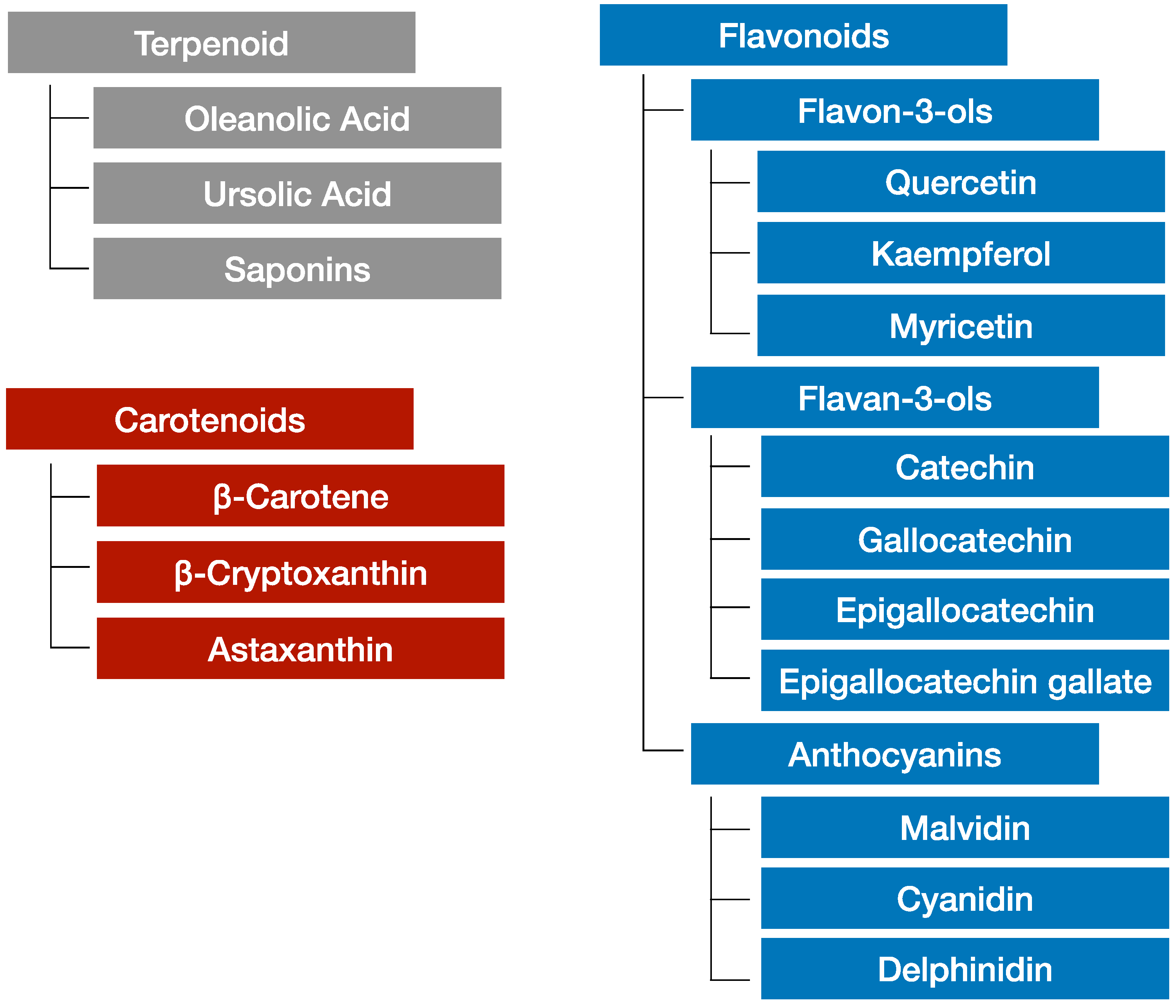
3.2. Phytochemicals in Grapes

Phytochemicals found in grapes have made headlines due to their role in the French diet, as red wine is associated with a reduced incidence of heart disease [74]. According to some advocates, people in France tend to have lower cardiovascular disease rates than their American counterparts, and one of the reasons for this phenomenon is considered to be the consumption of polyphenols. The effects of resveratrol, which are contained in red wine, became a focus of media reports at one time, and resveratrol was suggested to have positive effects on extending life span [75]. Recent research has shown that resveratrol reduces the risk of obesity and NAFLD and improves the condition of the gut microbiota [76]. The polyphenols in red wine also reportedly prevent arteriosclerosis at the cellular level [77]. In addition, one glass of wine a day is part of the Mediterranean diet, which is generally considered to be a healthy diet [78].
The phytochemicals contained in grapes can be divided into three classes: terpenoids, carotenoids, and flavonoids [79]. Figure 2 shows these classifications of grape phytochemicals [80]. Grape flavonoids are widely used in functional foods and pharmaceutical products. Table 2 summarizes the anti-metabolic syndrome and anti-inflammatory effects of grape phytochemicals. Typical examples of grape phytochemicals for pharmaceutical and nutraceutical use are grape seed extracts (GSE) [81,82]. Resveratrol is another well-known grape phytochemical [83]. Recently, the anti-inflammatory effects of flavonoids, in addition to their anti-oxidative effects, have become a focus of interest. Flavonoids suppress chronic inflammation induced by TNF-α [84]. Recent research shows that flavonoids and procyanidin suppress the senescence-associated secretory phenotype (SASP), and their anti-ageing effects are of interest to many researchers [85,86].

3.3. Combination of Vitamin D and Phytochemicals

In general, skin cells synthesize vitamin D from cholesterol after exposure to UV light. However, the amounts of vitamin D in the human body tend to become inadequate due to a variety of reasons including differences in season and latitude as well as differences in the amount of melanin pigment contained in the skin [87]. The concentration of 25-OH-D3 measured in the blood is regarded as the blood vitamin D level. The intake of vitamin D from foods and supplements is encouraged because a blood vitamin D level of >30 mg/mL is regarded as sufficient. Vitamin D is a fat-soluble vitamin. Even though vitamin D is named as a type of vitamin, it is also considered a type of steroid hormone. Vitamin D is a bone-related hormone involved in the formation and decomposition of bones, and vitamin D deficiency impairs bone calcification, resulting in rickets in children and osteomalacia in adults [88,89]. Recently, research has been focused on the immune regulatory effects and anti-inflammatory effects of vitamin D. Vitamin D is subdivided into vitamin D2, which originates from plants, and vitamin D3, which originates from animals. Vitamin D3 works more efficiently in the human body [90]. Cholesterol is converted into vitamin D in skin cells under UV exposure and is further converted into 25-OH-D3 (calcifediol) by a liver enzyme; it then circulates in the blood after bonding with vitamin D-binding proteins. Enzymes in the kidneys and immune cells convert 25-OH-D3 into 1α, 25-(OH)2-D3. Together with retinoid acid, a metabolite of vitamin A, activated 1α, 25-(OH)2-D3 binds to vitamin D receptors (VDR) in macrophages. Finally, the vitamin D-VDR complex attaches to the promotor region of the TNF-α gene and stops the production of TNF-α [91].
Table 3 shows the anti-metabolic syndrome and anti-inflammatory effects of vitamin D. The intake of vitamin D is helpful for the upregulation of natural immune responses and the maintenance of a diverse gut microbiota [92]. Vitamin D upregulates natural immunity through the induction of antimicrobial peptides, including cathelicidin LL-37 [93,94]. The intake of a high-fat diet and vitamin D deficiency concomitantly increase the population of the hepatic pathogenic Helicobacter hepaticus, and the oral administration of defensin, alpha 5 (DEFA5) suppresses their population [95]. Furthermore, vitamin D has immunosuppressive effects through IL-10 production by activated regulatory T cells (Treg) cells [96]. Activated Tregs suppress various disorders caused by chronic inflammation, including asthma [97]. In addition, vitamin D has protective effects against blood vessels, is involved in anti-oxidative activity, and suppresses proinflammatory cytokine production from inflammatory cells [98,99].

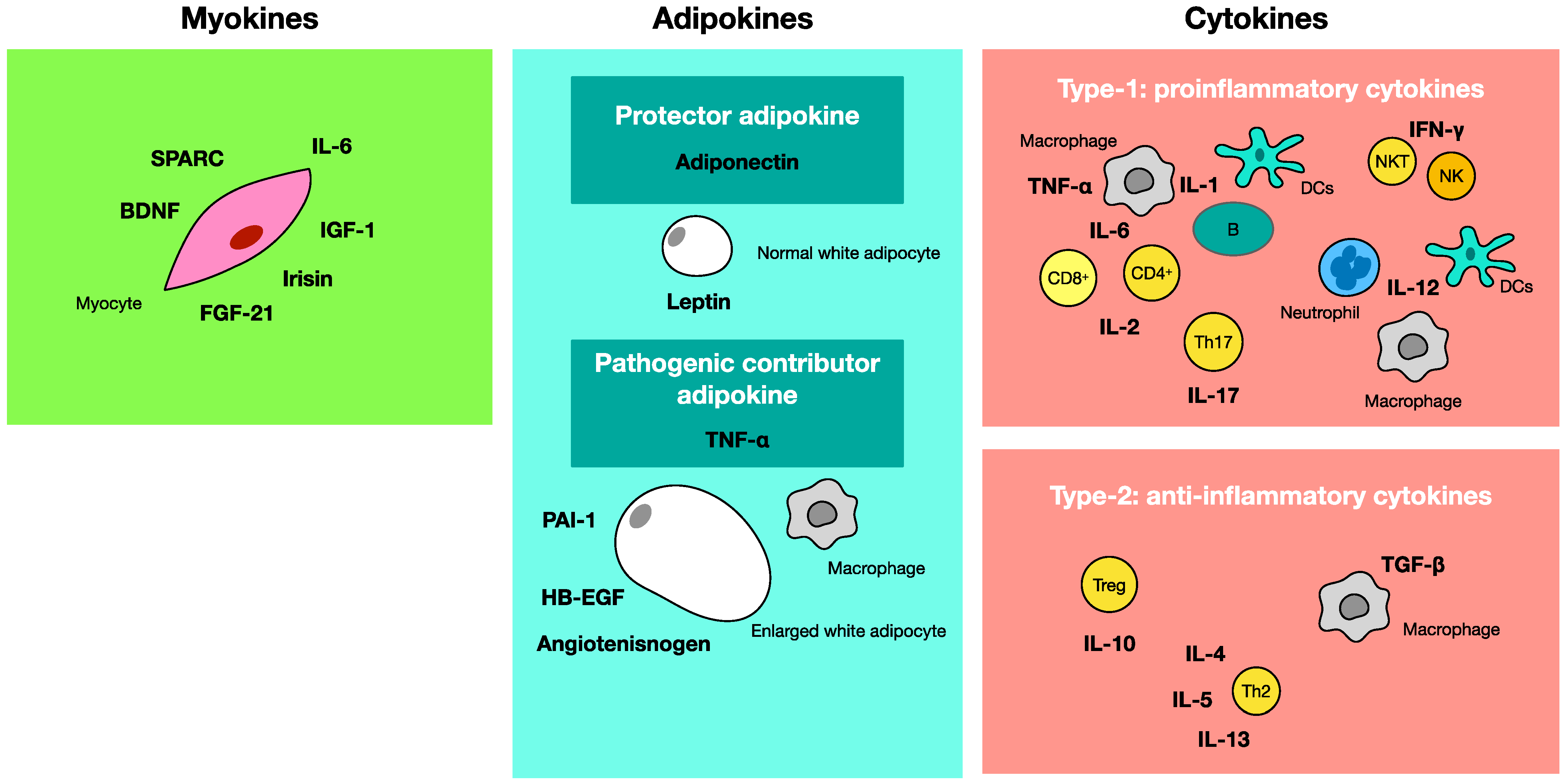
4. Adipokine, Myokine, Cytokine

Under obese conditions, inflammation is induced by the increased production of TNF-α and the decreased production of adiponectin [100]. The suppression of GLUT4 synthesis arising from the increased production of TNF-α in muscle cells induces insulin resistance and diabetes [101]. Increased adiponectin production improves metabolic syndrome because TNF-α suppression and GLUT4 production result in the improvement of glucose intake from cells [102]. On the other hand, muscle cells produce myokines through muscle stimulation from exercise. Increased myokines promote metabolism and prevent metabolic syndrome [103]. Furthermore, cytokines are produced from a variety of cells, and TNF-α is one of the proinflammatory cytokines [104]. Figure 3 shows the schematic relationship between adipokines, myokines, and cytokines.

4.1. Adipokine-Producing Adipocytes

There are three types of adipocytes: white adipocytes, brown adipocytes, and beige adipocytes. White adipocytes produce adipokines, physiologically active substances produced by adipocytes. Adiponectin produced from normal white adipocytes is a protector adipokine that suppresses metabolic syndrome, which works as a longevity-related hormone. Under normal conditions, white adipocytes are small and spheroidal and produce large amounts of adiponectin. White adipocytes also produce leptin (a peptide hormone) under normal conditions. In the presence of metabolic syndrome, however, enlarged white adipocytes produce TNF-α, which is an inflammatory pathogenic contributor adipokine [105]. Under obese conditions, however, adipocytes enlarge and change their shapes and produce TNF-α, thereby inducing inflammation, insulin resistance, and diabetes. Furthermore, the production of TNF-α reduces the production of adiponectin [106]. Enlarged white adipocytes also produce angiotensinogen, heparin-binding EGF-like growth factor (HB-EGF), and plasminogen activator inhibitor-1 (PAI-1).
White adipocytes/adipose tissue (WAT) exists in the subcutis and visceral fat, where it is involved in energy storage and release. White adipocytes change their color and function to become brown adipocytes. Only small numbers of brown adipocytes/adipose tissue (BAT) exist in the interscapulum and perirenal and periaortic abdominal aorta, where they participate in heat-producing metabolism. Transient receptor potential vanilloid 2 (TRPV2) is associated with this heat-producing mechanism in brown adipocytes [107]. Brown adipocytes decrease in the ageing process. On the other hand, beige adipocytes consume fatty acids and sugars as energy sources. Therefore, increasing the number of beige adipocytes is a good strategy for preventing metabolic syndrome [108,109].

4.2. Myokines Released from Skeletal Muscle

Myokines are produced from muscle cells in accordance with the contraction and extension of muscles during exercise. This substance has some useful properties for preventing metabolic syndrome. Currently known myokines include interleukin-6 (IL-6), fibroblast growth factor 21 (FGF-21), secreted protein acidic and rich in cysteine (SPARC), irisin, brain-derived neurotrophic factor (BDNF), and insulin-like growth factor 1 (IGF-1). IL-6 is a useful myokine for promoting metabolism and preventing obesity and diabetes. FGF21 participates in lipid, glucose, and energy metabolism [110]. SPARC is a newly identified myokine and is related to colon cancer suppression [111]. Skeletal muscle produces irisin which correlates with the browning of white adipocytes [112]. BDNF is a neurotrophic factor that promotes brain development. IGF-1 carries glucose, as does insulin, and activates nerve cells [113]. Building muscle is an effective way to increase myokine production, which prevents metabolic syndrome and ageing. A diet rich in the amino acid leucine is recommended for increasing myokine production [114].
The activation of metabolism through exercise is needed to prevent metabolic syndrome. The number of mitochondria is increased as a result of exercise, and metabolism is improved because muscle movement requires substantial amounts of ATP [115,116]. Adipose tissue releases free fatty acid (FFA) after enlargement, but muscle cells directly consume FFA as a source of energy, decreasing FFA in the body and muscle [117]. Without exercise, however, FFA accumulates in the liver as fat and induces metabolic syndrome and other chronic inflammation-related disorders [118]. Recent research shows that quercetin supplementation in combination with strength training improves the quality of muscle [119].
Mitochondria are important cytoplasmic organelles in cells that produce energy. As muscle requires a tremendous amount of energy, the role of mitochondria in muscle cells is very important. The activation of mitochondria is necessary to maintain a healthy condition because mitochondria produce ATP, an important energy source in the body. In addition to ageing, unhealthy lifestyle habits such as overeating and a lack of exercise decrease the number of mitochondria and progress the ageing of mitochondria. Aged mitochondria produce large amounts of reactive oxide. In this context, autophagy removes aged mitochondria and abnormal proteins from cells [120]. Epigallocatechin-gallate, (EGCG) a phytochemical, induces autophagy by inducing reactive oxide species in vitro [121]. To avoid metabolic syndrome, it is important not to reduce the number of mitochondria and to activate them instead. Research shows that 6 continuous weeks of dietary habits resulting in a 25% reduction of calories increases the number of mitochondria in the muscle [122].

4.3. Cytokines Produced by a Variety of Cells in the Body

Cytokine is the generic name for physiologically active substances composed of small molecular proteins produced by cells. The classification of cytokines varies because of the variety of cells producing them. Adipokines and myokines, which were mentioned previously, are cytokines in a broad sense. This section focusses mainly on type 1 proinflammatory and type 2 anti-inflammatory cytokines, cytokines produced by immune cells, and cytokines related to metabolic syndrome. Type 1 proinflammatory cytokines include IL-1, IL-2, IL-6, IL-12, IL-17, IFN-γ, and TNF-α. These cytokines are produced mainly by CD4+ type 1 helper T cells (Th1), macrophages, and dendritic cells, which characterise the type 1 immune response. In particular, IL-1, IL-6, IFN-γ, and TNF-α are regarded as important proinflammatory cytokines. These cytokines transmit signals via the type 1 cytokine receptor (CCR1), unlike other proinflammatory cytokines [123]. On the other hand, type 2 anti-inflammatory cytokines suppress proinflammatory responses. Recently, several reports have documented the effects of these cytokines in the type 2 immune response. IL-10 and TGF-β, as well as IL-4, IL-5, and IL-13, are considered anti-inflammatory cytokines. Cancer immune therapy requires the activation of cellular immunity [124], and the elimination of parasites that have infected the body requires the activation of humoral immunity [125]. Therefore, the balance of these cytokines is very important for maintaining a healthy condition [126]. In addition, recent research has shown intriguing findings suggesting a correlation between an increase in anti-inflammatory cytokines and learning and memory [127].
IL-10 production by peripherally induced-regulatory T cells (pTreg) suppresses various immune responses [128]. A good gut microbiota activates Tregs and upregulates immune responses. The butyrate-producing bacteria Faecalibacterium prausnitzii and Clostridium butyricum MIYAIRI produce a short fatty acid butyric acid and activate Tregs [129,130]. Secretory IgA antibody plays important roles in the intestinal mucosa [131]. In addition, the gut microbiota is reportedly related to the production of maternal IgA in milk [132]. The production of IgA is important for mucosal immune functions and requires activation through dendritic cells in both T cell-dependent and T cell-independent pathways [133].

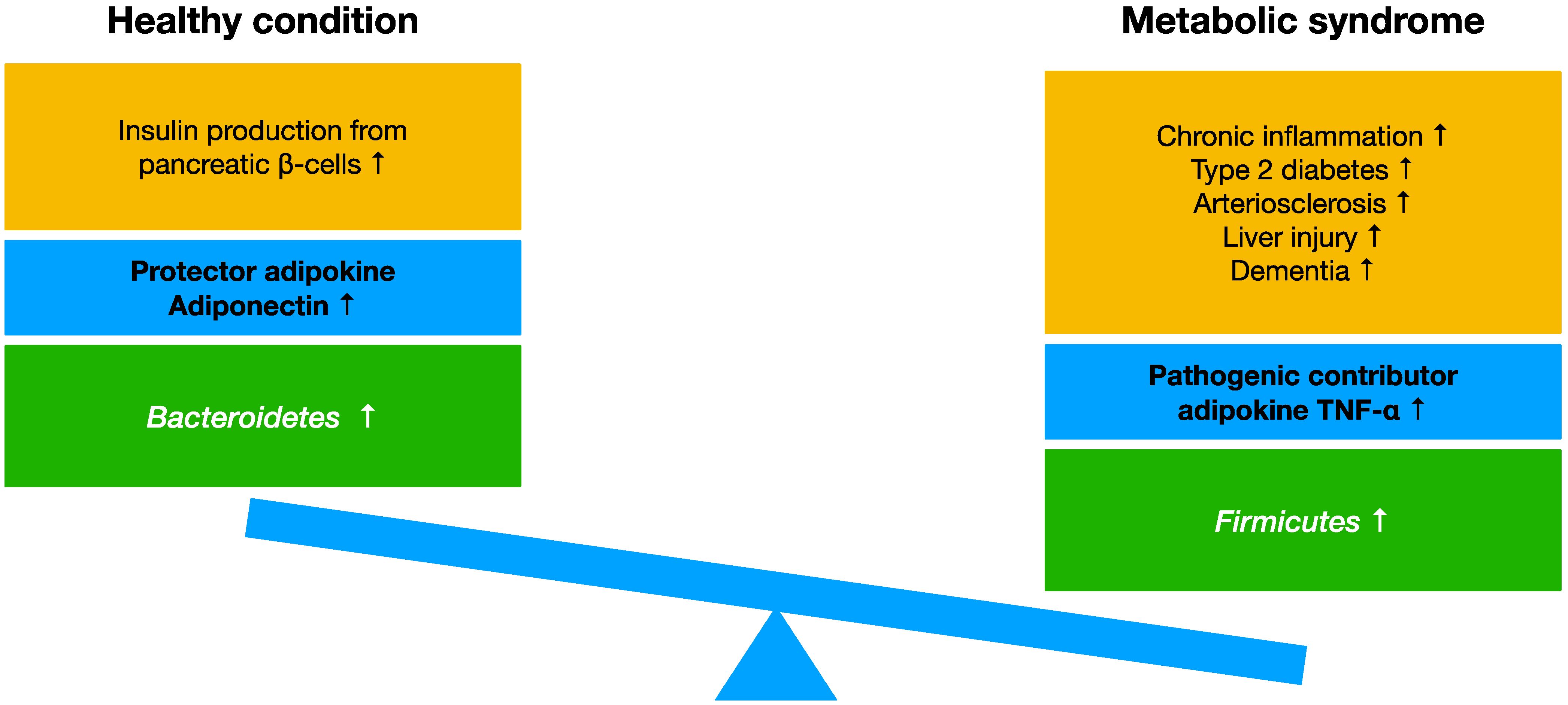
4.4. Effects of TNF-α and Adiponectin in Metabolic Syndrome

Under normal conditions, adipocytes produce adiponectin, which regulates energy consumption in the body through its actions in the hypothalamus, a critical part of the brain that controls endocrine function. This process suppresses liver glucose synthesis and promotes the burning of fat in the body. Adiponectin suppresses foam cell formation in blood vessels, vascular endothelium hyperplasia, and arteriosclerosis. Furthermore, adiponectin enhances insulin secretion from pancreatic β cells and glucose incorporation by skeletal muscle and promotes fat burning in the body [134]. On the other hand, TNF-α works as a pathogenic contributor and suppresses GLUT4 synthesis. This suppression induces insulin resistance, chronic inflammation, and metabolic syndrome. The increased production of TNF-α decreases the production of adiponectin and induces chronic inflammation [135]. To prevent metabolic syndrome, the suppression of TNF-α is important because chronic inflammation induced throughout the body leads to the onset of several disorders that in turn induce metabolic syndrome.
In metabolic syndrome, the level of TNF-α changes in accordance with the change of the gut microbiota [136]. Resveratrol contained in grapes suppresses proinflammatory factors including TNF-α and IL-17 via NF-κB modifications and changes gut microbiota. [137]. Furthermore, the deficiency of adiponectin decreases Bacteroidetes and stops the suppression of rhabdomyosarcoma [138]. It is known that patients undergoing cancer therapy tend to present with dysbiosis and a reduced ratio of adiponectin [139]. Thus, changes in TNF-α and adiponectin are highly associated with metabolic syndrome and the gut microbiota population.

5. Phytochemicals and Vitamin D Prevent Metabolic Syndrome and Improve Gut Microbiota

The consumption of phytochemicals and vitamin D with prebiotics and probiotics nurture good gut microbiota. Human gut microbiota changes with age [140], and microbiota in soil and in other creatures are related to microbiota in humans. Recently, these relationships of microbiota have been collectively called the microbiome and have been identified as important factors for understanding healthy longevity and the onset of several diseases in humans [141].
Athletes tend to have more gut microbiota diversity than other groups. They consume a greater variety of foods and perform much more exercise than other groups of people. This fact indicates that one’s gut microbiota is influenced by one’s diet [142]. In Japan, meat consumption has been increasing for several decades, whereas rice consumption has gradually decreased. A lack of exercise and excess caloric intake leads to obesity and fatty liver, which has a strong association with metabolic syndrome. In addition, reduced consumption of vegetables and a high salt intake result in imbalanced nutrition. Fast foods and sweetened diets are favorite choices of many people. On the other hand, the consumption of flavonoid-rich vegetables and fruits has decreased. A cohort study conducted in Japan revealed a relationship between the higher consumption of fruits and vegetables and a lower risk of death [143].
The typical Western diet, which is characterized by high fat and high sugar consumption, induces metabolic syndrome [144]. A large number of Gram-positive bacteria in the body are related to the cause of cellular senescence and liver cancer onset, and the consumption of a high-fat diet changes the gut microbiota [145]. The excess consumption of sweeteners induces dysbiosis [146]. Dysbiosis induced by high-fructose consumption is responsible for the induction of metabolic syndrome [147]. Recently, environmental factors such as endocrine-disrupting chemicals have been reported to induce inflammatory intestinal disorders, dysbiosis, and immunological disorders [148,149]. These factors are also related to obesity [150]. The intake of phytochemicals and vitamin D is associated with diverse and healthy gut microbiota, which prevents metabolic syndrome and maintains a healthy lifestyle.

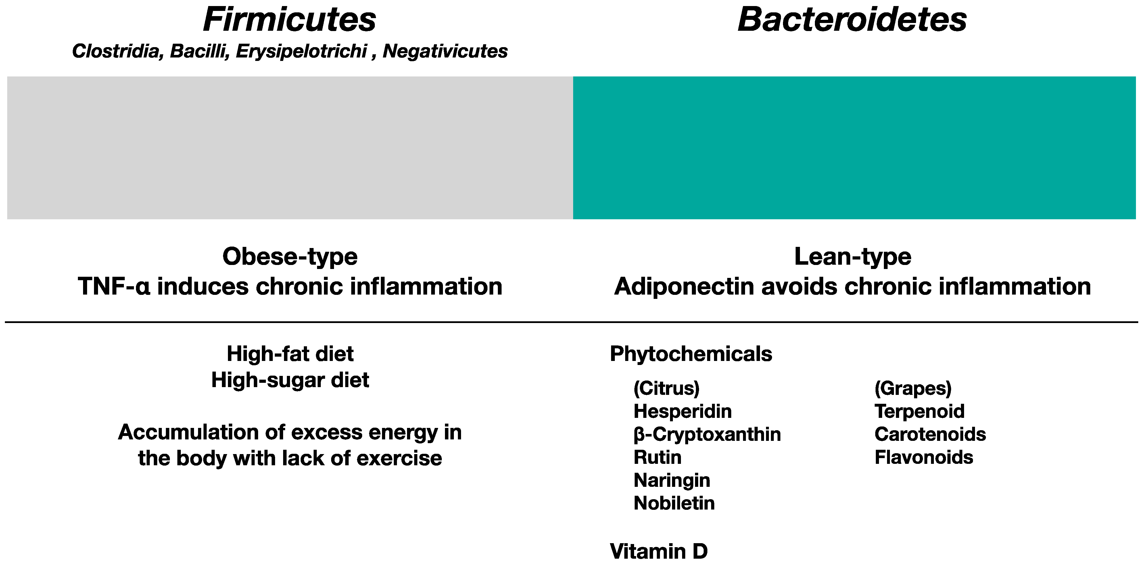
5.1. Gut Microbiota in Obese Type and Lean Type

Firmicutes bacteria are usually regarded as obese-type bacteria, whereas Bacteroidetes bacteria are regarded as lean-type bacteria [151]. Some obese-type gut microbiota produces enzymes to digest insoluble dietary fibers such as cellulose. Some decompose cellulose into glucose, inducing obesity. Reducing the F/B (Firmicutes vs. Bacteroidetes) ratio in gut microbiota is a good strategy for preventing obesity and metabolic syndrome [152]. For this purpose, reducing sugar consumption, changing the type of sugar consumed, considering the types of ingredients that are eaten together, and changing the timing of meals (e.g., avoiding eating dinner immediately before sleep as food is not digested during sleep), are good ideas. Figure 4 shows the influence of the consumption of phytochemicals and vitamin D in gut microbiota.
Firmicutes are Gram-positive bacteria and the most dominant type of gut microbiota. The phylum of Firmicutes is subdivided into four classes: Clostridia, Bacilli, Erysipelotrichi, and Negativicutes [153]. On the other hand, Bacteroidetes are Gram-negative bacteria that belong to the phylum of Bacteroidetes; Bacteroidetes are also a dominant type of gut microbiota. Decreasing Bacteroidetes bacteria in the gut causes dysbiosis and induces a thinner mucin layer and weakened cellular adhesion in the gut, leading to leaky gut syndrome; these effects can, in turn, lead to obesity, hypertension, diabetes, dyslipidemia, arteriosclerosis, NASH, and lifestyle-related diseases [154]. On the other hand, a good microbiota consists of bacteria that result in a low F/B ratio, the lactic acid bacteria Bifidobacterium [155,156], and butyrate-producing bacteria. Typical butyrate-producing bacteria include non-spore-bearing Faecalibacterium prausnitzii and spore-bearing Clostridium butyricum [157].
Maintaining a well-balanced gut microbiota is important for maintaining a healthy condition, and a gut microbiota survey of newborns to long-lived people in Japan showed an intriguing relationship between longevity and gut microbiota [158]. The colonization of Akkermansia muciniphila in the gut is associated with balanced gut immune responses [159]. Indole-3 propionate produced by gut microbiota promotes neural regeneration and restoration [160]. In addition, sleep time and the sleep rhythm influence the gut microbiota status. Metabolic syndrome disrupts the circadian rhythms, as some gut microbes produce sleep-related hormones such as melatonin. Dysbiosis reportedly induces a lack of sleep and insomnia caused by a lack of melatonin [161]. Healthy diets such as the Mediterranean diet are recommended because healthy dietary habits prevent metabolic syndrome and nurture good gut microbiota [162].

5.2. Influence of Phytochemicals and Vitamin D in Gut Microbiota

Prebiotics, probiotics [163], phytochemicals, and vitamin D (3PD) are important for maintaining healthy gut microbiota [164]. Prebiotics such as water-soluble dietary fibers, dietary fibers, and microbiota-accessible carbohydrates (MAC) nurture good gut microbiota [165]. Probiotics are beneficial bacteria for healthy gut conditions and include lactic acid bacteria and Bifidobacterium. Phytochemicals includes terpenoids, carotenoids, and flavonoids. Vitamin D is also important for nurturing good and diverse gut microbiota. Prebiotics and probiotics are effective for the recovery of the liver from conditions such as NAFLD and NASH [166]. Table 4 summarizes the influence of phytochemicals and vitamin D in gut microbiota.
In addition, the intake of both phytochemicals and vitamin D nurtures good gut microbiota. The effects of polyphenols such as quercetin on gut microbiota have been well reported. Gut microbiota metabolize these polyphenols, and the polyphenols change the components of gut microbiota [167]. The combined administration of Akkermansia muciniphila and quercetin established the colonization of A. muciniphila in mouse gut and changed the gut microbiota composition, ameliorating obesity and NAFLD by affecting bile acid metabolism [168]. Furthermore, quercetin suppressed obesity in mice that were administered sodium glutamate. In this model, quercetin ameliorated glutamate-induced hypothalamus injury and suppressed the retinol saturase (RetSat) levels that were induced by changing the composition of gut microbiota. Furthermore, quercetin administration increased the Bacteroides population and corrected the F/B ratio [169]. Other experiments show the synergy of quercetin and the water-soluble dietary fiber inulin, which led to massive bodyweight loss and the amelioration of metabolic syndrome in a high-fat-fed mouse model. The dietary fiber in inulin was thought to be decomposed into short-chain fatty acids and used as energy for intestinal endothelial cells, leading to fat decomposition and an improvement in insulin resistance. Furthermore, an increased population of the lactic acid-producing Faecalibaculum rodentium, a reduced F/B ratio, and increased GULT4 expression were also observed [170].
Vitamin D deficiency changes the balance of microbiota in the gut because of the strong relationship between vitamin D and gut microbiota [171]. The administration of vitamin D reportedly increased the Akkermansia and Faecalibacterium populations in a study examining multiple sclerosis [172]. In addition, a lack of vitamin D can cause dysbiosis and can result in some allergic responses. Vitamin D deficiency affects the gut microbiome by impairing both gut microbiota composition and the integrity of the gut epithelial barrier. In addition, vitamin D participates in immune responses via vitamin D receptor (VDR) signaling pathways [173]. Vitamin D modulates T cells and Paneth cells through VDR to modulate antimicrobial peptide release, which is involved in interactions between host and gut microbiota. Bacterial metabolites, including butyric acid, upregulate this VDR signaling pathway [174]. Because of these mechanisms, vitamin D and gut microbiota have a strong relationship. The intake of both phytochemicals and vitamin D participates in maintaining the diversity of gut microbiota, and the nurturing of good microbiota suppresses TNF-α production.

5.3. Effects of Phytochemicals and Vitamin D on the Suppression of Chronic Inflammation

Recently, the anti-inflammatory effects of phytochemicals and vitamin D have been a focus of interest, along with other effects including anti-oxidation processes and enzymatic reactions. In particular, the role of flavonoids in anti-inflammatory effects is considerable. The flavonoid quercetin suppresses metabolic syndrome, improves the condition of the gut microbiota in the presence of a high-fat diet [175], and prevents hyperlipidemia [176]. Quercetin administration also alleviates NASH, a liver disorder induced by chronic inflammation through LPS and FFA [177].
Quercetin is degraded from quercetin-glycoside by the gut microbiota and is absorbed in the gut in the same manner as other polyphenols. Then, quercetin forms quercetin-glucuronide and circulates in the blood. Macrophages incorporate oxidized-LDL and transform into TNF-α producing foam cells. These macrophages produce glucuronidase and decompose quercetin-glucuronide into its activated form. Activated quercetin overturns the states of macrophages and stops the production of TNF-α. The suppression of TNF-α by the flavonoid quercetin stops the induction of chronic inflammation [178]. In addition, a variety of immune cells in the gut express vitamin D receptor (VDR) and play a pivotal role in the interaction between gut microbiota and immune cells. Vitamin D works as a brake for excessive immune response and protects its host [179,180,181].

5.4. Molecular Mechanism for the Prevention of Metabolic Syndrome Requiring the Suppression of TNF-α and Chronic Inflammation

Metabolic syndrome, dyslipidemia, and diabetes induce continuous TNF-α production, inducing chronic inflammation in the blood vessels and the onset of atherosclerosis. Flavonoids suppress TNF-α production from macrophages [17]. Specifically, the flavonoids quercetin, hesperidin, and naringin in fruits and vegetables suppress TNF-α production by suppressing the expression of toll-like receptor 4 (TLR4) on macrophages. Flavonoids improve dysbiosis and suppress bacterial growth related to dyslipidemia, diabetes, and metabolic syndrome [182]. Fermented grape foods from Koshu, a Japanese grape strain (named K-FGF), contain the grape skin and seed paste of Vitis vinifera Koshu fermented with vegetable lactic acid bacteria. K-FGF suppresses TNF-α production and prevents chronic inflammation-induced disorders, including metabolic syndrome [183,184].
The molecular mechanism for the downregulation of macrophages related to chronic inflammation is hypothesized below (Figure 5). The flavonoid quercetin suppresses membrane fluidity and lipid raft formation on the cell surface membrane [185]. TLR4 expression on the cell surface is increased in cases with chronic inflammation. However, the suppression of TLR4 downregulates cellular signal transduction [84]. Next, activated vitamin D, 1α, 25-(OH)2-D3, binds to VDR and is transported into the nucleus. Finally, the complex of vitamin D-VDR binds to the TNF-α promoter gene and stops the production of TNF-α. This mechanism suppresses metabolic syndrome by ameliorating a variety of chronic inflammation-related disorders [15]. The consumption of citrus and grape phytochemicals together with vitamin D improves gut microbiota, suppresses chronic inflammation, and upregulates immune responses. In addition, enhanced production of LL-37 by vitamin D intake helps to suppress TNF-α production via the transcription factor NF-κB pathway, which is activated by TLR4 stimulation [186].

6. Conclusions

Metabolic syndrome is a comorbid condition associated with a variety of lifestyle-related disorders. Because this condition is induced by several diseases including obesity, diabetes, cardiovascular disorders, liver injury, and brain disorders, the prevention of metabolic syndrome is important for maintaining a healthy lifestyle and longevity. For this purpose, nurturing healthy gut microbiota, upregulating immune responses, and suppressing chronic inflammation is important. In addition, a healthy diet and moderate exercise are recommended. The intake of phytochemicals and vitamin D can help to achieve this goal. As discussed above, the consumption of phytochemicals and vitamin D is important to maintain a healthy life.

Author Contributions

Conceptualization, K.S., Y.K. and I.N.; investigation, K.S., Y.K. and I.N.; resources, K.S., Y.K. and I.N.; data curation, K.S., Y.K. and I.N.; writing—original draft preparation, K.S., Y.K. and I.N.; writing—review and editing, K.S., Y.K. and I.N.; visualization, K.S., Y.K. and I.N. All authors listed have made substantial, direct, and intellectual contributions to the work and have approved its publication. All authors have read and agreed to the published version of the manuscript.

Funding

This research received no external funding.

Conflicts of Interest

The authors declare no conflict of interest.

References

  1. Shai, I.; Schwarzfuchs, D.; Henkin, Y.; Shahar, D.R.; Witkow, S.; Greenberg, I.; Golan, R.; Fraser, D.; Bolotin, A.; Vardi, H.; et al. Dietary Intervention Randomized Controlled Trial (DIRECT) Group. Weight loss with a low-carbohydrate, Mediterranean, or low-fat diet. N. Engl. J. Med. 2008, 359, 229–241. [Google Scholar] [CrossRef] [PubMed] [Green Version]
  2. Ludwig, D.S.; Ebbeling, C.B. The Carbohydrate-Insulin Model of Obesity: Beyond “Calories In, Calories Out”. JAMA Intern. Med. 2018, 178, 1098–1103. [Google Scholar] [CrossRef] [PubMed]
  3. Cawthorn, W.P.; Sethi, J.K. TNF-alpha and adipocyte biology. FEBS Lett. 2008, 582, 117–131. [Google Scholar] [CrossRef] [PubMed] [Green Version]
  4. Li, X.; Zhang, D.; Vatner, D.F.; Goedeke, L.; Hirabara, S.M.; Zhang, Y.; Perry, R.J.; Shulman, G.I. Mechanisms by which adiponectin reverses high fat diet-induced insulin resistance in mice. Proc. Natl. Acad. Sci. USA 2020, 117, 32584–32593. [Google Scholar] [CrossRef] [PubMed]
  5. Wang, W.; Seale, P. Control of brown and beige fat development. Nat. Rev. Mol. Cell Biol. 2016, 17, 691–702. [Google Scholar] [CrossRef] [PubMed] [Green Version]
  6. Halle, M.; Berg, A.; Northoff, H.; Keul, J. Importance of TNF-alpha and leptin in obesity and insulin resistance: A hypothesis on the impact of physical exercise. Exerc. Immunol. Rev. 1998, 4, 77–94. [Google Scholar]
  7. Nedergaard, J.; Bengtsson, T.; Cannon, B. Unexpected evidence for active brown adipose tissue in adult humans. Am. J. Physiol. Endocrinol. Metab. 2007, 293, E444–E452. [Google Scholar] [CrossRef]
  8. Owens, B. Cell physiology: The changing colour of fat. Nature 2014, 508, S52–S53. [Google Scholar] [CrossRef]
  9. Wu, J.; Boström, P.; Sparks, L.M.; Ye, L.; Choi, J.H.; Giang, A.H.; Khandekar, M.; Virtanen, K.A.; Nuutila, P.; Schaart, G.; et al. Beige adipocytes are a distinct type of thermogenic fat cell in mouse and human. Cell 2012, 150, 366–376. [Google Scholar] [CrossRef] [Green Version]
  10. Chouchani, E.T.; Kazak, L.; Spiegelman, B.M. New Advances in Adaptive Thermogenesis: UCP1 and Beyond. Cell Metab. 2019, 29, 27–37. [Google Scholar] [CrossRef]
  11. Ley, R.E.; Turnbaugh, P.J.; Klein, S.; Gordon, J.I. Microbial ecology: Human gut microbes associated with obesity. Nature 2006, 444, 1022–1023. [Google Scholar] [CrossRef]
  12. Gomes, A.C.; Hoffmann, C.; Mota, J.F. The human gut microbiota: Metabolism and perspective in obesity. Gut Microbes 2018, 9, 308–325. [Google Scholar] [CrossRef] [Green Version]
  13. Chang, C.J.; Lin, T.L.; Tsai, Y.L.; Wu, T.R.; Lai, W.F.; Lu, C.C.; Lai, H.C. Next generation probiotics in disease amelioration. J. Food Drug Anal. 2019, 27, 615–622. [Google Scholar] [CrossRef] [Green Version]
  14. Di Daniele, N.; Noce, A.; Vidiri, M.F.; Moriconi, E.; Marrone, G.; Annicchiarico-Petruzzelli, M.; D’Urso, G.; Tesauro, M.; Rovella, V.; De Lorenzo, A. Impact of Mediterranean diet on metabolic syndrome, cancer and longevity. Oncotarget 2017, 8, 8947–8979. [Google Scholar] [CrossRef] [Green Version]
  15. Santa, K. Grape Phytochemicals and Vitamin D in Alleviation of Lung Disorders. Endocr. Metab. Immune. Disord. Drug Targets 2022, 22, 1276–1292. [Google Scholar] [CrossRef]
  16. Martinon, P.; Fraticelli, L.; Giboreau, A.; Dussart, C.; Bourgeois, D.; Carrouel, F. Nutrition as a Key Modifiable Factor for Periodontitis and Main Chronic Diseases. J. Clin. Med. 2021, 10, 197. [Google Scholar] [CrossRef]
  17. Kawaguchi, K.; Matsumoto, T.; Kumazawa, Y. Effects of antioxidant polyphenols on TNF-alpha-related diseases. Curr. Top. Med. Chem. 2011, 11, 1767–1779. [Google Scholar] [CrossRef] [Green Version]
  18. Dimitrov, V.; White, J.H. Vitamin D signaling in intestinal innate immunity and homeostasis. Mol. Cell Endocrinol. 2017, 453, 68–78. [Google Scholar] [CrossRef]
  19. White, J.H. Emerging Roles of Vitamin D-Induced Antimicrobial Peptides in Antiviral Innate Immunity. Nutrients 2022, 14, 284. [Google Scholar] [CrossRef]
  20. Thomas, R.L.; Jiang, L.; Adams, J.S.; Xu, Z.Z.; Shen, J.; Janssen, S.; Ackermann, G.; Vanderschueren, D.; Pauwels, S.; Knight, R.; et al. Vitamin D metabolites and the gut microbiome in older men. Nat. Commun. 2020, 11, 5997. [Google Scholar] [CrossRef]
  21. Eckel, R.H.; Grundy, S.M.; Zimmet, P.Z. The metabolic syndrome. Lancet 2005, 365, 1415–1428. [Google Scholar] [CrossRef] [PubMed]
  22. Huang, P.L. A comprehensive definition for metabolic syndrome. Dis. Model Mech. 2009, 2, 231–237. [Google Scholar] [CrossRef] [PubMed] [Green Version]
  23. Sato, J.; Kanazawa, A.; Watada, H. Type 2 Diabetes and Bacteremia. Ann. Nutr. Metab. 2017, 71, 17–22. [Google Scholar] [CrossRef] [PubMed]
  24. Kim, S.H.; Jo, S.H.; Kwon, Y.I.; Hwang, J.K. Effects of onion (Allium cepa L.) extract administration on intestinal α-glucosidases activities and spikes in postprandial blood glucose levels in SD rats model. Int. J. Mol. Sci. 2011, 12, 3757–3769. [Google Scholar] [CrossRef] [Green Version]
  25. Hube, F.; Hauner, H. The two tumor necrosis factor receptors mediate opposite effects on differentiation and glucose metabolism in human adipocytes in primary culture. Endocrinology 2000, 41, 2582–2588. [Google Scholar] [CrossRef]
  26. Zou, Y.; Zhong, L.; Hu, C.; Zhong, M.; Peng, N.; Sheng, G. LDL/HDL cholesterol ratio is associated with new-onset NAFLD in Chinese non-obese people with normal lipids: A 5-year longitudinal cohort study. Lipids Health Dis. 2021, 20, 28. [Google Scholar] [CrossRef]
  27. Luo, J.; Yang, H.; Song, B.L. Mechanisms and regulation of cholesterol homeostasis. Nat. Rev. Mol. Cell Biol. 2020, 21, 225–245. [Google Scholar] [CrossRef]
  28. Ganjali, S.; Gotto, A.M., Jr.; Ruscica, M.; Atkin, S.L.; Butler, A.E.; Banach, M.; Sahebkar, A. Monocyte-to-HDL-cholesterol ratio as a prognostic marker in cardiovascular diseases. J. Cell Physiol. 2018, 233, 9237–9246. [Google Scholar] [CrossRef]
  29. Suzuki, K.; Susaki, E.A.; Nagaoka, I. Lipopolysaccharides and Cellular Senescence: Involvement in Atherosclerosis. Int. J. Mol. Sci. 2022, 23, 11148. [Google Scholar] [CrossRef]
  30. Santos, G.M.; Ismael, S.; Morais, J.; Araújo, J.R.; Faria, A.; Calhau, C.; Marques, C. Intestinal Alkaline Phosphatase: A Review of This Enzyme Role in the Intestinal Barrier Function. Microorganisms 2022, 10, 746. [Google Scholar] [CrossRef]
  31. Eutamene, H.; Beaufrand, C.; Harkat, C.; Theodorou, V. The role of mucoprotectants in the management of gastrointestinal disorders. Expert Rev. Gastroenterol. Hepatol. 2018, 12, 83–90. [Google Scholar] [CrossRef]
  32. Duraisamy, P.; Ravi, S.; Krishnan, M.; Livya, C.M.; Manikandan, B.; Arunagirinathan, K.; Ramar, M. Dynamic Role of Macrophage Sub Types on Development of Atherosclerosis and Potential Use of Herbal Immunomodulators as Imminent Therapeutic Strategy. Cardiovasc. Hematol. Agents Med. Chem. 2022, 20, 2–12. [Google Scholar] [CrossRef]
  33. Hoving, L.R.; Katiraei, S.; Heijink, M.; Pronk, A.; van der Wee-Pals, L.; Streefland, T.; Giera, M.; Willems van Dijk, K.; van Harmelen, V. Dietary Mannan Oligosaccharides Modulate Gut Microbiota, Increase Fecal Bile Acid Excretion, and Decrease Plasma Cholesterol and Atherosclerosis Development. Mol. Nutr. Food Res. 2018, 62, e1700942. [Google Scholar] [CrossRef] [Green Version]
  34. Guan, B.; Tong, J.; Hao, H.; Yang, Z.; Chen, K.; Xu, H.; Wang, A. Bile acid coordinates microbiota homeostasis and systemic immunometabolism in cardiometabolic diseases. Acta. Pharm. Sin. B 2022, 12, 2129–2149. [Google Scholar] [CrossRef]
  35. Lanthier, N.; Delzenne, N. Targeting the Gut Microbiome to Treat Metabolic Dysfunction-Associated Fatty Liver Disease: Ready for Prime Time? Cells 2022, 11, 2718. [Google Scholar] [CrossRef]
  36. Tough, I.R.; Schwartz, T.W.; Cox, H.M. Synthetic G protein-coupled bile acid receptor agonists and bile acids act via basolateral receptors in ileal and colonic mucosa. Neurogastroenterol. Motil. 2020, 32, e13943. [Google Scholar] [CrossRef]
  37. Sugiyama, N.; Uehara, O.; Morikawa, T.; Paudel, D.; Ebata, K.; Hiraki, D.; Harada, F.; Yoshida, K.; Kato, S.; Nagasawa, T.; et al. Gut flora alterations due to lipopolysaccharide derived from Porphyromonas gingivalis. Odontology 2022, 110, 673–681. [Google Scholar] [CrossRef]
  38. Powell, E.E.; Wong, V.W.; Rinella, M. Non-alcoholic fatty liver disease. Lancet 2021, 397, 2212–2224. [Google Scholar] [CrossRef]
  39. Breuer, D.A.; Pacheco, M.C.; Washington, M.K.; Montgomery, S.A.; Hasty, A.H.; Kennedy, A.J. CD8+ T cells regulate liver injury in obesity-related nonalcoholic fatty liver disease. Am. J. Physiol. Gastrointest. Liver Physiol. 2020, 318, G211–G224. [Google Scholar] [CrossRef]
  40. Liu, Y.; Myojin, T.; Li, K.; Kurita, A.; Seto, M.; Motoyama, A.; Liu, X.; Satoh, A.; Munemasa, S.; Murata, Y.; et al. A Major Intestinal Catabolite of Quercetin Glycosides, 3-Hydroxyphenylacetic Acid, Protects the Hepatocytes from the Acetaldehyde-Induced Cytotoxicity through the Enhancement of the Total Aldehyde Dehydrogenase Activity. Int. J. Mol. Sci. 2022, 23, 1762. [Google Scholar] [CrossRef]
  41. Pal, K.; Mukadam, N.; Petersen, I.; Cooper, C. Mild cognitive impairment and progression to dementia in people with diabetes, prediabetes and metabolic syndrome: A systematic review and meta-analysis. Soc. Psychiatry Psychiatr. Epidemiol. 2018, 53, 1149–1160. [Google Scholar] [CrossRef] [PubMed]
  42. Nguyen, T.T.; Ta, Q.T.H.; Nguyen, T.K.O.; Nguyen, T.T.D.; Giau, V.V. Type 3 Diabetes and Its Role Implications in Alzheimer’s Disease. Int. J. Mol. Sci. 2020, 21, 3165. [Google Scholar] [CrossRef] [PubMed]
  43. Crowe, T.P.; Greenlee, M.H.W.; Kanthasamy, A.G.; Hsu, W.H. Mechanism of intranasal drug delivery directly to the brain. Life Sci. 2018, 195, 44–52. [Google Scholar] [CrossRef] [PubMed]
  44. Miyauchi, E.; Kim, S.W.; Suda, W.; Kawasumi, M.; Onawa, S.; Taguchi-Atarashi, N.; Morita, H.; Taylor, T.D.; Hattori, M.; Ohno, H. Gut microorganisms act together to exacerbate inflammation in spinal cords. Nature 2020, 585, 102–106. [Google Scholar] [CrossRef]
  45. Correale, J.; Hohlfeld, R.; Baranzini, S.E. The role of the gut microbiota in multiple sclerosis. Nat. Rev. Neurol. 2022, 18, 544–558. [Google Scholar] [CrossRef]
  46. Shimamura, Y.; Sei, S.; Nomura, S.; Masuda, S. Protective effects of dried mature Citrus unshiu peel (Chenpi) and hesperidin on aspirin-induced oxidative damage. J. Clin. Biochem. Nutr. 2021, 68, 149–155. [Google Scholar] [CrossRef]
  47. Pontifex, M.G.; Malik, M.M.A.H.; Connell, E.; Müller, M.; Vauzour, D. Citrus Polyphenols in Brain Health and Disease: Current Perspectives. Front. Neurosci. 2021, 15, 640648. [Google Scholar] [CrossRef]
  48. Yoshitomi, R.; Yamamoto, M.; Kumazoe, M.; Fujimura, Y.; Yonekura, M.; Shimamoto, Y.; Nakasone, A.; Kondo, S.; Hattori, H.; Haseda, A.; et al. The combined effect of green tea and α-glucosyl hesperidin in preventing obesity: A randomized placebo-controlled clinical trial. Sci. Rep. 2021, 11, 19067. [Google Scholar] [CrossRef]
  49. Kawaguchi, K.; Kikuchi, S.; Hasunuma, R.; Maruyama, H.; Yoshikawa, T.; Kumazawa, Y. A citrus flavonoid hesperidin suppresses infection-induced endotoxin shock in mice. Biol. Pharm. Bull. 2004, 27, 679–683. [Google Scholar] [CrossRef] [Green Version]
  50. Kometani, T.; Fukuda, T.; Kakuma, T.; Kawaguchi, K.; Tamura, W.; Kumazawa, Y.; Nagata, K. Effects of alpha-glucosylhesperidin, a bioactive food material, on collagen-induced arthritis in mice and rheumatoid arthritis in humans. Immunopharmacol. Immunotoxicol. 2008, 30, 117–134. [Google Scholar] [CrossRef]
  51. Yari, Z.; Cheraghpour, M.; Hekmatdoost, A. Flaxseed and/or hesperidin supplementation in metabolic syndrome: An open-labeled randomized controlled trial. Eur. J. Nutr. 2021, 60, 287–298. [Google Scholar] [CrossRef]
  52. Yari, Z.; Movahedian, M.; Imani, H.; Alavian, S.M.; Hedayati, M.; Hekmatdoost, A. The effect of hesperidin supplementation on metabolic profiles in patients with metabolic syndrome: A randomized, double-blind, placebo-controlled clinical trial. Eur. J. Nutr. 2020, 59, 2569–2577. [Google Scholar] [CrossRef]
  53. Burri, B.J. Beta-cryptoxanthin as a source of vitamin A. J. Sci. Food Agric. 2015, 95, 1786–1794. [Google Scholar] [CrossRef]
  54. Unno, K.; Noda, S.; Nii, H.; Kawasaki, Y.; Iguchi, K.; Yamada, H. Anti-stress Effect of β-Cryptoxanthin in Satsuma Mandarin Orange on Females. Biol. Pharm. Bull. 2019, 42, 1402–1408. [Google Scholar] [CrossRef] [Green Version]
  55. Yamaguchi, M. Role of carotenoid β-cryptoxanthin in bone homeostasis. J. Biomed. Sci. 2012, 19, 36. [Google Scholar] [CrossRef] [Green Version]
  56. Sodum, N.; Kumar, G.; Bojja, S.L.; Kumar, N.; Rao, C.M. Epigenetics in NAFLD/NASH: Targets and therapy. Pharmacol. Res. 2021, 167, 105484. [Google Scholar] [CrossRef]
  57. Dhuique-Mayer, C.; Gence, L.; Portet, K.; Tousch, D.; Poucheret, P. Preventive action of retinoids in metabolic syndrome/type 2 diabetic rats fed with citrus functional food enriched in β-cryptoxanthin. Food Funct. 2020, 11, 9263–9271. [Google Scholar] [CrossRef]
  58. Llopis, S.; Rodrigo, M.J.; González, N.; Genovés, S.; Zacarías, L.; Ramón, D.; Martorell, P. β-Cryptoxanthin Reduces Body Fat and Increases Oxidative Stress Response in Caenorhabditis elegans Model. Nutrients 2019, 11, 232. [Google Scholar] [CrossRef] [Green Version]
  59. Ghorbani, A. Mechanisms of antidiabetic effects of flavonoid rutin. Biomed. Pharmacother. 2017, 96, 305–312. [Google Scholar] [CrossRef]
  60. Gul, A.; Kunwar, B.; Mazhar, M.; Faizi, S.; Ahmed, D.; Shah, M.R.; Simjee, S.U. Rutin and rutin-conjugated gold nanoparticles ameliorate collagen-induced arthritis in rats through inhibition of NF-κB and iNOS activation. Int. Immunopharmacol. 2018, 59, 310–317. [Google Scholar] [CrossRef]
  61. Chen, M.; Liu, P.; Zhou, H.; Huang, C.; Zhai, W.; Xiao, Y.; Ou, J.; He, J.; El-Nezami, H.; Zheng, J. Formation and metabolism of 6-(1-acetol)-8-(1-acetol)-rutin in foods and in vivo, and their cytotoxicity. Front. Nutr. 2022, 9, 973048. [Google Scholar] [CrossRef] [PubMed]
  62. Maejima, Y.; Horita, S.; Yokota, S.; Ono, T.; Proks, P.; Yoshida-Komiya, H.; Ueta, Y.; Nishimori, K.; Misaka, S.; Shimomura, K. Identification of oxytocin receptor activating chemical components from traditional Japanese medicines. J. Food Drug Anal. 2021, 29, 653–675. [Google Scholar] [CrossRef] [PubMed]
  63. Micháliková, D.; Tyukos Kaprinay, B.; Lipták, B.; Švík, K.; Slovák, L.; Sotníková, R.; Knezl, V.; Gaspárová, Z. Natural substance rutin versus standard drug atorvastatin in a treatment of metabolic syndrome-like condition. Saudi. Pharm. J. 2019, 27, 1196–1202. [Google Scholar] [CrossRef] [PubMed]
  64. Blahova, J.; Martiniakova, M.; Babikova, M.; Kovacova, V.; Mondockova, V.; Omelka, R. Pharmaceutical Drugs and Natural Therapeutic Products for the Treatment of Type 2 Diabetes Mellitus. Pharmaceuticals 2021, 14, 806. [Google Scholar] [CrossRef] [PubMed]
  65. Den Hartogh, D.J.; Tsiani, E. Antidiabetic Properties of Naringenin: A Citrus Fruit Polyphenol. Biomolecules 2019, 9, 99. [Google Scholar] [CrossRef] [Green Version]
  66. Kawaguchi, K.; Kikuchi, S.; Hasegawa, H.; Maruyama, H.; Morita, H.; Kumazawa, Y. Suppression of lipopolysaccharide-induced tumor necrosis factor-release and liver injury in mice by naringin. Eur. J. Pharmacol. 1999, 368, 245–250. [Google Scholar] [CrossRef]
  67. Kawaguchi, K.; Maruyama, H.; Hasunuma, R.; Kumazawa, Y. Suppression of inflammatory responses after onset of collagen-induced arthritis in mice by oral administration of the Citrus flavanone naringin. Immunopharmacol. Immunotoxicol. 2011, 33, 723–729. [Google Scholar] [CrossRef]
  68. Chanet, A.; Milenkovic, D.; Deval, C.; Potier, M.; Constans, J.; Mazur, A.; Bennetau-Pelissero, C.; Morand, C.; Bérard, A.M. Naringin, the major grapefruit flavonoid, specifically affects atherosclerosis development in diet-induced hypercholesterolemia in mice. J. Nutr. Biochem. 2012, 23, 469–477. [Google Scholar] [CrossRef]
  69. Alam, M.A.; Kauter, K.; Brown, L. Naringin improves diet-induced cardiovascular dysfunction and obesity in high carbohydrate, high fat diet-fed rats. Nutrients 2013, 5, 637–650. [Google Scholar] [CrossRef] [Green Version]
  70. Nakajima, A.; Aoyama, Y.; Shin, E.J.; Nam, Y.; Kim, H.C.; Nagai, T.; Yokosuka, A.; Mimaki, Y.; Yokoi, T.; Ohizumi, Y.; et al. Nobiletin, a citrus flavonoid, improves cognitive impairment and reduces soluble Aβ levels in a triple transgenic mouse model of Alzheimer’s disease (3XTg-AD). Behav. Brain Res. 2015, 289, 69–77. [Google Scholar] [CrossRef]
  71. He, B.; Nohara, K.; Park, N.; Park, Y.S.; Guillory, B.; Zhao, Z.; Garcia, J.M.; Koike, N.; Lee, C.C.; Takahashi, J.S.; et al. The Small Molecule Nobiletin Targets the Molecular Oscillator to Enhance Circadian Rhythms and Protect against Metabolic Syndrome. Cell Metab. 2016, 23, 610–621. [Google Scholar] [CrossRef]
  72. Saini, R.K.; Ranjit, A.; Sharma, K.; Prasad, P.; Shang, X.; Gowda, K.G.M.; Keum, Y.S. Bioactive Compounds of Citrus Fruits: A Review of Composition and Health Benefits of Carotenoids, Flavonoids, Limonoids, and Terpenes. Antioxidants 2022, 11, 239. [Google Scholar] [CrossRef]
  73. Morrow, N.M.; Burke, A.C.; Samsoondar, J.P.; Seigel, K.E.; Wang, A.; Telford, D.E.; Sutherland, B.G.; O’Dwyer, C.; Steinberg, G.R.; Fullerton, M.D.; et al. The citrus flavonoid nobiletin confers protection from metabolic dysregulation in high-fat-fed mice independent of AMPK. J. Lipid Res. 2020, 61, 387–402. [Google Scholar] [CrossRef]
  74. Buja, L.M. The history, science, and art of wine and the case for health benefits: Perspectives of an oenophilic cardiovascular pathologist. Cardiovasc. Pathol. 2022, 60, 107446. [Google Scholar] [CrossRef]
  75. Baur, J.A.; Pearson, K.J.; Price, N.L.; Jamieson, H.A.; Lerin, C.; Kalra, A.; Prabhu, V.V.; Allard, J.S.; Lopez-Lluch, G.; Lewis, K.; et al. Resveratrol improves health and survival of mice on a high-calorie diet. Nature 2006, 444, 337–342. [Google Scholar] [CrossRef] [Green Version]
  76. Wang, P.; Wang, J.; Li, D.; Ke, W.; Chen, F.; Hu, X. Targeting the gut microbiota with resveratrol: A demonstration of novel evidence for the management of hepatic steatosis. J. Nutr. Biochem. 2020, 81, 108363. [Google Scholar] [CrossRef]
  77. Iijima, K.; Yoshizumi, M.; Ouchi, Y. Effect of red wine polyphenols on vascular smooth muscle cell function--molecular mechanism of the ‘French paradox’. Mech. Ageing. Dev. 2002, 123, 1033–1039. [Google Scholar] [CrossRef]
  78. Gao, Y.; Yu, X.A.; Wang, B.; Yin, G.; Wang, J.; Wang, T.; Bi, K. Based on Multi-Activity Integrated Strategy to Screening, Characterization and Quantification of Bioactive Compounds from Red Wine. Molecules 2021, 26, 6750. [Google Scholar] [CrossRef]
  79. Panche, A.N.; Diwan, A.D.; Chandra, S.R. Flavonoids: An overview. J. Nutr. Sci. 2016, 29, e47. [Google Scholar] [CrossRef] [Green Version]
  80. Santa, K.; Kumazawa, Y.; Nagaoka, I. The Potential Use of Grape Phytochemicals for Preventing the Development of Intestine-Related and Subsequent Inflammatory Diseases. Endocr. Metab. Immune. Disord. Drug Targets 2019, 19, 794–802. [Google Scholar] [CrossRef]
  81. Liu, Q.; Jiang, J.X.; Liu, Y.N.; Ge, L.T.; Guan, Y.; Zhao, W.; Jia, Y.L.; Dong, X.W.; Sun, Y.; Xie, Q.M. Grape seed extract ameliorates bleomycin-induced mouse pulmonary fibrosis. Toxicol. Lett. 2017, 273, 1–9. [Google Scholar] [CrossRef] [PubMed]
  82. Padilla-González, G.F.; Grosskopf, E.; Sadgrove, N.J.; Simmonds, M.S.J. Chemical Diversity of Flavan-3-Ols in Grape Seeds: Modulating Factors and Quality Requirements. Plants 2022, 11, 809. [Google Scholar] [CrossRef] [PubMed]
  83. Matsumoto, Y.; Katano, Y. Cardiovascular Protective Effects of Polyphenols Contained in Passion Fruit Seeds Namely Piceatannol and Scirpusin B: A Review. Tokai J. Exp. Clin. Med. 2021, 46, 151–161. [Google Scholar] [PubMed]
  84. Kumazawa, Y.; Kawaguchi, K.; Takimoto, H. Immunomodulating effects of flavonoids on acute and chronic inflammatory responses caused by tumor necrosis factor alpha. Curr. Pharm. Des. 2006, 12, 4271–4279. [Google Scholar] [CrossRef] [PubMed]
  85. Mbara, K.C.; Devnarain, N.; Owira, P.M.O. Potential Role of Polyphenolic Flavonoids as Senotherapeutic Agents in Degenerative Diseases and Geroprotection. Pharmaceut. Med. 2022, 13, 1–22. [Google Scholar] [CrossRef] [PubMed]
  86. Xu, Q.; Fu, Q.; Li, Z.; Liu, H.; Wang, Y.; Lin, X.; He, R.; Zhang, X.; Ju, Z.; Campisi, J.; et al. The flavonoid procyanidin C1 has senotherapeutic activity and increases lifespan in mice. Nat. Metab. 2021, 3, 1706–1726. [Google Scholar] [CrossRef]
  87. Cannell, J.J.; Vieth, R.; Umhau, J.C.; Holick, M.F.; Grant, W.B.; Madronich, S.; Garland, C.F.; Giovannucci, E. Epidemic influenza and vitamin D. Epidemiol. Infect. 2006, 134, 1129–1140. [Google Scholar] [CrossRef]
  88. Albaik, M.; Khan, J.A.; Sindi, I.; Akesson, K.E.; McGuigan, F.E.A. Bone mass in Saudi women aged 20–40 years: The association with obesity and vitamin D deficiency. Arch. Osteoporos. 2022, 17, 123. [Google Scholar] [CrossRef]
  89. Autier, P.; Mullie, P.; Macacu, A.; Dragomir, M.; Boniol, M.; Coppens, K.; Pizot, C.; Boniol, M. Effect of vitamin D supplementation on non-skeletal disorders: A systematic review of meta-analyses and randomised trials. Lancet Diabetes Endocrinol. 2017, 5, 986–1004. [Google Scholar] [CrossRef]
  90. Alayed Albarri, E.M.; Sameer Alnuaimi, A.; Abdelghani, D. Effectiveness of vitamin D2 compared with vitamin D3 replacement therapy in a primary healthcare setting: A retrospective cohort study. Qatar Med. J. 2022, 2022, 29. [Google Scholar] [CrossRef]
  91. Fakhoury, H.M.A.; Kvietys, P.R.; AlKattan, W.; Anouti, F.A.; Elahi, M.A.; Karras, S.N.; Grant, W.B. Vitamin D and intestinal homeostasis: Barrier, microbiota, and immune modulation. J. Steroid Biochem. Mol. Biol. 2020, 200, 105663. [Google Scholar] [CrossRef]
  92. Zeng, Y.; Luo, M.; Pan, L.; Chen, Y.; Guo, S.; Luo, D.; Zhu, L.; Liu, Y.; Pan, L.; Xu, S.; et al. Vitamin D signaling maintains intestinal innate immunity and gut microbiota: Potential intervention for metabolic syndrome and NAFLD. Am. J. Physiol. Gastrointest. Liver Physiol. 2020, 318, G542–G553. [Google Scholar] [CrossRef]
  93. Liu, P.T.; Stenger, S.; Li, H.; Wenzel, L.; Tan, B.H.; Krutzik, S.R.; Ochoa, M.T.; Schauber, J.; Wu, K.; Meinken, C.; et al. Toll-like receptor triggering of a vitamin D-mediated human antimicrobial response. Science 2006, 311, 1770–1773. [Google Scholar] [CrossRef]
  94. Adams, J.S.; Ren, S.; Liu, P.T.; Chun, R.F.; Lagishetty, V.; Gombart, A.F.; Borregaard, N.; Modlin, R.L.; Hewison, M. Vitamin d-directed rheostatic regulation of monocyte antibacterial responses. J. Immunol. 2009, 182, 4289–4295. [Google Scholar] [CrossRef] [Green Version]
  95. Su, D.; Nie, Y.; Zhu, A.; Chen, Z.; Wu, P.; Zhang, L.; Luo, M.; Sun, Q.; Cai, L.; Lai, Y.; et al. Vitamin D Signaling through Induction of Paneth Cell Defensins Maintains Gut Microbiota and Improves Metabolic Disorders and Hepatic Steatosis in Animal Models. Front. Physiol. 2016, 7, 498. [Google Scholar] [CrossRef] [Green Version]
  96. Mabrouk, R.R.; Amer, H.A.; Soliman, D.; Mohamed, N.A.; El-Ghoneimy, D.H.; Hamdy, A.M.; Atef, S.A. Vitamin D Increases Percentages of Interleukin-10 Secreting Regulatory T Cells in Children with Cow’s Milk Allergy. Egypt. J. Immunol. 2019, 26, 15–29. [Google Scholar]
  97. Huang, F.; Ju, Y.H.; Wang, H.B.; Li, Y.N. Maternal vitamin D deficiency impairs Treg and Breg responses in offspring mice and deteriorates allergic airway inflammation. Allergy Asthma Clin. Immunol. 2020, 16, 89. [Google Scholar] [CrossRef]
  98. Kim, D.H.; Meza, C.A.; Clarke, H.; Kim, J.S.; Hickner, R.C. Vitamin D and Endothelial Function. Nutrients 2020, 12, 575. [Google Scholar] [CrossRef] [Green Version]
  99. Xu, Y.; Baylink, D.J.; Chen, C.S.; Reeves, M.E.; Xiao, J.; Lacy, C.; Lau, E.; Cao, H. The importance of vitamin d metabolism as a potential prophylactic, immunoregulatory and neuroprotective treatment for COVID-19. J. Transl. Med. 2020, 18, 322. [Google Scholar] [CrossRef]
  100. Makarewicz, A.; Jamka, M.; Geltz, J.; Śmidowicz, A.; Kokot, M.; Kaczmarek, N.; Mądry, E.; Walkowiak, J. Comparison of the Effect of Endurance, Strength, and Endurance-Strength Training on Inflammatory Markers and Adipokines Levels in Overweight and Obese Adults: Systematic Review and Meta-Analysis of Randomised Trials. Healthcare 2022, 10, 1098. [Google Scholar] [CrossRef]
  101. Bourebaba, L.; Marycz, K. Pathophysiological Implication of Fetuin-A Glycoprotein in the Development of Metabolic Disorders: A Concise Review. J. Clin. Med. 2019, 8, 2033. [Google Scholar] [CrossRef] [PubMed] [Green Version]
  102. Shah, M.A.; Haris, M.; Faheem, H.I.; Hamid, A.; Yousaf, R.; Rasul, A.; Shah, G.M.; Khalil, A.A.K.; Wahab, A.; Khan, H.; et al. Cross-Talk between Obesity and Diabetes: Introducing Polyphenols as an Effective Phytomedicine to Combat the Dual Sword Diabesity. Curr. Pharm. Des. 2022, 28, 1523–1542. [Google Scholar] [CrossRef] [PubMed]
  103. Qi, C.; Song, X.; Wang, H.; Yan, Y.; Liu, B. The role of exercise-induced myokines in promoting angiogenesis. Front. Physiol. 2022, 13, 981577. [Google Scholar] [CrossRef] [PubMed]
  104. Sethi, J.K.; Hotamisligil, G.S. Metabolic Messengers: Tumour necrosis factor. Nat. Metab. 2021, 3, 1302–1312. [Google Scholar] [CrossRef]
  105. Kahn, C.R.; Wang, G.; Lee, K.Y. Altered adipose tissue and adipocyte function in the pathogenesis of metabolic syndrome. J. Clin. Investig. 2019, 129, 3990–4000. [Google Scholar] [CrossRef]
  106. Sahu, B.; Bal, N.C. Adipokines from white adipose tissue in regulation of whole body energy homeostasis. Biochimie 2022, 204, 92–107. [Google Scholar] [CrossRef]
  107. Sun, W.; Uchida, K.; Suzuki, Y.; Zhou, Y.; Kim, M.; Takayama, Y.; Takahashi, N.; Goto, T.; Wakabayashi, S.; Kawada, T.; et al. Lack of TRPV2 impairs thermogenesis in mouse brown adipose tissue. EMBO Rep. 2016, 17, 383–399. [Google Scholar] [CrossRef]
  108. Lizcano, F. The Beige Adipocyte as a Therapy for Metabolic Diseases. Int. J. Mol. Sci. 2019, 20, 5058. [Google Scholar] [CrossRef] [Green Version]
  109. Chen, Y.; Ikeda, K.; Yoneshiro, T.; Scaramozza, A.; Tajima, K.; Wang, Q.; Kim, K.; Shinoda, K.; Sponton, C.H.; Brown, Z.; et al. Thermal stress induces glycolytic beige fat formation via a myogenic state. Nature 2019, 565, 180–185. [Google Scholar] [CrossRef]
  110. Chen, Z.; Yang, L.; Liu, Y.; Huang, P.; Song, H.; Zheng, P. The potential function and clinical application of FGF21 in metabolic diseases. Front. Pharmacol. 2022, 13, 1089214. [Google Scholar] [CrossRef]
  111. Aoi, W.; Naito, Y.; Takagi, T.; Tanimura, Y.; Takanami, Y.; Kawai, Y.; Sakuma, K.; Hang, L.P.; Mizushima, K.; Hirai, Y.; et al. A novel myokine, secreted protein acidic and rich in cysteine (SPARC), suppresses colon tumorigenesis via regular exercise. Gut 2013, 62, 882–889. [Google Scholar] [CrossRef]
  112. Colaianni, G.; Cinti, S.; Colucci, S.; Grano, M. Irisin and musculoskeletal health. Ann. N. Y. Acad. Sci. 2017, 1402, 5–9. [Google Scholar] [CrossRef]
  113. Alizadeh Pahlavani, H. Exercise Therapy for People with Sarcopenic Obesity: Myokines and Adipokines as Effective Actors. Front. Endocrinol. 2022, 13, 811751. [Google Scholar] [CrossRef]
  114. Liu, C.; Liu, J.; Wang, T.; Su, Y.; Li, L.; Lan, M.; Yu, Y.; Liu, F.; Xiong, L.; Wang, K.; et al. Immunoglobulin Superfamily Containing Leucine-Rich Repeat (Islr) Participates in IL-6-Mediated Crosstalk between Muscle and Brown Adipose Tissue to Regulate Energy Homeostasis. Int. J. Mol. Sci. 2022, 23, 10008. [Google Scholar] [CrossRef]
  115. Hood, D.A.; Memme, J.M.; Oliveira, A.N.; Triolo, M. Maintenance of Skeletal Muscle Mitochondria in Health, Exercise, and Aging. Annu. Rev. Physiol. 2019, 81, 19–41. [Google Scholar] [CrossRef]
  116. Ruegsegger, G.N.; Booth, F.W. Health Benefits of Exercise. Cold Spring Harb. Perspect. Med. 2018, 8, a029694. [Google Scholar] [CrossRef] [Green Version]
  117. Lipke, K.; Kubis-Kubiak, A.; Piwowar, A. Molecular Mechanism of Lipotoxicity as an Interesting Aspect in the Development of Pathological States-Current View of Knowledge. Cells 2022, 11, 844. [Google Scholar] [CrossRef]
  118. Yoshiko, A.; Kaji, T.; Kozuka, T.; Sawazaki, T.; Akima, H. Evaluation of rehabilitation exercise effects by using gradation-based skeletal muscle echo intensity in older individuals: A one-group before-and-after trial study. BMC Geriatr. 2021, 21, 485. [Google Scholar] [CrossRef]
  119. Otsuka, Y.; Miyamoto, N.; Nagai, A.; Izumo, T.; Nakai, M.; Fukuda, M.; Arimitsu, T.; Yamada, Y.; Hashimoto, T. Effects of Quercetin Glycoside Supplementation Combined with Low-Intensity Resistance Training on Muscle Quantity and Stiffness: A Randomized, Controlled Trial. Front. Nutr. 2022, 9, 912217. [Google Scholar] [CrossRef]
  120. Abate, M.; Festa, A.; Falco, M.; Lombardi, A.; Luce, A.; Grimaldi, A.; Zappavigna, S.; Sperlongano, P.; Irace, C.; Caraglia, M.; et al. Mitochondria as playmakers of apoptosis, autophagy and senescence. Semin. Cell Dev. Biol. 2020, 98, 139–153. [Google Scholar] [CrossRef]
  121. Satoh, M.; Takemura, Y.; Hamada, H.; Sekido, Y.; Kubota, S. EGCG induces human mesothelioma cell death by inducing reactive oxygen species and autophagy. Cancer Cell Int. 2013, 13, 19. [Google Scholar] [CrossRef] [PubMed] [Green Version]
  122. Civitarese, A.E.; Carling, S.; Heilbronn, L.K.; Hulver, M.H.; Ukropcova, B.; Deutsch, W.A.; Smith, S.R.; Ravussin, E.; CALERIE Pennington Team. Calorie restriction increases muscle mitochondrial biogenesis in healthy humans. PLoS Med. 2007, 4, e76. [Google Scholar] [CrossRef]
  123. Megha, K.B.; Joseph, X.; Akhil, V.; Mohanan, P.V. Cascade of immune mechanism and consequences of inflammatory disorders. Phytomedicine 2021, 91, 153712. [Google Scholar] [CrossRef] [PubMed]
  124. Tekguc, M.; Wing, J.B.; Osaki, M.; Long, J.; Sakaguchi, S. Treg-expressed CTLA-4 depletes CD80/CD86 by trogocytosis, releasing free PD-L1 on antigen-presenting cells. Proc. Natl. Acad. Sci. USA 2021, 118, e2023739118. [Google Scholar] [CrossRef] [PubMed]
  125. Nishimura, T.; Santa, K.; Yahata, T.; Sato, N.; Ohta, A.; Ohmi, Y.; Sato, T.; Hozumi, K.; Habu, S. Involvement of IL-4-producing Vβ8.2+ CD4+ CD62L- CD45RB- T cells in non-MHC gene-controlled predisposition toward skewing into T helper type-2 immunity in BALB/c mice. J. Immunol. 1997, 158, 5698–56706. [Google Scholar] [CrossRef]
  126. Spellberg, B.; Edwards, J.E., Jr. Type 1/Type 2 immunity in infectious diseases. Clin. Infect. Dis. 2001, 32, 76–102. [Google Scholar] [CrossRef] [Green Version]
  127. Levin, S.G.; Pershina, E.V.; Bugaev-Makarovskiy, N.A.; Chernomorets, I.Y.; Konakov, M.V.; Arkhipov, V.I. Why Do Levels of Anti-inflammatory Cytokines Increase During Memory Acquisition? Neuroscience 2021, 473, 159–169. [Google Scholar] [CrossRef]
  128. Motomura, Y.; Kitamura, H.; Hijikata, A.; Matsunaga, Y.; Matsumoto, K.; Inoue, H.; Atarashi, K.; Hori, S.; Watarai, H.; Zhu, J.; et al. The transcription factor E4BP4 regulates the production of IL-10 and IL-13 in CD4+ T cells. Nat Immunol. 2011, 12, 450–459. [Google Scholar] [CrossRef] [Green Version]
  129. Lyu, M.; Suzuki, H.; Kang, L.; Gaspal, F.; Zhou, W.; Goc, J.; Zhou, L.; Zhou, J.; Zhang, W.; Shen, Z.; et al. ILC3s select microbiota-specific regulatory T cells to establish tolerance in the gut. Nature 2022, 610, 744–751. [Google Scholar] [CrossRef]
  130. Kedmi, R.; Najar, T.A.; Mesa, K.R.; Grayson, A.; Kroehling, L.; Hao, Y.; Hao, S.; Pokrovskii, M.; Xu, M.; Talbot, J.; et al. A RORγt+ cell instructs gut microbiota-specific Treg cell differentiation. Nature 2022, 610, 737–743. [Google Scholar] [CrossRef]
  131. Okai, S.; Usui, F.; Yokota, S.; Hori-i, Y.; Hasegawa, M.; Nakamura, T.; Kurosawa, M.; Okada, S.; Yamamoto, K.; Nishiyama, E.; et al. High-affinity monoclonal IgA regulates gut microbiota and prevents colitis in mice. Nat. Microbiol. 2016, 1, 16103. [Google Scholar] [CrossRef]
  132. Usami, K.; Niimi, K.; Matsuo, A.; Suyama, Y.; Sakai, Y.; Sato, S.; Fujihashi, K.; Kiyono, H.; Uchino, S.; Furukawa, M.; et al. The gut microbiota induces Peyer’s-patch-dependent secretion of maternal IgA into milk. Cell Rep. 2021, 36, 109655. [Google Scholar] [CrossRef]
  133. Tezuka, H.; Ohteki, T. Regulation of IgA Production by Intestinal Dendritic Cells and Related Cells. Front. Immunol. 2019, 10, 1891. [Google Scholar] [CrossRef]
  134. Unamuno, X.; Gómez-Ambrosi, J.; Rodríguez, A.; Becerril, S.; Frühbeck, G.; Catalán, V. Adipokine dysregulation and adipose tissue inflammation in human obesity. Eur. J. Clin. Investig. 2018, 48, e12997. [Google Scholar] [CrossRef] [Green Version]
  135. Tan, J.; Guo, L. Swimming intervention alleviates insulin resistance and chronic inflammation in metabolic syndrome. Exp. Ther. Med. 2019, 17, 57–62. [Google Scholar] [CrossRef]
  136. Hosseinkhani, F.; Heinken, A.; Thiele, I.; Lindenburg, P.W.; Harms, A.C.; Hankemeier, T. The contribution of gut bacterial metabolites in the human immune signaling pathway of non-communicable diseases. Gut Microbes. 2021, 13, 1–22. [Google Scholar] [CrossRef]
  137. Diaz-Gerevini, G.T.; Repossi, G.; Dain, A.; Tarres, M.C.; Das, U.N.; Eynard, A.R. Beneficial action of resveratrol: How and why? Nutrition 2016, 32, 174–178. [Google Scholar] [CrossRef]
  138. Peng, J.; Wang, J.Y.; Huang, H.F.; Zheng, T.T.; Li, J.; Wang, L.J.; Ma, X.C.; Xiao, H.T. Adiponectin Deficiency Suppresses Rhabdomyosarcoma Associated with Gut Microbiota Regulation. Biomed. Res. Int. 2021, 2021, 8010694. [Google Scholar] [CrossRef]
  139. Rotz, S.J.; Sangwan, N.; Nagy, M.; Tzeng, A.; Jia, M.; Moncaliano, M.; Majhail, N.S.; Eng, C. Fecal microbiota of adolescent and young adult cancer survivors and metabolic syndrome: An exploratory study. Pediatr. Hematol. Oncol. 2022, 39, 629–643. [Google Scholar] [CrossRef]
  140. Martino, C.; Dilmore, A.H.; Burcham, Z.M.; Metcalf, J.L.; Jeste, D.; Knight, R. Microbiota succession throughout life from the cradle to the grave. Nat. Rev. Microbiol. 2022, 20, 707–720. [Google Scholar] [CrossRef]
  141. Nagata, N.; Nishijima, S.; Miyoshi-Akiyama, T.; Kojima, Y.; Kimura, M.; Aoki, R.; Ohsugi, M.; Ueki, K.; Miki, K.; Iwata, E.; et al. Population-level Metagenomics Uncovers Distinct Effects of Multiple Medications on the Human Gut Microbiome. Gastroenterology 2022, 163, 1038–1052. [Google Scholar] [CrossRef] [PubMed]
  142. Donati Zeppa, S.; Agostini, D.; Gervasi, M.; Annibalini, G.; Amatori, S.; Ferrini, F.; Sisti, D.; Piccoli, G.; Barbieri, E.; Sestili, P.; et al. Mutual Interactions among Exercise, Sport Supplements and Microbiota. Nutrients 2019, 12, 17. [Google Scholar] [CrossRef] [PubMed]
  143. Sahashi, Y.; Goto, A.; Takachi, R.; Ishihara, J.; Kito, K.; Kanehara, R.; Yamaji, T.; Iwasaki, M.; Inoue, M.; Shoichiro, T.; et al. Inverse Association between Fruit and Vegetable Intake and All-Cause Mortality: Japan Public Health Center-Based Prospective Study. J. Nutr. 2022, 152, 2245–2254. [Google Scholar] [CrossRef] [PubMed]
  144. Johnson, A.R.; Wilkerson, M.D.; Sampey, B.P.; Troester, M.A.; Hayes, D.N.; Makowski, L. Cafeteria diet-induced obesity causes oxidative damage in white adipose. Biochem. Biophys. Res. Commun. 2016, 473, 545–550. [Google Scholar] [CrossRef] [Green Version]
  145. Yoshimoto, S.; Loo, T.M.; Atarashi, K.; Kanda, H.; Sato, S.; Oyadomari, S.; Iwakura, Y.; Oshima, K.; Morita, H.; Hattori, M.; et al. Obesity-induced gut microbial metabolite promotes liver cancer through senescence secretome. Nature 2013, 499, 97–101. [Google Scholar] [CrossRef]
  146. Suez, J.; Korem, T.; Zeevi, D.; Zilberman-Schapira, G.; Thaiss, C.A.; Maza, O.; Israeli, D.; Zmora, N.; Gilad, S.; Weinberger, A.; et al. Artificial sweeteners induce glucose intolerance by altering the gut microbiota. Nature 2014, 514, 181–186. [Google Scholar] [CrossRef]
  147. Kawano, Y.; Edwards, M.; Huang, Y.; Bilate, A.M.; Araujo, L.P.; Tanoue, T.; Atarashi, K.; Ladinsky, M.S.; Reiner, S.L.; Wang, H.H.; et al. Microbiota imbalance induced by dietary sugar disrupts immune-mediated protection from metabolic syndrome. Cell 2022, 185, 3501–3519.e20. [Google Scholar] [CrossRef]
  148. Magrone, T. Effects of Plastics on Human Health and Mechanisms of Action. Endocr. Metab. Immune. Disord. Drug Targets 2022, 22, 663–664. [Google Scholar] [CrossRef]
  149. Santa, K.; Ohsawa, T.; Sakimoto, T. Para-Nonylphenol Induces Apoptosis of U937 Human Monocyte Leukemia Cells in vitro. Endocr. Metab. Immune. Disord. Drug Targets 2016, 16, 213–223. [Google Scholar] [CrossRef]
  150. Murro, I.; Lisco, G.; Di Noia, C.; Lampignano, L.; Zupo, R.; Giagulli, V.A.; Guastamacchia, E.; Triggiani, V.; De Pergola, G. Endocrine Disruptors and Obesity: An Overview. Endocr. Metab. Immune. Disord. Drug Targets 2022, 22, 798–806. [Google Scholar]
  151. Bibbò, S.; Dore, M.P.; Pes, G.M.; Delitala, G.; Delitala, A.P. Is there a role for gut microbiota in type 1 diabetes pathogenesis? Ann. Med. 2017, 49, 11–22. [Google Scholar] [CrossRef]
  152. Murata, C.; Gutiérrez-Castrellón, P.; Pérez-Villatoro, F.; García-Torres, I.; Enríquez-Flores, S.; de la Mora-de la Mora, I.; Fernández-Lainez, C.; Werner, J.; López-Velázquez, G. Delivery mode-associated gut microbiota in the first 3 months of life in a country with high obesity rates: A descriptive study. Medicine 2020, 99, e22442. [Google Scholar] [CrossRef]
  153. Riva, A.; Borgo, F.; Lassandro, C.; Verduci, E.; Morace, G.; Borghi, E.; Berry, D. Pediatric obesity is associated with an altered gut microbiota and discordant shifts in Firmicutes populations. Environ. Microbiol. 2017, 19, 95–105. [Google Scholar] [CrossRef]
  154. Wilmanski, T.; Diener, C.; Rappaport, N.; Patwardhan, S.; Wiedrick, J.; Lapidus, J.; Earls, J.C.; Zimmer, A.; Glusman, G.; Robinson, M.; et al. Gut microbiome pattern reflects healthy ageing and predicts survival in humans. Nat. Metab. 2021, 3, 274–286. [Google Scholar] [CrossRef]
  155. De Filippis, F.; Pasolli, E.; Ercolini, D. The food-gut axis: Lactic acid bacteria and their link to food, the gut microbiome and human health. FEMS Microbiol. Rev. 2020, 44, 454–489. [Google Scholar] [CrossRef]
  156. Stojanov, S.; Berlec, A.; Štrukelj, B. The Influence of Probiotics on the Firmicutes/Bacteroidetes Ratio in the Treatment of Obesity and Inflammatory Bowel disease. Microorganisms 2020, 8, 1715. [Google Scholar] [CrossRef]
  157. Stoeva, M.K.; Garcia-So, J.; Justice, N.; Myers, J.; Tyagi, S.; Nemchek, M.; McMurdie, P.J.; Kolterman, O.; Eid, J. Butyrate-producing human gut symbiont, Clostridium butyricum, and its role in health and disease. Gut Microbes. 2021, 13, 1–28. [Google Scholar] [CrossRef]
  158. Odamaki, T.; Kato, K.; Sugahara, H.; Hashikura, N.; Takahashi, S.; Xiao, J.Z.; Abe, F.; Osawa, R. Age-related changes in gut microbiota composition from newborn to centenarian: A cross-sectional study. BMC Microbiol. 2016, 16, 90. [Google Scholar] [CrossRef] [Green Version]
  159. Bae, M.; Cassilly, C.D.; Liu, X.; Park, S.M.; Tusi, B.K.; Chen, X.; Kwon, J.; Filipčík, P.; Bolze, A.S.; Liu, Z.; et al. Akkermansia muciniphila phospholipid induces homeostatic immune responses. Nature 2022, 608, 168–173. [Google Scholar] [CrossRef]
  160. Serger, E.; Luengo-Gutierrez, L.; Chadwick, J.S.; Kong, G.; Zhou, L.; Crawford, G.; Danzi, M.C.; Myridakis, A.; Brandis, A.; Bello, A.T.; et al. The gut metabolite indole-3 propionate promotes nerve regeneration and repair. Nature 2022, 607, 585–592. [Google Scholar] [CrossRef]
  161. Matenchuk, B.A.; Mandhane, P.J.; Kozyrskyj, A.L. Sleep, circadian rhythm, and gut microbiota. Sleep Med. Rev. 2020, 53, 101340. [Google Scholar] [CrossRef] [PubMed]
  162. Khalil, M.; Shanmugam, H.; Abdallah, H.; John Britto, J.S.; Galerati, I.; Gómez-Ambrosi, J.; Frühbeck, G.; Portincasa, P. The Potential of the Mediterranean Diet to Improve Mitochondrial Function in Experimental Models of Obesity and Metabolic Syndrome. Nutrients 2022, 14, 3112. [Google Scholar] [CrossRef] [PubMed]
  163. Sanders, M.E.; Merenstein, D.J.; Reid, G.; Gibson, G.R.; Rastall, R.A. Probiotics and prebiotics in intestinal health and disease: From biology to the clinic. Nat. Rev. Gastroenterol. Hepatol. 2019, 16, 605–616. [Google Scholar] [CrossRef] [PubMed]
  164. Kikut, J.; Konecka, N.; Ziętek, M.; Kulpa, D.; Szczuko, M. Diet supporting therapy for inflammatory bowel diseases. Eur. J. Nutr. 2021, 60, 2275–2291. [Google Scholar] [CrossRef]
  165. Hryckowian, A.J.; Van Treuren, W.; Smits, S.A.; Davis, N.M.; Gardner, J.O.; Bouley, D.M.; Sonnenburg, J.L. Microbiota-accessible carbohydrates suppress Clostridium difficile infection in a murine model. Nat. Microbiol. 2018, 3, 662–669. [Google Scholar] [CrossRef]
  166. Carpi, R.Z.; Barbalho, S.M.; Sloan, K.P.; Laurindo, L.F.; Gonzaga, H.F.; Grippa, P.C.; Zutin, T.L.M.; Girio, R.J.S.; Repetti, C.S.F.; Detregiachi, C.R.P.; et al. The Effects of Probiotics, Prebiotics and Synbiotics in Non-Alcoholic Fat Liver Disease (NAFLD) and Non-Alcoholic Steatohepatitis (NASH): A Systematic Review. Int. J. Mol. Sci. 2022, 23, 8805. [Google Scholar] [CrossRef]
  167. Shabbir, U.; Rubab, M.; Daliri, E.B.; Chelliah, R.; Javed, A.; Oh, D.H. Curcumin, Quercetin, Catechins and Metabolic Diseases: The Role of Gut Microbiota. Nutrients 2021, 13, 206. [Google Scholar] [CrossRef]
  168. Juárez-Fernández, M.; Porras, D.; Petrov, P.; Román-Sagüillo, S.; García-Mediavilla, M.V.; Soluyanova, P.; Martínez-Flórez, S.; González-Gallego, J.; Nistal, E.; Jover, R.; et al. The Synbiotic Combination of Akkermansia muciniphila and Quercetin Ameliorates Early Obesity and NAFLD through Gut Microbiota Reshaping and Bile Acid Metabolism Modulation. Antioxidants 2021, 10, 2001. [Google Scholar] [CrossRef]
  169. Zhao, L.; Zhu, X.; Xia, M.; Li, J.; Guo, A.Y.; Zhu, Y.; Yang, X. Quercetin Ameliorates Gut Microbiota Dysbiosis That Drives Hypothalamic Damage and Hepatic Lipogenesis in Monosodium Glutamate-Induced Abdominal Obesity. Front. Nutr. 2021, 8, 671353. [Google Scholar] [CrossRef]
  170. Tan, S.; Caparros-Martin, J.A.; Matthews, V.B.; Koch, H.; O’Gara, F.; Croft, K.D.; Ward, N.C. Isoquercetin and inulin synergistically modulate the gut microbiome to prevent development of the metabolic syndrome in mice fed a high fat diet. Sci. Rep. 2018, 8, 10100. [Google Scholar] [CrossRef] [Green Version]
  171. Malaguarnera, L. Vitamin D and microbiota: Two sides of the same coin in the immunomodulatory aspects. Int. Immunopharmacol. 2020, 79, 106112. [Google Scholar] [CrossRef]
  172. Cantarel, B.L.; Waubant, E.; Chehoud, C.; Kuczynski, J.; DeSantis, T.Z.; Warrington, J.; Venkatesan, A.; Fraser, C.M.; Mowry, E.M. Gut microbiota in multiple sclerosis: Possible influence of immunomodulators. J. Investig. Med. 2015, 63, 729–734. [Google Scholar] [CrossRef] [Green Version]
  173. Murdaca, G.; Gerosa, A.; Paladin, F.; Petrocchi, L.; Banchero, S.; Gangemi, S. Vitamin D and Microbiota: Is There a Link with Allergies? Int. J. Mol. Sci. 2021, 22, 4288. [Google Scholar] [CrossRef]
  174. Battistini, C.; Ballan, R.; Herkenhoff, M.E.; Saad, S.M.I.; Sun, J. Vitamin D Modulates Intestinal Microbiota in Inflammatory Bowel Diseases. Int. J. Mol. Sci. 2020, 22, 362. [Google Scholar] [CrossRef]
  175. Etxeberria, U.; Arias, N.; Boqué, N.; Macarulla, M.T.; Portillo, M.P.; Martínez, J.A.; Milagro, F.I. Reshaping faecal gut microbiota composition by the intake of trans-resveratrol and quercetin in high-fat sucrose diet-fed rats. J. Nutr. Biochem. 2015, 26, 651–660. [Google Scholar] [CrossRef]
  176. Arai, Y.; Watanabe, S.; Kimira, M.; Shimoi, K.; Mochizuki, R.; Kinae, N. Dietary intakes of flavonols, flavones and isoflavones by Japanese women and the inverse correlation between quercetin intake and plasma LDL cholesterol concentration. J. Nutr. 2000, 130, 2243–2250. [Google Scholar] [CrossRef] [Green Version]
  177. Cano-Martínez, A.; Bautista-Pérez, R.; Castrejón-Téllez, V.; Carreón-Torres, E.; Pérez-Torres, I.; Díaz-Díaz, E.; Flores-Estrada, J.; Guarner-Lans, V.; Rubio-Ruíz, M.E. Resveratrol and Quercetin as Regulators of Inflammatory and Purinergic Receptors to Attenuate Liver Damage Associated to Metabolic Syndrome. Int. J. Mol. Sci. 2021, 22, 8939. [Google Scholar] [CrossRef]
  178. Kawai, Y.; Nishikawa, T.; Shiba, Y.; Saito, S.; Murota, K.; Shibata, N.; Kobayashi, M.; Kanayama, M.; Uchida, K.; Terao, J. Macrophage as a target of quercetin glucuronides in human atherosclerotic arteries: Implication in the anti-atherosclerotic mechanism of dietary flavonoids. J. Biol. Chem. 2008, 283, 9424–9434. [Google Scholar] [CrossRef] [Green Version]
  179. Ishisaka, A.; Kawabata, K.; Miki, S.; Shiba, Y.; Minekawa, S.; Nishikawa, T.; Mukai, R.; Terao, J.; Kawai, Y. Mitochondrial dysfunction leads to deconjugation of quercetin glucuronides in inflammatory macrophages. PLoS ONE 2013, 8, e80843. [Google Scholar] [CrossRef] [Green Version]
  180. Kawai, Y. Understanding metabolic conversions and molecular actions of flavonoids in vivo: Toward new strategies for effective utilization of natural polyphenols in human health. J. Med. Investig. 2018, 65, 162–165. [Google Scholar] [CrossRef] [Green Version]
  181. Gurkan, N. Vitamin D supplementation during pregnancy inhibits the activation of fetal membrane NF-κB pathway. Eur. Rev. Med. Pharmacol. Sci. 2022, 26, 5926–5931. [Google Scholar] [PubMed]
  182. Hu, Q.; Qu, C.; Xiao, X.; Zhang, W.; Jiang, Y.; Wu, Z.; Song, D.; Peng, X.; Ma, X.; Zhao, Y. Flavonoids on diabetic nephropathy: Advances and therapeutic opportunities. Chin. Med. 2021, 16, 74. [Google Scholar] [CrossRef] [PubMed]
  183. Tominaga, T.; Kawaguchi, K.; Kanesaka, M.; Kawauchi, H.; Jirillo, E.; Kumazawa, Y. Suppression of type-I allergic responses by oral administration of grape marc fermented with Lactobacillus plantarum. Immunopharmacol. Immunotoxicol. 2010, 32, 593–599. [Google Scholar] [CrossRef] [PubMed]
  184. Kumazawa, Y.; Takimoto, H.; Matsumoto, T.; Kawaguchi, K. Potential use of dietary natural products, especially polyphenols, for improving type-1 allergic symptoms. Curr. Pharm. Des. 2014, 20, 857–863. [Google Scholar] [CrossRef]
  185. Kaneko, M.; Takimoto, H.; Sugiyama, T.; Seki, Y.; Kawaguchi, K.; Kumazawa, Y. Suppressive effects of the flavonoids quercetin and luteolin on the accumulation of lipid rafts after signal transduction via receptors. Immunopharmacol. Immunotoxicol. 2008, 30, 867–882. [Google Scholar] [CrossRef]
  186. Molhoek, E.M.; den Hertog, A.L.; de Vries, A.M.; Nazmi, K.; Veerman, E.C.; Hartgers, F.C.; Yazdanbakhsh, M.; Bikker, F.J.; van der Kleij, D. Structure-function relationship of the human antimicrobial peptide LL-37 and LL-37 fragments in the modulation of TLR responses. Biol. Chem. 2009, 390, 295–303. [Google Scholar] [CrossRef]
Figure 1. The mechanism of onset and suppression of metabolic syndrome. High-fat, high-glucose and typical Western diet causes accumulation of excess energy in the body, reduction of protective adipokine adiponectin, and increase of pathogenic contributor adipokine TNF-α induces chronic inflammation and metabolic syndrome. Metabolic syndrome induces type 2 diabetes, arteriosclerosis, and dementia. In metabolic syndrome, gut microbiota induces dysbiosis. Increased Firmicutes and decreased Bacteroidetes in the gut microbiota induce chronic inflammation and metabolic-syndrome-related disorders. Healthy diet and exercise prevent obesity, maintain good gut microbiota, and suppress metabolic syndrome.
Ijms 24 02627 g001
Figure 2. Schematic view of grape phytochemicals in anti-metabolic syndrome and anti-inflammatory effects. This figure shows each grape phytochemical contained in grapes. Grape phytochemicals are classified into terpenoids, carotenoids, and flavonoids. Grape terpenoids are subdivided into oleanolic acid, ursolic acid, and saponins. Grape carotenoids are subdivided into β-carotene, β-cryptoxanthin, and astaxanthin. Grape flavonoids are subdivided into flavon-3-ols, flavan-3-ols, and anthocyanins.
Ijms 24 02627 g002
Figure 3. Schematic relationship of adipokines, myokines, and cytokines. This figure shows adipokines, myokines, and cytokines and their producing cells. Adipokines include protective adiponectin produced from normal white adipocytes, and pathogenic contributor TNF-α which is produced from enlarged white adipocytes and macrophages. Myokines are produced by muscle cells. Cytokines are physiologically active substances produced by cells in the body. This scheme shows immune regulatory cytokines, namely Type 1 proinflammatory cytokines and Type 2 anti-inflammatory cytokines. These cytokines are produced from macrophages, dendritic cells, neutrophils, NK cells, NKT cells, T cells, and B cells. CD4+ T cells are classified into Th1/Th2, Th17, and regulatory T cells (Treg), and each of them produces different cytokines. TNF-α belongs to both adipokines and proinflammatory cytokines. IL-6 is also classified into proinflammatory cytokines and myokines.
Ijms 24 02627 g003
Figure 4. The influence of the consumption of phytochemicals and vitamin D for the gut microbiota. High-fat, high-sugar, high-calorie diet is responsible for obesity; these conditions lead to metabolic syndrome due to the lack of exercise. This status causes chronic inflammation induced by pathogenic contributor TNF-α, and the state of microbiota becomes Firmicutes dominant. On the other hand, the intake of phytochemicals and vitamin D preferentially induces beneficial gut microbiota with increasing protector adiponectin. Mainly the increase of Bacteroidetes and the decrease of the F/B ratio is beneficial for maintaining a healthy condition.
Ijms 24 02627 g004
Figure 5. Molecular mechanism for the suppression of TNF-α and chronic inflammation. This figure shows molecular mechanism of TNF-α suppression and chronic inflammation by phytochemicals, flavonoid, and vitamin D. TNF-α induces chronic inflammation and related disorders and finally induces metabolic syndrome. Flavonoid quercetin reduces the expression of toll-like receptor 4 (TLR4) on the surface of macrophage and suppresses the formation of lipid raft on the cell membrane. Suppression of TLR4 stops activation of the NF-κB signalling pathway. In addition, vitamin D-enhanced LL-37 also suppress the activation of transcription factor NF-κB via TLR4 stimulation. Then, the active form of vitamin D, 1α, 25(OH)2-D3, conjugates with the vitamin D receptor (VDR) in the cytoplasm and is translocated to the nucleus. The vitamin D-VDR complex attaches to the gene promoter region of TNF-α and stops production of TNF-α. Interaction between phytochemicals and vitamin D suppress the production of TNF-α and induction of chronic inflammation. Finally, lowered TNF-α production suppresses the chronic-inflammation-related disorders including diabetes, cardiovascular diseases, and Alzheimer’s and suppresses metabolic syndrome.
Ijms 24 02627 g005
Table 2. Anti-metabolic syndrome and anti-inflammatory effects of grape phytochemicals.
Grape PhytochemicalsEffectsSubjectsRef
WineHealth-improving effects of wineHuman[74]
ResveratrolMaintaining health condition
in high calorie intake
Mouse/
D. melanogaster/
C. elegans/
S. cerevisiae
[75]
Reducing risks of NAFLD and gut dysbiosisMouse[76]
Atherosclerosis preventionHuman[83]
Red wine polyphenolAtherosclerosis prevention,
effects in vascular smooth muscle cells
Human and
Bovine
endothelial cells
[77]
Red wine bioactive compoundAnti-oxidative, thrombin inhibition,
lipase inhibition
Cells/Activity screening kit[78]
FlavonoidsAnti-oxidative, anti-inflammatory,
anti-carcinogenesis,
circulatory system disease prevention
Human[79]
Alleviating collagen-induced arthritis Mouse[84]
Anti-ageingHuman[85]
Grape phytochemicals, GSE,
K-FGF
Alleviating intestine related disorderedHuman/
Rat/Mouse
[80]
Grape seed extract (GSE)Lung fibrosis preventionMouse[81]
Grape seed flan-3-olsAnalysis of biosynthetic pathways
in nutraceuticals
Physical analysis[82]
ProcyanidinPreventing senescenceMouse/
Human cells
[86]
Table 3. Anti-metabolic syndrome and anti-inflammatory effects of vitamin D.
EffectsSubjectsRef
Boosting natural immunity, maintaining diversity of gut microbiotaHuman[92]
Antibacterial peptide LL-37 induction, upregulation of innate immunity Human[93]
Strengthen natural immunity by the induction of antibacterial peptideHuman[94]
Gut microbiota modification, insulin-resistance, NAFLD by defensinsMouse[95]
Treg activation by IL-10 production,
suppression of inflammatory immune response
Human/
Human cells
[96]
Suppression of chronic inflammation related disorders by Treg activationMouse[97]
Vascular vessel protection, anti-oxidative,
proinflammatory cytokine suppression
Human[98]
Upregulation of immunity against COVID-19 infectionHuman[99]
Table 4. The influence of phytochemicals and vitamin D in gut microbiota.
SourceEffectsSubjectsRef
PolyphenolsMicrobiota in metabolic disordersHuman/
Rat/Mouse
[167]
QuercetinImprovement of obesity and NAFLDMouse[168]
Correct F/B ratio, obesityMouse[169]
Insulin resistance,
increases Faecalibaculum rodentium,
improves F/B ratio, increases GULT4
Mouse[170]
Vitamin DGut microbiota modificationHuman[171]
Increasing Akkermansia and Faecalibacterium
(in multiple sclerosis)
Human[172]
Improvement of gut dysbiosisHuman[173]
Antimicrobial peptide release,
gut microbiota interaction
Human/Mouse[174]
Disclaimer/Publisher’s Note: The statements, opinions and data contained in all publications are solely those of the individual author(s) and contributor(s) and not of MDPI and/or the editor(s). MDPI and/or the editor(s) disclaim responsibility for any injury to people or property resulting from any ideas, methods, instructions or products referred to in the content.

