Review

Advances in the Biosynthesis and Molecular Evolution of Steroidal Saponins in Plants

State Key Laboratory of Hybrid Rice, College of Life Sciences, Wuhan University, Wuhan 430072, China
*
Author to whom correspondence should be addressed.
These authors contributed equally to this work.
Int. J. Mol. Sci. 2023, 24(3), 2620; https://doi.org/10.3390/ijms24032620
Submission received: 24 December 2022 / Revised: 24 January 2023 / Accepted: 26 January 2023 / Published: 30 January 2023
(This article belongs to the Special Issue Latest Research on Plant Steroids)

Abstract

:
Steroidal saponins are an important type of plant-specific metabolite that are essential for plants’ responses to biotic and abiotic stresses. Because of their extensive pharmacological activities, steroidal saponins are also important industrial raw materials for the production of steroidal drugs. In recent years, more and more studies have explored the biosynthesis of steroidal saponins in plants, but most of them only focused on the biosynthesis of their molecular skeleton, diosgenin, and their subsequent glycosylation modification mechanism needs to be further studied. In addition, the biosynthetic regulation mechanism of steroidal saponins, their distribution pattern, and their molecular evolution in plants remain unclear. In this review, we summarized and discussed recent studies on the biosynthesis, molecular regulation, and function of steroidal saponins. Finally, we also reviewed the distribution and molecular evolution of steroidal saponins in plants. The elucidation of the biosynthesis, regulation, and molecular evolutionary mechanisms of steroidal saponins is crucial to provide new insights and references for studying their distribution, diversity, and evolutionary history in plants. Furthermore, a deeper understanding of steroidal saponin biosynthesis will contribute to their industrial production and pharmacological applications.

1. Introduction

Saponins are a kind of natural secondary metabolite with various structures and functions, and are widely distributed in plants [1]. They are a group of compounds with complex chemical structures composed of a triterpenoid or steroidal aglycone molecular skeleton conjugated to a sugar moiety, classed as triterpenoid saponins and steroidal saponins, respectively [1]. Because of the combination of a hydrophobic aglycone skeleton and hydrophilic sugar chain, these compounds have water surface tension, amphiphilic (i.e., hydrophilic and hydrophobic), foaming, and emulsifying properties, which is why they are named saponins, derived from sapo, i.e., soap [1,2,3]. Saponins isolated from plants are considered to have a variety of properties, often acting to protect plants from pathogens and plant-feeding insects. For humans and animals, saponins can affect cell membrane permeability or the lysing of cells, have hypoglycemic activity, and can also affect cholesterol metabolism; they, therefore, have a wide range of applications in food, cosmetics, pharmaceuticals, and other fields [1,4].
Although saponins are ubiquitous in plants, the steroidal saponins discussed in this paper are mainly distributed in monocots, such as the Dioscoreaceae (Dioscorea zingiberensis), Melanthiaceae (Paris polyphylla), and Asparagaceae (Asparagus officinalis) [1,3]. Steroidal saponins isolated from plants can be divided into the following five types according to their molecular skeleton structures:
(1)
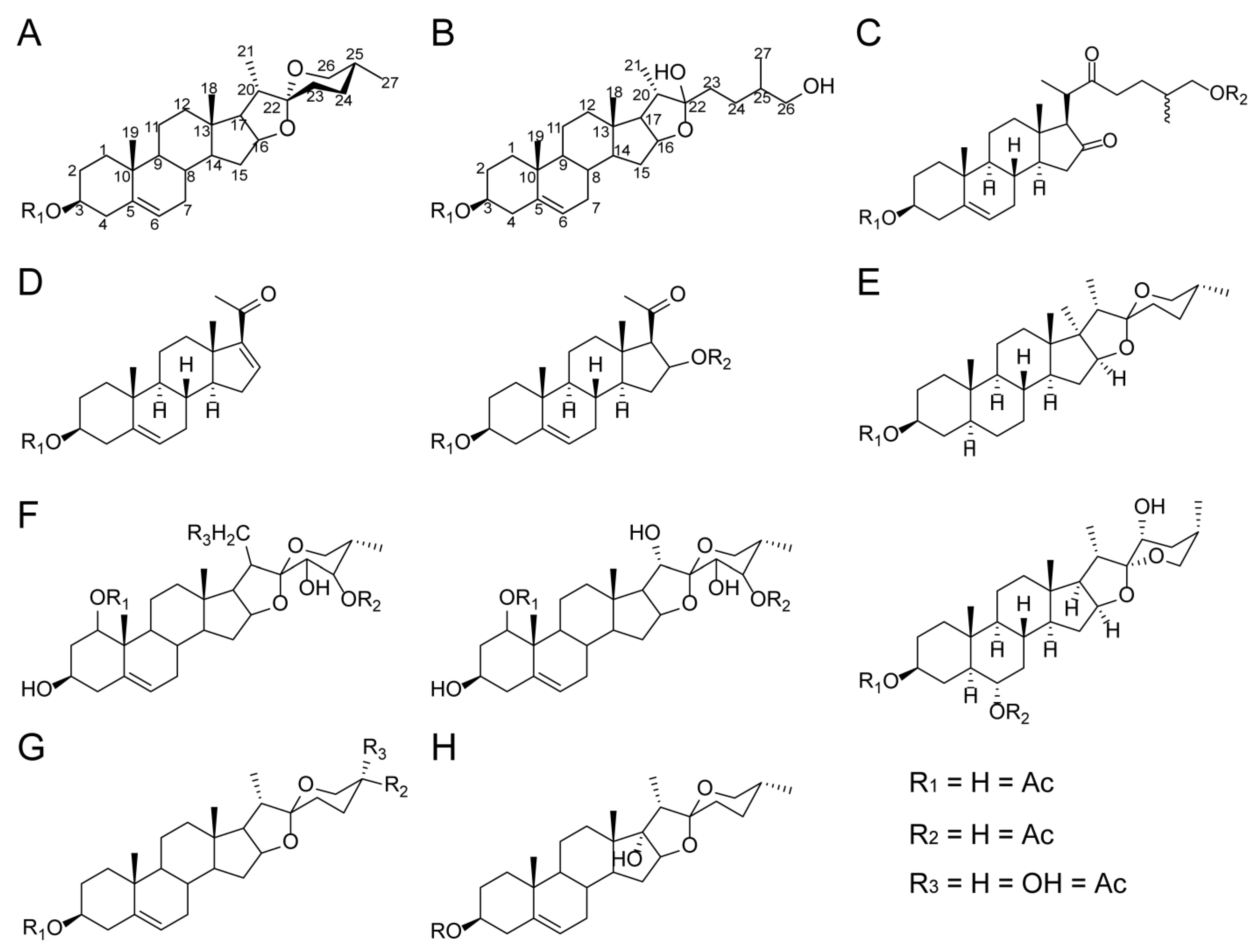
Spirostanol saponins: a hexacyclic ABCDEF-ring system characterized by an axial methyl or hydroxymethyl on the F ring (C-27) [5,6,7,8,9,10] (Figure 1A). The core aglycone of spirostanol saponins has cis- or trans-fusion between ring A and ring B, or has double bonds between ring C-5 and C-6 [7]. The common spirostanol saponins isolated from plants include dioscin, gracillin, and trillin [5,9].
(2)
Furostanol saponins: a pentacyclic ABCDE ring with a sixth open F ring [5,6,7,8,9,10] (Figure 1B). The common furostanol saponins include parvifloside, protogracillin, and protodioscin [5]. Furostanol saponins usually have 25(R) and 25(S) structures, or are saturated at the C-20 (22) or C-22 (23) positions on the open F ring [5,6]. The two sugar chains of furostanol saponins are usually connected at the C-3 and C-26 positions, while the β-glucoside at the C-26 position will produce a closed ring reaction under the catalysis of glycosidase and convert it into spirostanol saponin [8,11].
(3)
Cholestane saponins: produced by the oxidative cracking of the C-22/C-23 bond of the aglycone skeleton [7,8,9] (Figure 1C). Cholestane saponins (such as Anguivioside XV and Smilaxchinoside D) have been found in some Smilax species [7]; a homo-cholestane saponin with aromatic ring E (such as Paris pseudoside A and B) has also been found in some Paris species [8].
(4)
Pregnane saponins: a tetracyclic ABCD-ring system [5,7] (Figure 1D). They may be biosynthesized via the oxidative cleavage of the double bond between C-20 and C-22 in the structure of furostane [7,9]. Pregnane saponins isolated from plants include Spongipregnoloside A/B/C/D/E, Trinervuloside A, Riparoside B, and Timosaponin J/K [5,7].
(5)
Isospirostanol saponins: monosaccharide chain saponins. Their unique feature is the equatorial methyl or hydroxymethyl on the F ring (C-27) [7] (Figure 1E). Isospirostanol saponins include the following types: dehydrogenation between C-5 and C-6, carbonylation at C-6, hydroxylation at C-17 or C-27, and cis–trans fusion between the A ring and B ring [7].
(6)
Polyhydroxylated saponins: a six-ring system [8] (Figure 1F). They have hydroxyl groups at the C-1 position, and may also be hydroxylated at the C-1, C-3, C-21, C-23, and C-24 positions [8,12,13]. Moreover, the fructose at the C-24 position of the aglycone is unique in Paris plants [8].
(7)
Pseudospirostanol saponins, a kind of tetrahydropyran F ring, are rare in plants, and include nuatigenin and isonutigenin isolated from Paris species [8] (Figure 1G).
(8)
Pennogenin saponins are also rare in plants (Figure 1H). If diosgenin is also hydroxylated at C-17, C-23, C-24, and C-27, it can form pennogenin or hydroxy-penogenin saponin, such as polyphyllin D, Paris VI, and Paris VII isolated from Paris species [8].
Figure 1. Structural diagrams of eight types of steroidal saponins isolated from plants. (A) Spirostanol saponins, (B) furostanol saponins, (C) cholestane saponins, (D) pregnane saponins, (E) isospirostanol saponins, (F) polyhydroxylated saponins, (G) pseudospirostanol saponins, and (H) pennogenin saponins. Furostanol and spirostanol saponins are the two most common types of steroidal saponins in plants, while other types of steroidal saponins are unique to different plants.
Ijms 24 02620 g001
The diversity of steroidal saponins in plants is related to the differences in the aglycone structures, especially the differences in the aglycone oxidation mode, such as C-16, C-22, and C-26 sites in the aglycone skeleton of spirostanol saponins. They also occur rarely at sites such as C-1 and C-3, of spirostanol saponins; C-6, C-7, C-12, and C27 of furostanol saponins; and C-2 and C-5 of cholestane saponins. In addition, it is also possible to introduce double bonds between C-25 and C-27 of spirostanol’s aglycone skeleton, and between C-20 and C-22 of the aglycone skeleton of furostanol saponins [5,7,9]. According to the number of sugar units connected to the aglycone of steroidal saponins, they are categorized as monoglycosides, dioglycosides, trioglycosides, or tetraoglycosides, and the common sugar units are pyranose, rhamnose, and arabinopyranose [5,6,7,8,9]. The glycosylation sites of different types of steroidal saponins are different. Spirostanol saponins are usually in 1-OH, 3-OH, and 24-OH, and furostanol saponins are usually in 1-OH, 3-OH, and 26-OH [5,6,7,8,9].
Steroidal saponins have a wide range of functions and pharmacological activities due to their connections with different aglycone skeletons and different numbers of sugar chains, and there are also many different types of steroidal saponins in the same plant. Therefore, the distribution and existing forms of steroidal saponins in plants are diverse, and may also be related to the different functions of steroidal saponins in different plants. For plants, steroidal saponins can help them to cope with biological and abiotic stresses such as pathogens, herbivores, and drought [14]. Moses et al. believed that saponins in plants are produced in response to biological stress [1]. In stress reactions, such as when they are being attacked by herbivorous insects or during pathogen infection, the increase in the saponin contents in plants is usually mediated by hormones, such as jasmonic acid and salicylic acid [14,15,16]. For human beings, steroidal saponins isolated from plants have a variety of pharmacological uses, including antibacterial activity, antitumor activity, anti-inflammatory activity, and hypoglycemic and lipid-lowering effects, so they are an important industrial raw material for the production of steroidal drugs [1,3].
Due to the complexity of the chemical structure of steroidal saponins, their biosynthesis in plants and the formation and evolution of their synthetic pathways have not been thoroughly analyzed. Here, we introduce the biosynthesis, regulation, functions, and distribution of steroidal saponins in plants. At present, the research on steroidal saponin biosynthesis is focusing on the key enzymes involved in the side-chain hydroxylation, oxidation, and glycosylation of sterols (cholesterol or β-sitosterol). Although great progress has been made in the research of steroidal saponin biosynthesis, there may still be some unknown key steps, and their regulatory mechanisms are still unclear. The distribution, possible formation, and molecular evolution of steroidal saponins in plants are also reviewed here. Fully understanding the biosynthesis and molecular evolution of steroidal saponins can not only enhance our understanding of plants against biological and abiotic stresses, but also help promote the industrial production and pharmacological application of steroidal saponins.

2. Biosynthesis of Steroidal Saponins

Steroidal saponins are spirostanol or furostanol derivatives biosynthesized through a series of processes on the squalene molecular backbone, including oxidation, hydroxylation, and glycosylation [1,3,17]. Thus, the biosynthesis of steroidal saponins can be divided into three stages: the biosynthesis of 2,3-oxidosqualene via the mevalonate (MVA) pathway and 2-C-methyl-d-erythritol-4-phosphate (MEP) pathway, cholesterol/β-sitosterol generated by a series of catalytic reactions of 2,3-oxidosqualene, and steroidal saponins formed by the hydroxylation, oxidation, and glycosylation of cholesterol or β-sitosterol at side chains, such as the C-16, C-22, and C-26 positions [1,3,17,18,19] (Figure 2).

2.1. Biosynthesis of 2,3-Oxysqualene

The precursor of 2,3-oxsqualene is isopentenyl diphosphate (IPP). There are two ways to synthesize IPP in plants, namely MVA and MEP (Figure 2). The MVA pathway is found in the cytoplasm and mitochondria. Acetyl Co-A is catalyzed by acetyl Co-A acetyltransferase (ACAT) to form acetoacetyl Co-A, which is then catalyzed by 3-hydroxy-3-methylglutaryl-CoA synthase (HMGS), mevalonate kinase (MVA), phosphomevalonate kinase (PMK), and mevalonate-5-diphosphate decarboxylase (MVD) to generate IPP (isopentenyl-5-diphosphate) [1,3,20].
The MEP pathway is found in plastids. Glyceraldehyde-3-phosphate and pyruvate form 1-deoxy-D-xylulose 5-phosphate (DXP) under the catalysis of 1-deoxy-D-xylulose 5-phosphate synthase (DXS). Then, DXP is catalyzed by 1-deoxy-D-xylulose 5-phosphate reductoisomerase (DXR), 2-C-methyl-D-erythritol 4-phosphate cytidylyltransferase (MCT), 4-diphosphocytidyl-2-C-methyl-D-erythritol kinase (CMK), 2-C-methyl-D-erythritol 2,4-cyclodiphosphate synthase (MDS), 4-hydroxy-3-methylbut-2-enyl pyrophosphate (HMBPP) synthase (HDS), and HMBPP reductase (HDR) to generate IPP (isopentenyl -5-diphosphate) [1,3,20].
Subsequently, IPP is catalyzed by isopentenyl diphosphate isomerase (IDI), geranyl pyrophosphate synthase (GPPS), and farnesyl pyrophosphate synthase (FPPS) to synthesize farnesyl pyrophosphate (FPP). Finally, FPP is condensed by squalene synthase (SQS) to form squalene, which is further epoxidized to 2,3-oxidosqualene by squalene epoxidase (SQE) [1,3,20].

2.2. Biosynthesis of Cholesterol/β-Sitosterol

2,3-oxidosqualene is usually cyclized from a linear to a polycyclic structure by various oxidosqualene cyclases (OSCs), and the cyclization of 2,3-oxidosqualene also marks the demarcation point between sterol and triterpene metabolism in plants. 2,3-oxidosqualene is cyclized by cycloartenol synthase (CAS) to generate the plant sterol precursor cycloartenol [18]. Cholesterol/β-sitosterol are synthesized from cycloartenol under the catalysis of a series of enzymes, such as sterol side-chain reductase 1/2 (SSR1/2), sterol C-24 methyltransferase (SMT), C-4 sterol methyl oxidase (SMO), cyclopropylsterol isomerase (CPI), sterol C-14 demethylase (CYP51), sterol C-14 reductase (C14-R), sterol 8,7 isomerase (8,7 SI), sterol 5(6) desaturase (C5-SD), and 7-dehydrocholesterol reductase (7-DR) [18,20,21,22] (Figure 2).

2.3. Modification of Cholesterol or Sitosterol Side Chain to Form Steroidal Saponins

As steroidal saponins are generated by a series of complex catalytic reactions, such as the oxidation, hydroxylation, and glycosylation of cholesterol or the β-sitosterol molecular skeleton, the synthesis of steroidal saponins has been controversial at present [1,19,23] (Figure 2).
Some candidate genes related to the biosynthesis of the steroidal saponin precursor diosgenin (such as PpcyP90G4, PpCYP94D108, PpCYP94D109, PpCYP72A616, TfCYP90B50, TfCYP72A613, and TfCYP82J17) were screened from P. polyphylla and Trigonella foenum graecum (fenugreek), and the metabolite changes in transgenic Nicotiana benthamiana plants were verified by metabolomics [19]. Finally, the functions of two genes, PpCYP90G4 and TfCYP90B50, in plants were determined: they were involved in the formation of 22S-hydroxycholesterol by catalytic side chains of cholesterol [19]. The generated 22S-hydroxycholesterol was then catalyzed by CYPs and other enzymes to finally synthesize steroidal saponins [19,22].
However, Zhou et al. showed that the DzCYP90B71 gene was involved in cholesterol C22-R oxidation and that the DzCYP90G6 gene was involved in catalytic C16 oxidation through the transcriptome sequencing and targeted metabolome analysis of D. zingiberensis [17]. They believed that the CYP90 gene should catalyze the formation of cholesterol, which should be 22R hydroxycholesterol, rather than 22S hydroxycholesterol [17,19].
In general, diosgenin is generated from cholesterol through the catalysis of cholesterol C16/C22-monohydroxylase (CYP90B/CYP94), cholesterol 26-hydroxylase (CYP72A), and other enzymes. The first step of the biosynthesis of steroidal saponins from diosgenin is the glycosylation of 3-O-sterol glycosyltransferase (S3GT belongs to the UGT80 family), which transfers the glycosyl group to the 3-OH site of the diosgenin [24,25].
At present, there have been few studies on the late modification of steroidal saponin aglycones, such as diosgenin. In addition to UGT80, Song et al. found that the UGT73CR1 gene is involved in the later modification of steroidal saponins, such as the glycosylation of steroidal saponins, and has a higher affinity for diosgenin than UGT80 [25,26]. In addition, furostanol glycoside 26-O-β-glucosidase (F26G) can not only catalyze the conversion of furostanol saponins to spirostanol saponins, but is also involved in catalyzing diosgenin to form steroidal saponins [20,27]. Β-sitosterol may also be catalyzed by a series of enzymes to form steroidal saponins, which needs further exploration.
Although the current plant tissue culture technology is an effective and important means to explore and verify the biosynthesis of plant-specific metabolites [28,29], it remains difficult to construct the transgenic system of many medicinal plants, such as D. zingiberensis and P. polyphylla, which leads to difficulty in the verification of many biosynthetic pathway genes. Therefore, people use microorganisms to explore the biosynthetic mechanism of steroidal saponins [28,30], and the construction of engineering strains seems to be the most effective and economical method to produce natural plant-based drugs.
Cheng et al. first constructed a yeast chassis strain (DG-Cho) capable of producing cholesterol and then co-expressed the DzinCYP90G6 and DzinCYP94D144 (identified from D. zingiberensis) and VvCPR (cytochrome P450 reductases) genes from Vitis vinifera in the constructed strain. Finally, engineering strain DG002, which can convert cholesterol into diosgenin, was obtained [30]. The DG002 strain constructed by Cheng et al. significantly improved the conversion rate of cholesterol into diosgenin [30]. In addition, the constructed engineering strains can also be used to verify the function of the key genes involved in steroidal saponin biosynthesis isolated and identified from different plants.
It has also been found that, by modifying the synthetic pathway of ergosterol in microorganisms, they can synthesize cholesterol [31,32]. In addition to using cholesterol to synthesize steroidal saponins, microorganisms can also use their own enzyme system to convert steroidal saponins, thus increasing the types of steroidal saponins. Pgas-1 isolated and purified from Aspergillus oryzae can hydrolyze various sugars at different positions of protodioscin, thus producing various types of steroidal saponins [33]. It has been found that Absidia coerulea is able to convert the steroidal saponins in D. zingiberensis to produce five new steroidal saponins [34]. The endophytic fungus (strain 39) isolated from Dioscorea nipponica increased the content of steroidal saponins in the ethanol extract of the rhizome in vitro [35]. The production of steroidal saponins in P. polyphylla can be promoted by using Cylindrocarpon sp., Scutellospora calospora, and Trichoderma viride [36,37].

3. Molecular Regulation of Steroidal Saponin Biosynthesis in Plants

At present, there have been few in-depth studies on the regulation mechanism of steroidal saponin biosynthesis in plants. Steroidal saponins may be induced by a variety of elicitors, including abiotic and biotic elicitors. Methyl jasmonate (MeJA) is used as an abiotic elicitor to induce the synthesis of steroidal saponins in fenugreek and D. zingiberensis [38,39]. Ethylene, salt stress, and melatonin had the same effect as MeJA on increasing the production of steroidal saponins [40,41]. Biotic elicitors usually include fungi or bacteria; for example, Aspergillus niger, Fusarium oxysporum Dzf17, and Saccharomyces cerevisiae are used as fungal elicitors to increase diosgenin production in Helicteres isora, while Escherichia coli and Bacillus subtilis are used as bacterial elicitors to significantly increase diosgenin production [42]. However, the specific molecular regulation mechanism of steroidal saponin biosynthesis is still unknown. One of the main reasons is that it is difficult to establish an appropriate and efficient transgenic system in medicinal plants (such as D. zingiberensis, P. polyphylla, and fenugreek), which hinders the verification of gene functions and makes it difficult to conduct in-depth research.
Moses et al. believed that the molecular regulation mechanism of steroidal saponin biosynthesis in plants is multipronged, including environmental factors and various regulatory proteins, such as transcription factors (TFs), protein kinases, and phosphatases [1]. Among them, transcription factors are considered to play an important role in the regulation of the biosynthesis of steroidal saponins, steroidal glycoside alkaloids, and triterpene saponins [1,43,44,45].
Steroidal glycoside alkaloids (SGAs) and steroidal saponins have the same biosynthetic source; that is, they both use cholesterol as a synthetic precursor [1]. Therefore, the regulation of SGA biosynthesis can be used as a reference for research on the regulation of steroidal saponin biosynthesis. It has been reported that SGAs can also be regulated by multiple transcription factors. For example, GLYCOALKALOID METABOLISM 9 (GAME9) is an AP2/ERF transcription factor that can positively regulate the biosynthesis of SGAs in tomato and potato [46]. Specifically, GAME9 can change the expression levels of the upstream MVA pathway genes of SGA biosynthesis and cholesterol synthesis gene SSR2, and cholesterol synthesis gene C5-SD is the direct target of GAME9. Moreover, GAME9 plays a regulatory role in SGA synthesis by cooperating with the SlMYC2 transcription factor [18,46,47].
The GAME genes seem to be the target genes of the SGA biosynthetic pathway, and the silencing of the GAME11 gene leads to decreased levels of α-lycoposide in tomato [48]. In addition, MYB, AP2/EREBP, bHLH, WRKY, and other TFs may be involved in the biosynthesis of SGAs in Fritillaria imperialis and Fritillaria anhuiensis [43,44,49]. The jasmonate-responsive transcription factors of the ETHYLENE RESPONSE FACTOR (ERF) family (JREs) can regulate SGA biosynthesis by binding to the GCC box-like promoter elements of SGA biosynthesis genes in tomato [47].
Phytosterols are important metabolites of steroids, which include cholesterol, β-sitosterol, campesterol, and so on. Campesterol is a precursor of brassinosteroid (BR), which is a competitive synthesis compound of steroidal saponins and cholesterol or β-sitosterol, as a precursor of steroidal saponins [20,50]. The structure of BR is close to the molecular skeleton structure of steroidal saponins; moreover, we have found that BR is also regulated by drought and salt stress [25,51,52]. Therefore, the research results of BR biosynthesis regulation can also be used as a reference to explore the molecular regulation of steroidal saponins in plants.
The BRI1-EMS-SUPPRESSOR1 (BES1) and BRASSINAZOLE-RESISTANT1 (BZR1) families are the core TFs [53,54]; they can not only regulate BR synthesis, but also interact with other TFs to regulate BR synthesis. RD26 is an NAC transcription factor induced by drought and inhibiting the BR response [52]. WRKY46, WRKY54, and WRKY70, which are WRKY transcription factors, have the same effect as RD26 [51], and MYB TFs have been reported to regulate BR; for example, GmMYB14 overexpression decreases the BR content in soybean [55]. DWF4, which is a C-22 hydroxylase, is a target gene downstream of the regulatory pathway of BR biosynthesis. The PIF4-BES1 complex activates the promoter of the DWF4 gene to increase the accumulation of BRs and serve as a rate-limiting step of the BR biosynthetic pathway in Arabidopsis [53,56].

4. Functions of Steroidal Saponins in Plants

Steroidal saponins distribute and accumulate in tissue or organ-specific ways at different stages of plant growth and development, indicating that steroidal saponins may also play a role in plant growth and development [3,14,57,58]. The ability of plants to synthesize specific metabolites is essential for their survival. As far as steroidal saponins are concerned, the content and composition of steroidal saponins in plants are affected by the environment and growth stage [14].
In plants, steroidal saponins, like other plant antitoxins, act as a line of defense against infection by pathogens. The difference is that steroidal saponins are mainly accumulated and stored in plants and the biosynthesis and accumulation of steroidal saponins require a lot of energy, but plant antitoxins are more often produced by stress, so energy consumption is small [58]. However, there is another possibility: when plants are infected by pathogens, the content of precursor substances for steroidal saponin synthesis will increase due to stimulation, which will also lead to an increase in the steroidal saponin content, thus reducing energy consumption, to a certain extent [59]. Take the research on the relationship between saponins and pathogens in oat (Avena sativa) as an example: both triterpenoid saponins and steroidal saponins are present in oat, and the triterpenoid saponins (avenacins) are accumulated in root tip epidermal cells through active glycosylation, while avenacins have strong antifungal activity and can be released into the soil rhizosphere [60,61,62]. Compared with triterpenoid saponins, steroidal saponins in oats are stored in the vacuoles of plants as inactive disaccharides. When pathogenic bacteria infect plant tissues and destroy cell membranes, steroidal saponins will be activated so that β-Glucosidase hydrolyzes the D-glucose unit to form toxic single anthocyanin, which destroys the fungal plasma membrane by forming membrane pores, thus leading to fungal cell death [63,64].
Another role of steroidal saponins in plants is to protect them from phytophagous animals or insects. The main mechanism of action is to inhibit feeding by herbivores and insects as toxins and digestive inhibitors [65,66,67]. However, the specific mechanism may be that steroidal saponins have cytotoxicity and can destroy the cell membranes of blood cells and many animal cells [68]. Another possible explanation is that steroidal saponins lead to the penetration of insect cell membranes and the loss of cell integrity [58]. It has also been reported that steroidal saponins isolated from Allium porrum have a strong antifeedant effect on Lepidoptera insects, such as Peridroma saucia and Mamestra configurata [69].

5. Distribution of Steroidal Saponins in Plants

Steroidal saponins are synthesized and distributed in different plants. According to the current research, they are mainly distributed in monocots, such as the Dioscoreaceae (Dioscorea species), Melanthiaceae (Paris species), Smilacaceae (Smilax species), Asparagaceae (Asparagus species), Poaceae (Avena sativa), and Arecaceae (Phoenix dactylifera) [1,3]. Steroidal saponins are also distributed in a few eudicots, such as the Solanaceae (Solanum species) and Fabaceae (fenugreek) [1,3].
However, the composition of steroidal saponins in different plants varies greatly. For example, the main saponins that accumulate in Dioscorea are the five-ring furostanol saponins (parvifloside, protodioscin, and protobioside) and the six-ring spirostanol saponins (dioscin and gracillin). In addition to the accumulation of the above two steroidal saponins, four-ring pregnane saponins (such as parisyunnanoside J) and six-ring polyhydroxylated saponins (such as parisyunnanosides G/H/I and padelaoside B) accumulate in Paris plants [8,70] (Figure 1, Supplementary Table S1). In addition, the organs or tissues in which steroidal saponins accumulate in different plants are also different; for example, Dioscorea plants accumulate steroidal saponins in underground rhizomes (D. zingiberensis) or tubers (D. composita) and fenugreek accumulates them in its seeds [70,71,72,73].

5.1. Arecales Species

At present, there has been very little research on steroidal saponins in Arecaes plants, and the research that does exist mainly focuses on Phoenix dactylifera [74]. Based on ESI-MS analysis, Hamed et al. identified 21 steroidal saponins from methanol extracts of P. dactylifera pollen, among which Protodioscin was the most common. Two newly identified steroidal saponins were also reported: 3-o-histi-dine-26-o-hexosyl-dioscin and 3-o-histidine-26-o-dihexosyl hydroxydioscin [74] (Supplementary Table S1).

5.2. Asparagales Species

In contrast, Asparagales plants contain relatively more steroidal saponins, including the Amaryllidaceae, Agavaceae, Lomandroideae, Nolinoideae, Asparagoideae, and Asphodelaceae [10,75,76,77,78]. In addition, the type and distribution of steroidal saponins in these plants are also different.
At present, the main plant species containing steroidal saponins in the Amaryllidaceae include the Allioideae, Allium species. According to the structure of aglycone, steroidal saponins isolated from the flowers, bulbs, seeds, and underground parts of Allium plants can be divided into three types: spirostanol saponins with diosgenin or agigenin as aglycones, furostanol saponins with sugar chains at the C-3 and C-26 positions, and cholestane saponins with C-5 (C-6) unsaturated double bonds [10,79,80,81,82] (Figure 1, Supplementary Table S1). The sugar residues linked to steroidal saponins in Allium plants are mainly monosaccharides or disaccharides, but there are a few trisaccharides [10].
Compared with the Amaryllidaceae, Agave species are the main plants containing steroidal saponins in the Agavaceae, and a small number of steroidal saponins have also been identified from a few Agavaceae plants, such as Camascia and Yucca species [83,84,85]. The types of steroidal saponins isolated from Agavaceae plants are mainly furostanol saponins and spirosterol saponins. Meanwhile, steroidal saponins are mainly distributed in the leaves, fruits, flowers, and rhizomes of Agavaceae plants [75,86,87,88] (Supplementary Table S1). Spirostanol saponins can be divided into monosaccharides, disaccharides, and polysaccharides according to the linked glycosyl units, while furostanol saponins are mainly disaccharides and a few trisaccharides [75,86,87,88].
As for Lomandroideae plants, only five steroidal saponins have been identified in Cordyline stricta [89]. Among the Nolinoideae plants, the plants containing steroidal saponins are mainly Polygonatum, Dracaena, and Sansevieria species [9,64]. About 180 different saponins were isolated from Dracaena and Sansevieria plants, mainly furostanol saponins and spirostanol saponins, as well as a small number of cholestane saponins, pregnane saponins, and β- Sitosterol saponins [9]. In traditional Chinese medicine, the rhizomes of some Polygonatum plants are also known as “Yuzhu”, which are used to treat osteoporosis, diabetes, and lung disease [64,77,78]. Steroidal saponins isolated from the rhizomes of Polygonatum plants can be divided into three types: cholestanol-type (such as polygonatumoside A/B/C), furostanol-type (such as polygodoside G), and spirostanol-type (such as polygonoside A/B/C) [77,90,91] (Supplementary Table S1).
Among the Asparagoideae plants, steroidal saponins are mainly distributed in Asparagus species [92]. Not many steroidal saponins have been isolated from the roots and fruits of Asparagus plants, and the most common one is protodioscin [93,94]. Only a small number of steroidal saponins were isolated from the aerial parts of Hemerocallis fulva var. kwanso in the Asphodelaceae [78] (Supplementary Table S1).

5.3. Dioscoreales Plants

Dioscoreales plants are an ancient branch of monocots, among which Dioscoreaceae and Taccaceae contain steroidal saponins. Among the Dioscoreaceae plants, only Dioscorea plants contain steroidal saponins. It has been reported that, among the over 600 species of Dioscorea, about 137 contain steroidal saponins, of which 41 species of Dioscorea contain more than 1% diosgenin [95]. More than 190 steroidal saponins have been isolated and identified from Dioscorea plants, including spirostanol saponins and furostanol saponins. These steroidal saponins have also been used as chemical taxonomic markers of Dioscorea plants [70,96].
Among the nine major clades of Dioscorea pants, steroidal saponins were mainly distributed in the early diverged lineage of Dioscorea plants, such as Dioscorea sect. Stenophora [20]. The common plants containing steroidal saponins in Dioscorea sect. Stenophora are D. zingiberensis, D. panthaica, D. parviflora, D. futschauensis, D. septemloba, D. nipponica, and D. collettii var. hypoglauca [70,72,97]. More than 70 steroidal saponins have been isolated from the rhizomes of Dioscorea sect. Stenophora plants, mainly including the following two types: spirostanol saponins and furostanol saponins, and the content of spirostanol saponins is higher than that of furostanol saponins [72]. The most common of these steroidal saponins include dioscin, gracillin, protogracillin, parvifloside, protodeltonin, trillin, methyl protogracillin, and protobioside [70,72]. Species-specific steroidal saponins include progenin II and III and dioscoreside C/D/E in D. panthaica [97], and spongipregnoloside A-D and spongioside A/B in D. septemloba [98]. A few pregnane saponins have also been isolated from D. spongiosa [5,99] (Supplementary Table S1).
Other clades of Dioscorea species, such as Dioscorea sect. Combilium and Dioscorea sect. Opsophyton, also contain a few steroidal saponins, including D. esculenta and D. bulbifera [100,101]. However, there has been little research on steroidal saponins in the Dioscorea sect. Shannicorea, Dioscorea sect. Lasiophyton, Dioscorea sect. Botryosicyos, and Dioscorea sect. Enantiopheyllum plants are mainly distributed in China. Among the Dioscorea plants distributed outside China, D. composita, native to Mexico, also contains a high content of steroidal saponins, which is another important steroidal drug resource plant, in addition to D. zingiberensis [95]. It has been determined that the content of diosgenin in the tubers of D. composita is 3.68 ± 0.5% (dry weight) [94]. In Taccaceae, only Tacca chantrieri contains steroid saponins; about five furostanol saponins and two spirostanol saponins were identified from its rhizome [102] (Supplementary Table S1).

5.4. Liliales Plants

Liliales is an early diverged lineage of monocots, and the plants in the lineage containing steroidal saponins are the Liliaceae, Melanthiaceae, and Smilacaceae.
(1)
Liliaceae plants. At present, the research on steroidal saponins of Liliaceae plants has mainly focused on Lilium and Fritillaria plants [103]. More than 80 steroidal saponins have been isolated and identified from the bulbs of Lilium lancifolium, L pumilum, L. longiflorum, L. candidum, L. speciosum, L. tenuifolium, L. callosum, and other Lilium plants, which are mainly divided into four types: spirostanol-type, furostanol-type, isospirostanol-type, and pseudospirostanol saponins [103]. Specifically, spirostanol saponins isolated from Lilium plants mainly include lilioglycoside B/H and brownioside, which are unique to Lilium plants [103,104,105,106,107]; the isolated isospirostanol saponins include lilioglycoside C, D, and I, respectively; and furostanol saponins mainly include lilioglycoside K, N, R and pardarinoside A–D, F, and G [103,104,105,106,107]. Among Fritillaria plants, only 12 steroidal saponins have been isolated and identified from the bulbs of Fritillaria pallidiflora, such as pallidiflosides D/E/G/H/I, protobioside, and Polyphyllin V [108] (Supplementary Table S1).
(2)
Melanthiaceae plants. Among the plants in this family, Paris and Trillium are the largest two genera; they are important Chinese medicine plants with a long medicinal history [109,110]. A variety of steroidal saponins have been isolated and identified from the rhizomes of Paris plants, including spirostanol-type, furostanol-type, cholestane-type, pregnane-type, and polyhydroxylated-type saponins [8]. Steroidal saponins isolated and identified from Trillium plants can be divided into the following types: spirostanol, furostanol, and pennogenin saponins. Among them, the specific saponins of Trillium plants are trillins, trikamsteroside C, trillenoside A, parisapioside C, etc. [111] (Supplementary Table S1).
(3)
Smilacaceae plants. The most common plants containing steroidal saponins are Smilax plants [7]. A total of 104 steroidal saponins have been isolated from about 20 species of Smilax plants [7]. These steroidal saponins include five types: spirostanol-type, isospirostanol-type, furostanol-type, pregnane-type, and cholestane-type; most of the linked sugar groups are monosaccharides or disaccharides [7,112,113,114,115].
Steroidal saponins are also found in a few monocots, such as Poales and Zingiberales. Poaceae are only Avena plants (oats), such as Avena sativa and A. strigosa. A total of 16 steroidal saponins were isolated and identified from oats, including Avenacoside A, B, C, D, etc. [116]. Oats are also one of the few plants that contain both triterpenoid saponins and steroidal saponins. However, Costus Spiralis and C. speciosus are the only Zingiberales containing steroidal saponins in their rhizomes [117,118] (Supplementary Table S1).

5.5. Eudicots

In eudicots, steroidal saponins were identified mainly from a few plants, such as the Fabales, Ranunculales, and Solanales. Among them, Fabaceae plants only contained a small number of steroidal saponins in the seeds of fenugreek [71,73]. A small number of steroidal saponins was identified in two plants (Helleborus niger and H. orientalis) in the Ranunculaceae, including one polyhydroxy saponin: polyhydroxy hellebosaponins [118,119]. In Solanaceae plants, steroidal saponins are mainly distributed in Solanum plants. For example, three furostanol saponins, four spirostanol saponins, two cholestane-type saponins, and one spirostanol saponin were identified in Solanum abutilides, S. anguivi, and S. melongena [120,121,122,123] (Supplementary Table S1).
In addition, more than 17 steroidal saponins, including Terrestrinin C-T, were identified in the Zygophyllales plant Tribulus terrestris [124,125].
It can be seen from the above research results that steroidal saponins are mainly distributed in the early diverged lineages of monocots, including the Asparaceae (Asparagus), Dioscoreaceae (Dioscorea), Liliaceae (Lilium), Melanthiaceae (Paris and Trillium), and Smilacaceae (Smilax). There are also a few eudicots, mainly distributed in Solanaceae plants, that produce these compounds (Supplementary Table S1). Furthermore, Li et al. evaluated the evolution and distribution pattern of steroidal saponins in Dioscorea plants through ancestral states analysis [20]. Their results showed that steroidal saponins were distributed intensively in Dioscorea sect. Stenophora species. With the evolution of Dioscorea species, the frequency of steroidal saponins in plants is decreasing, with only small numbers in the more evolved clades, such as New world I, New world II, sect. Opsophython, and Malagasy clade [20,126]. A large number of ancestral nodes of steroidal saponins have been evaluated in the Dioscorea sect. Stenophora branches, indicating that steroidal saponins are an ancestral trait in Dioscorea species, mainly distributed in the early differentiated lineages of Dioscorea, and are phylogenetically conserved [120].
However, the mechanism of the distribution and accumulation of steroidal saponins in a few species of the above families and genera and how the distribution differences evolved are still unknown. The answers to these questions are of great significance for the systematic understanding of the distribution and evolution pattern of steroidal saponins in plants.

6. Molecular Evolution of Steroidal Saponin Biosynthetic Pathway Genes

Steroidal saponins are mainly distributed in Dioscorea, Paris, Asparagus, and other plants. At present, only D. zingiberensis, Dioscorea rotundata, asparagus, P. dactylifera, oats, and other plant genomes have been assembled and annotated. However, steroidal saponins are only abundant in D. zingiberensis, and the content is relatively small in other plants. Moreover, the genomes of most plants containing steroidal saponins, such as Allium, Paris, Smilax, Trillium, and Polygonatum, have not yet been reported. In the absence of species genome data, only comparative transcriptome analysis can be used to analyze the biosynthesis and evolution of plant steroidal saponins to a limited extent.
Therefore, it is the first/best choice to analyze the biosynthesis and evolution of steroidal saponins by sequencing and assembling the genome of D. zingiberensis. Other plants, such as fenugreek and trillium, are also suitable research materials. Li et al. assembled a high-quality chromosome-level genome of D. zingiberensis by combining Illumina and Oxford Nanopore technologies [20]. Through comparative genomic analysis, it was found that there were a large number of species-specific genes in the genome of D. zingiberensis, and these specific genes are significantly enriched in the biological processes of the responses to pathogens, oxidative stress, etc. This finding indicates that D. zingiberensis has evolved a large number of specific genes for coping with stress in order to adapt to the environment, and there are many D. zingiberensis-specific genes involved in the biosynthesis of steroidal saponins [20]. These results show that the genome of D. zingiberensis produced a relatively large number of genes involved in steroidal saponin biosynthesis during the evolutionary process, which may also be the premise or basis for steroidal saponin synthesis in D. zingiberensis [20].
Furthermore, there are a large number of expanded gene families in the D. zingiberensis genome, and these gene families are significantly enriched in UDP-glycosyltransferases activity, mono-oxygenase activity, steroid biosynthesis, stress response, and other processes related to steroidal saponin synthesis [20]. Moreover, these expanded gene families also include the gene family members of OSC, CYP450, and UGT, which play important roles in several key steps of steroidal saponin biosynthesis [1,20]. Thus, compared with other plants, the species-specific genes and significantly expanded gene families in the genome of D. zingiberensis provide abundant genetic and evolutionary resources related to steroidal saponin biosynthesis [20].
The expansion or contraction of gene families is an important factor in species differentiation, phenotypic diversification, and adaptation to natural changes [127,128]. Similarly, whole-genome duplication (WGD) events are also considered to be the major driving force of species diversity and phenotypic variation [129,130,131]. Specifically, the role of WGD and gene tandem duplication is mainly to produce a large number of gene copies in the genome. Further analysis showed that a large number of copies of key genes involved in the biosynthesis of steroidal saponins in D. zingiberensis, including CAS, CYP72A, CYP90B, CYP94, S3GT, and F26G, were produced through WGD and gene tandem duplication events [20]. Combined with the above results, gene tandem duplication coupled with WGD events in the genome may be the driving force to promote the evolution of the steroidal saponin biosynthetic pathway in D. zingiberensis [20].
Gene replication can help plants to obtain more gene copies, which may produce new gene functions that are more adaptive than single-copy or low-copy genes [130]. For example, sterol side-chain catalytic enzyme-encoding genes, such as CYP90B, CYP94, and CYP72A, play an important role in steroidal saponin biosynthesis, and the functions of these genes vary between different plants [5,18,132]. In plants without steroidal saponins, the CYP90 gene is involved in catalyzing the conversion of cholesterol into 22S hydroxycholesterol and then into brassinosteroids. However, in plants containing steroidal saponins, such as D. zingiberensis, P. polyphylla, and fenugreek, the new CYP90 gene generated through gene duplication has evolved new functions, such as sterol 16,22-polyhydroxylation during molecular evolution, which can catalyze the synthesis of cholesterol into diosgenin [19,20,133]. This also indicates that diosgenin and steroidal saponins, as new defense compounds, coordinate plant growth and defense in the evolution of plants containing steroidal saponins. Similarly, the main function of the CYP72A gene in plants is involved in the 13 hydroxylation of gibberellin, while in plants containing steroidal saponins, it is involved in the biosynthesis of steroidal saponins [19,38,134,135].
Duplicated genes usually have newly evolved functions that drive the evolution of the biosynthetic pathways of specific metabolites [79,136]. It can be inferred that the new members of the CYP450 and UGT gene families (such as CYP90, CYP94, CYP72A, UGT73, and UGT80 genes) generated by the gene tandem duplication and WGD event in the D. zingiberensis genome provide abundant genetic and evolutionary resources for the formation and evolution of the steroidal saponin biosynthetic pathway. Christ et al. also found that the CYP90 gene from P. polyphylla and fenugreek was mainly derived from gene duplication events in plants [19].
Phylogenetic analysis shows that members of the UGT80 family have been reported in algae, bryophytes, ferns, and angiosperms, indicating that UGT80 is the ancestor of the UGT family in plants [24,26]. Song et al. believe that the UGT73 family appeared after the emergence of ferns; however, a large number of members appear in Arabidopsis, O. sativa, and P. polyphylla [24,26,137]. The UGT73 family members in plants contain a variety of functional enzymes involved in the glycosylation of plant secondary metabolites, including flavonoids, triterpenes, steroidal saponins, and alkaloids [138,139]. In contrast, UGT80 has more diverse functions in plants, and is involved in the catalysis of sterols, steroidal glycoside alkaloids (SGAs), and steroidal saponins [25,26,140].
In conclusion, we speculate that the gene duplication events in the plant genome may provide the driving force for the species-specific evolution of steroidal saponin biosynthesis in plants; further, new defense substances, such as diosgenin and steroidal saponins, evolve to help plants coordinate their growth and defend against stress. In our opinion, it is necessary to verify the function of steroidal saponin biosynthesis genes in plants containing steroidal saponins using transgenic methods. It should also be noted that the current speculation needs further systematic exploration and verification in combination with phylogenetic analysis methods.

7. Conclusions and Perspectives

In this review, we systematically introduced the genes involved in the biosynthesis of steroidal saponins and their functions in plants. The key biosynthetic steps mainly focus on the biosynthesis of cholesterol and the hydroxylation, oxidation, and glycosylation of its side chains. The biosynthetic pathway of cholesterol has been thoroughly studied, but the specific catalytic step (i.e., the modification of the cholesterol C22 position) of diosgenin from cholesterol is still controversial. In addition, the production of different steroidal saponins by diosgenin under the catalysis of CYP450 and UGT enzymes is still under-studied. Therefore, it is necessary to further explore how diosgenin can synthesize steroidal saponins through glycosylation and other modifications by CYP450 and UGT. Moreover, there have been few in-depth studies on the regulation mechanism of steroidal saponin biosynthesis, and the identification of these various factors involved in the regulation of biosynthesis, including exogenous hormones, and transcription factors, can be used to enhance the biosynthesis of steroidal saponins in plants.
Due to the complex genetic background and metabolic composition of plants containing steroidal saponins, it is difficult to obtain plant calluses that can be used for transgenic verification, which is the greatest difficulty in the functional verification of genes related to steroidal saponin biosynthesis in plants. In addition to improving the content of steroidal saponins in plants using transgenic technology, it is also possible to use corresponding substrates in microbial systems (such as yeast) for the exogenous synthesis of steroidal saponins through synthetic biology methods. This strategy can also be used for the screening and identification of key genes for steroidal saponin biosynthesis. Therefore, the studies on steroidal saponin biosynthesis in the engineering bacteria system can provide important clues for screening and identifying new candidate genes that may also be applied to the industrial production of drugs.
We also reviewed the functions and distribution of steroidal saponins in plants and discussed the molecular evolutionary mechanism of steroidal saponin biosynthesis. These research advances can not only provide a reference for studying how steroidal saponins distribute and evolve in plants, but can also provide clues for exploring and mining new steroidal saponin resource plants.
In our opinion, without breaking through the technical problem of using transgenic plants to verify the function of pathway genes, the most effective research strategy to explore the biosynthesis and regulation mechanism of steroidal saponins is the use of microorganisms. At the same time, understanding the molecular evolution mechanism of steroidal saponins can also provide a reference for other specialized plant metabolites.

Supplementary Materials

The following supporting information can be downloaded at: https://www.mdpi.com/article/10.3390/ijms24032620/s1. References [141,142,143,144,145,146,147,148,149,150,151,152,153,154] are cited in the supplementary materials.

Author Contributions

Conceptualization, J.L. and Y.L.; writing—original draft preparation, Y.L., H.Y. and Z.L.; writing—review and editing, Y.L., H.Y., Z.L. and S.L.; supervision, J.L. All authors have read and agreed to the published version of the manuscript.

Funding

This work was supported by grants from the National Natural Science Foundation of China (30370152, 31270345, and 31470388, all to J.L.), and the project of Academician Expert Workstation Project of Yunnan Province (202005AF150019 to J.L.).

Institutional Review Board Statement

Not applicable.

Informed Consent Statement

Not applicable.

Data Availability Statement

Not applicable.

Conflicts of Interest

The authors declare no conflict of interest.

References

  1. Moses, T.; Papadopoulou, K.K.; Osbourn, A. Metabolic and functional diversity of saponins, biosynthetic intermediates and semi-synthetic derivatives. Crit. Rev. Biochem. Mol. Biol. 2014, 49, 439–462. [Google Scholar] [CrossRef] [PubMed] [Green Version]
  2. Anwar, Z.; Hussain, F. Steroidal Saponins: An Overview of Medicinal Uses. Ijcbs 2017, 11, 20–24. [Google Scholar]
  3. Upadhyay, S.; Jeena, G.S.; Shikha; Shukla, R.K. Recent advances in steroidal saponins biosynthesis and in vitro production. Planta 2018, 248, 519–544. [Google Scholar] [CrossRef] [PubMed]
  4. Desai, S.D.; Desai, D.G.; Kaur, H. Saponins and Their Biological Activities. Pharma Times 2009, 41, 13–16. [Google Scholar]
  5. Sautour, M.; Mitaine-Offer, A.C.; Lacaille-Dubois, M.A. The Dioscorea genus: A review of bioactive steroid saponins. J. Nat. Med. 2007, 61, 91–101. [Google Scholar] [CrossRef]
  6. Challinor, V.L.; De Voss, J.J. Open-chain steroidal glycosides, a diverse class of plant saponins. Nat. Prod. Rep. 2013, 30, 429–454. [Google Scholar] [CrossRef]
  7. Tian, L.W.; Zhang, Z.; Long, H.L.; Zhang, Y.J. Steroidal Saponins from the Genus Smilax and Their Biological Activities. Nat. Prod. Bioprospect. 2017, 7, 283–298. [Google Scholar] [CrossRef] [Green Version]
  8. Wang, Y.; Gao, W.; Li, X.; Wei, J.; Jing, S.; Xiao, P. Chemotaxonomic study of the genus Paris based on steroidal saponins. Biochem. Syst. Ecol. 2013, 48, 163–173. [Google Scholar] [CrossRef]
  9. Thu, Z.M.; Oo, S.M.; Nwe, T.M.; Aung, H.T.; Armijos, C.; Hussain, F.H.S.; Vidari, G. Structures and Bioactivities of Steroidal Saponins Isolated from the Genera Dracaena and Sansevieria. Molecules 2021, 26, 1916. [Google Scholar] [CrossRef]
  10. Sobolewska, D.; Michalska, K.; Podolak, I.; Grabowska, K. Steroidal Saponins from the Genus Allium. Phytochem. Rev. 2016, 15, 1–35. [Google Scholar] [CrossRef] [Green Version]
  11. Inoue, K.; Ebizuka, Y. Purification and characterization of furostanol glycoside 26- o- β-glucosidase from Costus speciosus rhizomes. FEBS Lett. 1996, 378, 157–160. [Google Scholar] [CrossRef] [PubMed] [Green Version]
  12. Ono, M.; Yanai, Y.; Ikeda, T.; Okawa, M.; Nohara, T. Steroids from the underground parts of Trillium kamtschaticum. Chem. Pharm. Bull. 2003, 51, 328–1331. [Google Scholar] [CrossRef]
  13. Ono, M.; Sugita, F.; Shigematsu, S.; Takamura, C.; Yoshimitsu, H.; Miyashita, H.; Ikeda, T.; Noara, T. Three new steroid glycosides from the underground parts of Trillium kamtschaticum. Chem. Pharm. Bull. 2007, 55, 1093–1096. [Google Scholar] [CrossRef] [Green Version]
  14. Szakiel, A.; Pączkowski, C.; Henry, M. Influence of environmental abiotic factors on the content of saponins in plants. Phytochem. Rev. 2011, 10, 471–491. [Google Scholar] [CrossRef]
  15. De Costa, F.; Yendo, A.C.A.; Fleck, J.D.; Gosmann, G.; Fett-Neto, A.G. Accumulation of a bioactive triterpene saponin fraction of Quillaja brasiliensis leaves is associated with abiotic and biotic stresses. Plant Physiol. Biochem. 2013, 66, 56–62. [Google Scholar] [CrossRef]
  16. Shabani, L.; Ehsanpour, A.A.; Asghari, G.; Emami, J. Glycyrrhizin production by in vitro cultured Glycyrrhiza glabra elicited by methyl Jasmonate and salicylic acid. Russ. J. Plant Physiol. 2009, 56, 621–626. [Google Scholar] [CrossRef]
  17. Zhou, C.; Yang, Y.; Tian, J.; Wu, Y.; An, F.; Li, C.; Zhang, Y. 22 R - but not 22 S -hydroxycholesterol is recruited for diosgenin biosynthesis. Plant J. 2021, 1, 940–951. [Google Scholar] [CrossRef]
  18. Sonawane, P.D.; Pollier, J.; Panda, S.; Szymanski, J.; Massalha, H.; Yona, M.; Unger, T.; Malitsky, S.; Arendt, P.; Pauwels, L.; et al. Plant cholesterol biosynthetic pathway overlaps with phytosterol metabolism. Nat. Plants 2016, 3, 16205. [Google Scholar] [CrossRef]
  19. Christ, B.; Xu, C.; Xu, M.; Li, F.S.; Wada, N.; Mitchell, A.J.; Han, X.L.; Wen, M.L.; Fujita, M.; Weng, J.K. Repeated evolution of cytochrome P450-mediated spiroketal steroid biosynthesis in plants. Nat. Commun. 2019, 10, 1–11. [Google Scholar] [CrossRef] [Green Version]
  20. Li, Y.; Tan, C.; Li, Z.; Guo, J.; Li, S.; Chen, X.; Wang, C.; Dai, X.; Yang, H.; Song, W.; et al. The genome of Dioscorea zingiberensis sheds light on the biosynthesis, origin and evolution of the medicinally important diosgenin saponins. Hortic. Res. 2022, 9, uhac165. [Google Scholar] [CrossRef]
  21. Gas-Pascual, E.; Berna, A.; Bach, T.J.; Schaller, H. Plant oxidosqualene metabolism: Cycloartenol synthase-dependent sterol biosynthesis in Nicotiana benthamiana. PLoS ONE 2014, 9, e109156. [Google Scholar] [CrossRef] [PubMed] [Green Version]
  22. Mohammadi, M.; Mashayekh, T.; Rashidi-Monfared, S.; Ebrahimi, A.; Abedini, D. New insights into diosgenin biosynthesis pathway and its regulation in Trigonella foenum-graecum L. Phytochem. Anal. 2020, 31, 229–241. [Google Scholar] [CrossRef] [PubMed]
  23. Thakur, M.; Melzig, M.; Fuchs, H.; Weng, A. Chemistry and pharmacology of saponins: Special focus on cytotoxic properties. Bot. Targets Ther. 2011, 1, 19–29. [Google Scholar]
  24. Song, W.; Zhang, C.; Wu, J.; Qi, J.; Hua, X.; Kang, L.; Yuan, Q.; Yuan, J.; Xue, Z. Characterization of three Paris polyphylla glycosyltransferases from different UGT families for Steroid Functionalization. ACS Synth. Biol. 2022, 11, 1669–1680. [Google Scholar] [CrossRef]
  25. Ye, T.; Song, W.; Zhang, J.J.; An, M.; Feng, S.; Yan, S.; Li, J. Identification and functional characterization of DzS3GT, a cytoplasmic glycosyltransferase catalyzing biosynthesis of diosgenin 3-O-glucoside in Dioscorea zingiberensis. Plant Cell. Tissue Organ Cult. 2017, 129, 399–410. [Google Scholar] [CrossRef]
  26. Yu, J.; Hu, F.; Dossa, K.; Wang, Z.; Ke, T. Genome-wide analysis of UDP-glycosyltransferase super family in Brassica rapa and Brassica oleracea reveals its evolutionary history and functional characterization. BMC Genom. 2017, 18, 474. [Google Scholar] [CrossRef] [Green Version]
  27. Nakayasu, M.; Kawasaki, T.; Lee, H.J.; Sugimoto, Y.; Onjo, M.; Muranaka, T.; Mizutani, M. Identification of furostanol glycoside 26-O-β-glucosidase involved in steroidal saponin biosynthesis from Dioscorea esculenta. Plant Biotechnol. 2015, 32, 299–308. [Google Scholar] [CrossRef] [Green Version]
  28. Chen, Y.; Wu, J.; Yu, D.; Du, X. Advances in steroidal saponins biosynthesis. Planta 2021, 254, 1–17. [Google Scholar] [CrossRef]
  29. Espinosa-Leal, C.A.; Puente-Garza, C.A.; García-Lara, S. In vitro plant tissue culture: Means for production of biological active compounds. Planta 2018, 248, 1–18. [Google Scholar] [CrossRef]
  30. Cheng, J.; Chen, J.; Liu, X.; Li, X.; Zhang, W.; Dai, Z.; Lu, L.; Zhou, X.; Cai, J.; Zhang, X.; et al. The origin and evolution of the diosgenin biosynthetic pathway in yam. Plant Commun. 2020, 2, 100079. [Google Scholar] [CrossRef]
  31. Pompon, D.; Dumas, B.; Spagnoli, R. Cholesterol-Producing Yeast Strains and Uses thereof. U.S. Patent 8,211,676, 3 July 2012. [Google Scholar]
  32. Souza, C.M.; Schwabe, T.M.E.; Pichler, H.; Ploier, B.; Leitner, E.; Guan, X.L.; Wenk, M.R.; Riezman, I.; Riezman, H. A stable yeast strain efficiently producing cholesterol instead of ergosterol is functional for tryptophan uptake, but not weak organic acid resistance. Metab. Eng. 2011, 13, 555–569. [Google Scholar] [CrossRef] [PubMed]
  33. Liu, T.; Yu, H.; Liu, C.; Wang, Y.; Tang, M.; Yuan, X.; Luo, N.; Xu, X.; Jin, F. Protodioscin-glycosidase-1 hydrolyzing 26-O-β-d-glucoside and 3-O-(1 → 4)-α-l-rhamnoside of steroidal saponins from Aspergillus oryzae. Appl. Microbiol. Biot. 2013, 97, 10035–10043. [Google Scholar] [CrossRef]
  34. Pang, X.; Wen, D.; Zhao, Y.; Xiong, C.Q.; Wang, X.Q.; Yu, L.Y.; Ma, B.P. Steroidal saponins obtained by biotransformation of total furostanol glycosides from Dioscora zingiberensis with Absidia coerulea. Carbohyd. Res. 2014, 402, 236–240. [Google Scholar] [CrossRef]
  35. Ding, C.-H.; Du, X.-W.; Xu, Y.; Xu, X.-M.; Mou, J.-C.; Yu, D.; Wu, J.-K.; Meng, F.-J.; Wang, W.-L.; Wang, L.-J. Screening for Differentially Expressed Genes in Endophytic Fungus Strain 39 During Co-culture with Herbal Extract of its Host Dioscorea nipponica Makino. Curr. Microbiol. 2014, 69, 517–524. [Google Scholar] [CrossRef]
  36. Li, Y.B.; Lin, L.; Liao, Q.H.; Yang, S.C.; Liu, T. Screening of endophytic fungi for promote the accumulation of active components of saponins from Pairs polyphylla var yunnanensis. J. Yunnan Agric. Univ. 2019, 34, 132–137. [Google Scholar]
  37. Wang, Q.; Huiju, Z.; Min, Y.; Hua, Z.; Zhou, N.; Li, Y. Effects of 28 species of AM fungi on diosgenin contents in Paris polyphylla var. yunnanensis. J. Dali Univ. 2018, 2, 22–25. [Google Scholar]
  38. Chaudhary, S.; Chikara, S.K.; Sharma, M.C.; Chaudhary, A.; Syed, B.A.; Chaudhary, P.S.; Mehta, A.; Patel, M.; Ghosh, A.; Iriti, M. Elicitation of diosgenin production in Trigonella foenum-graecum (fenugreek) seedlings by Methyl Jasmonate. Int. J. Mol. Sci. 2015, 16, 29889–29899. [Google Scholar] [CrossRef] [PubMed] [Green Version]
  39. Hou, L.; Yuan, X.; Li, S.; Li, Y.; Li, Z.; Li, J. Genome-wide Identification of CYP72A Gene Family and Expression Patterns Related to Jasmonic Acid Treatment and Steroidal Saponin Accumulation in Dioscorea zingiberensis. Int. J. Mol. Sci. 2021, 22, 10953. [Google Scholar] [CrossRef]
  40. De, D.; De, B. Elicitation of diosgenin production in Dioscorea floribunda by ethylene-generating agent. Fitoterapia 2005, 76, 153–156. [Google Scholar] [CrossRef] [PubMed]
  41. Mohamadi Esboei, M.; Ebrahimi, A.; Amerian, M.R.; Alipour, H. Melatonin confers fenugreek tolerance to salinity stress by stimulating the biosynthesis processes of enzymatic, non-enzymatic antioxidants, and diosgenin content. Front. Plant Sci. 2022, 13, 1–20. [Google Scholar] [CrossRef]
  42. Shaikh, S.; Shriram, V.; Khare, T.; Kumar, V. Biotic elicitors enhance diosgenin production in Helicteres isora L. suspension cultures via up-regulation of CAS and HMGR genes. Physiol. Mol. Biol. Plants 2020, 26, 593–604. [Google Scholar] [CrossRef] [PubMed]
  43. Eshaghi, M.; Shiran, B.; Fallahi, H.; Ravash, R.; Đeri, B.B. Identification of genes involved in steroid alkaloid Biosynthesis in Fritillaria imperialis via de novo transcriptomics. Genomics 2019, 111, 1360–1372. [Google Scholar] [CrossRef] [PubMed]
  44. Shan, T.; Shi, Y.; Xu, J.; Zhao, L.; Tao, Y.; Wu, J. Transcriptome analysis reveals candidate genes related to steroid alkaloid biosynthesis in Fritillaria anhuiensis. Physiol. Plant. 2022, 174, 1–15. [Google Scholar] [CrossRef]
  45. Yao, L.; Lu, J.; Wang, J.; Gao, W.Y. Advances in biosynthesis of triterpenoid saponins in medicinal plants. Chinese J. Nat. Med. 2020, 18, 417–424. [Google Scholar] [CrossRef] [PubMed]
  46. Cárdenas, P.D.; Sonawane, P.D.; Pollier, J.; Vanden Bossche, R.; Dewangan, V.; Weithorn, E.; Tal, L.; Meir, S.; Rogachev, I.; Malitsky, S.; et al. GAME9 regulates the biosynthesis of steroidal alkaloids and upstream isoprenoids in the plant mevalonate pathway. Nature Commun. 2016, 7, 10654. [Google Scholar] [CrossRef] [PubMed]
  47. Thagun, C.; Imanishi, S.; Kudo, T.; Nakabayashi, R.; Ohyama, K.; Mori, T.; Kawamoto, K.; Nakamura, Y.; Katayama, M.; Nonaka, S.; et al. Jasmonate-Responsive ERF Transcription Factors Regulate Steroidal Glycoalkaloid Biosynthesis in Tomato. Plant Cell Physiol. 2016, 57, 961–975. [Google Scholar] [CrossRef] [Green Version]
  48. Zhao, D.K.; Zhao, Y.; Chen, S.Y.; Kennelly, E.J. Solanum steroidal glycoalkaloids: Structural diversity, biological activities, and biosynthesis. Nat. Prod. Rep. 2021, 38, 1423–1444. [Google Scholar] [CrossRef]
  49. Li, Y.; Chen, Y.; Zhou, L.; You, S.; Deng, H.; Chen, Y.; Alseekh, S.; Yuan, Y.; Fu, R.; Zhang, Z.; et al. MicroTom Metabolic Network: Rewiring Tomato Metabolic Regulatory Network throughout the Growth Cycle. Mol. Plant 2020, 13, 1203–1218. [Google Scholar] [CrossRef]
  50. Du, Y.; Fu, X.; Chu, Y.; Wu, P.; Liu, Y.; Ma, L.; Tian, H.; Zhu, B. Biosynthesis and the Roles of Plant Sterols in Development and Stress Responses. Int. J. Mol. Sci. 2022, 23, 2332. [Google Scholar] [CrossRef]
  51. Chen, J.; Nolan, T.M.; Ye, H.; Zhang, M.; Tong, H.; Xin, P.; Chu, J. Arabidopsis WRKY46, WRKY54, and WRKY70 Transcription Factors Are Involved in Brassinosteroid-Regulated Plant Growth and Drought Responses. Plant Cell. 2017, 29, 1425–1439. [Google Scholar] [CrossRef] [Green Version]
  52. Planas-riverola, A.; Gupta, A.; Betego, I.; Bosch, N.; Iban, M. Brassinosteroid signaling in plant development and adaptation to stress. Development 2019, 1, 1–11. [Google Scholar] [CrossRef] [Green Version]
  53. Martínez, C.; Espinosa-ruíz, A.; De Lucas, M.; Bernardo-garcía, S.; Franco-zorrilla, J.M.; Prat, S. PIF 4 -induced BR synthesis is critical to diurnal and thermomorphogenic growth. EMBO J. 2018, 37, e99552. [Google Scholar] [CrossRef]
  54. Nolan, T.M.; Liu, D.; Russinova, E.; Yin, Y. Brassinosteroids: Multidimensional Regulators of Plant. Plant Cell. 2020, 32, 295–318. [Google Scholar] [CrossRef] [PubMed] [Green Version]
  55. Chen, L.; Yang, H.; Fang, Y.; Guo, W.; Chen, H.; Zhang, X.; Dai, W.; Chen, S.; Hao, Q.; Yuan, S.; et al. Overexpression of GmMYB14 improves high-density yield and drought tolerance of soybean through regulating plant architecture mediated by the brassinosteroid pathway. Plant Biochenol. J. 2021, 1, 702–716. [Google Scholar] [CrossRef] [PubMed]
  56. Kim, H.B.; Kwon, M.; Ryu, H.; Fujioka, S.; Takatsuto, S.; Yoshida, S. The Regulation of DWARF4 Expression Is Likely a Critical Mechanism in Maintaining the Homeostasis of Bioactive Brassinosteroids in Arabidopsis. Plant Physiol. 2006, 140, 548–557. [Google Scholar] [CrossRef] [PubMed]
  57. Papadopoulou, K.; Melton, R.E.; Leggett, M.; Daniels, M.J.; Osbourn, A.E. Compromised disease resistance in saponin-deficient plants. Proc. Natl. Acad. Sci. USA 1999, 96, 12923–12928. [Google Scholar] [CrossRef] [PubMed] [Green Version]
  58. Faizal, A.; Geelen, D. Saponins and their role in biological processes in plants. Phytochem. Rev. 2013, 12, 877–893. [Google Scholar] [CrossRef]
  59. Morrissey, J.P.; Osbourn, A.E. Fungal Resistance to Plant Antibiotics as a Mechanism of Pathogenesis. Microbiol. Mol. Biol. Rev. 1999, 63, 708–724. [Google Scholar] [CrossRef] [Green Version]
  60. Osbourn, A.E. Saponins in Cereals. Phytochemistry 2003, 62, 1–4. [Google Scholar] [CrossRef]
  61. Mugford, S.T.; Qi, X.; Bakht, S.; Hill, L.; Wegel, E.; Hughes, R.K.; Papadopoulou, K.; Melton, R.; Philo, M.; Sainsbury, F.; et al. A Serine Carboxypeptidase-Like Acyltransferase Is Required for Synthesis of Antimicrobial Compounds and Disease Resistance in Oats. Plant Cell 2009, 21, 2473–2484. [Google Scholar] [CrossRef] [Green Version]
  62. Hu, C.; Sang, S. Triterpenoid Saponins in Oat Bran and Their Levels in Commercial Oat Products. J. Agri. Food Chem. 2020, 68, 6381–6389. [Google Scholar] [CrossRef]
  63. Augustin, J.M.; Drok, S.; Shinoda, T.; Sanmiya, K.; Nielsen, J.K.; Khakimov, B.; Olsen, C.E.; Hansen, E.H.; Kuzina, V.; Ekstrøm, C.T.; et al. UDP-Glycosyltransferases from the UGT73C Subfamily in Barbarea Vulgaris Catalyze Sapogenin 3-O-Glucosylation in Saponin-Mediated Insect Resistance. Plant Physiol. 2012, 160, 1881–1895. [Google Scholar] [CrossRef] [PubMed] [Green Version]
  64. Morant, A.V.; Jørgensen, K.; Jørgensen, C.; Paquette, S.M.; Sánchez-Pérez, R.; Møller, B.L.; Bak, S. β-lucosidases as detonators of plant chemical defense. Phytochemistry 2008, 69, 1795–1813. [Google Scholar] [CrossRef] [PubMed]
  65. Wittstock, U.; Gershenzon, J. Constitutive plant toxins and their role in defense against herbivores and pathogens. Curr. Opin. Plant Biol. 2002, 5, 1–8. [Google Scholar] [CrossRef] [PubMed]
  66. Massad, T.J. Interactions in tropical reforestation—How plant defence and polycultures can reduce growth-limiting herbivory. Appl. Veg. Sci. 2012, 15, 338–348. [Google Scholar] [CrossRef]
  67. Mith, A.; Boland, W. Plant Defense Against Herbivores: Chemical Aspects. Annu. Rev. Plant Biol. 2012, 63, 431–450. [Google Scholar]
  68. Sparg, S.G.; Light, M.E.; Staden, J. Van. Biological activities and distribution of plant saponins. J. Ethnopharmacol. 2004, 94, 219–243. [Google Scholar] [CrossRef]
  69. Nawrot, B.J. Naturally occurring antifeedants: Effects on two polyphagous lepidopterans. J. Appl. Entomol. 1991, 12, 194–201. [Google Scholar] [CrossRef]
  70. Li, X.; Wang, Y.; Sun, J.; Li, X.; Zhao, C.; Zhao, P.; Man, S.; Gao, W. Chemotaxonomic studies of 12 Dioscorea species from China by UHPLC-QTOF-MS/MS analysis. Phytochem. Anal. 2020, 31, 164–182. [Google Scholar] [CrossRef]
  71. Ciura, J.; Szeliga, M.; Grzesik, M.; Tyrka, M. Next-generation sequencing of representational difference Analysis Products for identification of genes involved in diosgeninbBiosynthesis in fenugreek (Trigonella foenum-graecum). Planta 2017, 245, 977–991. [Google Scholar] [CrossRef] [Green Version]
  72. Zhang, X.; Jin, M.; Tadesse, N.; Dang, J.; Zhou, T.; Zhang, H.; Wang, S.; Guo, Z.; Ito, Y. Dioscorea zingiberensis C. H. Wright: An overview on its traditional use, phytochemistry, pharmacology, clinical applications, quality control, and toxicity. J. Ethnopharmacol. 2018, 220, 283–293. [Google Scholar] [CrossRef] [PubMed]
  73. Murakami, T.; Kishi, A.; Matsuda, H.; Yoshikawa, M. Medicinal Foodstuffs. XVII. Fenugreek Seed. (3): Structures of new furostanol-type steroid saponins, trigoneosides Xa, Xb, XIb, XIIa, XIIb, and XIIIa, from the seeds of Egyptian Trigonellafoenum-graecum L. Chem. Pharm. Bull. 2000, 48, 994–1000. [Google Scholar] [CrossRef] [PubMed] [Green Version]
  74. Hamed, A.I.; Ben Said, R.; Al-Ayed, A.S.; Moldoch, J.; Mahalel, U.A.; Mahmoud, A.M.; Elgebaly, H.A.; Perez, A.J.; Stochmal, A. Fingerprinting of strong spermatogenesis steroidal saponins in male flowers of Phoenix dactylifera (Date palm) by LC-ESI-MS. Nat. Prod. Res. 2017, 31, 2024–2031. [Google Scholar] [CrossRef]
  75. Sidana, J.; Singh, B.; Sharma, O.P. Saponins of Agave: Chemistry and bioactivity. Phytochemistry 2016, 130, 22–46. [Google Scholar] [CrossRef]
  76. Hamdi, A.; Jiménez-Araujo, A.; Rodríguez-Arcos, R.; Jaramillo-Carmona, S.; Lachaal, M.; Karray Bouraoui, N.; Guillén-Bejarano, R. Asparagus Saponins: Chemical Characterization, Bioavailability and Intervention in Human Health. Nutr. Food Sci. Int. J. 2018, 7, 26–31. [Google Scholar]
  77. Zhao, P.; Zhao, C.; Li, X.; Gao, Q.; Huang, L.; Xiao, P.; Gao, W. The genus Polygonatum: A review of ethnopharmacology, phytochemistry and pharmacology. J. Ethnopharmacol. 2018, 214, 274–291. [Google Scholar] [CrossRef] [PubMed]
  78. Onishi, T.K.; Ujiwara, Y.F.; Onoshima, T.K.; Iyosawa, S.K.; Ishi, M.N. Steroidal saponins from Hemerocallis fulva var. kwanso. Chem. Pharm. Bull. 2001, 49, 318–320. [Google Scholar] [CrossRef] [PubMed]
  79. Gang, D.R. Evolution of flavors and scents. Annu. Rev. Plant Biol. 2005, 56, 301–325. [Google Scholar] [CrossRef]
  80. Jabrane, A.; Ben, H.; Miyamoto, T.; Mirjolet, J.; Duchamp, O.; Harzallah-skhiri, F.; Lacaille-dubois, M. Spirostane and cholestane glycosides from the bulbs of Allium nigrum L. Food Chem. 2011, 125, 447–455. [Google Scholar] [CrossRef]
  81. Elias, R.; Pichette, A. Cytotoxic steroidal saponins from the flowers of Allium leucanthum. Molecules 2008, 13, 2925–2934. [Google Scholar]
  82. Lai, W.; Bo, Y.; Li, X.; Na, L.; Jun, Z.; Sheng, W. New Steroidal Sapogenins from the Acid Hydrolysis Product of the Whole Glycoside Mixture of Welsh Onion Seeds. Chinese Chem. Lett. 2012, 23, 193–196. [Google Scholar] [CrossRef]
  83. Montoro, P.; Skhirtladze, A.; Perrone, A.; Benidze, M.; Kemertelidze, E.; Piacente, S. Determination of steroidal glycosides in Yucca gloriosa flowers by LC/MS/MS. J. Pharm. Biomed. Anal. 2010, 52, 791–795. [Google Scholar] [CrossRef] [PubMed]
  84. Miyakoshi, M.; Tamura, Y.; Masuda, H.; Mizutani, K.; Tanaka, O.; Ikeda, T. Antiyeast Steroidal Saponins from Yucca schidigera (Mohave Yucca), a New Anti-Food-Deteriorating Agent. J. Nat. Prod. 2000, 25, 332–338. [Google Scholar] [CrossRef] [PubMed]
  85. Uroda, M.K.; Imaki, Y.M.; Asegawa, F.H.; Okosuka, A.Y.; Ashida, Y.S. Steroidal Glycosides from the Bulbs of Camassia Leichtlinii and Their Cytotoxic Activities. Chem. Pharm. Bull. 2001, 49, 726–731. [Google Scholar]
  86. Jin, J.-M.; Zhang, Y.-J.; Yang, C.-R. Four New Steroid Constituents from the Waste Residue of Fibre Separation from Agave americana Leaves. Chem. Pharm. Bull. 2004, 52, 654–658. [Google Scholar] [CrossRef] [Green Version]
  87. Yokosuka, A.; Mimaki, Y. Steroidal Saponins from the Whole Plants of Agave utahensis and Their Cytotoxic Activity. Phytochemistry 2009, 70, 807–815. [Google Scholar] [CrossRef]
  88. Sati, O.P.; Pant, G.J.; Miyahara, K.; Kawasaki, T. Cantalasaponin-1, a novel spirostanol bisdesmoside from Agave cantala. J. Nat. Prod. 1985, 48, 395–399. [Google Scholar] [CrossRef]
  89. Mimaki, Y.; Kuroda, M.; Takaashi, Y.; Sashida, Y. Steroidal saponins from the leaves of Cordyline stricta. Phytochemistry 1998, 47, 79–85. [Google Scholar] [CrossRef]
  90. Li, X.L.; Ma, R.H.; Zhang, F.; Ni, Z.J.; Thakur, K.; Wang, S.; Zhang, J.G.; Wei, Z.J. Evolutionary Research Trend of Polygonatum species: A comprehensive account of their transformation from traditional medicines to functional foods. Crit. Rev. Food Sci. Nutr. 2021, 20, 1–18. [Google Scholar] [CrossRef]
  91. Zhang, H.; Chen, L.; Kou, J.-P.; Zhu, D.-N.; Qi, J.; Yu, B.-Y. Steroidal sapogenins and glycosides from the Fibrous Roots of Polygonatum odoratum with inhibitory effect on tissue factor (TF) procoagulant activity. Steroids 2014, 89, 1–10. [Google Scholar] [CrossRef]
  92. Negi, J.S.; Singh, P.; Joshi, G.P.; Rawat, M.S.; Bisht, V.K. Chemical Constituents of Asparagus. Pharmacogn. Rev. 2010, 4, 215–220. [Google Scholar]
  93. Huang, X.; Kong, L. Steroidal saponins from roots of Asparagus officinalis. Steroids 2006, 71, 171–176. [Google Scholar] [CrossRef] [PubMed]
  94. Sun, Z.; Huang, X.; Kong, L. A new steroidal saponin from the dried stems of Asparagus officinalis L. Fitoterapia 2010, 81, 210–213. [Google Scholar] [CrossRef] [PubMed]
  95. Shen, L.; Xu, J.; Luo, L.; Hu, H.; Meng, X.; Li, X.; Chen, S. Predicting the potential global distribution of diosgenin-contained Dioscorea species. Chinese Med. 2018, 13, 1–10. [Google Scholar] [CrossRef] [PubMed]
  96. Jing, S.-S.; Wang, Y.; Li, X.-J.; Li, X.; Zhao, W.-S.; Zhou, B.; Zhao, C.-C.; Huang, L.-Q.; Gao, W.-Y. Phytochemical and chemotaxonomic studies on Dioscorea collettii. Biochem. Syst. Ecol. 2017, 71, 10–15. [Google Scholar] [CrossRef]
  97. Wang, W.; Zhao, Y.; Jing, W.; Zhang, J.; Xiao, H.; Zha, Q.; Liu, A. Ultrahigh-Performance Liquid Chromatography-Ion Trap Mass spectrometry characterization of the steroidal saponins of Dioscorea panthaica Prain et Burkill and its application for accelerating the isolation and structural elucidation of steroidal saponins. Steroids 2015, 95, 51–65. [Google Scholar] [CrossRef]
  98. Yi, T.; Fan, L.L.; Chen, H.L.; Zhu, G.Y.; Suen, H.M.; Tang, Y.N.; Zhu, L.; Chu, C.; Zhao, Z.Z.; Chen, H.B. Comparative analysis of diosgenin in Dioscorea species and related medicinal plants by UPLC-DAD-MS. BMC Biochem. 2014, 15, 1–6. [Google Scholar] [CrossRef] [Green Version]
  99. Yin, J.; Kouda, K.; Tezuka, Y.; Tran, Q.L.; Miyahara, T.; Chen, Y.; Kadota, S. Steroidal glycosides from the rhizomes of Dioscorea spongiosa. J. Nat. Prod. 2003, 66, 646–650. [Google Scholar] [CrossRef]
  100. Ghosh, S. Phytochemsitry and Therapeutic Potential of Medicinal Plant: Dioscorea bulbifera. Med. Chem. 2015, 5, 4. [Google Scholar] [CrossRef]
  101. Lee, H.J.; Watanabe, B.; Nakayasu, M.; Onjo, M.; Sugimoto, Y.; Mizutani, M. Novel steroidal saponins from Dioscorea esculenta (Togedokoro). Biosci. Biotechnol. Biochem. 2017, 81, 2253–2260. [Google Scholar] [CrossRef]
  102. Yokosuka, A.; Mimaki, Y.; Sashida, Y. Steroidal and Pregnane Glycosides from the Rhizomes of Tacca chantrieri. J. Nat. Prod. 2002, 65, 1293–1298. [Google Scholar] [CrossRef] [PubMed]
  103. Wang, P.; Li, J.; Attia, F.A.K.; Kang, W.; Wei, J.; Liu, Z.; Li, C. A critical review on chemical constituents and pharmacological effects of Lilium. Food Sci. Hum. Wellness 2019, 8, 330–336. [Google Scholar] [CrossRef]
  104. Mimaki, Y.; Satou, T.; Kuroda, M.; Sashida, Y.; Hatakeyama, Y. New steroidal constituents from the bulbs of Lilium candidum. Chem. Pharm. Bull. 1998, 46, 1829–1832. [Google Scholar] [CrossRef] [PubMed] [Green Version]
  105. Mimaki, Y.; Sashida, Y. Steroidal saponins and alkaloids from the bulbs of Lilium brownii var. colchesteri. Chem. Pharm. Bull. 1990, 38, 3055–3059. [Google Scholar] [CrossRef] [PubMed] [Green Version]
  106. Mimaki, Y.; Sashida, Y. Steroidal and phenolic constituents of Lilium speciosum. Phytochemistry 1991, 30, 937–940. [Google Scholar] [CrossRef]
  107. Jing-min, X. Steroidal saponins and phenylic constituents from Lilium lancifolium and their anti-oxidant activities. Chinese Tradit. Herb. Drugs 2011, 21, 21–24. [Google Scholar]
  108. Shen, S.; Li, G.; Huang, J.; Chen, C.; Ren, B.; Lu, G.; Tan, Y.; Zhang, J.; Li, X.; Wang, J. Steroidal saponins from Fritillaria pallidiflora Schrenk. Fitoterapia 2012, 83, 785–794. [Google Scholar] [CrossRef]
  109. Ur Rahman, S.; Ismail, M.; Khurram, M.; Ullah, I.; Rabbi, F.; Iriti, M. Bioactive Steroids and Saponins of the Genus Trillium. Molecules 2017, 22, 2156. [Google Scholar] [CrossRef] [Green Version]
  110. Yang, Y.; Jin, H.; Zhang, J.; Wang, Y. Determination of Total Steroid Saponins in Different Species of Paris Using Ftir Combined with Chemometrics. J. AOAC Int. 2018, 101, 732–738. [Google Scholar] [CrossRef]
  111. Yan, T.; Zhai, M.; Jia, J. Phytochemical and chemotaxonomic studies on the Trillium tschonoskii Maxim. J. Tradit. Med. 2018, 13, 61–67. [Google Scholar]
  112. Bernardo, R.R.; Pinto, A.V.; Parente, J.P. Steroidal saponins from Smilax officinalis. Phytochemistry 1996, 43, 465–469. [Google Scholar] [CrossRef] [PubMed]
  113. Challinor, V.L.; Parsons, P.G.; Chap, S.; White, E.F.; Blanchfield, J.T.; Lehmann, R.P.; De Voss, J.J. Steroidal saponins from the roots of Smilax Sp.: Structure and bioactivity. Steroids 2012, 77, 504–511. [Google Scholar] [CrossRef] [PubMed]
  114. Elhouchet, Z.B.; Autour, M.S.; Iyamoto, T.M.; Ubois, M.L.A. Steroidal Saponins from the Roots of Smilax aspera Subsp. mauritanica. Chem. Pharm. Bull. 2008, 56, 1324–1327. [Google Scholar] [CrossRef] [PubMed] [Green Version]
  115. Shao, B.; Guo, H.; Cui, Y.; Ye, M.; Han, J.; Guo, D. Steroidal Saponins from Smilax china and Their Anti-Inflammatory Activities. Phytochemistry 2007, 68, 623–630. [Google Scholar] [CrossRef]
  116. Yang, J.; Wang, P.; Wu, W.; Zhao, Y.; Idehen, E.; Sang, S. Steroidal Saponins in Oat Bran. J. Agric. Food Chem. 2016, 64, 1549–1556. [Google Scholar] [CrossRef]
  117. Da Silva, B.P.; Parente, J.P. New Steroidal Saponins from Rhizomes of Costus spiralis. Z. Fur Nat. -Sect. C J. Biosci. 2004, 59, 81–85. [Google Scholar] [CrossRef]
  118. Pawar, V.; Pawar, P. Costus speciosus: An Important Medicinal Plant. Ijsr. Net 2014, 3, 28–33. [Google Scholar]
  119. Watanabe, K.; Mimaki, Y.; Sakagami, H.; Sashida, Y. Bufadienolide and spirostanol glycosides from the rhizomes of helleborusorientalis. J. Nat. Prod. 2003, 66, 236–241. [Google Scholar] [CrossRef]
  120. Duckstein, S.M.; Lorenz, P.; Conrad, J.; Stintzing, F.C. Tandem mass spectrometric characterization of acetylated polyhydroxy hellebosaponins, the principal steroid saponins in Helleborus niger L. roots (#). Rapid commun. Mass spectrom. RCM 2014, 28, 1801–1812. [Google Scholar]
  121. Yoshimitsu, H.; Nishida, M.; Nohara, T. Steroidal glycosides from the fruits of Solanum abutiloides. Phytochemistry 2003, 64, 1361–1366. [Google Scholar] [CrossRef]
  122. Honbu, T.; Ikeda, T.; Zhu, X.-H.; Yoshihara, O.; Okawa, M.; Nafady, A.M.; Nohara, T. New steroidal glycosides from the fruits of Solanum anguivi. J. Nat. Prod. 2002, 65, 1918–1920. [Google Scholar] [CrossRef] [PubMed]
  123. Lee, C.-L.; Hwang, T.-L.; He, W.-J.; Tsai, Y.-H.; Yen, C.-T.; Yen, H.-F.; Chen, C.-J.; Chang, W.-Y.; Wu, Y.-C. Anti-neutrophilic inflammatory steroidal glycosides from Solanum torvum. Phytochemistry 2013, 95, 315–321. [Google Scholar] [CrossRef]
  124. Kang, L.P.; Wu, K.L.; Yu, H.S.; Pang, X.; Liu, J.; Han, L.F.; Zhang, J.; Zhao, Y.; Xiong, C.Q.; Song, X.B.; et al. Steroidal saponins from Tribulus terrestris. Phytochemistry 2014, 107, 182–189. [Google Scholar] [CrossRef] [PubMed]
  125. Wang, Z.-F.; Wang, B.-B.; Zhao, Y.; Wang, F.-X.; Sun, Y.; Guo, R.-J.; Song, X.-B.; Xin, H.-L.; Sun, X.-G. Furostanol and spirostanol saponins from Tribulus terrestris. Molecules 2016, 21, 429. [Google Scholar] [CrossRef]
  126. Viruel, J.; Segarra-Moragues, J.G.; Raz, L.; Forest, F.; Wilkin, P.; Sanmartín, I.; Catalán, P. Late Cretaceous-Early Eocene origin of yams (Dioscorea, Dioscoreaceae) in the Laurasian Palaearctic and their subsequent Oligocene-Miocene diversification. J. Biogeogr. 2016, 43, 750–762. [Google Scholar] [CrossRef]
  127. Chen, S.; Xu, J.; Liu, C.; Zhu, Y.; Nelson, D.R.; Zhou, S.; Li, C.; Wang, L.; Guo, X.; Sun, Y.; et al. Genome sequence of the model medicinal mushroom Ganoderma lucidum. Nat. Commun. 2012, 3, 1–9. [Google Scholar] [CrossRef] [PubMed] [Green Version]
  128. Guo, Y.L. Gene family evolution in green plants with emphasis on the origination and evolution of Arabidopsis thaliana genes. Plant J. 2013, 73, 941–951. [Google Scholar] [CrossRef]
  129. Jiao, Y.; Wickett, N.J.; Ayyampalayam, S.; Chanderbali, A.S.; Landherr, L.; Ralph, P.E.; Tomsho, L.P.; Hu, Y.; Liang, H.; Soltis, P.S.; et al. Ancestral polyploidy in seed plants and angiosperms. Nature 2011, 473, 97–100. [Google Scholar] [CrossRef] [PubMed]
  130. Clark, J.W.; Donoghue, P.C.J. Whole-Genome Duplication and Plant Macroevolution. Trends Plant Sci. 2018, 23, 933–945. [Google Scholar] [CrossRef] [PubMed] [Green Version]
  131. Ren, R.; Wang, H.; Guo, C.; Zhang, N.; Zeng, L.; Chen, Y.; Ma, H.; Qi, J. Widespread Whole Genome Duplications Contribute to Genome Complexity and Species Diversity in Angiosperms. Mol. Plant 2018, 11, 414–428. [Google Scholar] [CrossRef] [Green Version]
  132. Diener, A.C.; Li, H.; Zhou, W.X.; Whoriskey, W.J.; Nes, W.D.; Fink, G.R. STEROL METHYLTRANSFERASE 1 Controls the Level of Cholesterol in Plants. Plant Cell 2000, 12, 853–870. [Google Scholar] [CrossRef] [Green Version]
  133. Weng, J.K. The evolutionary paths towards complexity: A metabolic perspective. New Phytol. 2014, 201, 1141–1149. [Google Scholar] [CrossRef] [Green Version]
  134. Prall, W.; Hendy, O.; Thornton, L.E. Utility of a Phylogenetic Perspective in Structural Analysis of CYP72A Enzymes from Flowering Plants. PLoS ONE 2016, 11, e0163024. [Google Scholar] [CrossRef] [PubMed] [Green Version]
  135. He, J.; Chen, Q.; Xin, P.; Yuan, J.; Ma, Y.; Wang, X.; Xu, M.; Chu, J.; Peters, R.J.; Wang, G. CYP72A enzymes catalyse 13-hydrolyzation of gibberellins. Nat. Plants 2019, 5, 1057–1065. [Google Scholar] [CrossRef] [PubMed]
  136. Zhao, Q.; Yang, J.; Cui, M.Y.; Liu, J.; Fang, Y.; Yan, M.; Qiu, W.; Shang, H.; Xu, Z.; Yidiresi, R.; et al. The Reference Genome Sequence of Scutellaria baicalensis Provides Insights into the Evolution of Wogonin Biosynthesis. Mol. Plant 2019, 12, 935–950. [Google Scholar] [CrossRef] [Green Version]
  137. De Costa, F.; Barber, C.J.S.; Kim, Y.-B.; Reed, D.W.; Zhang, H.X.; Fett-Neto, A.G.; Covello, P.S. Molecular cloning of an ester-forming triterpenoid: UDP-glucose 28-O-glucosyltransferase involved in saponin biosynthesis from the medicinal plant Centella asiatica. Plant Sci. 2017, 262, 9–17. [Google Scholar] [CrossRef] [PubMed] [Green Version]
  138. Isayenkova, J.; Wray, V.; Nimta, M.; Strack, D.; Vogt, T. Cloning and functional characterization of two regioselective flavonoid glucosyltransferases from Beta vulgaris. Phytochemistry 2006, 67, 1598–1612. [Google Scholar] [CrossRef] [PubMed]
  139. Stucky, D.F.; Arpin, J.C.; Schrick, K. Functional diversification of two UGT80 enzymes required for steryl glucoside synthesis in Arabidopsis. J. Exp. Bot. 2015, 66, 189–201. [Google Scholar] [CrossRef] [PubMed] [Green Version]
  140. Itkin, M.; Heinig, U.; Tzfadia, O.; Bhide, A.J.; Shinde, B.; Cardenas, P.D.; Bocobza, S.E.; Unger, T.; Malitsky, S.; Finkers, R.; et al. Biosynthesis of antinutritional alkaloids in Solanaceous crops is mediated by clustered genes. Science 2013, 341, 175–179. [Google Scholar] [CrossRef]
  141. El-Hashash, M.A.; Amine, M.S.; Shoeb, H.A.; Refahy, L.A. ChemInform Abstract: Triterpenes from Agave kerchovei. Cheminform 1995, 26. [Google Scholar] [CrossRef]
  142. Hu, G.; Mao, R.; Ma, Z. A new steroidal saponin from the seeds of Allium tuberosum. Food Chem. 2009, 113, 1066–1068. [Google Scholar] [CrossRef]
  143. Yuan, L.; Ji, T.-F.; Li, C.-J.; Wang, A.-G.; Yang, J.-B.; Su, Y.-L. Two new steroidal saponins from the seeds of Allium cepa L. J. Asian Nat. Prod. Res. 2009, 11, 213–218. [Google Scholar] [CrossRef] [PubMed]
  144. Zhou, L.-B.; Chen, T.-H.; Bastow, K.F.; Shibano, M.; Lee, K.-H.; Chen, D.-F.; Filiasparosides, A.-D. Cytotoxic Steroidal Saponins from the Roots of Asparagus filicinus. J. Nat. Prod. 2007, 70, 1263–1267. [Google Scholar] [CrossRef]
  145. Wu, J.-J.; Cheng, K.-W.; Zuo, X.-F.; Wang, M.; Li, P.; Zhang, L.-Y.; Wang, H.; Ye, W.-C. Steroidal saponins and ecdysterone from Asparagus filicinus and their cytotoxic activities. Steroids 2010, 75, 734–739. [Google Scholar] [CrossRef] [PubMed]
  146. Sharma, U.; Kumar, N.; Singh, B. Furostanol Saponin and Diphenylpentendiol from the Roots of Asparagus racemosus. Nat. Prod. Commun. 2012, 7, 995–998. [Google Scholar] [CrossRef] [Green Version]
  147. Yang, D.-J.; Lu, T.-J.; Hwang, L.S. Isolation and Identification of Steroidal Saponins in Taiwanese Yam Cultivar (Dioscorea pseudojaponica Yamamoto). J. Agric. Food Chem. 2003, 51, 6438–6444. [Google Scholar] [CrossRef]
  148. Lin, S.; Wang, D.; Yang, D.; Yao, J.; Tong, Y.; Chen, J. Characterization of steroidal saponins in crude extract from Dioscorea nipponica Makino by liquid chromatography tandem multi-stage mass spectrometry. Anal. Chim. Acta 2007, 599, 98–106. [Google Scholar] [CrossRef]
  149. Yu, H.-S.; Ma, B.-P.; Song, X.-B.; Kang, L.-P.; Zhang, T.; Fu, J.; Zhao, Y.; Xiong, C.-Q.; Tan, D.-W.; Zhang, L.-J.; et al. Two New Steroidal Saponins from the Processed Polygonatum kingianum. Helvetica Chim. Acta 2010, 93, 1086–1092. [Google Scholar] [CrossRef]
  150. Zhao, Y.; Kang, L.-P.; Liu, Y.-X.; Zhao, Y.; Xiong, C.-Q.; Ma, B.-P.; Dong, F.-T. Three new steroidal saponins from the rhizome of Paris polyphylla. Org. Magn. Reson. 2007, 45, 739–744. [Google Scholar] [CrossRef]
  151. Yang, Q.-X.; Xu, M.; Zhang, Y.-J.; Li, H.-Z.; Yang, C.-R. Steroidal Saponins from Disporopsis pernyi. Helvetica Chim. Acta 2004, 87, 1248–1253. [Google Scholar] [CrossRef]
  152. Le Tran, Q.; Tezuka, Y.; Banskota, A.H.; Tran, Q.K.; Saiki, I.; Kadota, S. New Spirostanol Steroids and Steroidal Saponins from Roots and Rhizomes of Dracaena angustifolia and Their Antiproliferative Activity. J. Nat. Prod. 2001, 64, 1127–1132. [Google Scholar] [CrossRef] [PubMed]
  153. Wu, X.-H.; Wang, C.-Z.; Wang, S.-Q.; Mi, C.; He, Y.; Zhang, J.; Zhang, Y.-W.; Anderson, S.; Yuan, C.-S. Anti-hyperuricemia effects of allopurinol are improved by Smilax riparia, a traditional Chinese herbal medicine. J. Ethnopharmacol. 2015, 162, 362–368. [Google Scholar] [CrossRef] [PubMed]
  154. Mimaki, Y.; Watanabe, K.; Ando, Y.; Sakuma, C.; Sashida, Y.; Furuya, S.; Sakagami, H. Flavonol Glycosides and Steroidal Saponins from the Leaves of Cestrum nocturnum and Their Cytotoxicity. J. Nat. Prod. 2000, 64, 17–22. [Google Scholar] [CrossRef] [PubMed]
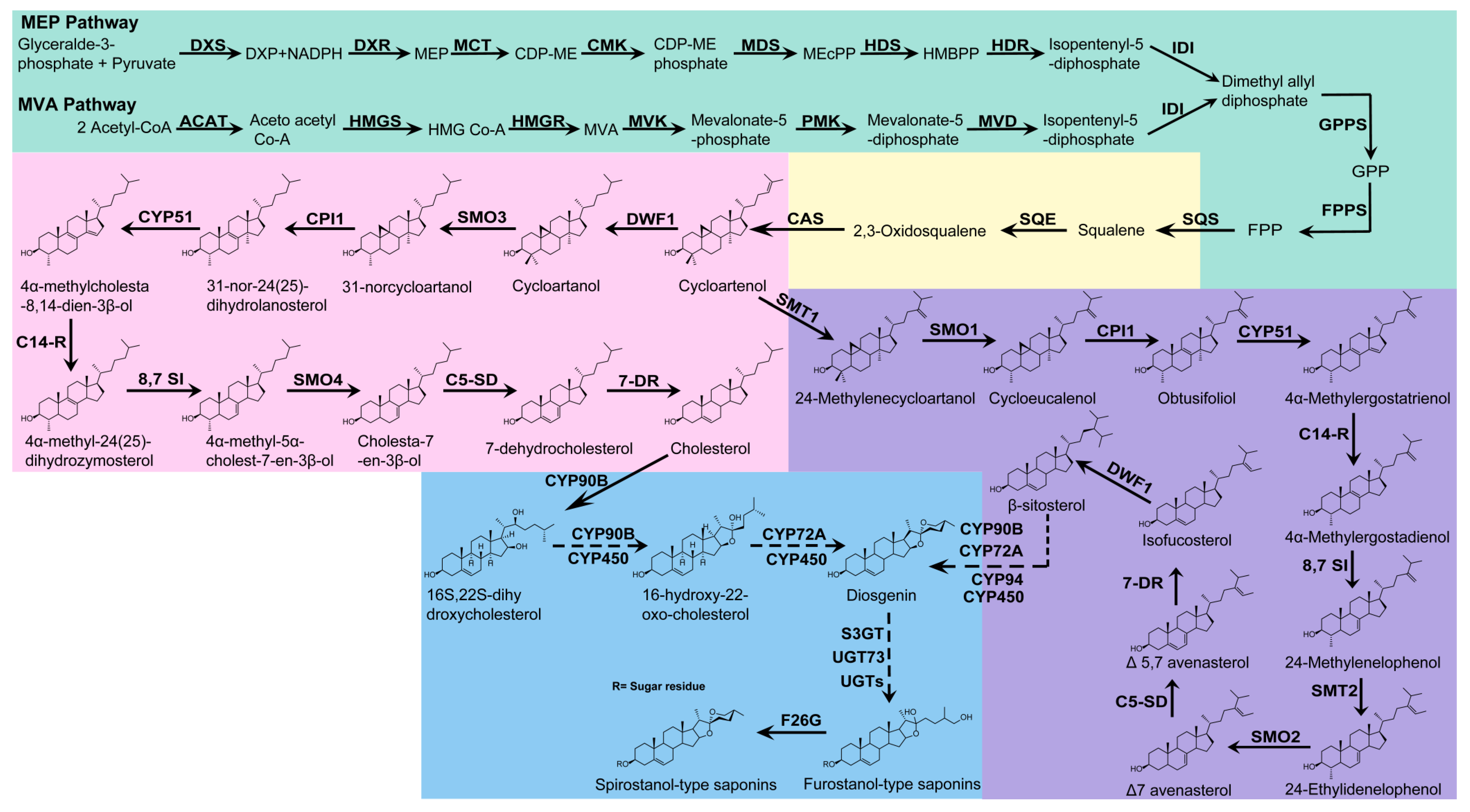
Figure 2. Possible biosynthetic pathways of steroidal saponins in plants. Biosynthetic pathway of 2,3-oxysqualene (light green); squalene is catalyzed by SQE to form 2, 3-oxide squalene (light yellow), which is also the demarcation point between sterol and triterpene metabolism. The biosynthetic pathway of cholesterol and β-sitosterol are depicted in pink and purple, respectively. The biosynthetic pathway of steroidal saponins is marked in blue. Dashed arrows indicate that there may be multiple catalytic steps.
Ijms 24 02620 g002
Disclaimer/Publisher’s Note: The statements, opinions and data contained in all publications are solely those of the individual author(s) and contributor(s) and not of MDPI and/or the editor(s). MDPI and/or the editor(s) disclaim responsibility for any injury to people or property resulting from any ideas, methods, instructions or products referred to in the content.