
Antimicrobial Performance of Innovative Functionalized Surfaces Based on Enamel Coatings: The Effect of Silver-Based Additives on the Antibacterial and Antifungal Activity

 ,
,  and
and 
Abstract
1. Introduction
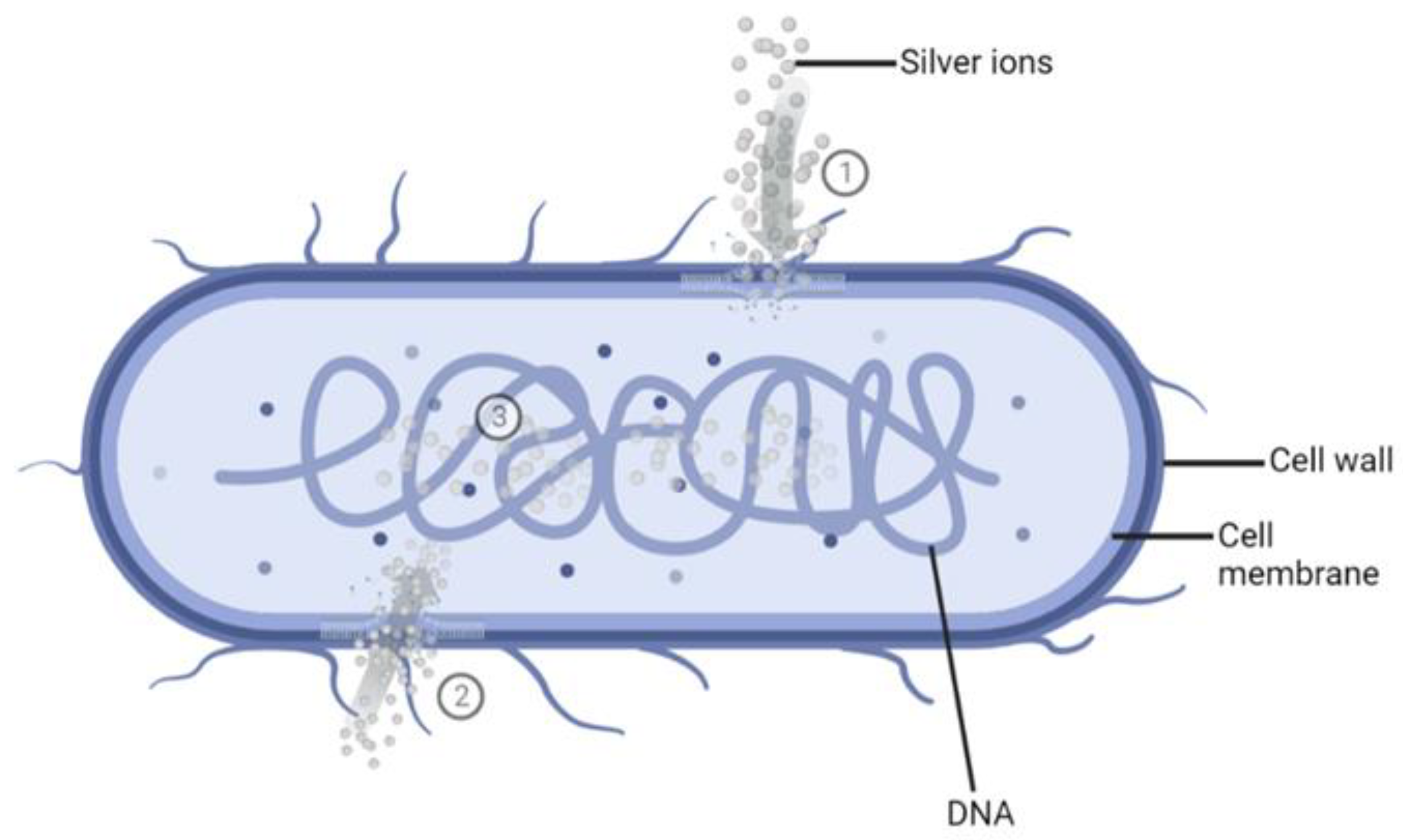
- The Ag+ ions may destroy the energy production of the bacterial cell by binding to the cellular proteins and enzymes through sulfhydryl groups (-SH), which leads to the inactivation of proteins and enzymes [33,35,36,37]. Subsequently, the bacterial cell will burst, leading to bacteria destruction [37].
2. Results
2.1. Coatings Characterization
2.2. In Vitro Analysis of Coatings Antimicrobial Effectiveness
2.2.1. Antibacterial Efficacy
2.2.2. Antifungal Activity
3. Discussion
4. Material and Methods
4.1. Samples Deposition and Morphological Characterization
4.2. Microbial Strains and Culture Media
4.3. Evaluation of Antimicrobial Activity
- Sterilization of test specimens and cover films: The test samples (reference, 1% AgNO3 coated, and 2% AgNO3 coated surface) and cover films were sterilized following the protocol described by Calovi et al. [66]. The polypropylene (PP) films (3.6 × 3.6 cm) were used to cover the suspension of test microbes dispersed on coated surfaces (4.0 × 4.0 cm) to prevent evaporation of microbial suspension. PP films and test specimens were kept in 70% ethanol for 10 min and then exposed to ultraviolet (UV) radiation for 1 h per sample side inside the laminar flow hood for sterilization.
- Preparation of the test inoculum: The microbial strains were sub-cultured on appropriate agar plates (NA for E. coli, TSA for S. aureus, and SDA for C. albicans) at 37 °C for 24–48 h, according to the microbial strain, to obtain fresh cultures. Isolated colonies of the test microorganisms from the fresh cultures were resuspended in 10 mL of 1:500 NB (E. coli and S. aureus) or TSB (C. albicans) using a sterile loop. Cell number was adjusted to ~1.5 × 10⁸ cells/mL, corresponding to 0.5 McFarland standard, and the optical density (OD) at 600 nm (OD600), ranging between 0.175 and 0.180 for E. coli, 0.210 for S. aureus and 0.250 for C. albicans, was determined with a UV-Vis spectrophotometer (Biochrom, Cambridge, USA). The suspensions were then diluted in PBS 1× to obtain the respective inoculum with a final concentration ranging from 3.9 × 10⁵ cells/mL to 1.6 × 10⁶ cells/mL (target concentration of 9.4 × 10⁵ cells/mL). Microbial counts were determined by measuring the colony-forming units per mL (CFU/mL), following 10-fold dilution in PBS 1× and inoculated onto agar plates using an L-shaped loop and incubated for 24–48 h at 37 °C.
- Inoculation of the test specimens: The sterilized enameled surfaces were placed in the middle of the petri dish, with the coated surface facing up. The test inoculum was pipetted onto each surface of the test samples. The contaminated surfaces were covered with a PP film, gently pressing down to spread the test inoculum on the coated surfaces but not to leak beyond the edges of the PP films. Then, the contaminated surfaces were incubated for different time intervals at 35 ± 1 °C, keeping the relative humidity above 90%. Reference samples used as controls to represent the test inoculum were processed instantly after microbial suspension at the T0.
- Recovery of microbial cells from test specimens: Microorganisms were recovered from each sample after different inoculation times by adding 10 mL of PBS 1× to the Petri dishes containing the contaminated samples. Upon adding 10 mL of PBS 1×, the Petri dishes containing the contaminated samples were placed on Major Science Orbital Shaker (Saratoga, CA, USA) for 5 min at 125 revolutions per minute (rpm), to ensure the dissociation of the microorganisms from the surfaces. Then, the contaminated surfaces were washed by collecting and releasing the PBS 1× using a pipette at least four times. After this step, 10-fold serial dilutions in PBS 1× were performed and, finally, 1 mL of the undiluted sample and of each dilution was spread over the Petri dishes, and 15 mL of PCA was included. The colonies grown on PCA plates were counted according to the microbial strain incubation time at 35 ± 1 °C.
- Determination of antimicrobial activity (R) of enamel surfaces contaminated for 24 h: The antimicrobial efficacy of enameled surfaces with 1% wt. AgNO3 and 2% wt. AgNO3 was evaluated by assessing the recovered microbial cells in contact with the enameled surfaces included for 24 h. The general formula for calculating the recovered cells/cm2 from each specimen as per ISO 22,196 standard is as follows:
- I.
- The average number of viable counts recovered at T0 from the control samples should range between 6.2 × 10³ cells/cm2 and 2.5 × 10⁴ cells/cm2.
- II.
- The logarithmic value of the number of viable counts recovered from control samples immediately after inoculation must meet the following criteria:
- III.
- The number of viable microbial counts recovered from each reference sample at T24 should be more than 6.2 × 10 cells/cm2.
5. Conclusions
Author Contributions
Funding
Institutional Review Board Statement
Informed Consent Statement
Data Availability Statement
Acknowledgments
Conflicts of Interest
References
- Mahira, S.; Jain, A.; Khan, W.; Domb, A.J. Antimicrobial materials—An overview. In Antimicrobial Materials for Biomedical Applications, 1st ed.; Abraham, J., Domb, K.R.K., Farah, S., Eds.; Royal Society of Chemistry: London, UK, 2019; pp. 1–37. [Google Scholar]
- Bures, S.; Fishbain, J.T.; Uyehara, C.F.; Parker, J.M.; Berg, B.W. Computer keyboards and faucet handles as reservoirs of nosocomial pathogens in the intensive care unit. Am. J. Infect. Control 2000, 28, 465–471. [Google Scholar] [CrossRef] [PubMed]
- Weber, D.J.; Rutala, W.A. Role of environmental contamination in the transmission of vancomycin-resistant enterococci. Infect. Control Hosp. Epidemiol. 1997, 18, 306–309. [Google Scholar] [CrossRef] [PubMed]
- Otter, J.A.; Yezli, S.; Salkeld, J.A.; French, G.L. Evidence that contaminated surfaces contribute to the transmission of hospital pathogens and an overview of strategies to address contaminated surfaces in hospital settings. Am. J. Infect. Control 2013, 41, S6–S11. [Google Scholar] [CrossRef]
- Moore, L.D.; Robbins, G.; Quinn, J.; Arbogast, J.W. The impact of COVID-19 pandemic on hand hygiene performance in hospitals. Am. J. Infect. Control 2021, 49, 30–33. [Google Scholar] [CrossRef] [PubMed]
- Bhat, S.A.; Sher, F.; Kumar, R.; Karahmet, E.; Haq, S.A.U.; Zafar, A.; Lima, E.C. Environmental and health impacts of spraying COVID-19 disinfectants with associated challenges. Environ. Sci. Pollut. Res. 2021, 29, 85648–85657. [Google Scholar] [CrossRef]
- Cheng, V.; Chau, P.; Lee, W.; Ho, S.; Lee, D.; So, S.; Wong, S.; Tai, J.; Yuen, K. Hand-touch contact assessment of high-touch and mutual-touch surfaces among healthcare workers, patients, and visitors. J. Hosp. Infect. 2015, 90, 220–225. [Google Scholar] [CrossRef]
- Burke, J.P. Infection control-a problem for patient safety. N. Engl. J. Med. 2003, 348, 651–656. [Google Scholar] [CrossRef]
- Pittet, D.; Donaldson, L. Clean care is safer care: A worldwide priority. Lancet 2005, 366, 1246–1247. [Google Scholar] [CrossRef]
- Adlhart, C.; Verran, J.; Azevedo, N.F.; Olmez, H.; Keinänen-Toivola, M.M.; Gouveia, I.; Melo, L.F.; Crijns, F. Surface modifications for antimicrobial effects in the healthcare setting: A critical overview. J. Hosp. Infect. 2018, 99, 239–249. [Google Scholar] [CrossRef] [PubMed]
- Dancer, S.J. Controlling hospital-acquired infection: Focus on the role of the environment and new technologies for decontamination. Clin. Microbiol. Rev. 2014, 27, 665–690. [Google Scholar] [CrossRef] [PubMed]
- Khan, A.; Dancer, S.; Humphreys, H. Priorities in the prevention and control of multidrug-resistant Enterobacteriaceae in hospitals. J. Hosp. Infect. 2012, 82, 85–93. [Google Scholar] [CrossRef]
- Datta, R.; Platt, R.; Yokoe, D.S.; Huang, S.S. Environmental cleaning intervention and risk of acquiring multidrug-resistant organisms from prior room occupants. Arch. Intern. Med. 2011, 171, 491–494. [Google Scholar] [CrossRef]
- Gold, N.A.; Mirza, T.M.; Avva, U. Alcohol sanitizer. 2018. Available online: https://europepmc.org/article/nbk/nbk513254 (accessed on 20 September 2022).
- Choi, H.; Chatterjee, P.; Lichtfouse, E.; Martel, J.A.; Hwang, M.; Jinadatha, C.; Sharma, V.K. Classical and alternative disinfection strategies to control the COVID-19 virus in healthcare facilities: A review. Environ. Chem. Lett. 2021, 19, 1945–1951. [Google Scholar] [CrossRef]
- Avershina, E.; Shapovalova, V.; Shipulin, G. Fighting antibiotic resistance in hospital-acquired infections: Current state and emerging technologies in disease prevention, diagnostics and therapy. Front. Microbiol. 2021, 12, 2044. [Google Scholar] [CrossRef] [PubMed]
- Goscé, L.; Johansson, A. Analysing the link between public transport use and airborne transmission: Mobility and contagion in the London underground. Environ. Health 2018, 17, 1–11. [Google Scholar] [CrossRef]
- Vardoulakis, S.; Oyarce, D.A.E.; Donner, E. Transmission of COVID-19 and other infectious diseases in public washrooms: A systematic review. Sci. Total Environ. 2022, 803, 149932. [Google Scholar] [CrossRef]
- Kagan, L.J.; Aiello, A.E.; Larson, E. The role of the home environment in the transmission of infectious diseases. J. Community Health 2002, 27, 247–267. [Google Scholar] [CrossRef] [PubMed]
- Russo, F.; Furlan, B.; Calovi, M.; Massidda, O.; Rossi, S. Silver-based vitreous enamel coatings: Assessment of their antimicrobial activity towards Escherichia coli and Staphylococcus aureus before and after surface degradation. Surf. Coat. Technol. 2022, 445, 128702. [Google Scholar] [CrossRef]
- Kramer, A.; Schwebke, I.; Kampf, G. How long do nosocomial pathogens persist on inanimate surfaces? A systematic review. BMC Infect. Dis. 2006, 6, 1–8. [Google Scholar] [CrossRef]
- Disinfection & Sterilization Guidelines | Guidelines Library | Infection Control | CDC. 2019. Available online: https://www.cdc.gov/infectioncontrol/guidelines/disinfection/index.html (accessed on 30 September 2022).
- Page, K.; Wilson, M.; Parkin, I.P. Antimicrobial surfaces and their potential in reducing the role of the inanimate environment in the incidence of hospital-acquired infections. J. Mater. Chem. 2009, 19, 3819–3831. [Google Scholar] [CrossRef]
- Crijns, F.R.; Keinänen-Toivola, M.M.; Dunne, C.P. Antimicrobial coating innovations to prevent healthcare-associated infection. J. Hosp. Infect. 2017, 95, 243–244. [Google Scholar] [CrossRef]
- Chen, X.; Hirt, H.; Li, Y.; Gorr, S.-U.; Aparicio, C. Antimicrobial GL13K peptide coatings killed and ruptured the wall of Streptococcus gordonii and prevented formation and growth of biofilms. PLoS ONE 2014, 9, e111579. [Google Scholar] [CrossRef]
- Whitehead, K.A.; Verran, J. The effect of surface topography on the retention of microorganisms. Food Bioprod. Process. 2006, 84, 253–259. [Google Scholar] [CrossRef]
- Rossi, S.; Russo, F.; Calovi, M. Durability of vitreous enamel coatings and their resistance to abrasion, chemicals, and corrosion: A review. J. Coat. Technol. Res. 2021, 18, 39–52. [Google Scholar] [CrossRef]
- Savvova, O. Effect of zinc and tin oxides on the bactericidal properties of glass enamel coatings. Glass Ceram. 2014, 71, 254–257. [Google Scholar] [CrossRef]
- Shippy, G. Reclamation of Scrap Frit. In Proceedings of the 41st Porcelain Enamel Technical Forum: Ceramic Engineering and Science Proceedings (1979); Smothers, W.J., Ed.; John Wiley & Sons, Inc.: Hoboken, NJ, USA, 1980; pp. 208–210. [Google Scholar]
- Elliott, R.H. Zero Discharge, Zero Pollution, and Source Reduction. In Proceedings of the 50th Porcelain Enamel Institute Technical Forum: Ceramic Engineering and Science Proceedings; Wachtman, J.B., Ed.; John Wiley & Sons, Inc.: Hoboken, NJ, USA, 1989; p. 513. [Google Scholar]
- Cameron, D.S. Waste Minimization. In Proceedings of the 50th Porcelain Enamel Institute Technical Forum: Ceramic Engineering and Science Proceedings (1989); Wachtman, J.B., Ed.; John Wiley & Sons, Inc.: Hoboken, NJ, USA, 1989. [Google Scholar]
- Silvestry-Rodriguez, N.; Sicairos-Ruelas, E.E.; Gerba, C.P.; Bright, K.R. Silver as a disinfectant. Rev. Environ. Contam. Toxicol. 2007, 191, 23–45. [Google Scholar] [PubMed]
- Thurman, R.B.; Gerba, C.P.; Bitton, G. The molecular mechanisms of copper and silver ion disinfection of bacteria and viruses. Crit. Rev. Environ. Sci. Technol. 1989, 18, 295–315. [Google Scholar] [CrossRef]
- Slawson, R.; Lee, H.; Trevors, J. Bacterial interactions with silver. Biol. Met. 1990, 3, 151–154. [Google Scholar] [CrossRef] [PubMed]
- Slawson, R.M.; Van Dyke, M.I.; Lee, H.; Trevors, J.T. Germanium and silver resistance, accumulation, and toxicity in microorganisms. Plasmid 1992, 27, 72–79. [Google Scholar] [CrossRef]
- Feng, Q.L.; Wu, J.; Chen, G.Q.; Cui, F.; Kim, T.; Kim, J. A mechanistic study of the antibacterial effect of silver ions on Escherichia coli and Staphylococcus aureus. J. Biomed. Mater. Res. 2000, 52, 662–668. [Google Scholar] [CrossRef] [PubMed]
- Liau, S.; Read, D.; Pugh, W.; Furr, J.; Russell, A. Interaction of silver nitrate with readily identifiable groups: Relationship to the antibacterialaction of silver ions. Lett. Appl. Microbiol. 1997, 25, 279–283. [Google Scholar] [CrossRef]
- Klueh, U.; Wagner, V.; Kelly, S.; Johnson, A.; Bryers, J. Efficacy of silver-coated fabric to prevent bacterial colonization and subsequent device-based biofilm formation. J. Biomed. Mater. Res. 2000, 53, 621–631. [Google Scholar] [CrossRef] [PubMed]
- Richards, R. Antimicrobial action of silver nitrate. Microbios 1981, 31, 83–91. [Google Scholar]
- Marambio-Jones, C.; Hoek, E. A review of the antibacterial effects of silver nanomaterials and potential implications for human health and the environment. J. Nanopar. Res. 2010, 12, 1531–1551. [Google Scholar] [CrossRef]
- Brady, M.J.; Lisay, C.M.; Yurkovetskiy, A.V.; Sawan, S.P. Persistent silver disinfectant for the environmental control of pathogenic bacteria. Am. J. Infect. Control 2003, 31, 208–214. [Google Scholar] [CrossRef] [PubMed]
- Silver, S. Bacterial silver resistance: Molecular biology and uses and misuses of silver compounds. FEMS Microbiol. Rev. 2003, 27, 341–353. [Google Scholar] [CrossRef] [PubMed]
- Takai, K.; Ohtsuka, T.; Senda, Y.; Nakao, M.; Yamamoto, K.; Matsuoka, J.; Hirai, Y. Antibacterial properties of antimicrobial-finished textile products. Microbiol. Immunol. 2002, 46, 75–81. [Google Scholar] [CrossRef]
- Kim, K.-J.; Sung, W.-S.; Moon, S.-K.; Choi, J.-S.; Kim, J.-G.; Lee, D.-G. Antifungal effect of silver nanoparticles on dermatophytes. J. Microbiol. Biotechnol. 2008, 18, 1482–1484. [Google Scholar]
- Hendiger, E.B.; Padzik, M.; Sifaoui, I.; Reyes-Batlle, M.; López-Arencibia, A.; Zyskowska, D.; Grodzik, M.; Pietruczuk-Padzik, A.; Hendiger, J.; Olędzka, G. Silver Nanoparticles Conjugated with Contact Lens Solutions May Reduce the Risk of Acanthamoeba Keratitis. Pathogens 2021, 10, 583. [Google Scholar] [CrossRef]
- Anwar, A.; Siddiqui, R.; Hussain, M.A.; Ahmed, D.; Shah, M.R.; Khan, N.A. Silver nanoparticle conjugation affects antiacanthamoebic activities of amphotericin B, nystatin, and fluconazole. Parasitol. Res. 2018, 117, 265–271. [Google Scholar] [CrossRef]
- Lee, B.; Lee, M.J.; Yun, S.J.; Kim, K.; Choi, I.-H.; Park, S. Silver nanoparticles induce reactive oxygen species-mediated cell cycle delay and synergistic cytotoxicity with 3-bromopyruvate in Candida albicans, but not in Saccharomyces cerevisiae. Int. J. Nanomed. 2019, 14, 4801. [Google Scholar] [CrossRef] [PubMed]
- González-Fernández, S.; Lozano-Iturbe, V.; Menéndez, M.F.; Ordiales, H.; Fernández-Vega, I.; Merayo, J.; Vazquez, F.; Quirós, L.M.; Martín, C. A Promising Antifungal and Antiamoebic Effect of Silver Nanorings, a Novel Type of AgNP. Antibiotics 2022, 11, 1054. [Google Scholar] [CrossRef] [PubMed]
- Poolman, J.T.; Anderson, A.S. Escherichia coli and Staphylococcus aureus: Leading bacterial pathogens of healthcare associated infections and bacteremia in older-age populations. Expert Rev. Vaccines 2018, 17, 607–618. [Google Scholar] [CrossRef] [PubMed]
- Kabir, M.A.; Hussain, M.A.; Ahmad, Z. Candida albicans: A model organism for studying fungal pathogens. ISRN Microbiol. 2012, 2012, 1–15. [Google Scholar] [CrossRef] [PubMed]
- Voges, D.L. Porcelain Enamel: Properties and Applications. In Proceedings of the 58th Porcelain Enamel Institute Technical Forum: Ceramic Engineering and Science Proceedings, Nashville, TN, USA, 12–16 May 1996; pp. 22–26. [Google Scholar]
- Aboualigalehdari, E.; Ghafourian, S.; Sadeghifard, N.; Sekawi, Z. Is Candida albicans a cause of nosocomial infection in Iran? Rev. Med. Microbiol. 2013, 24, 85–88. [Google Scholar] [CrossRef]
- Nabipour, Y.S.; Rostamzad, A. Comparing the antimicrobial effects of silver and copper nanoparticles against pathogenic and resistant bacteria of Klebsiella pneumonia, Pseudomonas aeruginosa and Staphylococcus aureus. Cumhur. Üniversitesi Fen Edeb. Fakültesi Fen Bilim. Derg. 2015, 36, 2541–2546. [Google Scholar]
- Schrand, A.M.; Rahman, M.F.; Hussain, S.M.; Schlager, J.J.; Smith, D.A.; Syed, A.F. Metal-based nanoparticles and their toxicity assessment. Wiley Interdiscip. Rev. Nanomed. 2010, 2, 544–568. [Google Scholar] [CrossRef]
- Lara, H.H.; Ayala-Núnez, N.V.; Ixtepan Turrent, L.d.C.; Rodríguez Padilla, C. Bactericidal effect of silver nanoparticles against multidrug-resistant bacteria. World J. Microbiol. Biotechnol. 2010, 26, 615–621. [Google Scholar] [CrossRef]
- Silhavy, T.J.; Kahne, D.; Walker, S. The bacterial cell envelope. Cold Spring Harb. Perspect. Biol. 2010, 2, a000414. [Google Scholar] [CrossRef]
- Graybill, J.; Burgess, D.; Hardin, T. Key issues concerning fungistatic versus fungicidal drugs. Eur. J. Clin. Microbiol. 1997, 16, 42–50. [Google Scholar] [CrossRef]
- Vazquez-Muñoz, R.; Avalos-Borja, M.; Castro-Longoria, E. Ultrastructural analysis of Candida albicans when exposed to silver nanoparticles. PLoS ONE 2014, 9, e108876. [Google Scholar] [CrossRef] [PubMed]
- Ahamad, I.; Bano, F.; Anwer, R.; Srivastava, P.; Kumar, R.; Fatma, T. Antibiofilm Activities of Biogenic Silver Nanoparticles Against Candida albicans. Front. Microbiol. 2021, 12, 4093. [Google Scholar] [CrossRef] [PubMed]
- Kim, K.-J.; Sung, W.S.; Suh, B.K.; Moon, S.-K.; Choi, J.-S.; Kim, J.G.; Lee, D.G. Antifungal activity and mode of action of silver nano-particles on Candida albicans. Biometals 2009, 22, 235–242. [Google Scholar] [CrossRef]
- Lara, H.H.; Romero-Urbina, D.G.; Pierce, C.; Lopez-Ribot, J.L.; Arellano-Jiménez, M.J.; Jose-Yacaman, M. Effect of silver nanoparticles on Candida albicans biofilms: An ultrastructural study. J. Nanobiotechnol. 2015, 13, 1–12. [Google Scholar] [CrossRef] [PubMed]
- Deyá, C.; Bellotti, N. Biosynthesized silver nanoparticles to control fungal infections in indoor environments. Adv. Nat. Sci. Nanosci. Nanotechnol. 2017, 8, 025005. [Google Scholar] [CrossRef]
- Ishida, K.; Cipriano, T.F.; Rocha, G.M.; Weissmüller, G.; Gomes, F.; Miranda, K.; Rozental, S. Silver nanoparticle production by the fungus Fusarium oxysporum: Nanoparticle characterisation and analysis of antifungal activity against pathogenic yeasts. Mem. Inst. Oswaldo Cruz 2013, 109, 220–228. [Google Scholar] [CrossRef]
- ISO 4287:1997; Geometrical Product Specifications (GPS)–Surface Texture: Profile Method–Terms, Definitions and Surface Texture Parameters. International Organization for Standardization: Geneva, Switzerland, 1997.
- Cashera, A.; Porosa, L. ISO 22196: 2011 Measurement of Antibacterial Activity on Plastics and Other Non-Porous Surfaces “Modified Large Droplet Inoculation Method”. Available online: http://www.skinguard24.com/data/SG24_Ryerson.pdf (accessed on 30 September 2022).
- Calovi, M.; Furlan, B.; Coroneo, V.; Massidda, O.; Rossi, S. Facile Route to Effective Antimicrobial Aluminum Oxide Layer Realized by Co-Deposition with Silver Nitrate. Coatings 2022, 12, 28. [Google Scholar] [CrossRef]
- Donkor, E.S. Nosocomial pathogens: An in-depth analysis of the vectorial potential of cockroaches. Trop. Med. Infect. Dis. 2019, 4, 14. [Google Scholar] [CrossRef] [PubMed]


| Sample Type | Ra (μm) | Rz (μm) |
|---|---|---|
| E0 | 0.15 ± 0.01 | 0.74 ± 0.05 |
| E1 | 0.15 ± 0.02 | 0.77 ± 0.16 |
| E2 | 0.11 ± 0.02 | 0.63 ± 0.09 |
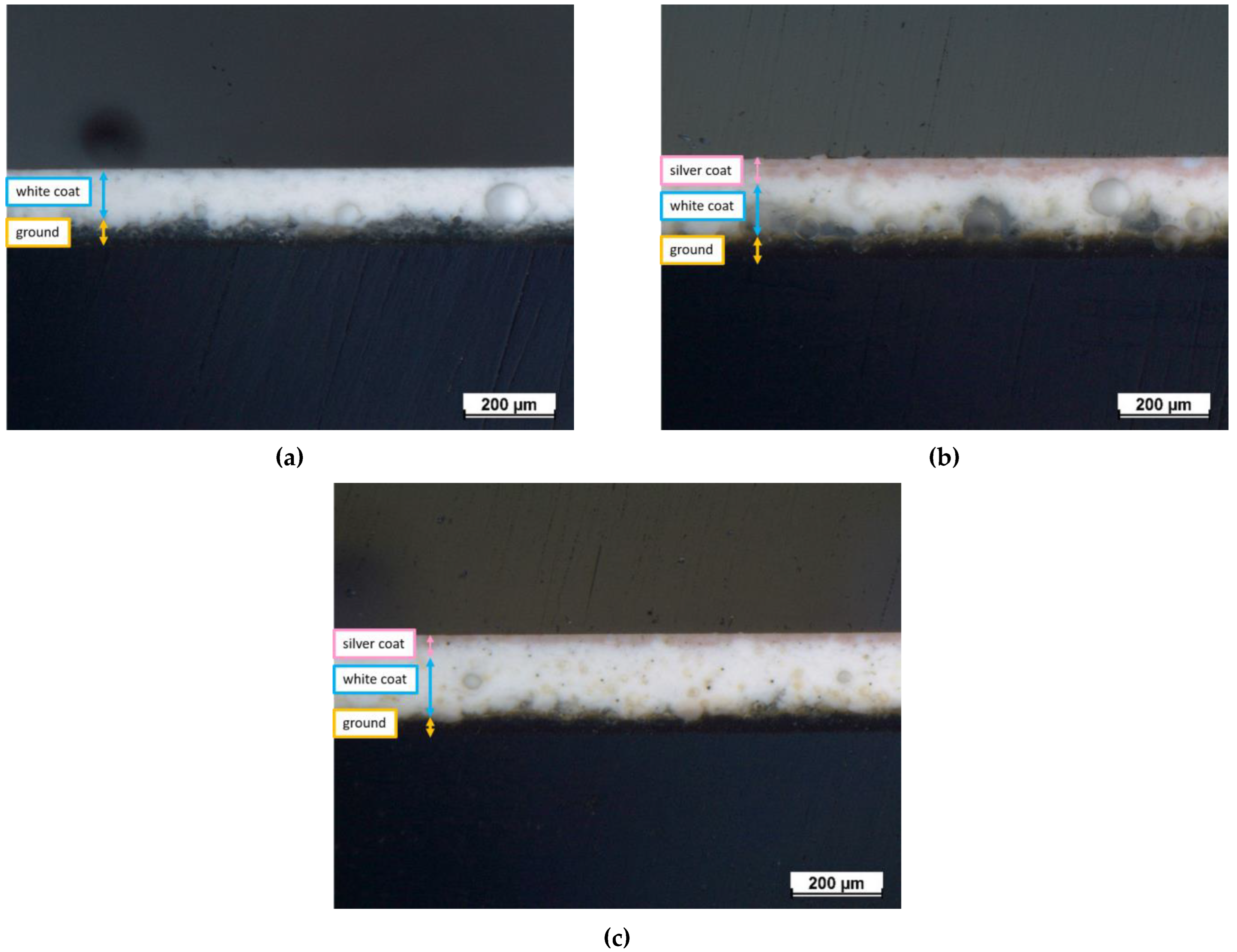
| Sample Type | Total Thickness (µm) | Ground Coat (µm) | White Coat (µm) | Silver Coat (µm) |
|---|---|---|---|---|
| E0 | 171 ± 1 | 51 ± 7 | 120 ± 6 | N/A |
| E1 | 217 ± 3 | 50 ± 5 | 137 ± 1 | 30 ± 2 |
| E2 | 214 ± 1 | 39 ± 7 | 151 ± 3 | 24 ± 3 |
| Reference Sample | 1 wt.% AgNO3 | 2 wt.% AgNO3 | |
|---|---|---|---|
| Sample nomenclature | E0 | E1 | E2 |
| Number of layers | 2 | 3 | 3 |
Disclaimer/Publisher’s Note: The statements, opinions and data contained in all publications are solely those of the individual author(s) and contributor(s) and not of MDPI and/or the editor(s). MDPI and/or the editor(s) disclaim responsibility for any injury to people or property resulting from any ideas, methods, instructions or products referred to in the content. |
© 2023 by the authors. Licensee MDPI, Basel, Switzerland. This article is an open access article distributed under the terms and conditions of the Creative Commons Attribution (CC BY) license (https://creativecommons.org/licenses/by/4.0/).
Share and Cite
Rehman, H.U.; Russo, F.; Calovi, M.; Massidda, O.; Rossi, S. Antimicrobial Performance of Innovative Functionalized Surfaces Based on Enamel Coatings: The Effect of Silver-Based Additives on the Antibacterial and Antifungal Activity. Int. J. Mol. Sci. 2023, 24, 2364. https://doi.org/10.3390/ijms24032364
Rehman HU, Russo F, Calovi M, Massidda O, Rossi S. Antimicrobial Performance of Innovative Functionalized Surfaces Based on Enamel Coatings: The Effect of Silver-Based Additives on the Antibacterial and Antifungal Activity. International Journal of Molecular Sciences. 2023; 24(3):2364. https://doi.org/10.3390/ijms24032364
Chicago/Turabian StyleRehman, Hamza Ur, Francesca Russo, Massimo Calovi, Orietta Massidda, and Stefano Rossi. 2023. "Antimicrobial Performance of Innovative Functionalized Surfaces Based on Enamel Coatings: The Effect of Silver-Based Additives on the Antibacterial and Antifungal Activity" International Journal of Molecular Sciences 24, no. 3: 2364. https://doi.org/10.3390/ijms24032364
APA StyleRehman, H. U., Russo, F., Calovi, M., Massidda, O., & Rossi, S. (2023). Antimicrobial Performance of Innovative Functionalized Surfaces Based on Enamel Coatings: The Effect of Silver-Based Additives on the Antibacterial and Antifungal Activity. International Journal of Molecular Sciences, 24(3), 2364. https://doi.org/10.3390/ijms24032364