Article

Comparative Analysis of Morphological and Functional Effects of 225Ac- and 177Lu-PSMA Radioligand Therapies (RLTs) on Salivary Glands

by
Benedikt Feuerecker
1,2,3,4,*,
Andrei Gafita
5,
Thomas Langbein
1,
Robert Tauber
6,
Christof Seidl
1,
Frank Bruchertseifer
7,
Jürgen E. Gschwendt
6,
Wolfgang A. Weber
1,2,
Calogero D’Alessandria
1,†,
Alfred Morgenstern
7,† and
Matthias Eiber
1,2,†
1
Department of Nuclear Medicine, School of Medicine, Technical University of Munich, 81675 München, Germany
2
Deutsches Konsortium für Translationale Krebsforschung (DKTK), Partnersite München, 69124 Heidelberg, Germany
3
Department of Radiology, University Hospital, LMU Munich, 81377 München, Germany
4
Department of Radiology, School of Medicine, Technical University of Munich, 81675 München, Germany
5
Division of Nuclear Medicine and Molecular Imaging, The Russell H. Morgan Department of Radiology and Radiological Science, Johns Hopkins University School of Medicine, Baltimore, MD 21287, USA
6
Department of Urology, School of Medicine, Klinikum Rechts der Isar, Technical University of Munich, 81675 München, Germany
7
European Commission, Joint Research Centre (JRC), 76344 Karlsruhe, Germany
*
Author to whom correspondence should be addressed.
These authors are joint senior authors.
Int. J. Mol. Sci. 2023, 24(23), 16845; https://doi.org/10.3390/ijms242316845
Submission received: 31 August 2023 / Revised: 24 October 2023 / Accepted: 13 November 2023 / Published: 28 November 2023

Abstract

:
Most Prostate Specific Membrane Antigens (PSMAs) targeting small molecules accumulate in the salivary glands (SGs), raising concerns about SG toxicity, especially after repeated therapies or therapy with 225Ac-labeled ligands. SG toxicity is assessed clinically by the severity of patient-reported xerostomia, but this parameter can be challenging to objectively quantify. Therefore, we explored the feasibility of using SG volume as a biomarker for toxicity. In 21 patients with late-stage metastatic resistant prostate cancer (mCRPC), the PSMA volume and ligand uptake of SG were analyzed retrospectively before and after two cycles of 177Lu-PSMA (LuPSMA; cohort A) and before and after one cycle of 225Ac-PSMA-617 (AcPSMA, cohort B). Mean Volume-SG in cohort A was 59 ± 13 vs. 54 ± 16 mL (−10%, p = 0.4), and in cohort B, it was 50 ± 13 vs. 40 ± 11 mL (−20%, p = 0.007), respectively. A statistically significant decrease in the activity concentration in the SG was only observed in group B (SUVmean: 9.2 ± 2.8 vs. 5.3 ± 1.8, p < 0.0001; vs. A: SUVmean: 11.2 ± 3.3 vs. 11.1 ± 3.5, p = 0.8). SG volume and PSMA-ligand uptake are promising markers to monitor the SG toxicity after a PSMA RLT.

1. Introduction

The treatment of metastatic castration-resistant prostate cancer (mCRPC) remains a major challenge. A prolonged overall survival with the radiopharmaceutical 177Lu-PSMA-617 has been recently proven in a phase III clinical trial compared to the standard of care (median OS 15.3 vs. 11.3 months) [1]. However, primary or secondary radioresistance to 177Lu-PSMA (LuPSMA) limits its effect [2]. It has been proposed that targeted alpha therapy (TAT) has the potential to overcome the radioresistance of beta emitters through its higher linear energy transfer [3,4]. TAT has been proven to be more effective than beta emitters in preclinical studies as it induces DNA double-strand breaks [3].
The alpha emitter Actinium-225 (225Ac) has been recently used for the PSMA-targeted treatment of mCRPC, and promising results have been reported using 225Ac-PSMA-617 (AcPSMA) [3,4,5]. However, xerostomia is a major limiting side effect for AcPSMA, which can lead to the discontinuation of treatment [4,6]. Deterioration of salivary function is a clinical problem described after an external beam radiation treatment [7,8] and after a radioiodine treatment [9,10,11]. Its extent has been related to the absorbed dose based on the data of external beam radiation therapy [12]. For alpha emitters, quantitative radiation dosimetry is not trivial, given the lack of direct gamma emissions. Therefore, a quantitative measurement of delivered dose to the salivary glands (SGs) is highly challenging. Dose estimations can be made based on the dosimetry of LuPSMA treatment and serial PET measurements. Salivary gland scintigraphy provides an objective measure to quantify SG function and has been reported as a tool to assess SG function in patients with thyroid diseases [13,14,15,16] and mCRPC [17]. Furthermore, an indirect measurement of the effects of radiation on SG can be made, based on pre- and post-therapeutic staging scans such as PSMA PET combined with morphological imaging.
Therefore, our aim of this retrospective analysis was to investigate the potential correlates in the morphological and molecular PET imaging of clinically observed xerostomia. Pre- and post-treatments hybrid PET imaging in patients who have undergone 225Ac-PSMA-617 radioligand treatment (RLT) and 177Lu-PSMA-I&T RLT were compared. We hypothesize that decreases in SG volumes and PSMA-ligand uptake (a) are dependent on the type of radiation (alpha vs. beta) and (b) are related to xerostomia.

2. Results

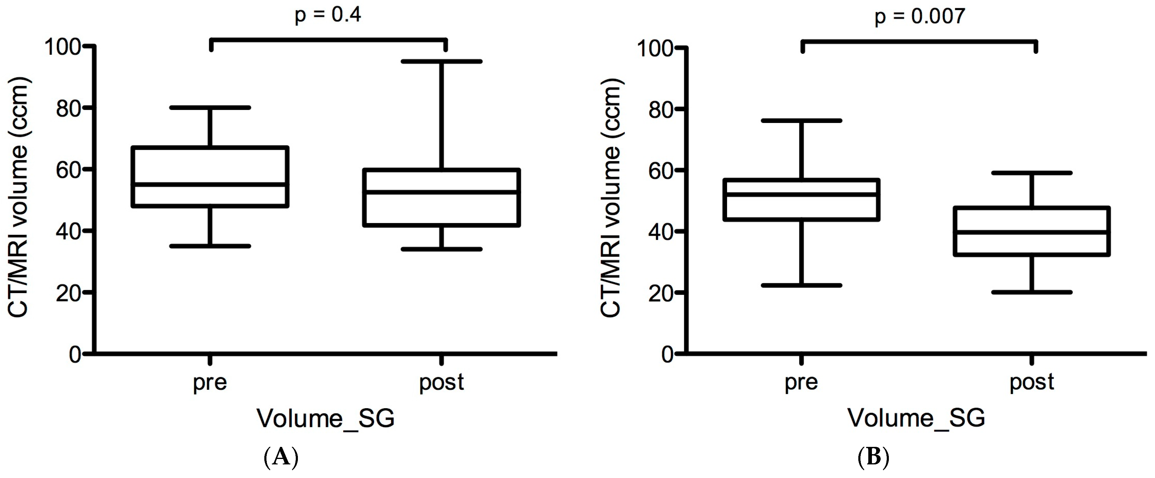
2.1. Volumetric Changes in Salivary Glands before and after LuPSMA and AcPSMA RLTs

In cohort A (before vs. after LuPSMA RLT), no significant volumetric size changes were observed: the mean Volume-SG of the SG was 59 ± 13 vs. 54 ± 16 mL (p = 0.4, Figure 1A). Mean relative and absolute changes in Volume-SG were 10% and 5 mL.
In contrast, a highly significant decrease in volumes was observed in cohort B (before vs. after AcPSMA RLT): the mean Volume-SG was 50 ± 13 mL vs. 40 ± 11 mL (p = 0.007, Figure 1B). Mean relative and absolute changes in Volume-SG were 20% and 10 mL.

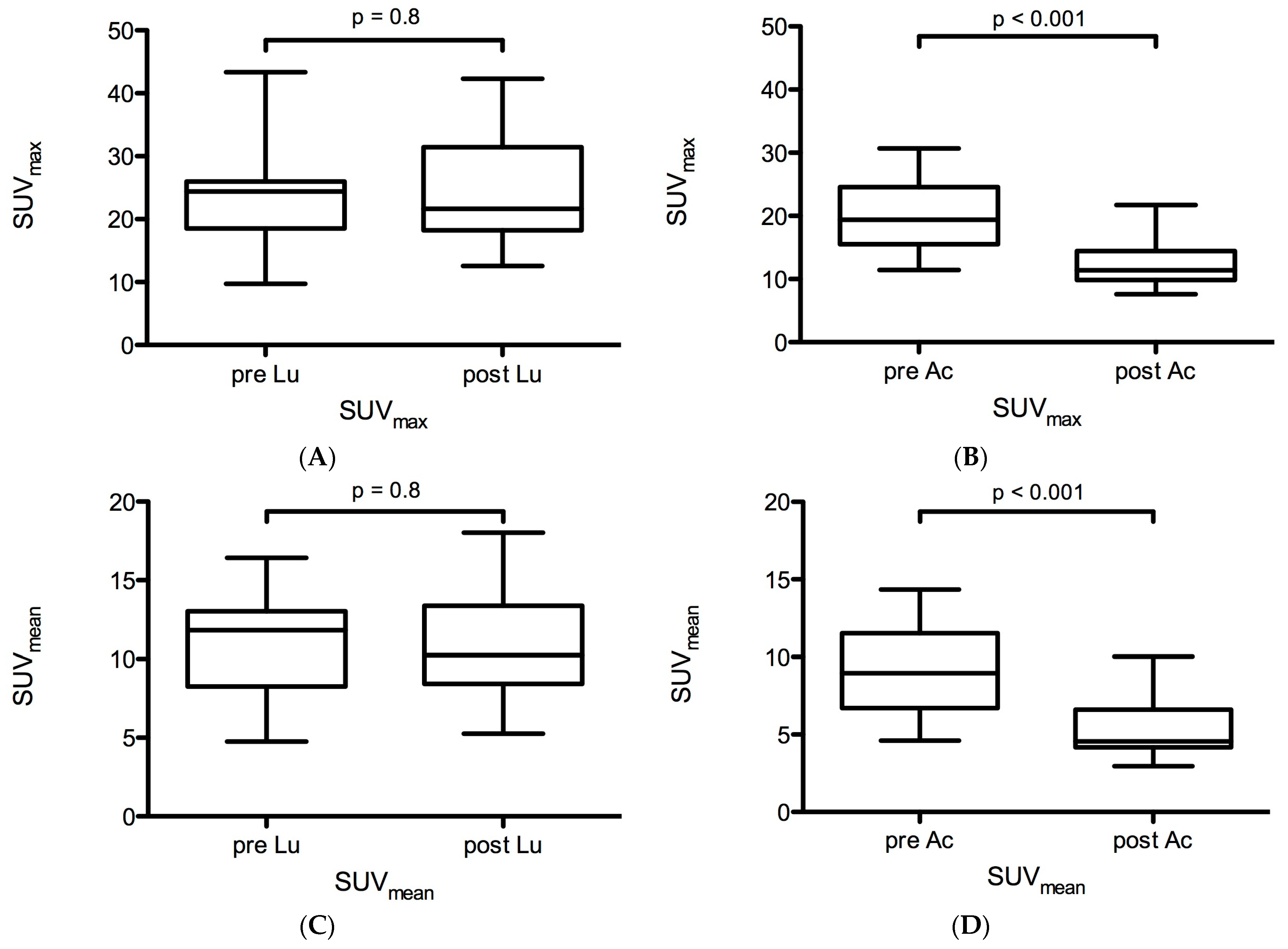
2.2. Functional Changes in PSMA-Ligand Uptake before and after LuPSMA and AcPSMA RLTs

In cohort A, no significant changes in PSMA-ligand uptake were observed: the mean SUVmax and SUVmean were 23.8 ± 7.7 vs. 24.7 ± 8.7 (p = 0.8) and 11.0 ± 3.3 vs. 10.8 ± 3.4 (p = 0.8), respectively (Figure 2A,C). Mean relative changes in SUVmax and SUVmean were +3.8% and −1.8%. The mean PSMA-SGU was 757 ± 264 vs. 721 ± 316 (p = 0.7, Figure 3A). Mean relative and absolute changes for PSMA-SGU were −5% and −30 (Figure 3A).
In contrast, a highly significant decrease in PSMA-ligand uptake was observed in cohort B: the mean SUVmax and SUVmean were 20.1 ± 5.4 vs. 12.3 ± 3.6 (p < 0.0001) and 9.2 ± 2.8 vs. 5.3 ± 1.8 (p < 0.0001), respectively (Figure 2B,D). Mean relative changes in SUVmax and SUVmean were −38.8% and −42.4%. The mean PSMA-SGU was 711 ± 268 vs. 276 ± 162 (p < 0.0001). Mean relative and absolute changes for PSMA-SGU were −61% and −435 (Figure 3B).

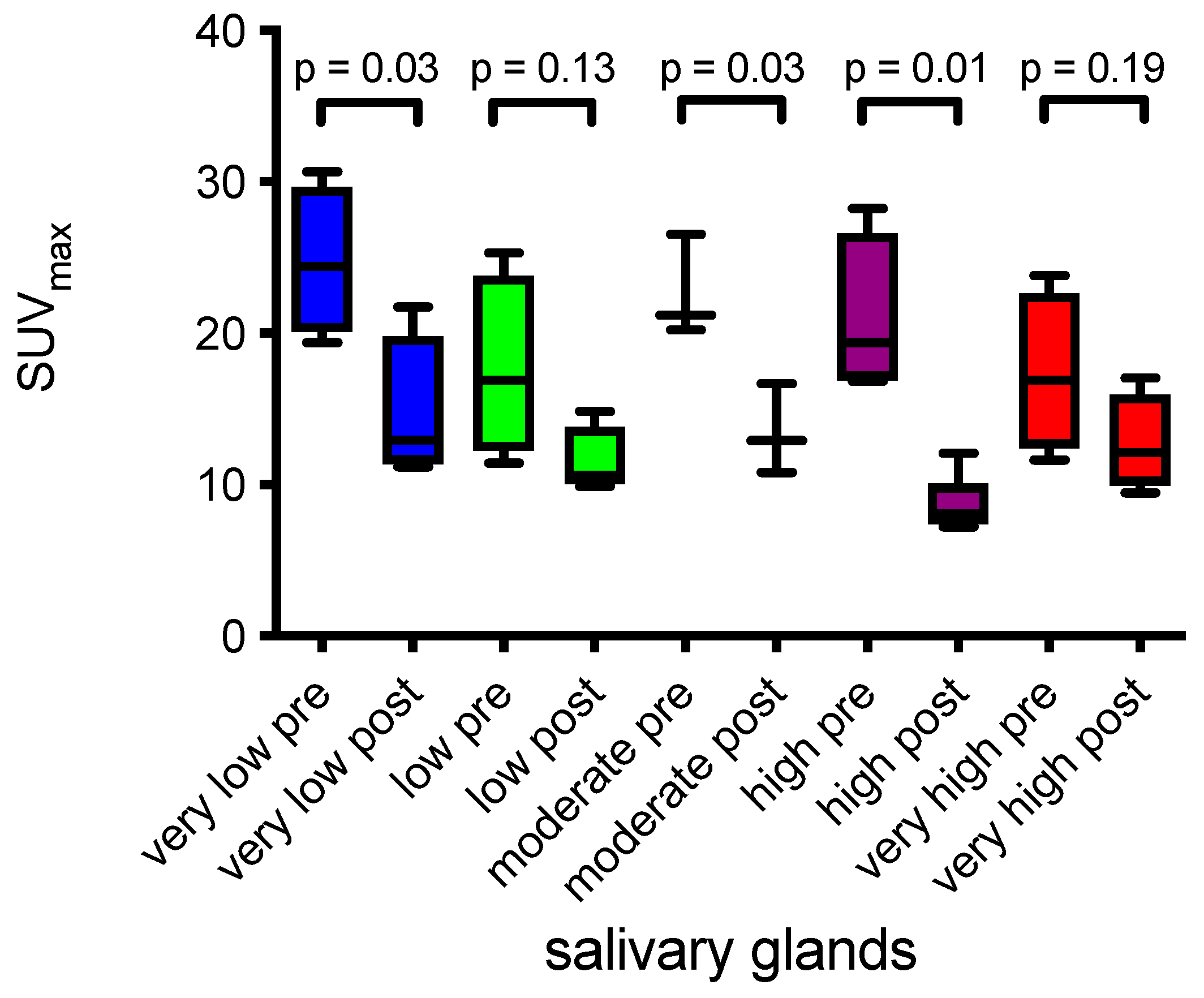
2.3. Salivary Glands and Tumor Burden

Based on the five quartiles of pre-therapeutic whole body tumor burden, changes in the salivary gland SUVmean and SUVmax pre- and post-AcPSMA were quantified. Statistically significant decreases in SUVmax of the SG were measured in groups with very low, moderate, high, and very high pre-therapeutic tumor burden (Table 1 and Figure 4). No correlation between SUVmean of the SG and tumor burden was observed in the low and very high groups (Table 1). In each of these five tumor burden groups, no statistically significant changes in whole body tumor burden were observed post-AcPSMA. No significant changes in SUVmax and SUVmean were observed in groups with very low, low, high, and very high tumor burden patients treated with LuPSMA.

3. Discussion

In this retrospective analysis, a treatment with one cycle of AcPSMA resulted in a significant decrease in morphological and functional surrogate parameters of salivary glands, which were assessed with PSMA PET. In contrast, no substantial differences could be observed after treatment with two cycles of LuPSMA in the same patients.
The deterioration of the salivary gland function is a clinically relevant side effect of AcPSMA reported in the literature [3,6,18]. Our retrospective study is the first to present quantitative data from imaging to potentially link it with objective measures. For the external beam radiation treatment [7,19] of the neck, different reports on potential xerostomia using imaging as a quantitative measure are available. In an MRI study including 52 patients with squamous cell carcinoma of the neck, the volume of the parotid glands decreased by an average of 26% at 30 Gy and approx. 40% at 70 Gy [20]. In another study with 15 head and neck cancer patients, the median parotid volume loss was 28.1% (range: 5.9–53.6%) [21]. Furthermore, in a study with 18 patients irradiated with a radiation dose of 38.1 to 64.4 Gy, a reduction of the parotid glands by approximately 35%, was observed [22].
The evaluation of delivered doses of Actinium-225 to the salivary gland remains challenging because radiation doses depend on the microscopic distribution of the radioactivity within the tissue, which is currently unknown. Based on a dose assumption, an administration of 10 kBq/kg of 225Ac-PSMA-617 would result in a mean salivary gland dose of approximately 67 Gy [23]. For LuPSMA, data on the dosimetry of the salivary glands for both LuPSMA-617 [24,25,26] and LuPSMA-I&T [27] exist, resulting in a dose of 8.1–21.9 Gy to the salivary glands (after two i.v. injections of 7.4 GBq LuPSMA).
In our retrospective analysis, Volume-SG was reduced by 10% in cohort A but by 20% in cohort B. Similarly, PSMA-SGU was reduced by −5% in cohort A but by −61% in cohort B. These data indicate that LuPSMA has only minor effects on the salivary glands, but AcPSMA induces profound physical and biological effects on the salivary glands. This is in line with the clinical observation that patients treated with LuPSMA rarely report a permanent xerostomia or request for a stop of treatment [28].
Based on the data presented here, both function (PSMA-SGU) and morphological size (Volume-SG) of the salivary glands decreased significantly after AcPSMA RLT. Considering the production of ca. 1 Liter/day of saliva (70% arising from the parotid, submandibular, and sublingual glands [29]), a reduction of ca. −20% (Volume-SG) to −61% (PSMA-SG) could hypothetically result in a daily production of ca. 390–800 mL of saliva. A range of 0.12–0.16 mL/min for salivary flow rate has been described as a critical range for patients and defines a clinically relevant hypofunction [30]. This would translate into a critical range of daily salivary production of approximately 172–230 mL. PSMA-SGU reduction after AcPSMA RLT reaches close to this critical range as shown by the above calculation. In fact, the relative morphological changes after AcPSMA RLT of the salivary glands were almost three times lower compared to the functional changes (Volume-SG −20% vs. PSMA-SGU −61%), and therefore, a reduction in Volume-SG may not fully explain the loss of salivary function. In summary, PSMA-SGU seems to correlate more closely to clinically observed xerostomia than Volume-SG and might be a more predictive parameter of salivary gland (dys)function.
With respect to the tumor sink effect, controversial results have been reported after LuPSMA RLT. In mCRPC patients that were visually classified based on 68Ga-PSMA uptake, a decline in the salivary glands of 36–43% was observed [31]. Gafita et al. report a decrease in SUVmax in patients with a very high PSMA-VOL by an average of −26.6% [32]. Werner et al. report no correlation between salivary gland uptake and tumor volume in a study with 50 patients using 18F-DCFPyL PET [33]. Given the already relatively high tumor burden in our cohort, the observation of no additional tumor sink effect in the very high PSMA-TUB group compared to the low volume group might be explainable. In the study by Gafita et al., the patient group with a very high tumor burden had a Volume-SG of ≥1355 mL, which corresponds to the second quintile (1095–1610 mL) of our study (the very high tumor burden group of our study exhibited a Volume-SG of ≥4039 mL). However, a tendency towards a tumor sink was observed (Figure 4).
Xerostomia as a result of PSMA treatment is a known side effect, which is caused by a physiological tracer uptake [34,35,36]. It has been reported that xerostomia is less pronounced after the first cycles of 177Lu-PSMA RLT and in patients with a higher tumor burden due to the tumor sink effect [31,37,38]. Xerostomia was also described after a 131I-labeled MIP-1095 PSMA therapy as the second most common side effect after hematological toxicity [39] with ca. 25% of the patients demonstrating a dry mouth [40]. However, xerostomia was also reported in patients treated with other PSMA ligands at a high variability of frequency [2,41,42,43,44]. Initial studies with 177Lu-PSMA-617 reported that 2/56 patients showed xerostomia [45], while the frequency of grade 1 xerostomia reached up to 80% as per a report of a prospective phase 2 trial [34]. In a recent study including 30 patients using 177Lu-PSMA-617, CTCAE grade 2 xerostomia occurred in 17% of the patients [46]. On the other hand, the frequency of transient dry mouth symptoms in 26 patients treated with repetitive cycles of 177Lu-PSMA was 46% [28]. In patients treated with 225Ac-PSMA-RLT, data indicate a higher frequency and a pronounced impact on quality of life of xerostomia, leading to the request of treatment in up to 25% of patients [6]. Interestingly, our morphological data show that, at the initiation of the AcPSMA treatment, the salivary glands were already reduced compared to the beginning of the LuPSMA treatment (ca. 59 vs. 50 mL, −15%), pointing to the fact that LuPSMA treatment results in a slow decrease in salivary gland sizes.

4. Materials and Methods

4.1. Patient Population

Data of mCRPC patients who underwent PSMA PET/CT or PET/MRI before and after 177Lu-PSMA-I&T—(LuPSMA) and 225Ac-PSMA-617—(AcPSMA) RLTs were retrospectively analyzed. Only patients who had comparable imaging data (which used similar PSMA-ligand pre- and post-treatments) with a sufficient coverage of the parotid gland were included.
First, 21 patients (cohort B), who were treated with AcPSMA as a salvage therapy after previous treatments (e.g., chemotherapy and the use of novel androgen receptor-targeted therapy) and who showed disease progression after LuPSMA RLT, were included. Tumor response and adverse events of these patients have been recently reported [6]. Second, out of these 21 patients, 15 patients (cohort A) were identified who underwent LuPSMA at our institution and for whom appropriate pairs of PSMA PET/CT or PET/MRI data (2 patients) were available.
Patients’ xerostomia was graded on a three-point Likert scale (no to only mild xerostomia: grade 1; moderate symptoms with minor effects on daily life: grade 2; and severe xerostomia with major impacts on daily life/food or drink intake: grade 3). In total, nine patients had grade 1 xerostomia, six patients had grade 2, and six patients had grade 3 xerostomia.
Patient and treatment details for AcPSMA and LuPSMA are given in Table 2. All patients signed an informed consent and were treated under compassionate use after a discussion of an interdisciplinary tumor board. The present retrospective analysis was approved by the local ethics committee under the reference number of 115/18S.

4.2. PSMA-Ligand PET Imaging

PET/CT and PET/MRI scans were acquired using the Siemens Biograph mCT and the Siemens Biograph mMR (Siemens Healthineers, Erlangen, Germany) in accordance with the EANM/SNMMI guideline for PSMA-ligand PET imaging.
18F-rhPSMA7.3 was used in 13 and 7 patients before and after AcPSMA (mean: 305 ± 47 MBq) and LuPSMA (mean: 310 ± 49 MBq), respectively. 68Ga-PSMA-11 was used in 8 and 8 patients before and after AcPSMA (mean: 121 ± 22 MBq) and LuPSMA (mean: 106 ± 20 MBq), respectively. Only patients with the pairs of imaging sets with the same radiotracer (18F-rhPSMA7.3 or 68Ga-PSMA-11) and imaging modality (PET/CT or PET/MRI) were included.

4.3. Image Analysis

The following parameters of the SG were analyzed in all patients to determine the morphological and molecular correlates of its function: a. the morphological volume determined with cross-sectional imaging datasets (Volume-SG), b. the total PSMA-ligand uptake of the SG (PSMA-SGU), which is similar to the total lesion glycolysis determined with 18F-FDG PET and represents the total PSMA activity from all tumor voxels [47], and c. SUVmean and SUVmax of the SG. d. in patients who underwent 225Ac-PSMA-617 RLT, the PSMA-avid tumor volume (PSMA-TV), which is similar to the metabolic tumor volume from 18F-FDG PET, was obtained as previously proposed in [47]. All segmentations were performed by one nuclear medicine physician. For all PET-measurements, values were not corrected for body surface or lean body mass.
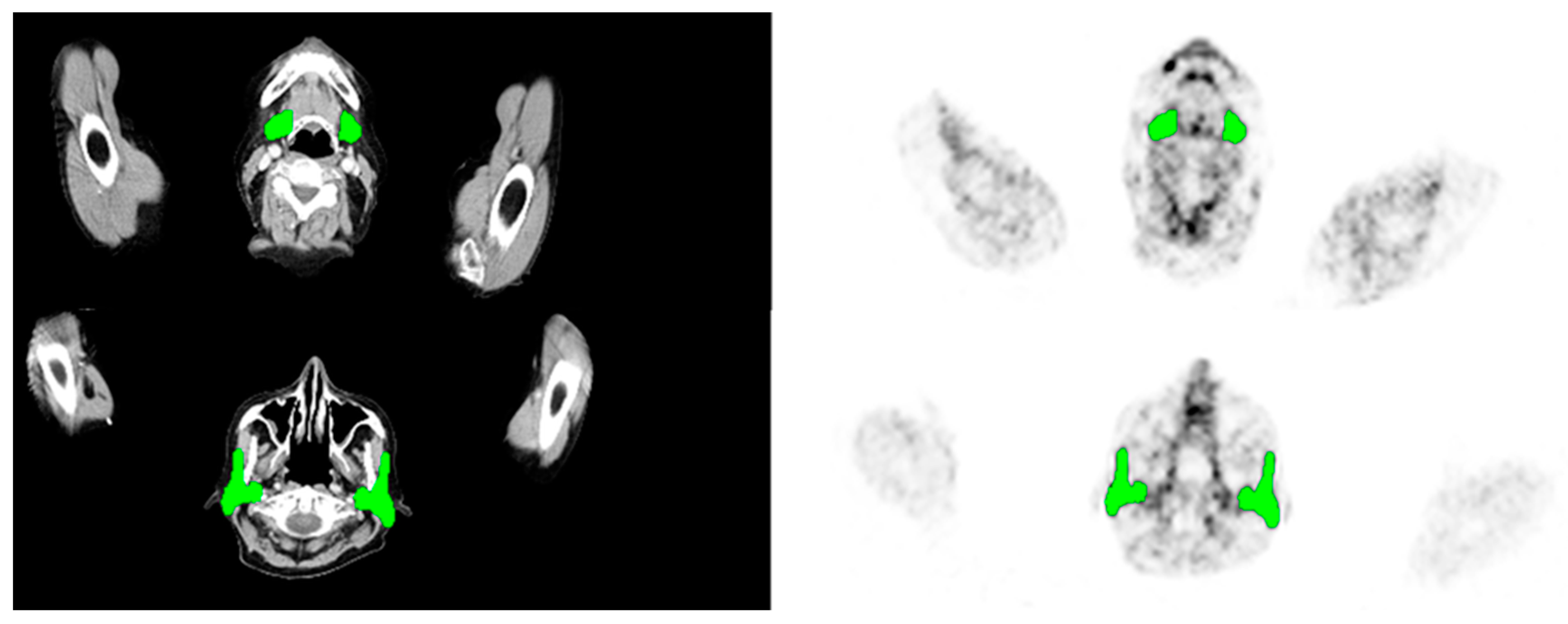
  • Volume-SG was determined in the simultaneously acquired anatomical data (CT or MRI) of the SG. Delineation of the submandibular and parotid glands was measured of each gland separately and on the basis of all available slices (Figure 5).
  • PSMA-SGU was quantified before the first and after the first two cycles of LuPSMA (cohort A) treatment and before and after the first cycle of AcPSMA (cohort B). SG was defined as the parotid and the submandibular glands. PSMA-SGU was determined using the in-house developed software qPSMA (with a threshold SUV of 4).
  • SUVmean and SUVmax was determined using Syngo.Via (Siemens Healthineers, Erlangen, Germany). For SUVmean, a 3D VOI using an isocontour of 20% of the SUVmax was used.
  • PSMA-TV was measured using qPSMA [47]. Bone lesions and soft tissue lesions were separately segmented, and obtained results were summed up. The PSMA-ligand uptake in normal organs was neglected before the quantification of whole-body tumor burden.

4.4. Statistical Analysis

To assess the alterations in morphological and functional parameters of the SG after AcPSMA and LuPSMA RLTs, means, standard deviations, and 95% confidence intervals (95%CI) of Volume-SG, PSMA-SG, and SUVmean and SUVmax of the salivary glands, and their relative and absolute changes were calculated for cohorts A and B.
To determine the impact of a PSMA positive tumor volume on SG changes in cohort B, PSMA-TV was classified into five groups based on quintiles: very low (Q1: ≤20th percentile), low (Q2: 20th–40th percentile), moderate (Q3: 40th–60th percentile), high (Q4: 60th–80th percentile), and very high (Q5: ≥80th percentile). These quintiles were compared with functional changes in the salivary glands.
T-tests using a two-sided unpaired T-Test with Welch correction were used to compare means of Volume-SG, PSMA-SG, and SUVmean and SUVmax of the SG in cohorts A and B and PSMA-TV in cohort B. A p-value of <0.05 was considered statistically significant. All calculations were performed using GraphPad Prism version 5.00 (GraphPad Software, San Diego, CA, USA).

5. Conclusions

Salivary gland volume and tracer uptake as measured from routine PSMA PET studies are potential biomarker for SG toxicity and should be further evaluated in clinical trials of PSMA radioligand therapy.

6. Limitations

One limitation of this retrospective analysis is that it includes both patients with 68Ga-PSMA11 and 18F-rhPSMA7.3, and this could potentially have an effect on the uptake characteristics of salivary glands. However, we only investigated patients who underwent the same radiotracer pre- and post-treatments, and an additional analysis of our data did not show statistically significant differences in the SUVmax and SUVmean in a sub-group analysis both before and after Lu- and Ac-PSMA-RLTs (refer to Supplementary Table S1 and Supplementary Figure S1). Notably, limiting the investigation to only one radiotracer would have substantially reduced the number of suitable patients. Moreover, the direct measurements of the salivary gland function, e.g., using salivary scintigraphy, were not available for analysis in this retrospective analysis.

Supplementary Materials

The following supporting information can be downloaded at: https://www.mdpi.com/article/10.3390/ijms242316845/s1.

Author Contributions

B.F., conceptualization, data curation, methodology, and writing—review and editing. A.G., writing—original draft, data curation, and methodology. T.L., investigation and review and editing. R.T., data curation, investigation, and review. C.S., data curation, and review and editing. F.B. and. J.E.G., review and editing. W.A.W., review and editing. C.D., investigation and review and editing. A.M., investigation and review and editing. M.E., writing—original draft, investigation, and review and editing. All authors have read and agreed to the published version of the manuscript.

Funding

This research received no external funding.

Institutional Review Board Statement

All procedures performed in studies involving human participants were in accordance with the ethical standards of the institutional and/or national research committee and with the 1964 Helsinki declaration and its later amendments or comparable ethical standards. The study was approved by the local ethics committee (115/18 S).

Informed Consent Statement

Informed consent was obtained from all subjects involved in the study.

Data Availability Statement

Data are contained within the article.

Acknowledgments

225Ac was kindly provided by the Joint Research Centre, European Commission, Directorate for Nuclear Safety and Security, Karlsruhe, Germany. PSMA-617 was kindly provided by Endocyte Inc., a subsidiary of Advanced Accelerator Applications, Saint-Genis-Pouilly, France.

Conflicts of Interest

ME reports fees from Blue Earth Diagnostics Ltd. (consultant, research funding), Novartis/AAA (consultant, speaker), Telix (consultant), Bayer (consultant, research funding), RayzeBio (consultant), Point Biopharma (consultant), Eckert-Ziegler (speaker), Janssen Pharmaceuticals (consultant, speakers bureau), Parexel (image review), and Bioclinica (image review) outside the submitted work and a patent application for rhPSMA. BF reports fees from Novartis (consultant). WW reports that he is on advisory boards and receives compensation from Bayer, Blue Earth Diagnostics, Endocyte, Reflexion, Rayzebio, Vida Ventures, ITM, and Pentixapharm. He has received research support from Siemens, BMS, Ipsen, Imaginab, and Piramal. The remaining authors declare that the research was conducted in the absence of any commercial or financial relationships that could be construed as potential conflict of interest.

References

  1. Sartor, O.; de Bono, J.; Chi, K.N.; Fizazi, K.; Herrmann, K.; Rahbar, K.; Tagawa, S.T.; Nordquist, L.T.; Vaishampayan, N.; El-Haddad, G.; et al. Lutetium-177-PSMA-617 for Metastatic Castration-Resistant Prostate Cancer. N. Engl. J. Med. 2021, 385, 1091–1103. [Google Scholar] [CrossRef] [PubMed]
  2. Rahbar, K.; Ahmadzadehfar, H.; Kratochwil, C.; Haberkorn, U.; Schafers, M.; Essler, M.; Baum, R.P.; Kulkarni, H.R.; Schmidt, M.; Drzezga, A.; et al. German Multicenter Study Investigating 177Lu-PSMA-617 Radioligand Therapy in Advanced Prostate Cancer Patients. J. Nucl. Med. 2017, 58, 85–90. [Google Scholar] [CrossRef] [PubMed]
  3. Kratochwil, C.; Bruchertseifer, F.; Rathke, H.; Hohenfellner, M.; Giesel, F.L.; Haberkorn, U.; Morgenstern, A. Targeted alpha-Therapy of Metastatic Castration-Resistant Prostate Cancer with 225Ac-PSMA-617: Swimmer-Plot Analysis Suggests Efficacy Regarding Duration of Tumor Control. J. Nucl. Med. 2018, 59, 795–802. [Google Scholar] [CrossRef] [PubMed]
  4. Kratochwil, C.; Bruchertseifer, F.; Giesel, F.L.; Weis, M.; Verburg, F.A.; Mottaghy, F.; Kopka, K.; Apostolidis, C.; Haberkorn, U.; Morgenstern, A. 225Ac-PSMA-617 for PSMA-Targeted alpha-Radiation Therapy of Metastatic Castration-Resistant Prostate Cancer. J. Nucl. Med. 2016, 57, 1941–1944. [Google Scholar] [CrossRef] [PubMed]
  5. Zacherl, M.J.; Gildehaus, F.J.; Mittlmeier, L.; Boning, G.; Gosewisch, A.; Wenter, V.; Unterrainer, M.; Schmidt-Hegemann, N.; Belka, C.; Kretschmer, A.; et al. First Clinical Results for PSMA-Targeted alpha-Therapy Using 225Ac-PSMA-I&T in Advanced-mCRPC Patients. J. Nucl. Med. 2021, 62, 669–674. [Google Scholar]
  6. Feuerecker, B.; Tauber, R.; Knorr, K.; Heck, M.; Beheshti, A.; Seidl, C.; Bruchertseifer, F.; Pickhard, A.; Gafita, A.; Kratochwil, C.; et al. Activity and Adverse Events of Actinium-225-PSMA-617 in Advanced Metastatic Castration-resistant Prostate Cancer After Failure of Lutetium-177-PSMA. Eur. Urol. 2020, 79, 343–350. [Google Scholar] [CrossRef]
  7. Hey, J.; Setz, J.; Gerlach, R.; Janich, M.; Hildebrandt, G.; Vordermark, D.; Gernhardt, C.R.; Kuhnt, T. Parotid gland-recovery after radiotherapy in the head and neck region--36 months follow-up of a prospective clinical study. Radiat. Oncol. 2011, 6, 125. [Google Scholar] [CrossRef]
  8. Dijkema, T.; Raaijmakers, C.P.; Ten Haken, R.K.; Roesink, J.M.; Braam, P.M.; Houweling, A.C.; Moerland, M.A.; Eisbruch, A.; Terhaard, C.H. Parotid gland function after radiotherapy: The combined michigan and utrecht experience. Int. J. Radiat. Oncol. Biol. Phys. 2010, 78, 449–453. [Google Scholar] [CrossRef]
  9. Fard-Esfahani, A.; Emami-Ardekani, A.; Fallahi, B.; Fard-Esfahani, P.; Beiki, D.; Hassanzadeh-Rad, A.; Eftekhari, M. Adverse effects of radioactive iodine-131 treatment for differentiated thyroid carcinoma. Nucl. Med. Commun. 2014, 35, 808–817. [Google Scholar] [CrossRef]
  10. Itonaga, T.; Tokuuye, K.; Mikami, R.; Shimizu, A.; Sato, H.; Yoshimura, M.; Tsukahara, K.; Saito, K. Mathematical evaluation of post-radiotherapy salivary gland function using salivary gland scintigraphy. Br. J. Radiol. 2022, 95, 20210718. [Google Scholar] [CrossRef]
  11. Allweiss, P.; Braunstein, G.D.; Katz, A.; Waxman, A. Sialadenitis following I-131 therapy for thyroid carcinoma: Concise communication. J. Nucl. Med. 1984, 25, 755–758. [Google Scholar] [PubMed]
  12. Wang, K.; Pearlstein, K.A.; Moon, D.H.; Mahbooba, Z.M.; Deal, A.M.; Wang, Y.; Sutton, S.R.; Motley, B.B.; Judy, G.D.; Holmes, J.A.; et al. Assessment of Risk of Xerostomia After Whole-Brain Radiation Therapy and Association With Parotid Dose. JAMA Oncol. 2019, 5, 221–228. [Google Scholar] [CrossRef] [PubMed]
  13. Badam, R.K.; Suram, J.; Babu, D.B.; Waghray, S.; Marshal, R.; Bontha, S.C.; Lavanya, R.; Kanth, S. Assessment of Salivary Gland Function Using Salivary Scintigraphy in Pre and Post Radioactive Iodine Therapy in Diagnosed Thyroid Carcinoma Patients. J. Clin. Diagn. Res. 2016, 10, ZC60. [Google Scholar] [CrossRef]
  14. Bohuslavizki, K.H.; Brenner, W.; Lassmann, S.; Tinnemeyer, S.; Tonshoff, G.; Sippel, C.; Wolf, H.; Clausen, M.; Henze, E. Quantitative salivary gland scintigraphy in the diagnosis of parenchymal damage after treatment with radioiodine. Nucl. Med. Commun. 1996, 17, 681–686. [Google Scholar] [CrossRef]
  15. Malpani, B.L.; Samuel, A.M.; Jaiswar, R.K. Salivary gland scintigraphy after radioiodine therapy. Nucl. Med. Commun. 1998, 19, 183–184. [Google Scholar] [CrossRef] [PubMed]
  16. Kim, J.W.; Kim, J.M.; Choi, M.E.; Kim, S.K.; Kim, Y.M.; Choi, J.S. Does Salivary Function Decrease in Proportion to Radioiodine Dose? Laryngoscope 2020, 130, 2173–2178. [Google Scholar] [CrossRef]
  17. Langbein, T.; Kulkarni, H.R.; Schuchardt, C.; Mueller, D.; Volk, G.F.; Baum, R.P. Salivary Gland Toxicity of PSMA-Targeted Radioligand Therapy with 177Lu-PSMA and Combined 225Ac- and 177Lu-Labeled PSMA Ligands (TANDEM-PRLT) in Advanced Prostate Cancer: A Single-Center Systematic Investigation. Diagnostics 2022, 12, 1926. [Google Scholar] [CrossRef]
  18. Yadav, M.P.; Ballal, S.; Sahoo, R.K.; Tripathi, M.; Seth, A.; Bal, C. Efficacy and safety of 225Ac-PSMA-617 targeted alpha therapy in metastatic castration-resistant Prostate Cancer patients. Theranostics 2020, 10, 9364–9377. [Google Scholar] [CrossRef]
  19. Konings, A.W.; Coppes, R.P.; Vissink, A. On the mechanism of salivary gland radiosensitivity. Int. J. Radiat. Oncol. Biol. Phys. 2005, 62, 1187–1194. [Google Scholar] [CrossRef]
  20. Nomayr, A.; Lell, M.; Sweeney, R.; Bautz, W.; Lukas, P. MRI appearance of radiation-induced changes of normal cervical tissues. Eur. Radiol. 2001, 11, 1807–1817. [Google Scholar] [CrossRef]
  21. Barker, J.L., Jr.; Garden, A.S.; Ang, K.K.; O′Daniel, J.C.; Wang, H.; Court, L.E.; Morrison, W.H.; Rosenthal, D.I.; Chao, K.S.; Tucker, S.L.; et al. Quantification of volumetric and geometric changes occurring during fractionated radiotherapy for head-and-neck cancer using an integrated CT/linear accelerator system. Int. J. Radiat. Oncol. Biol. Phys. 2004, 59, 960–970. [Google Scholar] [CrossRef]
  22. Wu, V.W.; Ying, M.T.; Kwong, D.L. Evaluation of radiation-induced changes to parotid glands following conventional radiotherapy in patients with nasopharygneal carcinoma. Br. J. Radiol. 2011, 84, 843–849. [Google Scholar] [CrossRef]
  23. Belli, M.L.; Sarnelli, A.; Mezzenga, E.; Cesarini, F.; Caroli, P.; Di Iorio, V.; Strigari, L.; Cremonesi, M.; Romeo, A.; Nicolini, S.; et al. Targeted Alpha Therapy in mCRPC (Metastatic Castration-Resistant Prostate Cancer) Patients: Predictive Dosimetry and Toxicity Modeling of 225Ac-PSMA (Prostate-Specific Membrane Antigen). Front. Oncol. 2020, 10, 531660. [Google Scholar] [CrossRef] [PubMed]
  24. Kratochwil, C.; Giesel, F.L.; Stefanova, M.; Benesova, M.; Bronzel, M.; Afshar-Oromieh, A.; Mier, W.; Eder, M.; Kopka, K.; Haberkorn, U. PSMA-Targeted Radionuclide Therapy of Metastatic Castration-Resistant Prostate Cancer with 177Lu-Labeled PSMA-617. J. Nucl. Med. 2016, 57, 1170–1176. [Google Scholar] [CrossRef] [PubMed]
  25. Violet, J.; Jackson, P.; Ferdinandus, J.; Sandhu, S.; Akhurst, T.; Iravani, A.; Kong, G.; Kumar, A.R.; Thang, S.P.; Eu, P.; et al. Dosimetry of 177Lu-PSMA-617 in Metastatic Castration-Resistant Prostate Cancer: Correlations Between Pretherapeutic Imaging and Whole-Body Tumor Dosimetry with Treatment Outcomes. J. Nucl. Med. 2019, 60, 517–523. [Google Scholar] [CrossRef]
  26. Kabasakal, L.; AbuQbeitah, M.; Aygun, A.; Yeyin, N.; Ocak, M.; Demirci, E.; Toklu, T. Pre-therapeutic dosimetry of normal organs and tissues of 177Lu-PSMA-617 prostate-specific membrane antigen (PSMA) inhibitor in patients with castration-resistant prostate cancer. Eur. J. Nucl. Med. Mol. Imaging 2015, 42, 1976–1983. [Google Scholar] [CrossRef] [PubMed]
  27. Feuerecker, B.; Chantadisai, M.; Allmann, A.; Tauber, R.; Allmann, J.; Steinhelfer, L.; Rauscher, I.; Wurzer, A.; Wester, H.J.; Weber, W.A.; et al. Pretherapeutic Comparative Dosimetry of 177Lu-rhPSMA-7.3 and 177Lu-PSMA I&T in Patients with Metastatic Castration-Resistant Prostate Cancer. J. Nucl. Med. 2022, 63, 833–839. [Google Scholar] [PubMed]
  28. Derlin, T.; Widjaja, L.; Werner, R.A.; Bengel, F.M. 177Lu-PSMA for Extended Treatment of Metastatic Castration-Resistant Prostate Cancer. J. Nucl. Med. 2023, 64, 54–58. [Google Scholar] [CrossRef] [PubMed]
  29. Taieb, D.; Foletti, J.M.; Bardies, M.; Rocchi, P.; Hicks, R.J.; Haberkorn, U. PSMA-Targeted Radionuclide Therapy and Salivary Gland Toxicity: Why Does It Matter? J. Nucl. Med. 2018, 59, 747–748. [Google Scholar] [CrossRef]
  30. Navazesh, M.; Christensen, C.; Brightman, V. Clinical criteria for the diagnosis of salivary gland hypofunction. J. Dent. Res. 1992, 71, 1363–1369. [Google Scholar] [CrossRef]
  31. Gaertner, F.C.; Halabi, K.; Ahmadzadehfar, H.; Kurpig, S.; Eppard, E.; Kotsikopoulos, C.; Liakos, N.; Bundschuh, R.A.; Strunk, H.; Essler, M. Uptake of PSMA-ligands in normal tissues is dependent on tumor load in patients with prostate cancer. Oncotarget 2017, 8, 55094–55103. [Google Scholar] [CrossRef] [PubMed]
  32. Gafita, A.; Wang, H.; Robertson, A.; Armstrong, W.R.; Zaum, R.; Weber, M.; Yagubbayli, F.; Kratochwil, C.; Grogan, T.R.; Nguyen, K.; et al. Tumor sink effect in 68Ga-PSMA-11 PET: Myth or Reality? J. Nucl. Med. 2021, 63, 226–232. [Google Scholar] [CrossRef] [PubMed]
  33. Werner, R.A.; Bundschuh, R.A.; Bundschuh, L.; Lapa, C.; Yin, Y.; Javadi, M.S.; Buck, A.K.; Higuchi, T.; Pienta, K.J.; Pomper, M.G.; et al. Semiquantitative Parameters in PSMA-Targeted PET Imaging with [18F]DCFPyL: Impact of Tumor Burden on Normal Organ Uptake. Mol. Imaging Biol. 2020, 22, 190–197. [Google Scholar] [CrossRef] [PubMed]
  34. Hofman, M.S.; Violet, J.; Hicks, R.J.; Ferdinandus, J.; Thang, S.P.; Akhurst, T.; Iravani, A.; Kong, G.; Ravi Kumar, A.; Murphy, D.G.; et al. [177Lu]-PSMA-617 radionuclide treatment in patients with metastatic castration-resistant prostate cancer (LuPSMA trial): A single-centre, single-arm, phase 2 study. Lancet Oncol. 2018, 19, 825–833. [Google Scholar] [CrossRef] [PubMed]
  35. Rupp, N.J.; Umbricht, C.A.; Pizzuto, D.A.; Lenggenhager, D.; Topfer, A.; Muller, J.; Muehlematter, U.J.; Ferraro, D.A.; Messerli, M.; Morand, G.B.; et al. First Clinicopathologic Evidence of a Non-PSMA-Related Uptake Mechanism for 68Ga-PSMA-11 in Salivary Glands. J. Nucl. Med. 2019, 60, 1270–1276. [Google Scholar] [CrossRef] [PubMed]
  36. Tonnesmann, R.; Meyer, P.T.; Eder, M.; Baranski, A.C. [177Lu]Lu-PSMA-617 Salivary Gland Uptake Characterized by Quantitative In Vitro Autoradiography. Pharmaceuticals 2019, 12, 18. [Google Scholar] [CrossRef] [PubMed]
  37. Filss, C.; Heinzel, A.; Miiller, B.; Vogg, A.T.J.; Langen, K.J.; Mottaghy, F.M. Relevant tumor sink effect in prostate cancer patients receiving 177Lu-PSMA-617 radioligand therapy. Nuklearmedizin 2018, 57, 19–25. [Google Scholar] [CrossRef]
  38. Tuncel, M.; Telli, T.; Tuncali, M.C.; Karabulut, E. Predictive factors of tumor sink effect: Insights from 177Lu-Prostate-specific membrane antigen therapy. Ann. Nucl. Med. 2021, 35, 529–539. [Google Scholar] [CrossRef]
  39. Afshar-Oromieh, A.; Haberkorn, U.; Zechmann, C.; Armor, T.; Mier, W.; Spohn, F.; Debus, N.; Holland-Letz, T.; Babich, J.; Kratochwil, C. Repeated PSMA-targeting radioligand therapy of metastatic prostate cancer with 131I-MIP-1095. Eur. J. Nucl. Med. Mol. Imaging 2017, 44, 950–959. [Google Scholar] [CrossRef]
  40. Zechmann, C.M.; Afshar-Oromieh, A.; Armor, T.; Stubbs, J.B.; Mier, W.; Hadaschik, B.; Joyal, J.; Kopka, K.; Debus, J.; Babich, J.W.; et al. Radiation dosimetry and first therapy results with a 124I/131I-labeled small molecule (MIP-1095) targeting PSMA for prostate cancer therapy. Eur. J. Nucl. Med. Mol. Imaging 2014, 41, 1280–1292. [Google Scholar] [CrossRef]
  41. Mahajan, S.; Grewal, R.K.; Friedman, K.P.; Schoder, H.; Pandit-Taskar, N. Assessment of salivary gland function after 177Lu-PSMA radioligand therapy: Current concepts in imaging and management. Transl. Oncol. 2022, 21, 101445. [Google Scholar] [CrossRef] [PubMed]
  42. Hofman, M.S.; Emmett, L.; Sandhu, S.; Iravani, A.; Joshua, A.M.; Goh, J.C.; Pattison, D.A.; Tan, T.H.; Kirkwood, I.D.; Ng, S.; et al. [177Lu]Lu-PSMA-617 versus cabazitaxel in patients with metastatic castration-resistant prostate cancer (TheraP): A randomised, open-label, phase 2 trial. Lancet 2021, 397, 797–804. [Google Scholar] [CrossRef] [PubMed]
  43. Wollenweber, T.; Zisser, L.; Kretschmer-Chott, E.; Weber, M.; Grubmuller, B.; Kramer, G.; Shariat, S.F.; Mitterhauser, M.; Schmitl, S.; Vraka, C.; et al. Renal and Salivary Gland Functions after Three Cycles of PSMA-617 Therapy Every Four Weeks in Patients with Metastatic Castration-Resistant Prostate Cancer. Curr. Oncol. 2021, 28, 3692–3704. [Google Scholar] [CrossRef]
  44. Rahbar, K.; Bode, A.; Weckesser, M.; Avramovic, N.; Claesener, M.; Stegger, L.; Bogemann, M. Radioligand Therapy With 177Lu-PSMA-617 as A Novel Therapeutic Option in Patients With Metastatic Castration Resistant Prostate Cancer. Clin. Nucl. Med. 2016, 41, 522–528. [Google Scholar] [CrossRef] [PubMed]
  45. Baum, R.P.; Kulkarni, H.R.; Schuchardt, C.; Singh, A.; Wirtz, M.; Wiessalla, S.; Schottelius, M.; Mueller, D.; Klette, I.; Wester, H.J. 177Lu-Labeled Prostate-Specific Membrane Antigen Radioligand Therapy of Metastatic Castration-Resistant Prostate Cancer: Safety and Efficacy. J. Nucl. Med. 2016, 57, 1006–1013. [Google Scholar] [CrossRef]
  46. Van Kalmthout, L.; Braat, A.; Lam, M.; van Leeuwaarde, R.; Krijger, G.; Ververs, T.; Mehra, N.; Bins, A.; Hunting, J.; de Keizer, B. First Experience With 177Lu-PSMA-617 Therapy for Advanced Prostate Cancer in the Netherlands. Clin. Nucl. Med. 2019, 44, 446–451. [Google Scholar] [CrossRef]
  47. Gafita, A.; Bieth, M.; Kronke, M.; Tetteh, G.; Navarro, F.; Wang, H.; Gunther, E.; Menze, B.; Weber, W.A.; Eiber, M. qPSMA: Semiautomatic Software for Whole-Body Tumor Burden Assessment in Prostate Cancer Using 68Ga-PSMA11 PET/CT. J. Nucl. Med. 2019, 60, 1277–1283. [Google Scholar] [CrossRef]
Figure 1. Morphological changes in SG volume based on CT/MRI quantification after 177Lu-PSMA (A) and 225Ac-PSMA-617 (B).
Ijms 24 16845 g001
Figure 2. Changes in SUVmax (the total of submandibular and parotid glands) after 177Lu-PSMA RLT (A) and after 225Ac-PSMA-617 RLT (B), respectively, and change in SUVmean (the total of submandibular and parotid glands) after 177Lu-PSMA RLT (C) and 225Ac-PSMA-617 RLT (D), respectively.
Ijms 24 16845 g002
Figure 3. Changes in PSMA-SGU after 177Lu-177-PSMA RLT (A) and after 225Ac-PSMA-617 RLT (B), respectively.
Ijms 24 16845 g003
Figure 4. SUVmax of the SG stratified by tumor burden before (pre) and after (post) 225Ac-PSMA-617 RLT and also stratified by tumor load (colors indicate groups). Group moderate, n = 3, all other groups, n = 4.
Ijms 24 16845 g004
Figure 5. CT based segmentation (left) of submandibular (upper row) and parotid glands (lower row), and the illustration of its transfer to the respective PET images (right). Green colored areas indicate the respective salivary glands.
Ijms 24 16845 g005
Table 1. Uptake characteristics of salivary glands (SUVmean and SUVmax) before and after Ac- and Lu-PSMA RLTs of patients from cohort B. Patients are stratified in five groups based on their whole body tumor volume prior to 225Ac-PSMA RLT. Statistically significant changes are marked in bold (* p = 0.03, ** p = 0.02, *** p = 0.04, # p = 0.01).
Whole Body
Tumor Volume Prior to AcPSMA
Very LowLowModerateHighVery High
prepostprepostprepostprepostprepost
AcPSMA RLT
Whole bodyPSMA-TV (mL)602 ± 354431 ± 2961393 ± 2171456 ± 3911848 ± 1562370 ± 10763378 ± 2883216 ± 6934869 ± 3424296 ± 1252
Salivary glandsSUVmean11.7 ± 2.46.7 ± 2.4 *8.1 ± 3.34.9 ± 1.111.1 ± 1.85.7 ± 1.6 **9.1 ± 2.94.5 ± 2.3 ***7.8 ± 2.75.3 ± 1.7
SUVmax24.8 ± 4.914.7 ± 4.8 *17.7 ± 6.111.5 ± 2.322.7 ± 3.413.5 ± 2.9 **20.9 ± 5.48.6 ± 1.9 #17.3 ± 5.312.7 ± 3.2
LuPSMA RLT
Salivary glandsSUVmean14.9 ± 2.913.5 ± 2.611.3 ± 2.211.2 + 5.39.0 ± 2.811.9 ± 1.9 *10.2 ± 2.68.8 ± 1.411.3 ± 2.810.3 ± 3.1
SUVmax33.6 ± 9.833.4 ± 14.323.9 ± 4.023.7 ± 10.318.6 ± 4.125.4 ± 5.9 **21.5 ± 5.318.8 ± 2.924.5 ± 5.626.1 ± 12.1
Table 2. Patient characteristics at the timepoint of initiation of 225Ac-PSMA RLT.
No.Number (Agents) of Previous mCRPC Lines Prior to 225Ac-RLTNumber (Agents) of
Previous mCRPC Lines Prior to 225Ac-RLT
Number of LuPSMA
Cycles
Activity LuPSMA RLT (GBq)/CycleECOG ScoreMetastasesActivity of First
AcPSMA RLT (MBq)
1 *4 (E, A, D, Lu)428/7.20B, LN8
28 (D, C, A, C, E, C, Ra, Lu)825.7/5.70B8
34 (D, E, A, Lu)487.4/7.4/7.3/7.3/7.1/7.1/7.1/7.31B, LN8
4 *5 (A, E, Lu, D, Cis/Eto)547.2/7.7/7.2/7.71B, LN8
5 *6 (D, A, Lu, C, E, Cis/Eto)627.6/7.41B, LN, Liver, Lungs8
6 *6 (D, Ra, E, C, A, Lu)646.9/7.3/7.4/7.52B, LN10
74 (D, Ra, A, Lu)457.5/7.3/7.5/7.8/7.71B, LN8
8 *8 (CureVac, A + CureVac, D, Study, C, Lu, E, A)827.3/7.51B, LN, Lungs8
9 *4 (A, D, Lu, E)467.2/7.4/7.3/7.4/7.3/6.71B, LN, Peritoneal10
10 *3 (A, E, Lu)367.3/7.6/7.7/7.0/7.5/ 7.41B, LN10
117 (A, E, D, A, D, C, Lu)768.3/7.9/8.3/7.9/7.4/7.31B8
126 (A, E, D, C, Lu, Cis/Eto)628.3/7.81B, LN13
13 *5 (E, D, A, E, Lu)565.1/7.4/7.3/7.6/7.4/6.71B, LN, Liver, Lung11
14 *5 (A, E, D, C, Lu)517.91B, LN, Liver, Brain6
15 *8 (CureVac, A, Ra, Lu, E, D, O, C)867.3/7.3/7.3/7.6/7.4/ 7.01B, LN10
16 *8 (D, C, A, D, E, A, C, Lu)887.3/7.8/7.2/7.2/7.5/7.5/7.5/7.40B, LN, Lungs12
17 *5 (D/C, A, D/C, Carbo, Lu)547.3/7.6/7.2/7.31LN, B, Peritoneal9
183 (A, Lu, D)353.7/3.7/5.5/5.5/4.21B, LN10
19 *6 (D, A, E, C, Lu, C)646.8/7.6/7.3/9.01B, LN, Liver 14
20 *5 (E, D, A, Lu, C)548.2/7.5/6.2/7.51B, LN8
21 *6 (A, E, D, Lu, Ra, C)643.3/3.3/3.4/3.51B8
Abbreviations: Gs = Gleason Score, AP = alkaline phosphatase, LDH = lactate dehydrogenase, AcPSMA = 225Ac-PSMA-617, LuPSMA = 177Lu-PSMA, RLT = radioligand therapy, E = Enzalutamide, A = Abiraterone, D = Docetaxel, Lu = 177Lu-PSMA I&T, RTx = Radiatio, C = Cabazitaxel, Ra = Ra-223-Dichloride, Cis/Eto = Cisplatin/Etoposide, Carbo = Carboplatin; I = immune therapy, O = Olaparib, CureVac = CureVac Study, B = bones, and LN = lymph nodes. * cohort A.
Disclaimer/Publisher’s Note: The statements, opinions and data contained in all publications are solely those of the individual author(s) and contributor(s) and not of MDPI and/or the editor(s). MDPI and/or the editor(s) disclaim responsibility for any injury to people or property resulting from any ideas, methods, instructions or products referred to in the content.

