Article

Antigenic and Structural Properties of the Lipopolysaccharide of the Uropathogenic Proteus mirabilis Dm55 Strain Classified to a New O85 Proteus Serogroup

by
Agata Palusiak
1,*,
Anna Turska-Szewczuk
2 and
Dominika Drzewiecka
1
1
Department of Biology of Bacteria, Faculty of Biology and Environmental Protection, University of Lodz, Banacha 12/16, 90-237 Lodz, Poland
2
Department of Genetics and Microbiology, Institute of Biological Sciences, Maria Curie-Sklodowska University, Akademicka 19, 20-033 Lublin, Poland
*
Author to whom correspondence should be addressed.
Int. J. Mol. Sci. 2023, 24(22), 16424; https://doi.org/10.3390/ijms242216424
Submission received: 17 October 2023 / Revised: 8 November 2023 / Accepted: 13 November 2023 / Published: 16 November 2023
(This article belongs to the Special Issue Lipopolysaccharide: Bacterial Endotoxin 2023)

Abstract

:
The aim of the study was the serological and structural characterization of the lipopolysaccharide (LPS) O antigen from P. mirabilis Dm55 coming from the urine of a patient from Lodz. The Dm55 LPS was recognized in ELISA only by the O54 antiserum, suggesting a serological distinction of the Dm55 O antigen from all the 84 Proteus LPS serotypes described. The obtained polyclonal rabbit serum against P. mirabilis Dm55 reacted in ELISA and Western blotting with a few LPSs (including O54), but the reactions were weaker than those observed in the homologous system. The LPS of P. mirabilis Dm55 was subjected to mild acid hydrolysis, and the obtained high-molecular-mass O polysaccharide was chemically studied using sugar and methylation analyses, mass spectrometry, and 1H and 13C NMR spectroscopy, including 1H,1H NOESY, and 1H,13C HMBC experiments. The Dm55 O unit is a branched three-saccharide, and its linear fragment contains α-GalpNAc and β-Galp, whereas α-GlcpNAc occupies a terminal position. The Dm55 OPS shares a disaccharide epitope with the Proteus O54 antigen. Due to the structural differences of the studied O antigen from the other described Proteus O polysaccharides, we propose to classify the P. mirabilis Dm55 strain to a new Proteus O85 serogroup.

1. Introduction

The genus Proteus, formerly belonging to the family Enterobacteriaceae, has been lately recognized as a member of the newly formed family Morganellaceae. It has been proposed to be placed in the new family based on taxonomic changes within the order Enterobacteriales, renamed as Enterobacterales [1]. Proteus bacteria are Gram-negative, facultatively anaerobic, heterotrophic, proteolytic, and peritrichously flagellated motile rods [2]. The last feature accounts for the swarming phenomenon, which distinguishes Proteus spp. from most Enterobacterales [3]. The genus Proteus includes two species—P. mirabilis and P. vulgaris—and was first described in 1885 by a German microbiologist Gustav Hauser and now covers more than ten species. The rods may be a member of the resident microbiota in the gastrointestinal tracts of a part of the human population, and the intestines are probably a reservoir of these organisms [2,4]. At the same time, Proteus spp. bacteria are opportunistic human pathogens, which, in favorable conditions, may lead to cross-infections and autoinfections. Among all described Proteus species, P. mirabilis is the most frequent cause of infections [2]. It is regarded as the third most common etiological factor of urinary tract infections (UTIs), among which catheter-associated urinary tract infections (CAUTIs) occur quite frequently [5,6]. According to the National Healthcare Safety Network (NHSN, Atlanta, GA, USA) review, Proteus spp. account for 4% of CAUTI cases [7]. Apart from UTIs, P. mirabilis has been implicated in pneumonia, neonatal meningoencephalitis, empyema and osteomyelitis, and eye, ear, nose, and wound infections [3]. These bacteria are also a causative agent of diarrhea, and some studies have shown their potential role in infective endocarditis and rheumatoid arthritis development [2,8,9]. Pathogenicity of P. mirabilis strains is associated with numerous virulence factors such as urease, fimbriae, flagella, proteinases, and lipopolysaccharide (LPS) endotoxin, which poses a significant threat since its interaction with the host immune system may contribute to a septic shock development [3,6]. A smooth (S) LPS of Proteus spp. bacilli is built of three regions differing in their structures and biological functions: lipid A (structurally the most conserved region), core oligosaccharide (more structurally diverse), and O-polysaccharide (structurally the most heterogenous). Interestingly, S-form strains also possess in their outer membrane rough (R) LPS, and there are some strains that may be completely deprived of three-part LPS. It should be mentioned that S strains are considered to be more pathogenic than R forms. O polysaccharide is the most outstanding part of the bacterial cell wall and exhibits many biological activities, like getting involved in glycocalyx formation or making the bacteria resistant to the active membrane attack complex [10,11]. It is also a highly immunogenic O antigen, and its fragments (epitopes), which are common to many different bacteria, may be used as vaccine antigens inducing cross-protection in humans [10]. To date, the O antigen of Shigella sonnei LPS has been successfully used in vaccines administered to humans in clinical studies [12]. Thus, it is crucial to gain a better knowledge of the OPS structure and epitope specificity, which are also a basis for serological classification of the strains to an appropriate O serogroup. To date, the Proteus classification scheme includes 84 O serogroups and 11 R serogroups, which are still being updated and completed with new representatives [13,14]. In this work, we report on a new OPS structure and the serological specificity of the P. mirabilis Dm55 LPS obtained from the isolate coming from a patient from central Poland.

2. Results

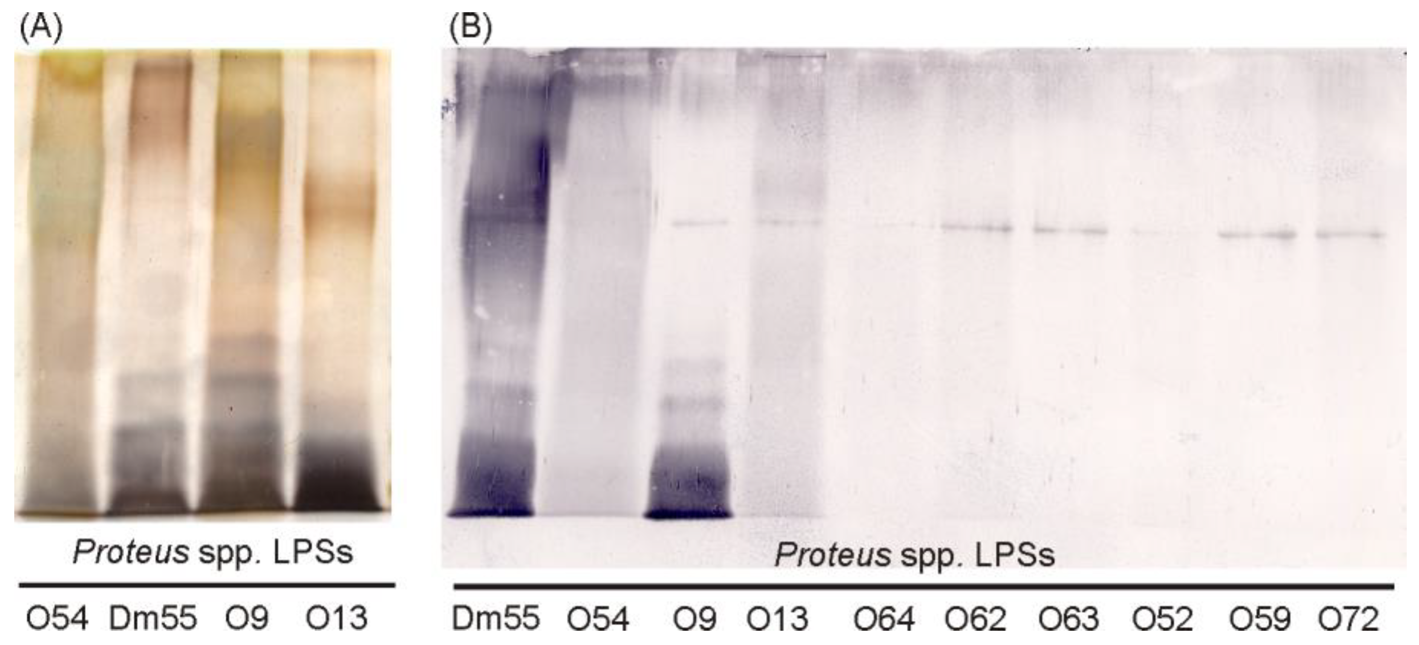
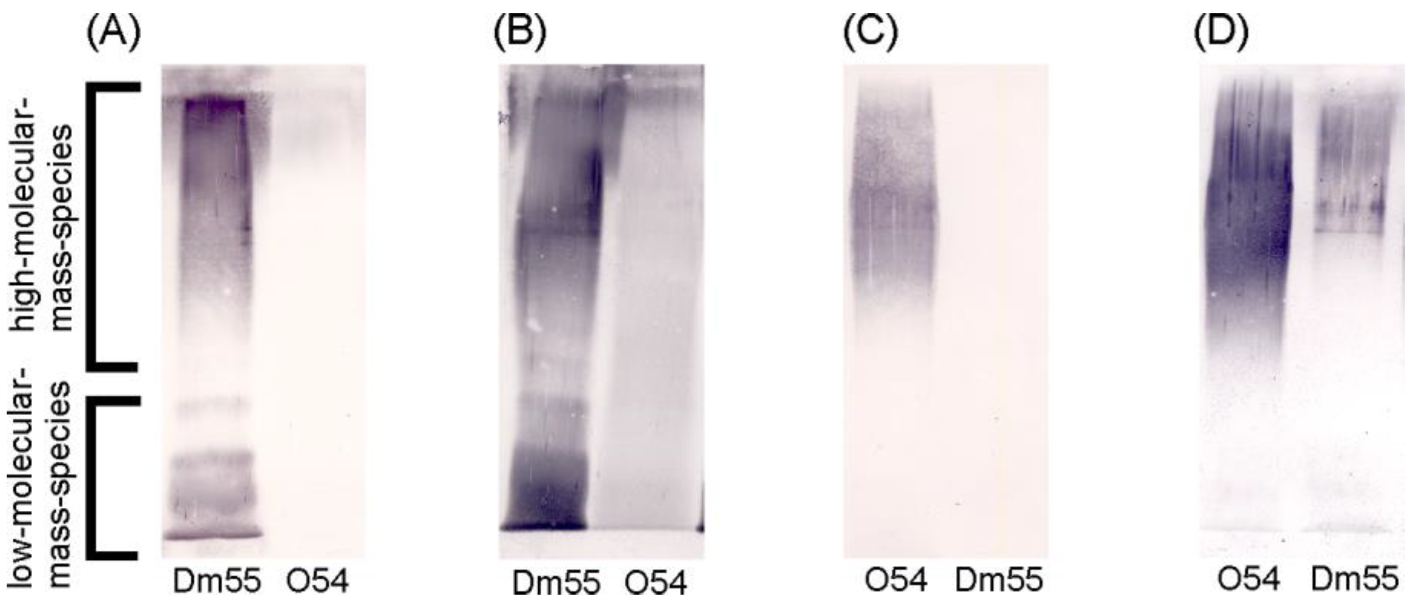
2.1. Serological Studies

The LPS of P. mirabilis Dm55 was extracted by the phenol–water method. It demonstrated a typical, ladder-like pattern in SDS-PAGE after Alcian blue–silver staining, thus proving the presence of high-molecular-mass molecules, including an O-specific long-chain polysaccharide (S form LPS) (Figure 1A).
To find out if the studied strain may be classified as one of the 84 Proteus O serotypes recognized so far, its LPS was tested in ELISA with the sera specific to each of the serotypes in dilutions 1:32,000 and 1:64,000. The only cross-reaction was observed with the O54 antiserum (1:64,000), but it was much weaker than in the homologous system of the O54 antiserum (1:256,000), which suggested only similarity between and not identity of both antigens (Dm55 and O54 LPSs).
At the next stage of the studies, a whole-cell vaccine containing bacterial cells of P. mirabilis Dm55 was prepared to obtain the polyclonal rabbit serum to confirm the suggested similarity between the Dm55 and O54 antigens. The serum reactivity was checked in ELISA with the homologous LPS as well as with the LPSs presenting each of the known 84 Proteus O serotypes. The P. mirabilis Dm55 antiserum appeared to be quite reactive since the cross-reactions were observed with many LPSs, but the serum titers were lower than in the homologous system (1:4,096,000) (Table 1).
Among the cross-reacting LPSs, five, P. mirabilis O54, O9, P. vulgaris O13, and P. penneri O62, O72, reacted to the titer 1:64,000 or higher. The remaining heterologous LPSs bound the antibodies with a lower intensity, and the reactivity titers of the sera specific to these LPSs were much higher (Table 1).
The reactions detected in ELISA (Table 1) were also analyzed by using the Western blotting method (Figure 1B). The homologous LPS, P. mirabilis Dm55, was strongly recognized by the P. mirabilis Dm55 serum in a way characteristic for both low- and high-migrating bands, which indicated a presence in the antiserum of both core-specific and O-polysaccharides-specific antibodies (Figure 1B). Three cross-reacting LPSs (O54, O13, and O9) were found to display in Western blotting quite intensive binding patterns, while the reactions of the remaining LPSs were less intense and concerned mainly the high-molecular-mass species containing the whole LPS molecules (Figure 1B).
The reaction patterns of low-molecular-mass species of P. mirabilis O9 and Dm55 LPSs were very similar in the reaction with the Dm55 antiserum, which suggests a high level of similarities in the core oligosaccharides of both LPSs. In the opposite system, the reaction of the O9 antiserum with the Dm55 LPS core region was weaker, but it was visible that the O9 antiserum was poor in anti-core antibodies (Figure S1A).
The P. vulgaris O13 LPS reacted in Western blotting with the P. mirabilis Dm55 serum at the level of both low- and high-molecular-mass species, but the intensity of its banding pattern was weaker than that of the homologous LPS, Dm55 (Figure 1B). However, in the opposite system, the O13 antiserum did not recognize the Dm55 LPS (Figure S1B).
The P. mirabilis O54 LPS was the one that cross-reacted with the studied Dm55 antiserum quite strongly both in ELISA and Western blotting (Table 1, Figure 1B). The cross-reaction was also observed for the opposite system, O54 antiserum with the Dm55 LPS, in the Western blot, where it was visible, especially in the region of slowly-migrating LPS molecules, rich in the OPS fractions (Figure 2D). However, the binding pattern of the Dm55 LPS was different than that of the O54 (homologous) LPS.
To confirm serological similarities between P. mirabilis O54 and Dm55 LPSs, the sera specific to both strains were adsorbed with the bacterial cells of the cross-reacting strain. Adsorption resulted in the weakening of the Western blotting reaction in each homologous system, which indicated some similarities but not the identity between the P. mirabilis Dm55 and O54 LPSs (Figure 2A–D). To see how much the reactivities of both tested sera with the homologous LPS changed after their adsorption, they were also tested in ELISA. It was demonstrated that the adsorption of the Dm55 antiserum with the cells of the P. mirabilis O54 strain resulted in an eightfold decrease in the reactivity titer from 4,096,000 (not adsorbed Dm55 serum) to 512,000 (adsorbed Dm55 serum). However, the reactivity of the O54 antiserum adsorbed with Dm55 biomass was reduced as much as 32-fold compared to the titer of not adsorbed O54 serum (Table 2).
Only the antibodies specific to a putative common epitope were removed from both antisera during the adsorption processes. The immunoglobulins that remained in the sera were specific to the fragments, which were different in the two OPSs (Table 2, Figure 2A,C).
The three heterologous LPSs (P. mirabilis O9, O54, and P. vulgaris O13), which strongly cross-reacted with the tested Dm55 antiserum in ELISA and Western blotting, when separated in polyacrylamide gel were also subjected to a silver staining procedure with prefixation of gel in Alcian blue. These LPSs presented different electrophoretic patterns than the Dm55 LPS (Figure 1A). The low-molecular-mass fractions of each LPS tested were silver stained with high intensity, but the most similar bands were observed for the Dm55 and O9 LPSs. What is more, these two LPSs, together with the LPS of the O13 strain, contained higher amounts of R-form molecules than the O54 LPS. As for the high-molecular-mass fractions of the LPSs, the most visible bands were obtained for the O9 and O54 LPSs, while for the O13 and Dm55 LPSs, less abundant fractions of long-chain molecules were observed (Figure 1A).

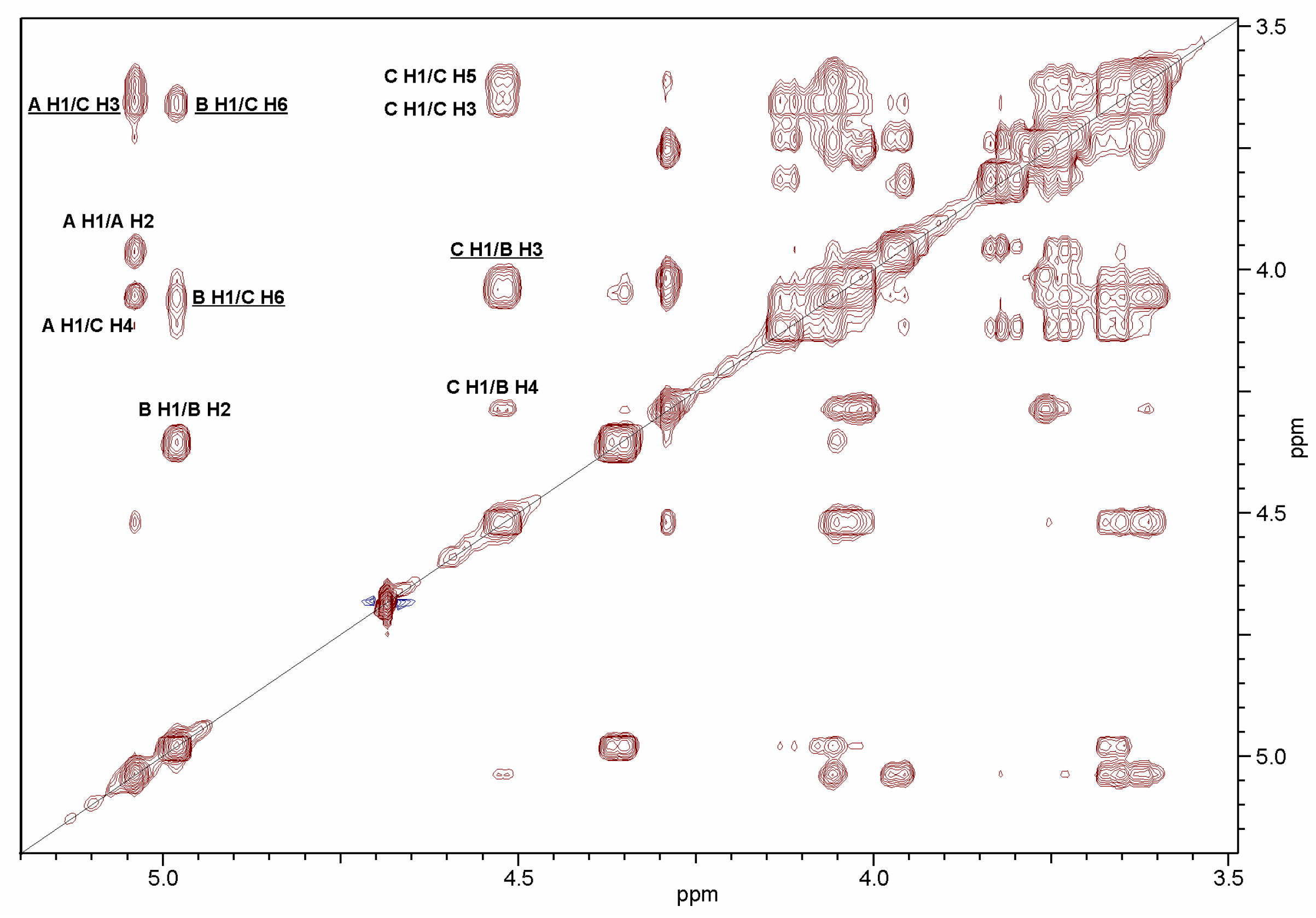
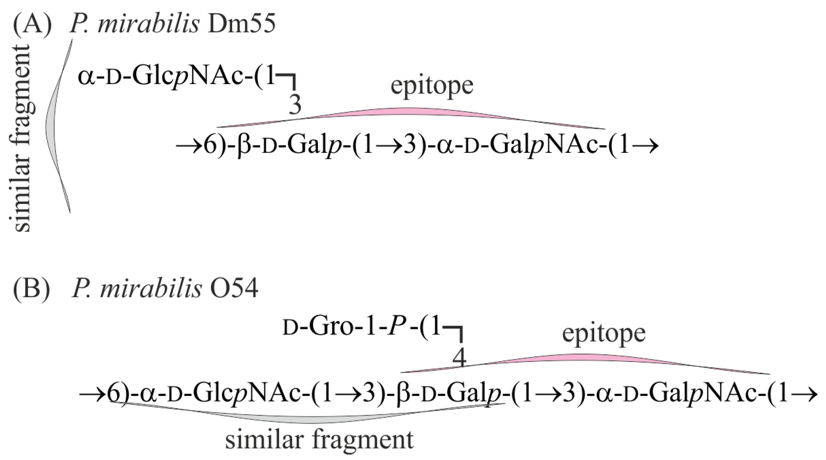
2.2. Structural Studies of O-Polysaccharide (OPS)

The OPS was released from the LPS of P. mirabilis Dm55 by mild-acid degradation followed by centrifugation of the lipid A precipitate and isolated, in a void volume, by gel-permeation-chromatography (GPC) on a Sephadex G50 fine column. The yield of the high-molecular-mass OPS fraction was 15% of the LPS mass subjected to hydrolysis. The GLC-MS sugar analysis of alditol acetates obtained after full acid hydrolysis of the OPS with 2 M CF3CO2H showed the presence of galactose (Gal), glucosamine (GlcN), and galactosamine (GalN) as the major components, in a peak area ratio of 1.0:1.0:1.4. The alditol acetates GLC-MS data have been shown in Supplementary Figure S2.
The determination of the absolute configuration of the monosaccharides by GLC of acetylated (S)-2-octyl glycosides indicated the presence of d-Gal, d-GlcN, and d-GalN.
The methylation analysis of the OPS completed the compositional data and resulted in identification of 1,3,5,6-tetra-O-acetyl-2,4-di-O-methylhexitol-1-d (derived from 3,6-disubstituted Gal), 1,5-di-O-acetyl-2-deoxy-3,4,6-tri-O-methyl-2-(N-methyl)acetamidohexitol-1-d (derived from terminal GlcN) and 1,3,5-tri-O-acetyl-2-deoxy-4,6-di-O-methyl-2-(N-methyl)acetamidohexitol-1-d (derived from 3-substituted GalN), with a peak area ratio of 1.0:1.2:1.3, identified by GLC-MS. The permethylated alditol acetates GLC-MS data have been shown in Supplementary Figure S3.
The O-polysaccharide structure of P. mirabilis Dm55 was then studied with the use of 1D and 2D NMR spectroscopy.
The 1H NMR spectrum of the O-polysaccharide (Figure 3) showed three signals for anomeric protons at δ 5.04, 4.99, and 4.53, with an integral intensity ratio of 1.0:1.04:0.98, indicating that the O-polysaccharide had a regular structure. There were also two signals for N-acetyl groups at δ 2.04 and the ring proton signals in the range of δ 3.62–4.37, some of which overlapped.
The analysis of the two-dimensional homonuclear (1H,1H DQF-COSY, TOCSY, and NOESY) and heteronuclear (1H,13C HSQC, 1H,13C H2BC, and 1H,13C HMBC) NMR experiments resulted in the assignment of the 1H and 13C resonances to the OPS of P. mirabilis Dm55. The 1H and 13C NMR chemical shifts are presented in Table 3.
The 1H,13C HSQC spectrum (Figure 4) contained two correlation signals at δ 3.97/54.7 and 4.36/49.7 of protons at the nitrogen-bearing carbons to the corresponding carbons and showed that the OPS repeating unit contained N-acetamido sugars. Moreover, the absence of signals at the 13C coordinate in the region of δ 83.0–88.0 characteristics of C-4 furanoses and the anomeric carbons, which should have had higher chemical shift values due to the less shielded nuclei than in their corresponding counterparts, demonstrated that all the sugars were pyranoses [15].
The 1H,1H TOCSY and DQF-COSY spectra revealed three spin systems for monosaccharides, which were labeled A, B, and C in the order of the decreasing chemical shifts of their H-1 protons. In the 1H,1H DQF COSY and TOCSY spectra, correlations of H-1/H-2 up to H-6 typical of monosaccharide having the gluco configuration were found for spin system A. Then, the carbon resonances were inferred from the 1H,13C HSQC spectrum, which also showed the correlations of H-2 at δ 3.97 with the nitrogen-bearing carbon at δ 54.7, and H-6 protons with C-6 at δ 3.76/62.2. Based on these data and after including 3JH1,H2 and 1JC1,H1 coupling constant values of ~3.3 Hz and 174 Hz, respectively, spin system A was assigned to α-GlcN [16].
In the 1H,1H TOCSY spectrum, starting from the H-1 proton, the correlations with H-2, H-3, and H-4 were visible for spin systems B and C, indicating monosaccharides with galacto configuration. The remaining resonances were assigned from the NOESY, DQF-COSY, and heteronuclear experiments.
In the 1H,13C HMBC spectrum (Figure 5), correlations of the anomeric proton at δ 4.99 with carbons C-3 and C-5 at δ 79.2 and 71.8, respectively, were found for spin system B, and then the proton resonances were assigned from the 1H,13C HSQC spectrum.
Moreover, in this spectrum (Figure 4), the correlation signal at δ 4.36/49.7 (H-2/C-2) of the proton at the nitrogen-bearing carbon to the corresponding carbon was assigned to spin system B. In the NOESY spectrum, correlations of H-4/H-5 and H-4/H-6 at δ 4.29/4.03 and 4.29/3.76, respectively, were visible for this residue. By including the 3JH1,H2 and 1JC1,H1 coupling constant values of 3.3 Hz and 174 Hz, respectively, spin system B was assigned to α-GalN [16].
On the other hand, for spin system C, in the NOESY spectrum (Figure 6), the intra-residue correlations of H-1/H-3 and H-1/H-5 characteristics of β-configurated sugars were found, and then the H-3/H-5, H-4/H-5, and H-4/H-6 correlations were analyzed.
However, given the coincidence of the H-2/H-3 and H-3/H-5 as well as H-4/H-5 and H-4/H-6 correlation signals, the chemical shifts of corresponding C-2-C-6 carbons, which were inferred from the 1H,13C HSQC spectrum, were assigned after consideration of the two-bond and long-range correlations in the 1H,13C H2BC and 1H,13C HMBC spectra, methylation analysis data and the glycosylation effects on the 13C NMR resonances [17]. The 3JH1,H2 and 1JC1,H1 coupling constant values of 7.7 Hz and 165 Hz, respectively, confirmed that spin system C was β-Gal [16].
The 13C resonance of the NAc carbonyl signals was assigned from the correlations between H-2 of residues A and B (δ 3.97 and 4.36) and the corresponding carbons in the HMBC spectrum (δ 175.6) and between the latter and the methyl proton signals at δ 2.04, respectively.
The NOESY spectrum (Figure 6) showed intra-residue cross-peaks: H-1/H-2 for α-GlcpNAc and α-GalNAc (A and B) and H-1/H-3, and H-1/H-5 for β-Galp C, which confirmed the anomeric configurations of the sugar residues [18].
The low-field displacement of the carbon atoms C-3 (δ 78.7) and C-6 (δ 65.8) of spin system C and C-3 of the B residue (δ 79.2), compared with their resonances in the corresponding non-substituted monosaccharides [17], unlike the C-2,3,4,6 carbon atoms of spin system A showing the insignificant displacements, confirmed the glycosylation pattern of the monosaccharides in the oligosaccharide unit [19]. These data demonstrated that the O-antigen repeating unit is branched with residue C at the branching point and a terminal position of residue A.
The sugar sequence in the OPS repeating unit was determined in the homo- and heteronuclear experiments. The 1H,1H NOESY spectrum (Figure 6) showed correlation signals for pairs of transglycosidic protons, i.e., B H-1/C H-6, C H-1/B H-3, A H-1/C H-3 at δ 4.99/3.66;4.06, 4.53/4.04, and 5.04/3.66, respectively, thus indicating a sequence BCB in the linear part of the O-unit, and the α-GlcpNAc residue (A) located at the terminal position and glycosylating the β-Galp residue (C) at position C-3. Moreover, in the spectrum, the additional inter-residue NOE contacts for C H-1/B H-4 at δ 4.53/4.29 and A H-1/C H-4 at δ 5.04/4.06 were observed, typical of (1 → 3)-linked sugars, which confirmed the glycosylation of α-GlcpNAc (B) and β-Galp (C) at carbon C-3 [18]. Full range of 1H,1H NOESY spectrum of the OPS of P. mirabilis strain Dm55 has been shown in Supplementary Figure S4.
In the 1H,13C HMBC spectrum (Figure 5), the following correlations between anomeric protons and transglycosidic carbons were observed: GalpNAc B H-1/ Galp C C-6 at δ 4.99/65.9, Galp C H-1/ GalpNAc B C-3 at δ 4.53/79.2, and GlcpNAc A H-1/ Galp C C-3 at δ 5.04/78.7. Furthermore, the following correlations between anomeric carbons and transglycosidic protons were also visible in the spectrum: GalpNAc B C-1/ Galp C H-6 at δ 98.4/4.06, Galp C C-1/ GalpNAc B H-3 at δ 105.9/4.04, and GlcpNAc A C-1/ Galp C H-3 at δ 95.6/3.66. Full range of 1H,13C HMBC spectrum of the OPS of P. mirabilis strain Dm55 has been shown in Supplementary Figure S5.
In conclusion, the data showed that the O-specific polysaccharide from P. mirabilis Dm55 had the structure presented below (Scheme 1).

3. Discussion

The aim of the study was the serological and structural characterization of the LPS O antigen from the strain P. mirabilis Dm55 coming from the urine of a patient from Lodz (Poland). To date, such characterization has been performed for ten Proteus spp. strains infecting patients from central Poland and isolated from different sources, among which four isolates were found as uropathogenic P. mirabilis [13,14,20]. Urine was the most common source (43%) of Proteus spp. isolates among 617 clinical strains coming from that area, and P. mirabilis appeared to be the predominant species (90.3%). Most of these strains have already been classified to numerous O serogroups, and the O serotypes most prevalent in central Poland have been indicated [13].
The scenario of the serological studies in this work was realized using reliable and complementary techniques successfully employed in our previous research and also in other studies (ELISA as a highly sensitive serological method and Western blotting due to its specificity).
The first stage of the serological classification of the P. mirabilis Dm55 LPS was an analysis of its similarity to Proteus LPS serotypes described so far by testing its specificity with the O1–O84 Proteus antisera in ELISA. A lack of strong cross-reactions with the set of the tested antisera observed for the Dm55 LPS suggested its serological distinction from all known Proteus O serotypes. Thus, in the next stage, a polyclonal rabbit antiserum was obtained for the studied Dm55 strain, which was rich in O-specific antibodies, but also anti-core antibodies were present in large amounts (Figure 1B). The situation is not rare, and such antiserum may be applied to the determination of similarities not only in O antigens but also in the LPS core region in various strains [13]. In ELISA, the P. mirabilis Dm55 antiserum strongly cross-reacted with a few Proteus spp. LPS (Table 1). However, in Western blotting (Figure 1B), cross-reactions with some LPSs (O52, O59, O62-O64, O72) were barely visible. Due to the lower specificity and, simultaneously, higher sensitivity of reactions in ELISA compared to Western blotting [21], the less specific cross-reactions were not confirmed by the latter method.
The LPSs for which the most intensive banding patterns were obtained in Western blotting with the P. mirabilis Dm55 antiserum were subjected to a silver staining procedure with prefixation of gel in Alcian blue after SDS-PAGE. Such a combination of staining methods allows detection of even very small amounts of acidic polysaccharides, components typical for Proteus spp. O-polysaccharides of LPSs, which makes the procedure more sensitive than silver staining without a previous gel fixation with Alcian blue [22]. The electrophoregram of LPS preparations from the strains Dm55, O9, O13, and O54 (Figure 1A) shows different staining patterns of separated LPS molecules varying in the numbers of the repeating units. The tested Dm55 LPS was less intensively stained within the molecules consisting of O-chains compared to the O9 and O54 LPSs. However, the studied Dm55 OPS does not contain cationic groups; thus, it may have been less clearly stained than the remaining ones containing some acidic components [11], which are the additional binding sites for the anionic dye Alcian blue (Figure 1A).
As was mentioned before, the only serum that cross-reacted with the P. mirabilis Dm55 LPS in ELISA was that specific to the P. mirabilis O54 strain (the first stage of the studies). This fact and quite strong serological activity observed for the opposite system (P. mirabilis Dm55 antiserum and O54 LPS) in ELISA and Western blotting suggested that these two LPSs, P. mirabilis Dm55 and O54, share some high structural similarities (Table 1, Figure 1B). The chemical and structural analyses of the P. mirabilis Dm55 OPS conducted in this work confirmed this hypothesis. Both Dm55 and O54 LPSs (this work, [11]) possess in their OPS repeating units two following common fragments: β-d-Galp-(1 → 3)-α-d-GalpNAc, which may play a role of a common epitope and α-d-GlcpNAc-(1 → 3)-β-d-Galp (indicated as a similar fragment in Figure 7).
The latter fragment in the studied Dm55 OPS is side-branched, while in P. mirabilis O54 OPS, it is part of a linear structure of its repeating unit. What is more, in O54 OPS, the d-β-Galp residue is additionally substituted with the d-Gro-1-P residue, which is absent in Dm55 OPS (Figure 7). Thus, despite the similar composition of the two OPSs, the structural differences between them strongly influenced their serological specificity and reactivity of both studied antisera in the cross-reactions (the lower reciprocal titers in ELISA and weaker electrophoretic bands in Western blots when compared to those observed for homologous systems—Table 1, Figure 1B and Figure 2D. The differences between the O antigens accounted for a slight decrease in the sera reactivity in the homologous systems after their adsorption with the cells of the cross-reacting strains (Table 2).
O13 and O9 LPSs were recognized by the Dm55 antiserum (Figure 1B), but the reactions in the opposite systems were hardly visible (Figure S1A,B). Moreover, no common structural fragments were found in their OPSs and Dm55 O antigen [11]. The data indicate that, most probably, only the core-specific antibodies contributed to the observed one-sided cross-reactions of the Dm55 antiserum (Figure 1B).

4. Materials and Methods

4.1. The Studied Strains, Lipopolysaccharides (LPSs), and Polyclonal O-Specific Sera

The studied strain, P. mirabilis Dm55, was isolated in 2009 from the urine of a 35-year-old man and was kindly provided by the Diagnostyka Laboratory in Lodz, Poland. According to Senior [23], the analyzed biochemical properties of the strain confirm that it belongs to the P. mirabilis species (positive in phenyloalanine, ornithine, and urea utilization, while tryptophane, mannose, and salicin—negative). The strain was stored at −80 °C and cultivated using a nutrient broth medium.
The LPS of the strain was obtained by applying the classical phenol–water method [24]. Briefly, the LPS was extracted from the lyophilized biomass of the strain using 45% phenol at 65 °C for 30 min. After the removal of the biomass residues (centrifugation) and phenol (dialysis to water), the LPS solution was concentrated and purified (to remove proteins and nucleic acids) using 2.5 M trichloroacetic acid (TCA) to achieve pH 2. The precipitated contaminations were removed by centrifugation (5285× g), and TCA was dialyzed to water. Then, the LPS sample was lyophilized.
The Dm55 antiserum was obtained according to a described procedure [25] via an 18-day vaccination of a White New Zealand rabbit by applying three doses of the 1.5 × 10¹⁰ suspension of heat-killed bacterial cells, with the approval of the local ethical committee from 17 July 2006 (The Ninth Local Ethical Committee on Animal Testing in Lodz, permission number 29/ŁB333/2006).
LPS samples representing O1–O84 Proteus serotypes, as well as the O1–O84-specific sera, belong to the Department of Biology of Bacteria.
Each selected antiserum was adsorbed three times using a wet biomass of a particular cross-reacting strain in a volume ratio of 1:10. Lack of reaction of an adsorbed serum with the homologous LPS confirmed that the adsorption was proper and complete [20].

4.2. Enzyme-Linked Immunosorbent Assay (ELISA)

ELISA was performed according to the method by Drzewiecka et al. [13], with some modifications. Flat-bottom 96-well-titrate plates were used, and the wells were coated with 50 ng of the studied LPS in 50 µL phosphate saline buffer. The analyzed antisera were serially diluted (q = 2), which allowed for determining the titer—the last dilution when the reaction was considered to be positive. The reactions were visualized using rabbit-IgG specific peroxidase-conjugated goat antibodies (Jackson ImmunoResearch, West Grove, PA, USA) and 2,2′-Azino-bis(3-ethylbenzothiazoline-6-sulfonic acid) diammonium salt (ABTS) (Sigma-Aldrich, St. Louis, MO, USA) as a substrate for peroxidase. The absorbance (A405) was measured with the use of a Multiskan Go microplate reader (Thermo Fisher Scientific, Vantaa, Finland).

4.3. Polyacrylamide Gel Electrophoresis (PAGE) of LPS and Western Blotting

The LPS samples were prepared by mixing equal volumes of LPS water solution (2 mg/mL) and a loading buffer (2% SDS and 50 mM Tris/HCl (pH 6.8), 25% glycerol, 0.1% bromophenol blue), followed by boiling for 10 min. Then, 6 μg of each LPS sample was added per lane of SDS-polyacrylamide gels and separated in the electric field (200 V). When separated, LPS bands were visualized in gels by staining with silver nitrate and 0.01% Alcian Blue [22,26] or transferred to nitrocellulose membranes in the electric field (100 V). Then, the serological reactions with analyzed O-antisera observed in ELISA were visualized, applying goat anti-rabbit-IgG antibodies conjugated with alkaline phosphatase (AP) (Jackson ImmunoResearch, West Grove, PA, USA) and a proper AP Conjugate Substrate Kit (Bio-Rad, Hercules, CA, USA) [13].

4.4. Degradation of LPS and Isolation of O-Polysaccharide

The LPS sample (120 mg) of P. mirabilis Dm55 was heated in 2% acetic acid at 100 °C for 3 h, and the lipid A precipitate was removed by centrifugation (12,000× g, 30 min). The supernatant was concentrated and then fractionated by gel-permeation chromatography (GPC) on a column (1.8 cm × 80 cm) of Sephadex G-50 fine (Pharmacia, Uppsala, Sweden) using 1% acetic acid as an eluent and monitoring with a differential refractometer (Knauer, Berlin, Germany).

4.5. Chemical Analyses

For neutral and amino sugar analysis, the OPS sample of P. mirabilis Dm55 was hydrolyzed with 2 M CF3CO2H (120 °C, 2 h), reduced with NaBD4, and peracetylated with a 1:1 (v/v) Ac2O-pyridine mixture (85 °C, 0.5 h). Alditol and aminoalditol acetate derivatives were analyzed by GLC-MS.
The absolute configuration of monosaccharides was determined by GLC of acetylated (S)-(+)-2-octyl- and racemic 2-octyl-glycoside derivatives using authentic sugar standards as described previously [27].
Methylation analysis of the OPS (1.5 mg) was carried out with methyl iodide in dimethyl sulfoxide in the presence of powdered sodium hydroxide as described elsewhere [28]. The products were recovered by extraction with chloroform/water (1:1, v/v), N-acetylated, hydrolyzed with 2 M CF3CO2H (120 °C, 2 h), N-acetylated, reduced with NaBD4 and peracetylated. The partially methylated alditol and aminoalditol acetates (PMAA) were analyzed by GLC-MS.
All the sugar derivatives were analyzed on an Agilent Technologies 7890A gas chromatograph (Wilmington, DE, USA) connected to a 5975C MSD detector (inert XL EI/CI, Agilent Technologies, Wilmington, DE, USA). The chromatograph was equipped with an HP-5MS capillary column (Agilent Technologies, 30 m × 0.25 mm, flow rate of 1 mL min−1, He as a carrier gas). The temperature program was as follows: 150 °C for 5 min, then 150 to 310 °C at 5 °C min−1, and the final temperature was maintained for 10 min.

4.6. NMR Spectroscopy

The OPS sample (7 mg) was deuterium-exchanged by freeze-drying from a 99.95% D2O solution and examined in 99.98% D2O. 1D and 2D NMR spectra were recorded at 32 °C on a 500 MHz NMR Varian Unity Inova instrument (Varian Associates, Palo Alto, CA, USA) and calibrated with internal acetone (δH 2.225, δC 31.45). Standard Varian software Vnmrj V. 4.2 rev. (Agilent Technologies, Santa Clara, CA, USA) was used to acquire and process the NMR data. Homonuclear and heteronuclear two-dimensional experiments: 1H,1H TOCSY, 1H,1H DQF-COSY, 1H,1H NOESY, 1H,13C HSQC, 1H,13C H2BC, and 1H,13C HMBC were conducted for signal assignments and determination of the sugar sequence in the repeating unit. The mixing time of 100 and 200 ms was used in the TOCSY and NOESY experiments, respectively. The 1H,13C HSQC spectrum (band-selective gHSQCAD) measured without 13C decoupling was used to determine the 1JC1,H1 coupling constants for the anomeric carbons. The heteronuclear multiple-bond correlation (1H,13C HMBC) experiment was optimized for JC,H = 8 Hz, with a 2-step low-pass filter of 130 and 165 Hz to suppress one-bond correlations.

5. Conclusions

The serological and structural results presented in this work proved the uniqueness of the P. mirabilis Dm55 O antigen, which is different from the 84 Proteus O serotypes described. These data encouraged us to create a new subsequent Proteus O85 serogroup, with the P. mirabilis Dm55 strain as its representative. So far, the Proteus spp. isolates from the patients living in central Poland have been classified into seven newly formed Proteus O serogroups, O77-O82 and O84, among which group O78 appeared to be the most numerous [13,14]. The uniqueness of the OPS structure of the P. mirabilis Dm55 LPS also concerns the number of components in the O-repeating unit since a branched three-saccharide unit has been found so far only in two Proteus O serotypes (O6 and O13) [11].
The serological and structural data presented in this paper show how important the particular configuration and position of OPS components are in the LPS specificity. Even small changes in the LPS structure may contribute to a better adaptation of bacteria to the host environment by avoiding being captured by specific antibodies [29]. The changeability and variety of clinical strains may be important properties, increasing their chances of evasion. The large number of the O serotypes found among the Proteus clinical isolates in central Poland, including the new ones that have been recently described, confirms the observation.

Supplementary Materials

The following supporting information can be downloaded at: https://www.mdpi.com/article/10.3390/ijms242216424/s1.

Author Contributions

Conceptualization, D.D. and A.P.; data curation, A.T.-S. and A.P.; formal analysis, D.D. and A.T.-S.; funding acquisition, D.D.; investigation A.T.-S. and A.P.; methodology, A.P. and A.T.-S.; project administration, D.D.; resources, D.D.; software, A.P. and A.T.-S.; supervision, D.D.; validation, A.P., D.D. and A.T.-S.; visualization, A.P. and A.T.-S.; writing original draft, A.P., D.D. and A.T.-S.; writing review and editing, A.P., D.D. and A.T.-S. All authors have read and agreed to the published version of the manuscript.

Funding

This research was financially supported partially by grant no. N N401 020135 from the Ministry of Science and Higher Education (Poland) and by the University of Lodz, Poland.

Institutional Review Board Statement

The animal study protocol was approved by the Ninth Local Ethical Committee on Animal Testing in Łódź, permission number 29/ŁB333/2006 (date of approval: 17 July 2006).

Informed Consent Statement

Not applicable.

Data Availability Statement

The data that support the findings of this study are available from the corresponding author on reasonable request.

Conflicts of Interest

The authors declare no conflict of interest. The funders had no role in the design of the study, in the collection, analyses, or interpretation of data, in the writing of the manuscript, or in the decision to publish the results.

References

  1. Adeolu, M.; Alnajar, S.; Naushad, S.; Gupta, R.S. Genome-based phylogeny and taxonomy of the ‘Enterobacteriales’: Proposal for Enterobacterales ord. nov. divided into the families Enterobacteriaceae, Erwiniaceae fam. nov., Pectobacteriaceae fam. nov., Yersiniaceae fam. nov., Hafniaceae fam. nov., Morganellaceae fam. nov., and Budviciaceae fam. nov. Int. J. Syst. Evol. Microbiol. 2016, 66, 5575–5599. [Google Scholar] [CrossRef] [PubMed]
  2. Drzewiecka, D. Significance and roles of Proteus spp. bacteria in natural environments. Microb. Ecol. 2016, 72, 741–758. [Google Scholar] [CrossRef] [PubMed]
  3. Schaffer, J.N.; Pearson, M.M. Proteus mirabilis and urinary tract infections. Microbiol. Spectr. 2015, 3, 1–65. [Google Scholar] [CrossRef] [PubMed]
  4. Hamilton, A.L.; Kamm, M.A.; Siew, C.N.; Morrisond, M. Proteus spp. as putative gastrointestinal pathogens. Clin. Microbiol. Rev. 2018, 31, e00085-17. [Google Scholar] [CrossRef]
  5. Armbruster, C.E.; Mobley, H.L.T.; Pearson, M.M. Pathogenesis of Proteus mirabilis infection. EcoSal Plus 2018, 8, 1–73. [Google Scholar] [CrossRef]
  6. Filipiak, A.; Chrapek, M.; Literacka, E.; Wawszczak, M.; Głuszek, S.; Majchrzak, M.; Wróbel, G.; Łysek-Gładysińska, M.; Gniadkowski, M.; Adamus-Białek, W. Pathogenic factors correlate with antimicrobial resistance among clinical Proteus mirabilis strains. Front. Microbiol. 2020, 11, 579389. [Google Scholar] [CrossRef]
  7. Flores-Mireles, A.; Hreha, T.N.; Hunstad, D.A. Pathophysiology, treatment, and prevention of catheter-associated urinary tract infection. Top. Spinal. Cord. Inj. Rehabil. 2019, 25, 228–240. [Google Scholar] [CrossRef]
  8. Ioannou, P.; Vougiouklakis, G. Infective endocarditis by Proteus species: A systematic review. GERMS 2020, 10, 229–239. [Google Scholar] [CrossRef]
  9. Rashid, T.; Ebringer, A. Rheumatoid arthritis is linked to Proteus—The evidence. Clin. Rheumatol. 2007, 26, 1036–1043. [Google Scholar] [CrossRef]
  10. Caroff, M.; Karibian, D. Structure of bacterial lipopolysaccharides. Carbohydr. Res. 2003, 338, 2431–2447. [Google Scholar] [CrossRef]
  11. Knirel, Y.A.; Perepelov, A.V.; Kondakova, A.N.; Senchenkova, S.N.; Sidorczyk, Z.; Rozalski, A.; Kaca, W. Structure and serology of O-antigens as the basis for classification of Proteus strains. Innate Immun. 2011, 17, 70–96. [Google Scholar] [CrossRef] [PubMed]
  12. Launay, O.; Lewis, D.J.M.; Anemona, A.; Loulergue, P.; Leahy, J.; Sciré, A.S.; Maugard, A.; Marchetti, E.; Zancan, S.; Huo, Z.; et al. Safety profile and immunologic responses of a novel vaccine against Shigella sonnei administered intramuscularly, intradermally and intranasally: Results from two parallel randomized phase 1 clinical studies in healthy adult volunteers in Europe. EBioMedicine 2017, 22, 164–172. [Google Scholar] [CrossRef] [PubMed]
  13. Drzewiecka, D.; Palusiak, A.; Siwińska, M.; Zabłotni, A. The prevailing O serogroups among the serologically differentiated clinical Proteus spp. strains in central Poland. Sci. Rep. 2021, 11, 18982. [Google Scholar] [CrossRef] [PubMed]
  14. Drzewiecka, D.; Siwińska, M.; Senchenkova, S.N.; Levina, E.A.; Shashkov, A.S.; Knirel, Y.A. Structural and serological characterization of the O antigen of Proteus mirabilis clinical isolates classified into a new Proteus serogroup, O84. Int. J. Mol. Sci. 2023, 24, 4699. [Google Scholar] [CrossRef]
  15. Bock, K.; Pedersen, C. Carbon-13 nuclear magnetic resonance spectroscopy of monosaccharides. Adv. Carbohydr. Chem. Biochem. 1983, 41, 27–66. [Google Scholar] [CrossRef]
  16. Jansson, P.-E.; Kenne, L.; Widmalm, G. Computer-assisted structural analysis of polysaccharides with an extended version of casper using 1H- and 13C-n.m.r. data. Carbohydr. Res. 1989, 188, 169–191. [Google Scholar] [CrossRef]
  17. Lipkind, G.M.; Shashkov, A.S.; Knirel, Y.A.; Vinogradov, E.V.; Kochetkov, N.K. A computer-assisted structural analysis of regular polysaccharides on the basis of 13C-N.M.R. data. Carbohydr. Res. 1988, 175, 59–75. [Google Scholar] [CrossRef]
  18. Lipkind, G.M.; Shashkov, A.S.; Mamyan, S.S.; Kochetkov, N.K. The Nuclear Overhauser effect and structural factors determining the conformations of disaccharide glycosides. Carbohydr. Res. 1988, 181, 522–531. [Google Scholar] [CrossRef]
  19. Shashkov, A.S.; Lipkind, G.M.; Knirel, Y.A.; Kochetkov, N.K. Stereochemical factors determining the effects of glycosylation on the 13C chemical shifts in carbohydrates. Magn. Reson. Chem. 1988, 26, 735–747. [Google Scholar] [CrossRef]
  20. Drzewiecka, D.; Arbatsky, N.P.; Shashkov, A.S.; Staczek, P.; Knirel, Y.A.; Sidorczyk, Z. Structure and serological properties of the O-antigen of two clinical Proteus mirabilis strains classified into a new Proteus O77 serogroup. FEMS Immunol. Med. Microbiol. 2008, 54, 185–194. [Google Scholar] [CrossRef]
  21. Schmitt, P.; Splettstösser, W.; Porsch-Ozcürümez, M.; Finke, E.J.; Grunow, R. A novel screening ELISA and a confirmatory Western blot useful for diagnosis and epidemiological studies of tularemia. Epidemiol. Infect. 2005, 133, 759–766. [Google Scholar] [CrossRef] [PubMed]
  22. Møller, H.J.; Poulsen, J.H. Improved method for silver staining of glycoproteins in thin sodium dodecyl sulfate polyacrylamide gels. Anal. Biochem. 1995, 226, 371–374. [Google Scholar] [CrossRef] [PubMed]
  23. Senior, B.W. Media and tests to simplify the recognition and identification of members of the Proteeae. J. Med. Microbiol. 1997, 46, 39–44. [Google Scholar] [CrossRef]
  24. Westphal, O.; Jann, K. Bacterial lipopolysaccharides. Extraction with phenol-water and further applications of the procedure. Methods Carbohydr. Chem. 1965, 5, 83–89. [Google Scholar]
  25. Sidorczyk, Z.; Zych, K.; Toukach, F.V.; Arbatsky, N.P.; Zabłotni, A.; Shashkov, A.S.; Knirel, Y.A. Structure of the O-polysaccharide and classification of Proteus mirabilis G1 in Proteus serogroup O3. Eur. J. Biochem. 2002, 269, 1406–1412. [Google Scholar] [CrossRef]
  26. Tsai, C.M.; Frasch, C.E. A Sensitive silver-stain for detecting lipopolysaccharide in polyacrylamide gels. Anal. Biochem. 1982, 119, 115–119. [Google Scholar] [CrossRef] [PubMed]
  27. Leontein, K.; Lindberg, B.; Lönngren, J. Assignment of absolute configuration of sugars by g.l.c. of their acetylated glycosides formed from chiral alcohols. Carbohydr. Res. 1978, 62, 359–362. [Google Scholar] [CrossRef]
  28. Ciucanu, I.; Kerek, F. A simple and rapid method for the permethylation of carbohydrates. Carbohydr. Res. 1984, 131, 209–217. [Google Scholar] [CrossRef]
  29. Matsuura, M. Structural modifications of bacterial lipopolysaccharide that facilitate Gram-negative bacteria evasion of host innate immunity. Front. Immunol. 2013, 4, 109. [Google Scholar] [CrossRef]
Figure 1. SDS-PAGE and Alcian blue–silver staining patterns of preparations strongly reacting in ELISA of the 95 tested LPSs (A), Western blot of Proteus spp. electrophoretically separated LPSs studied in reaction with P. mirabilis Dm55 antiserum (B). The LPS samples were prepared by mixing equal volumes of LPS water solution (2 mg/mL) and a loading buffer, followed by boiling for 10 min. Then, 6 μg of each LPS sample was added per lane of SDS-polyacrylamide gels and separated in SDS-PAGE.
Ijms 24 16424 g001
Figure 2. Western blotting of the P. mirabilis Dm55 and O54 LPSs with (A) the P. mirabilis Dm55 antiserum adsorbed with the cells of the P. mirabilis O54 strain, (B) the P. mirabilis Dm55 antiserum (not adsorbed), (C) the P. mirabilis O54 antiserum adsorbed with the cells of the P. mirabilis Dm55 strain, (D) the P. mirabilis O54 antiserum (not adsorbed).
Ijms 24 16424 g002
Figure 3. The 1H NMR spectrum of the OPS of P. mirabilis strain Dm55. The spectrum was recorded at 32 °C in D2O at 500 MHz. The capital letters and Arabic numerals refer to the atoms in the sugar residues denoted, as shown in Table 3. NAc, N-acetyl groups, IS—acetone as an internal standard (δ 2.225), asterisk—free acetic acid.
Ijms 24 16424 g003
Figure 4. 1H,13C HSQC spectrum (500 × 125 MHz) of the OPS of P. mirabilis strain Dm55. The spectrum was recorded at 32 °C in D2O as a solvent. The capital letters and Arabic numerals refer to proton/carbon pairs in the respective sugar residues denoted as follows: A, GlcpNAc; B, GalpNAc; C, Galp; IS—acetone as an internal standard (δH 2.225, δC 31.45).
Ijms 24 16424 g004
Figure 5. Regions of the 1H,13C HMBC spectrum of the OPS of P. mirabilis strain Dm55. The long-range heteronuclear correlations for (A) anomeric protons and (B) anomeric carbons are marked. Interresidue correlations important for establishing the sugar sequence in the O-antigen repeating unit are underlined. The capital letters and Arabic numerals refer to protons or carbons in the sugars denoted, as shown in Table 3.
Ijms 24 16424 g005
Figure 6. Part of the 1H,1H NOESY spectrum of the OPS of P. mirabilis strain Dm55. The NOE correlations between anomeric protons and protons at the glycosidic linkages are underlined. Some other important H/H correlations are depicted as well. The capital letters and Arabic numerals refer to protons in the sugar residues denoted as shown in Table 3.
Ijms 24 16424 g006
Scheme 1. Structure of the OPS repeating unit of P. mirabilis Dm55.
Ijms 24 16424 sch001
Figure 7. Structures of the O repeating unit of P. mirabilis Dm55 (A) and O54 [11]. (B) LPSs The common epitope and the similar fragment are indicated by arcs. d-Gal, d-galactose; d-GalNAc, 2-acetamido-2-deoxy-d-galactose (N-acetyl-d-galactosamine); d-GlcNAc, N-acetyl-d-glucosamine; d-Gro-1-P, d-Glycerol 1-phosphate.
Ijms 24 16424 g007
Table 1. Reactivity in ELISA of Dm55 antiserum with the most reactive LPSs studied compared to the homologous reactions of these LPSs. The raw data (absorbance values) obtained for the observed reactions can be found in the Supplementary Materials Tables S1 and S2.
Proteus spp. LPSsReciprocal Titre of:
Dm55 Antiserum
with the Cross-Reacting LPSs
Homologous Antisera
Dm55-4,096,000
O5464,000256,000
O9128,000256,000
O13512,000512,000
O6432,0002,048,000
O6264,000>4,096,000
O6316,000512,000
O5216,0002,048,000
O598000/16,000128,000
O7264,000128,000
Table 2. The reactivity of intact and adsorbed P. mirabilis Dm55 and O54 antisera with the studied LPSs in ELISA. The data obtained for each homologous system, after and without adsorption procedure, are in bold.
Reciprocal Titer in the Reaction with
P. mirabilis Dm55 AntiserumDm55 LPSO54 LPS
not adsorbed4,096,00064,000
adsorbed with P. mirabilis O54 biomass512,000<2000
P. mirabilis O54 Antiserum
not adsorbed64,000256,000
adsorbed with P. mirabilis Dm55 biomass<20008000
Table 3. 1H (500 MHz) and 13C NMR (125 MHz) data (δ, ppm) for the OPS of P. mirabilis Dm55.
Sugar ResidueH-1
C-1
H-2
C-2
H-3
C-3
H-4
C-4
H-5
C-5
H-6
C-6
NAc
α-d-GlcpNAcA5.043.973.823.744.133.762.04
95.654.772.270.671.962.223.3; 175.6
→3)-α-d-GalpNAcB4.994.364.044.294.033.762.04
98.449.779.269.871.862.223.3; 175.6
→3,6)-β-d-GalpC4.533.623.664.063.623.66; 4.06
105.970.378.766.476.065.8
A, B, C—the note sugar residues; the glycosylated carbon atoms are underlined.
Disclaimer/Publisher’s Note: The statements, opinions and data contained in all publications are solely those of the individual author(s) and contributor(s) and not of MDPI and/or the editor(s). MDPI and/or the editor(s) disclaim responsibility for any injury to people or property resulting from any ideas, methods, instructions or products referred to in the content.

