Article

Some Structural Elements of Bacterial Protein MF3 That Influence Its Ability to Induce Plant Resistance to Fungi, Viruses, and Other Plant Pathogens

by
Denis Erokhin
1,
Sophya Popletaeva
1,
Igor Sinelnikov
2,
Alexandra Rozhkova
2,
Larisa Shcherbakova
1,* and
Vitaly Dzhavakhiya
1
1
All-Russian Research Institute of Phytopathology, 143050 Bolshie Vyazemy, Russia
2
Federal Research Centre “Fundamentals of Biotechnology”, Russian Academy of Sciences, 119991 Moscow, Russia
*
Author to whom correspondence should be addressed.
Int. J. Mol. Sci. 2023, 24(22), 16374; https://doi.org/10.3390/ijms242216374
Submission received: 2 October 2023 / Revised: 1 November 2023 / Accepted: 8 November 2023 / Published: 15 November 2023
(This article belongs to the Special Issue New Advances in Plant-Fungal Interactions)

Abstract

:
The ability of the MF3 protein from Pseudomonas fluorescens to protect plants by inducing their resistance to pathogenic fungi, bacteria, and viruses is well confirmed both in greenhouses and in the field; however, the molecular basis of this phenomenon remains unexplored. To find a relationship between the primary (and spatial) structure of the protein and its target activity, we analyzed the inducing activity of a set of mutants generated by alanine scanning and an alpha-helix deletion (ahD) in the part of the MF3 molecule previously identified by our group as a 29-amino-acid peptide working as the inducer on its own. Testing the mutants’ inducing activity using the “tobacco–tobacco mosaic virus” pathosystem revealed that some of them showed an almost threefold (V60A and V62A) or twofold (G51A, L58A, ahD) reduction in inducing activity compared to the wild-type MF3 type. Interestingly, these mutations demonstrated close proximity in the homology model, probably contributing to MF3 reception in a host plant.

1. Introduction

The global population is expected to reach 10 billion people by the middle of the 21st century, and this growth will be accompanied by a permanent rise in food consumption [1]. To produce more food, farmers will have to accelerate the use of chemical pesticides to effectively control crop losses caused by plant diseases. However, the increased application of pesticides leads to issues with pests’ resistance and pollution caused by pesticide residues [2]. Thus, alternative approaches that make it possible to avoid or significantly reduce the use of pesticides may be employed to address their overuse.
Such approaches include a number of eco-based methods. For example, organic fertilization provides restoration of biodiversity of soil fungi, thus reducing the level of plant infection by pathogenic fungi [3]. The use of antagonistic fungi, yeasts, and bacteria provides some control of plant pathogenic fungi [4,5,6], while the combination of some antagonistic fungi or some compounds of natural origin with commercial fungicides makes it possible to significantly reduce the dosages of these chemicals, thus reducing the pesticide load on the environment [7,8].
Another group of methods is based on plant immunity improvement achieved by the stimulation of plants with electromagnetic fields [9] or the priming of plants with nonpathogenic microorganisms [10] as well as microbial or plant metabolites. Application of inducers for activation or priming of plant resistance to various pathogens is a strategy that relies on nonspecific plant defense responses, promoting more sustainable agriculture [11,12]. Inducers can be substances of different natures and origins. In many cases, inducers trigger one or both of the mainstream pathways leading to the development of nonspecific resistance in the entire plant: systemic acquired resistance (SAR) [13] and induced systemic resistance (ISR) [14].
Typical synthetic inducers of systemic resistance are analogues of some plant hormones. Most of them belong to the family of salicylic acid (SA) derivatives and its structural and/or functional analogues [15,16,17]. They are involved in SAR. Besides the SA derivative methyl salicylate, other functional analogues of SA such as glycerol-3 phosphate, dehydroabietinal, and azelaic acid can act as mobile signals of SAR [18]. Jasmonic acid and ethylene are the key plant hormones associated with ISR [19]; however, in the last decade, experiments showed some evidence that salicylic acid is also involved in this mechanism [14]. There are also a number of nonhormone-like inducers including vitamins [20,21,22], nonprotein amino acids [23,24], and even inorganic salts [25,26].
Most of the currently known biogenic inducers are microbial metabolites. They are produced by fungi, bacteria, viruses, or oomycetes and act as inducers of plant immunity when interacting with the host plant. They may have variable structures, the most common of which are oligosaccharides, peptides, glycopeptides, and proteins [11]. The following examples of well-studied biogenic inducers represent their main structural classes: β-glucans [27,28,29], chitin [30,31], and chitosan [32,33] are the typical fungal oligosaccharide inducers; flg22 [34,35,36,37,38] is a peptide inducer from bacterial flagella; BcGs1 [39] is a fungal glycopeptide inducer isolated from Botrytis cinerea; and bacterial harpins [40,41,42] are examples of protein inducers.
Although many synthetic inducers demonstrate high protective activity against crop diseases [11], their use in agriculture should be considered with caution, and, as with other xenobiotics, they should be carefully checked for phytotoxicity and biodegradability. Contrary to that, being ubiquitous in biomes, biogenic inducers may put less pressure on the environment because of their higher rate of biodegradation compared to xenobiotics and do not result in the accumulation of toxic residues. Moreover, in providing defense to plants, some biogenic inducers may also be beneficial for plant health. For example, harpin Hpa1 stimulates growth in rice with little cost to plant development [43,44].
The special interest in bacterial protein inducers is caused by the added benefit of leveraging powerful and flexible recombinant protein technology [45] to produce active ingredients for commercial use. One such inducer, a low-molecular-weight (16.9 kDa), thermostable protein named microbial factor 3 (MF3), was isolated from the plant-growth-promoting rhizobacterium Pseudomonas fluorescens [46]. MF3 ability to protect plants both in greenhouses and in the field by inducing resistance to some major groups of plant pathogens including fungi [46,47] and viruses [46] was confirmed. The protective properties of MF3 were primarily revealed in dicot plants in the following host–pathogen pairs: tobacco—Alternaria longipes, tobacco–tobacco mosaic virus (TMV), and white cabbage (Brassica oleracea var. capitata)–turnip mosaic virus [46]. Interestingly, MF3 did not directly affect TMV or A. longipes, and it also possessed no phytotoxicity. Further experiments with monocot plants confirmed the protective activity of MF3 in wheat—Septoria nodorum and rice—Magnaporthe oryzae (syn. Pyricularia oryzae) pathosystems [47]. Preliminary experiments also indicate the presence of a protective effect of MF3 in relation to some plant-infecting bacteria and oomycetes. After the identification of the MF3 gene structure, it was found that MF3 belongs to a family of FKBP-type peptidyl-prolyl cis–trans isomerases [46].
We supposed that the active center of MF3 (AC MF3) responsible for its ability to induce plant resistance is localized within the most conserved sequence of this protein. The calculation of this sequence using a PROSITE bioinformational resource showed this sequence consisted of 29 amino acid residues (Figure 1) and contained an arginine−lysine site that could be subjected to a trypsinolysis resulting in a complete lack of inducing activity of MF3; therefore, AC MF3 is suggested to be a mandatory part of the entire inducer [48]. We also showed that the synthetic AC MF3 peptide protected by bovine serum albumin from protease degradation possessed its own inducing activity comparable with that of the wild-type MF3.
Despite some progress in AC MF3 isolation and description, the molecular basis of its inducing activity and inducing activity of the entire MF3 remain poorly explored. The revealing of the relationship between the structural elements of the inducer and their influence on its inducing activity [49,50,51] is valuable on its own but also may become the first step towards identification of a relevant plant receptor, since the first molecular event that normally triggers inducer–plant interaction is the inducer recognition by receptors of plant cells [52].
The current study was aimed at the further molecular characterization of MF3 and its active center by investigating relationships between some elements of the primary and predicted secondary structures of AC MF3 and the inducing activity of the entire protein. For this purpose, a site-specific mutagenesis was realized within AC MF3, including a number of point mutations and a removal of the predicted alpha-helix fragment. The obtained mutants were tested for their inducing activity using a tobacco–TMV pathosystem. To determine the spatial distribution of the studied mutations, the tertiary MF3 structure was modeled. The results of this study may narrow the functional region of AC MF3 and, considering this region to be a putative receptor-binding site, they may be also helpful in its further in silico docking to plant receptors with the resolved 3D structures. Protein mutants associated with the maximum reduction of the inducing activity of MF3 may be further used as discriminators in affine chromatography experiments aimed at the identification of the corresponding receptors in plant cells.

2. Results

2.1. Design of AC MF3 Mutants

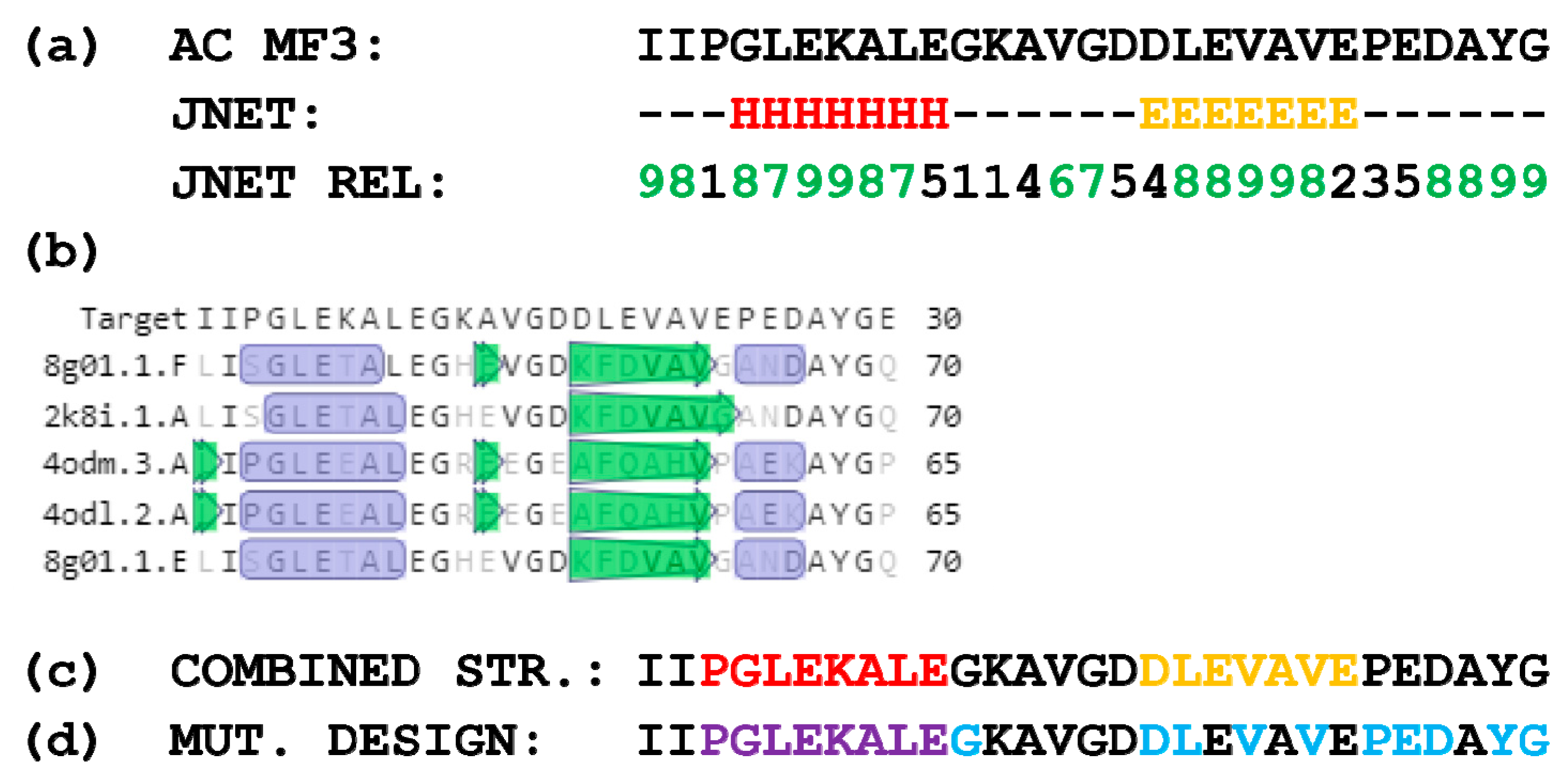
A prediction of the secondary structure of AC MF3 was carried out using both a computation method (Figure 2a) and multiple alignment of the target sequence with sequences with the known 2D structures (Figure 2b). The resulting combined secondary structure of AC MF3 is shown in Figure 2c. Eleven mutants including some point substitutions to alanine and deletion of the predicted alpha-helix region were designed (Figure 2d) and obtained.

2.2. Inducing Activity of AC MF3 Mutants

To reveal mutations influencing the MF3 protective properties, ten mutants of this protein with single amino acid substitutions (G51A, D57A, L58A, V60A, V62A, P64A, E65A, D66A, Y68A, G69A) generated by alanine scanning and one mutant with the alpha-helix deletion (ahD) were produced via heterologous expression. The ability of the obtained mutants to induce plant resistance was tested in parallel with wild-type MF3 using a tobacco–TMV pathosystem. In these experiments, the target activity of all obtained mutants except G69A turned out to be lower than that of MF3 (Figure 3).
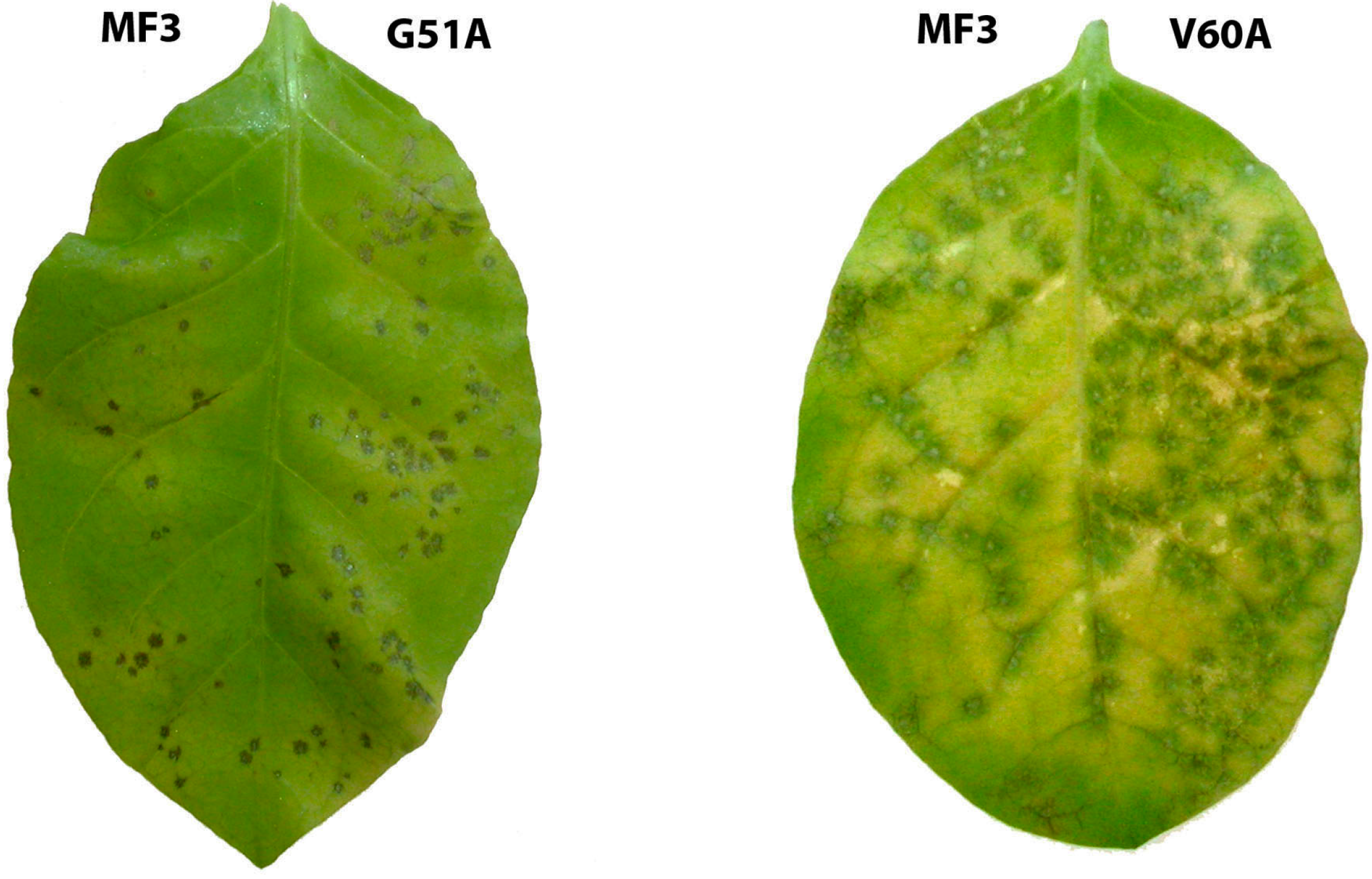
Among the studied mutants, V62A (valine substitution with alanine at position 62) was surprisingly the least active. A pre-inoculation application of V62A on tobacco leaves resulted in an almost threefold reduction of the number of developed necrotic lesions compared to wild-type MF3. Other mutants were divided into three groups according to their activity level. The first group included G51A, V60A, and L58A containing alanine-substituted glycine, valine, and leucine, respectively, as well as ahD; these mutants showed approximately twice lower target activity than wild-type MF3 (see Figure 4 as an illustrative example). Mutants from the second group (P64A and D66A) provided a 1.5-fold reduction of the number of developing necrotic lesions, while the third group (D57A, E65A, and Y68A) provided statistically significant (1.2–1.3-fold), but rather biologically unimportant suppression of the disease manifestation.

2.3. The 3D Model of MF3 and Its Analysis

A 3D model of MF3 was designed using a homology approach (Figure 5). The model showed a high confidence level (0.65–0.83) for the spatial structure of AC MF3.
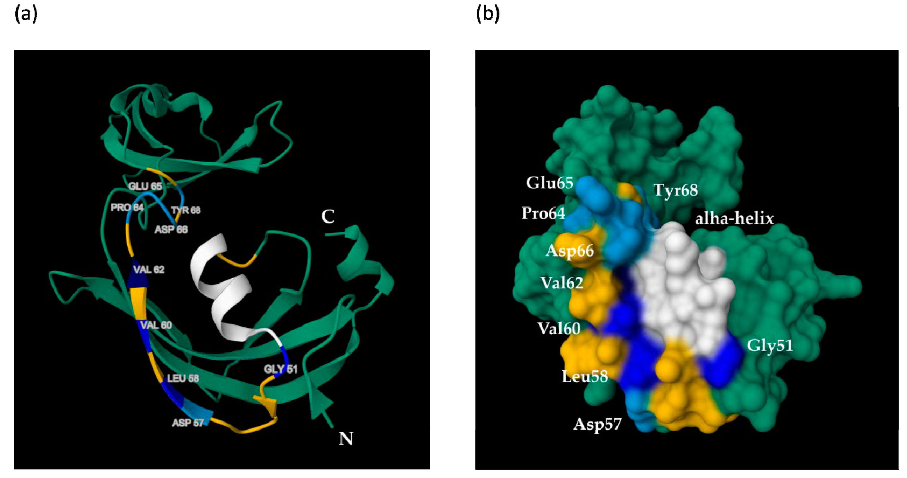
A spatial distribution of amino acids, whose mutations led to the maximum reduction of the inducing activity of MF3, is shown in Figure 6.
The performed analysis revealed that Gly 51, Leu 58, Val 60, Val 62, and the alpha-helix, which met the above-mentioned criterion, are located close to each other on the same side of the MF3 globule.

3. Discussion

Identification of the relationship between the structure of the protein under study and its function is one of the most important tasks in the molecular characterization of proteins. Such research may shed light on which elements of the primary, secondary, or tertiary structure of a protein are responsible for the manifestation of its functional activity. Earlier we showed that the 29-amino-acid region (AC MF3) of the MF3 molecule is obligatory for its inducing activity [48], although it remained unclear which specific elements of AC MF3 were associated with the manifestation of this activity. We designed and obtained 11 mutant proteins characterized by some point substitutions to alanine or a deletion of the predicted alpha-helix (ahD mutant) within AC MF3. The method of amino acid substitutions to alanine (alanine scanning approach [58]) was chosen because it helps to avoid any bias in the choice of the amino acid to be replaced. Alpha-helixes are known as common sites for the protein–receptor interaction [59]. Thus, the purpose of obtaining this ahD mutant was to test whether this particular alpha-helix is important for the inducing activity of MF3. In addition, we hypothesized that the deletion of this alpha-helix would result in a greater reduction of the target MF3 activity compared to point mutations, as it was suggested to more significantly interfere with the structure of AC MF3. The choice of the tobacco (var. Xanthi)–TMV assay for the validation of the inducing activity of MF3 was determined by its sensitivity and a large amount of necroses usually developing on the leaves of this tobacco variety, allowing the quantification of the results of the experiment. A 3D model of MF3 was developed to analyze the spatial distribution of the studied mutations. A homology method [55] was used for modeling since it relies on experimental data rather than on computational algorithms only. As a template for modeling, a SlyD PPI-ase (8g01.1.F) from Esherichia coli was chosen, since it represents the most relevant MF3 homologue with resolved 3D structure.
The results of the study of the inducing activity of MF3 mutants were rather unexpected for us. First, we did not expect that the maximum effect on the inducing activity of MF3 would be shown by the V62A mutation representing the alanine substitution of a hydrophobic valine, rather than by the substitution of a polar or charged amino acid or proline known to affect the rotation of polypeptide chains. Second, though the alpha-helix deletion resulted in a notable reduction of the target MF3 activity, this reduction was comparable to that for a glycine substitution and even weaker than the effect of a valine substitution. This result can be explained by the suggestion that the target activity of the protein is probably influenced not by the whole alpha-helix but rather some individual amino acid or amino acids within it. Or, perhaps, the answer is that AC MF3 is not the only active center in the MF3 molecule responsible for its target activity, and, for example, a plant cell receptor may bind not only to AC MF3. It is worthwhile to perform further alanine scanning experiments with the other amino acids composing AC MF3, including those within the alpha-helix, in order to refine the data on the contribution of individual amino acids to the plant resistance-inducing activity of MF3.
The obtained 3D MF3 model showed that the structure of AC MF3 has a high level of confidence. Thus, it can be used to make assumptions about the spatial distribution of the mutations of interest. Those amino acids (Gly 51, Leu 58, Val 60, Val 62) and alpha-helix, whose substitution or deletion led to a 2–2.5-fold decrease in the target MF3 activity, demonstrate proximity to each other and form a compact region on the surface of the protein globule, while amino acids associated with a reduction of 1.5-fold or less in the target MF3 activity are located away from this site (Figure 6). This may be interpreted as the presence of a putative receptor-binding site within the AC region of MF3. Combining data on the plant resistance-inducing activity of mutant proteins and the spatial distribution of the corresponding amino acids, one can assume that the functional domain of AC MF3, which is probably responsible for binding with a cell receptor, may be narrowed from 29 to about 15 amino acids. Note, however, that this assumption can be clarified only after the completion of more alanine scanning experiments.
The obtained data can be applied for the further molecular characterization of MF3. First, the putative receptor-binding site within AC MF3 can be used for in silico docking to plant receptors with known structures. Second, protein variants with mutations providing the maximum negative influence on the inducing activity of MF3 may be used as negative discriminators in affine chromatography experiments aimed at the identification of putative plant receptors.
Elucidation of the role of amino acids composing AC MF3 may be of both theoretical and practical significance. Such information may help to identify a putative corresponding plant receptor that, in turn, will provide a better understanding of the mechanisms of the inducing activity of MF3. At the same time, this information can be used to design an improved version of MF3 or its active center, which would be characterized by a higher activity and probably consist of fewer amino acids, thus requiring less effort for its biosynthesis and stability maintenance. The results of laboratory studies [46,60,61] and some data obtained in completed [62] and still ongoing greenhouse and field trials with such crops as potato, tomato, and wheat evidence a good protecting efficiency of MF3 against Phytophthora infestans and Stagonospora nodorum as well as potato and tomato viruses. The development of an improved (more efficient or more stable) version of this plant resistance inducer may accelerate the development of corresponding environmentally safe biopreparations intended to improve resistance of agricultural crops to various pathogens. Note that no chemical pesticides have been registered for direct use against plant viruses yet, though this type of plant pathogen is able to cause serious yield losses [63], especially in the case of a combination of several viruses or viruses with fungal pathogens [64]. The control of viral diseases in crops includes mainly risk-reducing measures, such as the use of virus-resistant crops and insecticides to kill carriers (the majority of plant viruses are carried by insects) and quality control of seed material to prevent the further spreading of infection [65]. To date, the main way to deal with virus-infected plants is to eradicate them to prevent the further spreading of infection. In view of these difficulties related to the nature of viral pathogens, the appearance of preparations able to directly reduce the level of viral (as well as fungal) infections in crops may become a new step in providing food safety and improving yield quality.

4. Materials and Methods

4.1. Design and Obtaining of AC MF3 and Alpha-Helix Deletion Mutants

A QuikChangeTM [66] method was chosen to obtain mutations in AC MF3 designed by the alanine scanning procedure [58]. QuikChangeTM uses a pair of partially complementary oligonucleotides with an introduced mutation. During PCR, oligonucleotides were annealed on matrix DNA representing the starting pET28-MF3 plasmid. Plasmid DNA containing the target mutation was further amplified. To avoid the occurrence of nontarget mutations, a high-precision Q5TM DNA polymerase (NEB, Ipswich, MA, USA) was used for amplification.
The amplification mode used in this study included 15 cycles at 98 °C for 10 s, 60 °C for 30 s, and 72 °C for 300 s. The linear PCR product represented a linearized vector with overlapping complementary ends. The resulting pool of DNA (original and mutant) was then treated with DpnI restriction endonuclease to degrade the original DNA. Linear mutant DNA obtained after such treatment was then transformed into E. coli NEB Turbo cells, where the break was further ligated along the homology site by a bacterial reparation system with the introduction of mutations embedded in primers. The primers used for mutagenesis are presented in Table 1.
The alpha-helix deletion was obtained by a similar method, using forward primers with two homology sites of 19 nucleotides complementary to the original sequence and a deletion between them.
As a result, 11 plasmids containing AC MF3 mutants were obtained. The plasmids were transformed into the expression strain (E. coli Rosetta (DE3)), and the homogeneous forms of 11 mutant proteins were isolated using Ni Sepharose immobilized metal ion affinity chromatography (IMAC) [67].
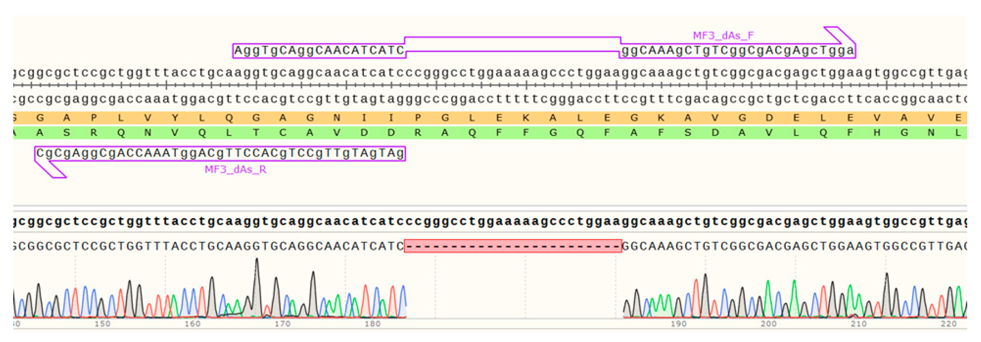
The results of a Sanger sequencing of the AC MF3 region containing the ahD deletion are shown in Figure 7.

4.2. Inducing Activity Assay

To assess the resistance-inducing activity of the obtained mutants compared to wild-type MF3 and to examine the relationship between this activity and the protein structure, a bioassay involving inoculation of detached leaves of necrotic-responding tobacco cultivar Xanthi NN with TMV was used (see [60] for details). In brief, five 10 µL drops of a tested mutant protein solution were applied on the left half of each detached leaf and spread on its adaxial surface (10 leaves per treatment). The right half of the same leaf was similarly treated with a solution of the wild-type MF3 protein. Protein solutions were prepared on the day of the experiment by dissolving freeze-dried preparations of MF3 or mutant proteins purified by IMAC (see Section 4.1) in distilled water. Prior to the experiments, the concentrations of mutant proteins were adjusted to that of MF3 using a Qubit 4 fluorometer (Thermo Fisher Scientific, Waltham, MA, USA). Treated leaves were incubated in a wet chamber for 24 h followed by a separate inoculation of left and right halves with equal concentrations of TMV by rubbing them with a mixture of a viral suspension and carborundum [46]. MF3 was used at a concentration of 10 µg/mL, which was determined earlier to result in a 50–70% reduction of the number of necrotic lesions compared to a water-treated control. After a 4–5-day incubation of inoculated leaves in a wet chamber at 20–22 °C, the level of inducing activity of each mutant was evaluated by a comparison of the number of necrotic lesions developed on a leaf half treated with a mutant with that obtained for the right leaf half treated with MF3; the result was expressed as a percentage of increase or reduction of this index. The number of necroses developed after a TMV infection of tobacco plants represents a common index used for evaluation of the disease development level [68,69].

4.3. The 2D Structure Prediction and 3D Modeling

A 2D structure prediction was performed using a JPred server [53] with a JNET algorithm [54] and SWISS-MODEL [55]. A 3D modeling was carried out using SWISS-MODEL with a GMQE score for choosing the most relevant template. The model visualization and the further analysis were carried out using RCSB PDB [56] and Mol* 3D Viewer [57] programs.

4.4. Statistical Analysis

For each mutant, evaluation of its inducing activity arranged in parallel with the wild-type MF3 testing included three independent experimental series. Mean values, standard errors (SEs), and the significance of differences at p ≤ 0.05 (t-test for independent variables) were calculated using STATISTICA v. 6.1 software (StatSoft Inc., Tulsa, OK, USA).

5. Conclusions

To study the relationship between the primary structure of the MF3 protein, its spatial configuration, and ability to induce plant resistance to pathogens, a set of 11 mutants generated by alanine scanning and alpha-helix deletion (ahD) in the earlier revealed 29-amino-acid region (AC MF3) responsible for the target activity were generated and tested for their protecting activity in a tobacco–TMV model system. Several mutants showed almost threefold (V60A and V62A) or twofold (G51A, L58A, ahD) reduction in their inducing activity compared to the wild-type MF3. A construction of the 3D MF3 model showed these mutations were located in close proximity to each other within AC that may be interpreted as the presence of a putative receptor-binding site in this region of the MF3 molecule. The data obtained can be applied for the further molecular characterization of MF3 and identification of a putative plant receptor for this protein, while further alanine scanning experiments with other amino acids composing AC MF3 will refine the data on the contribution of individual amino acids to the inducing activity of this protein.

Author Contributions

Conceptualization, D.E., L.S. and V.D.; methodology, D.E., L.S. and V.D.; formal analysis, D.E., L.S. and V.D.; investigation, S.P. and I.S.; writing—original draft preparation, D.E. and V.D.; writing—review and editing, L.S. and A.R.; visualization D.E., L.S. and S.P.; supervision, V.D.; project administration, V.D.; funding acquisition, V.D. All authors have read and agreed to the published version of the manuscript.

Funding

This work was financially supported by the Russian Science Foundation (project no. 22-16-00154).

Institutional Review Board Statement

Not applicable.

Informed Consent Statement

Not applicable.

Data Availability Statement

The authors declare that the data supporting the findings of this study are available within the main text of the manuscript. Raw data are available from the corresponding author upon reasonable request.

Conflicts of Interest

The authors declare no conflict of interest.

References

  1. Godfray, H.C.; Beddington, J.R.; Crute, I.R.; Haddad, L.; Lawrence, D.; Muir, J.F.; Pretty, J.; Robinson, S.; Thomas, S.M.; Toulmin, C. Food security: The challenge of feeding 9 billion people. Science 2010, 327, 812–818. [Google Scholar] [CrossRef] [PubMed]
  2. Tudi, M.; Ruan, H.D.; Wang, L.; Lyu, J.; Sadler, R.; Connell, D.; Chu, C.; Phung, D.T. Agriculture development, pesticide application and its impact on the environment. Int. J. Environ. Res. Public Health 2021, 18, 1112. [Google Scholar] [CrossRef]
  3. Gleń-Karolczyk, K.; Boligłowa, E.; Antonkiewicz, J. Organic fertilization shapes the biodiversity of fungal communities associated with potato dry rot. Appl. Soil Ecol. 2018, 129, 43–51. [Google Scholar] [CrossRef]
  4. Thambugala, K.M.; Daranagama, D.A.; Phillips, A.J.L.; Kannangara, S.D.; Promputtha, I. Fungi vs. fungi in biocontrol: An overview of fungal antagonists applied against fungal plant pathogens. Front. Cell Infect. Microbiol. 2020, 10, 604923. [Google Scholar] [CrossRef]
  5. Millan, A.F.-S.; Larraya, L.; Farran, I.; Ancin, M.; Veramendi, J. Successful biocontrol of major postharvest and soil-borne plant pathogenic fungi by antagonistic yeasts. Biol. Control 2021, 160, 104683. [Google Scholar] [CrossRef]
  6. Pellegrini, M.; Ercole, C.; Di Zio, C.; Matteucci, F.; Pace, L.; Del Gallo, M. In vitro and in planta antagonistic effects of plant growth-promoting rhizobacteria consortium against soilborne plant pathogens of Solanum tuberosum and Solanum lycopersicum. FEMS Microbiol. Lett. 2020, 367, fnaa099. [Google Scholar] [CrossRef]
  7. Hatem, A.; Yaderets, V.; Karpova, N.; Glagoleva, E.; Ovchinnikov, A.; Petrova, K.; Shibaeva, A.; Dzhavakhiya, V. Inhibition of the growth of Botrytis cinerea by Penicillium chrysogenum VKM F-4876D combined with fludioxonil-, difenoconazole-, or tebuconazole-based fungicides. Agronomy 2023, 13, 2602. [Google Scholar] [CrossRef]
  8. Shcherbakova, L.; Kartashov, M.; Statsyuk, N.; Pasechnik, T.; Dzhavakhiya, V. Assessment of the sensitivity of some plant pathogenic fungi to 6-demethylmevinolin, a putative natural sensitizer able to help overcoming the fungicide resistance of plant pathogens. Antibiotics 2020, 9, 842. [Google Scholar] [CrossRef]
  9. Ulbasheva, S.; Vorobyev, D.; Statsyuk, N.; Kuznetsova, M. Pre-planting and post-harvest treatment of potato with low-frequency pulse electric field suppresses the development of the leaf and tuber blight. In Agriculture Digitalization and Organic Production; Ronzhin, A., Kostyaev, A., Eds.; Smart Innovation, Systems and Technologies; Springer: Singapore, 2023; Volume 362, pp. 279–291. [Google Scholar] [CrossRef]
  10. Ebrahimi-Zarandi, M.; Saberi Riseh, R.; Tarkka, M.T. Actinobacteria as effective biocontrol agents against plant pathogens, an overview on their role in eliciting plant defense. Microorganisms 2022, 10, 1739. [Google Scholar] [CrossRef] [PubMed]
  11. Zhu, F.; Cao, M.Y.; Zhang, Q.P.; Mohan, R.; Schar, J.; Mitchell, M.; Chen, H.; Liu, F.; Wang, D.; Fu, Z.Q. Join the green team: Inducers of plant immunity in the plant disease sustainable control toolbox. J. Adv. Res. 2023. advance online publication. [Google Scholar] [CrossRef]
  12. Reglinski, T.; Havis, N.; Rees, H.J.; de Jong, H. The practical role of induced resistance for crop protection. Phytopathology 2023, 113, 719–731. [Google Scholar] [CrossRef] [PubMed]
  13. Durrant, W.E.; Dong, X. Systemic acquired resistance. Ann. Rev. Phytopathol. 2004, 42, 185–209. [Google Scholar] [CrossRef]
  14. Yu, Y.; Gui, Y.; Li, Z.; Jiang, C.; Guo, J.; Niu, D. Induced systemic resistance for improving plant immunity by beneficial microbes. Plants 2022, 11, 386. [Google Scholar] [CrossRef] [PubMed]
  15. Tripathi, D.; Raikhy, G.; Kumar, D. Chemical elicitors of systemic acquired resistance—Salicylic acid and its functional analogs. Curr. Plant Biol. 2019, 17, 48–59. [Google Scholar] [CrossRef]
  16. Bektas, Y.; Eulgem, T. Synthetic plant defense elicitors. Front. Plant Sci. 2015, 5, 804. [Google Scholar] [CrossRef]
  17. Faize, L.; Faize, M. Functional analogues of salicylic acid and their use in crop protection. Agronomy 2018, 8, 5. [Google Scholar] [CrossRef]
  18. Fu, Z.Q.; Dong, X. Systemic acquired resistance: Turning local infection into global defense. Ann. Rev. Plant Biol. 2013, 64, 839–863. [Google Scholar] [CrossRef]
  19. Van der Ent, S.; Van Wees, S.C.; Pieterse, C.M. Jasmonate signaling in plant interactions with resistance-inducing beneficial microbes. Phytochemistry 2009, 70, 1581–1588. [Google Scholar] [CrossRef]
  20. Boubakri, H.; Gargouri, M.; Mliki, A.; Brini, F.; Chong, J.; Jbara, M. Vitamins for enhancing plant resistance. Planta 2016, 244, 529–543. [Google Scholar] [CrossRef]
  21. Ahn, I.P.; Kim, S.; Lee, Y.H. Vitamin B1 functions as an activator of plant disease resistance. Plant Physiol. 2005, 138, 1505–1515. [Google Scholar] [CrossRef]
  22. Fitzpatrick, T.B.; Chapman, L.M. The importance of thiamine (vitamin B1) in plant health: From crop yield to biofortification. J. Biol. Chem. 2020, 295, 12002–12013. [Google Scholar] [CrossRef]
  23. Cohen, Y.; Vaknin, M.; Mauch-Mani, B. BABA-induced resistance: Milestones along a 55-year journey. Phytoparasitica 2016, 44, 513–538. [Google Scholar] [CrossRef]
  24. Návarová, H.; Bernsdorff, F.; Döring, A.C.; Zeier, J. Pipecolic acid, an endogenous mediator of defense amplification and priming, is a critical regulator of inducible plant immunity. Plant Cell 2012, 24, 5123–5141. [Google Scholar] [CrossRef] [PubMed]
  25. Mucharromah, E.; Kuc, J. Oxalate and phosphates induce systemic resistance against diseases caused by fungi, bacteria and viruses in cucumber. Crop Prot. 1991, 10, 265–270. [Google Scholar] [CrossRef]
  26. Deliopoulos, T.; Kettlewell, P.; Hare, M. Fungal disease suppression by inorganic salts: A review. Crop Prot. 2010, 29, 1059–1075. [Google Scholar] [CrossRef]
  27. Fesel, P.H.; Zuccaro, A. β-glucan: Crucial component of the fungal cell wall and elusive MAMP in plants. Fungal Genet. Biol. 2016, 90, 53–60. [Google Scholar] [CrossRef]
  28. Klarzynski, O.; Plesse, B.; Joubert, J.M.; Yvin, J.C.; Kopp, M.; Kloareg, B.; Fritig, B. Linear beta-1,3 glucans are elicitors of defense responses in tobacco. Plant Physiol. 2000, 124, 1027–1038. [Google Scholar] [CrossRef]
  29. Gauthier, A.; Trouvelot, S.; Kelloniemi, J.; Frettinger, P.; Wendehenne, D.; Daire, X.; Joubert, J.M.; Ferrarini, A.; Delledonne, M.; Flors, V.; et al. The sulfated laminarin triggers a stress transcriptome before priming the SA- and ROS-dependent defenses during grapevine’s induced resistance against Plasmopara viticola. PLoS ONE 2014, 9, e88145. [Google Scholar] [CrossRef]
  30. Pusztahelyi, T. Chitin and chitin-related compounds in plant-fungal interactions. Mycology 2018, 9, 189–201. [Google Scholar] [CrossRef]
  31. Gong, B.Q.; Wang, F.Z.; Li, J.F. Hide-and-seek: Chitin-triggered plant immunity and fungal counterstrategies. Trends Plant Sci. 2020, 25, 805–816. [Google Scholar] [CrossRef]
  32. Hadwiger, L.A. Multiple effects of chitosan on plant systems: Solid science or hype. Plant Sci. 2013, 208, 42–49. [Google Scholar] [CrossRef]
  33. Mukarram, M.; Ali, J.; Dadkhah-Aghdash, H.; Kurjak, D.; Kačík, F.; Ďurkovič, J. Chitosan-induced biotic stress tolerance and crosstalk with phytohormones, antioxidants, and other signalling molecules. Front. Plant Sci. 2023, 14, 1217822. [Google Scholar] [CrossRef]
  34. Felix, G.; Duran, J.D.; Volko, S.; Boller, T. Plants have a sensitive perception system for the most conserved domain of bacterial flagellin. Plant J. 1999, 18, 265–276. [Google Scholar] [CrossRef]
  35. Sun, W.; Dunning, F.M.; Pfund, C.; Weingarten, R.; Bent, A.F. Within-species flagellin polymorphism in Xanthomonas campestris pv. campestris and its impact on elicitation of Arabidopsis FLAGELLIN SENSING2-dependent defenses. Plant Cell 2006, 18, 764–779. [Google Scholar] [CrossRef]
  36. Sanguankiattichai, N.; Buscaill, P.; Preston, G.M. How bacteria overcome flagellin pattern recognition in plants. Curr. Opin. Plant Biol. 2022, 67, 102224. [Google Scholar] [CrossRef] [PubMed]
  37. Lopez-Gomez, M.; Sandal, N.; Stougaard, J.; Boller, T. Interplay of flg22-induced defence responses and nodulation in Lotus japonicus. J. Exp. Bot. 2012, 63, 393–401. [Google Scholar] [CrossRef]
  38. Czékus, Z.; Martics, A.; Pollák, B.; Kukri, A.; Tari, I.; Ördög, A.; Poór, P. The local and systemic accumulation of ethylene determines the rapid defence responses induced by flg22 in tomato (Solanum lycopersicum L.). J. Plant Physiol. 2023, 287, 154041. [Google Scholar] [CrossRef] [PubMed]
  39. Zhang, Y.; Zhang, Y.; Qiu, D.; Zeng, H.; Guo, L.; Yang, X. BcGs1, a glycoprotein from Botrytis cinerea, elicits defence response and improves disease resistance in host plants. Biochem. Biophys. Res. Commun. 2015, 457, 627–634. [Google Scholar] [CrossRef]
  40. Wei, Z.M.; Laby, R.J.; Zumoff, C.H.; Bauer, D.W.; He, S.Y.; Collmer, A.; Beer, S.V. Harpin, elicitor of the hypersensitive response produced by the plant pathogen Erwinia amylovora. Science 1992, 257, 85–88. [Google Scholar] [CrossRef]
  41. Choi, M.S.; Kim, W.; Lee, C.; Oh, C.S. Harpins, multifunctional proteins secreted by gram-negative plant-pathogenic bacteria. Mol. Plant Microbe Interact. 2013, 26, 1115–1122. [Google Scholar] [CrossRef]
  42. Wang, D.; Wang, B.; Wang, J.; Wang, S.; Wang, W.; Niu, Y. Exogenous application of harpin protein Hpa1 onto Pinellia ternata induces systemic resistance against tobacco mosaic virus. Phytopathology 2020, 110, 1189–1198. [Google Scholar] [CrossRef]
  43. Chen, L.; Qian, J.; Qu, S.; Long, J.; Yin, Q.; Zhang, C.; Wu, X.; Sun, F.; Wu, T.; Hayes, M.; et al. Identification of specific fragments of HpaG Xooc, a harpin from Xanthomonas oryzae pv. oryzicola, that induce disease resistance and enhance growth in plants. Phytopathology 2008, 98, 781–791. [Google Scholar] [CrossRef] [PubMed]
  44. Chen, L.; Zhang, S.J.; Zhang, S.S.; Qu, S.; Ren, X.; Long, J.; Yin, Q.; Qian, J.; Sun, F.; Zhang, C.; et al. A fragment of the Xanthomonas oryzae pv. oryzicola harpin HpaG Xooc reduces disease and increases yield of rice in extensive grower plantings. Phytopathology 2008, 98, 792–802. [Google Scholar] [CrossRef]
  45. Oliveira, C.; Domingues, L. Guidelines to reach high-quality purified recombinant proteins. Appl. Microbiol. Biotechnol. 2018, 102, 81–92. [Google Scholar] [CrossRef]
  46. Shumilina, D.; Krämer, R.; Klocke, E.; Dzhavakhiya, V. MF3 (peptidyl-prolyl cis-trans isomerase of FKBP type from Pseudomonas fluorescens)—An elicitor of non-specific plant resistance against pathogens. Phytopathol. Pol. 2006, 41, 39–49. [Google Scholar]
  47. Shumilina, D.; Dzhavakhiya, V. A new approach to development and practical application of bio-pesticides based on the Pseudomonas fluorescens protein—Inductor of non-specific plant resistance to fungal, viral and bacterial diseases. In Proceedings of the 10th ISTC/Korea Workshop “Industrial Applications of Russia’s New Biotechnologies”, Mokpo, Republic of Korea, 14–15 March 2006. [Google Scholar]
  48. Dzhavakhiya, V.G.; Voinova, T.M.; Shumilina, D.V. Search for the active center pf peptidyl-prolyl cys/trans isomerase from Pseudomonas fuorescens responsible for the induction of tobacco (Nicotiana tabacum L.) plant resistance to tobacco mosaic virus. Sel’skokhozyaistvennaya Biol. Agric. Biol. 2016, 51, 392–400. [Google Scholar] [CrossRef]
  49. Kunze, G.; Zipfel, C.; Robatzek, S.; Niehaus, K.; Boller, T.; Felix, G. The N terminus of bacterial elongation factor Tu elicits innate immunity in Arabidopsis plants. Plant Cell 2004, 16, 3496–3507. [Google Scholar] [CrossRef]
  50. Noda, J.; Brito, N.; González, C. The Botrytis cinerea xylanase Xyn11A contributes to virulence with its necrotizing activity, not with its catalytic activity. BMC Plant Biol. 2010, 10, 38. [Google Scholar] [CrossRef] [PubMed]
  51. Frías, M.; González, M.; González, C.; Brito, N. A 25-residue peptide from Botrytis cinerea xylanase BcXyn11A elicits plant defenses. Front. Plant Sci. 2019, 10, 474. [Google Scholar] [CrossRef]
  52. Yu, T.Y.; Sun, M.K.; Liang, L.K. Receptors in the induction of the plant innate immunity. Mol. Plant Microbe Interact. 2021, 34, 587–601. [Google Scholar] [CrossRef]
  53. Cuff, J.A.; Clamp, M.E.; Siddiqui, A.S.; Finlay, M.; Barton, G.J. JPred: A consensus secondary structure prediction server. Bioinformatics 1998, 14, 892–893. [Google Scholar] [CrossRef] [PubMed]
  54. Cuff, J.A.; Barton, G.J. Application of multiple sequence alignment profiles to improve protein secondary structure prediction. Proteins 2000, 40, 502–511. [Google Scholar] [CrossRef]
  55. Waterhouse, A.; Bertoni, M.; Bienert, S.; Studer, G.; Tauriello, G.; Gumienny, R.; Heer, F.T.; de Beer, T.A.P.; Rempfer, C.; Bordoli, L.; et al. SWISS-MODEL: Homology modelling of protein structures and complexes. Nucleic Acids Res. 2018, 46, W296–W303. [Google Scholar] [CrossRef] [PubMed]
  56. Berman, H.M.; Westbrook, J.; Feng, Z.; Gilliland, G.; Bhat, T.N.; Weissig, H.; Shindyalov, I.N.; Bourne, P.E. The Protein Data Bank. Nucleic Acids Res. 2000, 28, 235–242. [Google Scholar] [CrossRef] [PubMed]
  57. Sehnal, D.; Bittrich, S.; Deshpande, M.; Svobodová, R.; Berka, K.; Bazgier, V.; Velankar, S.; Burley, S.K.; Koča, J.; Rose, A.S. Mol* Viewer: Modern web app for 3D visualization and analysis of large biomolecular structures. Nucleic Acids Res. 2021, 49, W431–W437. [Google Scholar] [CrossRef] [PubMed]
  58. Morrison, K.L.; Weiss, G.A. Combinatorial alanine-scanning. Curr. Opin. Chem. Biol. 2001, 5, 302–307. [Google Scholar] [CrossRef]
  59. Edwards, T.A.; Wilson, A.J. Helix-mediated protein--protein interactions as targets for intervention using foldamers. Amino Acids 2011, 41, 743–754. [Google Scholar] [CrossRef]
  60. Popletaeva, S.B.; Statsyuk, N.V.; Voinova, T.M.; Arslanova, L.R.; Zernov, A.L.; Bonartsev, A.P.; Bonartseva, G.A.; Dzhavakhiya, V.G. Evaluation of eliciting activity of peptidil prolyl cys/trans isomerase from Pseudonomas fluorescens encapsulated in sodium alginate regarding plant resistance to viral and fungal pahogens. AIMS Microbiol. 2018, 12, 192–208. [Google Scholar] [CrossRef]
  61. Voinova, T.; Kartashov, M.; Pasechnik, T.; Shcherbakova, L.; Statsyuk, N.; Dzhavakhiya, V. Peptidyl prolyl cis/trans isomerase from Pseudomonas fluorescens encapsulated into biodegradable natural polymers: A potential plant protection agent inducing plant resistance to fungal pathogens. Biocatal. Agric. Biotechnol. 2021, 36, 102112. [Google Scholar] [CrossRef]
  62. Dzhavakhiya, V.G.; Filippov, A.V.; Skryabin, K.G.; Voinova, T.M.; Kuznetsova, M.A.; Shulga, O.A.; Pridannikov, M.V.; Shumilina, D.V.; Kromina, K.A.; Battchikova, N.V.; et al. Proteins Inducing Multiple Resistance of Plants to Phytopathogens and Pests; World Intellectual Property Organization (WIPO): Geneva, Switzerland, 2005. [Google Scholar]
  63. Rao, G.P.; Reddy, M.G. Overview of yield losses due to plant viruses. In Applied Plant Virology. Advances, Detection, and Antiviral Strategies; Awasthi, L.P., Ed.; Academic Press: London, UK, 2020; pp. 531–562. [Google Scholar] [CrossRef]
  64. Nie, X.; Singh, M. Response of potato, tobacco and Physalis floridana plants to mixed infection with PVX, PVYNTN and PVYO strains. Can. J. Plant Pathol. 2013, 35, 390–401. [Google Scholar] [CrossRef]
  65. Tatineni, S.; Hein, G.L. Plant viruses of agricultural importance: Current and future perspectives of virus disease management strategies. Phytopathology 2023, 113, 117–141. [Google Scholar] [CrossRef] [PubMed]
  66. Xia, Y.; Chu, W.; Qi, Q.; Xun, L. New insights into the QuikChange™ process guide the use of Phusion DNA polymerase for site-directed mutagenesis. Nucleic Acids Res. 2015, 43, e12. [Google Scholar] [CrossRef]
  67. Yip, T.T.; Hutchens, T.W. Immobilized metal ion affinity chromatography. Methods Mol. Biol. 1992, 11, 17–31. [Google Scholar] [CrossRef] [PubMed]
  68. Holmes, F.O. Local lesions in tobacco mosaic. Bot. Gaz. 1929, 87, 39–55. [Google Scholar] [CrossRef]
  69. Nourinejhad Zarghani, S.; Ehlers, J.; Monavari, M.; von Bargen, S.; Hamacher, J.; Büttner, C.; Bandte, M. Applicability of different methods for quantifying virucidal efficacy using MENNO florades and tomato brown rugose fruit virus as an example. Plants 2023, 12, 894. [Google Scholar] [CrossRef]
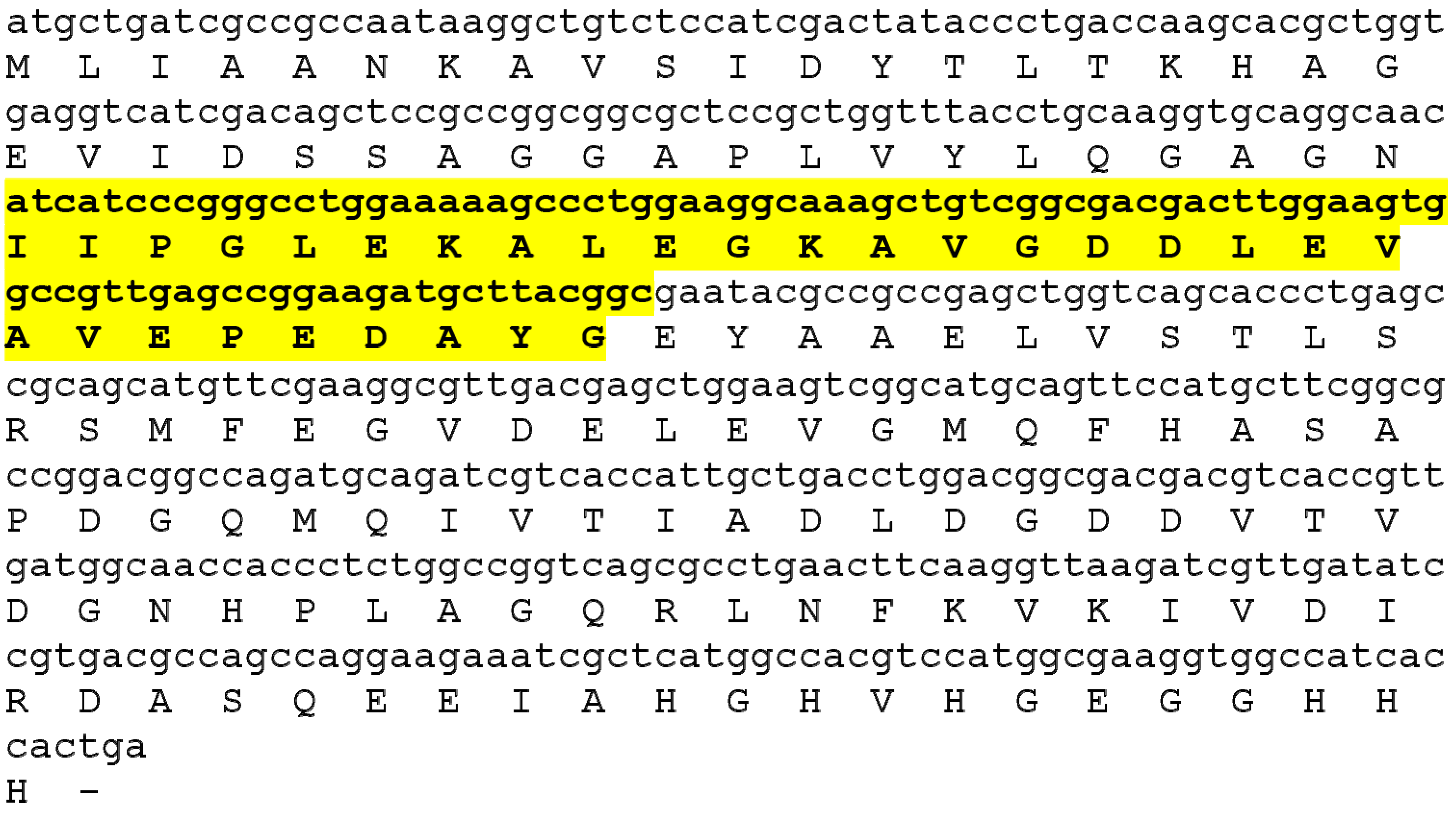
Figure 1. MF3-encoding gene and amino acid sequence. The MF3 active center is marked in bold and highlighted with yellow.
Ijms 24 16374 g001
Figure 2. Prediction of the AC MF3 secondary structure and design of mutants. (a) Predicted secondary structure of AC MF3 calculated using a JPred server [53] and a JNET algorithm [54]: “H” indicates the alpha-helix region, “E” indicates a beta-strand; numbers represent the confidence level increasing from 1 to 9. (b) Predicted secondary structure of AC MF3 obtained using SWISS-MODEL [55]: purple boxes indicate alpha-helixes, green arrows indicate beta-strands. (c) Combined secondary structure: the alpha-helix and beta-strand are indicated with red and orange colors, respectively. (d) Mutation design: point substitutions to alanine are indicated with blue; the deleted alpha-helix motif is indicated with purple.
Ijms 24 16374 g002
Figure 3. Comparison of the effect of the MF3 protein and its mutants on the formation of necrotic lesions on tobacco leaves artificially inoculated with TMV. LAm, the least active mutant; MAm, the most active one. Y-bars indicate standard error (SE) of the mean at p ≤ 0.05 (10 leaves per treatment, 3 replications per experiment). * Leaves treated with distilled water prior to inoculation were used as the control.
Ijms 24 16374 g003
Figure 4. Examples of necrotic lesions developed on tobacco leaves, which were separately treated with wild-type MF3 (left halves) and G51A or V60A mutants (right halves) prior to inoculation with tobacco mosaic virus.
Ijms 24 16374 g004
Figure 5. A 3D model of MF3 built with SWISS-MODEL [55] (alpha-helix regions are indicated with arrows). (a) Sequence alignment with the template 8g01.1.F. Here and in (b): more or less confident 3D structures are indicated with blue or red, respectively. (b) A 3D model of MF3, 2D structure representation. (c) AC MF3 region (indicated in yellow) visualized using RCSB PDB [56] and Mol* 3D Viewer [57]. N and C indicate the N- and C-end of the MF3 molecule, respectively.
Ijms 24 16374 g005
Figure 6. Spatial distribution of substituted amino acids within the MF3 molecule modeled using RCSB PDB and Mol* 3D Viewer: (a) 2D structure representation, (b) molecular surface representation. The alpha helix is marked with a white color. Amino acids associated with a <1.5-, 2-, and >2.5-fold reduction of the inducing activity of a protein are indicated with light-blue, blue, and dark-blue (Val62) colors, respectively. The rest part of the AC MF3 region is indicated with yellow. The rest of the protein molecule is indicated with green.
Ijms 24 16374 g006
Figure 7. The target sequence of the MF3 region of the ahD mutant protein confirmed by Sanger sequencing of the plasmid used for its production. Amino acid sequences corresponding to the direct and complementary DNA sequences of a plasmid are indicated with yellow and green, respectively. Red indicates the alpha-helix deletion.
Ijms 24 16374 g007
Table 1. Primers used for the AC MF3 mutagenesis.
No.Primer5′–3′ Sequence
123
111(G_A)Fcctggaagccaaagctgtcggcg
211(G_A)Rctttggcttccagggctttttc
317(D_A)Ftcggcgacgccttggaagtggc
417(D_A)Racttccaaggcgtcgccgacag
518(L_A)Fgcgacgacgcggaagtggccgttg
618(L_A)Rccacttccgcgtcgtcgccgacag
720(V_A)Fttggaagcggccgttgagcc
820(V_A)Raacggccgcttccaagtcgtc
922(V_A)Fgtggccgctgagccggaagatg
1022(V_A)Rtccggctcagcggccacttccaag
1124(P_A)Fgttgaggcggaagatgcttacg
1224(P_A)Rtcttccgcctcaacggccacttc
1325(E_A)Fttgagccggcagatgcttacggc
1425(E_A)Raagcatctgccggctcaacggcc
1526(D_A)Fcggaagctgcttacggcgaatac
1626(D_A)Rgtaagcagcttccggctcaacgg
1728(Y_L)Rgatgctttaggcgaatacgccg
1828(Y_L)Fattcgcctaaagcatcttccgg
1929(G_A)Ftgcttacgccgaatacgccgcc
2029(G_A)Rcgtattcggcgtaagcatcttcc
21MF3_dAs_FAggTgCAggCAACATCATCggCAAAgCTgTCggCgACgAgcTgga
22MF3_dAs_RgATgATgTTgCCTgCACCTTgCAggTAAACCAgCggAgCgC
Disclaimer/Publisher’s Note: The statements, opinions and data contained in all publications are solely those of the individual author(s) and contributor(s) and not of MDPI and/or the editor(s). MDPI and/or the editor(s) disclaim responsibility for any injury to people or property resulting from any ideas, methods, instructions or products referred to in the content.