Article

Garden Cress Seed Oil Abrogates Testicular Oxidative Injury and NF-kB-Mediated Inflammation in Diabetic Mice

by
Rasha Abu-Khudir
1,2,*,
Gehan M. Badr
3,
Heba Ibrahim Abd El-Moaty
4,5,
Rabab S. Hamad
4,6,
Najla K. Al Abdulsalam
4,
Aml Sayed Ali Abdelrahem
7,
Saleha Alqarni
8,
Mayyadah Abdullah Alkuwayti
4,
Sherine Abdel Salam
9,† and
Hanaa F. Abd El-Kareem
3,†
1
Department of Chemistry, College of Science, King Faisal University, P.O. Box 380, Al-Ahsa 31982, Saudi Arabia
2
Department of Chemistry, Biochemistry Division, Faculty of Science, Tanta University, Tanta 31527, Egypt
3
Department of Zoology, Faculty of Science, Ain Shams University, Cairo 11566, Egypt
4
Department of Biological Sciences, College of Science, King Faisal University, P.O. Box 380, Al-Ahsa 31982, Saudi Arabia
5
Medicinal and Aromatic Plants Department, Desert Research Center El-Mataria, Cairo 11753, Egypt
6
Central Laboratory, Theodor Bilharz Research Institute, Giza 12411, Egypt
7
Department of Nursing, College of Applied Medical Science, King Faisal University, P.O. Box 380, Al-Ahsa 31982, Saudi Arabia
8
Department of Clinical Nutrition, College of Applied Medical Science King Faisal University, P.O. Box 380, Al-Ahsa 31982, Saudi Arabia
9
Department of Zoology, Faculty of Science, Alexandria University, Alexandria 21511, Egypt
*
Author to whom correspondence should be addressed.
These authors contributed equally to this work.
Int. J. Mol. Sci. 2023, 24(20), 15478; https://doi.org/10.3390/ijms242015478
Submission received: 30 September 2023 / Revised: 19 October 2023 / Accepted: 20 October 2023 / Published: 23 October 2023
(This article belongs to the Section Bioactives and Nutraceuticals)

Abstract

:
Diabetes mellitus is a metabolic disorder associated with various complications encompassing male reproductive dysfunction. The present study aimed to investigate the therapeutic potential of biologically active Lepidium sativum seed oil (LSO) against the testicular dysfunction associated with streptozotocin (STZ)-induced diabetes. Male adults (n = 24) were divided into four groups: control, LSO-administered, diabetic (D), and LSO-treated diabetic (D+LSO) groups. LSO was extracted from L. sativum seeds, and its chemical composition was determined using GC-MS. Serum testosterone levels, testicular enzymatic antioxidants (catalase (CAT) and superoxide dismutase (SOD)), an oxidative stress (OS) biomarker, malondialdehyde (MDA), pro-inflammatory markers (NF-kB, IL-1, IL-6, and TNF-α), and the expression level of NF-kB were assessed. In addition, histopathological changes were evaluated in testicular tissues. The results obtained showed that the chemical composition of LSO indicated its enrichment mainly with γ-tocopherol (62.1%), followed by 2-methylhexacosane (8.12%), butylated hydroxytoluene (8.04%), 10-Methylnonadecane (4.81%), and δ-tocopherol (3.91%). Moreover, LSO administration in the D+LSO mice significantly increased testosterone levels and ameliorated the observed testicular oxidative damage, inflammatory response, and reduced NF-kB expression compared to the diabetic mice. Biochemical and molecular analyses confirmed the histological results. In conclusion, LSO may prevent the progression of diabetes-induced impairment in the testes through inhibition of the OS- and NF-kB-mediated inflammatory response.

1. Introduction

Diabetes mellitus (DM) is an intricate metabolic disorder that results in persistent high blood glucose levels or hyperglycemia [1]. Being linked to an increased risk of micro- and macrovascular diabetic complications, prolonged hyperglycemia is generally associated with lower quality of life and responsible for the morbidity and mortality assigned to diabetes [2,3]. Multiple body organs are affected by micro- and/or macrovascular diabetic complications, resulting in their damage, dysfunction, and eventual failure [4,5]. Reproductive dysfunction is one of the adverse effects of diabetes in both humans and animal models [6,7,8,9]. Recently, it was reported that type 1 and type 2 diabetes (T1DM and T2DM, respectively) adversely affected the sperm quality and histopathology of reproductive organs in T1DM and T2DM mice with a great extent of similarity. However, the severity of reduced sperm concentration and precocious acrosome exocytosis were significant in T1DM compared to T2DM animals [10]. A substantial number of male diabetic subjects of reproductive age experience infertility, where the prevalence of infertility in diabetic men ranges from 35% to 51% [11,12,13]. In DM patients, various mechanisms are involved in the etiology of male infertility, including pre-, testicular, and post-testicular ones. The involvement of such mechanisms may differ depending on the type of diabetes (T1DM or T2DM), disease duration, and metabolic compensation of glycemic impairment [6]. Pre-testicular mechanisms involve central as well as peripheral hypogonadism, leading to decreased serum levels of gonadotropins, including follicle-stimulating hormone (FSH) and luteinizing hormone (LH), and testosterone (T) [14,15]. Indeed, high incidence of hypogonadism has been previously reported among type 2 diabetic men [16,17,18,19]. Moreover, a large amount of pre-clinical and clinical evidence supports substantial association between DM-related metabolic derangements and reduced serum testosterone levels [20,21]. At the testicular level, changes resulting from DM encompass increased OS consequent to overproduction of ROS and eventual lipid peroxidation (LPO) in seminal fluid, DNA fragmented sperms, altered mitochondrial bioenergetics, and the formation of advanced glycated end-products (AGEs) [22,23,24,25]. Moreover, plausible post-testicular mechanisms associated with DM include male accessory gland infection/inflammation (MAGI) and erectile and/or ejaculatory dysfunction. These pathogenic mechanisms result in altered sperm parameters, damaged sperms, and/or premature or delayed ejaculation, anejaculation, or retro-ejaculation [26,27,28].
Glucose metabolism in Sertoli cells (SCs) is crucial for the normal development of germ cells [29,30]. In addition, it is involved in maintaining fundamental cell activity as well as the motility and fertilizing ability of mature sperms [31]. However, chronic hyperglycemia triggers the activation of many pathological pathways associated with the development of diabetic complications, including elevated production of advanced glycated end-products (AGEs), the polyol pathway, the protein kinase C (PKC) pathway, the hexosamine pathway, and mitochondrial damage [32,33]. Upregulation of these pathways results in the overproduction of free radicals, including reactive oxygen species (ROS) and the decreased efficiency of antioxidant defense systems, which eventually lead to oxidative stress (OS) and increased cell death [34,35,36]. Indeed, increased OS is one of the major sources of the hyperglycemia-induced triggers of diabetic complications [37,38,39]. In this regard, it has been reported that many of the diabetes-induced molecular changes, that greatly influence sperm quality and function, are attributed to the increase in OS [40,41,42]. In fact, the male reproductive system is extremely vulnerable to OS mostly due to the restricted antioxidant enzyme systems in spermatozoa as well as the enrichment of sperm cell membranes with polyunsaturated fatty acids (PUFAs), rendering sperms extremely sensitive to ROS assault which ultimately leads to lipid peroxidation [43]. Consequent production of lipid peroxides and the disruption of membrane integrity extremely affect the viability and function of sperm cells, resulting in impaired motility and reduced fertilization capacity [44]. In addition to lipid peroxidation, sperm motility as well as fertilization ability are adversely affected by the ROS-induced oxidative damage of the proteins and nuclear DNA of sperm cells [45,46]. Moreover, hyperglycemia-induced overproduction of ROS generation has the potential to exacerbate mitochondrial dysfunction, eventually resulting in mitochondrial OS which ultimately leads to the apoptosis of spermatozoa and reduced viability of sperms [47,48]. In addition to disruption of antioxidant and mitochondrial mechanisms, hyperglycemia-induced ROS overproduction triggers the production of downstream inflammatory factors. OS is known to modulate NF-kB signaling pathway, thus initiating the activation of downstream inflammatory signaling pathways, resulting in the release of inflammatory factors such as IL-1, IL-6, and TNF-α [49,50]. Besides OS, inflammation is one of the common influencing causes that trigger damage to the male reproductive system as a result of diabetic complications [51,52]. Many studies have stated that hyperglycemia-induced pro-inflammatory factors could influence testicular cells or tissues [23,53]. Moreover, they can stimulate ROS generation, including nitric oxide (NO), which prolongs inflammatory reactions and can affect spermatogenesis, sperm maturation, and diminish semen quality [54,55,56,57].
Previous research has shown that conventional treatment of diabetes, whether using insulin or oral hypoglycemic medications, can have serious side effects and is ineffective in preventing the onset of numerous complications related to diabetes [58]. Therefore, there is an urgent need to implement safe compounds with plausible protective effects against oxidative and inflammatory changes induced by diabetes. In this regard, several studies have demonstrated the benefits of various medicinal plants and their derived bioactive phytochemicals in the management of diabetes-induced male infertility [59,60,61,62]. Lepidium sativum L., also known as Garden cress/Hab El Rashad, is a comparatively fast-growing annual herb that belongs to the family Brassicaceae [63]. It is native to Southeast Asia and Northeast Africa, and it is cultivated all over the world nowadays [64]. L. sativum possesses numerous pharmacological effects, namely antioxidant and anti-inflammatory activities; hence, it is commonly used in traditional medicine worldwide [65,66]. Secondary metabolites such as flavonoids, tannins, glycosides, polyphenols, lectin, and the mucilage were revealed by a phytochemical analysis of L. sativum extract [67,68,69]. The chemical composition of the plant’s seeds encompasses, among others, essential oils, fatty oils, fatty acids, carbohydrates, proteins, vitamins, flavonoids, alkaloids, isothiocyanate, and glycosides [68,70]. Moreover, natural antioxidants, such as tochopherols, are enriched in the seed extract [64]. In the field of folk medicine, the leaves and seed oil of L. sativum are known for their role in the treatment of various inflammatory disorders, including diabetes mellitus, asthma, and hepatitis [71,72,73,74,75]. Regarding diabetes-induced adverse effects on the male reproductive system, it was shown that L. sativum seed extract, as an antioxidant, played a beneficial protective role against the STZ-induced histopathological damage of the prostate gland and epididymis by preventing OS [76,77]. Moreover, improvement of semen parameters, sperm function, and sexual behavior have been observed in diabetic mice due to the cooperative ameliorative effects of L. sativum and co-enzyme Q10 (CoQ10). Such protective effects are partially mediated, at least, via their regulatory actions on the HPG axis through the triggering of the overexpression of the gonadotropin-releasing hormone (GnRH) gene and elevation in the production or secretion of LH, FSH, and testosterone [78]. Nevertheless, the preventive effect of L. sativum seed oil (LSO) on the basal cellular and molecular parameters of the male reproductive system has not been demonstrated at present. Hence, we hypothesize that L. sativum may be a plausible therapeutic alternative to conventional therapy for male infertility. In fact, there are now no specific treatments for diabetic testis lesions. The current study aimed to investigate the chemical compounds of L. sativum seed oil (LSO) and its potential role in mitigating the progression of diabetes-induced testicular impairment in a STZ-diabetes-induced mouse model.

2. Results

2.1. Identification of the Bioactive Compounds in LSO by GC-MS Analysis

Ten components representing 100% of the total fixed oil were identified in LSO. The GC-MS chromatogram in Figure 1 depicts peaks based on retention time (RT) and area %.
The data revealed high percentage of two forms of vitamin E (66.01%), including γ tocopherol (γT; 62.1%) and δ-tocopherol (δT; 3.91%) (Table 1). In addition, alkanes (24.22%), antioxidant phenol derivatives (8.04%), and alcohol (1.73%) were also detected.

2.2. Serum Testosterone Level in Diabetic Mice Treated with/without LSO

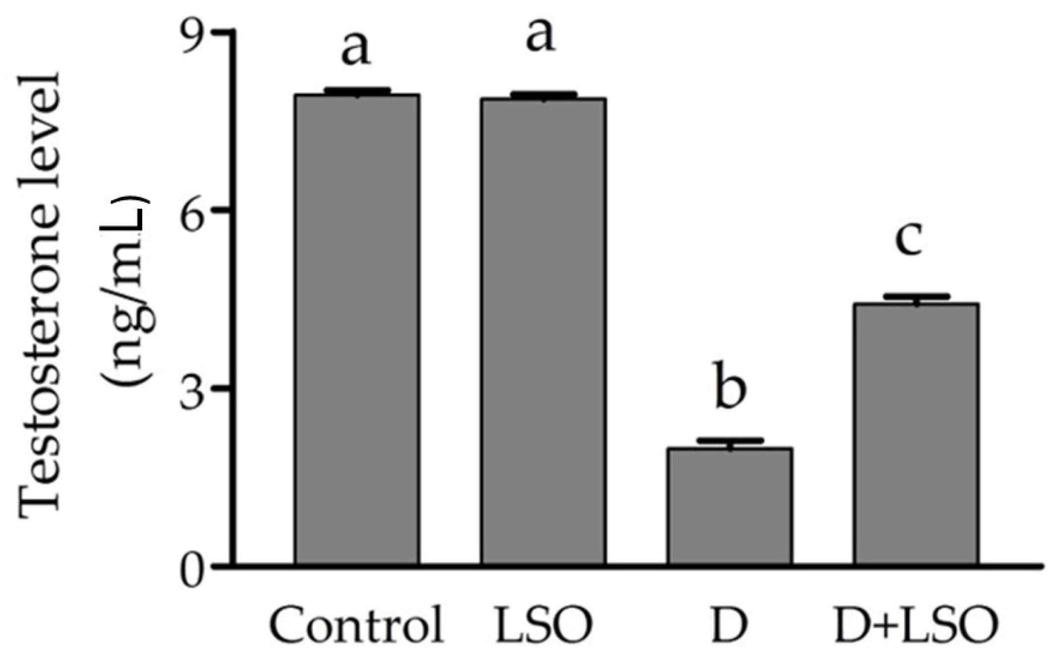
As shown in Figure 2, STZ-induced diabetes led to a significant decrease (−75%) in serum testosterone (T) in the diabetic mice compared to the control group. Meanwhile, the administration of LSO to the diabetic mice produced a significant increase (+128.5%) in serum testosterone levels compared to the diabetic mice group. However, serum testosterone was not completely restored to normal levels, and a significant decrease was observed in the D+LSO mice compared to the control and LSO groups.

2.3. Redox Status in the Testes of Diabetic Mice Treated with/without LSO

The activities of the testicular enzymatic antioxidants SOD and CAT in the diabetic group demonstrated significant decreases of −71.2% and −55.9%, respectively, compared to the control group (Figure 3A,B). Conversely, treatment of the diabetic mice with LSO resulted in a significant increase in SOD (+108.4%) and CAT (+52.6%) activities compared to the diabetic mice. Moreover, it was observed that the testicular MDA level was significantly increased (+270.7%) in the diabetic mice compared to that of the control. On the other hand, the MDA level in the D+LSO group was significantly decreased (−28.9%) compared to the diabetic mice (Figure 3C).

2.4. NF-kB and Pro-Inflammatory Cytokines in the Testes of Diabetic Mice Treated with/without LSO

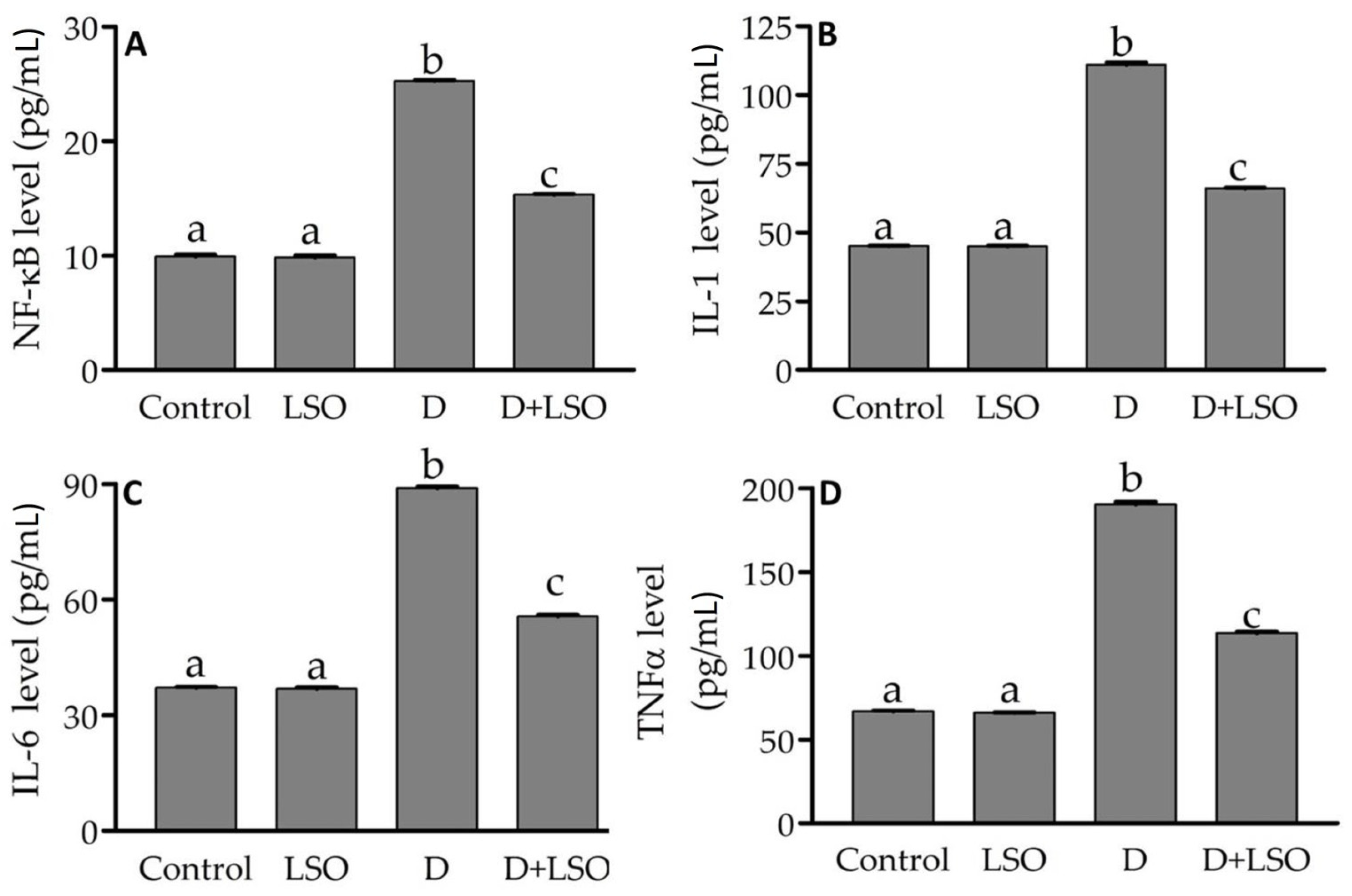
To assess the anti-inflammatory role of LSO, the testicular protein level of the NF-kB p65 subunit was determined using ELISA, as shown in Figure 4A. It was significantly increased (+254.7%) in the diabetic mice compared to the control and LSO. However, it was significantly decreased (−39.4%) in the D+LSO-treated group compared to the diabetic one. In addition, the level of testicular pro-inflammatory cytokines, IL-1, IL-6, and TNF-α, was also assessed. Our data showed a significant increase in the levels of IL-1 (+146.5%), IL-6 (+139.1%), and TNF-α (+184.4%) in the diabetic mice compared to the control (Figure 4B–D). Conversely, in the D+LSO mice, treatment of the diabetic mice with LSO significantly reduced IL-1 by −40.5%, IL-6 by −37.4%, and TNF-α by −40.3% when compared to the corresponding levels in the diabetic group.

2.5. NF-kB p65 Subunit Expression in the Testes of Diabetic Mice Treated with/without LSO

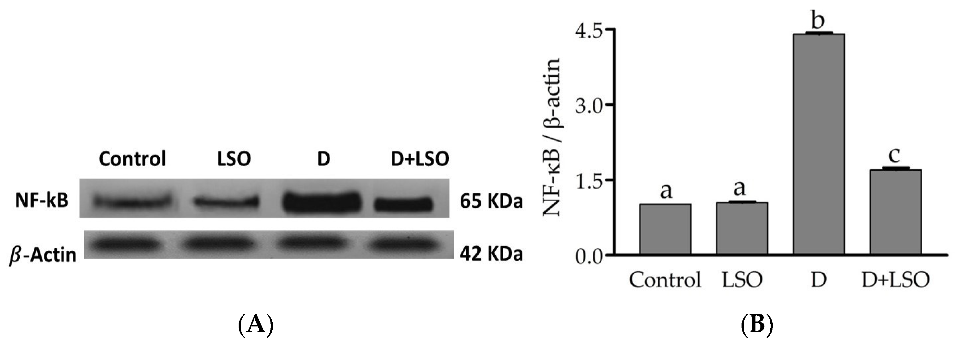
To further confirm the involvement of NF-kB in the pathway via which LSO modulates the inflammatory response, the expression of testicular NF-kB was assessed using Western blot analysis (Figure 5A). NF-kB p65 subunit expression was notably upregulated in the diabetic mice compared to the control and LSO groups (by +76.81% and +76.13%, respectively). Whereas downregulation of NF-kB expression was observed upon LSO administration in the D+LSO mice by −31.12% compared to the diabetic mice (Figure 5B). The data suggested the impact of the NF-kB pathway on the LSO therapeutic action against testicular damage in the diabetic mice model.

2.6. Effect of LSO on Testicular Histoarchitecture in the Diabetic Mice Treated with/without LSO

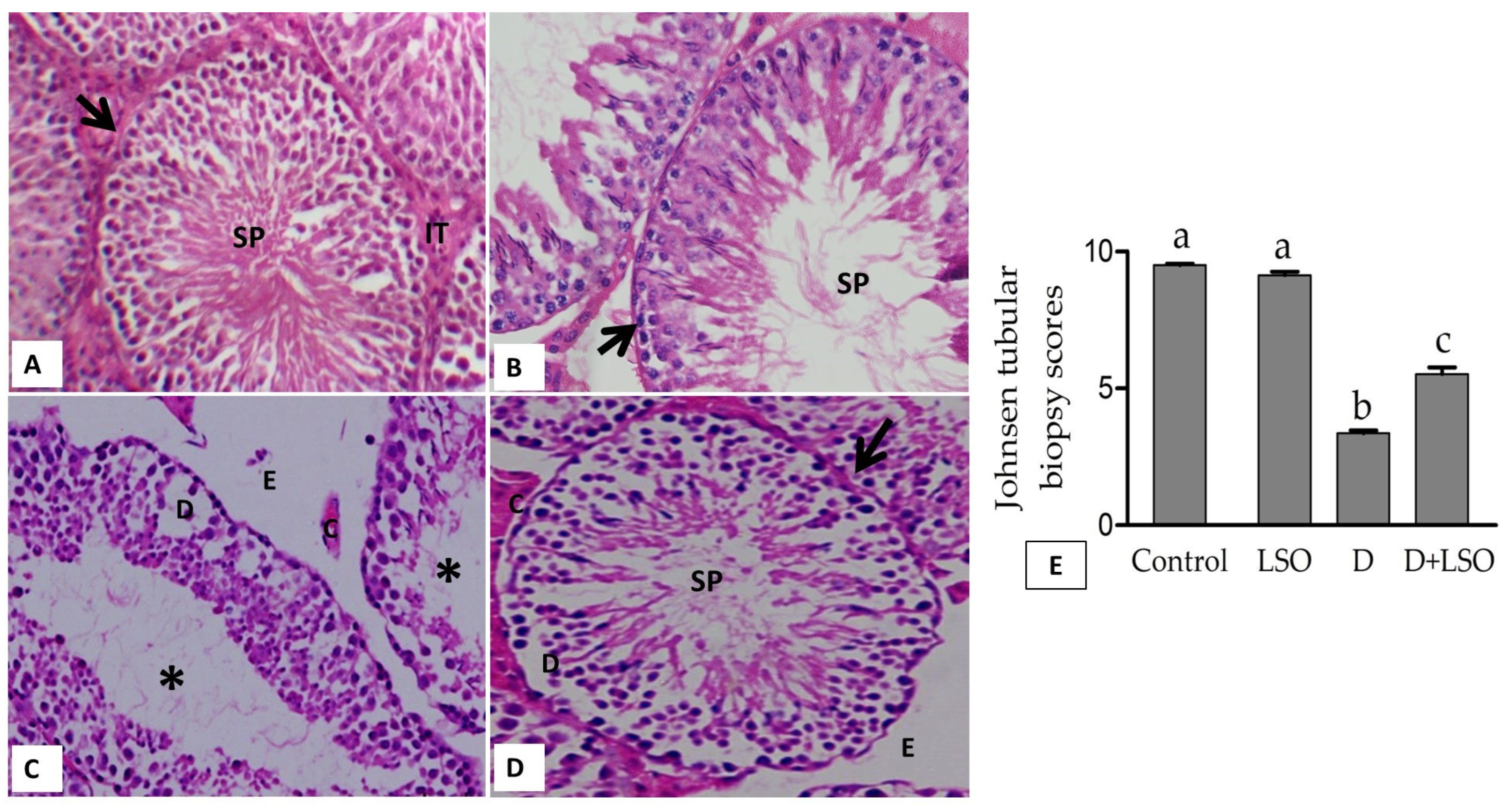
Figure 6 shows the representative photomicrographs and histopathological scoring of testes in all studied groups. Seminiferous tubules with normal spermatogenesis stages were observed in the control group (Figure 6A). The LSO group revealed no changes similar to the control (Figure 6B). STZ-induced diabetes resulted in marked damage in the seminiferous epithelium, and empty/atrophied tubules were predominant in this group. Germinal cell death was also notable. In addition, there was obvious edema in the interstitial space and sloughing (detachment) in the germinal epithelial layer (Figure 6C). Conversely, the testes of the D+LSO mice exhibited little degeneration in the interstitial space and lesser detachment in the germinal layer, and the tubular lumen was filled with spermatids, indicating improvement in spermatogenesis series. However, edema, congestion, and few epithelial detachments were still found (Figure 6D). The mean JTBS (Figure 6E) in the diabetic group (D) significantly decreased by −64.5% compared to the control group, revealing atrophy and hypospermatogenesis. However, treatment of the diabetic mice with LSO significantly increased the mean JTBS value by +63.85% in the D+LSO group compared to that recorded in the diabetic (D) group alone.

3. Discussion

Diabetes mellitus (DM) is a recognized cause of male sexual dysfunction, which may lead ultimately to sub/infertility. Nearly half of diabetic men have reduced semen quality and exhibit reproductive dysfunction [9]. The underlying mechanisms involve, among others, chronic hyperglycemia-induced oxidative stress, inflammation, neuropathy, and insulin resistance (IR), resulting in a reduction in the level of reproductive hormones (FSH, LH, and testosterone) as well as structurally injured male reproductive organs [9,79]. Thus far, the treatment of DM-associated male reproductive dysfunction is challenging, as no established medication that specifically addresses the reproductive health of diabetic patients is available [9]. Knowing that OS is a main cause of damaged reproductive organs, natural products (NPs) with antioxidant capacity can thus be used as supplements for the prevention and/or treatment of diabetic complications, including reproductive dysfunction. To our knowledge, the current study is the first to demonstrate the protective effect of L. sativum seed oil (LSO) against diabetic-induced male reproductive dysfunction. LSO exhibited antioxidant activity as it restored normal levels of enzymatic antioxidants and oxidative stress biomarkers. Moreover, LSO showed anti-inflammatory effects via modulating the levels of inflammatory molecules, including NF-kB, IL-1, IL-6, and TNF-α. In addition, LSO was able to restore normal levels of testosterone.
GC-MC spectral analysis revealed the presence of ten bioactive components in LSO, the major of which were two forms of vitamin E, namely γ-tocopherol (62.1%) and δ- tocopherol (3.91%), followed by six alkanes (24.22%), the antioxidant phenol derivative butylated hydroxytoluene (BHT; 8.04%), and an alcoholic compound (1.73%). Many of these bioactive components possess antioxidant and/or anti-inflammatory activities and hence a wide range of applications, including potential therapeutic benefits. Our data are consistent with those of Umesha and Naidu, who found that feeding rats on a diet with LSO increased blood tocopherol levels and elevated the activity of antioxidant enzymes [80]. Indeed, forms of vitamin E such as γ-tocopherol (γT), δ-tocopherol (δT), and γ-tocotrienol (γ-T3) are strong natural therapeutic antioxidants and known to possess anti-inflammatory effects against various diseases with an underlying inflammatory, redox, or malignant component [81,82]. Furthermore, butylated hydroxytoluene (BHT) is a lipophilic organic substance widely used to prevent free radical-mediated oxidation in organic fluids, and its therapeutic implications have been previously suggested [83,84,85]. In addition to the above-mentioned components, the 2-methyltetracosane alkane detected in the LSO extract is known to possess free radical scavenging activity and proposed antibacterial activity [86,87]. Also, LSO contains tetracosane which has previously been reported as an antioxidant and anticancer alkane compound [88]. The presence of such bioactive compounds in LSO may be a key factor in reducing the free radicals resulting from hyperglycemia in STZ-induced diabetic mice. Oxidative stress is a key mechanism associated with the onset of diabetic issues. In diabetes, hyperglycemia increases mitochondrial glucose oxidation which results in ROS release into the cell cytoplasm, promoting pro-oxidants over antioxidants [35,89,90]. In the present research, the administration of LSO to the STZ-induced diabetic mice impeded OS in testicular tissues, as evidenced by the increase in the enzymatic activities of CAT and SOD and the decrease in the levels of MDA. Similarly, other reports have shown the potent antioxidant properties and improvement in testicular antioxidants exerted by other seed oils via increasing the overall levels of endogenous antioxidants [91,92].
Indeed, inflammation is known to play an emerging role in the pathophysiology of diabetes, whereby various inflammatory pathways and biomarkers of inflammation are involved [93]. Moreover, it has been previously reported that the innate immune and inflammatory responses are involved in the progression of hyperglycemia-induced secondary diabetic complications [94]. The transcription factor nuclear factor kappa B (NF-kB) plays a major role in innate immunity and inflammatory responses. Moreover, it is also involved in the modulation of responses to various stimuli, including free radicals and cytokines [95,96]. Remarkably, chronic hyperglycemia and the concurrent increased levels of advanced glycation end products (AGEs) and their receptors (RAGE) as well as OS activate NF-kB by various pathways that subsequently trigger the expression of various pro-inflammatory cytokines, including IL-1, IL-6, and TNF-α, which are crucial for the development of inflammation in diabetes and other chronic diseases [97,98,99]. Our data revealed a substantial increase in the levels of testicular inflammatory markers, NF-kB, IL-1, IL-6, and TNF-α, in the diabetic mice compared to the control. In accordance with our findings, previous studies have reported high levels of inflammation in diabetic rat testes [100,101,102]. Notably, the administration of LSO to the diabetic mice resulted in anti-inflammatory effects, as indicated by reduction in the elevated pro-inflammatory cytokines in the D+LSO mice, a modulatory effect associated with decline in NF-kB expression.
Previous studies have reported the anti-inflammatory effects of LSO [73,103,104], which is probably attributed to the inhibition of the NF-kB signaling pathway. Moreover, vitamin E forms, representing the major components in LSO extracted in the current study, scavenge reactive nitrogen species (RNS) and suppress pro-inflammatory signaling such as NF-kB [105,106]. In addition, it has been reported that butylated hydroxytoluene (BHT) and γ-tocopherol (γT) contribute to L. sativum’s anti-inflammatory, antioxidant, and immunomodulatory properties [65].
It has been demonstrated that spermatogenesis and steroidogenesis are inhibited by hyperglycemia-induced OS and increased levels of pro-inflammatory markers, IL-1 and TNF-α [56,107,108]. Moreover, previous in vitro and in vivo studies have revealed the inhibition of testosterone (T) production induced by LH consequent to the inhibitory effect exerted by IL-1 and TNF-α on Leydig cells [109,110]. In line with these findings, a significant decrease in serum testosterone levels in the STZ-induced diabetic mice was observed compared to the control. Treatment with LSO markedly restored testosterone levels in the D+LSO mice compared to the diabetic group. Comparably, improvement in testosterone levels in diabetic animal models has been previously reported upon treatment with seed oil extracted from various medicinal plants [91,92]. The restoration of the serum testosterone levels in the diabetic mice treated with LSO (D+LSO) when compared to the STZ-induced diabetic mice affirms the therapeutic potential of LSO on testicular tissues in diabetic mice. Owing to the reported antioxidant and anti-inflammatory effects of LSO [73,80,111], the protective effect of LSO on testicular tissues is attained via the reduction of oxidative damage and suppression of inflammation induced by hyperglycemia. Moreover, STZ induction of diabetes resulted in damaged testicular tissue and hypospermatogenesis as indicated by JTBS. This may be explained by the hyperglycemia-induced increased generation of free radicals and the reduced activity of antioxidant enzymes in the testicular tissues, the elevated levels of pro-inflammatory markers, the suppression of gonadotropin and testosterone release, and the inhibition of steroidogenesis [91,112,113].

4. Materials and Methods

4.1. Plant Material

The Lepidium sativum L. seeds were purchased from a local market in Hofuf city, Al-Ahsa, Saudi Arabia.

4.2. Extraction of Fixed Oil

The Lepidium sativum L. seeds were ground using an electrical grinder to a fine powder; then, 470 g of the seed powder was extracted with petroleum ether (40–60 °C):diethyl ether (1:1 v/v) until it was clear using the soxhlet apparatus at 40 °C. The solvent was removed under reduced pressure. The total fixed oil was quantified as previously described [114] and kept at 4 °C for further analysis.

4.3. Gas Chromatography-Mass Spectrometry (GC-MS) Analysis of LSO

The chemical composition of LSO was identified using a GCMS-QP2010 SE gas chromatograph-mass spectrometer (SHIMADZU, Kyoto, Japan) with a direct capillary column Rtx–5MS (30 m × 0.25 mm × 0.25 µm film thickness). The temperature of the column oven was first maintained at 50 °C (2 min hold), then raised by 10 °C/min to 150 °C, and after that to 300 °C by 10 °C/min. The injector temperature was maintained at 250 °C. At a constant flow rate (1 mL/min), helium was employed as a carrier gas. Samples of 1.0 µL were inoculated automatically by an AOC-20S Auto sampler attached with GC in the splitless mode. Electron ionization (EI) mass spectra were obtained at 70 eV ionization voltages over the range of m/z 20–500 in full scan mode. The temperatures of the ion source and transfer line were set to 250 and 300 °C, respectively. The constituents were recognized by comparing their retention times and mass spectra with those of the NIST 05 mass spectral database.

4.4. Animals

Twenty-four adult (8 weeks old) male mice (25~28 g) obtained from an animal facility at King Faisal University, Saudi Arabia, were used in this study. The mice were main-tained under standard conditions with a 12 h light/dark cycle, a temperature of (~20–22 °C), and 40–60% relative humidity. The animals were given regular food with water ad libitum. All experimental procedures were followed according to relevant guidelines and regulations for experimental animal use in research established by the Deanship of Scientific Research, Vice Presidency for Graduate Studies and Scientific Research, King Faisal University (KFU-REC-2023-MAR-ETHICS734).

4.5. Induction of Diabetes

Overnight-fasted mice were induced by a single intraperitoneal (i.p.) injection of 50 mg/kg in 0.1 mol/L citric acid buffer, pH 4.5 streptozotocin (STZ; Sigma, St. Louis, MO, USA). The diabetic state was confirmed within 7 days after STZ injection by determining tail vein blood glucose levels. Mice with a non-fasting blood glucose concentration over 16.67 mmol/L for three consecutive days were diagnosed as diabetic models [115].

4.6. Experimental Design

After two weeks of diabetes induction, the experimental mice were separated into four groups (6 mice in each).
  • Control: The mice were injected intraperitoneally (i.p.) with citric acid–sodium citrate buffer solution.
  • Diabetic (D): Diabetes was induced by a single intraperitoneal (i.p.) injection of streptozotocin (STZ; 50 mg/kg/day) for five consecutive days [115].
  • Lepidium seed oil (LSO): The mice were administrated intragastrically with 0.5 mL/kg LSO twice a week for four weeks.
  • D+LSO: The diabetic mice administrated with LSO as mentioned in the LSO-alone group.

4.7. Tissue Sampling

The mice were euthanized under anesthesia and sacrificed to their collect blood. The blood samples were collected and centrifuged at 5000× g for 10 min; the separated sera were subjected to testosterone analysis. Immediately, the paired testes were excised. The right testes were stored in 10% formaldehyde fixative fluid for histomorphology, whereas the left testes were stored at −80 °C for biochemical and molecular (western blotting) analyses.

4.8. Assay of Serum Testosterone

Serum samples were used to determine the levels of testosterone (T) using a mouse testosterone enzyme-linked immunosorbent assay (ELISA) kit (Cat. No. MBS494055; MyBioSource, Inc., San Diego, CA, USA) according to manufacturer’s instructions. Spectrophotometric analysis was used to evaluate the optical density at 450 nm, and the testosterone level (ng/mL) was evaluated against the reference stranded assessed in a similar manner.

4.9. Assessment of Redox Status

The colorimetric assay kits for determining the antioxidant enzymes catalase (CAT; Cat. No. CA 2516) and superoxide dismutase (SOD; Cat. No. SD 2520) as well as the LPO biomarker, malondialdehyde (MDA; Cat. No. MD 2528), were purchased from Bio Diagnostic (Dokki, Giza, Egypt). The tissues from the left testes were homogenized in 0.1 M phosphate buffer (pH 7.4). The homogenates were centrifuged at 2800× g for 30 min at 4 °C. The obtained supernatants were utilized for biochemical estimations of CAT, SOD, and MDA as per the guidelines provided by the manufacturer. The protein content in the testicular homogenates was determined as previously described [116], employing bovine serum albumin (BSA) as the standard.

4.10. Determination of NF-kB Factor and Pro-Inflammatory Cytokines

The testicular protein levels of nuclear factor-kappa B (NF-kB) p65 subunit, interleukin-1 (IL-1), interleukin-6 (IL-6), and tumor necrosis factor-alpha (TNF-α) were measured using ELISA kits (NF-kB, Cat. No.: orb409109; Biorbyt Ltd., Cambridge, UK) and (IL-1, Cat. No.: MBS264984; IL-6, Cat. No., MBS843429; TNF-α, Cat. No.: MBS825002; My Bio-Source, Inc., San Diego, CA, USA), respectively, in accordance with the manufacturer’s instructions.

4.11. Western Blotting

Approximately 50 mg of testicular tissues were added to RIPA lysis buffer (Cat. No.: PL005, BIO BASIC INC., Markham, ON, Canada). The tissue samples were homogenized, and protein quantifications were carried out by a Bradford protein assay kit (Cat. No.: SK304, BIO BASIC INC., Markham, ON, Canada). Equal quantities of protein per specimen were exposed to 10% sodium dodecyl sulfate–polyacrylamide gel electrophoresis (SDS-PAGE), and the resolved proteins were electrophoretically transmitted onto polyvinylidene difluoride (PVDF) membranes using the Trans-Blot Turbo Transfer System (Bio-Rad Laboratories, Inc., CA, USA). These membranes were blocked in tris-buffered saline with 0.1% Tween 20 (TBST) and 3% BSA for 60 min at room temperature. Subsequently, the membranes were probed overnight at 4 °C with primary antibodies against NF-kB p65 (Cat. No. ab31481, 1:1000) and β-actin as an internal loading control (Cat. No. ab8226, 1:10,000), both purchased from Abcam (Cambridge, UK). After washing with TBST, the membranes were incubated with horseradish peroxidase-conjugated goat anti-rabbit IgG secondary antibodies at room temperature for 1 h. The blots were washed three times between the primary as well as secondary antibody incubations. Finally, the immunoreactive bands of the NF-kB p65 subunit were imaged using the ChemiDoc MP imager with Image LabTM software (version 6.1) (Bio-Rad Laboratories, Inc., Hercules, CA, USA) to validate the Western blot data via total protein normalization by the β-actin housekeeping protein.

4.12. Histological Study on the Testes

The excised right testes were fixed in 10% formaldehyde solution at 28 °C for 24 h, dehydrated, and inserted in paraffin wax. Traditional H&E (hematoxylin and eosin) dye was used to stain the thin sections (4 μm thick). A typical light microscope (Nikon Corporation, Tokyo, Japan) was used for examining the slides. Twenty seminiferous tubules from each sample were magnified at 400× to determine the testicular maturity and damage using the Johnsen’s tubular biopsy score (JTBS) method [117]. Each seminiferous tubule was given a score between 1 and 10 to indicate its level of germinal epithelial maturity according to the following description: score (1), no cells appear in the tubular section; score (2), no germ cells exist; score (3), just the spermatogonia can be seen; score (4), limited primary spermatocytes; score (5), numerous primary spermatocytes but no spermatozoa or spermatids; score (6), only a few spermatids; score (7), a large number of early spermatids deprived of differentiation; score (8), a few differentiated spermatids; score (9), several differentiated spermatids; and finally, score (10), regular tubules with lots of sperms.

4.13. Statistical Analyses

The results were expressed as the mean ± standard error (SE). The sample size (n = 6) was identified using the G power program (version 3.1.9.7). Graph Pad prism software(version 4.03, San Diego, CA, USA) was used for the statistical analysis. Using the Shapiro–Wilk normality test, the data’s normality was evaluated. One-way ANOVA was used to analyze the statistical variance between the groups, and Tukey’s post hoc test was used to compare the groups. Statistics were determined to be significant for all p values < 0.05.

5. Conclusions

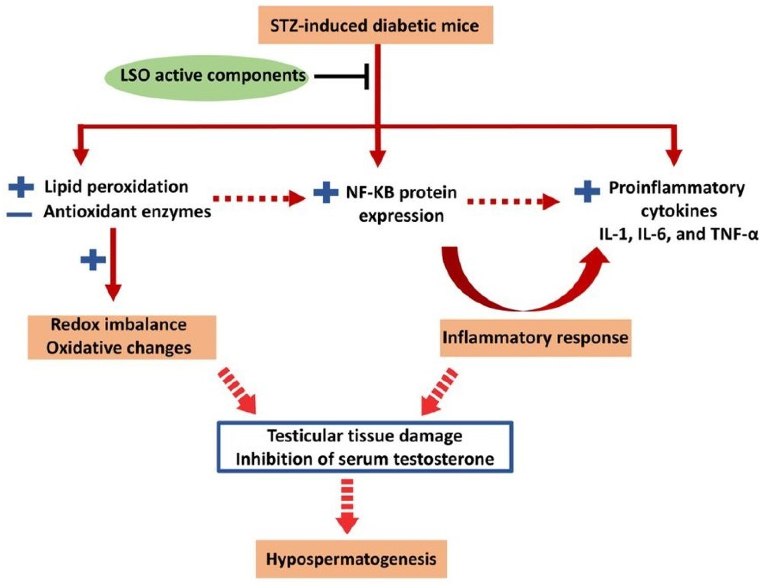
The obtained results show, for the first time, the protective effect of LSO against male reproductive dysfunction observed in diabetic mice. Alleviation of diabetic-induced reproductive dysfunction might be attributed to the improvement in the testicular antioxidant defense system, lowering the levels of pro-inflammatory markers, and the restoration of serum testosterone levels (Figure 7). The findings of the current study support the previously reported antioxidant and anti-inflammatory activities of LSO and pave the way for its therapeutic application in the management of diabetes-associated male reproductive dysfunction. We believe that administering LSO to male diabetic patients will effectively ameliorate the process of spermatogenesis and enhance the architecture of the testicular tissue.

Author Contributions

Conceptualization, R.A.-K., G.M.B., H.I.A.E.-M., S.A.S. and H.F.A.E.-K.; methodology, R.A.-K., G.M.B., H.I.A.E.-M., R.S.H., N.K.A.A., A.S.A.A., S.A., M.A.A., S.A.S. and H.F.A.E.-K.; formal analysis, R.A.-K., G.M.B., H.I.A.E.-M., S.A.S. and H.F.A.E.-K.; investigation, R.A.-K., G.M.B., H.I.A.E.-M., R.S.H., N.K.A.A., A.S.A.A., S.A., M.A.A., S.A.S. and H.F.A.E.-K.; data curation, R.A.-K., G.M.B., H.I.A.E.-M., R.S.H., N.K.A.A., A.S.A.A., S.A., M.A.A., S.A.S. and H.F.A.E.-K.; writing—original draft preparation, R.A.-K., G.M.B., H.I.A.E.-M., R.S.H., S.A.S. and H.F.A.E.-K.; writing—review and editing, R.A.-K., G.M.B., H.I.A.E.-M., R.S.H., N.K.A.A., A.S.A.A., S.A., M.A.A., S.A.S. and H.F.A.E.-K.; funding acquisition, R.A.-K. All authors have read and agreed to the published version of the manuscript.

Funding

This research was funded by the Deanship of Scientific Research, Vice Presidency for Graduate Studies and Scientific Research, King Faisal University, Saudi Arabia (GRANT4583).

Institutional Review Board Statement

The study was reviewed and approved by the Deanship of Scientific Research, Vice Presidency for Graduate Studies and Scientific Research, King Faisal University (KFU-REC-2023-MAR-ETHICS734).

Informed Consent Statement

Not applicable.

Data Availability Statement

All data analyzed during this study are included in this published article.

Acknowledgments

The authors acknowledge the Deanship of Scientific Research, Vice Presidency for Graduate Studies and Scientific Research, King Faisal University, Saudi Arabia, for the financial support under the annual funding track (GRANT4583).

Conflicts of Interest

The authors declare no conflict of interest.

References

  1. Henninger, J.; Hammarstedt, A.; Rawshani, A.; Eliasson, B. Metabolic Predictors of Impaired Glucose Tolerance and Type 2 Diabetes in a Predisposed Population—A Prospective Cohort Study. BMC Endocr. Disord. 2015, 15, 51. [Google Scholar] [CrossRef]
  2. Beckman, J.A.; Creager, M.A. Vascular Complications of Diabetes. Circ. Res. 2016, 118, 1771–1785. [Google Scholar] [CrossRef]
  3. Iacobini, C.; Vitale, M.; Pesce, C.; Pugliese, G.; Menini, S. Diabetic Complications and Oxidative Stress: A 20-Year Voyage Back in Time and Back to the Future. Antioxidants 2021, 10, 727. [Google Scholar] [CrossRef]
  4. Bereda, G. Complication of Diabetes Mellitus: Microvascular and Macrovascular Complications. Int. J. Diabetes 2022, 3, 123–128. [Google Scholar]
  5. Nellaiappan, K.; Preeti, K.; Khatri, D.K.; Singh, S.B. Diabetic Complications: An Update on Pathobiology and Therapeutic Strategies. Curr. Diabetes Rev. 2022, 18, e030821192146. [Google Scholar] [CrossRef]
  6. Condorelli, R.A.; La Vignera, S.; Mongioi, L.M.; Alamo, A.; Calogero, A.E. Diabetes Mellitus and Infertility: Different Pathophysiological Effects in Type 1 and Type 2 on Sperm Function. Front. Endocrinol. 2018, 9, 268. [Google Scholar] [CrossRef]
  7. Sansone, A.; Mollaioli, D.; Ciocca, G.; Limoncin, E.; Colonnello, E.; Jannini, E.A. Sexual Dysfunction in Men and Women with Diabetes: A Reflection of their Complications? Curr. Diabetes Rev. 2022, 18, e030821192147. [Google Scholar] [CrossRef]
  8. Tsampoukas, G.; Tharakan, T.; Narayan, Y.; Khan, F.; Cayetano, A.; Papatsoris, A.; Buchholz, N.; Minhas, S. Investigating the Therapeutic Options for Diabetes-Associated Male Infertility as Illustrated in Animal Experimental Models. Andrologia 2022, 54, e14521. [Google Scholar] [CrossRef]
  9. Andlib, N.; Sajad, M.; Kumar, R.; Thakur, S.C. Abnormalities in Sex Hormones and Sexual Dysfunction in Males with Diabetes Mellitus: A Mechanistic Insight. Acta Histochem. 2023, 125, 151974. [Google Scholar] [CrossRef]
  10. Sampannang, A.; Arun, S.; Burawat, J.; Sukhorum, W.; Iamsaard, S. Comparison of Male Reproductive Parameters in Mice with Type 1 and Type 2 Diabetes. Clin. Exp. Reprod. Med. 2020, 47, 20–33. [Google Scholar] [CrossRef]
  11. Bener, A.; Al-Ansari, A.A.; Zirie, M.; Al-Hamaq, A.O. Is Male Fertility Associated with Type 2 Diabetes Mellitus? Int. Urol. Nephrol. 2009, 41, 777–784. [Google Scholar] [CrossRef] [PubMed]
  12. La Vignera, S.; Calogero, A.E.; Condorelli, R.; Lanzafame, F.; Giammusso, B.; Vicari, E. Andrological Characterization of the Patient with Diabetes Mellitus. Minerva Endocrinol. 2009, 34, 1–9. [Google Scholar] [PubMed]
  13. Lotti, F.; Maggi, M. Effects of Diabetes Mellitus on Sperm Quality and Fertility Outcomes: Clinical Evidence. Andrology 2023, 11, 399–416. [Google Scholar] [CrossRef] [PubMed]
  14. Chan, J.L.; Mantzoros, C.S. Leptin and the Hypothalamic-Pituitary Regulation of the Gonadotropin-Gonadal Axis. Pituitary 2001, 4, 87–92. [Google Scholar] [CrossRef]
  15. Pitteloud, N.; Hardin, M.; Dwyer, A.A.; Valassi, E.; Yialamas, M.; Elahi, D.; Hayes, F.J. Increasing Insulin Resistance Is Associated with a Decrease in Leydig Cell Testosterone Secretion in Men. J. Clin. Endocrinol. Metab. 2005, 90, 2636–2641. [Google Scholar] [CrossRef] [PubMed]
  16. Serwaa, D.; Bello, F.A.; Osungbade, K.O.; Nkansah, C.; Osei-Boakye, F.; Appiah, S.K.; Antwi, M.H.; Danquah, M.; Buckman, T.A.; Owusu, E. Prevalence and Determinants of Low testosterone Levels in Men with Type 2 Diabetes Mellitus; a Case-Control Study in A District Hospital in Ghana. PLoS Glob. Public Health 2021, 1, e0000052. [Google Scholar] [CrossRef] [PubMed]
  17. Zhou, Y.; Tian, R.; Wang, X.; Sun, J.; Zhu, L.; An, X. The Occurrence of Hypogonadotropic Hypogonadism in Chinese Men with Type 2 Diabetes. Clin. Endocrinol. 2022, 96, 837–846. [Google Scholar] [CrossRef]
  18. Musa, E.; El-Bashir, J.M.; Sani-Bello, F.; Bakari, A.G. Clinical and Biochemical Correlates of Hypogonadism in Men with Type 2 Diabetes Mellitus. Pan Afr. Med. J. 2021, 38, 292. [Google Scholar] [CrossRef] [PubMed]
  19. Costanzo, P.R.; Knoblovits, P. Male Gonadal Axis Function in Patients with Type 2 Diabetes. Horm. Mol. Biol. Clin. Investig. 2016, 26, 129–134. [Google Scholar] [CrossRef] [PubMed]
  20. Corona, G.; Vena, W.; Pizzocaro, A.; Vignozzi, L.; Sforza, A.; Maggi, M. Testosterone Therapy in Diabetes and Pre-Diabetes. Andrology 2023, 11, 204–214. [Google Scholar] [CrossRef] [PubMed]
  21. Gianatti, E.J.; Grossmann, M. Testosterone Deficiency in Men with Type 2 Diabetes: Pathophysiology and Treatment. Diabet. Med. 2020, 37, 174–186. [Google Scholar] [CrossRef]
  22. Karimi, J.; Goodarzi, M.T.; Tavilani, H.; Khodadadi, I.; Amiri, I. Increased Receptor for Advanced Glycation End Products in Spermatozoa of Diabetic Men and Its Association with Sperm Nuclear DNA Fragmentation. Andrologia 2012, 44, 280–286. [Google Scholar] [CrossRef]
  23. Barkabi-Zanjani, S.; Ghorbanzadeh, V.; Aslani, M.; Ghalibafsabbaghi, A.; Chodari, L. Diabetes Mellitus and the Impairment of Male Reproductive Function: Possible Signaling Pathways. Diabetes Metab. Syndr. 2020, 14, 1307–1314. [Google Scholar] [CrossRef]
  24. Amaral, S.; Oliveira, P.J.; Ramalho-Santos, J. Diabetes and the Impairment of Reproductive Function: Possible Role of Mitochondria and Reactive Oxygen Species. Curr. Diabetes Rev. 2008, 4, 46–54. [Google Scholar] [CrossRef] [PubMed]
  25. Agbaje, I.M.; Rogers, D.A.; McVicar, C.M.; McClure, N.; Atkinson, A.B.; Mallidis, C.; Lewis, S.E. Insulin Dependant Diabetes Mellitus: Implications for Male Reproductive Function. Hum. Reprod. 2007, 22, 1871–1877. [Google Scholar] [CrossRef]
  26. Calogero, A.E.; Duca, Y.; Condorelli, R.A.; La Vignera, S. Male Accessory Gland Inflammation, Infertility, and Sexual Dysfunctions: A Practical Approach to Diagnosis and Therapy. Andrology 2017, 5, 1064–1072. [Google Scholar] [CrossRef]
  27. Mostafa, T.; Abdel-Hamid, I.A. Ejaculatory Dysfunction in Men with Diabetes Mellitus. World J. Diabetes 2021, 12, 954–974. [Google Scholar] [CrossRef]
  28. Condorelli, R.A.; Calogero, A.E.; Vicari, E.; Duca, Y.; Favilla, V.; Morgia, G.; Cimino, S.; Di Mauro, M.; La Vignera, S. Prevalence of Male Accessory Gland Inflammations/Infections in Patients with Type 2 Diabetes Mellitus. J. Endocrinol. Invest. 2013, 36, 770–774. [Google Scholar] [CrossRef]
  29. Rato, L.; Alves, M.G.; Socorro, S.; Duarte, A.I.; Cavaco, J.E.; Oliveira, P.F. Metabolic Regulation Is Important for Spermatogenesis. Nat. Rev. Urol. 2012, 9, 330–338. [Google Scholar] [CrossRef]
  30. Mateus, I.; Feijo, M.; Espinola, L.M.; Vaz, C.V.; Correia, S.; Socorro, S. Glucose and Glutamine Handling in the Sertoli Cells of Transgenic Rats Overexpressing Regucalcin: Plasticity towards Lactate Production. Sci. Rep. 2018, 8, 10321. [Google Scholar] [CrossRef]
  31. Ding, G.L.; Liu, Y.; Liu, M.E.; Pan, J.X.; Guo, M.X.; Sheng, J.Z.; Huang, H.F. The Effects of Diabetes on Male Fertility and Epigenetic Regulation During Spermatogenesis. Asian J. Androl. 2015, 17, 948–953. [Google Scholar] [CrossRef] [PubMed]
  32. Babel, R.A.; Dandekar, M.P. A Review on Cellular and Molecular Mechanisms Linked to the Development of Diabetes Complications. Curr. Diabetes Rev. 2021, 17, 457–473. [Google Scholar] [CrossRef]
  33. Maranta, F.; Cianfanelli, L.; Cianflone, D. Glycaemic Control and Vascular Complications in Diabetes Mellitus Type 2. Adv. Exp. Med. Biol. 2021, 1307, 129–152. [Google Scholar] [CrossRef] [PubMed]
  34. Yaribeygi, H.; Atkin, S.L.; Sahebkar, A. A Review of the Molecular Mechanisms of Hyperglycemia-Induced Free Radical Generation Leading to Oxidative Stress. J. Cell. Physiol. 2019, 234, 1300–1312. [Google Scholar] [CrossRef] [PubMed]
  35. Gonzalez, P.; Lozano, P.; Ros, G.; Solano, F. Hyperglycemia and Oxidative Stress: An Integral, Updated and Critical Overview of Their Metabolic Interconnections. Int. J. Mol. Sci. 2023, 24, 9352. [Google Scholar] [CrossRef] [PubMed]
  36. Khalid, M.; Petroianu, G.; Adem, A. Advanced Glycation End Products and Diabetes Mellitus: Mechanisms and Perspectives. Biomolecules 2022, 12, 542. [Google Scholar] [CrossRef]
  37. Bhatti, J.S.; Sehrawat, A.; Mishra, J.; Sidhu, I.S.; Navik, U.; Khullar, N.; Kumar, S.; Bhatti, G.K.; Reddy, P.H. Oxidative stress in the Pathophysiology of Type 2 Diabetes and Related Complications: Current Therapeutics Strategies and Future Perspectives. Free Radic. Biol. Med. 2022, 184, 114–134. [Google Scholar] [CrossRef]
  38. Singh, H.; Singh, R.; Singh, A.; Singh, H.; Singh, G.; Kaur, S.; Singh, B. Role of oxidative stress in diabetes-induced complications and their management with antioxidants. Arch. Physiol. Biochem. 2023, 1–26. [Google Scholar] [CrossRef]
  39. Darenskaya, M.A.; Kolesnikova, L.I.; Kolesnikov, S.I. Oxidative Stress: Pathogenetic Role in Diabetes Mellitus and Its Complications and Therapeutic Approaches to Correction. Bull. Exp. Biol. Med. 2021, 171, 179–189. [Google Scholar] [CrossRef]
  40. Rato, L.; Oliveira, P.; Sousa, M.; Silva, B.; Alves, M. Chapter 2.6—Role of Reactive Oxygen Species in Diabetes-Induced Male Reproductive Dysfunction. In Oxidants, Antioxidants and Impact of the Oxidative Status in Male Reproduction; Henkel, R., Samanta, L., Agarwal, A., Eds.; Academic Press: Cambridge, MA, USA, 2019; pp. 135–147. [Google Scholar]
  41. Abbasihormozi, S.H.; Babapour, V.; Kouhkan, A.; Niasari Naslji, A.; Afraz, K.; Zolfaghary, Z.; Shahverdi, A.H. Stress Hormone and Oxidative Stress Biomarkers Link Obesity and Diabetes with Reduced Fertility Potential. Cell J. 2019, 21, 307–313. [Google Scholar] [CrossRef]
  42. Leisegang, K. Oxidative Stress in Men with Obesity, Metabolic Syndrome and Type 2 Diabetes Mellitus: Mechanisms and Management of Reproductive Dysfunction. Adv. Exp. Med. Biol. 2022, 1358, 237–256. [Google Scholar] [CrossRef] [PubMed]
  43. Mannucci, A.; Argento, F.R.; Fini, E.; Coccia, M.E.; Taddei, N.; Becatti, M.; Fiorillo, C. The Impact of Oxidative Stress in Male Infertility. Front. Mol. Biosci. 2021, 8, 799294. [Google Scholar] [CrossRef]
  44. Aitken, R.J.; Drevet, J.R. The Importance of Oxidative Stress in Determining the Functionality of Mammalian Spermatozoa: A Two-Edged Sword. Antioxidants 2020, 9, 111. [Google Scholar] [CrossRef]
  45. Nowicka-Bauer, K.; Nixon, B. Molecular Changes Induced by Oxidative Stress That Impair Human Sperm Motility. Antioxidants 2020, 9, 134. [Google Scholar] [CrossRef]
  46. Hosen, M.B.; Islam, M.R.; Begum, F.; Kabir, Y.; Howlader, M.Z. Oxidative Stress Induced Sperm DNA Damage, a Possible Reason for Male Infertility. Iran. J. Reprod. Med. 2015, 13, 525–532. [Google Scholar] [PubMed]
  47. Chianese, R.; Pierantoni, R. Mitochondrial Reactive Oxygen Species (ROS) Production Alters Sperm Quality. Antioxidants 2021, 10, 92. [Google Scholar] [CrossRef]
  48. Costa, J.; Braga, P.C.; Rebelo, I.; Oliveira, P.F.; Alves, M.G. Mitochondria Quality Control and Male Fertility. Biology 2023, 12, 827. [Google Scholar] [CrossRef] [PubMed]
  49. Su, S.C.; Hung, Y.J.; Huang, C.L.; Shieh, Y.S.; Chien, C.Y.; Chiang, C.F.; Liu, J.S.; Lu, C.H.; Hsieh, C.H.; Lin, C.M.; et al. Cilostazol Inhibits Hyperglucose-Induced Vascular Smooth Muscle Cell Dysfunction by Modulating the RAGE/ERK/NF-kB Signaling Pathways. J. Biomed. Sci. 2019, 26, 68. [Google Scholar] [CrossRef]
  50. Liu, X.; Lin, R.; Zhao, B.; Guan, R.; Li, T.; Jin, R. Correlation Between Oxidative Stress and the NF-kB Signaling Pathway in the Pulmonary Tissues of Obese Asthmatic Mice. Mol. Med. Rep. 2016, 13, 1127–1134. [Google Scholar] [CrossRef]
  51. Arkali, G.; Aksakal, M.; Kaya, S.O. Protective Effects of Carvacrol against Diabetes-Induced Reproductive Damage in Male Rats: Modulation of Nrf2/HO-1 Signalling Pathway and Inhibition of NF-kB-Mediated Testicular Apoptosis and Inflammation. Andrologia 2021, 53, e13899. [Google Scholar] [CrossRef]
  52. Zhu, X.; Guo, F.; Tang, H.; Huang, C.; Xie, G.; Huang, T.; Li, Y.; Liu, C.; Wang, H.; Chen, B. Islet Transplantation Attenuating Testicular Injury in Type 1 Diabetic Rats Is Associated with Suppression of Oxidative Stress and Inflammation via Nrf-2/HO-1 and NF-kB Pathways. J. Diabetes Res. 2019, 2019, 8712492. [Google Scholar] [CrossRef]
  53. Hasan, H.; Bhushan, S.; Fijak, M.; Meinhardt, A. Mechanism of Inflammatory Associated Impairment of Sperm Function, Spermatogenesis and Steroidogenesis. Front. Endocrinol. 2022, 13, 897029. [Google Scholar] [CrossRef]
  54. Luo, Y.; Zhu, Y.; Basang, W.; Wang, X.; Li, C.; Zhou, X. Roles of Nitric Oxide in the Regulation of Reproduction: A Review. Front. Endocrinol. 2021, 12, 752410. [Google Scholar] [CrossRef]
  55. Han, X.X.; Jiang, Y.P.; Liu, N.; Wu, J.; Yang, J.M.; Li, Y.X.; Sun, M.; Sun, T.; Zheng, P.; Jian-Qiang, Y. Protective Effects of Astragalin on Spermatogenesis in Streptozotocin-Induced Diabetes in Male Mice by Improving Antioxidant Activity and Inhibiting Inflammation. Biomed. Pharmacother. 2019, 110, 561–570. [Google Scholar] [CrossRef]
  56. Dutta, S.; Sengupta, P.; Slama, P.; Roychoudhury, S. Oxidative Stress, Testicular Inflammatory Pathways, and Male Reproduction. Int. J. Mol. Sci. 2021, 22, 10043. [Google Scholar] [CrossRef]
  57. Fraczek, M.; Kurpisz, M. Inflammatory Mediators Exert Toxic Effects of Oxidative Stress on Human Spermatozoa. J. Androl. 2007, 28, 325–333. [Google Scholar] [CrossRef]
  58. Prabhakar, P.K.; Doble, M. Effect of Natural Products on Commercial Oral Antidiabetic Drugs in Enhancing 2-Deoxyglucose Uptake by 3T3-L1 Adipocytes. Ther. Adv. Endocrinol. Metab. 2011, 2, 103–114. [Google Scholar] [CrossRef] [PubMed]
  59. Abdi, M.; Alizadeh, F.; Daneshi, E.; Abouzaripour, M.; Fathi, F.; Rahimi, K. Ameliorative Effect of Stevia rebaudiana Bertoni on Sperm Parameters, In Vitro Fertilization, and Early Embryo Development in a Streptozotocin-Induced Mouse Model of Diabetes. Zygote 2023, 31, 475–482. [Google Scholar] [CrossRef]
  60. Khazaei, M.R.; Gravandi, E.; Ghanbari, E.; Niromand, E.; Khazaei, M. Trifolium pratense Extract Increases Testosterone and Improves Sperm Characteristics and Antioxidant Status in Diabetic Rats. Biotech. Histochem. 2022, 97, 576–583. [Google Scholar] [CrossRef] [PubMed]
  61. Ostovan, F.; Gol, A.; Javadi, A. Protective Properties of Rydingia persica in Reproductive Complications Induced by Diabetes in Male Rats: An Experimental Study. Int. J. Reprod. Biomed. 2022, 20, 123–136. [Google Scholar] [CrossRef] [PubMed]
  62. Oroojan, A.A.; Ahangarpour, A.; Paknejad, B.; Zareian, P.; Hami, Z.; Abtahi, S.R. Effects of Myricitrin and Solid Lipid Nanoparticle-Containing Myricitrin on Reproductive System Disorders Induced by Diabetes in Male Mouse. World J. Mens Health 2021, 39, 147–157. [Google Scholar] [CrossRef]
  63. Gupta, R.; Choudhary, P. Botanical Description of Garden Cress (Lepidium sativum L.) Plant and Physical Characteristics of Its Seeds. J. Pharmacogn. Phytochem. 2020, 9, 2424–2428. [Google Scholar]
  64. Ramadan, M.F.; Oraby, H.F. Chapter 20—Lepidium sativum Seeds: Therapeutic Significance and Health-Promoting Potential. In Nuts and Seeds in Health and Disease Prevention, 2nd ed.; Preedy, V.R., Watson, R.R., Eds.; Academic Press: Cambridge, MA, USA, 2020; pp. 273–289. [Google Scholar]
  65. Vazifeh, S.; Kananpour, P.; Khalilpour, M.; Eisalou, S.V.; Hamblin, M.R. Anti-Inflammatory and Immunomodulatory Properties of Lepidium sativum. Biomed. Res. Int. 2022, 2022, 3645038. [Google Scholar] [CrossRef]
  66. Ahmad, A.; Nabi, R.; Mishra, A.; Ahmad, I.Z. A Panoramic Review on Lepidium sativum L. Bioactives as Prospective Therapeutics. Drug Res. 2021, 71, 233–242. [Google Scholar] [CrossRef] [PubMed]
  67. Kaiyrkulova, A.; Li, J.; Aisa, H.A. Chemical Constituents of Lepidium sativum Seeds. Chem. Nat. Compd. 2019, 55, 736–737. [Google Scholar] [CrossRef]
  68. Hekmatshoar, Y.; Ozkan, T.; Rahbar Saadat, Y. Evidence for Health-Promoting Properties of Lepidium sativum L.: An Updated Comprehensive Review. Turk. J. Pharm. Sci. 2022, 19, 714–723. [Google Scholar] [CrossRef]
  69. Painuli, S.; Quispe, C.; Herrera-Bravo, J.; Semwal, P.; Martorell, M.; Almarhoon, Z.M.; Seilkhan, A.; Ydyrys, A.; Rad, J.S.; Alshehri, M.M.; et al. Nutraceutical Profiling, Bioactive Composition, and Biological Applications of Lepidium sativum L. Oxid. Med. Cell. Longev. 2022, 2022, 2910411. [Google Scholar] [CrossRef] [PubMed]
  70. Shah, M.B.; Dudhat, V.A.; Gadhvi, K.V. Lepidium sativum: A Potential Functional Food. J. Ayurvedic Herb. Med. 2021, 7, 140–149. [Google Scholar] [CrossRef]
  71. Bigoniya, P.; Shukla, A. Phytopharmacological Screening of Lepidium sativum Seeds Total Alkaloid: Hepatoprotective, Antidiabetic and In Vitro Antioxidant Activity Along with Identification by LC/MS/MS. PharmaNutrition 2014, 2, 90. [Google Scholar] [CrossRef]
  72. Sakran, M.; Selim, Y.; Zidan, N. A New Isoflavonoid From Seeds of Lepidium sativum L. and Its Protective Effect on Hepatotoxicity Induced by Paracetamol in Male Rats. Molecules 2014, 19, 15440–15451. [Google Scholar] [CrossRef]
  73. Alqahtani, F.Y.; Aleanizy, F.S.; Mahmoud, A.Z.; Farshori, N.N.; Alfaraj, R.; Al-Sheddi, E.S.; Alsarra, I.A. Chemical composition and antimicrobial, antioxidant, and anti-inflammatory activities of Lepidium sativum seed oil. Saudi J. Biol. Sci. 2019, 26, 1089–1092. [Google Scholar] [CrossRef] [PubMed]
  74. Getahun, T.; Sharma, V.; Gupta, N. Chemical Composition, Antibacterial and Antioxidant Activities of oils Obtained by Different Extraction Methods from Lepidium sativum L. seeds. Ind. Crops Prod. 2020, 156, 112876. [Google Scholar] [CrossRef]
  75. Kumar, V.; Tomar, V.; Ranade, S.A.; Yadav, H.K.; Srivastava, M. Phytochemical, Antioxidant Investigaations and Fatty Acid Composition of Lepidium sativum Seeds. J. Environ. Biol. 2020, 41, 59–65. [Google Scholar] [CrossRef]
  76. Kamani, M.; Hosseini, E.S.; Kashani, H.H.; Atlasi, M.A.; Nikzad, H. Protective Effect of Lepidium sativum Seed Extract on Histopathology and Morphology of Epididymis in Diabetic Rat Model. Int. J. Morphol. 2017, 35, 603–610. [Google Scholar] [CrossRef]
  77. Kamani, M.; Mhabadi, J.A.; Atlasi, M.A.; Seyedi, F.; Kamani, E.; Nikzad, H. Protective Effect of Alcoholic Extract of Garden Cress Seeds on the Histopathological Changes of the Ventral Prostate in Streptozotocin Diabetic Rats. Int. J. Morphol. 2017, 35, 1118–1178. [Google Scholar] [CrossRef]
  78. Asl, F.R.; Khosravi, M.; Hajikhani, R.; Solati, J.; Fahimi, H. Complementary Effects of Coenzyme Q10 and Lepidium sativum Supplementation on the Reproductive Function of Mice: An Experimental Study. Int. J. Reprod. Biomed. 2021, 19, 607–618. [Google Scholar] [CrossRef]
  79. He, Z.; Yin, G.; Li, Q.Q.; Zeng, Q.; Duan, J. Diabetes Mellitus Causes Male Reproductive Dysfunction: A Review of the Evidence and Mechanisms. In Vivo 2021, 35, 2503–2511. [Google Scholar] [CrossRef]
  80. Umesha, S.S.; Naidu, K.A. Antioxidants and Antioxidant Enzymes Status of Rats Fed on n-3 PUFA Rich Garden Cress (Lepidium Sativum L.) Seed Oil and Its Blended Oils. J. Food Sci. Technol. 2015, 52, 1993–2002. [Google Scholar] [CrossRef]
  81. Ungurianu, A.; Zanfirescu, A.; Nitulescu, G.; Margina, D. Vitamin E Beyond Its Antioxidant Label. Antioxidants 2021, 10, 634. [Google Scholar] [CrossRef]
  82. Ciarcia, G.; Bianchi, S.; Tomasello, B.; Acquaviva, R.; Malfa, G.A.; Naletova, I.; La Mantia, A.; Di Giacomo, C. Vitamin E and Non-communicable Diseases: A Review. Biomedicines 2022, 10, 2473. [Google Scholar] [CrossRef]
  83. Hossain, K.F.B.; Hosokawa, T.; Saito, T.; Kurasaki, M. Amelioration of Butylated Hydroxytoluene against Inorganic Mercury Induced Cytotoxicity and Mitochondrial Apoptosis in PC12 Cells via Antioxidant Effects. Food Chem. Toxicol. 2020, 146, 111819. [Google Scholar] [CrossRef] [PubMed]
  84. Yehye, W.A.; Rahman, N.A.; Ariffin, A.; Abd Hamid, S.B.; Alhadi, A.A.; Kadir, F.A.; Yaeghoobi, M. Understanding the Chemistry Behind the Antioxidant Activities of Butylated Hydroxytoluene (BHT): A Review. Eur. J. Med. Chem. 2015, 101, 295–312. [Google Scholar] [CrossRef] [PubMed]
  85. Yehye, W.A.; Abdul Rahman, N.; Saad, O.; Ariffin, A.; Abd Hamid, S.B.; Alhadi, A.A.; Kadir, F.A.; Yaeghoobi, M.; Matlob, A.A. Rational Design and Synthesis of New, High Efficiency, Multipotent Schiff Base-1,2,4-triazole Antioxidants Bearing Butylated Hydroxytoluene Moieties. Molecules 2016, 21, 847. [Google Scholar] [CrossRef] [PubMed]
  86. Zubair, M.F.; Ajibade, S.O.; Lawal, A.Z.; Yusuf, S.A.; Babalola, J.B.; Mukadam, A.A.; Hamid, A.A. GC-MS Analysis, Antioxidant and Antimicrobial Properties of Eclipta prostrata Leaves. Int. J. Chem. Biochem. Sci. 2017, 11, 25–43. [Google Scholar]
  87. Ramya, B.; Malarvili, T.; Velavan, S. GC-MS Analysis of Bioactive Compounds in Bryonopsis laciniosa Fruit Extract. Int. J. Pharm. Sci. Res. 2015, 6, 3375–3379. [Google Scholar]
  88. Barupal, T.; Meena, M.; Sharma, K. Inhibitory Effects of Leaf Extract of Lawsonia inermis on Curvularia lunata and Characterization of Novel Inhibitory Compounds by GC-MS Analysis. Biotechnol. Rep. 2019, 23, e00335. [Google Scholar] [CrossRef]
  89. Black, H.S. A Synopsis of the Associations of Oxidative Stress, ROS, and Antioxidants with Diabetes Mellitus. Antioxidants 2022, 11, 2003. [Google Scholar] [CrossRef]
  90. Singh, A.; Kukreti, R.; Saso, L.; Kukreti, S. Mechanistic Insight Into Oxidative Stress-Triggered Signaling Pathways and Type 2 Diabetes. Molecules 2022, 27, 950. [Google Scholar] [CrossRef]
  91. Kermani, J.; Goodarzi, N.; Bakhtiari, M. An Experimental Study to Evaluate the Protective Effects of Solanum lycopersicum Seed Essential Oil on Diabetes-Induced Testicular Injuries. Medicina 2019, 55, 499. [Google Scholar] [CrossRef]
  92. Nasiri, K.; Akbari, A.; Nimrouzi, M.; Ruyvaran, M.; Mohamadian, A. Safflower Seed Oil Improves Steroidogenesis and Spermatogenesis in Rats with Type II Diabetes Mellitus by Modulating the Genes Expression Involved in Steroidogenesis, Inflammation and Oxidative Stress. J. Ethnopharmacol. 2021, 275, 114139. [Google Scholar] [CrossRef]
  93. Tsalamandris, S.; Antonopoulos, A.S.; Oikonomou, E.; Papamikroulis, G.A.; Vogiatzi, G.; Papaioannou, S.; Deftereos, S.; Tousoulis, D. The Role of Inflammation in Diabetes: Current Concepts and Future Perspectives. Eur. Cardiol. 2019, 14, 50–59. [Google Scholar] [CrossRef]
  94. Plowman, T.J.; Shah, M.H.; Fernandez, E.; Christensen, H.; Aiges, M.; Ramana, K.V. Role of Innate Immune and Inflammatory Responses in the Development of Secondary Diabetic Complications. Curr. Mol. Med. 2022, 23, 901–920. [Google Scholar] [CrossRef] [PubMed]
  95. Liu, T.; Zhang, L.; Joo, D.; Sun, S.C. NF-κB Signaling in Inflammation. Signal Transduct. Target. Ther. 2017, 2, 17023. [Google Scholar] [CrossRef] [PubMed]
  96. Liu, D.; Zhong, Z.; Karin, M. NF-kB: A Double-Edged Sword Controlling Inflammation. Biomedicines 2022, 10, 1250. [Google Scholar] [CrossRef]
  97. Suryavanshi, S.V.; Kulkarni, Y.A. NF-κβ: A Potential Target in the Management of Vascular Complications of Diabetes. Front. Pharmacol. 2017, 8, 798. [Google Scholar] [CrossRef] [PubMed]
  98. Lingappan, K. NF-κB in Oxidative Stress. Curr. Opin. Toxicol. 2018, 7, 81–86. [Google Scholar] [CrossRef]
  99. Yousef, H.; Khandoker, A.H.; Feng, S.F.; Helf, C.; Jelinek, H.F. Inflammation, Oxidative Stress and Mitochondrial Dysfunction in the Progression of Type II Diabetes Mellitus with Coexisting Hypertension. Front. Endocrinol. 2023, 14, 1173402. [Google Scholar] [CrossRef] [PubMed]
  100. Khalil, A.S.M.; Giribabu, N.; Yelumalai, S.; Shahzad, H.; Kilari, E.K.; Salleh, N. Myristic Acid Defends against Testicular Oxidative Stress, Inflammation, Apoptosis: Restoration of Spermatogenesis, Steroidogenesis in Diabetic Rats. Life Sci. 2021, 278, 119605. [Google Scholar] [CrossRef]
  101. Nna, V.U.; Abu Bakar, A.B.; Ahmad, A.; Eleazu, C.O.; Mohamed, M. Oxidative Stress, NF-kB-mediated Inflammation and Apoptosis in the Testes of Streptozotocin-Induced Diabetic Rats: Combined Protective Effects of Malaysian Propolis and Metformin. Antioxidants 2019, 8, 465. [Google Scholar] [CrossRef]
  102. Khosravi, Z.; Sedaghat, R.; Baluchnejadmojarad, T.; Roghani, M. Diosgenin Ameliorates Testicular Damage in Streptozotocin-Diabetic Rats Through Attenuation of Apoptosis, Oxidative Stress, and Inflammation. Int. Immunopharmacol. 2019, 70, 37–46. [Google Scholar] [CrossRef]
  103. Diwakar, B.T.; Lokesh, B.R.; Naidu, K.A. Modulatory Effect of α-Linolenic Acid-Rich Garden Cress (Lepidium sativum L.) Seed Oil on Inflammatory Mediators in Adult Albino Rats. Br. J. Nutr. 2011, 106, 530–539. [Google Scholar] [CrossRef] [PubMed]
  104. Reddy, K.V.; Maheswaraiah, A.; Naidu, K.A. Rice Bran Oil and N-3 Fatty Acid-Rich Garden Cress (Lepidium sativum) Seed Oil Attenuate Murine Model of Ulcerative Colitis. Int. J. Colorectal Dis. 2014, 29, 267–269. [Google Scholar] [CrossRef] [PubMed]
  105. Jiang, Q. Natural Forms of Vitamin E: Metabolism, Antioxidant, and Anti-Inflammatory Activities and Their Role in Disease Prevention and Therapy. Free Radic. Biol. Med. 2014, 72, 76–90. [Google Scholar] [CrossRef]
  106. Di Vincenzo, A.; Tana, C.; El Hadi, H.; Pagano, C.; Vettor, R.; Rossato, M. Antioxidant, Anti-Inflammatory, and Metabolic Properties of Tocopherols and Tocotrienols: Clinical Implications for Vitamin E Supplementation in Diabetic Kidney Disease. Int. J. Mol. Sci. 2019, 20, 5101. [Google Scholar] [CrossRef]
  107. Agarwal, A.; Rana, M.; Qiu, E.; AlBunni, H.; Bui, A.D.; Henkel, R. Role of Oxidative Stress, Infection and Inflammation in Male Infertility. Andrologia 2018, 50, e13126. [Google Scholar] [CrossRef] [PubMed]
  108. Guzmán, C.; Hernández-Bello, R.; Morales-Montor, J. Regulation of Steroidogenesis in Reproductive, Adrenal and Neural Tissues by Cytokines. Open Neuroendocrinol. J. 2010, 3, 161–169. [Google Scholar]
  109. Syriou, V.; Papanikolaou, D.; Kozyraki, A.; Goulis, D.G. Cytokines and Male Infertility. Eur. Cytokine Netw. 2018, 29, 73–82. [Google Scholar] [CrossRef] [PubMed]
  110. Hales, D.B. Testicular Macrophage Modulation of Leydig Cell Steroidogenesis. J. Reprod. Immunol. 2002, 57, 3–18. [Google Scholar] [CrossRef]
  111. Chatoui, K.; Harhar, H.; El Kamli, T.; Tabyaoui, M. Chemical Composition and Antioxidant Capacity of Lepidium sativum Seeds from Four Regions of Morocco. Evid. Based Complement. Alternat. Med. 2020, 2020, 7302727. [Google Scholar] [CrossRef]
  112. Rezaei, N.; Mardanshahi, T.; Shafaroudi, M.M.; Abedian, S.; Mohammadi, H.; Zare, Z. Effects of L-Carnitine on the Follicle-Stimulating Hormone, Luteinizing Hormone, Testosterone, and Testicular Tissue Oxidative Stress Levels in Streptozotocin-Induced Diabetic Rats. J. Evid. Based Integr. Med. 2018, 23, 2515690X18796053. [Google Scholar] [CrossRef]
  113. Fajri, M.; Ahmadi, A.; Sadrkhanlou, R. Protective Effects of Equisetum arvense Methanolic Extract on Testicular Tissue Disorders in Streptozotocin-Induced Diabetic Murine Model. Vet. Res. Forum 2021, 12, 497–503. [Google Scholar] [CrossRef] [PubMed]
  114. AOAC. Official Methods of Analysis, 15th ed.; Association of Official Analytical Chemists, INC.: Arlington, VA, USA, 1990. [Google Scholar]
  115. Liu, Y.; Yang, Z.; Kong, D.; Zhang, Y.; Yu, W.; Zha, W. Metformin Ameliorates Testicular Damage in Male Mice with Streptozotocin-Induced Type 1 Diabetes Through the PK2/PKR Pathway. Oxid. Med. Cell. Longev. 2019, 2019, 5681701. [Google Scholar] [CrossRef] [PubMed]
  116. Lowry, O.H.; Rosebrough, N.J.; Farr, A.L.; Randall, R.J. Protein Measurement with the Folin Phenol Reagent. J. Biol. Chem. 1951, 193, 265–275. [Google Scholar] [CrossRef]
  117. Johnsen, S.G. Testicular Biopsy Score Count—A Method for Registration of Spermatogenesis in Human Testes: Normal Values and Results in 335 Hypogonadal Males. Hormones 1970, 1, 2–25. [Google Scholar] [CrossRef] [PubMed]
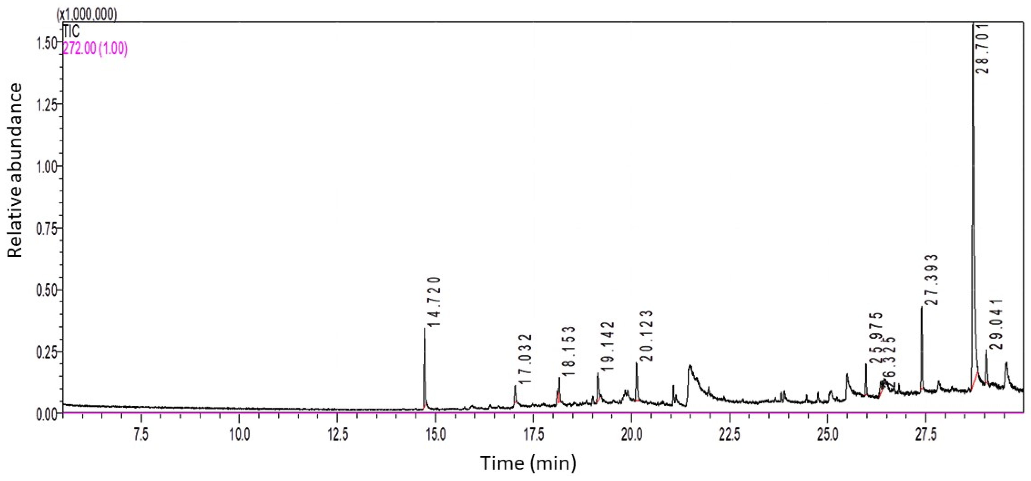
Figure 1. GC-MS spectral analysis of LSO.
Ijms 24 15478 g001
Figure 2. Effect of LSO on serum testosterone levels in diabetic mice with STZ-induced testicular toxicity. Data are expressed as the mean ± SE (n = 6 per group). Bars with different letters are significantly different from each other (p < 0.05).
Ijms 24 15478 g002
Figure 3. Effect of LSO on the testicular biomarkers of oxidative stress: (A) SOD; (B) CAT; and (C) MDA in diabetic mice with STZ-induced testicular toxicity. Data expressed as the mean ± SE (n = 6 per group). Bars with different letters are significantly different from each other (p < 0.05).
Ijms 24 15478 g003
Figure 4. Effect of LSO on the testicular protein levels of NF-kB and the pro-inflammatory cytokines in the diabetic mice with STZ-induced testicular toxicity. (A) NF-kB; (B) IL-1; (C) IL-6; and (D) TNF-α. The results are expressed as the mean ± SE (n = 6 per group). Bars with different letters are significantly different from each other (p < 0.05).
Ijms 24 15478 g004
Figure 5. (A) Effect of LSO on testicular NF-kB expression in diabetic mice with STZ-induced testicular toxicity assessed by Western blot analysis. (B) The quantification histogram of NF-kB p65 protein expression normalized by β-actin. The data are expressed as the mean ± SE (n = 6 per group). Bars with different letters are significantly different from each other (p < 0.05).
Ijms 24 15478 g005
Figure 6. Light micrographs of testicular tissues of the experimental groups. Control group (A) exhibiting a regular seminiferous tubule with complete spermatogenic series, the LSO group (B) showing a normal spermatogenic pattern, the diabetic group (C) demonstrating marked tubular atrophy and interstitial edema, the D+LSO group (D) indicating noticeable preservation in seminiferous tubule histoarchitecture, and (E) the JTBS score. Bars with different letters are significantly different (p < 0.05) between groups. Hematoxylin and eosin (H, E) staining, original magnification 400×. Spermatozoa (SP), seminiferous tubules (arrows), interstitial tissue (IT), atrophic seminiferous tubules (asterisks), desquamation (D), edema (E), and congestion (C).
Ijms 24 15478 g006
Figure 7. Proposed mechanisms underlying the therapeutic effects of LSO against testicular damage and dysfunction in diabetic mice.
Ijms 24 15478 g007
Table 1. Chemical components identified in LSO using GC-MS spectral analysis.
Peak No.Retention Time (RT)Area%Compound Name
114.728.04Butylated Hydroxytoluene
217.0321.82Tridecane, 1-iodo-
318.1532.722-methyloctacosane
419.1423.91δ-Tocopherol
520.1234.8110-Methylnonadecane
625.9752.672-methyltetracosane
726.3251.73Z-6-Pentadecen-1-ol acetate
827.3938.122-methylhexacosane
928.70162.1γ-Tocopherol
1029.0414.08Tetracosane, 1-bromo-
Disclaimer/Publisher’s Note: The statements, opinions and data contained in all publications are solely those of the individual author(s) and contributor(s) and not of MDPI and/or the editor(s). MDPI and/or the editor(s) disclaim responsibility for any injury to people or property resulting from any ideas, methods, instructions or products referred to in the content.