Article

The Root-Colonizing Endophyte Piriformospora indica Supports Nitrogen-Starved Arabidopsis thaliana Seedlings with Nitrogen Metabolites

1
Department of Plant Physiology, Matthias-Schleiden-Institute, Friedrich-Schiller-University Jena, 07743 Jena, Germany
2
Bioinformatics Core Facility, Friedrich-Schiller-University Jena, 07743 Jena, Germany
3
Université Paris-Saclay, INRAE, AgroParisTech, Institut Jean-Pierre Bourgin (IJPB), 78000 Versailles, France
4
Institute of Botany, Technische Universität Dresden, 01217 Dresden, Germany
5
Graduate School of Bioagricultural Sciences, Nagoya University, Nagoya 464-8601, Japan
6
Centro de Biotechnología y Genómica de Plantas, Instituto Nacional de Investigación y Tecnología Agraria y Alimentación (INIA), Universidad Politécnica de Madrid (UPM), Campus de Montegancedo, 28223 Madrid, Spain
7
Departamento de Biotecnología-Biología Vegetal, Escuela Técnica Superior de Ingeniería Agronómica, Alimentaria y de Biosistemas, Universidad Politécnica de Madrid (UPM), 28040 Madrid, Spain
*
Author to whom correspondence should be addressed.
Int. J. Mol. Sci. 2023, 24(20), 15372; https://doi.org/10.3390/ijms242015372
Submission received: 9 September 2023 / Revised: 12 October 2023 / Accepted: 13 October 2023 / Published: 19 October 2023
(This article belongs to the Special Issue Abiotic Stresses in Plants: From Molecules to Environment)

Abstract

:
The root-colonizing endophytic fungus Piriformospora indica promotes the root and shoot growth of its host plants. We show that the growth promotion of Arabidopsis thaliana leaves is abolished when the seedlings are grown on media with nitrogen (N) limitation. The fungus neither stimulated the total N content nor did it promote 15NO3 uptake from agar plates to the leaves of the host under N-sufficient or N-limiting conditions. However, when the roots were co-cultivated with 15N-labelled P. indica, more labels were detected in the leaves of N-starved host plants but not in plants supplied with sufficient N. Amino acid and primary metabolite profiles, as well as the expression analyses of N metabolite transporter genes suggest that the fungus alleviates the adaptation of its host from the N limitation condition. P. indica alters the expression of transporter genes, which participate in the relocation of NO3, NH4+ and N metabolites from the roots to the leaves under N limitation. We propose that P. indica participates in the plant’s metabolomic adaptation against N limitation by delivering reduced N metabolites to the host, thus alleviating metabolic N starvation responses and reprogramming the expression of N metabolism-related genes.

1. Introduction

Nitrogen is a key mineral nutrient playing a crucial role in plant growth and development [1,2,3,4,5]. The soil microbiome contributes to nitrogen acquisition, and among the best studied endosymbiotic interactions are those with N-fixing rhizobia and arbuscular mycorrhizal (AM) fungi. Legumes gain access to N through symbiotic association with rhizobia, which convert N2 gas into ammonia in nodules. Although several efforts have been made to incorporate biological N fixation capacity into non-legume plants [6], agricultural crop production without N fertilization is currently not conceivable. AM fungi help plants in nutrient acquisition and much progress has been made in understanding the molecular basis of P and N transfer from the fungal partner to the host plant (cf. [7]). Less is known about endophytes, although they show relatively little host specificity and have therefore a great potential for agricultural applications [8,9].
A well-studied endophytic fungus is Piriformospora (Serendipita) indica, which interacts with numerous host plants and promotes their growth and resistance against biotic and abiotic stresses [10,11]. The stimulation of the growth of its hosts suggests that the fungus promotes nutrient acquisition, including nitrogen. An effect of P. indica on nitrate uptake and the nitrogen metabolism in the hosts has been reported repeatedly. On a full medium, the fungus promotes nitrogen accumulation and the expression of nitrate reductase in Arabidopsis thaliana [12]. In sunflower, P. indica increases the absorption of nitrogen by the root [13]. Strehmel et al. [14] showed that the concentration of nitrogen-rich amino acids decreased in inoculated A. thaliana plants. Ghaffari et al. [15] proposed that the nitrogen metabolism plays an important role in systemic salt-tolerance in leaves of P. indica-colonized barley. Furthermore, Lahrmann et al. [16] showed that the P. indica ammonium transporter Amt1 functions as a nitrogen sensor mediating the signal that triggers the in planta activation of the saprotrophic program. In Chinese cabbage, the amino acid γ-amino butyrate in particular is de novo synthesized in colonized roots [17]. Bandyopadhyay et al. [18] demonstrated that P. indica, together with Azotobacter chroococcum, facilitates the higher acquisition of N and P in rice. P. indica also improves chickpea productivity and N metabolism in a tripartite combination with Mesorhizobium [19]. Finally, Serendipita williamsii does not affect P status but C and N dynamics in AM tomato plants [20]. These examples highlight the importance of the N metabolism on numerous beneficial effects of P. indica for different plant species; however, how the fungus influences the host N metabolism is not clear. In this study, we use the model plant A. thaliana to investigate how P. indica interferes with N uptake and metabolism under N-limiting conditions.

2. Results

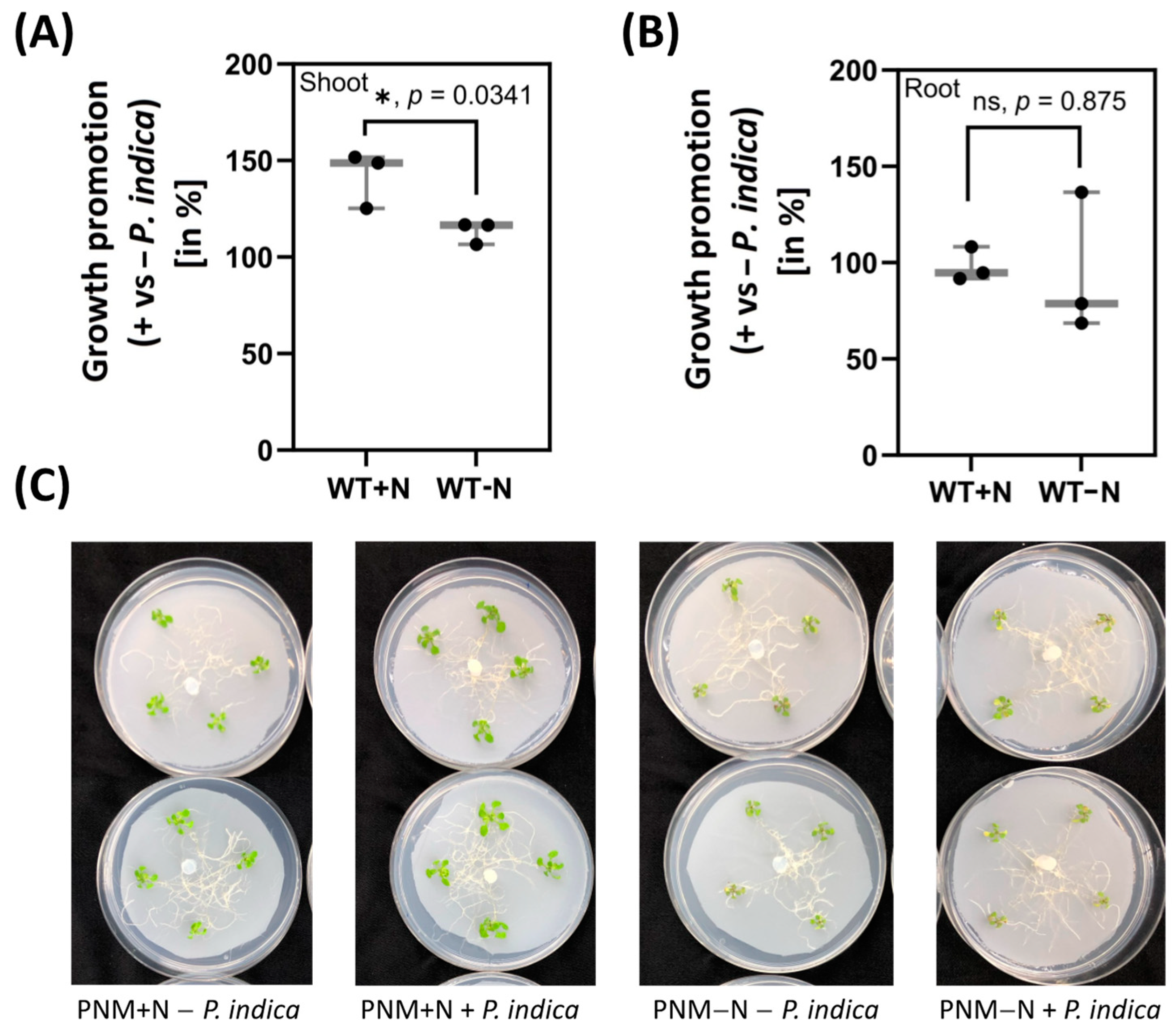
2.1. Shoot Growth Promotion by P. indica Requires External N Supply

P. indica colonizes A. thaliana roots and induces the visible growth promotion of A. thaliana seedlings after 4–7 days in full medium [21]. After 5 days, the fresh weight of the shoots was significantly increased (+41.9%) by the fungus, while barely any growth promotion was detectable on N-limited medium (Figure 1A). Root growth was neither affected by the fungus nor by N availability (Figure 1B). We conclude that under these experimental conditions, the shoot growth but not the root growth of A. thaliana seedlings is promoted by P. indica, and this requires N in the medium.

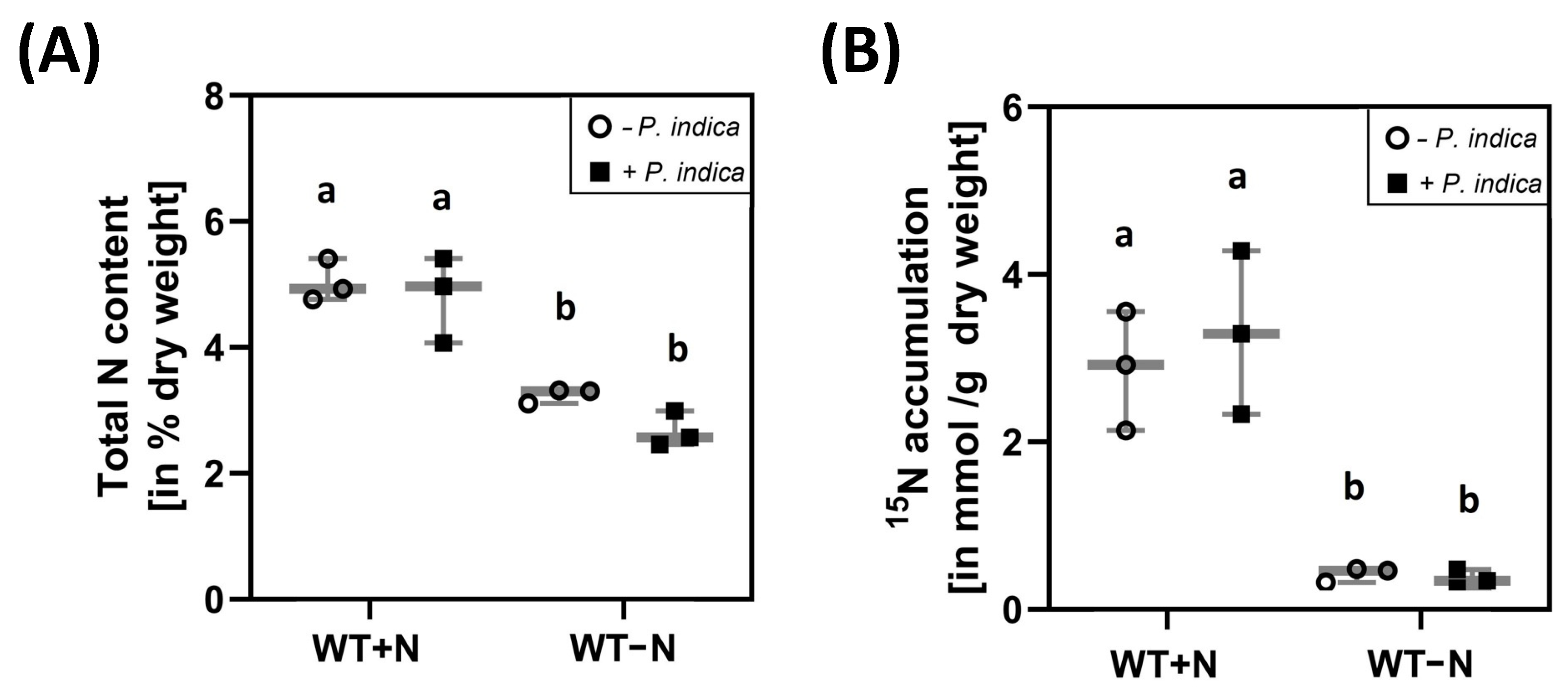
2.2. P. indica Colonisation Did Not Change the Total N Content in the Shoots and Transfer of 15N from the Medium to the Shoots

To test whether P. indica interferes with N accumulation or uptake into the plant under N-limiting conditions, the total N content in the shoots and the amount of 15N from 15NO3-labelled growth medium in the shoots were compared for uncolonized and colonized seedlings, grown on either full or N-limiting media. As expected, the total N content in the shoots of seedlings that were exposed to N limitation was lower than in the shoots of seedlings grown on full medium (Figure 2A). Furthermore, the accumulation of 15N in the shoots was much higher on full medium than on medium with low N (Figure 2B). However, we did not observe significant differences for uncolonized and colonized seedlings. This suggests that the fungus does not stimulate nitrate uptake from the medium under N-sufficient and N-limiting growth conditions.
N limitation might influence the colonisation of the roots. We observed that roots on N-limiting conditions were around two times more colonized than roots on full medium (Supplementary Figure S1), although the difference was not significant. This indicates that in spite of a higher colonisation rate, the transport of 15N label from the 15NO3-containing medium to the leaves was not stimulated by the fungus under N-limiting conditions.

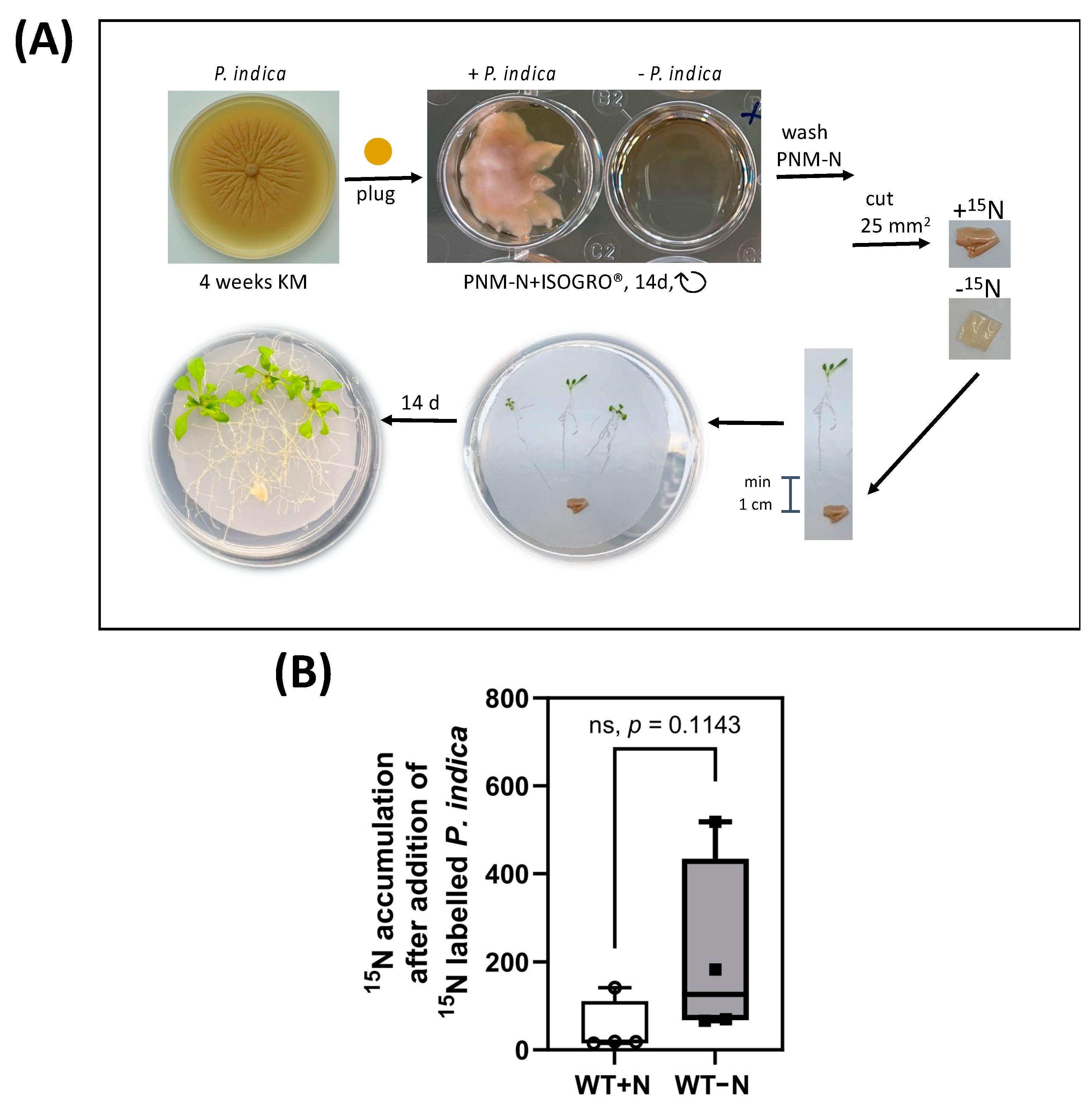
2.3. 15N Label Is Transferred from P. idica to the Host under N-Limiting Conditions

Since P. indica did not promote NO3 uptake, we tested whether labelled 15N metabolites are translocated from the fungus to the plant. As shown in Figure 3A, P. indica was cultured on 15N-containing medium for 14 days before co-culturing with A. thaliana seedlings on full or N-limited media.
The 15N-labelled fungal mycelium was positioned about 1 cm away from the roots. Establishing contact between the two partners and the initiation of root colonisation started approximately 24 h later, after the growing hyphae have reached the roots (Figure 3A). Since the label could be detected in the aerial parts of all analysed seedlings, which were in contact with P. indica, the fungus transfers N-containing metabolites to the roots of its host, and the label is further translocated to the aerial parts of the seedlings (Figure 3B). Interestingly, more 15N accumulated in the aerial parts of the plants under N-limiting conditions although the data were not significantly different (Figure 3B). This suggests that the fungus helps the host with reduced N metabolites to compensate N limitation during growth on NO3-limiting medium.

2.4. Reprogramming of the Metabolite Profiles to N Limitation Conditions Is Alleviated by P. indica

Next, we tested whether the fungus affects a host’s N metabolism under sufficient N and N-limitation conditions. We measured the levels of primary metabolites using GC-MS in the rosettes after 2 days of transfer to N-limitation conditions compared to N-sufficient conditions in the absence and presence of P. indica (Supplementary Table S1). We then calculated the metabolite ratios for plants grown under limiting versus sufficient N and compared these ratios for plants grown in the absence and the presence of the fungus. Although the amino acid profiles were comparable in colonized and uncolonized shoots, we observed slight differences for several amino acids (Table 1A). The content of aspartate and alanine decreased under N-limiting conditions in both the absence and the presence of the fungus, and these decreases were less pronounced in colonized shoots (Supplementary Figure S2). Similar tendencies were observed for amino acid content that increased under N-limitation conditions; these increases were less distinct in the presence of the fungus in the case of isoleucine, lysine, tryptophan, phenylalanine, leucine, and arginine (Supplementary Figure S2). The alterations in serin contents that were triggered by N limitation varied strongly in colonized plants in comparison to uncolonized plants. We then analysed the effect of N-limitation on soluble sugars (Table 1B). In the presence of P. indica, N-limitation triggered stronger increases in monosaccharides—in particular in glucose and fructose. The stress-related sugars trehalose and raffinose showed strong variations between the three independent replicates. However, raffinose tended to accumulate at higher levels under N limitation when the roots were colonized (see the Section 3). Overall, the slight alteration of the metabolite profiles in response to N-limitation by the colonisation with P. indica suggest a lessening of the effects of N limitation on several steps of the central metabolism.

2.5. P. indica Stimulates Expression of Specific Host´s Transporter Genes under N Limitation

The incorporation of 15N from the agar plate into the aerial parts of colonized seedlings is lower under N starvation when compared to seedlings grown on full medium (Figure 2), while a stimulation is observed for the translocation of labelled 15N from P. indica to the leaves under N limitation (Figure 3). To test whether genes for N metabolite transporters are regulated by P. indica, we performed expression profiles with RNA from the roots and shoots of seedlings, which were either grown on full or N-limited medium in the presence or absence of P. indica (Table 2).
Of the 56 investigated genes, which code for NO3, NH4+, amino acid or peptide transporters, 33 genes were differentially expressed in either the roots and shoots or both of colonized and uncolonized seedlings grown on full or N-limited media (Table 2). In the shoots, this included genes for two NH4 transporters (AMT1-3 and AMT1-5), three NO3 transporters (NRT2.2, NRT2.4 and NRT2.5), five members of the NITRATE TRANSPORTER 1/PEPTIDE TRANSPORTERs gene family (NPF2.6, NPF2.13, NPF5.3, NPF5.12 and NPF5.14) as well as the urea transporter DUR3. Furthermore, 21 amino acid transporters, including members of the LHT and AAP families, as well as 12 UmamiT putative amino acid transporters responded to the fungus. In contrast, seven transporter genes were downregulated by P. indica in the shoots of N-starved seedlings (Table 2). In the roots, six of these genes showed the same regulation (Table 2). This clearly demonstrates that the expression of genes for NO3, NH4+, amino acid and peptide transporters are major targets of the fungus under N-limited conditions (see the Section 3).

3. Discussion

N limitation has severe consequences for plant performance [22], and endophytes may help plants to better adapt to the shortage. We used the well-investigated symbiotic interaction between the model plant A. thaliana and P. indica to address this question. We demonstrate that under severe N limitation, the fungus does not stimulate the uptake of nitrate into the host plant but rather the N-label from fungal metabolites appears in the leaves of the host. Since our N-limiting medium contains barely any nitrate, the absence of a detectable stimulatory effect of the fungus on nitrate uptake into the host is not surprising. The N metabolites that are translocated from the hyphae to the plants under N limitation conditions did not results in fungus-induced growth promotion (Figure 1), suggesting that the N supply to the host by the fungus might only compensate deficits. Furthermore, N-translocation from the fungus to the host occurs only under N limitation conditions, suggesting the involvement of an N-sensing system (cf. [16]). The successful transfer of 15N by arbuscular mycorrhizal fungi to host plants has been shown previously [23,24,25]. More recently, Hoysted et al. [26] investigated clover (Trifolium repens) colonized by Mucoromycotina fungi and showed that the host gained both 15N and 33P tracers directly from the fungus in exchange for plant-fixed C. Whether the N supply to the host in our study system has comparable symbiotic features with profit for both partners or whether it is just the stress-related withdrawal of N from the fungus by the plant without any profit for the microbe remains to be investigated. However, since the fungus can grow and propagate on the host under our –N conditions, the N translocation to the host does not restrict hyphal growth. It appears that the conditions are not strong enough to induce changes in the symbiotic interaction [16]. It is also not clear which metabolites are transported from the microbe to the plant or how this occurs. In Medicago truncatula, three AMT2 family ammonium transporters (AMT2;3, AMT2;4, and AMT2;5) are involved in the uptake of N in form of ammonium from the periarbuscular space between the fungal plasma membrane and the plant-derived periarbuscular membrane [27]. In exchange, host plants transfer reduced carbon to the fungi [28,29,30]. Additionally, Cope et al. [31] showed that the colonization of M. truncatula with R. irregularis led to the elevated expression of the mycorrhiza-induced AMT2;3 and the nitrate transporter NPF4.12 as well as the putative ammonium transporter NIP1;5 in the roots. A dipeptide transporter from the arbuscular mycorrhizal fungus Rhizophagus irregularis is upregulated in the intraradical phase [32]. To investigate how the fungus manipulates the host N metabolism, we performed a comprehensive metabolome and transcriptome analysis for N-related metabolites and genes (Table 1 and Table 2).
No major impact of the colonization by P. indica on the changes of shoot metabolite levels in response to N limitation has been observed in this study. Liu et al. [33] demonstrated that raffinose positively regulates maize drought tolerance by reducing leaf transpiration. The raffinose family oligosaccharides are associated with various abiotic and biotic stress responses in different plant species (e.g., [34,35,36,37,38]). It is conceivable that the stimulatory effect of P. indica on the raffinose level in N-limited leaves reduces stress.
Nitrate transporter genes are often upregulated under N starvation; however, the role of endophytic microorganisms in nitrate acquisition is not fully understood. In rice, the arbuscular mycorrhizal fungus R. irregularis remarkably promoted growth and N acquisition, and about 42% of the overall N could be delivered via the symbiotic route under nitrate-limiting conditions [39]. Nitrate uptake occurs via NITRATE TRANSPORTER1/PEPTIDE TRANSPORTER FAMILY (NPF)4.5, a member of the low affinity nitrate transporter family, which is exclusively expressed in the arbuscles of the Gramineous species [39]. A comparable mechanism does not exist in our endophyte/A. thaliana model, and the putative A. thaliana NPF4.5 homolog is not upregulated in colonized roots under N-limited conditions. However, we observed a highly specific response of several NPF/NRT1 and NRT2 family members to P. indica colonisation, which suggest conclusions regarding how the fungus interferes with the plant N metabolism. The nitrate transporters NRT2.2 and NRT2.4 [40] are only upregulated in the rosettes when the roots are colonized by P. indica, while their expression in the roots does not respond to the fungus. This suggests that the fungus promotes nitrate scavenging, which is released from the vacuole in response to N starvation. In fact, NRT2.4 has been shown to be expressed close to the phloem in rosettes and to contribute to nitrate homeostasis in the phloem under limiting nitrate supply, since in nitrate-starved nrt2.4 mutants, nitrate content in shoot phloem exudates was decreased [40]. Likewise, NRT1.7 (NPF2.13) and NRT1.8 (NPF5.3) are upregulated by P. indica in leaves, but not in roots. NRT1.7 loads excess nitrate stored in source leaves into phloem and facilitates nitrate allocation to sink leaves. Under N starvation, the nrt1.7 mutant exhibits growth retardation, indicating that the NRT1.7-mediated source-to-sink remobilization of stored nitrate is important for sustaining growth in plants [41]. NRT1.8 is expressed predominantly in xylem parenchyma cells within the vasculature and functional disruption of NRT1.8 significantly increased the nitrate concentration in xylem sap [42]. In contrast, NRT2.3 and -2.6 are downregulated under N-limiting conditions and this is further promoted by the fungus. NRT2.6 has been linked to biotic and abiotic stress responses [43], and it appears that the downregulation of NRT2.6 expression by P. indica alleviates the stress responses in the roots. Finally, NRT1.9 (NPF2.9) is strongly downregulated by P. indica in the leaves. NRT1.9 is expressed in the companion cells of phloem. In NRT1.9 mutants, downward nitrate transport was reduced, suggesting that NRT1.9 facilitates the loading of nitrate into the phloem and enhances downward nitrate transport to the roots [44], apparently a process that is restricted by the fungus. Taken together, the analysis of the regulation of the A. thaliana nitrate transporter genes by P. indica suggests that the root-colonizing fungus supports nitrate transport to and availability in the aerial parts of the host under our nitrate-limiting conditions. This is further supported by the upregulation of NRT1.15 (NPF5.14) by P. indica in the leaves. NRT1.15 is a tonoplast-localized low-affinity nitrate transport [45] and the overexpression of the gene significantly decreased vacuolar nitrate contents and nitrate accumulation in A. thaliana shoots. NRT1.15 regulates vacuolar nitrate efflux, and the reallocation might also contribute to osmotic stress responses other than mineral nutrition [45].
Since the medium does not contain NH4+, the plant can only receive NH4+ from the fungus via ammonium transporters (AMTs) [41,46,47,48]. AMT1-4 expression is upregulated by P. indica in roots under N-limited conditions. Since AMT1-4 is root-specific [47], this suggests that the plant tries to compensate its N limitation by stimulating NH4+ uptake. NH4+ might also originate from the fungus, and it is conceivable that withdrawal of this ion from the fungus might ultimately result in a change of the symbiotic interaction towards saprophytism (cf. [16]). Furthermore, the expression of AMT1-3 and AMT1-5 as well as DUR3-coding for a urea transporter [49,50] is stimulated by P. indica in the shoots. Root colonization might create a metabolite environment in the host that requires these transporters for the proper distribution of the N metabolites in the aerial parts.
Seven amino acid transporters are regulated >log2-fold by P. indica colonisation in nitrate-deprived A. thaliana seedlings. In roots, the fungus prevents the downregulation of the gene for glutamine secreting GLUTAMINE DUMPER (GDU)1 [51], suggesting that the microbe wants to access to the plant glutamine. Furthermore, the broad-specificity high affinity amino acid transporter LYSINE HISTIDINE TRANSPORTER (LHT)1 [52], AMINO ACID PERMEASE (AAP)4, γ-AMINOBUTYRIC ACID TRANSPORTER (GAT)1, and CATIONIC AMINO ACID TRANSPORTER (CAT)5 are upregulated in the leaves of P. indica-colonized seedlings. These transporters have been proposed to be involved in nitrogen recycling in plants [53]. Apparently, a better or different N metabolism management is required for the plant when the roots are associated with the endophyte. LHT1 and -2 are also involved in the transport of 1-aminocyclopropane carboxylic acid, a biosynthetic precursor of ethylene [54], which might indicate an increased stress caused by the interaction with the fungus under N-limiting conditions. An involvement in nitrogen recycling has also been proposed for 5 of the 10 USUALLY MULTIPLE ACIDS MOVE IN AND OUT TRANSPORTERS (UMANIT13, −20, −40, −45 and −47) [53], which are regulated >log2-fold in either the roots or shoots of P. indica-colonized seedlings under N-limitations.

4. Materials and Methods

4.1. Plant and Fungus Material and Corresponding Growth Conditions

A. thaliana seeds (Col-0) were surface-sterilized and sown on N-free MGRL medium supplemented with 2.5 mM NH4NO3 and 3 g/L gelrite [55]. The KNO3 and Ca(NO3)2 in the MGRL medium were replaced by KCl and CaCl2 to ensure ion equilibrium. After 48 h of stratification at 4 °C in the dark, the seeds were transferred to long-day conditions with 22 °C, 16 h light/8 h dark, 80 μmol m−2 s−1 for 10 days.
Piriformospora indica was cultured on Kaefer’s medium as described previously [56,57]. As described previously, plugs of a 4-week-old fungal culture were used for co-cultures with the seedlings. The fungus was pre-grown for 7 days on PNM medium (PNM+N) with a nylon membrane in the dark at 22 °C. For N-limiting conditions (0 mM total N, PNM−N), KNO3 and Ca(NO3)2 were replaced by KCl and CaCl2. For control plates without fungus, only empty KM plugs were placed on top of the membrane.

4.2. Plant-Fungus Co-Cultures and the Determination of Growth Promotion

For plant-fungus co-cultures for 5 days, 4 plants (per Petri dish) were placed on top of the pre-grown fungal lawn, as described previously [57], with some adaptations. Plates were sealed with 3MTM Micropore tape to reduce the condensation and 10-day-old plants were used for co-cultivation to reduce the amount of N, which accumulated in the plants on MGRL medium before the co-culture. In pilot experiments, we showed that the reduced age did not affect the establishment of the symbiosis with the fungus. The co-cultures were incubated at 22 °C, 16 h light/8 h dark and 80 μmol m−2 s−1 with light from the top.
After 5 days, the roots and shoots of the plants were harvested separately. For that, 5 plates (=20 plants) were harvested as 1 sample. Both roots and shoots were washed in sterile distilled water and carefully dried before weighing and direct freezing in liquid nitrogen. Samples were stored in −80 °C until further use. These experiments were repeated 3–4 times independently.
To determine growth promotion by the fungus, the weight of the sample with fungus was normalized (divided) to the weight without fungus. This was performed for the total weights sampled from the full medium (PNM+N) as well as from the N-limited medium (PNM−N). Final growth promotion values presented in the figures are averages of 3 replicates from independent cultures.

4.3. 15N Labelling Experiments in the Medium

To analyse the uptake of nitrogen by the plant, 2.5% of the total KNO3 (which equals 0.125 mM KNO3) of the PNM medium was replaced by K15NO3 (Eurisotop, Saint-Aubin, France) dissolved in distilled water. For proper comparison, the 2.5% of K15NO3 was also added to the N-free medium (PNM−N) resulting in a final concentration of 0.125 mM nitrate. Finally, PNM−N control plates without 15N were used and contained 0.125 mM unlabelled KNO3. Plants grown on these plates were compared to those grown on PNM+N plates to analyse the natural abundance of 15N in the plant tissue. As described before, the fungus or control plug was pre-incubated on the PNM with nylon membrane for 1 week before plants were placed on the plates. The co-cultures were incubated for 5 days to ensure that enough 15N was taken up by the plant.

4.4. 15N Fungus-Labelling Experiments

To analyse whether the fungus can directly transfer N or N-containing metabolites to the plant, it was labelled with 15N before the co-culture. A modified KM medium without the N-containing components (20 g/L dextrose, 50 mL/L macronutrients, 10 mL/L micronutrients and 1 mL/L Fe-EDTA, 1 mL/L vitamin mix, pH 6.5) was prepared and supplemented with 10 g/L ISOGRO®-15N (CortecNet, Les Ulis, France) according to the manufacturers protocol. P. indica plugs of 2 mm diameter were incubated (23 °C, 50 rpm, dark) in 2 mL of KMISOGRO in Greiner CELLSTAR® 12-well plates (Greiner Bio-One, Frickenhausen, Germany) sealed with 3MTM Micropore tape. After 14 days of growth, the fungal tissue was separated from the remaining medium and carefully washed 3 times with N-free liquid PNM to remove the 15N bound to the hyphal surface. A 76.66% enrichment in 15N was achieved using this protocol. The fungus was carefully cut in 5 × 5 mm pieces and placed on PNM−N and PNM+N plates to start the co-cultures. To minimize 15N uptake by the plant from dead fungal material during the washing and handling procedure, the fungal plugs were placed in minimum 1 cm distance from the roots. Under these conditions, contact between the two symbionts requires the active growth of the hyphae towards the roots. Co-cultivation was performed with 3 plants per plate for 14 days to ensure that enough 15N was taken up by the plant.

4.5. Isolation and Clean-Up of RNA

RNA was isolated from the roots or shoots of 10-day-old seedlings, which were either co-cultured with the fungus for additional 5 days (root colonization results), or for an additional 4 days for expression profiling. Samples of root or shoot material were stored in −80 °C. For homogenization, the samples were ground with mortar and pistil in liquid nitrogen. A maximum of 100 mg material was used for RNA extraction. RNA was extracted with TrizolTM (ThermoFisher Scientific, Waltham, MA, USA) and chloroform according to the manufacturers protocol. Briefly, the plant material was mixed with 1 mL of TrizolTM and incubated on a shaker at room temperature for 15 min. After the addition of 250 μL chloroform and a second incubation phase, the sample was centrifuged (30 min, 4 °C). The supernatant was mixed with isopropanol and incubated on ice, followed by centrifugation. The pellet was washed twice with 80% ethanol, dried and resuspended in RNAse-free water. The RNA isolation was followed by an additional cleaning step to remove access salts originating from the fungus tissue. For this, the sample was mixed with 3 M sodium acetate (1/10 (v/v) in RNAse-free water, pH = 5.2) and 600 µL of ice-cold 100% ethanol and incubated at −20 °C for at least 1 hr. After centrifugation and 2 cleaning steps with 80% ethanol, the sample was resuspended in RNAse-free water. The quality and concentration of the extracted RNA was tested via absorbance analysis using a NanoVue (GE Healthcare, Uppsala, Sweden).

4.6. RNAseq and Data Analysis

After the transfer of samples to Novogene Genomics Service (Cambridge, UK), the RNA sample integrity was checked with a Bioanalyzer 2100 (Agilent). After samples passed the quality check, the service laboratory proceeded with the library construction and RNA sequencing (PE150) on Illumina NovaSeq™ 6000 platforms, as described in a previous study [58].
The RNAseq libraries were filtered and quality-trimmed with fastp (v0.23.2) [59], i.e., read ends were truncated to achieve a Phred quality score of 30 or more. Reads below 15 nt length or those comprising at least 2 ambiguous N bases were removed from the dataset. Read qualities were monitored by FastQC (v0.11.3; https://www.bioinformatics.babraham.ac.uk/projects/fastqc/, accessed on 15 December 2022). Hisat2 (v2.2.1) [60] was used with default parameters to map the quality-trimmed RNAseq libraries to the A. thaliana reference genome (TAIR10, Ensembl release 51). The mapping allowed spliced reads and single-read mapping to multiple best-fitting locations. FeatureCounts (v1.5.3) [61] was applied to perform read-counting based on the A. thaliana reference annotation (TAIR10, Ensembl release 51). The Bioconductor DESeq2 (v1.10.0) package [62] was utilized to identify DEGs in the different pairwise mutant and wild type comparisons. Benjamini and Hochberg’s false discovery rate (FDR) approach [63] was employed to adjust the calculated p-values for multiple testing.
To identify DEGs of transporters predicted to transport major N compounds, the obtained results were initially filtered according to their p-value (p < 0.05). Next, DEGs were sorted according to their log2-fold change—here only changes with numbers ≥ +1.5 and ≤ −1.5 were further analysed. This list was cross-checked with targets identified from a search in the UniProt database (https://www.uniprot.org, accessed on 15 December 2022) using keywords like “NH4 transport”.

4.7. Analysis of Fungal Colonization via qPCR

A total of 1 mg of RNA was used for the synthesis of cDNA. The Omniscript RT Kit (Qiagen, Hilden, Germany) was used according to the manufacturers protocol with the oligo(dT)18 primer (ThermoFisher Scientific, Waltham, USA). qPCR was performed with fifty nanograms of the synthesized cDNA as template in a Bio-Rad CFX96 Real-Time PCR Detection System (Feldkirchen, Germany) via DreamTaq Polymerase (ThermoFisher Scientific, Waltham, USA) and Evagreen (Biotium, Fremont, CA, USA). The data were normalized with respect to the A. thaliana RPS18B (At1g34030) gene using the 2−∆∆CT method [64]. To quantify the P. indica colonization level of A. thaliana roots, the expression of PiTEF1 [65] was analysed in comparison to the plant´s housekeeping gene RPS18B (At1g 34030). The following primers were used: PiTEF1: CGCAGAATACAAGGAGGCC and CGTATCGTAGCTCGCCTGC; RPS18B: GTCTCCAATGCCCTTGACAT and TCTTTCCTCTGCGACCAGTT [66]. The colonization was compared between plants grown on PNM−N and PNM+N media (set as 1.0) using the 2−∆∆CT method.

4.8. Determination of Total Nitrogen and 15N Enrichment

Total N and 15N contents were quantified on 1–2 mg aliquots of dry tissue, after drying a ground tissue aliquot at 65–70 °C for at least 48 h. N elements were detected using gas chromatography on a FLASH 2000 Organic Elemental Analyzer (Thermo Fisher Scientific, Villebon, France). The 15N/14N isotopic ratio was subsequently quantified using a coupled mass spectroscope (Delta V advantage IRMS; Thermo Fisher Scientific, Villebon, France). The total N content was only determined in plant shoots because the discrimination of N from plant or fungus was not possible in colonized root material.

4.9. Metabolomic Analysis

For GC-MS-based quantifications, 25 mg of finely ground plant material was resuspended in 1 mL of frozen (−20 °C) water:acetonitrile:isopropanol (2:3:3, v/v/v) containing Ribitol at 4 ug/mL and analysed as described in [67].

5. Conclusions

We observed an unexpected complexity in the plant N metabolism when N-deprived A. thaliana seedlings were colonized by P. indica. Our data suggest that the fungus neither stimulated the total N content nor promoted 15NO3 uptake from agar plates to the host. Rather, reduced N metabolites were transported from the fungus to the plant. Furthermore, gene expression and metabolite profiles suggest that N-containing metabolites were redistributed by P. indica in A. thaliana seedlings exposed to N-limitation.
Our initial observations highlight a few aspects that need to be investigated in greater detail. (1) Which N metabolites are transported from the fungus to a plant suffering under N limitation? (2) The plant appears to adapt its N metabolism under N limitation by transporting N metabolites shootwards, a process that is supported by the fungus. Is the fungal support for the plant specific for the symbiotic phase of the interaction? (3) Are our observations P. indica-specific or do they occur also in other endophyte/plant interactions?

Supplementary Materials

The following supporting information can be downloaded at: https://www.mdpi.com/article/10.3390/ijms242015372/s1.

Author Contributions

S.S.S., E.B., G.C., A.M. and A.K. performed the experiments and analysed the data, J.L.-M., H.S., T.K., J.V.-C., S.P., A.K. and R.O. developed the concept and wrote the manuscript. All authors have read and agreed to the published version of the manuscript.

Funding

This project was financially supported by the collaborative ICPS research project executed in the framework of the EIG CONCERT-Japan joint call on Food Crops and Biomass Production Technologies and the related national funding agencies: grants 01DR17007A and 01DR17007B from the Federal Ministry of Education and Research (BMBF), Germany, to R.O.; grant EIG_JC1JAPAN-045 from the Centre National de la Recherche Scientifique (CNRS), France, to A.K.; grant PCIN-2016–037 from the Ministry of Economy and Competitiveness (MINECO), Spain, to J.V.-C. and S.P.; and grant JPMJSC16C3 from the Japan Science and Technology Agency (JST) to H.S. This work was further supported by the Deutsche Forschungsgemeinschaft (DFG, German Research Foundation) CRC1127 ChemBioSys (project ID: 239748522) for R.O. This work benefited from the support of IJPB’s Plant Observatory technological platforms. The IJPB benefits from additional support of Saclay Plant Sciences-SPS (ANR-17-EUR-0007).

Institutional Review Board Statement

Not applicable.

Informed Consent Statement

Not applicable.

Data Availability Statement

The dataset is available in the NCBI GEO repository, under accession number GSE239281.

Acknowledgments

We thank Sarah Mußbach, Claudia Röppischer and Christin Weilandt for the excellent technical support in the lab.

Conflicts of Interest

The authors declare no conflict of interest.

References

  1. Engels, C.; Marschner, H. Plant uptake and utilization of nitrogen. In Nitrogen Fertilization in the Environment; Bacan, P.E., Ed.; Marcel Dekker, Inc.: New York, NY, USA, 1995; pp. 41–81. [Google Scholar]
  2. Vidal, E.A.; Alvarez, J.M.; Araus, V.; Riveras, E.; Brooks, M.D.; Krouk, G.; Ruffel, S.; Lejay, L.; Crawford, N.M.; Coruzzi, G.M.; et al. Nitrate in 2020: Thirty Years from Transport to Signaling Networks. Plant Cell 2020, 32, 2094–2119. [Google Scholar] [CrossRef] [PubMed]
  3. Lyzenga, W.J.; Liu, Z.; Olukayode, T.; Zhao, Y.; Kochian, L.V.; Ham, B.-K. Getting to the roots of N, P, and K uptake. J. Exp. Bot. 2023, 74, 1784–1805. [Google Scholar] [CrossRef] [PubMed]
  4. Wang, Y.-Y.; Cheng, Y.-H.; Chen, K.-E.; Tsay, Y.-F. Nitrate Transport, Signaling, and Use Efficiency. Annu. Rev. Plant Biol. 2018, 69, 85–122. [Google Scholar] [CrossRef] [PubMed]
  5. Aluko, O.O.; Kant, S.; Adedire, O.M.; Li, C.; Yuan, G.; Liu, H.; Wang, Q. Unlocking the potentials of nitrate transporters at improving plant nitrogen use efficiency. Front. Plant Sci. 2023, 14, 1074839. [Google Scholar] [CrossRef] [PubMed]
  6. Guo, K.; Yang, J.; Yu, N.; Luo, L.; Wang, E. Biological nitrogen fixation in cereal crops: Progress, strategies, and perspectives. Plant Commun. 2023, 4, 100499. [Google Scholar] [CrossRef]
  7. Rui, W.; Mao, Z.; Li, Z. The Roles of Phosphorus and Nitrogen Nutrient Transporters in the Arbuscular Mycorrhizal Symbiosis. Int. J. Mol. Sci. 2022, 23, 11027. [Google Scholar] [CrossRef]
  8. Chaudhary, P.; Agri, U.; Chaudhary, A.; Kumar, A.; Kumar, G. Endophytes and their potential in biotic stress management and crop production. Front. Microbiol. 2022, 13, 933017. [Google Scholar] [CrossRef]
  9. Zhang, Y.; Feng, H.; Druzhinina, I.S.; Xie, X.; Wang, E.; Martin, F.; Yuan, Z. Phosphorus/nitrogen sensing and signaling in diverse root-fungus symbioses. Trends Microbiol. 2023. [Google Scholar] [CrossRef]
  10. Saleem, S.; Sekara, A.; Pokluda, R. Serendipita indica—A Review from Agricultural Point of View. Plants 2022, 11, 3417. [Google Scholar] [CrossRef]
  11. Del Barrio-Duque, A.; Ley, J.; Samad, A.; Antonielli, L.; Sessitsch, A.; Compant, S. Beneficial Endophytic Bacteria-Serendipita indica Interaction for Crop Enhancement and Resistance to Phytopathogens. Front. Microbiol. 2019, 10, 2888. [Google Scholar] [CrossRef]
  12. Sherameti, I.; Shahollari, B.; Venus, Y.; Altschmied, L.; Varma, A.; Oelmüller, R. The Endophytic Fungus Piriformospora indica Stimulates the Expression of Nitrate Reductase and the Starch-degrading Enzyme Glucan-water Dikinase in Tobacco and Arabidopsis Roots through a Homeodomain Transcription Factor That Binds to a Conserved Motif in Their Promoters. J. Biol. Chem. 2005, 280, 26241–26247. [Google Scholar] [CrossRef]
  13. Eliaspour, S.; Sharifi, R.S.; Shirkhani, A. Evaluation of interaction between Piriformospora indica, animal manure and NPK fertilizer on quantitative and qualitative yield and absorption of elements in sunflower. Food Sci. Nutr. 2020, 8, 2789–2797. [Google Scholar] [CrossRef] [PubMed]
  14. Strehmel, N.; Mönchgesang, S.; Herklotz, S.; Krüger, S.; Ziegler, J.; Scheel, D. Piriformospora indica Stimulates Root Metabolism of Arabidopsis thaliana. Int. J. Mol. Sci. 2016, 17, 1091. [Google Scholar] [CrossRef]
  15. Ghaffari, M.R.; Ghabooli, M.; Khatabi, B.; Hajirezaei, M.R.; Schweizer, P.; Salekdeh, G.H. Metabolic and transcriptional response of central metabolism affected by root endophytic fungus Piriformospora indica under salinity in barley. Plant Mol. Biol. 2016, 90, 699–717. [Google Scholar] [CrossRef] [PubMed]
  16. Lahrmann, U.; Ding, Y.; Banhara, A.; Rath, M.; Hajirezaei, M.R.; Döhlemann, S.; von Wirén, N.; Parniske, M.; Zuccaro, A. Host-related metabolic cues affect colonization strategies of a root endophyte. Proc. Natl. Acad. Sci. USA 2013, 110, 13965–13970. [Google Scholar] [CrossRef] [PubMed]
  17. Hua, M.D.-S.; Kumar, R.S.; Shyur, L.-F.; Cheng, Y.-B.; Tian, Z.; Oelmüller, R.; Yeh, K.-W. Metabolomic compounds identified in Piriformospora indica-colonized Chinese cabbage roots delineate symbiotic functions of the interaction. Sci. Rep. 2017, 7, 9291. [Google Scholar] [CrossRef] [PubMed]
  18. Bandyopadhyay, P.; Yadav, B.G.; Kumar, S.G.; Kumar, R.; Kogel, K.-H.; Kumar, S. Piriformospora indica and Azotobacter chroococcum Consortium Facilitates Higher Acquisition of N, P with Improved Carbon Allocation and Enhanced Plant Growth in Oryza sativa. J. Fungi 2022, 8, 453. [Google Scholar] [CrossRef]
  19. Mansotra, P.; Sharma, P.; Sharma, S. Bioaugmentation of Mesorhizobium cicer, Pseudomonas spp. and Piriformospora indica for Sustainable Chickpea Production. Physiol. Mol. Biol. Plants 2015, 21, 385–393. [Google Scholar] [CrossRef]
  20. Hallasgo, A.M.; Spangl, B.; Steinkellner, S.; Hage-Ahmed, K. The Fungal Endophyte Serendipita williamsii Does Not Affect Phosphorus Status But Carbon and Nitrogen Dynamics in Arbuscular Mycorrhizal Tomato Plants. J. Fungi 2020, 6, 233. [Google Scholar] [CrossRef]
  21. Abdelaziz, M.E.; Kim, D.; Ali, S.; Fedoroff, N.V.; Al-Babili, S. The endophytic fungus Piriformospora indica enhances Arabidopsis thaliana growth and modulates Na+/K+ homeostasis under salt stress conditions. Plant Sci. 2017, 263, 107–115. [Google Scholar] [CrossRef]
  22. Krapp, A.; Berthomé, R.; Orsel, M.; Mercey-Boutet, S.; Yu, A.; Castaings, L.; Elftieh, S.; Major, H.; Renou, J.-P.; Daniel-Vedele, F. Arabidopsis Roots and Shoots Show Distinct Temporal Adaptation Patterns toward Nitrogen Starvation. Plant Physiol. 2011, 157, 1255–1282. [Google Scholar] [CrossRef]
  23. Hodge, A.; Stewart, J.; Robinson, D.; Griffiths, B.S.; Fitter, A.H. Competition between roots and soil microorganisms for nutrientsfrom nitrogen-rich patches of varying complexity. J. Ecol. 2000, 88, 150–164. [Google Scholar] [CrossRef]
  24. Hodge, A.; Campbell, C.D.; Fitter, A.H. An arbuscular mycorrhizal fungus accelerates decomposition and acquires nitrogen directly from organic material. Nature 2001, 413, 297–299. [Google Scholar] [CrossRef]
  25. Thirkell, T.J.; Cameron, D.D.; Hodge, A. Resolving the ‘nitrogen paradox’ of arbuscular mycorrhizas: Fertilisation with organic matter brings considerable benefits for plant nutrition and growth. Plant Cell Environ. 2016, 39, 1683–1690. [Google Scholar] [CrossRef] [PubMed]
  26. Hoysted, G.A.; Field, K.J.; Sinanaj, B.; Bell, C.A.; Bidartondo, M.I.; Pressel, S. Direct nitrogen, phosphorus and carbon exchanges between Mucoromycotina ‘fine root endophyte’ fungi and a flowering plant in novel monoxenic cultures. New Phytol. 2023, 238, 70–79. [Google Scholar] [CrossRef] [PubMed]
  27. Breuillin-Sessoms, F.; Floss, D.S.; Gomez, S.K.; Pumplin, N.; Ding, Y.; Levesque-Tremblay, V.; Noar, R.D.; Daniels, D.A.; Bravo, A.; Eaglesham, J.B.; et al. Suppression of Arbuscule Degeneration in Medicago truncatula phosphate transporter4 Mutants Is Dependent on the Ammonium Transporter 2 Family Protein AMT2;3. Plant Cell 2015, 27, 1352–1366. [Google Scholar] [CrossRef]
  28. Paul, E.A.; Kucey, R.M.N. Carbon flow in plant microbial associations incriminated in perinatal morbidity and mortality. Science 1981, 213, 473–474. [Google Scholar] [CrossRef]
  29. Wright, D.P.; Read, D.J.; Scholes, J.D. Mycorrhizal sink strength influences whole plant carbon balance of Trifolium repens L. Plant Cell Environ. 1998, 21, 881–891. [Google Scholar] [CrossRef]
  30. Lendenmann, M.; Thonar, C.; Barnard, R.L.; Salmon, Y.; Werner, R.A.; Frossard, E.; Jansa, J. Symbiont identity matters: Carbon and phosphorus fluxes between Medicago truncatula and different arbuscular mycorrhizal fungi. Mycorrhiza 2011, 21, 689–702. [Google Scholar] [CrossRef]
  31. Cope, K.R.; Kafle, A.; Yakha, J.K.; Pfeffer, P.E.; Strahan, G.D.; Garcia, K.; Subramanian, S.; Bücking, H. Physiological and transcriptomic response of Medicago truncatula to colonization by high- or low-benefit arbuscular mycorrhizal fungi. Mycorrhiza 2022, 32, 281–303. [Google Scholar] [CrossRef]
  32. Belmondo, S.; Fiorilli, V.; Pérez-Tienda, J.; Ferrol, N.; Marmeisse, R.; Lanfranco, L. A dipeptide transporter from the arbuscular mycorrhizal fungus Rhizophagus irregularis is upregulated in the intraradical phase. Front. Plant Sci. 2014, 5, 436. [Google Scholar] [CrossRef] [PubMed]
  33. Liu, Y.; Li, T.; Zhang, C.; Zhang, W.; Deng, N.; Dirk, L.M.A.; Downie, A.B.; Zhao, T. Raffinose positively regulates maize drought tolerance by reducing leaf transpiration. Plant J. 2023, 114, 55–67. [Google Scholar] [CrossRef] [PubMed]
  34. ElSayed, A.I.; Rafudeen, M.S.; Golldack, D. Physiological aspects of raffinose family oligosaccharides in plants: Protection against abiotic stress. Plant Biol. 2014, 16, 1–8. [Google Scholar] [CrossRef] [PubMed]
  35. Gu, L.; Jiang, T.; Zhang, C.; Li, X.; Wang, C.; Zhang, Y.; Li, T.; Dirk, L.M.A.; Downie, A.B.; Zhao, T. Maize HSFA2 and HSBP2 antagonistically modulate raffinose biosynthesis and heat tolerance in Arabidopsis. Plant J. 2019, 100, 128–142. [Google Scholar] [CrossRef]
  36. Han, Q.; Qi, J.; Hao, G.; Zhang, C.; Wang, C.; Dirk, L.M.A.; Downie, A.B.; Zhao, T. ZmDREB1A Regulates RAFFINOSE SYNTHASE Controlling Raffinose Accumulation and Plant Chilling Stress Tolerance in Maize. Plant Cell Physiol. 2020, 61, 331–341. [Google Scholar] [CrossRef]
  37. Li, C.; Tien, H.; Wen, M.; Yen, H.E. Myo-inositol transport and metabolism participate in salt tolerance of halophyte ice plant seedlings. Physiol. Plant. 2021, 172, 1619–1629. [Google Scholar] [CrossRef]
  38. Yang, J.; Ling, C.; Liu, Y.; Zhang, H.; Hussain, Q.; Lyu, S.; Wang, S.; Liu, Y. Genome-Wide Expression Profiling Analysis of Kiwifruit GolS and RFS Genes and Identification of AcRFS4 Function in Raffinose Accumulation. Int. J. Mol. Sci. 2022, 23, 8836. [Google Scholar] [CrossRef]
  39. Wang, S.; Chen, A.; Xie, K.; Yang, X.; Luo, Z.; Chen, J.; Zeng, D.; Ren, Y.; Yang, C.; Wang, L.; et al. Functional analysis of the OsNPF4.5 nitrate transporter reveals a conserved mycorrhizal pathway of nitrogen acquisition in plants. Proc. Natl. Acad. Sci. USA 2020, 117, 16649–16659. [Google Scholar] [CrossRef]
  40. Kiba, T.; Feria-Bourrellier, A.-B.; Lafouge, F.; Lezhneva, L.; Boutet-Mercey, S.; Orsel, M.; Bréhaut, V.; Miller, A.; Daniel-Vedele, F.; Sakakibara, H.; et al. The Arabidopsis Nitrate Transporter NRT2.4 Plays a Double Role in Roots and Shoots of Nitrogen-Starved Plants. Plant Cell 2012, 24, 245–258. [Google Scholar] [CrossRef]
  41. Chen, H.-Y.; Chen, Y.-N.; Wang, H.-Y.; Liu, Z.-T.; Frommer, W.B.; Ho, C.-H. Feedback inhibition of AMT1 NH4+-transporters mediated by CIPK15 kinase. BMC Biol. 2020, 18, 196. [Google Scholar] [CrossRef]
  42. Li, J.-Y.; Fu, Y.-L.; Pike, S.M.; Bao, J.; Tian, W.; Zhang, Y.; Chen, C.-Z.; Zhang, Y.; Li, H.-M.; Huang, J.; et al. The Arabidopsis Nitrate Transporter NRT1.8 Functions in Nitrate Removal from the Xylem Sap and Mediates Cadmium Tolerance. Plant Cell 2010, 22, 1633–1646. [Google Scholar] [CrossRef] [PubMed]
  43. Dechorgnat, J.; Patrit, O.; Krapp, A.; Fagard, M.; Daniel-Vedele, F. Characterization of the Nrt2.6 Gene in Arabidopsis thaliana: A Link with Plant Response to Biotic and Abiotic Stress. PLoS ONE 2012, 7, e42491. [Google Scholar] [CrossRef] [PubMed]
  44. Wang, Y.-Y.; Tsay, Y.-F. Arabidopsis Nitrate Transporter NRT1.9 Is Important in Phloem Nitrate Transport. Plant Cell 2011, 23, 1945–1957. [Google Scholar] [CrossRef] [PubMed]
  45. Lu, Y.-T.; Liu, D.-F.; Wen, T.-T.; Fang, Z.-J.; Chen, S.-Y.; Li, H.; Gong, J.-M. Vacuolar nitrate efflux requires multiple functional redundant nitrate transporter in Arabidopsis thaliana. Front. Plant Sci. 2022, 13, 926809. [Google Scholar] [CrossRef]
  46. Wu, X.; Liu, T.; Zhang, Y.; Duan, F.; Neuhäuser, B.; Ludewig, U.; Schulze, W.X.; Yuan, L. Ammonium and nitrate regulate NH4+ uptake activity of Arabidopsis ammonium transporter AtAMT1;3 via phosphorylation at multiple C-terminal sites. J. Exp. Bot. 2019, 70, 4919–4930. [Google Scholar] [CrossRef]
  47. Yuan, L.; Loqué, D.; Kojima, S.; Rauch, S.; Ishiyama, K.; Inoue, E.; Takahashi, H.; von Wirén, N. The Organization of High-Affinity Ammonium Uptake in Arabidopsis Roots Depends on the Spatial Arrangement and Biochemical Properties of AMT1-Type Transporters. Plant Cell 2007, 19, 2636–2652. [Google Scholar] [CrossRef]
  48. Yuan, L.; Graff, L.; Loqué, D.; Kojima, S.; Tsuchiya, Y.N.; Takahashi, H.; von Wirén, N. AtAMT1;4, a Pollen-Specific High-Affinity Ammonium Transporter of the Plasma Membrane in Arabidopsis. Plant Cell Physiol. 2009, 50, 13–25. [Google Scholar] [CrossRef]
  49. Zanin, L.; Tomasi, N.; Wirdnam, C.; Meier, S.; Komarova, N.Y.; Mimmo, T.; Cesco, S.; Rentsch, D.; Pinton, R. Isolation and functional characterization of a high affinity urea transporter from roots of Zea mays. BMC Plant Biol. 2014, 14, 222. [Google Scholar] [CrossRef]
  50. Wang, W.-H.; Köhler, B.; Cao, F.-Q.; Liu, G.-W.; Gong, Y.-Y.; Sheng, S.; Song, Q.-C.; Cheng, X.-Y.; Garnett, T.; Okamoto, M.; et al. Rice DUR3 mediates high-affinity urea transport and plays an effective role in improvement of urea acquisition and utilization when expressed in Arabidopsis. New Phytol. 2012, 193, 432–444. [Google Scholar] [CrossRef]
  51. Yu, S.; Pratelli, R.; Denbow, C.; Pilot, G. Suppressor mutations in the Glutamine Dumper1 protein dissociate disturbance in amino acid transport from other characteristics of the Gdu1D phenotype. Front. Plant Sci. 2015, 6, 593. [Google Scholar] [CrossRef]
  52. Hirner, A.; Ladwig, F.; Stransky, H.; Okumoto, S.; Keinath, M.; Harms, A.; Frommer, W.B.; Koch, W. Arabidopsis LHT1 Is a High-Affinity Transporter for Cellular Amino Acid Uptake in Both Root Epidermis and Leaf Mesophyll. Plant Cell 2006, 18, 1931–1946. [Google Scholar] [CrossRef] [PubMed]
  53. Havé, M.; Marmagne, A.; Chardon, F.; Masclaux-Daubresse, C. Nitrogen remobilisation during leaf senescence: Lessons from Arabidopsis to crops. J. Exp. Bot. 2017, 68, 2513–2529. [Google Scholar] [CrossRef] [PubMed]
  54. Choi, J.; Eom, S.; Shin, K.; Lee, R.-A.; Choi, S.; Lee, J.-H.; Lee, S.; Soh, M.-S. Identification of Lysine Histidine Transporter 2 as an 1-Aminocyclopropane Carboxylic Acid Transporter in Arabidopsis thaliana by Transgenic Complementation Approach. Front. Plant Sci. 2019, 10, 1092. [Google Scholar] [CrossRef] [PubMed]
  55. Fujiwara, T.; Hirai, M.Y.; Chino, M.; Komeda, Y.; Naito, S. Effects of Sulfur Nutrition on Expression of the Soybean Seed Storage Protein Genes in Transgenic Petunia. Plant Physiol. 1992, 99, 263–268. [Google Scholar] [CrossRef]
  56. Johnson, J.M.; Sherameti, I.; Ludwig, A.; Nongbri, P.L.; Sun, C.; Varma, A.; Oelmüller, R. Protocols for Arabidopsis thaliana and Piriformospora indica co-cultivation: A model system to study plant beneficial traits. Endocyt. Cell Res. 2011, 21, 101–113. [Google Scholar]
  57. Johnson, J.M.; Sherameti, I.; Nongbri, P.L.; Oelmüller, R. Standardized conditions to study beneficial and nonbeneficial traits in the Piriformospora indica/Arabidopsis thaliana interaction. In Piriformospora indica: Sebacinales and Their Biotechnological Applications; Varma, A., Kost, G., Oelmüller, R., Eds.; Soil Biology; Springer: Berlin/Heidelberg, Germany, 2013; Volume 33, pp. 325–343. [Google Scholar]
  58. Pérez-Alonso, M.; Guerrero-Galán, C.; Ortega-Villaizán, A.G.; Ortiz-García, P.; Scholz, S.S.; Ramos, P.; Sakakibara, H.; Kiba, T.; Ludwig-Müller, J.; Krapp, A.; et al. The calcium sensor CBL7 is required for Serendipita indica -induced growth stimulation in Arabidopsis thaliana, controlling defense against the endophyte and K+ homoeostasis in the symbiosis. Plant Cell Environ. 2022, 45, 3367–3382. [Google Scholar] [CrossRef]
  59. Chen, A.; Gu, M.; Wang, S.; Chen, J.; Xu, G. Transport properties and regulatory roles of nitrogen in arbuscular mycorrhizal symbiosis. Semin. Cell Dev. Biol. 2018, 74, 80–88. [Google Scholar] [CrossRef]
  60. Kim, D.; Paggi, J.M.; Park, C.; Bennett, C.; Salzberg, S.L. Graph-based genome alignment and genotyping with HISAT2 and HISAT-genotype. Nat. Biotechnol. 2019, 37, 907–915. [Google Scholar] [CrossRef]
  61. Liao, Y.; Smyth, G.K.; Shi, W. feature Counts: An efficient general purpose program for assigning sequence reads to genomic features. Bioinformatics 2014, 30, 923–930. [Google Scholar] [CrossRef]
  62. Love, M.I.; Huber, W.; Anders, S. Moderated estimation of fold change and dispersion for RNA-seq data with DESeq2. Genome Biol. 2014, 15, 550. [Google Scholar] [CrossRef]
  63. Benjamini, Y.; Hochberg, Y. Controlling the False Discovery Rate: A Practical and Powerful Approach to Multiple Testing. J. R. Stat. Soc. Ser. B Methodol. 1995, 57, 289–300. [Google Scholar] [CrossRef]
  64. Pfaffl, M.W. A new mathematical model for relative quantification in real-time RT-PCR. Nucleic Acids Res. 2001, 29, e45. [Google Scholar] [CrossRef] [PubMed]
  65. Bütehorn, B.; Rhody, D.; Franken, P. Isolation and Characterisation of Pitef1 Encoding the Translation Elongation Factor EF-1α of the Root Endophyte Piriformospora indica. Plant Biol. 2000, 2, 687–692. [Google Scholar] [CrossRef]
  66. Scholz, S.S.; Vadassery, J.; Heyer, M.; Reichelt, M.; Bender, K.W.; Snedden, W.A.; Boland, W.; Mithöfer, A. Mutation of the Arabidopsis Calmodulin-Like Protein CML37 Deregulates the Jasmonate Pathway and Enhances Susceptibility to Herbivory. Mol. Plant 2014, 7, 1712–1726. [Google Scholar] [CrossRef]
  67. Forzani, C.; Duarte, G.T.; Van Leene, J.; Clément, G.; Huguet, S.; Paysant-Le-Roux, C.; Mercier, R.; De Jaeger, G.; Leprince, A.-S.; Meyer, C. Mutations of the AtYAK1 Kinase Suppress TOR Deficiency in Arabidopsis. Cell Rep. 2019, 27, 3696–3708.e5. [Google Scholar] [CrossRef]
Figure 1. Shoot (A) and root (B) growth promotion of A. thaliana seedlings, which were either grown on full medium (+N) or N-limited medium (−N), in the presence of P. indica for 5 days. The % of growth promotion by the fungus was determined for 20 shoots and roots, the plant material grown on the respective media without the fungus was set as 100%. The percentage was determined for each replicate separately since the starting weight of plants was slightly different for the three independent replicates. All three replicates show the same trend of growth promotion. (C) shows representative pictures of the co-cultures at harvesting time. Statistic significant differences were analysed using one-way ANOVA (Holm–Sidak test). * significant, ns = not significant.
Ijms 24 15372 g001
Figure 2. Total N in the shoots of uncolonized (circles) and colonized (squares) A. thaliana seedlings. (A) Total N in the shoots of seedlings grown with or without P. indica on either full or N-limiting conditions for 5 days. The % of N was determined in the dried material of 20 shoots. (B) 15N accumulation in the shoots of seedlings grown with or without P. indica on either full or N-limiting conditions. 15N was determined in dried material of 20 shoots. Based on three independent experiments. Statistic significant differences were analysed via one-way ANOVA (Holm–Sidak test). Different small letters indicate statistic significant differences.
Ijms 24 15372 g002
Figure 3. (A) Experimental set-up of sterile A. thaliana seedlings co-cultivated with 15N-labelled P. indica. To obtain labelled fungal material, fungal plugs grown on KM plate were transferred to a modified liquid N-free KM medium supplemented with 10 g/L ISOGRO®-15N and incubated for 14 days in a well plate. The fungal material was separated from the medium, washed carefully with PNM−N and cut into 25 mm2 pieces. The fungus was placed onto the nylon membranes with 1 cm distance to the roots. The co-cultures were incubated for 14 days. For further details, see the Section 4. (B) 15N label in the shoots of seedlings that were exposed to the 15N-labelled hyphae on either full (white) or N-limited (grey) media. The accumulation of 15N was determined in the dried leaf material of 20 colonized shoots, 14 days after the beginning of the co-culture. For experimental details, see the Section 4. Based on three independent experiments. ns, not significant; analysed via ranked t-test (Mann–Whitney test).
Ijms 24 15372 g003
Table 1. Differentially accumulated metabolites (DAMs) in A. thaliana shoots. DAMs regulated by N limitation in A. thaliana rosettes without or with P. indica colonization. (A) amino acids; (B) soluble carbohydrates.
(A)Metabolite Ratio
Limiting vs. Sufficient N Supply
without P. indicawith P. indica
CompoundMeanSEMeanSE
Aspartate0.360.070.440.17
Alanine0.500.060.660.14
Homoserine0.690.170.550.10
Glutamine0.890.240.640.19
Glutamate0.980.210.760.07
Glycine1.100.460.720.12
Asparagine1.220.390.670.11
Proline1.450.191.420.05
Cystein1.450.301.200.04
Methionine1.510.710.720.04
Agmatine(-NH3)1.640.271.400.52
beta-Alanine1.670.461.250.12
Threonine1.860.572.640.73
Valine2.050.591.550.22
Arginine2.080.860.980.33
Leucine2.310.501.820.41
Histidine2.460.882.130.97
Tyrosine2.640.382.460.65
Phenylalanine2.650.991.460.22
Tryptophan2.700.992.020.48
Lysine2.780.762.030.65
Isoleucine3.241.102.040.45
Serine4.400.426.002.75
(B)Metabolite Ratio
Limiting vs. Sufficient N Supply
without P.indicawith P. indica
CompoundMeanSEMeanSE
Rhamnose1.220.081.200.09
Arabinose1.240.191.560.32
Gentiobiose1.290.171.410.13
Ribose1.320.071.610.24
Xylose1.420.322.210.58
Mannose1.430.281.890.15
Galactose1.440.342.020.22
Sucrose1.550.372.300.77
Glucose1.590.673.340.73
Fructose1.720.633.320.75
Maltose2.221.201.450.29
Trehalose2.882.212.020.54
Raffinose3.461.5510.213.77
Ijms 24 15372 i001
Values are given as the ratio of the (relative) content in N-limiting to N-sufficient growth conditions, as measured by GC-MS profiling. Data are the means +/− SE of three replicates from independent cultures on pools of 20 plantlets. There is no statistical difference with and without P. indica with p < 0.1. The gradual colour scale is indicated below the table (red, metabolite contents that decreased under limiting N supply; blue, metabolite contents that increased under limiting N supply).
Table 2. Differentially regulated transporters in A. thaliana. DEGs regulated by nitrogen limitation (−N) in A. thaliana tissues without (w/o) or with (w) P. indica colonization.
DEGsShootRoot
CategoryGeneAtg Number−N vs. +N−N vs. +N−N vs. +N−N vs. +N
w/o P. indicaw P. indicaw/o P. indicaw P. indica
Nitrate (NRT2 family)AtNRT2.2At1g08100x3.68xx
Nitrate (NRT2 family)AtNRT2.3At5G60780−1.55−5.8x−5.22
Nitrate (NRT2 family)AtNRT2.4At5g60770x3.612.92.58
Nitrate (NRT2 family)AtNRT2.5At1g129402.173.855.633.61
Nitrate (NRT2 family)AtNRT2.6At3g45060xx−3.85−5.14
Nitrate (NPF family)NPF2.6At3g45660x3.34xx
Nitrate (NPF family)NPF2.8/NRT1.9At5g28470x−4.49xx
Nitrate (NPF family)NPF2.13/NRT1.7At1g69870x2.48xx
Nitrate (NPF family)NPF4.1/AIT3At3g25260xxx2.3
Nitrate (NPF family)NPF5.3/NRT1.8At5g46040x4.1xx
Nitrate (NPF family)NPF5.6At2g37900xx−3.57x
Nitrate (NPF family)NPF5.12At1g72140−2.03xxx
Nitrate (NPF family)NPF5.14/NRT1.15At1g72120x1.83xx
Nitrate (NPF family)NPF6.2/NRT1.4At2g26690xx−2.11−1.61
Ammonium (AMT family)AMT1−3At3g24300x2.42xX
Ammonium (AMT family)AMT1−4At4g28700xxx2.51
Ammonium (AMT family)AMT1−5At3g24290x 4.184.122.51
UreaDUR3At5g45380x2.462.872.04
Amino acid (GDU family) GDU1At4g31730xx−2.1x
Amino acid (GDU family) GDU4At2g24762−1.77−1.99−2.54−1.96
Amino acid (GDU family) GDU5At5g24920xx−1.8x
Amino acid (GDU family) GDU6At3g30725xx−2.89−2.03
Amino acid (GDU family) GDU7At5g38770xx−1.81x
Amino acid (LHT family)LHT1At5g40780x2.16xx
Amino acid (LHT family)LHT2/AATL2At1g24400xxx−2.06
Amino acid (LHT family)LHT3At1g61270xxx1.53
Amino acid (LHT family)LHT7At4g351802.092.52xX
Amino acid (AAP family)AAP3At1g77380x1.84xX
Amino acid (AAP family)AAP4At5g63850x2.15xX
Amino acid (AAP family)AAP6At5g49630x1.9xX
Amino acid (AAP family)AAP7At5g23810xxx1.53
Amino acid (AVT family)AVT1EAt5g02170x−4.51−1.77X
Amino acid (AVT family)AVT1HAt5g167406.417.52.252.72
Amino acid (AVT family)AVT3BAt2g42005−2.89−1.65xX
Amino acid (CAT family)GAT1/BAT1At1g08230x2.02xX
Amino acid (CAT family)CAT1/AAT1At4g211201.653.39xX
Amino acid (CAT family)CAT5At2g34960x2.18xX
Amino acid (UmamiT family)UmamiT 4At3G18200x4.31xX
Amino acid (UmamiT family)UmamiT 8At4G16620x1.99−1.69X
Amino acid (UmamiT family)UmamiT 10At3G56620x1.89xX
Amino acid (UmamiT family)UmamiT 13At2G37450x−2.07x−1.74
Amino acid (UmamiT family)UmamiT 14At2G39510xx−1.54X
Amino acid (UmamiT family)UmamiT 17At4G08300x1.76xX
Amino acid (UmamiT family)UmamiT 19At1G21890x2.27x3.02
Amino acid (UmamiT family)UmamiT 20At4G08290x2−2.01−1.57
Amino acid (UmamiT family)UmamiT 25At1G09380x2.4xX
Amino acid (UmamiT family)UmamiT 26At1G11460x−1.9xX
Amino acid (UmamiT family)UmamiT 29At4G01430x1.57xX
Amino acid (UmamiT family)UmamiT 35At1G60050x1.75−2.5X
Amino acid (UmamiT family)UmamiT 36At1G70260xxx1.95
Amino acid (UmamiT family)UmamiT 40At5G40240x2.14xX
Amino acid (UmamiT family)UmamiT 42At5G40210x1.84xX
Amino acid (UmamiT family)UmamiT 43At3G28060x−2.34xX
Amino acid (UmamiT family)UmamiT 45At3G28100x1.85xX
Amino acid (UmamiT family)UmamiT 46At3G28070xx−5.76X
Amino acid (UmamiT family)UmamiT 47At3G28080xx−2.94X
Ijms 24 15372 i002
Values are given as log2-fold differential expression identified by RNAseq analysis, p < 0.05. x = not differentially expressed compared to full N (+N). The gradual colour scale from red to blue is indicated (red, transcript level decreased under limited N; blue, transcript level increased under limited N supply).
Disclaimer/Publisher’s Note: The statements, opinions and data contained in all publications are solely those of the individual author(s) and contributor(s) and not of MDPI and/or the editor(s). MDPI and/or the editor(s) disclaim responsibility for any injury to people or property resulting from any ideas, methods, instructions or products referred to in the content.

