Review

Overview of Recent Advances in Nano-Based Ocular Drug Delivery

Department of Ophthalmology, Tri-Service General Hospital, National Defense Medical Center, Taipei 11490, Taiwan
*
Author to whom correspondence should be addressed.
Int. J. Mol. Sci. 2023, 24(20), 15352; https://doi.org/10.3390/ijms242015352
Submission received: 19 September 2023 / Revised: 12 October 2023 / Accepted: 17 October 2023 / Published: 19 October 2023
(This article belongs to the Special Issue Recent Advances of Targeted Drug Delivery and Nanocarriers)

Abstract

:
Ocular diseases profoundly impact patients’ vision and overall quality of life globally. However, effective ocular drug delivery presents formidable challenges within clinical pharmacology and biomaterial science, primarily due to the intricate anatomical and physiological barriers unique to the eye. In this comprehensive review, we aim to shed light on the anatomical and physiological features of the eye, emphasizing the natural barriers it presents to drug administration. Our goal is to provide a thorough overview of various characteristics inherent to each nano-based drug delivery system. These encompass nanomicelles, nanoparticles, nanosuspensions, nanoemulsions, microemulsions, nanofibers, dendrimers, liposomes, niosomes, nanowafers, contact lenses, hydrogels, microneedles, and innovative gene therapy approaches employing nano-based ocular delivery techniques. We delve into the biology and methodology of these systems, introducing their clinical applications over the past decade. Furthermore, we discuss the advantages and challenges illuminated by recent studies. While nano-based drug delivery systems for ophthalmic formulations are gaining increasing attention, further research is imperative to address potential safety and toxicity concerns.

1. Introduction

Ocular diseases profoundly impact vision and quality of life with over 2.2 billion people worldwide experiencing vision impairment according to the World Health Organization (WHO) [1]. The human eye, a complex and delicate organ, is shielded by numerous barriers to protect the visual axis from infections and inflammation [2]. The ocular structure could be broadly divided into the anterior segment and posterior segment. The former contains the cornea, conjunctiva, iris, ciliary body, and lens, while the latter includes the vitreous humor, retina, choroid, sclera, and optic nerve [3]. The unique structures of each layer create significant obstacles for ocular drug administration. The dynamic barriers such as choroidal and conjunctival blood flow, lymphatic clearance, tear dilution, and efflux pumps also interfere with precise dosage maintenance [4]. The conventional drug administration route includes topical administration, systemic administration, intraocular, and periocular administration [5,6]. Topical eye drops and ointments are the most common and convenient drug delivery methods. However, their effectiveness is limited by low drug penetration into the eye, reducing their therapeutic impact [7]. Therefore, a targeted drug delivery system (DDS) to overcome both anatomical barriers and physiological barriers in ocular tissue is always a major challenge for pharmacologists and researchers.
In the last two decades, ophthalmic research has rapidly advanced to develop secure, patient-oriented formulations, delivery methods, and devices. These aim to address previous challenges and ensure consistent drug levels in ocular tissues. Nanocarrier-based therapeutic delivery systems have proven to be a promising option to enhance drug retention and permeation and prolong drug release in ocular tissue [8]. Diverse nanocarrier types, such as nanodispersion systems, nanomicelles, solid lipid nanoparticles (SLN), nanostructured lipid carriers (NLC), polymeric nanoparticles, liposomes, niosomes, and dendrimers, have been extensively studied to enhance drug penetration and facilitate precise, targeted delivery to different parts of the eye [9]. Nonetheless, the process of transitioning these nanotechnology-driven DDSs from experimental stages to practical clinical applications faces hurdles related to upscaling production and ensuring consistent quality control [10].
In this review, we elucidate ocular anatomical and physiological features, emphasizing natural barriers. We explore limitations in conventional formulations and alternative drug delivery methods. Recognizing deficiencies in existing treatments, we spotlight advanced nanocarriers’ success against ocular diseases. Our review offers a comprehensive overview of various nanocarrier-based therapeutic systems, encompassing nanomicelles, nanoparticles, nanosuspensions, nanoemulsions, microemulsions, nanofibers, dendrimers, liposomes, niosomes, nanowafers, contact lenses, hydrogels, microneedles, and nano-based gene therapy. We highlight their unique traits and present current clinical applications for anterior and posterior segment ocular diseases, summarizing benefits and challenges.

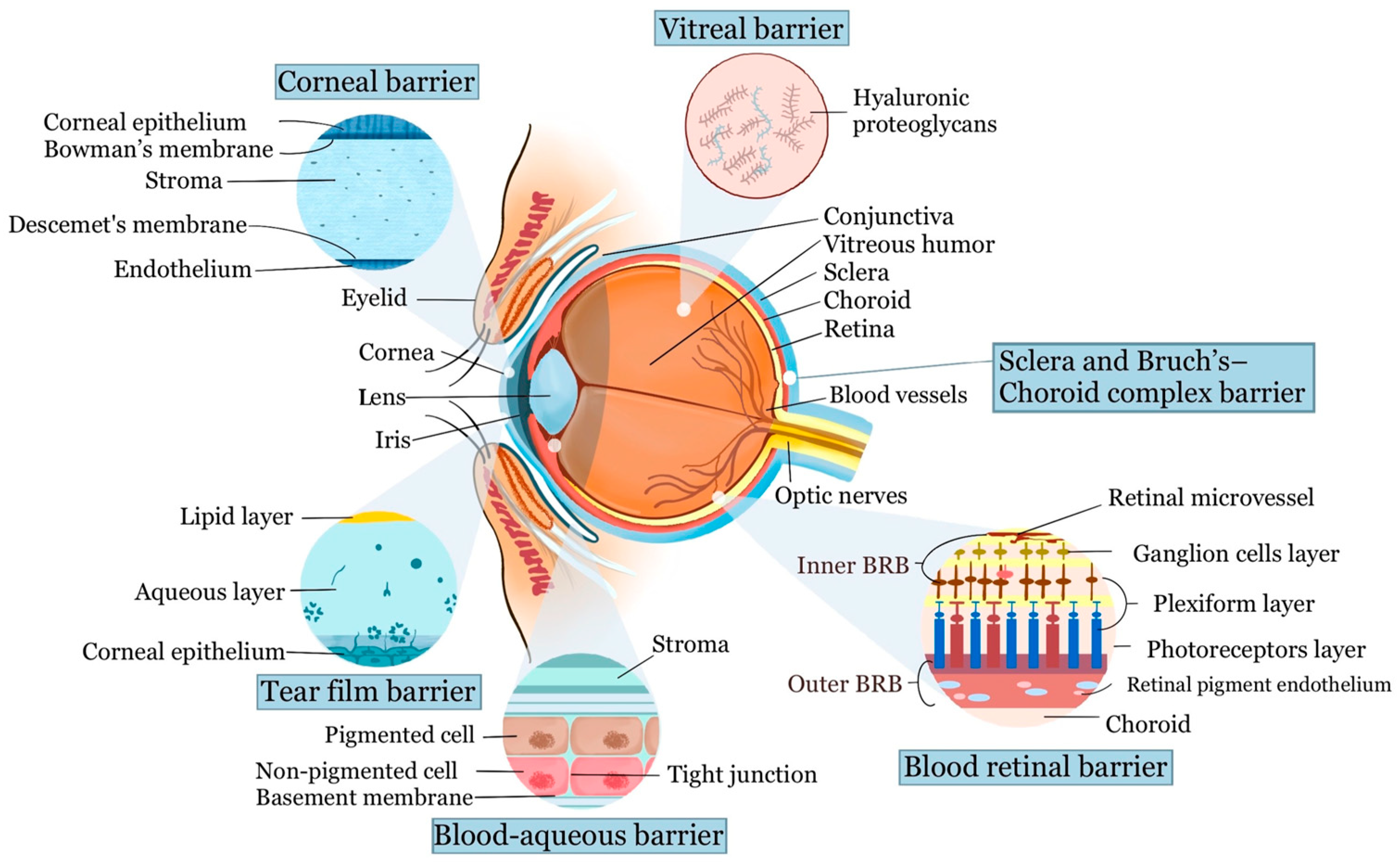
2. Anatomy and Barriers of Ocular Drug Delivery

The normal human eye measures approximately 22 to 27 mm axially and 69 to 85 mm in circumference [11]. It can be divided into two segments: anterior and posterior, separated by the ciliary body and lens. The anterior segment comprises the cornea, conjunctiva, iris, ciliary body, and lens, while the posterior segment contains the vitreous humor, retina, choroid, sclera, and optic nerve. The eye’s intricate anatomy and protective barriers pose challenges for drug administration (Figure 1). It shares characteristics with immune-privileged organs like the brain, isolating it from the body’s circulation with a blood–retinal barrier, making systemic therapy difficult, particularly for posterior segment disorders.

2.1. Barriers of the Anterior Segment

2.1.1. Tear Film Barrier

The tear film on the ocular surface forms an initial barrier, impeding drug delivery, while drainage through the nasolacrimal system can dilute and remove drugs, affecting their efficacy. This tear film, around 3 μm thick and 3 μL in volume, comprises three layers: an outer lipid layer, a middle aqueous layer, and an inner mucous layer [12]. The outer lipid layer prevents water evaporation but also hinders drug absorption into the cornea and sclera [13]. Meanwhile, the mucous layer in the tear film acts protectively, forming a hydrophilic barrier that efficiently removes debris and pathogens.
The lacrimal turnover rate is approximately 1~3 μL/min, causing drug loss from the ocular surface to be 500 to 700 times greater than the drug absorption rate into the anterior chamber. Irritant drugs, certain excipients, and pH deviations can trigger lacrimation, increasing tear production to about 300 mL per minute [14]. This rapid increase leads to immediate drainage through the nasolacrimal duct, causing over 85% of the administered drug dose to be lost before reaching the corneal surface. The retained drug may also undergo further dilution due to rapid tear turnover, reducing the concentration gradient and diffusion rate. This results in low bioavailability of intraocular drugs within the aqueous humor, leading to poor drug bioavailability with topical delivery, typically ranging from 0.1% to 5% [15].

2.1.2. Cornea and Conjunctival Barrier

The cornea, the outermost transparent avascular layer of the eye, has essential refractive and barrier functions. It consists of three cell layers: the lipophilic epithelium, the hydrophilic stroma, and the lipophilic endothelium, along with two interfaces: The Bowman layer and Descemet’s membrane. The corneal epithelium, comprising 5–7 lipid-rich cell layers with tight junctions and desmosomes, forms a robust barrier against drug penetration and microbial invasion [14,16]. The Bowman layer between the epithelium and stroma consists of acellular condensation of type I and type III collagen fibrils [17]. The Bowman layer allows drug and particle passage into the stroma, which makes up most of the cornea’s volume. It consists of hydrated type I collagen, providing structural support, optical clarity, and ocular immunity, facilitating the permeation and diffusion of hydrophilic drugs. Descemet’s membrane contains collagen type IV and VIII fibrils that provide support for the monolayer of corneal endothelial cells. Despite larger pore sizes in Descemet’s membrane reducing its barrier function, it can still filter macromolecules and particles that are directly administered into the stroma, protecting the endothelium. The corneal endothelium is a monolayer of cells that maintains stromal dehydration and allows the transportation of water and solute to the anterior chamber through both active (sodium–potassium ATPase pumps) and passive (endothelial intercellular tight junctions) mechanisms [16,18].
In contrast to the cornea, drug absorption through the conjunctiva is hindered by conjunctival capillaries and the lymphatic system, leading to drug leakage into the bloodstream and reduced bioavailability. Tight junctions in the conjunctival epithelium also impede the passive movement of hydrophilic molecules. The sclera, primarily composed of collagen fibers and proteoglycans, has a permeability similar to the corneal stroma. Recent studies suggest that drug permeation through the sclera inversely correlates with molecular size [19]. Linear dextrans exhibit lower permeability than globular proteins, and positively charged molecules have limited permeability due to interactions with the negatively charged proteoglycan matrix [4].

2.1.3. Blood–Aqueous Barrier

The blood–aqueous barrier (BAB) is formed by tight junctions in the ciliary process’s non-pigmented epithelium, endothelial cells in the iris vasculature, and the inner wall endothelium of Schlemm’s canal. The tight junctions regulate paracellular transport, controlling the movement of ions and small substances between adjacent cells. The BAB is not completely impermeable; instead, it serves as a specialized gateway for controlled molecule movement [20].

2.2. Barriers of the Posterior Segment

2.2.1. Vitreal Barrier

The vitreous is a gel-like, transparent substance that fills the space between the lens and the retina. It mainly consists of water, collagen types II, IX, V/XI, hyaluronic acid, and other extracellular matrix components. Positively charged nanomaterials may interact with the negatively charged components of the vitreal network and thus block its diffusion ability, while negatively charged particles, based on the example of poly lactic-co-glycolic acid (PLGA) or human serum albumin, can distribute successfully across the vitreous humor [21]. The vitreous provides structural support to the eye, maintaining its shape against intraocular pressure. The vitreoretinal interface acts as a barrier, restricting substances from passing into the retinal layers [22]. This interface comprises three main components: (1) The cortical vitreous, a thin layer (100–300 μm) rich in collagen parallel to the inner limiting membrane (ILM). (2) The ILM, at the innermost boundary of the retina, is primarily composed of collagen type IV, laminin, and fibronectin, serving as a physical barrier. (3) Expanded Müller cell footplates, glial cells extending from the vitreous side to the outer nuclear layer of the retina.

2.2.2. Blood–Retinal Barrier

The blood–ocular barrier (BOB) system includes two key barriers: the BAB and the blood–retinal barrier (BRB). The BRB is highly selective, controlling the passage of ions, proteins, and water to and from the retina. It comprises two parts: the outer BRB (oBRB), which includes the choroid, Bruch’s membrane (BM), and the retinal pigment epithelium (RPE), and the inner BRB (iBRB), formed by tight junctions among retinal capillary endothelial cells [23].
Starting from the outermost layer, the choroid includes the suprachoroid, large and medium blood vessel layers, and the choriocapillaris. The choriocapillaris play a role in nutrient supply and waste removal from the outer retinal layers. The Bruch’s membrane (BM), positioned between the choriocapillaris basement membrane and the RPE basement membrane, comprises outer and inner collagenous layers separated by a central elastic layer. BM allows size-selective passive diffusion but can block larger molecules. The RPE is a single layer of pigment-containing cells located beneath the neural retina layer. Its tight junctions maintain the integrity of the oBRB. For the iBRB, the retina vasculature penetrates at three main plexuses: the nerve fiber layer, inner plexiform layer, and outer plexiform layer. The iBRB mainly consists of the neurovascular unit, similar in structure and function to the blood–brain barrier, providing a barrier from systemic circulation. Molecule permeation is restricted based on size, charge, and lipophilicity. Small hydrophilic compounds can pass through junctions, while lipophilic molecules use the transcellular route [24].
The iBRB and oBRB have specific systems that allow substances to enter (influx transporters) or leave (efflux pumps) the retina. Developing drugs that mimic the substances recognized by influx transporters can help deliver drugs better into the retina. Additionally, designing drugs that are unrecognizable to the efflux pumps, or using inhibitors for these pumps, can help retain drugs in the desired location [21].

2.2.3. Sclera and Bruch’s–Choroid Complex Barrier

The choroid serves as a densely vascularized barrier situated between the retinal pigment epithelium (RPE) and the sclera. With a thickness of approximately 200 μm, it is structured into five distinct layers: Bruch’s membrane, the choriocapillaris layer, two vascular layers, and the suprachoroidal layer [21,25]. The choroid acts as a barrier against hydrophilic compounds, while positively charged lipophilic drugs can bind with the tissue to create slow-release depots. Additionally, drugs’ molecule sizes impact their ability to diffuse into the posterior eye segment. Bruch’s membrane, approximately 2–4 μm thick, consists mainly of collagen and elastin fibers. The choriocapillaris layer comprises highly fenestrated capillaries with pores ranging from 6 to 12 nm, allowing the passage of larger molecules [26].
The sclera, the eye’s outer opaque layer, is primarily made up of collagen fibers, proteoglycans, and glycoproteins, with an average thickness of 0.5–1 mm. Drug permeability through the sclera is influenced by factors such as molecular weight, size, charge, and lipophilicity. For instance, hydrophilic compounds like methazolamide can penetrate the sclera. The proteoglycan matrix in the sclera carries a negative charge under normal pH conditions, aiding the passage of negatively charged solutes through this barrier [27].

3. The Conventional Routes for Ocular Drug Administration

The conventional ocular drug delivery methods include topical, regional, and systemic administrations. Topical delivery using solutions, gels, ointments, and suspensions is common and cost-effective, primarily targeting anterior segment eye diseases. However, less than 5% of the administered dose typically reaches the aqueous humor, as it can be washed off by mechanisms like lacrimation, tear dilution, and tear turnover [4,6,28]. In regional administration, the formulation is given intraocularly or periocularly to minimize systemic side effects while enhancing target tissue delivery [29]. Intracameral administration involves direct injection into the anterior chamber and is commonly used in cataract surgery and for treating anterior segment diseases. Another method, intravitreal injection (IVI), has gained popularity in the past two decades for treating retinal disorders such as age-related macular degeneration (AMD), diabetic macular edema (DME), retinal vein occlusion (RVO), and endophthalmitis. While IVI allows for direct drug delivery to the posterior segment, frequent eye punctures may increase the risk of complications like endophthalmitis, hemorrhage, retinal detachment, and patient discomfort [30]. Other intraocular injection deliveries include intrastromal, subconjunctival, subretinal, and subchoroidal. Periorbital injection methods include retrobulbar, peribulbar, posterior juxtascleral, and subtenon injection. This is widely used for local anesthesia in ocular surgery.
Systemic administration, via intravenous and oral routes, is used in various ophthalmic conditions like delivering antibiotics for endophthalmitis, carbonic anhydrase inhibitors for intraocular pressure, and methotrexate and corticosteroids for uveitis treatment [30]. However, as mentioned before, BAB and BRB restrict access to specific eye segments, reducing systemic administration’s bioavailability. This often necessitates higher dosages for clinical efficacy, potentially resulting in systemic side effects. Additionally, factors like plasma protein binding, lipophilicity, and distribution clearance can complicate predicting accurate therapeutic concentrations [31]. Table 1 summarizes various routes of ocular drug administration and their application in clinical treatment and describes their benefits and challenges in bioavailability. The pathways of drug metabolism are shown in Figure 2 [32].

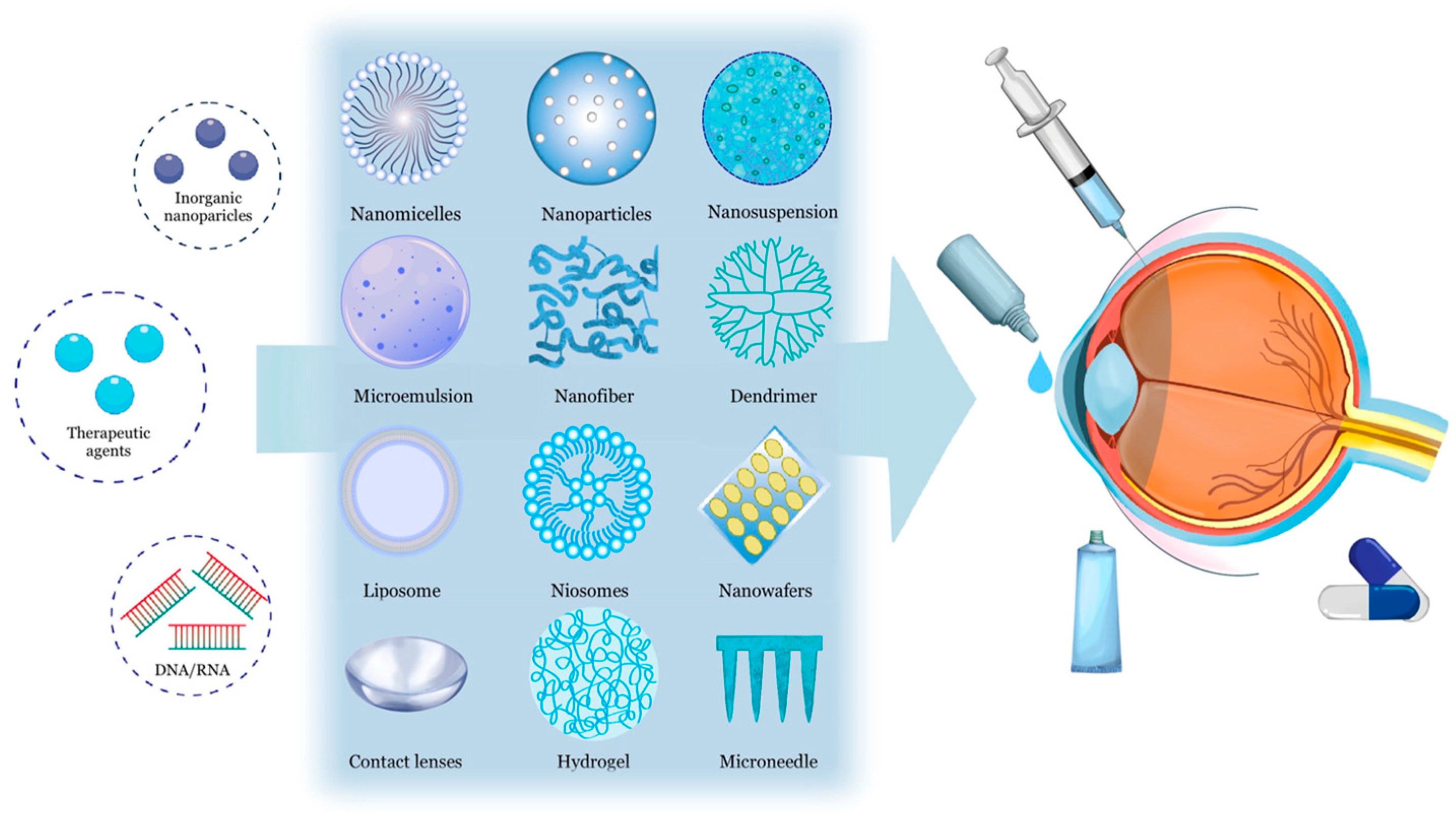
4. Nanotechnology-Based Ocular Drug Delivery Systems

Ocular nanotechnology-based drug delivery is a specialized method for precisely administering therapeutic agents to the eye using nanoscale carriers or systems (Figure 3). These carriers transport drugs to the target site, improve solubility and stability, extend release, and minimize side effects. This approach has gained attention over the past two decades for overcoming ocular barriers and enhancing drug bioavailability [10,14,36]. Some nanocarriers have received FDA approval and are commercially available for use in the clinical treatment of ocular diseases (Table 2). We provide a comprehensive introduction and list some recent exploratory studies in each nanotechnology-based ocular delivery method, highlighting their clinical applications in various eye disorders to offer treatment insights and explore future developments in this field.

4.1. Nanomicelles

4.1.1. Main Characteristics

Nanomicelles are self-assembling nanoscale (particle size usually within a range of 5 to 100 nm) colloidal dispersions with a hydrophobic core and hydrophilic shell. The micelles can be divided into three categories: polymers, surfactants, and multi-ion composite nanomicelles [37]. Amphiphilic molecules forming micelles exhibit properties influenced by factors like temperature, concentration, and the number of monomer molecules. In aqueous solutions, these molecules self-assemble into spherical or cylindrical nanoparticles, with their hydrophobic tails in the core and hydrophilic heads on the surface interacting with the aqueous environment. This structure allows for loading hydrophobic drugs into the core, improving solubility, stability, and targeted delivery to specific tissues [38]. Conversely, when amphiphilic molecules assemble in a nonpolar solvent, the hydrophobic part of the molecule faces outward while the hydrophilic portion is embedded inward, constituting so-called reverse nanomicelles, which can be used to load soluble drugs [39]. Stimuli-responsive nanomicelles release drugs in response to various cues, such as pH, temperature, ultrasound, light, and redox potential. These diverse nanomicelle structures enhance the solubility, stability, and delivery of hydrophobic compounds, generating significant interest in pharmaceutical and biomedical research.
Polymer nanomicelle carrier materials fall into two categories: natural and synthetic. Natural polymers like hyaluronic acid, albumin, and chitosan are widely available, biocompatible, and known for their non-toxic breakdown products in the human body, making them attractive for chemical modifications. Synthetic polymer nanomicelle carrier materials, such as polyethylene glycol (PEG), polyethylene oxide (PEO), polyacrylamide (PAM), polyvinylpyrrolidone (PVP), polyvinyl alcohol (PVA), and polyethyleneimine (PEI), comprise the hydrophilic sections of polymer nanomicelles. Hydrophobic components include biodegradable polyesters and amino acids like polylactide (PLLA), polyglycolide (PGA), polycaprolactone (PCL), polylactic glycolate (PLGA), polyaspartic acid (PAsp), polybenzylaspartic acid (PBLA), and polyglutamic acid (PGlu). Polymer nanomicelles can be categorized into three types based on the arrangement of amphiphilic molecules: amphiphilic block copolymers, graft polymer micelles, and amphiphilic random copolymers [40,41,42].

4.1.2. Clinical Applications

In recent years, nanomicelles have made significant strides in ocular drug delivery. These advancements include approved treatments like rapamycin nanomicelle eye drops to prevent immune rejection [43], terbinafine hydrochloride nanomicelle formulations for fungal eye infections [44], and Cequa® (a 0.09% cyclosporine eye solution) for dry eye syndrome management [45]. Table 3 summarizes recent in vitro and in vivo studies of nanomicelles in ophthalmic DDS applications. However, it is crucial to assess potential cytotoxicity, genotoxicity, and immune response before clinical use, especially considering the presence of multiple neuronal cells in the retina. Additionally, evaluating the ability of nanomicelles to cross the BRB and reach different retinal layers is essential to understand their potential toxicity. Thus, comprehensive toxicology testing is essential for assessing new drug carrier candidates in the future [46].

4.2. Nanoparticles

4.2.1. Main Characteristics

Nanoparticles (NPs) are tiny particles ranging from 1 to 1000 nm and can be composed of various materials like metals (e.g., gold or silver), polymers, lipids, ceramics, or other substances. They come in different shapes, such as spheres, rods, tubes, or irregular forms, depending on their material and fabrication method. They may or may not have a core–shell structure, which is different from micelles. NPs can be divided into nanocapsules and nanospheres based on their morphological structure. Nanospheres have a solid polymeric structure, whereas nanocapsules consist of a thin polymeric envelope, approximately 5 nm thick, surrounding the oily core. NPs can encapsulate both hydrophobic and hydrophilic drugs, safeguarding them from degradation, and improving targeted delivery, efficient drug absorption, and controlled drug release, thereby enhancing bioavailability [59]. In ophthalmic DDSs, NPs are usually composed of lipids, such as fatty acids and triglycerides, or polymers, which could be further divided into natural and synthetic. Natural polymers including cellulose, sodium alginate, hyaluronic acid, albumin, gelatin, and chitosan have the advantages of biocompatibility and minimal toxicity. Different natural polymers possess unique characteristics. For example, chitosan, a cationic polysaccharide derived from chitin, exhibits favorable mucoadhesive and antimicrobial properties in ocular drug delivery systems. However, it is insoluble in both water and alkaline solutions. On the other hand, hyaluronic acid boasts a high hydration capacity but comes with a higher cost [60]. The synthetic polymers, such as polylactic acid (PLA), polymethacrylic acid (PMAA), polycyanoacrylate, Eudragit® (RS100 and RL100), poly(ε-caprolactone) (PCL), poly(lactic acid-co-glycolide) (PLGA), and polyacrylamide, can be engineered to have versatile properties to improve biodegradability, biocompatibility, and controlled release manners which can meet specific therapeutic needs. However, they may also face some clinical concerns such as toxicity of monomers or byproducts and may provoke an immune response in vivo.
Solid lipid nanoparticles (SLNs) and nanostructured lipid carriers (NLCs) are both lipid-based nanoparticles used in drug delivery. The former is characterized by a solid matrix that enables controlled drug release, alongside improved stability and cost-effectiveness, while the latter is regarded as a second-generation lipid nanoparticle, consisting of both liquid and solid lipids without any crystalline structure [61].
NP size significantly affects drug loading and delivery in ocular drug delivery. Smaller NPs generally offer better stability and biodistribution. NPs in the range of 50–400 nm are preferred for ocular drug delivery, with sizes around 200 nm easily absorbed by the cornea and conjunctiva through topical administration. They exhibit improved mucoadhesion, better penetration through ocular barriers, and reduced ocular irritation [42,62]. The surface charge of NPs is essential; cationic NPs tend to stay longer on the ocular surface due to their interaction with negatively charged tissues, while anionic NPs have different characteristics [63].

4.2.2. Clinical Applications

Recent advancements have made significant progress in using NPs for detecting ocular diseases. For example, Nguyen et al. developed ultrapure chain-like gold nanoparticles (CGNPs) conjugated with RGD peptides for multimodal photoacoustic microscopy (PAM) and optical coherence tomography (OCT) to visualize choroidal neovascularization (CNV) in a rabbit model. Intravenous administration of CGNP clusters-RGD bound to CNV resulted in significantly enhanced PAM and OCT signals, offering a more sensitive diagnostic tool for retinal neovascularization [64,65,66]. Gold nanoparticles (GNPs) have been employed in detecting and treating retinoblastoma. Moradi et al. showed that combining brachytherapy and hyperthermia with GNPs increased retinoblastoma necrosis and significantly reduced its size in rabbit eyes [67].
Nanoparticles in ocular treatment come in two types: polymer-based and lipid-based. Polymersomes are vesicular structures characterized by a bilayer composition consisting of hydrophilic and hydrophobic block copolymers, with a central core filled with an aqueous solution. The polymer NPs are characterized by their biodegradable properties, extended circulation in the bloodstream, improved drug-loading capacity, and easily modified surfaces through the attachment of ligands which lead to targeted delivery [68]. By adjusting the composition and processing conditions, researchers can tailor the size and shape of polymersomes to meet specific requirements. Common shapes include spherical, oblong, and tubular. Therefore, polymersomes are widely used in the biomedical field for both therapeutics and diagnosis. In ophthalmic applications, formulating pranoprofen with PLGA polymer has improved ophthalmic delivery and enhanced the drug’s local anti-inflammatory and analgesic effects [69]. Varshochian et al. developed albuminated-PLGA-NPs containing bevacizumab for intravitreal injection in rabbit models. This formulation showed a sustained release of bevacizumab for 2 months, maintaining vitreous concentrations above 500 ng/mL (the minimum concentration required to block VEGF-induced angiogenesis) for over 8 weeks. This suggests that these NPs can sustain drug release, reducing the need for frequent dosing [70]. Chitosan-based polymeric nanoparticles loaded with drugs such as cefuroxime, diclofenac, atorvastatin, or dexamethasone have shown improved ocular bioavailability [71,72,73]. Hyaluronic acid-based nanoparticles have demonstrated favorable attributes, including size, pH, osmolarity, and entrapment efficiency, leading to enhanced corneal permeation and increased accumulation of cyclosporine A. This holds promise for the treatment of dry eye disease compared to commercial emulsions in a rabbit model [74].
Polymeric NPs face challenges related to cytotoxicity and a lack of efficient large-scale production methods. In contrast, lipid NPs pose a lower risk of toxicity since they rely on biodegradable and non-toxic lipid components. Both SLNs and NLCs raise great interest in DDSs due to their outstanding biocompatibility, tolerability, and scaling-up capabilities. SLNs were first reported in the 1990s by Professor Müller and Professor Gasco. SLNs represent a mixture of solid-state lipids under ambient and physiological temperatures while NLCs are the second generation of lipid nanoparticles that include liquid lipids in their structure [75]. Lipid-based NPs loaded with brimonidine and latanoprost have been employed for the treatment of glaucoma [62,76]. Antimicrobials such as natamycin and ofloxacin coated with SLNs have been shown to increase transcorneal permeation, prolong drug release rate, and enhance antifungal activity without cytotoxic effects [77,78]. In retinal vascular diseases such as AMD and DME, SLN and NLC-modified medications also improved bioavailability in aqueous and vitreous humor in animal models [79,80]. These nanoparticles can interact with the ocular surface while safeguarding the drugs from metabolic degradation and extending their residence time on the pre-corneal surface [81].

4.3. Nanosuspension

4.3.1. Main Characteristics

Nanosuspensions (NSs) consist of pure drug nanoparticles and stabilizers, typically with an average diameter below 1 μm (often in the range of 200–500 nm). They can be formulated in either aqueous or non-aqueous liquid phases, enhancing drug solubility in both aqueous and organic environments [82,83]. NSs are a versatile approach to improving the delivery of hydrophobic drugs and show promise in enhancing the performance of poorly water-soluble drugs, particularly those from natural sources [84]. NSs require careful formulation with stabilizing agents to maintain drug particle stability in the liquid medium. Various fabrication methods, including top-down techniques like wet milling, dry milling, high-pressure homogenization, and co-grinding, as well as bottom-up methods like anti-solvent precipitation, liquid emulsion, and sono-precipitation, are used. A combination of these approaches is often employed. NSs can be administered in liquid forms, or post-production processes like freeze drying, spray drying, or spray freezing can convert them into solid forms, allowing for diverse dosage forms like powders, pellets, tablets, capsules, films, or gels [85].
In ocular DDSs, nanosuspensions offer a method to deliver higher concentrations of poorly soluble drugs and extend residence time to the cul-de-sac [86]. NSs hold promise for improving ocular disease treatment through enhanced drug delivery efficiency, bioavailability, and patient comfort. However, challenges related to stability, particle size distribution control, and safety and toxicity concerns require rigorous testing and optimization to fully realize their potential and ensure their safety and efficacy in clinical applications.

4.3.2. Clinical Applications

Extensive research is ongoing to explore the use of NSs in various drug delivery systems, including oral, ocular, brain-targeted, topical, buccal, nasal, and transdermal routes [86]. In ocular drug delivery, NSs are being investigated for treating diverse eye conditions, such as glaucoma, macular degeneration, diabetic retinopathy, and uveitis. Table 4 provides examples of recent exploratory studies of NSs for both anterior segment and posterior segment disease treatment.

4.4. Nanoemulsions and Microemulsions

4.4.1. Main Characteristics

Nanoemulsions (NEs) are emulsions with sizes ranging from 20 to 500 nm, composed of two immiscible liquids (usually water and oil) stabilized by an amphiphilic surfactant [93]. They are transparent or translucent and thermodynamically unstable but kinetically stable systems. NEs are categorized based on the nature of the dispersed phase system, including oil-in-water (o/w) NEs, water-in-oil (w/o) NEs, and bi-continuous NEs, which feature both oil microdomains and water in the system. Among these, oil-in-water (o/w) NEs have gained prominence in ocular drug delivery due to their unique characteristics, such as easy dilution with tear fluid and the capacity to encapsulate lipophilic drugs in the oil phase.
Microemulsions (MEs), first introduced by Hoar and Schulman in the 1940s, are isotropic and thermodynamic stable dispersions made from water, oil, surfactants, and cosurfactants with small droplet sizes usually within the range of 5–200 nm [94]. While NEs and MEs share similar formulation components, including oil and water phases, surfactants, and potentially cosurfactants, their ratios differ. Generally, MEs require a higher surfactant-to-oil ratio compared to NEs [95]. In addition, NEs typically feature spherical-shaped particles due to the significant Laplace pressure, whereas MEs may display both spherical and non-spherical particles owing to their notably low interfacial tension. Ophthalmic NE formulations have demonstrated extended pre-corneal retention times, increased ability to penetrate ocular tissues, enhanced ocular drug bioavailability, and consistent drug levels in the eye. These benefits surpass those of previously reported gels or ointments. Cationic NEs extend drug dwelling time through electrostatic interactions with the anionic surface of the cornea’s mucin layer. This interaction enhances drug penetration through corneal tight junctions, ultimately increasing bioavailability [96,97].

4.4.2. Clinical Applications

NEs have broad applications in biomedicine owing to their small droplet sizes, offering stability and rheology control. They are widely employed in pharmaceutical formulations for topical, ocular, and intravenous delivery. NEs also serve as templates for producing nanocrystals of hydrophobic pharmaceutical ingredients [98]. Due to their excellent permeability through the cornea and conjunctival barriers, NEs are extensively used in treating anterior segment disorders. Fardous et al. created a gel-in-water (G/W) NE by ultrasonication using beeswax as an organogelator aiming to generate a delivery system of hydrophobic drugs to the posterior ocular region. In vitro testing demonstrated the biocompatibility of the G/W NE. Meanwhile, in vivo, application as eye drops revealed no signs of ocular irritation [99]. Despite the growing literature and products in this field, there are still challenges with NEs, including the need for significant amounts of surfactants and concerns about their potential toxicity, limited capacity to solubilize high-melting-point substances, and susceptibility to environmental factors affecting stability. Therefore, careful component selection and safety evaluation are essential for pharmaceutical development.

4.5. Nanofibers

4.5.1. Main Characteristics

Nanostructures are generally classified into four types: zero-dimensional (nanoparticles), one-dimensional (nanofibers, nanotubes, nanowires), two-dimensional (nanofilms), and three-dimensional (polycrystals). One-dimensional nanostructures are particularly intriguing due to their high surface-to-volume ratio, porous structure, mechanical strength, flexibility, and resemblance to the extracellular matrix [100,101]. Nanofibers, with diameters typically ranging from tens to hundreds of nanometers, are composed of various materials, including natural polymers (e.g., hyaluronic acid, chitosan, dextran, gelatin, collagen), synthetic polymers (e.g., PLA, PLGA, PCL), carbon, or ceramics. They exhibit a high aspect ratio and can be fabricated using various methods, categorized as either electrospinning or non-electrospinning. Electrospinning, in particular, has gained significant attention due to its ability to produce ultrafine fibers through the application of high voltage and low current, allowing for potential commercial-scale production [98,102].

4.5.2. Clinical Applications

Nanofiber composites are widely used in various biomedical applications such as medical implants, wound dressing, tissue scaffolds, and drug delivery systems. Drugs ranging from antibiotics and anticancer agents to proteins, DNA, RNA, living cells, and various growth factors can be loaded into electrospun nanofibers [103]. The drug release rate of nanofibers can be influenced by their morphology, porosity, and composition. Nanofibers, with their large surface area, offer significant drug loading capacity and extended drug release profiles, reducing the need for frequent dosing. Table 5 summarizes some recent research on nanofiber-based ocular drug delivery systems for both anterior and posterior segment disorders.

4.6. Dendrimers

4.6.1. Main Characteristics

Dendrimers are highly branched nanoscale polymer structures with a three-dimensional design and numerous functional groups on their surface, making them versatile and biocompatible for various applications [112]. Over a hundred distinct dendritic architectures exist, with polyamidoamine (PAMAM) and polypropyleneimine (PPI) dendrimers being widely recognized among them [113]. T Dendrimers can be synthesized to control their size, surface charge, peripheral functional groups, and solubility, making them highly adaptable for carrying various therapeutic drugs [114]. Dendrimer-drug conjugates have the potential to reduce systemic side effects, improve drug targeting, and prolong drug half-lives.
The cytotoxicity of dendrimers depends on factors like generation, surface group number, and terminal moiety type. Higher-generation dendrimers and those with positive surface charges tend to be more cytotoxic. PAMAM dendrimers at concentrations above 50 μg/mL may cause ocular functional damage [115]. Dendrimers modified with substances like polyethylene glycol (PEG), acetyl groups, carbohydrates, and other moieties have either minimal or no adverse effects on cells. Furthermore, some dendrimers possess inherent biological properties, such as anti-fungal, anti-bacterial, or cytotoxic effects on cancer cells while sparing normal cells. Therefore, the potential intrinsic cytotoxicity should be evaluated on a case-by-case basis [116].

4.6.2. Clinical Applications

Recent studies have shown promising results using dendrimer-based carriers for ocular drug delivery via various administration methods, including topical, intravitreal, and subconjunctival routes [114]. For instance, DenTimol, a dendrimer-based polymeric timolol analog, demonstrated efficient corneal penetration and a significant intraocular pressure (IOP) reduction in adult male rats, achieving an average reduction of 7.3 mmHg (approximately 30% from baseline) within 30 min [117]. Soiberman et al. developed a subconjunctival injectable gel using G4-PAMAM dendrimer and hyaluronic acid loaded with dendrimer dexamethasone (D-Dex) conjugate in a rat corneal inflammation model, leading to a significant reduction in corneal neovascularization. This injectable D-Dex gel holds promise as a drug delivery platform for treating various inflammatory ocular surface disorders, including dry eye, autoimmune keratitis, and post-surgical complications, reducing the need for frequent steroid administration. Additionally, dendrimers are utilized as scaffolds in corneal tissue engineering [118]. Duan et al. reported that a dendrimer crosslinked collagen gels supported human corneal epithelial cell growth and adhesion, without cell toxicity [119].
In ocular posterior segment diseases, dendrimers are also a promising method for drug delivery. Kambhampati et al. reported that systemic hydroxyl-terminated polyamidoamine dendrimer-triamcinolone acetonide conjugate (D-TA) could be selectively taken up by the injured mi/ma and RPE causing choroidal pro-inflammatory cytokines and pro-angiogenic factor suppression by limiting macrophage infiltration, resulting in significant CNV suppression in a rat model. The dendrimer was also taken up by choroidal macrophages in human postmortem diabetic eyes [120]. Kannan et al. highlighted the progress in therapeutics using hydroxyl polyamidoamine dendrimers, which can target cells systemically without the need for specific ligands. This offers a novel treatment option for wet AMD [121]. Recently, genome-editing technology for genetic disorders or cancers has gained lots of interest. Inoue et al. developed a folate-modified polyamidoamine dendrimer (FP-CDE) as a potential carrier for TTR-CRISPR pDNA therapy in hereditary amyloidogenic transthyretin ocular amyloidosis [122].

4.7. Liposomes and Niosomes

4.7.1. Main Characteristics

Liposomes and niosomes are vesicular drug delivery systems designed to encapsulate and deliver drugs in a controlled manner. They have similar structures but differ in composition. Liposomes are spherical vesicles made of phospholipid bilayers with a central water compartment diameter of 0.025 to 10 μm [123]. Liposomes can encapsulate hydrophilic and lipophilic drugs in their aqueous core or lipid bilayer, respectively. They can be modified to enhance specificity for specific cells or tissues by adjusting surface molecules, size, and lamellarity, and by adding targeting ligands or polymers [124,125]. Furthermore, liposomes are biocompatible and biodegradable, making them suitable for drug delivery applications and are widely used in pharmaceuticals, particularly for delivering chemotherapy drugs and vaccines, as well as in cosmetic products. Apart from the above benefits, liposomes present some disadvantages such as limited stability which may require a specific storage environment, limited capacity for loading certain hydrophobic compounds, and a short circulation half-life due to clearing by the immune system [126].
Niosomes are similar to liposomes in structure and function but are composed of non-ionic amphiphilic molecules in certain aqueous solutions by self-assembly technology instead of natural or synthetic phospholipids. The absence of phospholipids in niosomes makes them more stable than liposomes and less prone to oxidation or degradation. Niosomes can co-deliver both hydrophilic and lipophilic drugs in one vesicle. The formulation process is easier due to the good stability of the niosomes and the cost of preparation is also much cheaper than liposomes. Thus, niosomes are gaining importance in drug delivery systems [127]. However, niosomes can suffer from drug leakage over time, especially when exposed to physiological conditions. This leakage can affect the controlled release of drugs and reduce their therapeutic efficacy.

4.7.2. Clinical Applications

The choice between liposomes and niosomes hinges on factors like the drug type and desired system properties. They are versatile and customizable to regulate drug release rates and target specific cells or tissues through surface modifications. Several liposomal products, including Visudyne®, an FDA-approved formulation of Verteporfin, are already available for clinical use in photodynamic therapy for the treatment of patients with predominantly classic subfoveal CNV due to AMD, pathologic myopia, or presumed ocular histoplasmosis [128]. Additional FDA-approved liposomal ophthalmic formulations include Lacrisek®, a liposome-based formulation of vitamin A palmitate and vitamin E, and Artelac Rebalance®, a vitamin B12 eye drop formulation with PEG and hyaluronic acid, used for the clinical treatment of dry eye disease [36,129]. There is growing preclinical research on liposome-based and noisome-based ophthalmic formulations. Table 6 offers some of the recent studies in this field.

4.8. Nanowafers

4.8.1. Main Characteristics

A nanowafer is a small, transparent, circular disk containing nanoreservoirs filled with medication. These nanoreservoirs release the drug gradually, improving its absorption into the nearby eye tissue. The nanowafer dissolves after the desired drug release duration. PVA is a suitable non-immunostimulatory polymer for ocular nanowafer fabrication [140]. Nanowafer drug delivery offers sustained release, reducing the need for frequent dosing, minimizing systemic exposure, and reducing potential side effects. The use of biodegradable components enhances safety and minimizes long-term impacts. However, material choice in nanowafer formulations is critical for biocompatibility and to avoid toxicity. Dissolution rates can vary with formulation, affecting drug release kinetics and dosing regimens.

4.8.2. Clinical Applications

Nanowafer technology finds applications in various fields, including drug delivery. A study by Yuan et al. showcased the effectiveness of an axitinib-loaded nanowafer in treating corneal neovascularization. A once-a-day administration of the nanowafer outperformed twice-daily eye drops in therapeutic efficacy without impacting wound healing or causing toxicity in experimental models [140]. Marcano et al. created a nanowafer for cysteamine delivery to treat corneal cystinosis, a condition typically managed with frequent and irritative eye drops. In vivo tests on cystinosis knockout mice showed that the nanowafer, containing 10 μg of cysteamine and administered once daily, was twice as effective as twice-daily eye drops containing 44 μg of cysteamine [141]. Coursey et al. also proposed that a dexamethasone (Dex)-loaded nanowafer could release the drug on the ocular surface for a longer duration than aqueous eye drops. They found out that administering the Dex-NW treatment once daily, every other day over five days, effectively restored a normal ocular surface and corneal barrier function. This regimen exhibited a level of effectiveness comparable to that of applying dexamethasone eye drops twice daily. Additionally, Dex-NW significantly reduced the expression of inflammatory cytokines (e.g., TNF-α and IFN-γ), chemokines (such as CXCL-10 and CCL-5), and MMP-3, which are typically elevated in response to dry eye conditions [142].

4.9. Contact Lenses

4.9.1. Main Characteristics

Nanocarriers in contact lenses involve integrating nanoparticles or nanoscale materials into the contact lens structure. This technology aims to improve contact lens performance and comfort while facilitating controlled drug delivery directly to the ocular surface. This approach can extend drug retention time and enhance ocular bioavailability by over 50% [143,144]. There are mainly two types of contact lenses depending on the designed material: (1) soft contact lenses (SCL), composed of hydrogel or silicone hydrogel polymers, and (2) rigid gas-permeable contact lenses (RGP) [145]. The parameters of soft contact lenses include transparency, oxygen permeability, and glass transition temperature. SCL surface modifying methods include dip-coating (soaking); diffusion barrier insertion (Vitamin E); incorporation of functional monomers, ligands, and a polymeric matrix; molecular imprinting, incorporation of colloidal, drug-loaded nanoparticles or other colloidal nanostructured systems; and surface coating by multilayer film deposition of colloidal nanoparticles or ligands [146,147].
Contact lenses as drug delivery systems have numerous advantages for ocular drug delivery, including sustained drug release, targeted delivery, ease of wear for improved patient compliance, reduced tear dilution, protection of sensitive drugs, and potential for combination therapy. Despite these advantages, contact lens drug delivery systems also face challenges such as controlling drug release rates precisely, potential infection risk from improper handling, and the need for material and design optimization [147].

4.9.2. Clinical Applications

Recent applications of nanoparticle medicines in soft contact lenses include antibiotics, antihistamines, immunosuppressants, corticosteroids, and glaucoma drugs, with a focus on anterior segment ocular disorders. While contact lenses offer precise drug delivery control, they have limitations in terms of a short drug release duration, storage stability, and drug capacity [148]. Research is growing in the field of ophthalmic formulation techniques and lens material design to improve bioavailability in clinical applications. Table 7 summarizes recent preclinical research on soft contact lenses for various ocular diseases.

4.10. Hydrogels

4.10.1. Main Characteristics

Hydrogels are three-dimensional networks of hydrophilic monomers and multifunctional linkers that form a flexible and water-laden structure. Hydrogels can undergo swelling and shrinkage suitable for facilitating controlled drug release. Hydrogels can be made from either natural or synthetic monomers. Natural hydrogels, known for their biocompatibility and ability to degrade into non-toxic components, have been extensively studied in tissue engineering. Examples of natural polymers include hyaluronic acid, chitosan, and collagen. However, natural hydrogels often exhibit weaker mechanical strength, challenges in precise formulation and drug loading, and potential immunogenicity [155]. In contrast, synthetic hydrogels are composed of polymers not found in nature. Common synthetic polymers include poly(ethylene glycol) (PEG), poly(vinyl alcohol) (PVA), and poly(hydroxyethyl methacrylate) (PHEMA) [156]. Synthetic polymer hydrogels are highly customizable for specific applications, exhibit good batch-to-batch reproducibility, and have extended stability. However, they may face challenges related to clearance and the formation of toxic by-products during degradation.
Hydrogels achieve cross-linking through physical mechanisms such as chain entanglement, hydrogen bonding, hydrophobic interactions, complexation, or crystallite formation. They can also undergo chemical cross-linking through covalent interactions between the polymer and crosslinker. Furthermore, hydrogels can integrate functionalized components that respond to biological stimuli, making them adaptable to the surrounding environment. This adaptability is significant for in situ forming hydrogels, which crosslink as the temperature increases from room temperature to body temperature, as well as for controlled drug release triggered by factors like pH or photostimulation [157,158,159,160].

4.10.2. Clinical Applications

Hydrogels, owing to their biocompatibility and adjustable drug release profiles, have been studied for drug delivery in various ocular conditions. They can transport drugs to various target locations through different administration methods. Hydrogels are also widely used in combination with different nano-based formulations like NPs, nanomicelles, microneedles, and nanofibers to prolong drug retention and release on the human eye. Swarup et al. used a PNP hydrogel in a mouse model of alkali injury-induced symblephara to prevent fornical shortening and conjunctival fibrosis after injury [161]. Yazdanpanah et al. developed a thermoresponsive hydrogel using decellularized porcine cornea ECM (COMatrix) and demonstrated that this hydrogel improved the attachment and proliferation of human corneal epithelial cells and reduced TNF-α expression in vitro. This suggests its potential use as an ocular surface bandage [162]. Gau et al. explored an anti-VEGF-loaded supramolecular hydrogel that inhibited vascular growth in the retina and reduced CNV by providing extended and controlled release of the anti-VEGF agent. These hydrogels also exhibited the ability to reduce reactive oxygen species and local inflammation. This suggested the potential to replace current anti-VEGF therapy [163]. Cocarta et al. reported a hydrogel implant for transscleral drug delivery to treat retinoblastoma. A bi-layered design with an inner hydrophilic layer bound to a chemotherapeutic agent and the outer hydrophobic layer forms a barrier to prevent cytotoxicity of the delivered chemotherapeutics [164]. Researchers continue to explore and innovate in this field to improve the effectiveness and safety of ocular DDSs using hydrogels.

4.11. Microneedles

4.11.1. Main Characteristics

Microneedles (MNs) are small patches with tiny needles under 1 mm in length. They enable localized drug delivery in ocular DDSs, enhancing penetration through ocular barriers for improved therapy. MNs are minimally invasive, making them more acceptable to patients than intravitreal injections [165]. MNs come in various types based on their shape and drug delivery method: (1) Solid MNs create micro-holes to enhance drug permeability. (2) Hollow MNs use pressure to deliver liquid-loaded drugs. (3) Coated MNs have a drug layer that dissolves upon administration. (4) Dissolving MNs release drugs as their matrix dissolves. (5) Hydrogel-forming MNs absorb tissue fluids, promoting drug diffusion [166,167,168].
Although MNs provide minimally invasive and relatively painless methods for direct drug delivery, MNs for ocular DDSs face clinical limitations, including challenges in ensuring precise and consistent insertion and drug delivery, potential for eye damage, and patient acceptance. Regulatory approvals, standardization, and manufacturing costs are also factors to consider. More clinical studies and safety assessments are needed to establish their effectiveness and safety [165,167].

4.11.2. Clinical Applications

Recent progress in pharmaceutical technology has enabled microneedles (MNs) to offer targeted, less invasive, and highly effective drug delivery for various ocular conditions. Over the past decade, a broad spectrum of clinical applications has been explored, spanning from the treatment of keratitis, glaucoma, age-related macular degeneration, uveitis, and retinal vascular occlusion to retinitis pigmentosa [169]. Shi et al. created a dissolving MN patch loaded with fluconazole for treating fungal keratitis in rabbits. The MN patches, made from 30% PLA-HA, penetrated the corneal epithelium without irritating, increased drug residence time in the conjunctival sac by 2.5 h, and provided higher drug bioavailability than conventional eye drops and intrastromal injection [170]. Amer and his colleagues also found that MNs with interlocking features provided an 80% increase in adhesion strength and a slight increase in penetration force compared to microneedles without such features [171]. Matadh and colleagues showed that polymer-coated polymeric (PCP) MNs, a novel approach for controlled drug delivery, released drugs gradually and over a more extended period compared to uncoated MNs, which released the entire drug instantaneously in an ex vivo porcine eye mode [172].

4.12. Novel Gene Therapy with Nanotechnology-Based Ocular Delivery Techniques

Gene therapy has emerged as a promising approach to address genetic diseases since the 1960s and is propelled by advances in recombinant DNA technology. In the realm of ophthalmology, gene therapy has gained substantial traction, particularly in targeting retinal diseases, notably those affecting the retinal pigment epithelium [173]. Gene therapy relies on vectors to transport the desired genetic material into host cells. There are two primary categories of vectors: nonviral and viral vectors.
Nonviral delivery systems offer several advantages, including the ability to carry large plasmid DNA, minimal immunogenicity, and a reduced risk of side effects. Nanoparticles (NPs) have been widely used in nonviral gene delivery. These NPs can accommodate sizable plasmid DNA, exhibit safety profiles, sustain long-term protein expression, and pose minimal risk of insertional mutagenesis. Among the most stable NPs are those primarily composed of cationic lipids, PEG lipids, and cholesterol, which are also effective carriers of RNA [174]. Carbon dots (CDs) represent an emerging class of non-viral nanocarriers designed for delivering genes to retinal cells. These CDs possess distinctive physicochemical characteristics, including optical, electronic, and catalytic properties, which render them highly suitable for a wide range of applications, including biosensing, imaging, drug delivery, and, notably, gene delivery [175]. Despite the above advantages of nonviral vectors, challenges related to retinal anatomical barriers and pH sensitivity, which impact the efficiency of gene delivery and the duration of gene expression, are still important issues that need more effort.
Viral vectors use replication-deficient viruses to introduce genetic material into cells, both in vivo and in vitro. Adenoviruses, retroviruses, lentiviruses, and adeno-associated viruses (AAV) are commonly used in gene therapy, with rAAVs being considered a safe and reliable gene delivery method [176]. The CRISPR/Cas system, a revolutionary genome engineering tool, holds promise for treating diverse human diseases, with AAV vectors serving as the primary delivery method for CRISPR applications in the retina [177].
RNA interference (RNAi) therapy has been widely studied for retinal conditions like age-related macular degeneration and glaucoma, which involve specific retinal cell dysfunctions. The effective delivery of therapeutic RNAi to targeted retinal tissues is crucial for success. Lipid-based nanoparticles have shown promise as carriers for RNAi therapeutics, achieving efficient gene silencing in the retinal ganglion cell layer [178]. In summary, gene therapy, RNAi therapy, and CRISPR/Cas technology are emerging as promising strategies for the treatment of various retinal diseases. The development of precise delivery systems to target these gene therapeutics to specific sites within the human eye is of paramount importance.

5. Controlled Drug Delivery Systems

The sustained release characteristics in conventional DDSs are mainly based on a diffusion-controlled mechanism which might be affected by the in vivo metabolism environment and lead to probable acute adverse effects. Therefore, controlled drug delivery systems (CDDSs) are gaining attention nowadays for being able to more precisely deliver drugs while minimizing the possible side effects [179]. The majority of CDDSs rely on drug carriers that can react to various types of triggers, whether they are physical or chemical, such as magnetic fields, temperature changes, ultrasound, light, redox reactions, pH shifts, and biological signals like metabolites and enzymes [180]. Among these stimuli, the use of light as an external trigger in CDDSs offers distinct advantages.
Photoactivated nanomaterials for targeted DDSs enables precise focusing, allowing for both spatial and temporal control over the release of therapeutic agents in diseased cells or tissues. Typically, an effective light-triggered DDS involves processes including photolysis, photoisomerization, photo-crosslinking/de-crosslinking, and photoreduction. Both ultraviolet and visible light irradiation can be used as the trigger but the high energy photons also decrease the penetrating ability and might cause high phototoxicity. Therefore, development focuses particularly on low-energy photon irradiation such as near-infrared (NIR), which has a wavelength around 700–900 nm with minimal absorbance to hemoglobin, water, and lipids, allowing for safer and more effective drug delivery [181].
In ocular DDSs, light activation is also an attractive remote triggering method for precise drug release. Various mechanisms for initiating the action of light on liposomes have been devised. The effectiveness of light-activated liposomes, when applied topically, hinges on their capacity to undergo endocytosis within the corneal and conjunctival epithelial cells. In such scenarios, light activation serves as a means to initiate the release of drugs within these cells and facilitate delivery into the cytoplasm [182]. Giannos et al. conducted an in vitro study on photokinetic drug delivery and concluded that pulsed NIR light irradiation can be used to enhance the permeation of Bevacizumab, Ranibizumab, and Aflibercept through human sclera tissue [183]. Wang et al. also developed a multifunctional anti-scarring platform (PVA@rGO-Ag/5-Fu) that combines photothermal capabilities, antibacterial properties, and drug delivery functionality. PVA@rGO-Ag exhibited precise localized photothermal lethality towards both conjunctival fibroblasts and bacteria when exposed to 808 nm NIR radiation, showing the potential of an efficacious anti-scarring strategy for glaucoma surgery [184]. Kari and his colleagues developed a novel drug delivery system by creating a hyaluronic acid–lipid conjugate for light-activated indocyanine green liposomes. This system demonstrated both stability and mobility in vitreous and plasma samples, suggesting its potential applicability for intravenous and intravitreal administration routes [185].

6. Conclusions

Nanotechnology-based drug delivery systems are increasingly prominent in clinical pharmacology and biomaterial science, particularly in ophthalmic treatments. These nanocarriers enhance drug permeability, stability, and targeted release, improving drug bioavailability and reducing dosing frequency. For examples, topical cyclosporine A was developed to enhance tear production for patients with dry eye disease. However, the initial cyclosporine A solutions were prepared using oil-based solvents like castor oil or corn oil which led to adverse effects like blurred vision and low bioavailability. Cequa® is an FDA approved aqueous nanomicellar solution composite with cyclosporine A 0.09% that showed better efficacy than the oil-based vehicle group in phase II/III studies [186]. FDA approvals of various ophthalmic formulations with nano-based delivery systems underscore their clinical potential (Table 2). Nevertheless, clinical concerns persist, primarily regarding potential nanomaterial toxicity and degradation byproducts, prompting safety scrutiny. Previous studies have shown that tetrazolium-based assays, including MTT, MTS, and WST-1, are commonly used methods to assess cell viability and cytotoxicity. Biomarkers such as IL-8, IL-6, and the tumor necrosis factor may be measured to evaluate inflammatory or immune responses. A lactate dehydrogenase assay could be used in detecting cell integrity. Diverse cell lines are used in nanocarrier research for in vitro studies while several animal models are used as in vivo models. The toxicity or safety data generated using these different models have been inconsistent and sometimes conflicting. Consequently, it is difficult to quantify and definitively categorize these nanomaterials as more or less toxic to biological systems [187]. The complexity of nanocarrier materials may entail cost challenges, affecting affordability and scalability. There are many different upscaling methods that can sometimes lead to unexpected changes in nanoparticle properties. For instance, in an early study involving the upscaling of nanoparticles using an emulsion method, it was observed that increasing the impeller speed and agitation time resulted in reduced particle size [188]. Furthermore, in ocular DDSs, there is still a limitation on nanocarriers for treatment of posterior segment diseases via noninvasive administration routes. Addressing this challenge requires further innovative research to enhance nanocarrier penetration and achieve targeted delivery. In conclusion, while nanocarriers offer promise for ocular drug delivery, a thorough evaluation, including of safety, therapeutic requirements, market demand, scale-up capabilities, and regulatory aspects, is essential to understand their advantages and limitations.

Author Contributions

Conceptualization, L.-C.L. and D.-W.L.; software, L.-C.L.; validation, Y.-H.C. and D.-W.L.; resources, Y.-H.C. and D.-W.L.; writing—original draft preparation, L.-C.L.; writing—review and editing, Y.-H.C. and D.-W.L.; visualization, L.-C.L.; supervision, Y.-H.C. and D.-W.L.; funding acquisition, D.-W.L. All authors have read and agreed to the published version of the manuscript.

Funding

This work was supported by grants from the Teh-Tzer Study Group for Human Medical Research Foundation, the Tri-Service General Hospital (TSGH-E-112243), and the Ministry of National Defense-Medical Affairs Bureau (NMD-MAB-112-153).

Institutional Review Board Statement

Not applicable.

Informed Consent Statement

Not applicable.

Data Availability Statement

Not applicable.

Conflicts of Interest

The authors declare no conflict of interest.

References

  1. World Health Organization. World Report on Vision; WHO: Geneva, Switzerland, 8 October 2019. [Google Scholar]
  2. Taylor, A.W. Ocular immune privilege. Eye 2009, 23, 1885–1899. [Google Scholar] [CrossRef] [PubMed]
  3. Dave, R.S.; Goostrey, T.C.; Ziolkowska, M.; Czerny-Holownia, S.; Hoare, T.; Sheardown, H. Ocular drug delivery to the anterior segment using nanocarriers: A mucoadhesive/mucopenetrative perspective. J. Control. Release 2021, 336, 71–88. [Google Scholar] [CrossRef] [PubMed]
  4. Gaudana, R.; Ananthula, H.K.; Parenky, A.; Mitra, A.K. Ocular drug delivery. Aaps J. 2010, 12, 348–360. [Google Scholar] [CrossRef]
  5. Gote, V.; Sikder, S.; Sicotte, J.; Pal, D. Ocular Drug Delivery: Present Innovations and Future Challenges. J. Pharmacol. Exp. Ther. 2019, 370, 602–624. [Google Scholar] [CrossRef] [PubMed]
  6. Tsai, C.H.; Wang, P.Y.; Lin, I.C.; Huang, H.; Liu, G.S.; Tseng, C.L. Ocular Drug Delivery: Role of Degradable Polymeric Nanocarriers for Ophthalmic Application. Int. J. Mol. Sci. 2018, 19, 2830. [Google Scholar] [CrossRef] [PubMed]
  7. Adrianto, M.F.; Annuryanti, F.; Wilson, C.G.; Sheshala, R.; Thakur, R.R.S. In vitro dissolution testing models of ocular implants for posterior segment drug delivery. Drug Deliv. Transl. Res. 2022, 12, 1355–1375. [Google Scholar] [CrossRef]
  8. Kaushal, U.; Kaur, M.; Nagpal, M.; Bhuyan, M.; Gounder, K.P. Nanocarriers Based Ocular Therapeutics: Updates, Challenges and Future Prospectives. Curr. Drug Res. Rev. 2023, 15, 15–28. [Google Scholar]
  9. Qamar, Z.; Qizilbash, F.F.; Iqubal, M.K.; Ali, A.; Narang, J.K.; Ali, J.; Baboota, S. Nano-Based Drug Delivery System: Recent Strategies for the Treatment of Ocular Disease and Future Perspective. Recent. Pat. Drug Deliv. Formul. 2019, 13, 246–254. [Google Scholar] [CrossRef]
  10. Zhang, J.; Jiao, J.; Niu, M.; Gao, X.; Zhang, G.; Yu, H.; Yang, X.; Liu, L. Ten Years of Knowledge of Nano-Carrier Based Drug Delivery Systems in Ophthalmology: Current Evidence, Challenges, and Future Prospective. Int. J. Nanomed. 2021, 16, 6497–6530. [Google Scholar] [CrossRef]
  11. Kels, B.D.; Grzybowski, A.; Grant-Kels, J.M. Human ocular anatomy. Clin. Dermatol. 2015, 33, 140–146. [Google Scholar] [CrossRef]
  12. Dartt, D.A.; Willcox, M.D. Complexity of the tear film: Importance in homeostasis and dysfunction during disease. Exp. Eye Res. 2013, 117, 1–3. [Google Scholar] [CrossRef] [PubMed]
  13. Mandal, A.; Bisht, R.; Rupenthal, I.D.; Mitra, A.K. Polymeric micelles for ocular drug delivery: From structural frameworks to recent preclinical studies. J. Control. Release 2017, 248, 96–116. [Google Scholar] [CrossRef] [PubMed]
  14. Gorantla, S.; Rapalli, V.K.; Waghule, T.; Singh, P.P.; Dubey, S.K.; Saha, R.N.; Singhvi, G. Nanocarriers for ocular drug delivery: Current status and translational opportunity. RSC Adv. 2020, 10, 27835–27855. [Google Scholar] [CrossRef]
  15. Subrizi, A.; Del Amo, E.M.; Korzhikov-Vlakh, V.; Tennikova, T.; Ruponen, M.; Urtti, A. Design principles of ocular drug delivery systems: Importance of drug payload, release rate, and material properties. Drug Discov. Today 2019, 24, 1446–1457. [Google Scholar] [CrossRef] [PubMed]
  16. Mofidfar, M.; Abdi, B.; Ahadian, S.; Mostafavi, E.; Desai, T.A.; Abbasi, F.; Sun, Y.; Manche, E.E.; Ta, C.N.; Flowers, C.W. Drug delivery to the anterior segment of the eye: A review of current and future treatment strategies. Int. J. Pharm. 2021, 607, 120924. [Google Scholar] [CrossRef]
  17. Urtti, A. Challenges and obstacles of ocular pharmacokinetics and drug delivery. Adv. Drug Deliv. Rev. 2006, 58, 1131–1135. [Google Scholar] [CrossRef]
  18. Eghrari, A.O.; Riazuddin, S.A.; Gottsch, J.D. Overview of the Cornea: Structure, Function, and Development. Prog. Mol. Biol. Transl. Sci. 2015, 134, 7–23. [Google Scholar]
  19. Geroski, D.H.; Edelhauser, H.F. Transscleral drug delivery for posterior segment disease. Adv. Drug Deliv. Rev. 2001, 52, 37–48. [Google Scholar] [CrossRef]
  20. Coca-Prados, M. The blood-aqueous barrier in health and disease. J. Glaucoma 2014, 23, S36–S38. [Google Scholar] [CrossRef]
  21. Peynshaert, K.; Devoldere, J.; De Smedt, S.C.; Remaut, K. In vitro and ex vivo models to study drug delivery barriers in the posterior segment of the eye. Adv. Drug Deliv. Rev. 2018, 126, 44–57. [Google Scholar] [CrossRef]
  22. Tavakoli, S.; Peynshaert, K.; Lajunen, T.; Devoldere, J.; Del Amo, E.M.; Ruponen, M.; De Smedt, S.C.; Remaut, K.; Urtti, A. Ocular barriers to retinal delivery of intravitreal liposomes: Impact of vitreoretinal interface. J. Control. Release 2020, 328, 952–961. [Google Scholar] [CrossRef]
  23. Cunha-Vaz, J.; Bernardes, R.; Lobo, C. Blood-retinal barrier. Eur. J. Ophthalmol. 2011, 21 (Suppl. 6), S3–S9. [Google Scholar] [CrossRef] [PubMed]
  24. O’Leary, F.; Campbell, M. The blood-retina barrier in health and disease. FEBS J. 2023, 290, 878–891. [Google Scholar] [CrossRef] [PubMed]
  25. Hussain, A.A.; Starita, C.; Hodgetts, A.; Marshall, J. Macromolecular diffusion characteristics of ageing human Bruch’s membrane: Implications for age-related macular degeneration (AMD). Exp. Eye Res. 2010, 90, 703–710. [Google Scholar] [CrossRef] [PubMed]
  26. Cheruvu, N.P.; Amrite, A.C.; Kompella, U.B. Effect of eye pigmentation on transscleral drug delivery. Investig. Ophthalmol. Vis. Sci. 2008, 49, 333–341. [Google Scholar] [CrossRef]
  27. Varela-Fernández, R.; Díaz-Tomé, V.; Luaces-Rodríguez, A.; Conde-Penedo, A.; García-Otero, X.; Luzardo-Álvarez, A.; Fernández-Ferreiro, A.; Otero-Espinar, F.J. Drug Delivery to the Posterior Segment of the Eye: Biopharmaceutic and Pharmacokinetic Considerations. Pharmaceutics 2020, 12, 269. [Google Scholar] [CrossRef]
  28. Gaudana, R.; Jwala, J.; Boddu, S.H.; Mitra, A.K. Recent perspectives in ocular drug delivery. Pharm. Res. 2009, 26, 1197–1216. [Google Scholar] [CrossRef]
  29. Sen, H.N.; Vitale, S.; Gangaputra, S.S.; Nussenblatt, R.B.; Liesegang, T.L.; Levy-Clarke, G.A.; Rosenbaum, J.T.; Suhler, E.B.; Thorne, J.E.; Foster, C.S.; et al. Periocular corticosteroid injections in uveitis: Effects and complications. Ophthalmology 2014, 121, 2275–2286. [Google Scholar] [CrossRef]
  30. Jager, R.D.; Aiello, L.P.; Patel, S.C.; Cunningham, E.T., Jr. Risks of intravitreous injection: A comprehensive review. Retina 2004, 24, 676–698. [Google Scholar] [CrossRef]
  31. Del Amo, E.M.; Rimpelä, A.K.; Heikkinen, E.; Kari, O.K.; Ramsay, E.; Lajunen, T.; Schmitt, M.; Pelkonen, L.; Bhattacharya, M.; Richardson, D.; et al. Pharmacokinetic aspects of retinal drug delivery. Prog. Retin. Eye Res. 2017, 57, 134–185. [Google Scholar] [CrossRef]
  32. Hornof, M.; Toropainen, E.; Urtti, A. Cell culture models of the ocular barriers. Eur. J. Pharm. Biopharm. 2005, 60, 207–225. [Google Scholar] [CrossRef] [PubMed]
  33. Irigoyen, C.; Amenabar Alonso, A.; Sanchez-Molina, J.; Rodríguez-Hidalgo, M.; Lara-López, A.; Ruiz-Ederra, J. Subretinal Injection Techniques for Retinal Disease: A Review. J. Clin. Med. 2022, 11, 4717. [Google Scholar] [CrossRef] [PubMed]
  34. Mark, R.; Barakat, M. Suprachoroidal Injection Update-A novel drug delivery method with potential applications in gene therapy and ocular oncology. Retin. Physician Symp. 2019, 2019, E1–E6. [Google Scholar]
  35. Galindo, S.; de la Mata, A.; López-Paniagua, M.; Herreras, J.M.; Pérez, I.; Calonge, M.; Nieto-Miguel, T. Subconjunctival injection of mesenchymal stem cells for corneal failure due to limbal stem cell deficiency: State of the art. Stem Cell Res. Ther. 2021, 12, 60. [Google Scholar] [CrossRef]
  36. Li, S.; Chen, L.; Fu, Y. Nanotechnology-based ocular drug delivery systems: Recent advances and future prospects. J. Nanobiotechnol. 2023, 21, 232. [Google Scholar] [CrossRef] [PubMed]
  37. Bose, A.; Roy Burman, D.; Sikdar, B.; Patra, P. Nanomicelles: Types, properties and applications in drug delivery. IET Nanobiotechnol. 2021, 15, 19–27. [Google Scholar] [CrossRef] [PubMed]
  38. Trivedi, R.; Kompella, U.B. Nanomicellar formulations for sustained drug delivery: Strategies and underlying principles. Nanomedicine 2010, 5, 485–505. [Google Scholar] [CrossRef] [PubMed]
  39. Tawfik, S.M.; Azizov, S.; Elmasry, M.R.; Sharipov, M.; Lee, Y.I. Recent Advances in Nanomicelles Delivery Systems. Nanomaterials 2020, 11, 70. [Google Scholar] [CrossRef]
  40. Cai, R.; Zhang, L.; Chi, H. Recent development of polymer nanomicelles in the treatment of eye diseases. Front. Bioeng. Biotechnol. 2023, 11, 1246974. [Google Scholar] [CrossRef]
  41. Tsung, T.H.; Tsai, Y.C.; Lee, H.P.; Chen, Y.H.; Lu, D.W. Biodegradable Polymer-Based Drug-Delivery Systems for Ocular Diseases. Int. J. Mol. Sci. 2023, 24, 12976. [Google Scholar] [CrossRef]
  42. Han, H.; Li, S.; Xu, M.; Zhong, Y.; Fan, W.; Xu, J.; Zhou, T.; Ji, J.; Ye, J.; Yao, K. Polymer- and lipid-based nanocarriers for ocular drug delivery: Current status and future perspectives. Adv. Drug Deliv. Rev. 2023, 196, 114770. [Google Scholar] [CrossRef] [PubMed]
  43. Shah, M.; Edman, M.C.; Reddy Janga, S.; Yarber, F.; Meng, Z.; Klinngam, W.; Bushman, J.; Ma, T.; Liu, S.; Louie, S.; et al. Rapamycin Eye Drops Suppress Lacrimal Gland Inflammation in a Murine Model of Sjögren’s Syndrome. Invest. Ophthalmol. Vis. Sci. 2017, 58, 372–385. [Google Scholar] [CrossRef] [PubMed]
  44. Zhou, T.; Zhu, L.; Xia, H.; He, J.; Liu, S.; He, S.; Wang, L.; Zhang, J. Micelle carriers based on macrogol 15 hydroxystearate for ocular delivery of terbinafine hydrochloride: In vitro characterization and in vivo permeation. Eur. J. Pharm. Sci. 2017, 109, 288–296. [Google Scholar] [CrossRef] [PubMed]
  45. Nagai, N.; Otake, H. Novel drug delivery systems for the management of dry eye. Adv. Drug Deliv. Rev. 2022, 191, 114582. [Google Scholar] [CrossRef] [PubMed]
  46. Kamaleddin, M.A. Nano-ophthalmology: Applications and considerations. Nanomedicine 2017, 13, 1459–1472. [Google Scholar] [CrossRef] [PubMed]
  47. Terreni, E.; Chetoni, P.; Tampucci, S.; Burgalassi, S.; Al-Kinani, A.A.; Alany, R.G.; Monti, D. Assembling Surfactants-Mucoadhesive Polymer Nanomicelles (ASMP-Nano) for Ocular Delivery of Cyclosporine-A. Pharmaceutics 2020, 12, 253. [Google Scholar] [CrossRef]
  48. Shen, Y.; Yu, Y.; Chaurasiya, B.; Li, X.; Xu, Y.; Webster, T.J.; Tu, J.; Sun, R. Stability, safety, and transcorneal mechanistic studies of ophthalmic lyophilized cyclosporine-loaded polymeric micelles. Int. J. Nanomed. 2018, 13, 8281–8296. [Google Scholar] [CrossRef]
  49. Varela-Garcia, A.; Concheiro, A.; Alvarez-Lorenzo, C. Soluplus micelles for acyclovir ocular delivery: Formulation and cornea and sclera permeability. Int. J. Pharm. 2018, 552, 39–47. [Google Scholar] [CrossRef]
  50. Sun, X.; Sheng, Y.; Li, K.; Sai, S.; Feng, J.; Li, Y.; Zhang, J.; Han, J.; Tian, B. Mucoadhesive phenylboronic acid conjugated chitosan oligosaccharide-vitamin E copolymer for topical ocular delivery of voriconazole: Synthesis, in vitro/vivo evaluation, and mechanism. Acta Biomater. 2022, 138, 193–207. [Google Scholar] [CrossRef]
  51. Osouli, M.; Abdollahizad, E.; Alavi, S.; Mahboubi, A.; Abbasian, Z.; Haeri, A.; Dadashzadeh, S. Biocompatible phospholipid-based mixed micelles for posaconazole ocular delivery: Development, characterization, and in vitro antifungal activity. J. Biomater. Appl. 2023, 37, 969–978. [Google Scholar] [CrossRef]
  52. Li, X.; Fang, J.; Xin, M.; Li, Q.; Wang, J.; Yang, H.; Wu, X. Rebaudioside A/TPGS mixed nanomicelles as promising nanocarriers for nimodipine ocular delivery. Drug Deliv. Transl. Res. 2021, 11, 1119–1132. [Google Scholar] [CrossRef] [PubMed]
  53. Lin, H.R.; Chang, P.C. Novel pluronic-chitosan micelle as an ocular delivery system. J. Biomed. Mater. Res. B Appl. Biomater. 2013, 101, 689–699. [Google Scholar] [CrossRef] [PubMed]
  54. Xu, X.; Sun, L.; Zhou, L.; Cheng, Y.; Cao, F. Functional chitosan oligosaccharide nanomicelles for topical ocular drug delivery of dexamethasone. Carbohydr. Polym. 2020, 227, 115356. [Google Scholar] [CrossRef] [PubMed]
  55. Mehra, N.; Aqil, M.; Sultana, Y. A grafted copolymer-based nanomicelles for topical ocular delivery of everolimus: Formulation, characterization, ex-vivo permeation, in-vitro ocular toxicity, and stability study. Eur. J. Pharm. Sci. 2021, 159, 105735. [Google Scholar] [CrossRef]
  56. Ponnusamy, C.; Sugumaran, A.; Krishnaswami, V.; Palanichamy, R.; Velayutham, R.; Natesan, S. Development and Evaluation of Polyvinylpyrrolidone K90 and Poloxamer 407 Self-Assembled Nanomicelles: Enhanced Topical Ocular Delivery of Artemisinin. Polymers 2021, 13, 3038. [Google Scholar] [CrossRef]
  57. Gote, V.; Mandal, A.; Alshamrani, M.; Pal, D. Self-Assembling Tacrolimus Nanomicelles for Retinal Drug Delivery. Pharmaceutics 2020, 12, 1072. [Google Scholar] [CrossRef]
  58. Zhao, X.; Seah, I.; Xue, K.; Wong, W.; Tan, Q.S.W.; Ma, X.; Lin, Q.; Lim, J.Y.C.; Liu, Z.; Parikh, B.H.; et al. Antiangiogenic Nanomicelles for the Topical Delivery of Aflibercept to Treat Retinal Neovascular Disease. Adv. Mater. 2022, 34, e2108360. [Google Scholar] [CrossRef]
  59. Zielińska, A.; Carreiró, F.; Oliveira, A.M.; Neves, A.; Pires, B.; Venkatesh, D.N.; Durazzo, A.; Lucarini, M.; Eder, P.; Silva, A.M.; et al. Polymeric Nanoparticles: Production, Characterization, Toxicology and Ecotoxicology. Molecules 2020, 25, 3731. [Google Scholar] [CrossRef]
  60. Tsung, T.H.; Chen, Y.H.; Lu, D.W. Updates on Biodegradable Formulations for Ocular Drug Delivery. Pharmaceutics 2023, 15, 734. [Google Scholar] [CrossRef]
  61. Sánchez-López, E.; Espina, M.; Doktorovova, S.; Souto, E.B.; García, M.L. Lipid nanoparticles (SLN, NLC): Overcoming the anatomical and physiological barriers of the eye—Part I—Barriers and determining factors in ocular delivery. Eur. J. Pharm. Biopharm. 2017, 110, 70–75. [Google Scholar] [CrossRef]
  62. Yetisgin, A.A.; Cetinel, S.; Zuvin, M.; Kosar, A.; Kutlu, O. Therapeutic Nanoparticles and Their Targeted Delivery Applications. Molecules 2020, 25, 2193. [Google Scholar] [CrossRef]
  63. Akhter, S.; Anwar, M.; Siddiqui, M.A.; Ahmad, I.; Ahmad, J.; Ahmad, M.Z.; Bhatnagar, A.; Ahmad, F.J. Improving the topical ocular pharmacokinetics of an immunosuppressant agent with mucoadhesive nanoemulsions: Formulation development, in-vitro and in-vivo studies. Colloids Surf. B Biointerfaces 2016, 148, 19–29. [Google Scholar] [CrossRef]
  64. Nguyen, V.P.; Qian, W.; Li, Y.; Liu, B.; Aaberg, M.; Henry, J.; Zhang, W.; Wang, X.; Paulus, Y.M. Chain-like gold nanoparticle clusters for multimodal photoacoustic microscopy and optical coherence tomography enhanced molecular imaging. Nat. Commun. 2021, 12, 34. [Google Scholar] [CrossRef]
  65. Nguyen, V.P.; Li, Y.; Qian, W.; Liu, B.; Tian, C.; Zhang, W.; Huang, Z.; Ponduri, A.; Tarnowski, M.; Wang, X.; et al. Contrast Agent Enhanced Multimodal Photoacoustic Microscopy and Optical Coherence Tomography for Imaging of Rabbit Choroidal and Retinal Vessels in vivo. Sci. Rep. 2019, 9, 5945. [Google Scholar] [CrossRef]
  66. Afarid, M.; Mahmoodi, S.; Baghban, R. Recent achievements in nano-based technologies for ocular disease diagnosis and treatment, review and update. J. Nanobiotechnol. 2022, 20, 361. [Google Scholar] [CrossRef]
  67. Moradi, S.; Mokhtari-Dizaji, M.; Ghassemi, F.; Sheibani, S.; Asadi Amoli, F. Increasing the efficiency of the retinoblastoma brachytherapy protocol with ultrasonic hyperthermia and gold nanoparticles: A rabbit model. Int. J. Radiat. Biol. 2020, 96, 1614–1627. [Google Scholar] [CrossRef] [PubMed]
  68. Kotha, R.; Kara, D.D.; Roychowdhury, R.; Tanvi, K.; Rathnanand, M. Polymersomes Based Versatile Nanoplatforms for Controlled Drug Delivery and Imaging. Adv. Pharm. Bull. 2023, 13, 218–232. [Google Scholar] [CrossRef] [PubMed]
  69. Abrego, G.; Alvarado, H.; Souto, E.B.; Guevara, B.; Bellowa, L.H.; Parra, A.; Calpena, A.; Garcia, M.L. Biopharmaceutical profile of pranoprofen-loaded PLGA nanoparticles containing hydrogels for ocular administration. Eur. J. Pharm. Biopharm. 2015, 95, 261–270. [Google Scholar] [CrossRef] [PubMed]
  70. Varshochian, R.; Riazi-Esfahani, M.; Jeddi-Tehrani, M.; Mahmoudi, A.R.; Aghazadeh, S.; Mahbod, M.; Movassat, M.; Atyabi, F.; Sabzevari, A.; Dinarvand, R. Albuminated PLGA nanoparticles containing bevacizumab intended for ocular neovascularization treatment. J. Biomed. Mater. Res. A 2015, 103, 3148–3156. [Google Scholar] [CrossRef]
  71. Asasutjarit, R.; Theerachayanan, T.; Kewsuwan, P.; Veeranodha, S.; Fuongfuchat, A.; Ritthidej, G.C. Development and Evaluation of Diclofenac Sodium Loaded-N-Trimethyl Chitosan Nanoparticles for Ophthalmic Use. AAPS PharmSciTech 2015, 16, 1013–1024. [Google Scholar] [CrossRef]
  72. Arafa, M.G.; Girgis, G.N.S.; El-Dahan, M.S. Chitosan-Coated PLGA Nanoparticles for Enhanced Ocular Anti-Inflammatory Efficacy of Atorvastatin Calcium. Int. J. Nanomed. 2020, 15, 1335–1347. [Google Scholar] [CrossRef] [PubMed]
  73. Yu, A.; Shi, H.; Liu, H.; Bao, Z.; Dai, M.; Lin, D.; Lin, D.; Xu, X.; Li, X.; Wang, Y. Mucoadhesive dexamethasone-glycol chitosan nanoparticles for ophthalmic drug delivery. Int. J. Pharm. 2020, 575, 118943. [Google Scholar] [CrossRef]
  74. Liu, Y.; Wang, Y.; Yang, J.; Zhang, H.; Gan, L. Cationized hyaluronic acid coated spanlastics for cyclosporine A ocular delivery: Prolonged ocular retention, enhanced corneal permeation and improved tear production. Int. J. Pharm. 2019, 565, 133–142. [Google Scholar] [CrossRef] [PubMed]
  75. Musielak, E.; Feliczak-Guzik, A.; Nowak, I. Optimization of the Conditions of Solid Lipid Nanoparticles (SLN) Synthesis. Molecules 2022, 27, 2202. [Google Scholar] [CrossRef]
  76. Dang, H.; Dong, C.; Zhang, L. Sustained latanoprost release from PEGylated solid lipid nanoparticle-laden soft contact lens to treat glaucoma. Pharm. Dev. Technol. 2022, 27, 127–133. [Google Scholar] [CrossRef]
  77. Eid, H.M.; Elkomy, M.H.; El Menshawe, S.F.; Salem, H.F. Development, Optimization, and In Vitro/In Vivo Characterization of Enhanced Lipid Nanoparticles for Ocular Delivery of Ofloxacin: The Influence of Pegylation and Chitosan Coating. AAPS PharmSciTech 2019, 20, 183. [Google Scholar] [CrossRef]
  78. Khames, A.; Khaleel, M.A.; El-Badawy, M.F.; El-Nezhawy, A.O.H. Natamycin solid lipid nanoparticles—sustained ocular delivery system of higher corneal penetration against deep fungal keratitis: Preparation and optimization. Int. J. Nanomed. 2019, 14, 2515–2531. [Google Scholar] [CrossRef]
  79. Sharma, D.S.; Wadhwa, S.; Gulati, M.; Kumar, B.; Chitranshi, N.; Gupta, V.K.; Alrouji, M.; Alhajlah, S.; AlOmeir, O.; Vishwas, S.; et al. Chitosan modified 5-fluorouracil nanostructured lipid carriers for treatment of diabetic retinopathy in rats: A new dimension to an anticancer drug. Int. J. Biol. Macromol. 2023, 224, 810–830. [Google Scholar] [CrossRef]
  80. Zhu, R.; Chen, W.; Gu, D.; Wang, T.; Li, J.; Pan, H. Chondroitin sulfate and L-Cysteine conjugate modified cationic nanostructured lipid carriers: Pre-corneal retention, permeability, and related studies for dry eye treatment. Int. J. Biol. Macromol. 2023, 228, 624–637. [Google Scholar] [CrossRef]
  81. Janagam, D.R.; Wu, L.; Lowe, T.L. Nanoparticles for drug delivery to the anterior segment of the eye. Adv. Drug Deliv. Rev. 2017, 122, 31–64. [Google Scholar] [CrossRef]
  82. Khanuja, H.K.; Awasthi, R.; Mehta, M.; Satija, S.; Aljabali, A.A.A.; Tambuwala, M.M.; Chellappan, D.K.; Dua, K.; Dureja, H. Nanosuspensions—An Update on Recent Patents, Methods of Preparation, and Evaluation Parameters. Recent. Pat. Nanotechnol. 2021, 15, 351–366. [Google Scholar] [CrossRef]
  83. Manca, M.L.; Lai, F.; Pireddu, R.; Valenti, D.; Schlich, M.; Pini, E.; Ailuno, G.; Fadda, A.M.; Sinico, C. Impact of nanosizing on dermal delivery and antioxidant activity of quercetin nanocrystals. J. Drug Deliv. Sci. Technol. 2020, 55, 101482. [Google Scholar] [CrossRef]
  84. Ma, Y.; Cong, Z.; Gao, P.; Wang, Y. Nanosuspensions technology as a master key for nature products drug delivery and In vivo fate. Eur. J. Pharm. Sci. 2023, 185, 106425. [Google Scholar] [CrossRef] [PubMed]
  85. Pınar, S.G.; Oktay, A.N.; Karaküçük, A.E.; Çelebi, N. Formulation Strategies of Nanosuspensions for Various Administration Routes. Pharmaceutics 2023, 15, 1520. [Google Scholar] [CrossRef] [PubMed]
  86. Jacob, S.; Nair, A.B.; Shah, J. Emerging role of nanosuspensions in drug delivery systems. Biomater. Res. 2020, 24, 3. [Google Scholar] [CrossRef]
  87. Kim, J.H.; Jang, S.W.; Han, S.D.; Hwang, H.D.; Choi, H.G. Development of a novel ophthalmic ciclosporin A-loaded nanosuspension using top-down media milling methods. Pharmazie 2011, 66, 491–495. [Google Scholar]
  88. Soltani, S.; Zakeri-Milani, P.; Barzegar-Jalali, M.; Jelvehgari, M. Comparison of Different Nanosuspensions as Potential Ophthalmic Delivery Systems for Ketotifen Fumarate. Adv. Pharm. Bull. 2016, 6, 345–352. [Google Scholar] [CrossRef]
  89. Qin, T.; Dai, Z.; Xu, X.; Zhang, Z.; You, X.; Sun, H.; Liu, M.; Zhu, H. Nanosuspension as an Efficient Carrier for Improved Ocular Permeation of Voriconazole. Curr. Pharm. Biotechnol. 2021, 22, 245–253. [Google Scholar] [CrossRef]
  90. Ali, H.S.; York, P.; Ali, A.M.; Blagden, N. Hydrocortisone nanosuspensions for ophthalmic delivery: A comparative study between microfluidic nanoprecipitation and wet milling. J. Control. Release 2011, 149, 175–181. [Google Scholar] [CrossRef]
  91. Jadhav, P.A.; Yadav, A.V. Design, development and characterization of ketorolac tromethamine polymeric nanosuspension. Ther. Deliv. 2019, 10, 585–597. [Google Scholar] [CrossRef]
  92. Donia, M.; Osman, R.; Awad, G.A.S.; Mortada, N. Polypeptide and glycosaminoglycan polysaccharide as stabilizing polymers in nanocrystals for a safe ocular hypotensive effect. Int. J. Biol. Macromol. 2020, 162, 1699–1710. [Google Scholar] [CrossRef] [PubMed]
  93. Singh, Y.; Meher, J.G.; Raval, K.; Khan, F.A.; Chaurasia, M.; Jain, N.K.; Chourasia, M.K. Nanoemulsion: Concepts, development and applications in drug delivery. J. Control. Release 2017, 252, 28–49. [Google Scholar] [CrossRef] [PubMed]
  94. Hegde, R.R.; Verma, A.; Ghosh, A. Microemulsion: New insights into the ocular drug delivery. ISRN Pharm. 2013, 2013, 826798. [Google Scholar] [CrossRef]
  95. Singh, M.; Bharadwaj, S.; Lee, K.E.; Kang, S.G. Therapeutic nanoemulsions in ophthalmic drug administration: Concept in formulations and characterization techniques for ocular drug delivery. J. Control. Release 2020, 328, 895–916. [Google Scholar] [CrossRef] [PubMed]
  96. Henostroza, M.A.B.; Melo, K.J.C.; Yukuyama, M.N.; Löbenberg, R.; Bou-Chacra, N.A. Cationic rifampicin nanoemulsion for the treatment of ocular tuberculosis. Colloids Surf. A Physicochem. Eng. Asp. 2020, 597, 124755. [Google Scholar] [CrossRef]
  97. Patel, N.; Nakrani, H.; Raval, M.; Sheth, N. Development of loteprednol etabonate-loaded cationic nanoemulsified in-situ ophthalmic gel for sustained delivery and enhanced ocular bioavailability. Drug Deliv. 2016, 23, 3712–3723. [Google Scholar] [CrossRef]
  98. Gupta, A. Chapter 21—Nanoemulsions. In Nanoparticles for Biomedical Applications; Chung, E.J., Leon, L., Rinaldi, C., Eds.; Elsevier: Amsterdam, The Netherlands, 2020; pp. 371–384. [Google Scholar]
  99. Fardous, J.; Inoue, Y.; Yoshida, K.; Ono, F.; Higuchi, A.; Ijima, H. Delivery of Hydrophobic Drugs to the Posterior Ocular Region by Gel-in-Water Nanoemulsion. Transl. Vis. Sci. Technol. 2022, 11, 16. [Google Scholar] [CrossRef]
  100. Bavatharani, C.; Muthusankar, E.; Wabaidur, S.M.; Alothman, Z.A.; Alsheetan, K.M.; Al-Anazy, M.M.; Ragupathy, D. Electrospinning technique for production of polyaniline nanocomposites/nanofibres for multi-functional applications: A review. Synth. Met. 2021, 271, 116609. [Google Scholar] [CrossRef]
  101. Uzel, E.; Durgun, M.E.; Esentürk-Güzel, İ.; Güngör, S.; Özsoy, Y. Nanofibers in Ocular Drug Targeting and Tissue Engineering: Their Importance, Advantages, Advances, and Future Perspectives. Pharmaceutics 2023, 15, 1062. [Google Scholar] [CrossRef]
  102. Omer, S.; Forgách, L.; Zelkó, R.; Sebe, I. Scale-up of Electrospinning: Market Overview of Products and Devices for Pharmaceutical and Biomedical Purposes. Pharmaceutics 2021, 13, 286. [Google Scholar] [CrossRef]
  103. Hu, X.; Liu, S.; Zhou, G.; Huang, Y.; Xie, Z.; Jing, X. Electrospinning of polymeric nanofibers for drug delivery applications. J. Control. Release 2014, 185, 12–21. [Google Scholar] [CrossRef] [PubMed]
  104. Tawfik, E.A.; Alshamsan, A.; Abul Kalam, M.; Raish, M.; Alkholief, M.; Stapleton, P.; Harvey, K.; Craig, D.Q.M.; Barker, S.A. In vitro and in vivo biological assessment of dual drug-loaded coaxial nanofibers for the treatment of corneal abrasion. Int. J. Pharm. 2021, 604, 120732. [Google Scholar] [CrossRef]
  105. Göttel, B.; Lucas, H.; Syrowatka, F.; Knolle, W.; Kuntsche, J.; Heinzelmann, J.; Viestenz, A.; Mäder, K. In situ Gelling Amphotericin B Nanofibers: A New Option for the Treatment of Keratomycosis. Front. Bioeng. Biotechnol. 2020, 8, 600384. [Google Scholar] [CrossRef] [PubMed]
  106. Da Silva, G.R.; Lima, T.H.; Fernandes-Cunha, G.M.; Oréfice, R.L.; Da Silva-Cunha, A.; Zhao, M.; Behar-Cohen, F. Ocular biocompatibility of dexamethasone acetate loaded poly(ɛ-caprolactone) nanofibers. Eur. J. Pharm. Biopharm. 2019, 142, 20–30. [Google Scholar] [CrossRef]
  107. Taka, E.; Karavasili, C.; Bouropoulos, N.; Moschakis, T.; Andreadis, D.D.D.; Zacharis, C.K.K.; Fatouros, D.G.G. Ocular co-Delivery of Timolol and Brimonidine from a Self-Assembling Peptide Hydrogel for the Treatment of Glaucoma: In Vitro and Ex Vivo Evaluation. Pharmaceuticals 2020, 13, 126. [Google Scholar] [CrossRef] [PubMed]
  108. Cegielska, O.; Sierakowski, M.; Sajkiewicz, P.; Lorenz, K.; Kogermann, K. Mucoadhesive brinzolamide-loaded nanofibers for alternative glaucoma treatment. Eur. J. Pharm. Biopharm. 2022, 180, 48–62. [Google Scholar] [CrossRef]
  109. Andreadis, I.I.; Karavasili, C.; Thomas, A.; Komnenou, A.; Tzimtzimis, M.; Tzetzis, D.; Andreadis, D.; Bouropoulos, N.; Fatouros, D.G. In Situ Gelling Electrospun Ocular Films Sustain the Intraocular Pressure-Lowering Effect of Timolol Maleate: In Vitro, Ex Vivo, and Pharmacodynamic Assessment. Mol. Pharm. 2022, 19, 274–286. [Google Scholar] [CrossRef]
  110. de Souza, S.O.L.; Guerra, M.C.A.; Heneine, L.G.D.; de Oliveira, C.R.; Cunha Junior, A.D.S.; Fialho, S.L.; Oréfice, R.L. Biodegradable core-shell electrospun nanofibers containing bevacizumab to treat age-related macular degeneration. J. Mater. Sci. Mater. Med. 2018, 29, 173. [Google Scholar] [CrossRef]
  111. Romeo, A.; Kazsoki, A.; Omer, S.; Pinke, B.; Mészáros, L.; Musumeci, T.; Zelkó, R. Formulation and Characterization of Electrospun Nanofibers for Melatonin Ocular Delivery. Pharmaceutics 2023, 15, 1296. [Google Scholar] [CrossRef]
  112. Sherje, A.P.; Jadhav, M.; Dravyakar, B.R.; Kadam, D. Dendrimers: A versatile nanocarrier for drug delivery and targeting. Int. J. Pharm. 2018, 548, 707–720. [Google Scholar] [CrossRef]
  113. Wang, J.; Li, B.; Qiu, L.; Qiao, X.; Yang, H. Dendrimer-based drug delivery systems: History, challenges, and latest developments. J. Biol. Eng. 2022, 16, 18. [Google Scholar] [CrossRef] [PubMed]
  114. Lancina, M.G., 3rd; Yang, H. Dendrimers for Ocular Drug Delivery. Can. J. Chem. 2017, 95, 897–902. [Google Scholar] [CrossRef] [PubMed]
  115. Qin, C.; Wen, S.; Zhu, S.; Liu, D.; Chen, S.; Qie, J.; Chen, H.; Lin, Q. Are Poly(amidoamine) Dendrimers Safe for Ocular Applications? Toxicological Evaluation in Ocular Cells and Tissues. J. Ocul. Pharmacol. Ther. 2020, 36, 715–724. [Google Scholar] [CrossRef]
  116. Janaszewska, A.; Lazniewska, J.; Trzepiński, P.; Marcinkowska, M.; Klajnert-Maculewicz, B. Cytotoxicity of Dendrimers. Biomolecules 2019, 9, 330. [Google Scholar] [CrossRef] [PubMed]
  117. Lancina III, M.G.; Wang, J.; Williamson, G.S.; Yang, H. DenTimol as a dendrimeric timolol analogue for glaucoma therapy: Synthesis and preliminary efficacy and safety assessment. Mol. Pharm. 2018, 15, 2883–2889. [Google Scholar] [CrossRef] [PubMed]
  118. Orash Mahmoud Salehi, A.; Heidari-Keshel, S.; Poursamar, S.A.; Zarrabi, A.; Sefat, F.; Mamidi, N.; Behrouz, M.J.; Rafienia, M. Bioprinted Membranes for Corneal Tissue Engineering: A Review. Pharmaceutics 2022, 14, 2797. [Google Scholar] [CrossRef]
  119. Duan, X.; Sheardown, H. Dendrimer crosslinked collagen as a corneal tissue engineering scaffold: Mechanical properties and corneal epithelial cell interactions. Biomaterials 2006, 27, 4608–4617. [Google Scholar] [CrossRef]
  120. Kambhampati, S.P.; Bhutto, I.A.; Wu, T.; Ho, K.; McLeod, D.S.; Lutty, G.A.; Kannan, R.M. Systemic dendrimer nanotherapies for targeted suppression of choroidal inflammation and neovascularization in age-related macular degeneration. J. Control. Release 2021, 335, 527–540. [Google Scholar] [CrossRef]
  121. Kannan, R.M.; Pitha, I.; Parikh, K.S. A new era in posterior segment ocular drug delivery: Translation of systemic, cell-targeted, dendrimer-based therapies. Adv. Drug Deliv. Rev. 2023, 200, 115005. [Google Scholar] [CrossRef]
  122. Inoue, M.; Muta, K.; Mohammed, A.F.A.; Onodera, R.; Higashi, T.; Ouchi, K.; Ueda, M.; Ando, Y.; Arima, H.; Jono, H.; et al. Feasibility Study of Dendrimer-Based TTR-CRISPR pDNA Polyplex for Ocular Amyloidosis in Vitro. Biol. Pharm. Bull. 2022, 45, 1660–1668. [Google Scholar] [CrossRef]
  123. Ge, X.; Wei, M.; He, S.; Yuan, W.E. Advances of Non-Ionic Surfactant Vesicles (Niosomes) and Their Application in Drug Delivery. Pharmaceutics 2019, 11, 55. [Google Scholar] [CrossRef] [PubMed]
  124. De Leo, V.; Maurelli, A.M.; Giotta, L.; Catucci, L. Liposomes containing nanoparticles: Preparation and applications. Colloids Surf. B Biointerfaces 2022, 218, 112737. [Google Scholar] [CrossRef] [PubMed]
  125. López-Cano, J.J.; González-Cela-Casamayor, M.A.; Andrés-Guerrero, V.; Herrero-Vanrell, R.; Molina-Martínez, I.T. Liposomes as vehicles for topical ophthalmic drug delivery and ocular surface protection. Expert. Opin. Drug Deliv. 2021, 18, 819–847. [Google Scholar] [CrossRef] [PubMed]
  126. Lu, H.; Zhang, S.; Wang, J.; Chen, Q. A Review on Polymer and Lipid-Based Nanocarriers and Its Application to Nano-Pharmaceutical and Food-Based Systems. Front. Nutr. 2021, 8, 783831. [Google Scholar] [CrossRef]
  127. Moghtaderi, M.; Sedaghatnia, K.; Bourbour, M.; Fatemizadeh, M.; Salehi Moghaddam, Z.; Hejabi, F.; Heidari, F.; Quazi, S.; Farasati Far, B. Niosomes: A novel targeted drug delivery system for cancer. Med. Oncol. 2022, 39, 240. [Google Scholar] [CrossRef]
  128. Ghosh, S.; Carter, K.A.; Lovell, J.F. Liposomal formulations of photosensitizers. Biomaterials 2019, 218, 119341. [Google Scholar] [CrossRef]
  129. Meng, T.; Kulkarni, V.; Simmers, R.; Brar, V.; Xu, Q. Therapeutic implications of nanomedicine for ocular drug delivery. Drug Discov. Today 2019, 24, 1524–1538. [Google Scholar] [CrossRef]
  130. Ren, T.; Lin, X.; Zhang, Q.; You, D.; Liu, X.; Tao, X.; Gou, J.; Zhang, Y.; Yin, T.; He, H.; et al. Encapsulation of Azithromycin Ion Pair in Liposome for Enhancing Ocular Delivery and Therapeutic Efficacy on Dry Eye. Mol. Pharm. 2018, 15, 4862–4871. [Google Scholar] [CrossRef]
  131. Zhang, Z.H.; Teng, F.; Sun, Q.X.; Wang, S.Z.; Liu, C.; Zhao, G.Q. Rapamycin liposome gutta inhibiting fungal keratitis of rats. Int. J. Ophthalmol. 2019, 12, 536–541. [Google Scholar]
  132. El-Nabarawi, M.A.; Abd El Rehem, R.T.; Teaima, M.; Abary, M.; El-Mofty, H.M.; Khafagy, M.M.; Lotfy, N.M.; Salah, M. Natamycin niosomes as a promising ocular nanosized delivery system with ketorolac tromethamine for dual effects for treatment of candida rabbit keratitis; in vitro/in vivo and histopathological studies. Drug Dev. Ind. Pharm. 2019, 45, 922–936. [Google Scholar] [CrossRef]
  133. Allam, A.; El-Mokhtar, M.A.; Elsabahy, M. Vancomycin-loaded niosomes integrated within pH-sensitive in-situ forming gel for treatment of ocular infections while minimizing drug irritation. J. Pharm. Pharmacol. 2019, 71, 1209–1221. [Google Scholar] [CrossRef] [PubMed]
  134. Dai, Y.; Zhou, R.; Liu, L.; Lu, Y.; Qi, J.; Wu, W. Liposomes containing bile salts as novel ocular delivery systems for tacrolimus (FK506): In vitro characterization and improved corneal permeation. Int. J. Nanomed. 2013, 8, 1921–1933. [Google Scholar]
  135. Fathalla, D.; Fouad, E.A.; Soliman, G.M. Latanoprost niosomes as a sustained release ocular delivery system for the management of glaucoma. Drug Dev. Ind. Pharm. 2020, 46, 806–813. [Google Scholar] [CrossRef] [PubMed]
  136. Gonzalez-De la Rosa, A.; Navarro-Partida, J.; Altamirano-Vallejo, J.C.; Hernandez-Gamez, A.G.; Garcia-Bañuelos, J.J.; Armendariz-Borunda, J.; Santos, A. Novel Triamcinolone Acetonide-Loaded Liposomes Topical Formulation for the Treatment of Cystoid Macular Edema After Cataract Surgery: A Pilot Study. J. Ocul. Pharmacol. Ther. 2019, 35, 106–115. [Google Scholar] [CrossRef]
  137. Lai, S.; Wei, Y.; Wu, Q.; Zhou, K.; Liu, T.; Zhang, Y.; Jiang, N.; Xiao, W.; Chen, J.; Liu, Q.; et al. Liposomes for effective drug delivery to the ocular posterior chamber. J. Nanobiotechnol. 2019, 17, 64. [Google Scholar] [CrossRef] [PubMed]
  138. Khalil, M.; Hashmi, U.; Riaz, R.; Rukh Abbas, S. Chitosan coated liposomes (CCL) containing triamcinolone acetonide for sustained delivery: A potential topical treatment for posterior segment diseases. Int. J. Biol. Macromol. 2020, 143, 483–491. [Google Scholar] [CrossRef]
  139. Kattar, A.; Quelle-Regaldie, A.; Sánchez, L.; Concheiro, A.; Alvarez-Lorenzo, C. Formulation and Characterization of Epalrestat-Loaded Polysorbate 60 Cationic Niosomes for Ocular Delivery. Pharmaceutics 2023, 15, 1247. [Google Scholar] [CrossRef]
  140. Yuan, X.; Marcano, D.C.; Shin, C.S.; Hua, X.; Isenhart, L.C.; Pflugfelder, S.C.; Acharya, G. Ocular drug delivery nanowafer with enhanced therapeutic efficacy. ACS Nano 2015, 9, 1749–1758. [Google Scholar] [CrossRef]
  141. Marcano, D.C.; Shin, C.S.; Lee, B.; Isenhart, L.C.; Liu, X.; Li, F.; Jester, J.V.; Pflugfelder, S.C.; Simpson, J.; Acharya, G. Synergistic Cysteamine Delivery Nanowafer as an Efficacious Treatment Modality for Corneal Cystinosis. Mol. Pharm. 2016, 13, 3468–3477. [Google Scholar] [CrossRef]
  142. Coursey, T.G.; Henriksson, J.T.; Marcano, D.C.; Shin, C.S.; Isenhart, L.C.; Ahmed, F.; De Paiva, C.S.; Pflugfelder, S.C.; Acharya, G. Dexamethasone nanowafer as an effective therapy for dry eye disease. J. Control. Release 2015, 213, 168–174. [Google Scholar] [CrossRef]
  143. Filipe, H.P.; Henriques, J.; Reis, P.; Silva, P.C.; Quadrado, M.J.; Serro, A.P. Contact lenses as drug controlled release systems: A narrative review. Rev. Bras. De Oftalmol. 2016, 75, 241–247. [Google Scholar] [CrossRef]
  144. Choi, S.W.; Kim, J. Therapeutic Contact Lenses with Polymeric Vehicles for Ocular Drug Delivery: A Review. Materials 2018, 11, 1125. [Google Scholar] [CrossRef]
  145. Peral, A.; Martinez-Aguila, A.; Pastrana, C.; Huete-Toral, F.; Carpena-Torres, C.; Carracedo, G. Contact lenses as drug delivery system for glaucoma: A review. Appl. Sci. 2020, 10, 5151. [Google Scholar] [CrossRef]
  146. ElShaer, A.; Ghatora, B.; Mustafa, S.; Alany, R.G. Contact lenses as drug reservoirs & delivery systems: The successes & challenges. Ther. Deliv. 2014, 5, 1085–1100. [Google Scholar] [PubMed]
  147. Rykowska, I.; Nowak, I.; Nowak, R. Soft Contact Lenses as Drug Delivery Systems: A Review. Molecules 2021, 26, 5577. [Google Scholar] [CrossRef] [PubMed]
  148. Zhu, Q.; Liu, C.; Sun, Z.; Zhang, X.; Liang, N.; Mao, S. Inner layer-embedded contact lenses for pH-triggered controlled ocular drug delivery. Eur. J. Pharm. Biopharm. 2018, 128, 220–229. [Google Scholar] [CrossRef]
  149. Bajgrowicz, M.; Phan, C.M.; Subbaraman, L.N.; Jones, L. Release of Ciprofloxacin and Moxifloxacin from Daily Disposable Contact Lenses from an In Vitro Eye Model. Investig. Ophthalmol. Vis. Sci. 2015, 56, 2234–2242. [Google Scholar] [CrossRef]
  150. Phan, C.M.; Bajgrowicz, M.; McCanna, D.J.; Subbaraman, L.N.; Jones, L. Effects of Antifungal Soaked Silicone Hydrogel Contact Lenses on Candida albicans in an Agar Eye Model. Eye Contact Lens 2016, 42, 313–317. [Google Scholar] [CrossRef]
  151. Rad, M.S.; Sajadi Tabassi, S.A.; Moghadam, M.H.; Mohajeri, S.A. Controlled release of betamethasone from vitamin E-loaded silicone-based soft contact lenses. Pharm. Dev. Technol. 2016, 21, 894–899. [Google Scholar] [CrossRef]
  152. Soeken, T.A.; Ross, A.E.; Kohane, D.S.; Kuang, L.; Legault, G.L.; Caldwell, M.C.; Brundridge, W.L.; Merkley, M.B.; Ciolino, J.B.; Townley, J.R. Dexamethasone-Eluting Contact Lens for the Prevention of Postphotorefractive Keratectomy Scar in a New Zealand White Rabbit Model. Cornea 2021, 40, 1175–1180. [Google Scholar] [CrossRef]
  153. Maulvi, F.A.; Soni, P.D.; Patel, P.J.; Desai, A.R.; Desai, D.T.; Shukla, M.R.; Shah, S.A.; Shah, D.O.; Willcox, M.D.P. Controlled bimatoprost release from graphene oxide laden contact lenses: In vitro and in vivo studies. Colloids Surf. B Biointerfaces 2021, 208, 112096. [Google Scholar] [CrossRef] [PubMed]
  154. Xu, J.; Ge, Y.; Bu, R.; Zhang, A.; Feng, S.; Wang, J.; Gou, J.; Yin, T.; He, H.; Zhang, Y.; et al. Co-delivery of latanoprost and timolol from micelles-laden contact lenses for the treatment of glaucoma. J. Control. Release 2019, 305, 18–28. [Google Scholar] [CrossRef] [PubMed]
  155. Kirchhof, S.; Goepferich, A.M.; Brandl, F.P. Hydrogels in ophthalmic applications. Eur. J. Pharm. Biopharm. 2015, 95, 227–328. [Google Scholar] [CrossRef] [PubMed]
  156. Yu, J.; Xu, X.; Yao, F.; Luo, Z.; Jin, L.; Xie, B.; Shi, S.; Ma, H.; Li, X.; Chen, H. In situ covalently cross-linked PEG hydrogel for ocular drug delivery applications. Int. J. Pharm. 2014, 470, 151–157. [Google Scholar] [CrossRef] [PubMed]
  157. Torres-Luna, C.; Fan, X.; Domszy, R.; Hu, N.; Wang, N.S.; Yang, A. Hydrogel-based ocular drug delivery systems for hydrophobic drugs. Eur. J. Pharm. Sci. 2020, 154, 105503. [Google Scholar] [CrossRef]
  158. Cooper, R.C.; Yang, H. Hydrogel-based ocular drug delivery systems: Emerging fabrication strategies, applications, and bench-to-bedside manufacturing considerations. J. Control. Release 2019, 306, 29–39. [Google Scholar] [CrossRef]
  159. Ahmed, E.M. Hydrogel: Preparation, characterization, and applications: A review. J. Adv. Res. 2015, 6, 105–121. [Google Scholar] [CrossRef]
  160. Narayanaswamy, R.; Torchilin, V.P. Hydrogels and Their Applications in Targeted Drug Delivery. Molecules 2019, 24, 603. [Google Scholar] [CrossRef]
  161. Swarup, A.; Grosskopf, A.K.; Stapleton, L.M.; Subramaniam, V.R.; Li, B.; Weissman, I.L.; Appel, E.A.; Wu, A.Y. PNP Hydrogel Prevents Formation of Symblephara in Mice After Ocular Alkali Injury. Transl. Vis. Sci. Technol. 2022, 11, 31. [Google Scholar] [CrossRef]
  162. Yazdanpanah, G.; Shah, R.; Raghurama, R.S.S.; Anwar, K.N.; Shen, X.; An, S.; Omidi, M.; Rosenblatt, M.I.; Shokuhfar, T.; Djalilian, A.R. In-situ porcine corneal matrix hydrogel as ocular surface bandage. Ocul. Surf. 2021, 21, 27–36. [Google Scholar] [CrossRef]
  163. Gao, H.; Chen, M.; Liu, Y.; Zhang, D.; Shen, J.; Ni, N.; Tang, Z.; Ju, Y.; Dai, X.; Zhuang, A.; et al. Injectable Anti-Inflammatory Supramolecular Nanofiber Hydrogel to Promote Anti-VEGF Therapy in Age-Related Macular Degeneration Treatment. Adv. Mater. 2023, 35, e2204994. [Google Scholar] [CrossRef] [PubMed]
  164. Cocarta, A.I.; Hobzova, R.; Sirc, J.; Cerna, T.; Hrabeta, J.; Svojgr, K.; Pochop, P.; Kodetova, M.; Jedelska, J.; Bakowsky, U.; et al. Hydrogel implants for transscleral drug delivery for retinoblastoma treatment. Mater. Sci. Eng. C Mater. Biol. Appl. 2019, 103, 109799. [Google Scholar] [CrossRef] [PubMed]
  165. Glover, K.; Mishra, D.; Gade, S.; Vora, L.K.; Wu, Y.; Paredes, A.J.; Donnelly, R.F.; Singh, T.R.R. Microneedles for advanced ocular drug delivery. Adv. Drug Deliv. Rev. 2023, 201, 115082. [Google Scholar] [CrossRef] [PubMed]
  166. Huang, D.; Chen, Y.-S.; Rupenthal, I.D. Overcoming ocular drug delivery barriers through the use of physical forces. Adv. Drug Deliv. Rev. 2018, 126, 96–112. [Google Scholar] [CrossRef]
  167. Gadziński, P.; Froelich, A.; Wojtyłko, M.; Białek, A.; Krysztofiak, J.; Osmałek, T. Microneedle-based ocular drug delivery systems—recent advances and challenges. Beilstein. J. Nanotechnol. 2022, 13, 1167–1184. [Google Scholar] [CrossRef] [PubMed]
  168. Larrañeta, E.; Lutton, R.E.; Woolfson, A.D.; Donnelly, R.F. Microneedle arrays as transdermal and intradermal drug delivery systems: Materials science, manufacture and commercial development. Mater. Sci. Eng. R. Rep. 2016, 104, 1–32. [Google Scholar] [CrossRef]
  169. Gupta, P.; Yadav, K.S. Applications of microneedles in delivering drugs for various ocular diseases. Life Sci 2019, 237, 116907. [Google Scholar] [CrossRef]
  170. Shi, H.; Zhou, J.; Wang, Y.; Zhu, Y.; Lin, D.; Lei, L.; Vakal, S.; Wang, J.; Li, X. A Rapid Corneal Healing Microneedle for Efficient Ocular Drug Delivery. Small 2022, 18, e2104657. [Google Scholar] [CrossRef]
  171. Amer, M.; Chen, R.K. Self-Adhesive Microneedles with Interlocking Features for Sustained Ocular Drug Delivery. Macromol. Biosci. 2020, 20, e2000089. [Google Scholar] [CrossRef]
  172. Matadh, A.V.; Jakka, D.; Pragathi, S.G.; Poornima, K.; Shivakumar, H.N.; Murthy, R.N.; Rangappa, S.; Shivanna, M.; Murthy, S.N. Polymer coated polymeric microneedles for intravitreal delivery of dexamethasone. Exp. Eye Res. 2023, 231, 109467. [Google Scholar] [CrossRef]
  173. Tawfik, M.; Chen, F.; Goldberg, J.L.; Sabel, B.A. Nanomedicine and drug delivery to the retina: Current status and implications for gene therapy. Naunyn. Schmiedebergs. Arch. Pharmacol. 2022, 395, 1477–1507. [Google Scholar] [CrossRef] [PubMed]
  174. Chapa González, C.; Martínez Saráoz, J.V.; Roacho Pérez, J.A.; Olivas Armendáriz, I. Lipid nanoparticles for gene therapy in ocular diseases. Daru 2023, 31, 75–82. [Google Scholar] [CrossRef] [PubMed]
  175. Biswal, M.R.; Bhatia, S. Carbon Dot Nanoparticles: Exploring the Potential Use for Gene Delivery in Ophthalmic Diseases. Nanomaterials 2021, 11, 935. [Google Scholar] [CrossRef]
  176. Sahu, B.; Chug, I.; Khanna, H. The Ocular Gene Delivery Landscape. Biomolecules 2021, 11, 1135. [Google Scholar] [CrossRef] [PubMed]
  177. Salman, A.; Kantor, A.; McClements, M.E.; Marfany, G.; Trigueros, S.; MacLaren, R.E. Non-Viral Delivery of CRISPR/Cas Cargo to the Retina Using Nanoparticles: Current Possibilities, Challenges, and Limitations. Pharmaceutics 2022, 14, 1842. [Google Scholar] [CrossRef] [PubMed]
  178. Huang, X.; Chau, Y. Enhanced Delivery of siRNA to Retinal Ganglion Cells by Intravitreal Lipid Nanoparticles of Positive Charge. Mol. Pharm. 2021, 18, 377–385. [Google Scholar] [CrossRef]
  179. Tang, Y.; Wang, G. NIR light-responsive nanocarriers for controlled release. J. Photochem. Photobiol. C Photochem. Rev. 2021, 47, 100420. [Google Scholar] [CrossRef]
  180. Yang, G.; Liu, J.; Wu, Y.; Feng, L.; Liu, Z. Near-infrared-light responsive nanoscale drug delivery systems for cancer treatment. Coord. Chem. Rev. 2016, 320, 100–117. [Google Scholar] [CrossRef]
  181. Zhao, W.; Zhao, Y.; Wang, Q.; Liu, T.; Sun, J.; Zhang, R. Remote Light-Responsive Nanocarriers for Controlled Drug Delivery: Advances and Perspectives. Small 2019, 15, e1903060. [Google Scholar] [CrossRef]
  182. Lajunen, T.; Nurmi, R.; Kontturi, L.; Viitala, L.; Yliperttula, M.; Murtomäki, L.; Urtti, A. Light activated liposomes: Functionality and prospects in ocular drug delivery. J. Control. Release 2016, 244, 157–166. [Google Scholar] [CrossRef]
  183. Giannos, S.A.; Kraft, E.R.; Zhao, Z.Y.; Merkley, K.H.; Cai, J. Photokinetic Drug Delivery: Near infrared (NIR) Induced Permeation Enhancement of Bevacizumab, Ranibizumab and Aflibercept through Human Sclera. Pharm. Res. 2018, 35, 110. [Google Scholar] [CrossRef] [PubMed]
  184. Wang, Y.; Xu, Z.; Li, W.; Wei, W.; Qin, M.; Li, Q.; Liu, X.; Zhang, X.; Wang, X. A graphene-Ag based near-infrared defined accurate anti-scarring strategy for ocular glaucoma surgery. Biomater. Sci. 2022, 10, 1281–1291. [Google Scholar] [CrossRef] [PubMed]
  185. Kari, O.K.; Tavakoli, S.; Parkkila, P.; Baan, S.; Savolainen, R.; Ruoslahti, T.; Johansson, N.G.; Ndika, J.; Alenius, H.; Viitala, T.; et al. Light-Activated Liposomes Coated with Hyaluronic Acid as a Potential Drug Delivery System. Pharmaceutics 2020, 12, 763. [Google Scholar] [CrossRef] [PubMed]
  186. Periman, L.M.; Mah, F.S.; Karpecki, P.M. A Review of the Mechanism of Action of Cyclosporine A: The Role of Cyclosporine A in Dry Eye Disease and Recent Formulation Developments. Clin. Ophthalmol. 2020, 14, 4187–4200. [Google Scholar] [CrossRef]
  187. Huang, Y.W.; Cambre, M.; Lee, H.J. The Toxicity of Nanoparticles Depends on Multiple Molecular and Physicochemical Mechanisms. Int. J. Mol. Sci. 2017, 18, 2702. [Google Scholar] [CrossRef]
  188. Colombo, A.P.; Briançon, S.; Lieto, J.; Fessi, H. Project, design, and use of a pilot plant for nanocapsule production. Drug Dev. Ind. Pharm. 2001, 27, 1063–1072. [Google Scholar] [CrossRef]
Figure 1. Schematic diagram of the ocular anatomy and the physiological barriers to ocular drug delivery. Reproduced with permission from reference [7]. Copyright 2021, Springer Nature (Creative Commons Attribution 4.0 International License).
Ijms 24 15352 g001
Figure 2. Schematic diagram of the ocular pharmacokinetics. There are multiple pathways for drugs to enter and exit the eye, such as (1) drug absorption via the cornea from tears, (2) drug absorption through the sclera and conjunctiva, (3) drug distribution from the bloodstream to the anterior segment through BAB, and (4) drug elimination via the trabecular meshwork and Schlemm’s canal and (5) from the aqueous humor across the BAB. Moreover, (6) drugs may be directly administered into the vitreous or (7) gain access to the posterior segment of the eye through the BRB. Lastly, drugs can be eliminated from the vitreous via both (8) posterior and (9) anterior routes. (Solid arrows: entering route; Dotted arrows: route of elimination).
Ijms 24 15352 g002
Figure 3. Diagram of nanotechnology-based ocular drug delivery systems. Adapted from Ref. [36]. Copyright 2023, Springer Nature (Creative Commons Attribution 4.0 International License).
Ijms 24 15352 g003
Table 1. Summary of routes of administration, applications, benefits, and challenges in ocular delivery.
TypeMethodAreaClinical ApplicationsBenefitsChallenges
SystemicIntravenous/OralOcular infection,
Ocular hypertension, Uveitis, Optic neuritis
High patient
compliance
BOB, low bioavailability
systemic toxicity caused by high dosing
TopicalOn the surface of the corneaKeratitis, uveitis,
conjunctivitis, scleritis, episcleritis, blepharitis
High patient compliance, self-administration, non-invasivenessTear dilution/turnover
tear film and cornea
barriers, efflux pumps
IntraocularIntracameralInto the anterior chamberAnesthesia, pupil dilation, endophthalmitisDirect delivery to the target location,
lower dosing,
BRB avoidance,
higher efficiency
Poor patient compliance, invasiveness,
drug toxicity, puncture-related complications (pain, bleeding, vitreous hemorrhage, ocular hypertension, retinal detachment, endophthalmitis, lens and optic nerve damage)
IntravitrealInto the vitreal bodyAMD, RVO, DME, endophthalmitis, uveitis, CMV retinitis
SubretinalBetween neurosensory retina and RPEAMD, DME, cell therapy for inherited retinal dystrophies [33]
IntrastromalInto the corneal stromaKeratitis
SuprachoroidalBetween the sclera and choroidUveitic macular edema and DME [34]
SubconjunctivalBeneath
conjunctiva
Keratitis, corneal neovascularization [35]
PeriocularPosterior juxta scleralPosterior to the supertemporal limbus down to the scleraAnecortave acetate
(Retaane®) for AMD,
triamcinolone for DME
Selective delivery to both anterior and
posterior segments, avoidance of corneal and conjunctival
barriers, long
duration of action
Poor patient compliance, invasiveness, drug
deposition, puncture-
related complications (pain, bleeding, infection), risk of globe
rupture or scarring, nerve/muscle damage
RetrobulbarIntraconal spaceAnesthesia
PeribulbarOutside the four rectus muscles and their intramuscular septumAnesthesia
Sub-tenonBeneath
tenon capsule
Chronic uveitis,
macular telangiectasia, anesthesia
Table 2. Some examples of FDA-approved products related to nanotechnology-based ocular drug delivery systems.
ProductNanocarrier TypesConstituentsIndications
Hylo® gelHydrogelHyaluronate, sorbitolDry eye disease
Dextenza®HydrogelDexamethasoneOcular inflammation, allergic conjunctivitis
ReSure®HydrogelPolyethylene glycolCorneal incisions
Retisert®Intravitreal implantFluocinolone acetonideUveitis and macular edema
Artelac Rebalance®LiposomeVitamin B12Dry eye disease
Lacrisek®LiposomeVitamin A, EDry eye disease
Visudyne®LiposomeVerteporfinWet age macular degeneration
Clinitas Hydrate®LiposomeCarbomer 980Dry eye disease
Cequa®MicellesCyclosporine ADry eye disease
AzaSite®MicellesAzithromycinDry eye disease, keratitis, eye inflammation
Durezol®NanoemulsionDifluprednatePostoperative ocular inflammation
Restasis®NanoemulsionCyclosporine ADry eye disease
Durezol®NanoemulsionDifluprednateEye infection and pain
Cationorm®NanoemulsionMedical deviceDry eye disease
Ikervis®NanoemulsionCyclosporine AKeratitis
Xelpros®NanoemulsionLatanoprosOpen-angle glaucoma
Verkazia®NanoemulsionCyclosporineVernal keratoconjunctivitis
Cyclokat®NanoemulsionCyclosporine ADry eye disease
Systane®NanoemulsionAminomethyl propanolRelieve dryness of the eye
Trivaris™NanoparticlesTriamcinolone acetonideUveitis
BromSite®NanoparticlesBromfenacPostoperative inflammation and pain
Besivance®NanosuspensionBesifloxacinOcular bacterial infection
Tobradex ST®NanosuspensionTobramycin dexamethasoneOcular inflammation and bacterial infection
Inveltys®NanosuspensionLoteprednol etabonatePostoperative ocular inflammation and pain
Eysuvis®NanosuspensionLoteprednol etabonateDry eye disease
Lacrisert®Ocular insertsHydroxypropyl celluloseDry eye disease
Biofinity®Soft contact lensSilicone hydrogelCorrection of ametropia
Table 3. Examples of recent exploratory studies on nanomicelles for the treatment of ocular disorders.
Ocular DisordersLoaded AgentsMicellesDescriptionReference, Year
DEDCyclosporine AVitE-TPGS and OPEEIncrease residence time in tear fluid with a t1/2 value four times greater than Ikervis.[47], 2020
Cyclosporin AmPEG–hexPLAIn vivo transcorneal permeability was improved and nanomicelle formulation was significantly
efficacious in preventing corneal graft rejection.
[48], 2018
KeratitisAcyclovirPVCL-PVA-PEG, Soluplus®Acyclovir-loaded Soluplus micelles showed
homogeneous nanometric particle size and slightly negative Z-potential values, facilitating penetration through the cornea and sclera.
[49], 2018
VoriconazolePBA-CSVEPBA-CSVE-loaded voriconazole polymeric micelles proved to have good therapeutic effects, water solubility, biodegradability, low toxicity, and robust mucosal adhesion.[50], 2022
PosaconazoleEPC-TPGSDesirable stability over a month, slow release
without an initial burst, and a significantly higher in vitro antifungal activity in comparison with the drug suspension.
[51], 2023
GlaucomaNimodipine (NMD)Rebaudioside A/TPGSNMD micelles improved the in vivo permeation,
intraocular pressure reduction, and miosis.
[52], 2021
MetipranololPluronic F127 with Chitosan (0.3–0.8%)Pharmacological response significantly improved upon the incorporation of chitosan.[53], 2013
UveitisDexamethasoneCSO-VV-SABoth CSO-VV-SA nanomicelles and HCO-40/OC-40 mixed nanomicelles showed good retention in rabbit tears and equal delivering efficiency.[54], 2020
EverolimusPVCL-PVA-PEG, Soluplus®Everolimus nanomicelles showed significantly higher permeation across goat cornea than
everolimus suspension (p < 0.001).
[55], 2021
AMDArtemisininPVP K90 and Poloxamer 40796.0–99.0% artemisinin was released from the
nanomicelles within 8 h in vitro. Artemisinin-loaded nanomicelles show superior anti-angiogenic activity compared to artemisinin suspension.
[56], 2021
TacrolimusPEG- HCO-40 and OC-40 (Cequa®)Tacrolimus nanomicellar formulation lowers the pro-inflammatory cytokines and ROS.[57], 2020
CNVAfliberceptPEG, PPG, and PCL copolymer EPC (nEPC)Aflibercept-loaded nEPCs can penetrate the cornea in ex vivo models and deliver a significant amount of aflibercept to the retina in laser-induced CNV
murine models, causing CNV regression.
[58], 2022
DED = dry eye disease; AMD = age-related macular degeneration; CNV = choroidal neovascularization; VitE-TPGS = D-α-Tocopherol polyethylene glycol succinate; OPEE = octylphenoxy poly(ethyleneoxy)ethanol; mPEG–hexPLA = methoxy poly(ethylene glycol)-hexyl substituted poly(lactide); PVCL-PVA-PEG = polyvinyl caprolactam-polyvinyl acetate-polyethylene glycol graft copolymer; PBA-CSVE = phenylboronic acid-coupled chitosan-vitamin E copolymer; EPC-TPGS = egg phosphatidylcholine combined d-a-tocopheryl polyethylene glycol 1000 succinate; CSO-VV-SA = chitosan oligosaccharide-valylvaline-stearic acid; HCO-40 and OC-40 = hydrogenated castor oil-40 and octyxonyl-40; PVP = polyvinylpyrrolidone; ROS = reactive oxygen species; PPG = poly(propylene glycol).
Table 4. Examples of recent exploratory studies on nanosuspensions for the treatment of ocular disorders.
Ocular DisordersLoaded AgentsStabilizersDescriptionReference, Year
DEDCiclosporin APVA, PVP, HPMC, HPC, HECNS was physically and chemically stable for at least two months and caused less irritation to the rabbits’ eyes compared to the commercial product noted by the Schirmer tear test.[87], 2011
ConjunctivitisKetotifen FumaratePLGA
Eudragit RL100
Both NSs provide a useful dosage form for ocular drug delivery which can enhance the permeability of ketotifen fumarate.[88], 2016
KeratitisVoriconazoleEudragit RS 100Voriconazole-loaded NS enhances permeability and antifungal activity, effectively inhibiting Candida albicans growth at a lower concentration (2.5 μg/mL, p < 0.05) compared to the commercial voriconazole injection.[89], 2021
InflammationHydrocortisonePVP, HPMC, Tween 80NS sustained drug action was maintained up to 9 h compared to 5 h for the drug solution and showed good stability in room-temperature storage.[90], 2011
KetorolacEudragit RL-100NS increases viscosity and avoids drug loss from the precorneal surface and rapid drainage through nasolacrimal areas.[91], 2019
GlaucomaAcetazolamideAnionic polypeptide, poly-γ-glutamic acid (PG), and the glycosaminoglycan, hyaluronic acidEnhanced saturation solubility, higher reduction of IOP with a longer duration of spray-dried acetazolamide NS, and sustained drug release were confirmed.[92], 2020
Table 5. Examples of recent exploratory studies on nanofibers for the treatment of ocular disorders.
Ocular DisordersLoaded AgentsPolymersDescriptionReference, Year
Corneal abrasionMoxifloxacin pirfenidonePLGA and polyvinylpyrrolidone (PVP)The antimicrobial activity of moxifloxacin remained effective when encapsulated in the nanofibers, with a sustained release over 24 h. This nanofiber system holds promise for once-daily dosing for the treatment of corneal abrasions[104], 2021
KeratomycosisAmphotericin-BPLGA/Eu-L/Gellan Gum/PullulanAmphotericin B complex retained the antifungal activity with sufficient stability against irradiation-sterilization-induced drug degradation and was less toxic to cornea cells in vitro.[105], 2020
InflammationDexamethasone
(DX)
poly(ε-caprolactone) (PCL)DX PCL nanofibers exhibited ocular biocompatibility and safety by SD-OCT images and histological analysis of neuroretina and choroid in the rodent eye. The nanofiber could provide controlled DX release for 10 days.[106], 2019
GlaucomaTimolol maleate (TM)
Brimonidine (BR)
Self-assembling
peptide ac-(RADA)4-CONH2
A rapid and complete release of both drugs was achieved within 8 h, while a 2.8-fold and 5.4-fold higher corneal permeability was achieved for TM and BR, respectively[107], 2020
Brinzolamideβ-cyclodextrin, hydroxypropyl cellulose, and polycaprolactoneThe nanofiber provides more precise dosing and permeation through sheep corneas was almost linear in time, achieving therapeutic concentrations in the receptor medium over 6 h.[108], 2022
Timolol maleate (TM)In situ gelling nanofiber films (PVA and Poloxamer 407)In vivo administration of the ocular films in rabbits induced a faster onset and a sustained IOP-lowering effect up to 24 h.[109], 2022
AMDBevacizumabPCL and gelatin form the shell of the nanofibers, PVA in the coreBevacizumab retained its antiangiogenic activity when loaded into the biodegradable core–shell electrospun nanofibers. These nanofibers have the potential to be an alternative treatment
option to frequent intravitreal injections of antiangiogenic agents for AMD
[110], 2018
AMD, DR, and glaucomaMelatonin
(MEL)
PVA, PLAMEL release rates are based on the nature of the polymer. The fast and complete release was
observed in PVA-based samples, while the PLA
polymer showed slow, controlled MEL release.
[111], 2023
DR = diabetic retinopathy; IOP = intraocular pressure.
Table 6. Examples of recent exploratory studies on liposome-based and noisome-based ophthalmic formulations for the treatment of ocular disorders.
Ocular DisordersLoaded AgentsNanocarriersDescriptionReference, Year
DEDAzithromycin (AZM)LiposomeThe corneal permeation of AZM-liposome is approximately 2-fold greater than that of the AZM solution, and AZM-liposome significantly improved symptoms of dry eye in rats compared to hyaluronic acid sodium eye drops[130], 2018
Fungal
keratitis
RapamycinLiposomeThe severity of corneal lesions in the rapamycin-liposome treatment group was reduced[131], 2019
Natamycin (NAT)NiosomeNAT niosomal dispersion exhibited prolonged drug release (40.96–77.49% over 24 h) in vitro. Superiority in treatment of candida keratitis and better results on corneal infiltration and hypopyon level in vivo.[132], 2019
Ocular
infection
VancomycinNiosomeIn rabbits infected with MRSA, vancomycin niosomal gels showed a 180-fold increase in antibacterial effectiveness compared to untreated animals and a 2.5-fold improvement compared to those treated with the free vancomycin solution.[133], 2019
GVHDTacrolimusLiposomes containing bile saltsLiposomes containing bile salts increase the corneal transport of tacrolimus to 3–4-fold compared with conventional liposomes.[134], 2013
GlaucomaLatanoprostNiosomeNonspecific interactions between latanoprost and different
niosomal components vary drug encapsulation efficiency.
Latanoprost niosomal Pluronic® F127 gel had the best ability
to sustain drug release in rabbits’ eyes without toxic and irritant effects and significantly reduced IOP.
[135], 2020
PCMETriamcinolone acetonide (TA)LiposomePatients with refractory PCME under TA-liposome formulation therapy showed a significant improvement in BCVA and central foveal thickness without IOP increase.[136], 2019
AMDBerberine
hydrochloride
chrysophanol
PAMAM G3.0-coated compound liposomesPAMAM G3.0-coated compound liposomes exhibited good cellular permeability in human corneal epithelial cells and enhanced bio-adhesion to the corneal epithelium in a rabbit model. Liposomes were proven to possess protective effects in human retinal pigment epithelial cells.[137], 2019
CNVTriamcinolone acetonideChitosan coated liposomes (CCL)CCL showed a higher encapsulation efficiency with a highly positive surface charge (+41.1 Mv) that increased retention time, sustained release, and penetration via the corneal mucosal
barrier to the vitreous body.
[138], 2020
DREpalrestatCationic
niosomes
Niosomal had higher permeation than an unencapsulated drug in the sclera and showed the capability to encapsulate and carry epalrestat through the ocular barrier to treat the diabetic eye.[139], 2023
MRSA = methicillin-resistant Staphylococcus aureus; GVHD = graft versus host disease; PCME = pseudophakic cystoid macular edema; PAMAM G3.0 = G3 poly(amidoamine) dendrimer.
Table 7. Examples of recent exploratory studies on nanotechnology-based contact lenses for the treatment of ocular disorders.
Ocular
Disorders
Loaded
Agents
Method UsedDescriptionReference, Year
InfectionCiprofloxacin
Moxifloxacin
Soaking commercial hydrogel and silicone hydrogel lensesRelease in the vial for both drugs was rapid, reaching a plateau between 15 min and 2 h while under physiological flow conditions; a constant and slow release was observed over 24 h.[149], 2015
Natamycin (NA)
Fluconazole (FL)
Soaking commercial silicone hydrogel contact lensesLimited yeast growth.[150], 2016
InflammationBetamethasoneCommercial contact lenses soaked in
vitamin E solutions
Vitamin E can be applied as a hydrophobic diffusion barrier for controlling and sustaining BMZ release from silicone-based soft contact lenses.[151], 2016
DexamethasoneDrug-eluting
contact lenses
In a rabbit model following photorefractive keratectomy, weekly use of dexamethasone-
eluting contact lenses for 4 weeks proved safe and equally effective as applying 0.1% dexamethasone eye drops four times a day over the same duration in preventing corneal haze.
[152], 2021
GlaucomaBimatoprostGraphene oxide-loaded silicone hydrogel contact lensesSignificant improvement in mean residence time and area under the curve with DL-GO-0.2 μg-BMT-100 contact lens was found in the rabbit tear fluid in comparison to the eye drop solution. [153], 2021
Timolol
Latanoprost
Micelles-laden contact lenses (CLs-M)Significant improvement of the mean residence time and bioavailability of CLs-M compared with eye drops. The relative pharmacological availability of CLs-M was 9.8 times as high as the eye drops.[154], 2019
Disclaimer/Publisher’s Note: The statements, opinions and data contained in all publications are solely those of the individual author(s) and contributor(s) and not of MDPI and/or the editor(s). MDPI and/or the editor(s) disclaim responsibility for any injury to people or property resulting from any ideas, methods, instructions or products referred to in the content.

