
Research Progress on the Preparation and Function of Antioxidant Peptides from Walnuts

 ,
,
Abstract
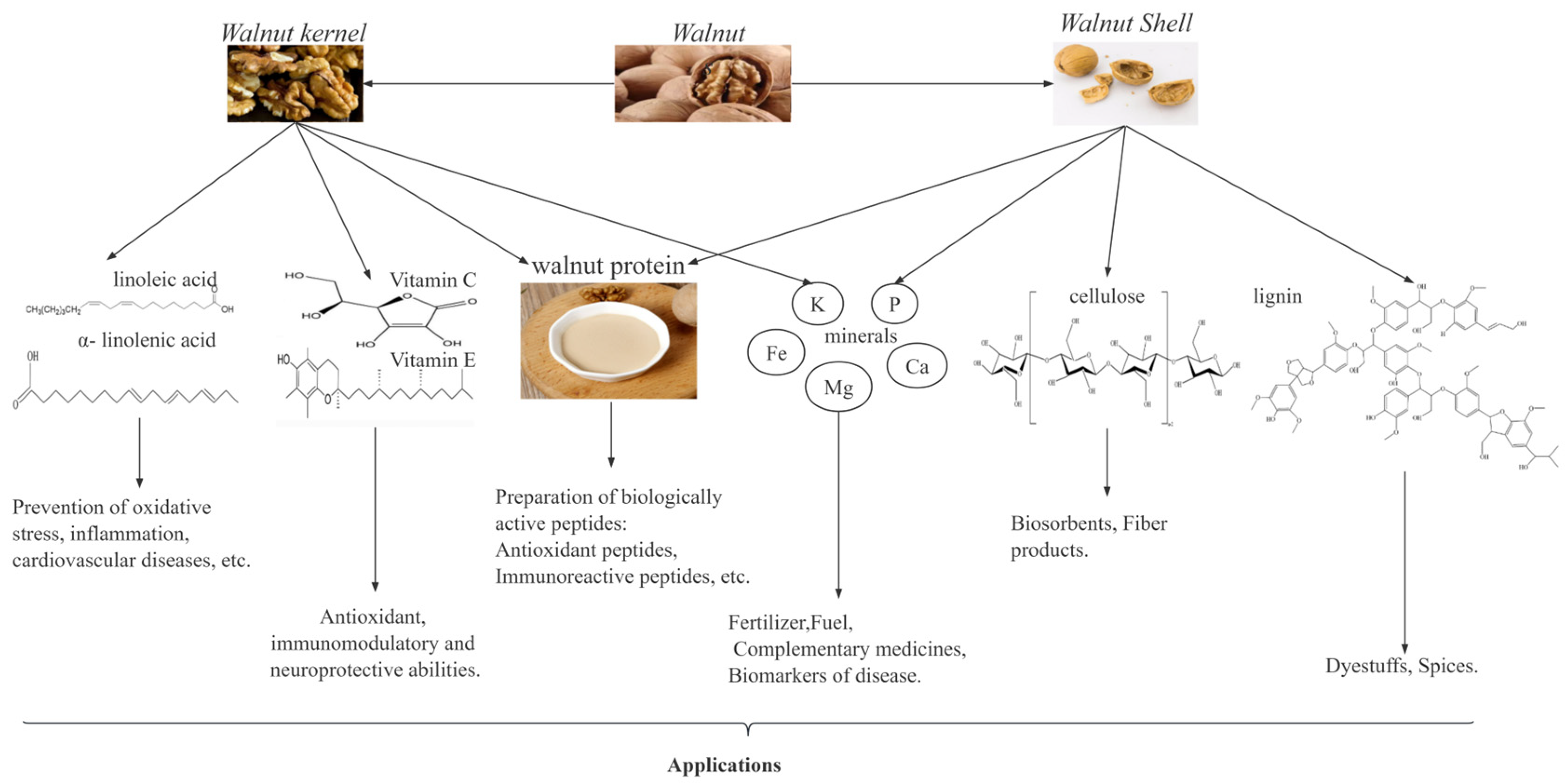
:1. Introduction
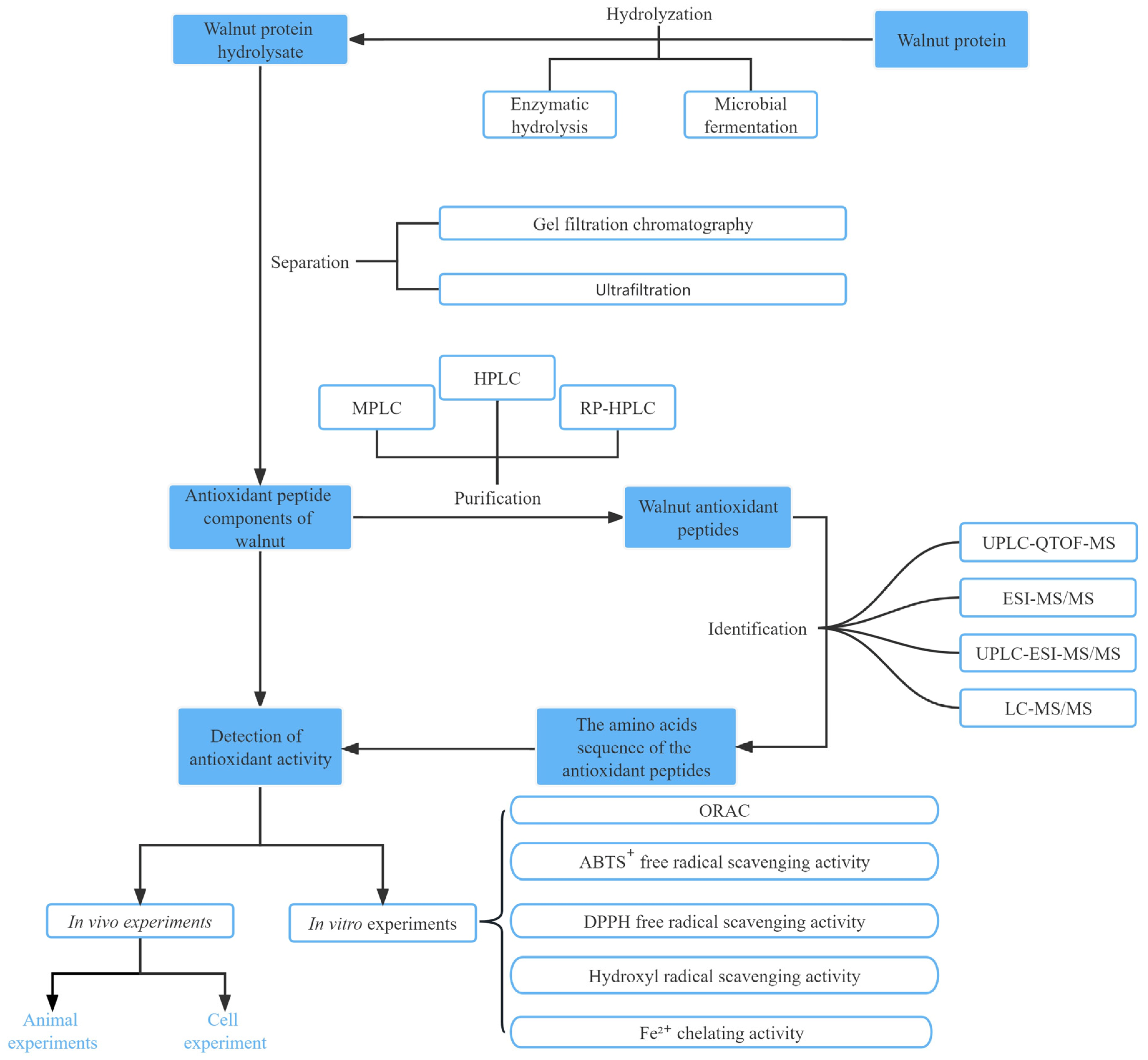
2. Preparation, Separation, Purification, and Identification of Antioxidant Peptides
2.1. Preparation of Antioxidant Peptides
2.1.1. Chemical Extraction
2.1.2. Microbial Fermentation
2.1.3. Enzymatic Hydrolysis
2.1.4. Chemical Synthesis
2.1.5. Gene Recombination
2.1.6. Other Methods
2.2. Purification, Isolation, and Identification of Walnut Antioxidant Peptides
3. Activity Analysis and Functional Study of Antioxidant Peptides
3.1. Regulation of ROS
3.2. Regulation of Endogenous Antioxidant Enzyme Activity
3.3. Regulation of Antioxidant-Related Signaling Pathways
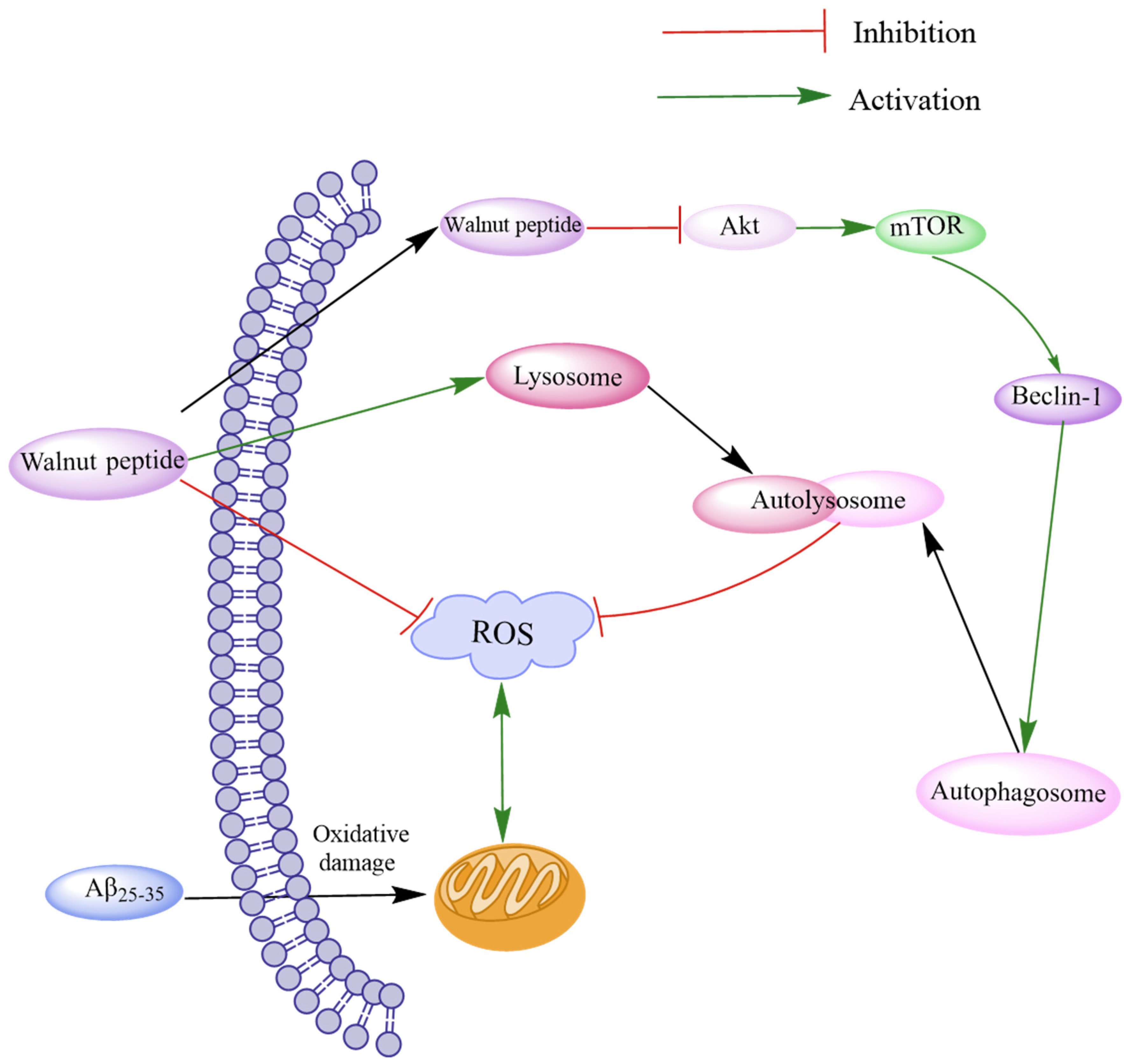
3.3.1. The Akt/mTOR Signaling Pathway
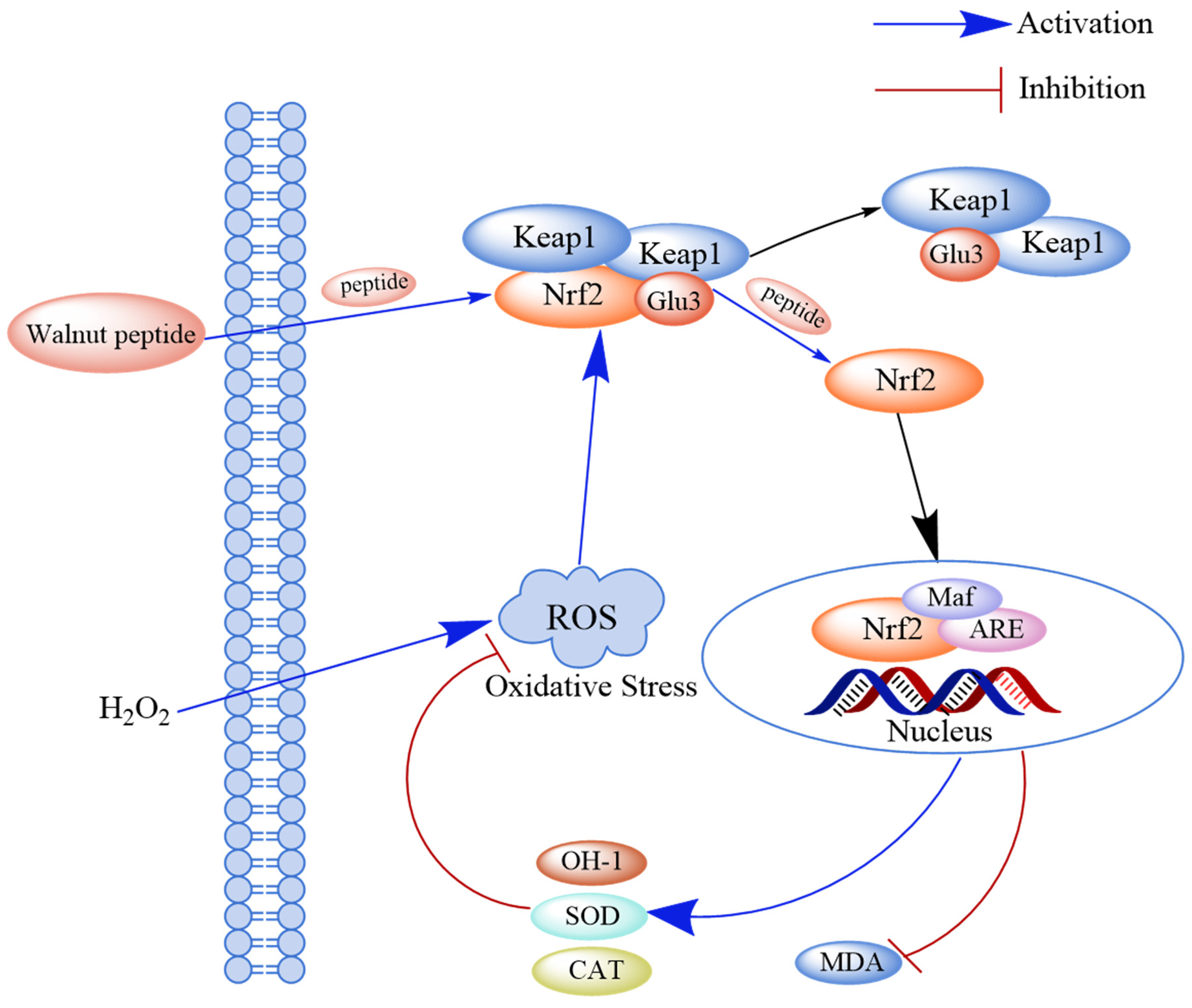
3.3.2. The Keap1-Nrf2-ARE Pathway
3.3.3. The Nrf2/HO-1 Pathway
4. Structure–Activity Relationship Study of Walnut Antioxidant Peptides
4.1. Molecular Weight
4.2. Amino Acid Composition and Sequence
4.2.1. Amino Acid Composition
4.2.2. Amino Acid Sequence
4.3. Secondary Structure
5. Conclusions
Author Contributions
Funding
Acknowledgments
Conflicts of Interest
References
- Karami, Z.; Akbari-Adergani, B. Bioactive food derived peptides: A review on correlation between structure of bioactive peptides and their functional properties. J. Food Sci. Technol. 2019, 56, 535–547. [Google Scholar] [CrossRef] [PubMed]
- Kehinde, B.A.; Sharma, P. Recently isolated antidiabetic hydrolysates and peptides from multiple food sources: A review. Crit. Rev. Food Sci. Nutr. 2020, 60, 322–340. [Google Scholar] [CrossRef]
- Wen, L.; Jiang, Y.; Zhou, X.; Bi, H.; Yang, B. Structure identification of soybean peptides and their immunomodulatory activity. Food Chem. 2021, 359, 129970. [Google Scholar] [CrossRef] [PubMed]
- Ren, L.; Fan, J.; Yang, Y.; Xu, Y.; Chen, F.; Bian, X.; Xing, T.; Liu, L.; Yu, D.; Zhang, N. Enzymatic hydrolysis of broken rice protein: Antioxidant activities by chemical and cellular antioxidant methods. Front. Nutr. 2021, 8, 788078. [Google Scholar] [CrossRef] [PubMed]
- Wang, C.; Tu, M.; Wu, D.; Chen, H.; Chen, C.; Wang, Z.; Jiang, L. Identification of an ACE-inhibitory peptide from walnut protein and its evaluation of the inhibitory mechanism. Int. J. Mol. Sci. 2018, 19, 1156. [Google Scholar] [CrossRef] [PubMed]
- Pereira, J.A.; Oliveira, I.; Sousa, A.; Ferreira, I.C.; Bento, A.; Estevinho, L. Bioactive properties and chemical composition of six walnut (Juglans regia L.) cultivars. Food Chem. Toxicol. 2008, 46, 2103–2111. [Google Scholar] [CrossRef]
- Tapia, M.I.; Sanchez-Morgado, J.R.; Garcia-Parra, J.; Ramírez, R.; Hernández, T.; González-Gómez, D. Comparative study of the nutritional and bioactive compounds content of four walnut (Juglans regia L.) cultivars. J. Food Compos. Anal. 2013, 31, 232–237. [Google Scholar] [CrossRef]
- Sheng, J.; Yang, X.; Chen, J.; Peng, T.; Yin, X.; Liu, W.; Liang, M.; Wan, J.; Yang, X. Antioxidative effects and mechanism study of bioactive peptides from defatted walnut (Juglans regia L.) meal hydrolysate. J. Agric. Food Chem. 2019, 67, 3305–3312. [Google Scholar] [CrossRef]
- Burin, M.A.; Ferronato, C.; Amorim, M.P.; Pereira, L.M.; Bilibio, D.; Bender, J.P.; Echevarria, D.P.; Garcia, H.O.; Núñez, J.G.; Bruno, A.N.; et al. Extraction of Pecan nut (Carya illinoinensis) oil using different techniques and its antitumor potential in human cancer cells. J. Supercrit. Fluids 2022, 179, 105409. [Google Scholar] [CrossRef]
- Zhi, T.; Hong, D.; Zhang, Z.; Li, S.; Xia, J.; Wang, C.; Wu, Y.; Jia, Y.; Ma, A. Anti-inflammatory and gut microbiota regulatory effects of walnut protein derived peptide LPF in vivo. Food Res. Int. 2022, 152, 110875. [Google Scholar] [CrossRef]
- Sanchis, P.; Molina, M.; Berga, F.; Muñoz, E.; Fortuny, R.; Costa-Bauzá, A.; Grases, F.; Buades, J.M. A pilot randomized crossover trial assessing the safety and short-term effects of walnut consumption by patients with chronic kidney disease. Nutrients 2020, 12, 63. [Google Scholar] [CrossRef] [PubMed]
- Guarneiri, L.L.; Paton, C.M.; Cooper, J.A. Pecan-enriched diets decrease postprandial lipid peroxidation and increase total antioxidant capacity in adults at-risk for cardiovascular disease. Nutr. Res. 2021, 93, 69–78. [Google Scholar] [CrossRef] [PubMed]
- Li, X.Y.; Guo, M.L.; Chi, J.T.; Ma, J. Bioactive peptides from walnut residue protein. Molecules 2020, 25, 1285. [Google Scholar] [CrossRef] [PubMed]
- Masoodi, L.; Gull, A.; Masoodi, F.A.; Gani, A.; Nissar, J.; Ahad, T.; Nayik, G.A.; Mukarram, S.A.; Kovács, B.; Prokisch, J.; et al. An overview on traditional vs. green technology of extraction methods for producing high quality walnut oil. Agronomy 2022, 12, 2258. [Google Scholar] [CrossRef]
- Fan, L.H.; Mao, X.Y.; Wu, Q.Z. Purification, identification and molecular docking of novel antioxidant peptides from walnut (Juglans regia L.) protein hydrolysates. Molecules 2022, 27, 8423. [Google Scholar] [CrossRef]
- Li, Y.; Jiang, B.; Zhang, T.; Mu, W.; Liu, J. Antioxidant and free radical-scavenging activities of chickpea protein hydrolysate (CPH). Food Chem. 2008, 106, 444–450. [Google Scholar] [CrossRef]
- Liu, M.C.; Yang, S.J.; Hong, D.; Yang, J.P.; Liu, M.; Lin, Y.; Huang, C.H.; Wang, C.J. A simple and convenient method for the preparation of antioxidant peptides from walnut (Juglans regia L.) protein hydrolysates. Chem. Cent. J. 2016, 10, 39. [Google Scholar] [CrossRef]
- Lv, R.; Dong, Y.; Bao, Z.; Zhang, S.; Lin, S.; Sun, N. Advances in the activity evaluation and cellular regulation pathways of food-derived antioxidant peptides. Trends Food Sci. Technol. 2022, 122, 171–186. [Google Scholar] [CrossRef]
- Wang, S.; Su, G.; Zhang, Q.; Zhao, T.; Liu, Y.; Zheng, L.; Zhao, M. Walnut (Juglans regia) peptides reverse sleep deprivation-induced memory impairment in rat via alleviating oxidative stress. J. Agric. Food Chem. 2018, 66, 10617–10627. [Google Scholar] [CrossRef]
- Su, S.; Wan, Y.; Guo, S.; Zhang, C.; Zhang, T.; Liang, M. Effect of peptide–phenolic interaction on the antioxidant capacity of walnut protein hydrolysates. Int. J. Food Sci. Technol. 2018, 53, 508–515. [Google Scholar] [CrossRef]
- Zhu, Y.; Lao, F.; Pan, X.; Wu, J. Food protein-derived antioxidant peptides: Molecular mechanism, stability and bioavailability. Biomolecules 2022, 12, 1162. [Google Scholar] [CrossRef] [PubMed]
- Wu, W.; Zhao, S.; Chen, C.; Ge, F.; Liu, D.; He, X. Optimization of production conditions for antioxidant peptides from walnut protein meal using solid-state fermentation. Food Sci. Biotechnol. 2014, 23, 1941–1949. [Google Scholar] [CrossRef]
- Wang, J.; Liu, J.; John, A.; Jiang, Y.; Zhu, H.; Yang, B.; Wen, L. Structure identification of walnut peptides and evaluation of cellular antioxidant activity. Food Chem. 2022, 388, 132943. [Google Scholar] [CrossRef] [PubMed]
- Orio, L.P.; Boschin, G.; Recca, T.; Morelli, C.F.; Ragona, L.; Francescato, P.; Arnoldi, A.; Speranza, G. New ACE-inhibitory peptides from hemp seed (Cannabis: Sativa L.) proteins. J. Agric. Food Chem. 2017, 65, 10482–10488. [Google Scholar] [CrossRef]
- Wen, Y. Plant protein-derived antioxidant peptides: Isolation, identification, mechanism of action and application in food systems: A review. Trends Food Sci. Technol. 2020, 105, 308–322. [Google Scholar] [CrossRef]
- Rojo, E.M.; Filipigh, A.A.; Bolado, S. Assisted-enzymatic hydrolysis vs. chemical hydrolysis for fractional valorization of microalgae biomass. Process Saf. Environ. Protect. 2023, 174, 276–285. [Google Scholar] [CrossRef]
- Onuh, J.O.; Girgih, A.T.; Aluko, R.E.; Aliani, M. In vitro antioxidant properties of chicken skin enzymatic protein hydrolysates and membrane fractions. Food Chem. 2014, 150, 366–373. [Google Scholar] [CrossRef]
- Cruz-Casas, D.E.; Aguilar, C.N.; Ascacio-Valdés, J.A.; Rodríguez-Herrera, R.; Chávez-González, M.L.; Flores-Gallegos, A.C. Enzymatic hydrolysis and microbial fermentation: The most favorable biotechnological methods for the release of bioactive peptides. Food chemistry. Mol. Sci. 2021, 3, 100047. [Google Scholar] [CrossRef]
- LeBlanc, J.G.; Matar, C.; Valdéz, J.C.; LeBlanc, J.; Perdigon, G. Immunomodulating effects of peptidic fractions issued from milk fermented with Lactobacillus helveticus. J. Dairy Sci. 2002, 85, 2733–2742. [Google Scholar] [CrossRef]
- Chen, N.; Yang, H.; Sun, Y.; Niu, J.; Liu, S. Purification and identification of antioxidant peptides from walnut (Juglans regia L.) protein hydrolysates. Peptides 2012, 38, 344–349. [Google Scholar] [CrossRef]
- Melini, F.; Melini, V.; Luziatelli, F.; Ficca, A.G.; Ruzzi, M. Health-promoting components in fermented foods: An up-to-date systematic review. Nutrients 2019, 11, 1189. [Google Scholar] [CrossRef] [PubMed]
- Chen, H.; Zhao, M.; Lin, L.; Wang, J.; Sun-Waterhouse, D.; Dong, Y.; Zhuang, M.; Su, G. Identification of antioxidative peptides from defatted walnut meal hydrolysate with potential for improving learning and memory. Food Res. Int. 2015, 78, 216–223. [Google Scholar] [CrossRef] [PubMed]
- Feng, L.; Peng, F.; Wang, X.; Li, M.; Lei, H.; Xu, H. Identification and characterization of antioxidative peptides derived from simulated in vitro gastrointestinal digestion of walnut meal proteins. Food Res. Int. 2019, 116, 518–526. [Google Scholar] [CrossRef] [PubMed]
- Feng, L.; Wang, Y.; Yang, J.; Sun, Y.F.; Li, Y.W.; Ye, Z.H.; Lin, H.B.; Yang, K. Overview of the preparation method, structure and function, and application of natural peptides and polypeptides. Biomed. Pharmacother. 2022, 153, 113493. [Google Scholar] [CrossRef]
- Liu, M.; Yang, S.; Yang, J.; Lee, Y.; Kou, J.; Wang, C. Neuroprotective and memory-enhancing effects of antioxidant peptide from walnut (Juglans regia L.) protein hydrolysates. Nat. Prod. Commun. 2019, 14, 1934578X19865838. [Google Scholar] [CrossRef]
- Hu, F.; Ci, A.T.; Wang, H.; Zhang, Y.Y.; Zhang, J.G.; Thakur, K.; Wei, Z.J. Identification and hydrolysis kinetic of a novel antioxidant peptide from pecan meal using Alkaline protease. Food Chem. 2018, 261, 301–310. [Google Scholar] [CrossRef]
- Zhang, S.; Zheng, H.; Zhang, R.; Shi, M.; Ren, R.; Cheng, S.; Dun, C. Extraction optimization and antioxidant activity evaluation of se-enriched walnut proteins. J. Food Process. Preserv. 2022, 46, e16719. [Google Scholar] [CrossRef]
- Sibilska-Kaminski, I.K.; Yin, J. Toward molecular cooperation by de novo peptides. Orig. Life Evol. Biosph. 2021, 51, 71–82. [Google Scholar] [CrossRef]
- Liu, C.; Guo, Y.; Zhao, F.; Qin, H.; Lu, H.; Fang, L.; Wang, J.; Min, W. Potential mechanisms mediating the protective effects of a peptide from walnut (Juglans mandshurica Maxim.) against hydrogen peroxide induced neurotoxicity in PC12 cells. Food Funct. 2019, 10, 3491–3501. [Google Scholar] [CrossRef]
- Fuse, S.; Otake, Y.; Nakamura, H. Peptide synthesis utilizing micro-flow technology. Chem. Asian J. 2018, 13, 3818–3832. [Google Scholar] [CrossRef]
- Titani, K. Protein design based on the structure and function relationship and its clinical application. Rinshō Ketsueki Jpn. J. Clin. Hematol. 1994, 35, 323–331. [Google Scholar]
- Goto, M.; Endo, T. High-molecular-weight poly(Gly-Val-Gly-Val-Pro) synthesis through microwave irradiation. J. Peptide Sci. 2016, 22, 452–460. [Google Scholar] [CrossRef] [PubMed]
- Wang, X.L.; Ma, S.N.; Yuan, Y.H.; Ding, Y.; Li, D.S. Expression and purification recombinant antihypertensive peptide ameliorates hypertension in rats with spontaneous hypertension. Protein Expr. Purif. 2015, 113, 30–34. [Google Scholar] [CrossRef] [PubMed]
- Liang, R.; Cheng, S.; Dong, Y.; Ju, H. Intracellular antioxidant activity and apoptosis inhibition capacity of PEF-treated KDHCH in HepG2 cells. Food Res. Int. 2019, 121, 336–347. [Google Scholar] [CrossRef]
- He, L.; Gao, Y.; Wang, X.; Han, L.; Yu, Q.; Shi, H.; Song, R. Ultrasonication promotes extraction of antioxidant peptides from oxhide gelatin by modifying collagen molecule structure. Ultrason. Sonochem. 2021, 78, 105738. [Google Scholar] [CrossRef]
- Mala, T.; Sadiq, M.B.; Anal, A.K. Comparative extraction of bromelain and bioactive peptides from pineapple byproducts by ultrasonic- and microwave-assisted extractions. J. Food Process Eng. 2021, 44, e13709. [Google Scholar] [CrossRef]
- Golly, M.K.; Ma, H.; Yuqing, D.; Wu, P.; Dabbour, M.; Sarpong, F.; Farooq, M. Enzymolysis of walnut (Juglans regia L.) meal protein: Ultrasonication-assisted alkaline pretreatment impact on kinetics and thermodynamics. J. Food Biochem. 2019, 43, e12948. [Google Scholar] [CrossRef]
- Wang, Q.; Wang, Y.; Huang, M.; Hayat, K.; Kurtz, N.C.; Wu, X.; Ahmad, M.; Zheng, F. Ultrasound-assisted alkaline proteinase extraction enhances the yield of pecan protein and modifies its functional properties. Ultrason. Sonochem. 2021, 80, 105789. [Google Scholar] [CrossRef]
- Ishak, N.H.; Sarbon, N.M. A review of protein hydrolysates and bioactive peptides deriving from wastes generated by fish processing. Food Bioprocess Technol. 2017, 27, 1365–1381. [Google Scholar] [CrossRef]
- Sila, A.; Bougatef, A. Antioxidant peptides from marine by-products: Isolation, identification and application in food systems. A review. J. Funct. Foods 2016, 21, 10–26. [Google Scholar] [CrossRef]
- Wang, C.; Song, W.; Jiang, L.Z.; Du, M. Purification and identification of an ACE-inhibitory peptide from walnut protein hydrolysate. Eur. Food Res. Technol. 2014, 239, 333–338. [Google Scholar] [CrossRef]
- Agyei, D.; Tsopmo, A.; Udenigwe, C.C. Bioinformatics and peptidomics approaches to the discovery and analysis of food-derived bioactive peptides. Anal. Bioanal. Chem. 2018, 410, 3463–3472. [Google Scholar] [CrossRef] [PubMed]
- Demelbauer, U.M.; Zehl, M.; Plematl, A.; Allmaier, G.; Rizzi, A. Determination of glycopeptide structures by multistage mass spectrometry with low-energy collision-induced dissociation: Comparison of electrospray ionization quadrupole ion trap and matrix-assisted laser desorption/ionization quadrupole ion trap reflectron time-of-flight approaches. Rapid Commun. Mass Spectrom. 2004, 18, 1575–1582. [Google Scholar] [PubMed]
- Meng, D.; Zhang, P.; Zhang, L.; Wang, H.; Ho, C.-T.; Li, S.; Shahidi, F.; Zhao, H. Detection of cellular redox reactions and antioxidant activity assays. J. Funct. Foods 2017, 37, 467–479. [Google Scholar] [CrossRef]
- Munteanu, I.G.; Apetrei, C. Analytical methods used in determining antioxidant activity: A review. Int. J. Mol. Sci. 2021, 22, 3380. [Google Scholar] [CrossRef]
- Liu, R.; Xing, L.; Fu, Q.; Zhou, G.H.; Zhang, W.G. A review of antioxidant peptides derived from meat muscle and by-products. Antioxidants 2016, 5, 32. [Google Scholar] [CrossRef]
- Mao, X.; Gu, C.; Chen, D.; Yu, B.; He, J. Oxidative stress-induced diseases and tea polyphenols. Oncotarget 2017, 8, 81649–81661. [Google Scholar] [CrossRef]
- Hancock, J.T.; Desikan, R.; Neill, S.J. Role of reactive oxygen species in cell signalling pathways. Biochem. Soc. Trans. 2001, 29, 345–349. [Google Scholar] [CrossRef]
- Prasad, S.; Gupta, S.C.; Tyagi, A.K. Reactive oxygen species (ROS) and cancer: Role of antioxidative nutraceuticals. Cancer Lett. 2016, 3835, 95–105. [Google Scholar] [CrossRef]
- Ren, D.; Zhao, F.; Liu, C.; Wang, J.; Guo, Y.; Liu, J.; Min, W. Antioxidant hydrolyzed peptides from Manchurian walnut (Juglans mandshurica Maxim.) attenuate scopolamine-induced memory impairment in mice. J. Sci. Food Agric. 2018, 98, 5142–5152. [Google Scholar] [CrossRef]
- Jahanbani, R.; Ghaffari, S.M.; Salami, M.; Vahdati, K.; Sepehri, H.; Sarvestani, N.N.; Sheibani, N.; Moosavi-Movahedi, A.A. Antioxidant and anticancer activities of Walnut (Juglans regia L.) protein hydrolysates using different proteases. Plant Foods Hum. Nutr. 2016, 71, 402–409. [Google Scholar] [CrossRef] [PubMed]
- Xue, Z.; Yu, W.; Liu, Z.; Wu, M.; Kou, X.; Wang, J. Preparation and antioxidative properties of a rapeseed (Brassica napus) protein hydrolysate and three peptide fractions. J. Agric. Food Chem. 2009, 57, 5287–5293. [Google Scholar] [CrossRef] [PubMed]
- Zhao, F.; Wang, J.; Lu, H.; Fang, L.; Qin, H.; Liu, C.; Min, W. Neuroprotection by walnut-derived peptides through autophagy promotion via Akt/mTOR signaling pathway against oxidative stress in PC12 Cells. J. Agric. Food Chem. 2020, 68, 3638–3648. [Google Scholar] [CrossRef] [PubMed]
- Jia, Q.; Yuan, J.F.; Liu, H.P.; Li, M.-Y.; Wu, Y.-R. Purification and identification of dual-enzyme hydrolysates obtained from defatted walnut and its antioxidant effects on d-galactose-induced aging mice. J. Food Meas. Charact. 2021, 15, 1034–1043. [Google Scholar] [CrossRef]
- Liu, D.; Guo, Y.; Zhu, J.; Tian, W.; Chen, M.; Ma, H. The necessity of enzymatically hydrolyzing walnut protein to exert antihypertensive activity based on in vitro simulated digestion and in vivo verification. Food Funct. 2021, 12, 3647–3656. [Google Scholar] [CrossRef]
- Gu, L.; Yu, Q.; Li, Q.; Zhang, L.; Lu, H.; Zhang, X. Andrographolide protects PC12 cells against β-Amyloid-induced autophagy-associated cell death through activation of the Nrf2-mediated p62 signaling pathway. Int. J. Mol. Sci. 2018, 19, 2844. [Google Scholar] [CrossRef]
- Shi, X.; Liu, Y.; Zhang, D.; Xiao, D. Valproic acid attenuates sepsis-induced myocardial dysfunction in rats by accelerating autophagy through the PTEN/AKT/mTOR pathway. Life Sci. 2019, 232, 116613. [Google Scholar] [CrossRef]
- Kubben, N.; Zhang, W.; Wang, L.; Voss, T.C.; Yang, J.; Qu, J.; Liu, G.H.; Misteli, T. Repression of the antioxidant nrf2 pathway in premature aging. Cell 2016, 165, 1361–1374. [Google Scholar] [CrossRef]
- Sun, Y.; Huang, J.; Chen, Y.; Shang, H.; Zhang, W.; Yu, J.; He, L.; Xing, C.; Zhuang, C. Direct inhibition of Keap1-Nrf2 protein-protein interaction as a potential therapeutic strategy for Alzheimer’s disease. Bioorg. Chem. 2020, 103, 104172. [Google Scholar] [CrossRef]
- Zhang, J.; Wu, S.; Wang, Q.; Yuan, Q.; Li, Y.; Reboredo-Rodríguez, P.; Varela-López, A.; He, Z.; Wu, F.; Hu, H.; et al. Oxidative stress amelioration of novel peptides extracted from enzymatic hydrolysates of Chinese pecan cake. Int. J. Mol. Sci. 2022, 23, 12086. [Google Scholar] [CrossRef]
- Gao, Y.; Qin, H.; Wu, D.; Liu, C.; Fang, L.; Wang, J.; Liu, X.; Min, W. Walnut peptide WEKPPVSH in alleviating oxidative stress and inflammation in lipopolysaccharide-activated BV-2 microglia via the Nrf2/HO-1 and NF-κB/p38 MAPK pathways. J. Biosci. Bioeng. 2021, 132, 496–504. [Google Scholar] [CrossRef] [PubMed]
- Zhao, F.; Liu, C.; Fang, L.; Lu, H.; Wang, J.; Gao, Y.; Gabbianelli, R.; Min, W. Walnut-derived peptide activates PINK1 via the NRF2/KEAP1/HO-1 pathway, promotes mitophagy, and alleviates learning and memory impairments in a mice model. J. Agric. Food Chem. 2021, 69, 2758–2772. [Google Scholar] [CrossRef]
- Wang, J.; Wu, T.; Fang, L.; Liu, C.; Liu, X.; Li, H.; Shi, J.; Li, M.; Min, W. Peptides from walnut (Juglans mandshurica Maxim.) protect hepatic HepG2 cells from high glucose-induced insulin resistance and oxidative stress. Food Funct. 2020, 11, 8112–8121. [Google Scholar] [CrossRef]
- Sun, G.Y.; Li, R.; Cui, J.; Hannink, M.; Gu, Z.; Fritsche, K.L.; Lubahn, D.B.; Simonyi, A. Withania somnifera and its withanolides attenuate oxidative and inflammatory responses and up-regulate antioxidant responses in BV-2 Microglial Cells. Neuromolecular Med. 2016, 18, 241–252. [Google Scholar] [CrossRef] [PubMed]
- Chi, C.F.; Hu, F.Y.; Wang, B.; Ren, X.J.; Deng, S.G.; Wu, C.W. Purification and characterization of three antioxidant peptides from protein hydrolyzate of croceine croaker (Pseudosciaena crocea) muscle. Food Chem. 2015, 168, 662–667. [Google Scholar] [CrossRef]
- Pan, X.Y.; Wang, Y.M.; Li, L.; Chi, C.F.; Wang, B. Four antioxidant peptides from protein hydrolysate of red stingray (Dasyatis akajei) cartilages: Isolation, identification, and In Vitro activity evaluation. Mar. Drugs 2019, 17, 263. [Google Scholar] [CrossRef] [PubMed]
- Jin, J.E.; Ahn, C.B.; Je, J.Y. Purification and characterization of antioxidant peptides from enzymatically hydrolyzed ark shell (Scapharca subcrenata). Process Biochem. 2018, 72, 170–176. [Google Scholar] [CrossRef]
- Dieterich, F.; Boscolo, W.; Leivas, C.; Kiatkoski, E.; Waszczynskyj, N. Potential Use of Tuna (Thunnus albacares) by-product: Production of Antioxidant Peptides and Recovery of Unsaturated Fatty Acids from Tuna Head. Int. J. Food Eng. 2017, 13, 20150365. [Google Scholar]
- Wang, B. Purification, identification, activity evaluation, and stability of antioxidant peptides from Alkaline protease hydrolysate of antarctic krill (Euphausia superba) proteins. Mar. Drugs 2021, 19, 347. [Google Scholar]
- Elias, R.J.; Kellerby, S.S.; Decker, E.A. Antioxidant activity of proteins and peptides. Crit. Rev. Food Sci. Nutr. 2008, 48, 430–441. [Google Scholar] [CrossRef]
- Liu, Z.; Shi, Y.; Liu, H.; Jia, Q.; Liu, Q.; Tu, J. Purification and identification of pine nut (Pinus yunnanensis Franch.) protein hydrolysate and its antioxidant activity in Vitro and in Vivo. Chem. Biodiv. 2020, 18, e2000710. [Google Scholar] [CrossRef] [PubMed]
- Chan, K.M.; Decker, E.A.; Feustman, C. Endogenous skeletal muscle antioxidants. Crit. Rev. Food Sci. Nutr. 1994, 34, 403–426. [Google Scholar] [CrossRef] [PubMed]
- Li, Q.; Shi, C.; Wang, M.; Zhou, M.; Liang, M.; Zhang, T.; Yuan, E.; Wang, Z.; Yao, M.; Ren, J. Tryptophan residue enhances in vitro walnut protein-derived peptides exerting xanthine oxidase inhibition and antioxidant activities. J. Funct. Foods 2019, 53, 276–285. [Google Scholar] [CrossRef]
- Ren, J.; Zhao, M.; Shi, J.; Wang, J.; Jiang, Y.; Cui, C.; Kakuda, Y.; Xue, S.J. Purification and identification of antioxidant peptides from grass carp muscle hydrolysates by consecutive chromatography and electrospray ionization-mass spectrometry. Food Chem. 2008, 108, 727–736. [Google Scholar] [CrossRef] [PubMed]
- Gu, M.; Chen, H.P.; Zhao, M.M.; Wang, X.; Yang, B.; Ren, J.-Y.; Su, G.-W. Identification of antioxidant peptides released from defatted walnut (Juglans Sigillata Dode) meal proteins with pancreatin. LWT Food Sci. Technol. 2015, 60, 213–220. [Google Scholar] [CrossRef]
- Zheng, L.; Zhao, Y.J.; Dong, H.Z.; Su, G.; Zhao, M. Structure-activity relationship of antioxidant dipeptides: Dominant role of Tyr, Trp, Cys and Met residues. J. Funct. Foods 2016, 21, 485–496. [Google Scholar] [CrossRef]
- Xiang, Z.; Xue, Q.; Gao, P.; Yu, H.; Wu, M.; Zhao, Z.; Li, Y.; Wang, S.; Zhang, J.; Dai, L. Antioxidant peptides from edible aquatic animals: Preparation method, mechanism of action, and structure-activity relationships. Food Chem. 2023, 404 Pt B, 134701. [Google Scholar] [CrossRef]
- Bongirwar, V.; Mokhade, A.S. Different methods, techniques and their limitations in protein structure prediction: A review. Prog. Biophys. Mol. Biol. 2022, 173, 72–82. [Google Scholar] [CrossRef]

| Enzyme Name | In Vitro Test Index | Isolation, Purification, and Identification | Enzymatic Conditions | Peptide Amino Sequences | Reference |
|---|---|---|---|---|---|
| Neutral protease, pineapple protease, pepsin, alkaline protease, papain, pancreatic protease | ABTS+ scavenging activity assay; DPPH free radical scavenging activity; Superoxide radical scavenging activity | Ultrafiltration membrane PVDF flat microporous membrane (MWCO of 200 kDa) | Neutral protease: pH: 7.0. Temperature: 50 °C, substrate concentration: 1:30 Alkaline protease: pH: 8.0. Temperature: 50 °C, substrate concentration 2:30 Pepsin: pH: 2.0. Temperature: 37 °C, substrate concentration: 1:30 Trypsin. pH: 8.0; temperature: 50 °C Enzyme/substrate: 2:100 (w/w) Pineapple protease: pH: 7.0. Temperature: 50 °C, substrate concentration: 3:30 Papain: pH: 7.0. Temperature: 50 °C, substrate concentration: 2:30 | Walnut peptide | [1,2] |
| Alkaline protease | ORAC DPPH scavenging activity assay | Ultrafiltration UPLC-MS/MS | Alkaline protease: 23,400 U/mg pH: 8.0, temperature: 55 °C | QGRPWG, PSRADIY, AYNIPVNIAR | [3] |
| Trypsin | DPPH scavenging activity assay Fe2+ chelating capacity ABTS+ scavenging activity assay ORAC | Gel filtration chromatography RP-HPLC UPLC-ESI-MS/MS | Trypsin: 4.6 × 10⁶ U/g pH: 7.5, temperature: 37 °C Enzyme/substrate: 2:100 (w/w) | YS, YK, YT, EM, CA, GR, YA, YG, LPC, CHC, SEK, GHC, YSVH | [4] |
| Pepsin, pancreatic protease | ABTS+ free radical scavenging activity. Oxygen radical absorbance capacity (ORAC). | Ultrafiltration Gel filtration chromatography. RP-HPLC. UPLC-QTOF-MS | Pepsin/substrate: 1:10 (w/w) pH: 2.0; temperature: 37 °C Trypsin/substrate: 1:10 (w/w) pH: 7.4; temperature: 37 °C | TY, SGGY. | [5] |
| Neutral protease, flavor protease, pepsin, and alkaline protease | DPPH free radical scavenging activity; ABTS+ Free radical scavenging activity; Hydroxyl radical scavenging activity; | Gel filtration chromatography HPLC LC-MS/MS | Neutral protease: 100 U/mg pH: 7.0; temperature: 50 °C Alkaline protease: 200 U/mg pH: 9.0; temperature: 55 °C Flavor protease: 30 U/mg pH: 7.5; temperature: 45 °C Pepsin: 500 U/mg pH: 2.0; temperature: 37 °C Enzyme/protein: 1:50 (w/w) | HADMVFY, NHCQYYL, NLFHKRP. | [6] |
| Alkaline protease | ABTS+ free radical scavenging activity; DPPH free radical scavenging activity; Hydroxyl radical scavenging activity Fe2+ chelating activity. | Ultrafiltration. Ion exchange chromatography. Gel filtration chromatography. RP-HPLC ESI-MS/MS | Alkaline protease: 62,000 U/g pH: 10.0; temperature: 55 °C Enzyme/substrate: 1:20 (w/w) | LAYLQYTDFETR | [7] |
| Alkaline protease | DPPH free radical scavenging activity; Hydroxyl radical scavenging activity; | SDS-PAGE Ion exchange column LC-HG-AFS | Alkaline protease: 4000 U/g, pH: 10.0, temperature: 45 °C, time: 4 h | Walnut peptide | [8] |
| Neutral protease, alkaline protease | DPPH free radical scavenging activity; Hydroxyl radical scavenging activity; Fe2+ chelating capacity | Ultrafiltration Gel filtration chromatography RP-HPLC ESI-MS/MS | Neutral protease: pH: 7.0, temperature: 50 °C Alkaline protease: pH: 8.0, temperature: 50 °C Pepsin: pH: 2.0; temperature: 37 °C | ADAF. | [9] |
| Alkaline protease | DPPH free radical scavenging activity; ORAC | Ion exchange column UPLC-MS/MS | Alkaline protease: 23,400 U/mg pH: 8.0, temperature:55 °C, time: 8 h | QGRPWG, GGEPRN, PSRADIY, AYNIPVNIAR, | [10] |
| Trypsin | Hydroxyl radical scavenging activity; ORAC | SP-825 macroporous absorption resin Medium-pressure liquid chromatography UPLC-ESI-MS/MS | Trypsin: 4.6 × 10⁶ U/g pH: 8.0; temperature: 55 °C Enzyme/substrate: 1:20 (w/w) | WSREEQEREE, ADIYTEEAGR. | [11] |
Disclaimer/Publisher’s Note: The statements, opinions and data contained in all publications are solely those of the individual author(s) and contributor(s) and not of MDPI and/or the editor(s). MDPI and/or the editor(s) disclaim responsibility for any injury to people or property resulting from any ideas, methods, instructions or products referred to in the content. |
© 2023 by the authors. Licensee MDPI, Basel, Switzerland. This article is an open access article distributed under the terms and conditions of the Creative Commons Attribution (CC BY) license (https://creativecommons.org/licenses/by/4.0/).
Share and Cite
Hu, Y.; Ni, C.; Wang, Y.; Yu, X.; Wu, H.; Tu, J.; Li, C.; Xiao, Z.; Wen, L. Research Progress on the Preparation and Function of Antioxidant Peptides from Walnuts. Int. J. Mol. Sci. 2023, 24, 14853. https://doi.org/10.3390/ijms241914853
Hu Y, Ni C, Wang Y, Yu X, Wu H, Tu J, Li C, Xiao Z, Wen L. Research Progress on the Preparation and Function of Antioxidant Peptides from Walnuts. International Journal of Molecular Sciences. 2023; 24(19):14853. https://doi.org/10.3390/ijms241914853
Chicago/Turabian StyleHu, Yuxi, Ce Ni, Yingying Wang, Xun Yu, Hao Wu, Jia Tu, Changzhu Li, Zhihong Xiao, and Li Wen. 2023. "Research Progress on the Preparation and Function of Antioxidant Peptides from Walnuts" International Journal of Molecular Sciences 24, no. 19: 14853. https://doi.org/10.3390/ijms241914853
APA StyleHu, Y., Ni, C., Wang, Y., Yu, X., Wu, H., Tu, J., Li, C., Xiao, Z., & Wen, L. (2023). Research Progress on the Preparation and Function of Antioxidant Peptides from Walnuts. International Journal of Molecular Sciences, 24(19), 14853. https://doi.org/10.3390/ijms241914853