Article

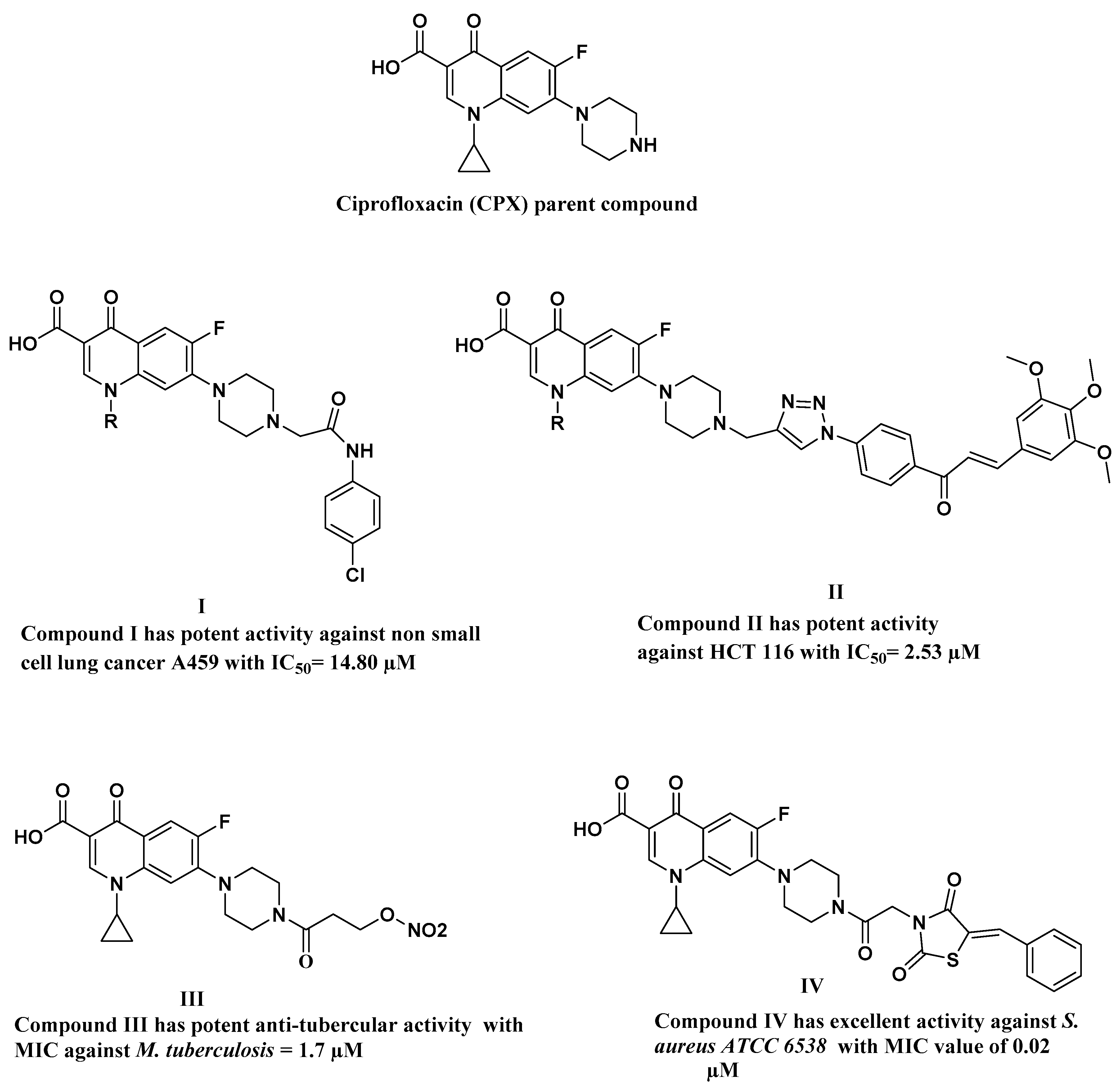
Synthesis, Physicochemical Characterization using a Facile Validated HPLC Quantitation Analysis Method of 4-Chloro-phenylcarbamoyl-methyl Ciprofloxacin and Its Biological Investigations

by
Mostafa F. Al-Hakkani
1,*,
Nourhan Ahmed
1,
Alaa A. Abbas
1,
Mohammad H. A. Hassan
2,
Hossameldin A. Aziz
3,
Ali M. Elshamsy
4,
Hazim O. Khalifa
5,6,
Mohamed A. Abdelshakour
7,
Mohammed S. Saddik
8,
Mahmoud M. A. Elsayed
8,
Marwa A. Sabet
9,
Mohamed A. El-Mokhtar
10,
Mosa Alsehli
11,
M. S. Amin
11,12,
Ahmed M. Abu-Dief
11,13,* and
Hamada H. H. Mohammed
14
1
Department of Research, Development, and Stability, UP Pharma, Industrial Zone, Arab El Awamer, Abnoub 76, Assiut 71745, Egypt
2
Department of Medical Laboratory Technology, Higher Technological Institute for Applied Health Sciences in Minya, Minya 71511, Egypt
3
Department of Pharmaceutical Chemistry, Faculty of Pharmacy, New Valley University, New Valley 72511, Egypt
4
Department of Pharmaceutical Chemistry, Faculty of Pharmacy, Deraya University, Mina, New Minia 61768, Egypt
5
Department of Veterinary Medicine, College of Agriculture and Veterinary Medicine, United Arab Emirates University, Al Ain P.O. Box 1555, United Arab Emirates
6
Department of Pharmacology, Faculty of Veterinary Medicine, Kafr Elsheikh University, Kafr El Sheikh 33516, Egypt
7
Department of Pharmaceutical Analytical Chemistry, Faculty of Pharmacy, Sohag University, Sohag 82524, Egypt
8
Department of Pharmaceutics and Clinical Pharmacy, Faculty of Pharmacy, Sohag University, Sohag 82524, Egypt
9
Department of Microbiology and Immunology, Faculty of Pharmacy, Sphinx University, New-Assiut 71684, Egypt
10
Department of Medical Microbiology and Immunology, Faculty of Medicine, Assiut University, Assiut 71515, Egypt
11
Department of Chemistry, College of Science, Taibah University, Madinah P.O. Box 344, Saudi Arabia
12
Chemistry Department, Faculty of science, Ain Shams University, Cairo 11566, Egypt
13
Department of Chemistry, Faculty of Science, Sohag University, Sohag 82524, Egypt
14
Department of Pharmaceutical Chemistry, Faculty of Pharmacy, Sohag University, Sohag 82524, Egypt
*
Authors to whom correspondence should be addressed.
Int. J. Mol. Sci. 2023, 24(19), 14818; https://doi.org/10.3390/ijms241914818
Submission received: 8 September 2023 / Revised: 26 September 2023 / Accepted: 30 September 2023 / Published: 1 October 2023

Abstract

:
A novel derivative of ciprofloxacin (Cpx) was synthesized and characterized using various analytical techniques, including FT-IR spectroscopy, UV-Vis spectroscopy, TEM and SEM analysis, 1H NMR, 13C NMR, and HPLC analysis. The newly prepared Cpx derivative (Cpx-Drv) exhibited significantly enhanced antibacterial properties compared to Cpx itself. In particular, Cpx-Drv demonstrated a 51% increase in antibacterial activity against S. aureus and a 30% improvement against B. subtilis. It displayed potent inhibitory effects on topoisomerases II (DNA gyrase and topoisomerase IV) as potential molecular targets, with IC50 values of 6.754 and 1.913 µg/mL, respectively, in contrast to Cpx, which had IC50 values of 2.125 and 0.821 µg/mL, respectively. Docking studies further supported these findings, showing that Cpx-Drv exhibited stronger binding interactions with the gyrase enzyme (PDB ID: 2XCT) compared to the parent Cpx, with binding affinities of −10.3349 and −7.7506 kcal/mole, respectively.

1. Introduction

Recently, the world has experienced rapid advancement and competition in the pharmaceutical industry. This is evident within the context of the evolving situation surrounding the coronavirus and diverse cancer ailments. Faced with this worldwide crisis, the resilience and harmonious collaboration among diverse scientific disciplines in their pursuit of identifying a secure solution were prominently demonstrated [1,2]. Antibiotic resistance has emerged as one of the most significant global health issues of the 21st century. Antibiotic resistance is reaching dangerously high levels worldwide, especially in developing countries [3].
Therefore, there is an urgent need to discover new, more appropriate antibiotics in light of the mutations of pathogenic and deadly microorganisms. Antibiotics are essential for contemporary human patients because they cure and prevent diseases. The use of pharmaceuticals has grown throughout history [4]. Among the most famous families of antibiotics common in modern times are the family of macrolides, beta-lactams, cephalosporins, and fluoroquinolones. Fluoroquinolones are synthetic broad-spectrum bactericidal agents that include the vast majority of antibiotics being used in various treatment approaches, such as ciprofloxacin (Cpx), garenoxacin, gatifloxacin, gemifloxacin, levofloxacin, and moxifloxacin [2]. Fluoroquinolones act by inhibiting the activities of DNA topoisomerase II, resulting in the accumulation of double-stranded DNA breaks and bacterial cell death. In addition, fluoroquinolones have various biological activities including antitubercular, antifungal [5], anti-inflammatory [6], anti-Alzheimer’s [7], antiviral [8], anti-malarial [9], and antitumor activities [10,11]. Cpx is one of the most important examples of the second-generation fluoroquinolones and the fifth most widely used generic antibiotic in the world due to its broad antibacterial activity [12] against both Gram-positive and Gram-negative bacteria and because it can also be used in wound dressing [13,14,15,16,17,18]. The importance of Cpx is also due to its application in managing prevalent conditions such as respiratory tract infections, urinary tract infections, prostatitis, cellulitis, malignant otitis externa, chancroid, anthrax, endocarditis, and gastroenteritis [19]. Cpx’s full chemical name, according to IUPAC, is 1-cyclopropyl-6-fluoro-1,4-dihydro-4-oxo-7-(1piperazinyl)-3-quinoline carboxylic acid (Figure 1), and it has a molecular mass of 331.35 g/mol and empirical formula C17H18FN3O3 [20].
Several types of research have indicated that N-4 piperazinyl substitution of quinolones’ nucleus reduces zwitterion characteristics, improves physiochemical properties, and enhances antibacterial activity towards Gram-positive strains, and increases lipophilicity, which is considered one of the most important factors taken into consideration for the design of new anticancer agents.
Based on the above-mentioned aspects, various derivatives have been synthesized, aiming to increase antibacterial activity or shift biological activity towards anticancer activity [9], as presented in Figure 1.
From this point, scientists started thinking about finding more derivatives of Cpx to achieve the desired results in terms of their superiority over its basic counterpart. Recently, Hamada et al. synthesized several derivatives of Cpx as listed in their published paper [21], which reported the preparation of seven derivatives to improve the weak anticancer activity of Cpx. Among these derivatives is compound I (Figure 1), which has a mass of 498.94 g/mol and the empirical formula C25H24FN4ClO4.
The efficacy of Cpx-Drv (Figure 1, compound I) has been evaluated and found to manifest a very good efficacy in this regard as an anticancer agent [21] against fifteen cell lines. However, Hamada and co-authors did not examine the anti-microbiological activity of Cpx-Drv against different species of Gram-negative and Gram-positive bacteria. They also did not provide a direct analysis method to detect and determine Cpx-Drv in a quick way that expresses the assay of this derivative and compares it with the main drug, the parent from which it is derived, i.e., Cpx.
Therefore, the purpose of this work is to prepare Cpx-Drv using the same approach as previously reported [21], but with more physicochemical characterizations. A UV-RP-HPLC analysis method with full validation procedures was implemented. Moreover, anti-microbiological activity against different bacterial species was demonstrated.

2. Results and Discussion
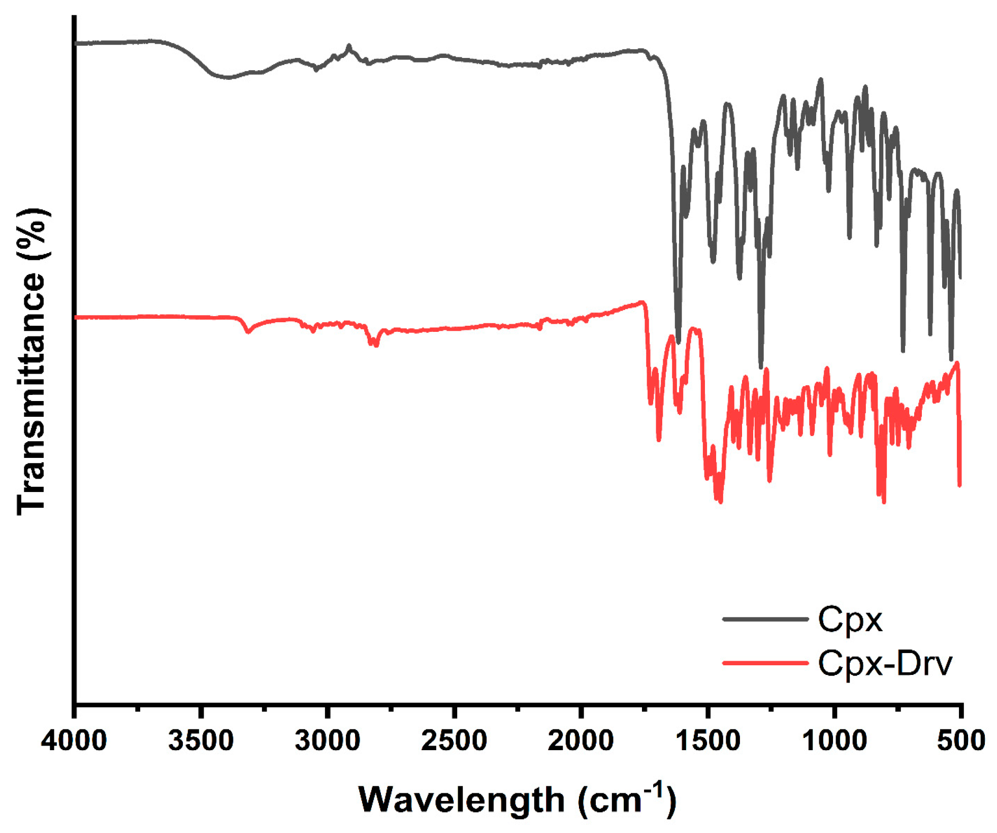
2.1. FTIR Analysis

The FTIR spectra of the as-prepared Cpx and Cpx-Drv are recorded in the mid-infrared region in the range of (500–4000 cm−1). The frequency of the bands for the different functional groups could be used to identify the molecule’s structure. Figure 2 and Table 1 provide a rapid summary for the identification of the functional groups of Cpx and Cpx-Drv. Table 1 shows some small deviations in the band positions, which indicate that the majority of peaks are shared between the original molecule Cpx and Cpx-Drv. The changes in the frequencies are attributed to the difference among the intermolecular distances according to the change in the dipole moment of the molecule [1].

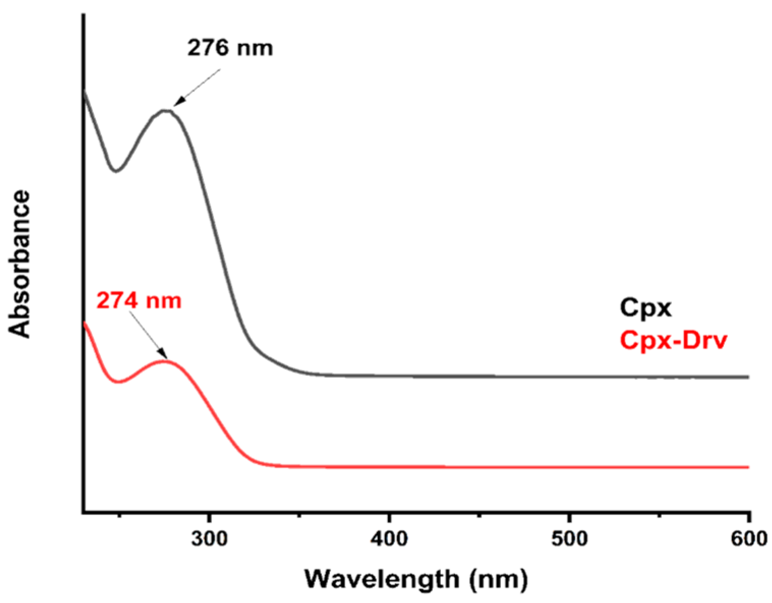
2.2. UV-Vis Spectroscopy Identification

Individual scan analysis of Cpx and Cpx-Drv samples was conducted in the range of 230–600 nm. Figure 3 shows the hypsochromic shift as a shorter wavelength in the maximum spectral band position was achieved from 276 nm to 274 nm. This change could be attributed to the higher molecular mass, including the structure relationship change according to Figure 1, compound I, where higher absorption is illustrated for Cpx-Drv.

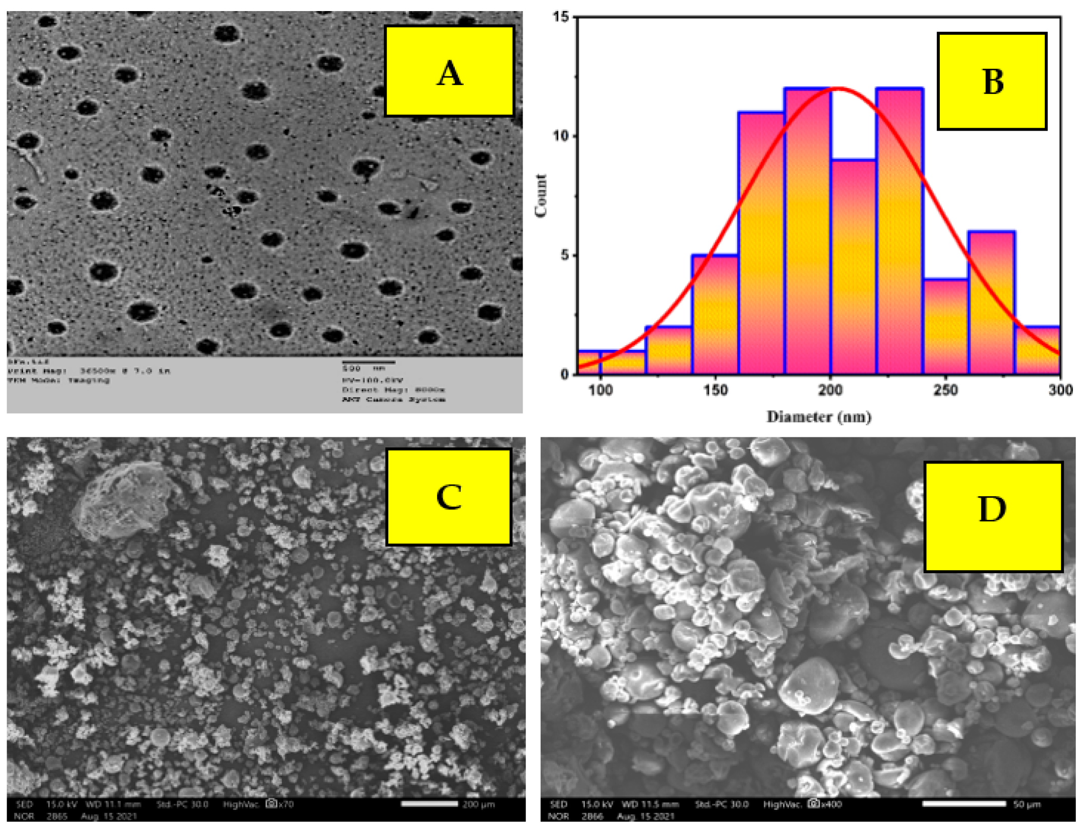
2.3. TEM and SEM Analysis

TEM and SEM analyses of Cpx-Drv are shown in Figure 4. The nanostructure integrity of the created Cpx-Drv was proven by TEM and SEM imaging on particles with a size range of 97.9–286.8 nm; the average particle size was found to be 203.3 nm and the median was 200.9 nm with a standard deviation of 42.3 nm. The monodispersed spherical shape was investigated in the absence of any agglomeration. Due to its monodispersibility and small particle size, the as-prepared Cpx-Drv can penetrate micro-organism cells as an antimicrobial, antiviral, or anticancer agent [22]. Moreover, due to these features, Cpx-Drv has the ability to become suspended in solution to form a suspension for oral administration of a drug.

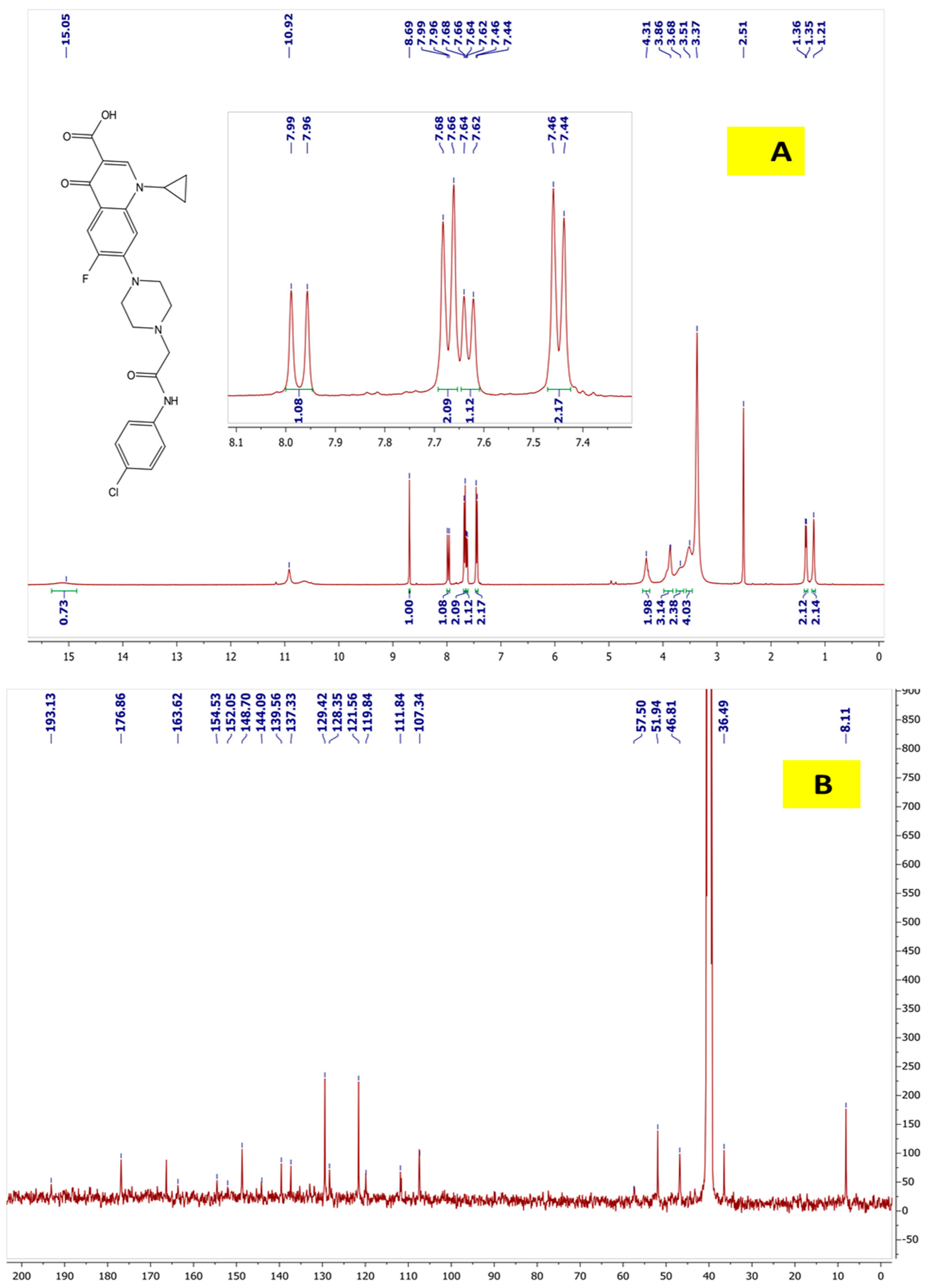
2.4. 1H NMR and 13C NMR for Cpx-Drv “1-Cyclopropyl-6-fluoro-4-oxo-7-(4-(2-oxo-2-(4-chlorophenyl)amino)ethyl) piperazin-1-yl)-1,4-dihydroquinoline-3carboxylic Acid”

Pale yellow crystal; (0.90 g, 88.30% yield); mp 237–239 °C; (Figure 5) manifested the 1H-NMR (400 MHz, DMSO-d6) δ 1.19–1.22 (2H, m, cyclopropyl-H), 1.34–1.37 (2H, m, cyclopropyl-H), 3.50–3.55 (4H, m, piperazinyl-H), 3.65–3.67 (2H, m, piperazinyl-H), 3.85–3.89 (3H, m, piperazinyl-H and cyclopropyl-H), 4.31 (2H, s, -N-CH2-CO), 7.45 (2H, d, J = 8 Hz, Ar-H), 7.63 (1H, d, J = 8 Hz, H-8), 7.67 (2H, d, J = 8 Hz, Ar-H), 7.98 (1H, d, J = 12 Hz, H-5), 8.69 (1H, s, H-2), 10.92 (1H, s, -NH-CO) 15.05 (1H, brs, COOH); 13C-NMR (100 MHz, DMSO-d6) δ 8.11, 36.49, 46.81, 51.94, 57.50, 107.34, 111.84, 119.84, 121.56, 128.35, 129.42, 137.33, 139.56, 144.09, 148.70, 152.05, 154,53, 163.62, 176.86, 193.13.

2.5. Water Content Determination

The moisture or water content can be quantified using a semi-micro technique, allowing for the measurement of water content, which can be represented as either moisture or crystalline water, expressed as a percentage. This method finds widespread application, particularly in the analysis of water content in pharmaceuticals and dietary supplements, whether supplied as solids or liquids [3]. The Karl Fischer method (KFT) was employed to measure the water content and was found to be 8.13%.

2.6. HPLC Quantitative, Qualitative Analysis, and Method Validation Check

According to the molecular structure as depicted in Figure 1, the elution of Cpx had a lower retention time, whereas Cpx-Drv has a higher molecular mass, so it was eluted at the end. In RP-HPLC, the basic rule is “likes dissolve likes or likes attract likes”. Because Cpx-Drv is more hydrophobic than Cpx, Cpx-Drv was eluted after Cpx, and this is the reason to use a high ratio of organic solvent. Cpx and Cpx-Drv peaks appeared at about 2.2 and 3.5 min, respectively, as seen in Figure 6. Under the optimized parameters of the analytical method, these ranged from 2.1 to 2.4 and 3.4 to 3.9 minutes for Cpx and Cpx-Drv peaks, respectively, over all the parameter changes. Table 2 and Table 3 show the high performance of the intended analysis method where the peak area RSD % ≤ 2.0%, USP tailing ≤ 2.0, theoretical plates ≥ 2000, and resolution ≥ 1.5 [23,24,25]. Hence, based on the obtained system suitability testing data, the method showed excellent validity through a wide range of retention times.
The results manifested excellent linearity, with R2 = 0.99999 for both Cpx and Cpx-Drv peak concentrations in the range of 5–200 µg/mL against their intensity responses. Moreover, LOD and LOQ limits were estimated, as shown in Table 4, where it was found that the current method could be used for both of the components, especially Cpx-Drv to determine low levels. The accuracy findings of the studied range (35–60 µg/mL) from the targeted concentration of 100% = 50 µg/mL for each of Cpx and Cpx-Drv were found to be within the acceptance criteria (98–102%), as displayed in Table 4 [26,27]. Moreover, there were no undesirable effects as a result of the presence of the two components, in addition to a high recovery percentage.
The RSD% of peak areas was employed to assess the repeatability of the analyte using six different preparations at the same desired concentration (50 µg/mL of Cpx and Cpx-Drv), as shown in Table 5. The RSD% for the intra-precision and the inter-precision measures were found to be within the repeatability criteria requirements of ≤2.0%. Moreover, the method was robust, rugged, selective, and specific according to the output data, as manifested in Table 5, where the major two peaks were found to be separated with a highly satisfactory and reasonable resolution of ≥1.5. The forced degradation not only reveals any further degradation or new peaks for both Cpx and Cpx-Drv, but also represents strong evidence for drug stability under aggressive degradation conditions.

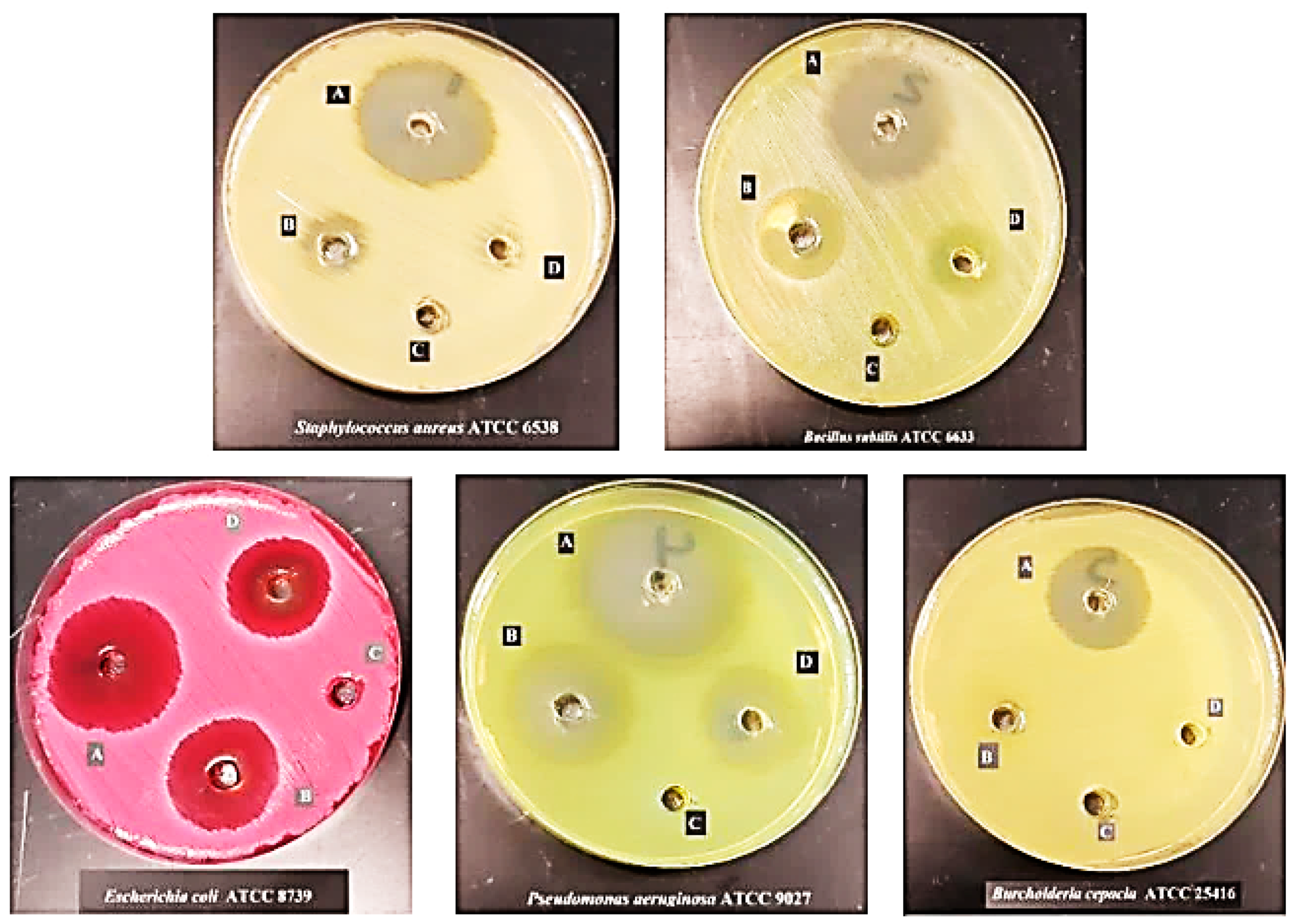
2.7. Antimicrobial Activity

The antimicrobial activity of Cpx-Drv against Gram-positive, Gram-negative, and yeast standard strains is shown in Figure 7. The results revealed that there was a notable outperformance in the antibacterial activity for Cpx-Drv in comparison with Cpx. This was obvious from measuring the inhibition zone around each concentration against the tested strains. The high activity of Cpx-Drv against Gram-positive bacteria in comparison to Gram-negative bacteria was noted. The antibacterial activity of Cpx-Drv was found to be higher than that of Cpx by 51-fold against S. aureus and 30-fold against B. subtilis for Gram-positive bacteria.
Regarding Gram-negative bacteria, Cpx-Drv exhibited the greatest effect against B. cepacia. The fold increase reached 100%, whereas the standard Cpx did not show any antimicrobial activity against the tested strain of Burkholderia sp. In the case of the E. coli strain, the fold increase reached 26.3% and increased to 35.5% for P. aeruginosa compared to Cpx only. On the other hand, Cpx-Drv used in this study did not show any antifungal activity for each of Cpx-Drv or Cpx, as shown in Table 5. This enhancement of antibacterial activity is most likely due to the bacterial aggregation of Cpx-Drv, which increased the exposure of bacteria cells to Cpx-Drv and consequently increased the concentration of Cpx-Drv inside the cell. The increase in the inhibition zone diameter indicates enhanced antibacterial activity, as shown in Table 6.

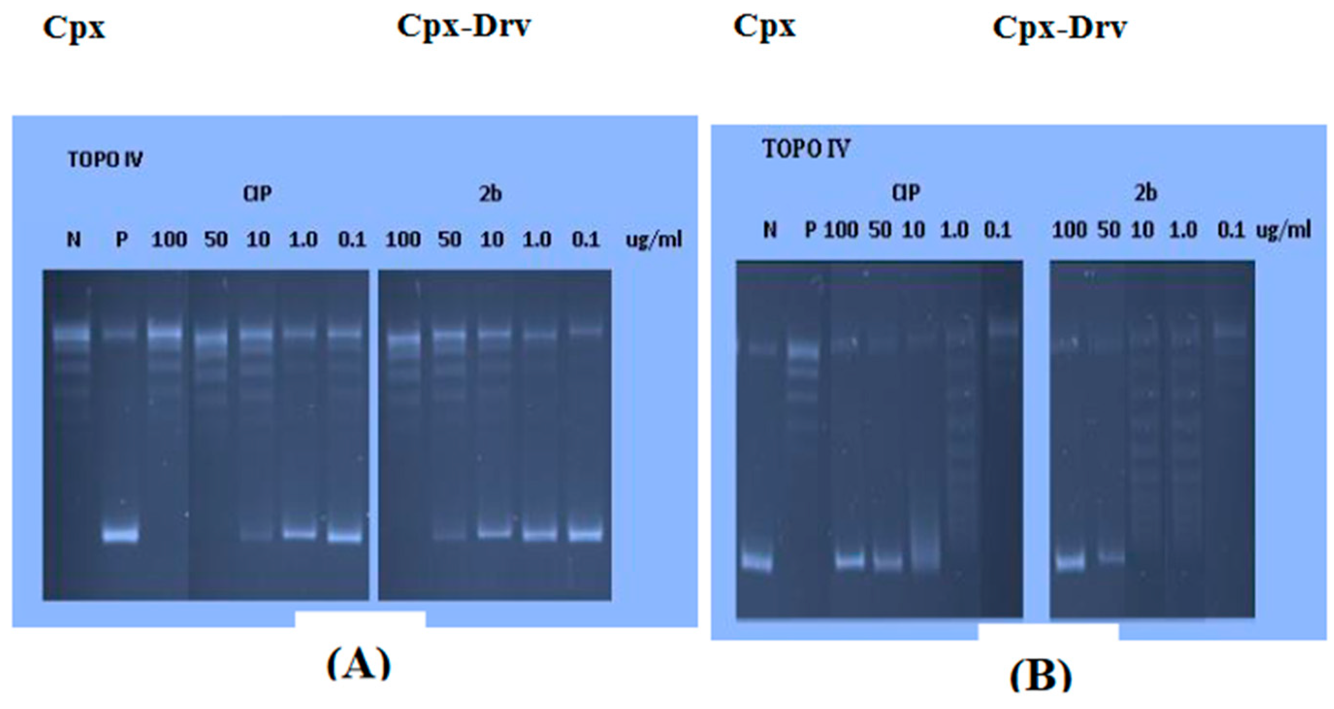
2.8. DNA Gyrase and Topoisomerase IV Inhibition Assays

Fluoroquinolones block DNA topoisomerase II enzymes (DNA gyrase and topoisomerase IV), as shown in Figure 8, which transiently cause DNA double-strand breaks as they negatively supercoil DNA. Fluoroquinolones inhibit DNA strands from passing through breaks and halt DNA replication, which causes cell death, by stabilizing the covalent enzyme-DNA adducts known as the cleaved complex. As a result, strong evidence for the inhibition of DNA topoisomerases can be found in the ability of the target compound, Cpx-Drv, to create a cleaved complex starting from the supercoiled plasmid. Using Cpx as a reference drug, agarose gel electrophoresis was used to assess the target compound’s ability to produce cleaved complexes starting with supercoiled pBR322. Ethidium bromide, a DNA intercalating chemical, was utilized in gel experiments to evaluate the stimulation of DNA breakage and inhibition of DNA supercoil relaxation. It is simple to distinguish relaxed DNA from nicked and linear species when ethidium bromide is added because closed circular DNA species positively supercoil when it is present. The results demonstrated that Cpx-Drv exhibits significant activity against DNA gyrase and topoisomerase IV, with IC50 values of 6.754 and 1.913 g/mL, but is less potent than Cpx. This is due to the role of N-4-piperazinyl substitution of Cpx in the enhancement of antibacterial activities by improving physiochemical parameters, decreasing zwitterions, and increasing lipophilicity. The improvement in physiochemical parameters has greater effect on antibacterial activity than the decrease in topoisomerase II inhibition of compound Cpx-Drv.
Cpx and Cpx-Drv were tested for their capacity to prevent DNA breakage by topoisomerase II enzymes. Results showed that Cpx-Drv is less powerful than Cpx against DNA gyrase and topoisomerase IV, with IC50 values of 6.754 and 1.913 g/mL, respectively. This suggests that the novel Cpx-Drv may have an additional antibacterial mechanism [28,29] (Table 7).

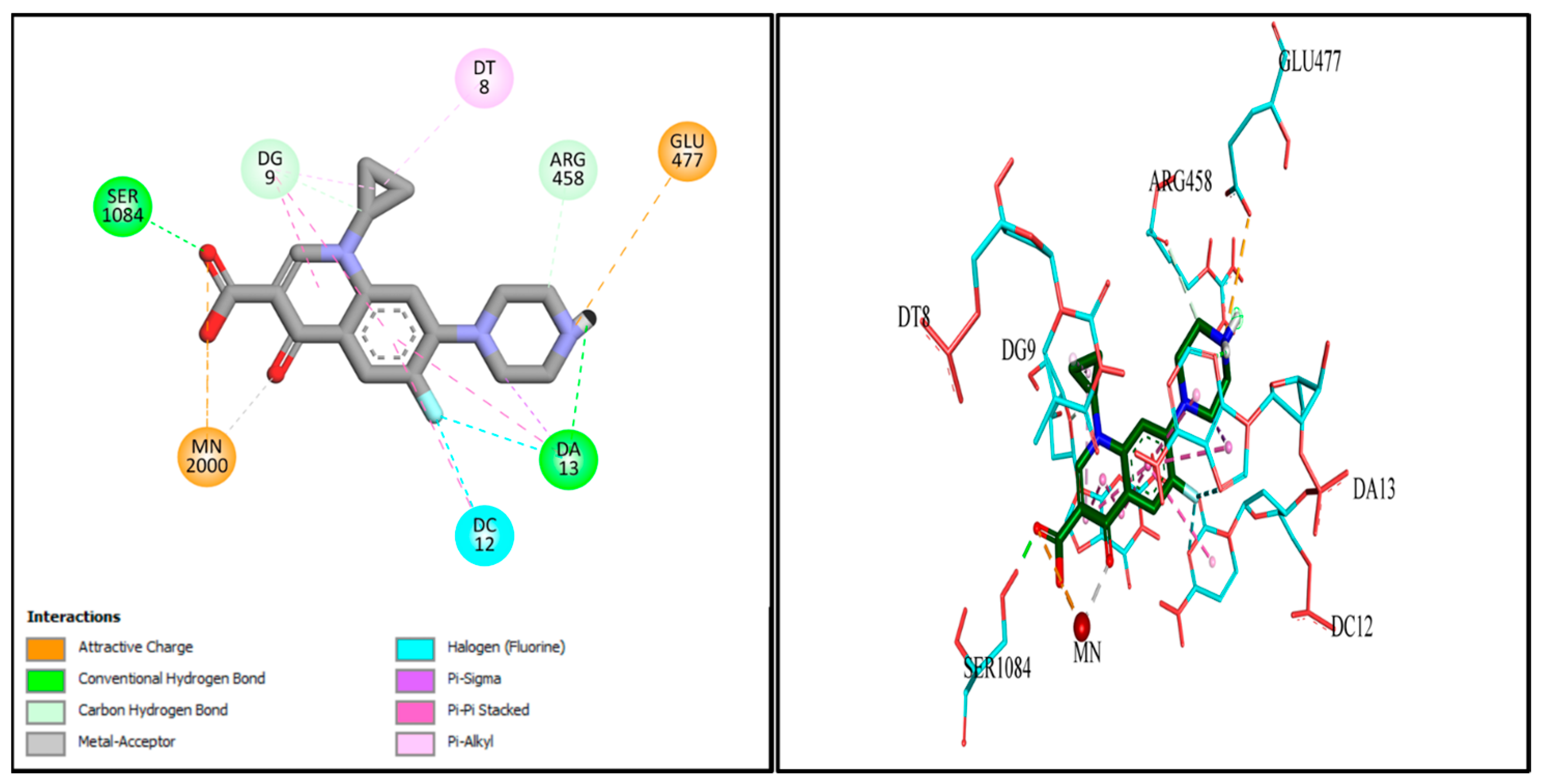
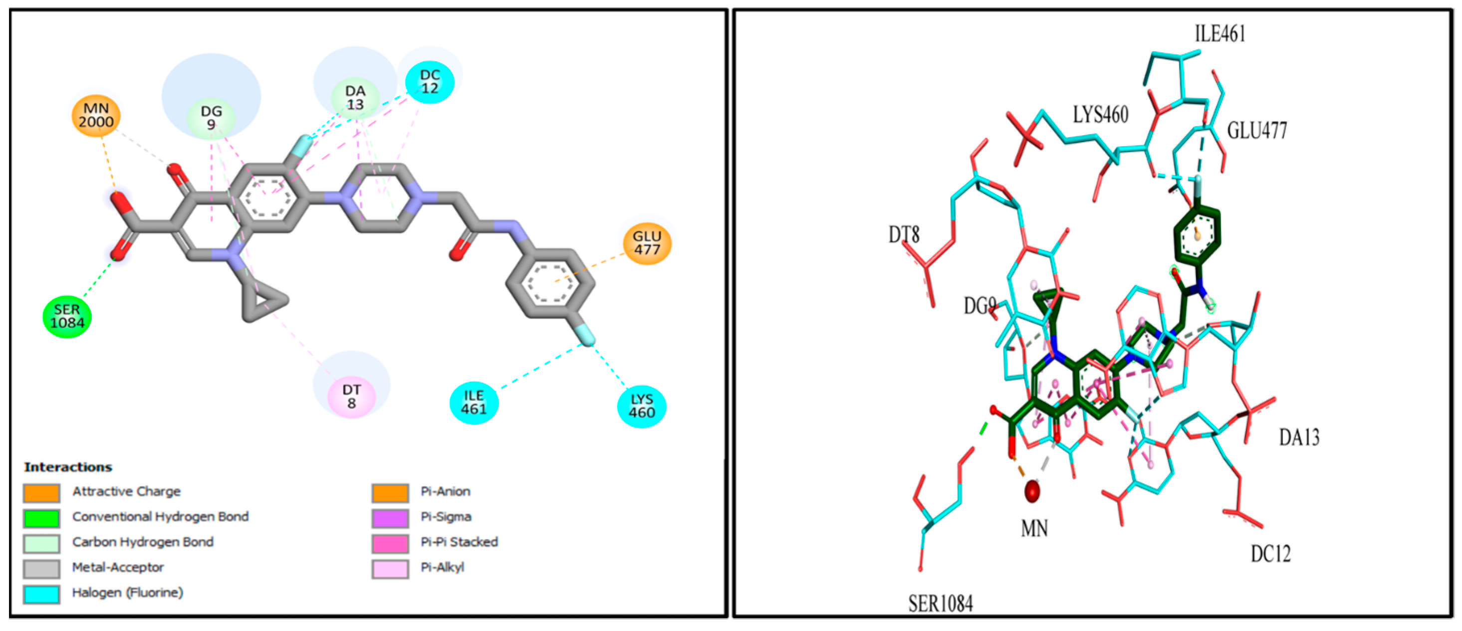
2.9. Docking Studies

Molecular docking investigations were conducted to explore the potential interactions of Cpx-Drv in the active site of the S. aureus gyrase enzyme (PDB: 2XCT). Redocking of the co-crystallized Cpx revealed several distinct interactions with the target enzyme’s active site, including chelation with Mn2+, hydrophobic interactions, hydrogen bonding, and pi-cationic interactions with DNA nucleotide bases (Figure 9). Furthermore, Cpx-Drv demonstrated its ability to interact with the gyrase-active site through hydrophobic interactions and additional hydrogen bonding with DNA nucleotide bases and amino acid residues (Figure 10). These findings align with the observation that Cpx-Drv exhibits stronger antibacterial activity compared to its parent compound Cpx. A summary of all possible binding interactions between Cpx and Cpx-Drv is provided in Table 8. Notably, the binding affinity of Cpx-Drv to the gyrase-active site was superior to that of Cpx, with respective values of −10.3349 and −7.7506 kcal/mol.

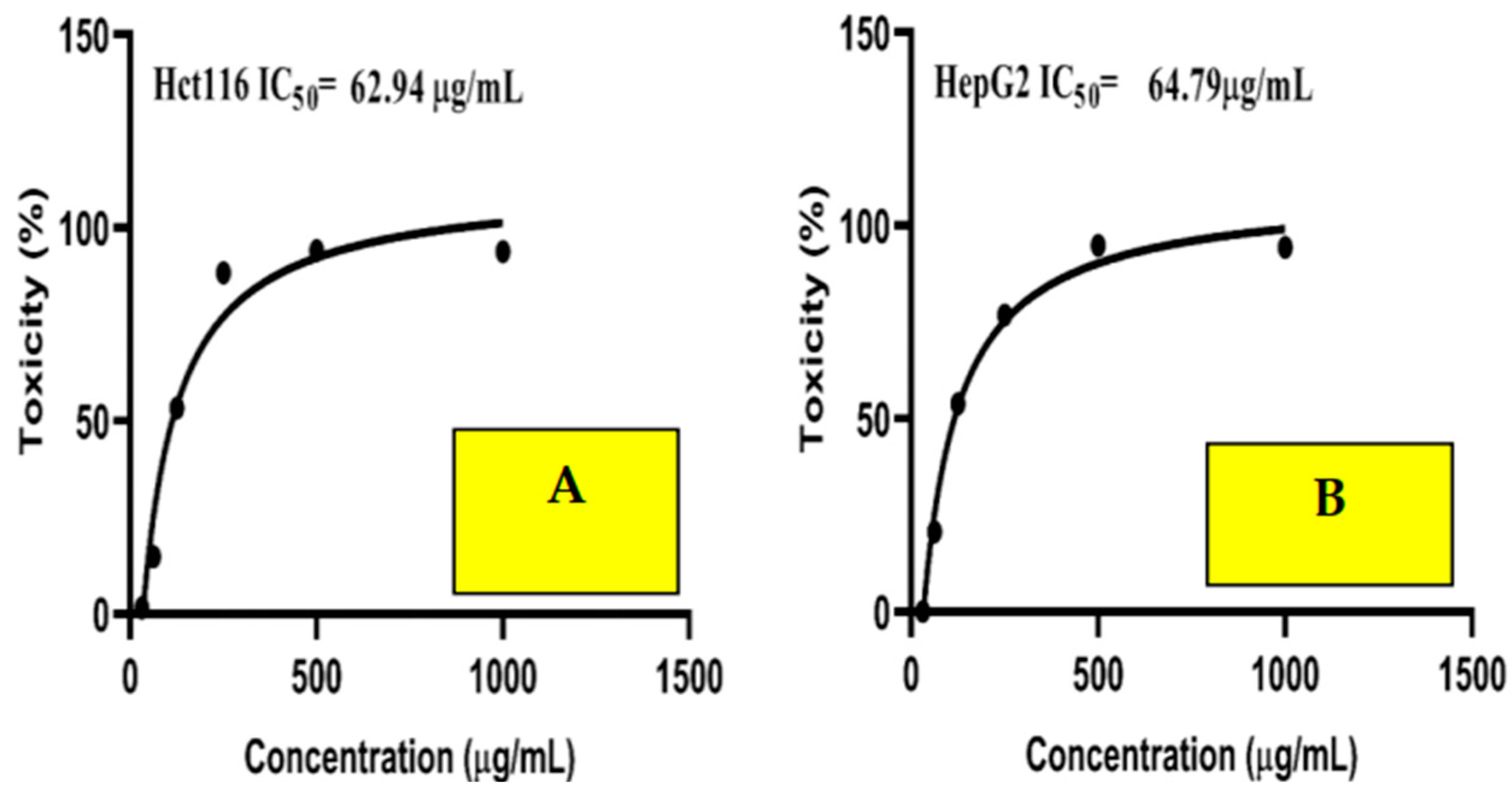
2.10. Anticancer Activity

The highly concentration-sensitive nature of Cpx-Drv, even at low concentrations, is revealed in Figure 11. Furthermore, the cytotoxicity of cells was found to be 93.9% and 93.3% at a dose of 1000 µg/mL in the HCT116 and HepG2 cell lines, respectively. The IC50 was estimated and found to be 62.94 and 64.79 µg/mL, respectively. The results revealed that Cpx-Drv has significant cytotoxic activity against HCT116 and HepG2. This result confirmed the previous finding of anticancer activity for the as-prepared Cpx-Drv [21]. Moreover, another reason for the anticancer activity of Cpx-Drv may be its particle size, which was found to be 203.3 nm, as revealed by TEM analysis. This small particle size was believed to facilitate the penetration into the cell line to destroy its biosystem and prevent tumor cells from reproducing. The anticancer mechanisms of Cpx-Drv were investigated and results showed that it exhibited four different mechanisms, including inhibition of MDR1, human topoisomerase I, and human topoisomerase II, as well as enhancement of the effects of paclitaxel (PTX) on microtubule assembly.

3. Materials and Methods

3.1. Materials

Cpx material was provided as a complimentary sample by UP Pharma (Assuit, Egypt). Acetonitrile HPLC-grade (ACN), potassium dihydrogen phosphate, HCl 37%, and NaOH (Scharlau, Spain) were used. Deionized water was filtered through a 0.45 μm nylon membrane filter prior to use.

3.2. Synthesis of the Target Compound Cpx-Drv

Cpx-Drv. was prepared as previously reported by Hamada et al. in their approach to new derivatives of Cpx [21] as follows. A mixture of the N-acyl-4-chloroaniline (2.2 mmol) in acetonitrile (10 mL), ciprofloxacin hydrochloride (2 mmol), and TEA (0.404 g, 4 mmol) was heated under reflux for 12-18 h. The formed precipitate was filtered off while hot, washed with acetonitrile, and dried under vacuum to produce the target compound Cpx-Drv.

3.3. Characterizations

FTIR was conducted using a KBr disc method using a Thermo Fisher Nicolet iS10 FTIR spectrometer with a wavenumber range of 4000–500 cm−1. UV-Vis absorption measurements were carried out in the range of 230–600 nm utilizing a PerkinElmer (LAMBDA 40 UV/Vis) spectrophotometer with a quartz cell having a 1 cm path length at room temperature. Morphological examination of Cpx-Drv was performed by scanning electron microscopy (SEM; JSM IT 200) and transmission electron microscopy (TEM; JEOL JEM-100C XII)).
Cpx and Cpx-Drv assay analyses were implemented using the HPLC model HP 1100 series.
Melting points were determined using a Stuart’s electrothermal melting point instrument and were uncorrected. NMR spectra (400 MHz for 1H, 100 MHz for 13C) were observed using a DMSO-d6 on Bruker AM400 spectrometer with tetramethyl silane as the internal standard. Chemical shift (d) results are provided in parts per million using DMSO-d6 as solvent, and coupling constants are designated as (J) in Hz. Splitting patterns are designated as follows: s, singlet; d, doublet; dd, doublet of doublet; t, triplet; q, quartet; m, multiplet; br s, broad singlet.
The semi-micro determination of water was performed using KFT (701-703 KF titrinio). A reaction as oxidation/reduction of the Karl Fischer reagent was used for estimation of the water percentage. The reagent was composed of sulfur dioxide, iodine, and resin that includes a nitrogen atom with at least one unshared electron pair free from pyridine [30]. The as-synthesized Cpx-Drv mass of about 100 mg was presented to the KFT in the presence of methanol as a reaction medium. The following equations were used for determination of water percentage:
Titer (mg/mL) = (mg of added water standard)/ (mL of KFT)
Water (%) = (Titer × mL of the KFT for Cpx-Drv × 100)/ (Cpx-Drv mass)

3.4. HPLC Quantitative, Qualitative Analysis and Method Validation Check

The HPLC method was developed and validated on the basis of the International Conference on Harmonization (ICH) validation guidelines [26,27]. Several trials were conducted to reach the optimum parameters for better separation and detection of the two peaks of Cpx and Cpx-Drv in one chromatogram. The trials to obtain Cpx and Cpx-Drv started together in one chromatogram.

3.4.1. Chromatographic System Configuration

Cpx and Cpx-Drv assay determinations were performed using the HPLC model HP 1100 series with variable wavelengths. The separation was achieved using a RP C18 BDS column (250 mm × 4.6 mm × 5 μm) (Thermo Scientific). The mobile phase consisted of 0.07 M of KH2PO4 in deionized water: ACN in a ratio (3:7, v/v) at a flow rate of 1.2 mL/min, with UV detection at 230 nm at room temperature and injection volume 20 μL.

3.4.2. System Suitability Check

First, both Cpx and Cpx-Drv were injected individually to determine the dedicated retention time. A system suitability test was conducted via analysis of six replicates of the same sample solution prepared by dissolving an amount of Cpx and Cpx-Drv to obtain a solution concentration of 50 µg/mL using mobile phase as a solvent.

3.4.3. Linearity and Range

The linearity test was conducted using different seven concentrations ranging from 10% to 400% of Cpx and Cpx-Drv. The working concentrations were prepared from 5–200 µg/mL. Every solution was injected twice.

3.4.4. LOD and LOQ

LOD and LOQ were estimated from the linearity calibration curve using the following equations:
LOD = 3.3 σ/S
LOQ = 10 σ/S
where σ is the standard error of concentration and area response and S is the slope of the linearity calibration curve.

3.4.5. Accuracy and Recovery

Both are two sides of the same coin and are used interchangeably. Accuracy describes the degree of closeness of the measured concentration value to the claimed theoretical concentration [23,24]. Accuracy was determined by weighing three different weights for each of Cpx and Cpx-Drv to produce theoretical concentrations at 35 µg/mL, 50 µg/mL, and 60 µg/mL. The linearity equation was used to determine the actual concentration, then the accuracy % was calculated using the following equation:
Accuracy %= Actual Conc.%/Theoretical Conc.% × 100
The accuracy acceptance limit for the assay method lay within 98.0–102% of the claimed concentration.

3.4.6. Precision (Repeatability and Intermediate Precision)

Repeatability was demonstrated using six different individual determinations of the desired concentration of the intended method (100% = 50 µg/mL) of Cpx and Cpx-Drv, which were performed using the same instrument on the same day by the same analyst [27,30,31,32]. The repeatability acceptance limit for the assay method should not be greater than 2.0%, which is expressed as RSD% of the rear response or assay content [27].
Other aspects to the intermediate precision may appear and be assessed as robustness and ruggedness. If the analytical method resists the slight changes that are deliberately applied to it and shows approximately the same separation efficiency, the method is called robust. These changes include changing the flow rate, the organic solvent ratio, the buffer ratio, and the temperature. Contrary to the definition of robustness, the analysis method is defined to be rugged if it manifests the results with the same efficiency when the remarkable changes are applied that are believed to affect the desired result. Ruggedness was assessed via a change in the HPLC column, analysis on another instrument, analysis by another analyst, or analysis on different days. Both robustness and ruggedness were assessed at Cpx and Cpx-Drv concentrations of 50 µg/mL for each, which were performed as system suitability and repeatability tests.

3.4.7. Specificity and Selectivity

Accelerated degradation was assessed by utilizing acid hydrolysis as 0.1 M of HCl, base hydrolysis as 0.1 M of NaOH, and light degradation over 6 h to reveal the stability-indicating features. The main reason for this test is that the analysis method can distinguish and separate between the principle intended components of Cpx and Cpx-Drv that we are looking for among the others that co-eluted with them. The acceptance criteria for the assay method’s specificity and selectivity are that the resolution value should be greater than 1.5 [26].

3.5. Antimicrobial Activity

The prepared antibiotic Cpx-Drv was subjected to evaluation of its antimicrobial activity against some reference standard bacterial and fungal strains, including 3 species of Gram-negative bacteria: Escherichia coli ATCC 8739 (E. coli), Burkholderia cepacia ATCC 25416 (B. cepacia), and Pseudomonas aeruginosa ATCC 9027 (P. aeruginosa). Two species of Gram-positive bacteria, Staphylococcus aureus ATCC 6538 (S. aureus) and Bacillus subtilis ATCC 6633 (B. subtilis), were also included. Furthermore, the yeast of Candida albicans ATCC 10231 (C. albicans) was also included [33,34,35,36]. The antimicrobial activity against the selected standard strain was evaluated and compared to the activity of Cpx. An overnight incubated microbial culture was suspended and the turbidity was adjusted to the equivalent 0.5 McFarland standard (1.5 × 10⁸ cfu/mL) [4,37,38].

3.5.1. Agar Well Diffusion Method

The antimicrobial activity of Cpx-Drv and Cpx against the tested standard strain was determined using the agar well diffusion method. Briefly, Cpx-Drv was dissolved in sterile distilled water and serial dilution was undertaken to obtain various concentrations (25, 50, and 100 μg/mL) used for testing their antimicrobial activities [17].

3.5.2. Antimicrobial Activity Bioassay

The prepared inoculums of standard strains (1 mL) were spread using a glass spreader to ensure an even distribution of the inoculums in Muller Hinton agar as a general medium, Cetrimide agar for Pseudomonas sp., MacConkey agar for E. Coli, and Sabouraud Dextrose agar for yeast. Wells were made by punching into the agar surface with a sterile cork borer. Using a micropipette, 150 L from each concentration was separately added to a single well. For bacterial strains, the inoculation plates were incubated at 37 °C for 24 h, and at 28 °C for 48 h for the yeast strain. The antimicrobial activity was estimated by measuring the diameter of the inhibition zone surrounding the wells. Cpx reference standard antibiotic was used as a positive control. Finally, the minimum inhibitory concentration (MIC) was defined as the lowest concentration that inhibits the growth of each strain [39,40,41]. All tests were carried out in triplicate.

3.6. Topoisomerase II Inhibition Assays

3.6.1. S. aureus Gyrase Supercoiling Assay

In accordance with the prescribed procedures derived from the literature, S. aureus DNA gyrase assay was carried out [42]. In three separate replicate runs, the novel chemical Cpx-Drv was dissolved in DMSO, serially diluted at doses of 100, 10, 1, and 0.1 M, and then tested in reaction mixtures. A total solution volume of 30 L was used containing 40 mM HEPES, 10 mM magnesium acetate, 500 mM potassium glutamate, 2 mM ATP, 0.05 mg/mL albumin, and Relaxed pBR322. At 37 °C, the DNA gyrase from S. aureus was incubated for 30 to 60 min. Staph. aureus gyrase DNA gyrase supercoiling reactions were stopped by adding 30 L of STEB and 30 L of chloroform/isoamyl alcohol (v:v, 24:1); the result was then centrifuged for one minute, and 20 L of this was loaded on a 1% agarose gel and run at 75V for roughly two hours. The gel was stained using ethidium bromide in water (0.5 mg/L). The fluorescent images were recorded using a UV transilluminator imaging device at a wavelength of 300 nm. The fluorescence intensity of the supercoiled plasmid reaction result was quantitated using the Imag Quant program (Molecular Dynamics). By applying nonlinear regression analysis in Graph Pad Prism, the results as IC50 values (concentration of the tested substance that results in 50% inhibition of enzyme activity) were calculated [42].

3.6.2. S. aureus Topoisomerase IV Decatenation Assay

IC50 values for compound Cpx-Drv and Cpx for Topo IV decatenation were determined. The following substances were utilized in this test: Topo IV Assay Buffer (provided as 5X), 50 mM Tris-HCl (pH 7.5), 5 mM magnesium chloride, 350 mM potassium glutamate, 5 mM DTT, and 1.5 m MATP, which were stored at or below −200 C. Quantities of 50 mM Tris-HCl (pH 7.5), 1 mM EDTA, 1 mM DTT, and 40% (v/v) glycerol (provided as 1X) made up the dilution buffer. The S. aureus Topo IV enzyme kDNA (100 ng/L) served as the substrate. STEB: S. aureus Topo IV, 40 percent sucrose (w/v), kDNA, 10 mM EDTA. Briefly, 100 mM Tris-HCl Bromophenol Blue, 0.5 mg/mL, pH 8, and various concentrations of the investigated substances were combined. For 30 min, the mixtures were incubated at 37 °C. STEB was used to halt the responses. Agarose gel electrophoresis was used to analyze the effects of the process. The gel was then colored with ethidium bromide and photographed under UV light [13,17,43].

3.7. Docking Studies

The structure of bacterial DNA gyrase (PDB code: 2XCT) was downloaded from the Protein Data Bank. The structure of compound Cpx-Drv was drawn and optimized using the molecular editors MarvinSketch and Avogadro. The protein was prepared using Autodock tools, where the co-crystallized water molecules and Cpx were removed, and Kollman charges and polar hydrogens were then added. The grid coordinates for DNA gyrase were set to 4.800, 44.477, and 67.943 for the x, y, and z axes, respectively, with grid dimensions of 56, 58, and 54. Autodock vina was used for molecular docking and the best docking pose was visualized using Discovery Studio Visualizer [44].

3.8. Anticancer Activity

MTT assay was implemented to evaluate the cell proliferation of human colorectal carcinoma Hct116 and human liver cancer “HepG2” cell lines, which were implemented at the Science way company and purchased from the ATCC and Asterand. The investigations were conducted as previously mentioned in the study of Al-Hakkani et al. [3,45,46,47,48]. The cytotoxicity profile using the newly prepared Cpx-Drv was demonstrated in the range of concentrations from 1000 to 31.25 μg/mL. A visible spectrophotometer at 560 nm was utilized to test activity.

4. Conclusions

A new derivative of Cpx was prepared and identified using various spectroscopic tools. A simple, sensitive, and accurate RP-HPLC method was developed and validated for the concurrent determination of Cpx and Cpx-Drv. Cpx-Drv antibacterial activity was greater against Gram-positive bacteria than against Gram-negative bacteria. Moreover, Cpx-Drv showed remarkable inhibitory activities against DNA gyrase and topoisomerase IV as potential molecular targets. Molecular modeling investigations of Cpx-Drv revealed higher binding interactions for gyrase enzyme (PDB ID: 2XCT) than Cpx, with greater binding affinity.

Author Contributions

Conceptualization: M.F.A.-H., H.H.H.M., A.M.A.-D., N.A., A.A.A., M.H.A.H., M.A.S., H.A.A. Methodology: M.F.A.-H., H.H.H.M., N.A., A.A.A., M.H.A.H., M.A.S., H.A.A. Software: M.F.A.-H., H.H.H.M., N.A., A.A.A., M.H.A.H., M.A.S., H.A.A. Data curation: M.F.A.-H., H.H.H.M., N.A., M.S.A., A.A.A., M.H.A.H., M.A.S., H.A.A. Visualization: M.F.A.-H., H.H.H.M., M.H.A.H., A.M.E., H.O.K., M.A.A., M.S.S., M.M.A.E., A.M.A.-D., M.A.E.-M., M.A.S., H.A.A. Investigation: M.F.A.-H., H.H.H.M., M.H.A.H., A.M.E., H.O.K., M.A., M.A.A., M.S.S., M.M.A.E., M.A.E.-M., M.A.S., H.A.A., M.S.A., Supervision: M.F.A.-H., H.H.H.M., M.H.A.H., M.A., M.A.E.-M., A.M.A.-D., H.A.A. Writing—Reviewing and Editing: M.F.A.-H., A.M.A.-D., H.H.H.M., M.H.A.H., M.A.S., H.A.A. All authors have read and agreed to the published version of the manuscript.

Funding

This research was funded by Deputyship for Research & Innovation, Ministry of Education in Saudi Arabia with grant number [445-9-333].

Institutional Review Board Statement

We confirm that all methods were carried out following relevant guidelines and regulations and the research did not involve human participants or animals.

Informed Consent Statement

Not applicable.

Data Availability Statement

All data generated or analyzed during this study are included in this article.

Acknowledgments

The authors extend their appreciation to the Deputyship for Research & Innovation, Ministry of Education in Saudi Arabia for funding this research work through the project number: 445-9-333.

Conflicts of Interest

The authors declare no conflict of interest.

References

  1. Al-Hakkani, M.F.; Gouda, G.A.; Hassan, S.H.A.; Saddik, M.S.; El-Mokhtar, M.A.; Ibrahim, M.A.; Mohamed, M.M.A.; Nagiub, A.M. Cefotaxime removal enhancement via bio- nanophotocatalyst α-Fe2O3 using photocatalytic degradation technique and its echo-biomedical applications. Sci. Rep. 2022, 12, 11881. [Google Scholar] [CrossRef] [PubMed]
  2. Hunge, Y.M.; Yadav, A.A.; Kang, S.-W.; Jun Lim, S.; Kim, H. Visible light activated MoS2/ZnO composites for photocatalytic degradation of ciprofloxacin antibiotic and hydrogen production. J. Photochem. Photobiol. A 2023, 434, 114250. [Google Scholar] [CrossRef]
  3. Al-Hakkani, M.F.; Gouda, G.A.; Hassan, S.H.A.; Mohamed, M.M.A.; Nagiub, A.M. Environmentally azithromycin pharmaceutical wastewater management and synergetic biocompatible approaches of loaded azithromycin@hematite nanoparticles. Sci. Rep. 2022, 12, 10970. [Google Scholar] [CrossRef] [PubMed]
  4. Ali, S.; Perveen, S.; Shah, M.R.; Zareef, M.; Arslan, M.; Basheer, S.; Ullah, S.; Ali, M. Bactericidal potentials of silver and gold nanoparticles stabilized with cefixime: A strategy against antibiotic-resistant bacteria. J. Nanopart. Res. 2020, 22, 201. [Google Scholar] [CrossRef]
  5. Khan, K.M.; Siddiqui, R.; Ambreen, N.; Sultana, N.; Tauseef, S.; Ahmad, A.; Perveen, S. Synthesis, antibacterial, and antifungal evaluation of norfloxacin derivatives. J. Pharm. Res. 2012, 5, 666–671. [Google Scholar]
  6. Dey, R.; Bishayi, B. Dexamethasone along with ciprofloxacin modulates S. aureus induced microglial inflammation via glucocorticoid (GC)-GC receptor-mediated pathway. Microb. Pathog. 2020, 145, 104227. [Google Scholar] [CrossRef]
  7. Pudlo, M.; Luzet, V.; Ismaïli, L.; Tomassoli, I.; Iutzeler, A.; Refouvelet, B. Quinolone–benzylpiperidine derivatives as novel acetylcholinesterase inhibitor and antioxidant hybrids for Alzheimer Disease. Bioorg. Med. Chem. 2014, 22, 2496–2507. [Google Scholar] [CrossRef]
  8. Dalhoff, A. Antiviral, antifungal, and antiparasitic activities of fluoroquinolones optimized for treatment of bacterial infections: A puzzling paradox or a logical consequence of their mode of action? Eur. J. Clin. Microbiol. Infect. Dis. 2015, 34, 661–668. [Google Scholar] [CrossRef]
  9. Li, J.; Li, S.; Bai, C.; Liu, H.; Gramatica, P. Structural requirements of 3-carboxyl-4 (1H)-quinolones as potential antimalarials from 2D and 3D QSAR analysis. J. Mol. Graph. Model. 2013, 44, 266–277. [Google Scholar] [CrossRef] [PubMed]
  10. Mohammed, H.H.; Abbas, S.H.; Hayallah, A.M.; Abuo-Rahma, G.E.-D.A.; Mostafa, Y.A. Novel urea linked ciprofloxacin-chalcone hybrids having antiproliferative topoisomerases I/II inhibitory activities and caspases-mediated apoptosis. Bioorg. Chem. 2021, 106, 104422. [Google Scholar] [CrossRef]
  11. Shabani, F.; Saghatforoush, L.A.; Ghammamy, S. Synthesis, characterization and anti-tumour activity of Iron (III) Schiff base complexes with unsymmetric tetradentate ligands. Bull. Chem. Soc. Ethiop. 2010, 24, 193–199. [Google Scholar] [CrossRef]
  12. Nunes, B.; Leal, C.; Rodrigues, S.; Antunes, S.C. Assessment of ecotoxicological effects of ciprofloxacin in Daphnia magna: Life-history traits, biochemical and genotoxic effects. Water Sci. Technol. 2018, 2017, 835–844. [Google Scholar] [CrossRef] [PubMed]
  13. Cacicedo, M.L.; Pacheco, G.; Islan, G.A.; Alvarez, V.A.; Barud, H.S.; Castro, G.R. Chitosan-bacterial cellulose patch of ciprofloxacin for wound dressing: Preparation and characterization studies. Int. J. Biol. Macromol. 2020, 147, 1136–1145. [Google Scholar] [CrossRef] [PubMed]
  14. Moharram, A.; Ismail, M.; Shoreit, A.; Hassan, M. Biodiversity of microbiota in cephalosporin-manufacturing environments at T3A factory, Assiut, Egypt. J. Basic Appl. Mycol. 2014, 5, 1–13. [Google Scholar]
  15. Hassan, M.; Ismail, M.; Moharram, A.; Shoreit, A. Synergistic Effect of Biogenic Silver-nanoparticles with β lactam Cefotaxime against Resistant Staphylococcus arlettae AUMC b-163 Isolated from T3A Pharmaceutical Cleanroom, Assiut, Egypt. Am. J. Microbiol. Res. 2016, 4, 132–137. [Google Scholar]
  16. Seeman, P. The Membrane Actions of Anesthetics and Tranquilizers. Pharmacol. Rev. 1972, 24, 583–655. [Google Scholar]
  17. Cho, K.-H.; Park, J.-E.; Osaka, T.; Park, S.-G. The study of antimicrobial activity and preservative effects of nanosilver ingredient. Electrochim. Acta 2005, 51, 956–960. [Google Scholar] [CrossRef]
  18. Azhdarzadeh, M.; Lotfipour, F.; Zakeri-Milani, P.; Mohammadi, G.; Valizadeh, H. Anti-bacterial performance of azithromycin nanoparticles as colloidal drug delivery system against different gram-negative and gram-positive bacteria. Adv. Pharm. Bull. 2012, 2, 17. [Google Scholar]
  19. Rong, X.; Qiu, F.; Jiang, Z.; Rong, J.; Pan, J.; Zhang, T.; Yang, D. Preparation of ternary combined ZnO-Ag2O/porous g-C3N4 composite photocatalyst and enhanced visible-light photocatalytic activity for degradation of ciprofloxacin. Chem. Eng. Res. Des. 2016, 111, 253–261. [Google Scholar] [CrossRef]
  20. Mocanu, A.-G.; Belu, I.; Croitoru, O.; Ciocîlteu, M.V.; Manda, C.; Neamtu, J. Formulation and characterization of ciprofloxacin loaded PLGA microspheres for applications in orthopedic infections. Curr. Health Sci. J. 2017, 43, 306. [Google Scholar]
  21. Mohammed, H.H.; Abd El-Hafeez, A.A.; Abbas, S.H.; Abdelhafez, E.-S.M.; Abuo-Rahma, G.E.-D.A. New antiproliferative 7-(4-(N-substituted carbamoylmethyl) piperazin-1-yl) derivatives of ciprofloxacin induce cell cycle arrest at G2/M phase. Bioorg. Med. Chem. 2016, 24, 4636–4646. [Google Scholar] [CrossRef] [PubMed]
  22. Moosa, M.H.; Abu-Okail, M.; Abu-Oqail, A.; Al-Shelkamy, S.A.; Shewakh, W.M.; Ghafaar, M.A. Structural and Tribological Characterization of Carbon and Glass Fabrics Reinforced Epoxy for Bushing Applications Safety. Polymers 2023, 15, 2064. [Google Scholar] [CrossRef] [PubMed]
  23. Al-Hakkani, M.F.; Ahmed, N.; Hassan, M.H.A. Rapidly, sensitive quantitative assessment of thiopental via forced stability indicating validated RP-HPLC method and its in-use stability activities. Sci. Rep. 2023, 13, 10294. [Google Scholar] [CrossRef]
  24. Al-Hakkani, M.F.; Ahmed, N.; Abbas, A.A.; Hassan, M.H.A. Cefoperazone rapidly and sensitive quantitative assessment via a validated RP-HPLC method for different dosage forms, in-use stability, and antimicrobial activities. BMC Chem. 2023, 17, 72. [Google Scholar] [CrossRef] [PubMed]
  25. Al-Hakkani, M.F. A new validated facile HPLC analysis method to determine methylprednisolone including its derivatives and practical application. Sci. Rep. 2023, 13, 11548. [Google Scholar] [CrossRef]
  26. ICH Steering Committee. ICH Q2B Validation of Analytical Procedures: Methodology; European Agency for the Evaluation of Medicinal Products: London, UK, 1996; Volume 281, p. 95. [Google Scholar]
  27. Tripartite, G.I.H. Validation of analytical procedures: Text and methodology. Q2 (R1) 2005, 1, 11–12. [Google Scholar]
  28. AHMA, G.E.D.A.; Abbas, S.; Shoman, M.; SAMIR, I.; EL-BAKY, R.A. New $ N $-4 piperazinyl derivatives of norfloxacin: Design, synthesis, and correlation of calculated physicochemical parameters with antibacterial activity. Turk. J. Chem. 2018, 42, 1072–1085. [Google Scholar]
  29. Vazquez, J.; Merino, S.; Doměnech, Ò.; Berlanga, M.; Vinas, M.; Montero, M. Hernandez-Borrell: Determination of the partition coefficients of a homologous series of ciprofloxacin: Influence of the N-4 piperazinyl alkylation on the antimicrobial activity. Int. J. Pharm. 2001, 220, 53–62. [Google Scholar] [CrossRef]
  30. Bhaskaran, N.A.; Kumar, L.; Reddy, M.S.; Pai, G.K. An analytical “quality by design” approach in RP-HPLC method development and validation for reliable and rapid estimation of irinotecan in an injectable formulation. Acta Pharm. 2021, 71, 57–79. [Google Scholar] [CrossRef]
  31. Shabir, G.A. Step-by-step analytical methods validation and protocol in the quality system compliance industry. J. Val. Technol. 2005, 10, 314–325. [Google Scholar]
  32. Devaraj, S.; Sivaperuman, A.; Nagarajan, N.C. RP-UPLC Method Development and Validation for Simultaneous Estimation of Mometasone Furoate and Miconazole Nitrate in Semisolid Dosage Form. Acta Pharm. Sci. 2020, 58, 335. [Google Scholar] [CrossRef]
  33. Alangari, A.; Alqahtani, M.S.; Mateen, A.; Kalam, M.A.; Alshememry, A.; Ali, R.; Kazi, M.; AlGhamdi, K.M.; Syed, R. Iron Oxide Nanoparticles: Preparation, Characterization, and Assessment of Antimicrobial and Anticancer Activity. Adsorpt. Sci. Technol. 2022, 2022, 1562051. [Google Scholar] [CrossRef]
  34. Arsalani, N.; Mashkouri, S.; Hosseini, M.G.; Ramazani, A.; Mostafavi, H. Green synthesis of water-soluble graphene nanosheets under solvent-free condition and in-situ anchored with MnO2 as supercapacitor. J. Mater. Sci. Mater. Electron. 2018, 29, 6692–6701. [Google Scholar] [CrossRef]
  35. Ocsoy, I.; Temiz, M.; Celik, C.; Altinsoy, B.; Yilmaz, V.; Duman, F. A green approach for formation of silver nanoparticles on magnetic graphene oxide and highly effective antimicrobial activity and reusability. J. Mol. Liq. 2017, 227, 147–152. [Google Scholar] [CrossRef]
  36. Mukherjee, D.; Ghosh, S.; Majumdar, S.; Annapurna, K. Green synthesis of α-Fe2O3 nanoparticles for arsenic(V) remediation with a novel aspect for sludge management. J. Env. Chem. Eng. 2016, 4, 639–650. [Google Scholar] [CrossRef]
  37. Amer, M.W.; Awwad, A.M. Green synthesis of copper nanoparticles by Citrus limon fruits extract, characterization and antibacterial activity. Chem. Int. 2021, 7, 1–8. [Google Scholar]
  38. Madubuonu, N.; Aisida, S.O.; Ali, A.; Ahmad, I.; Zhao, T.-K.; Botha, S.; Maaza, M.; Ezema, F.I. Biosynthesis of iron oxide nanoparticles via a composite of Psidium guavaja-Moringa oleifera and their antibacterial and photocatalytic study. J. Photochem. Photobiol. B 2019, 199, 111601. [Google Scholar] [CrossRef]
  39. Rodriguez-Mozaz, S.; Chamorro, S.; Marti, E.; Huerta, B.; Gros, M.; Sànchez-Melsió, A.; Borrego, C.M.; Barceló, D.; Balcázar, J.L. Occurrence of antibiotics and antibiotic resistance genes in hospital and urban wastewaters and their impact on the receiving river. Water Res. 2015, 69, 234–242. [Google Scholar] [CrossRef]
  40. Ziko, L.; Saqr, A.-H.A.; Ouf, A.; Gimpel, M.; Aziz, R.K.; Neubauer, P.; Siam, R. Antibacterial and anticancer activities of orphan biosynthetic gene clusters from Atlantis II Red Sea brine pool. Microb. Cell Fact. 2019, 18, 56. [Google Scholar] [CrossRef]
  41. Sekhavati, E.; Jafari, F.; SeyedAlinaghi, S.; Jamalimoghadamsiahkali, S.; Sadr, S.; Tabarestani, M.; Pirhayati, M.; Zendehdel, A.; Manafi, N.; Hajiabdolbaghi, M. Safety and effectiveness of azithromycin in patients with COVID-19: An open-label randomised trial. Int. J. Antimicrob. Agents 2020, 56, 106143. [Google Scholar] [CrossRef]
  42. Alt, S.; Mitchenall, L.A.; Maxwell, A.; Heide, L. Inhibition of DNA gyrase and DNA topoisomerase IV of Staphylococcus aureus and Escherichia coli by aminocoumarin antibiotics. J. Antimicrob. Chemother. 2011, 66, 2061–2069. [Google Scholar] [CrossRef] [PubMed]
  43. Some, S.; Bulut, O.; Biswas, K.; Kumar, A.; Roy, A.; Sen, I.K.; Mandal, A.; Franco, O.L.; İnce, İ.A.; Neog, K.; et al. Effect of feed supplementation with biosynthesized silver nanoparticles using leaf extract of Morus indica L. V1 on Bombyx mori L. (Lepidoptera: Bombycidae). Sci. Rep. 2019, 9, 14839. [Google Scholar] [CrossRef] [PubMed]
  44. Trott, O.; Olson, A. Software news and update AutoDock Vina: Improving the speed and accuracy of docking with a new scoring function. Effic. Optim. Multithreading. 2009, 31, 455–461. [Google Scholar]
  45. Hosny, S.; Gouda, G.A.; Abu-El-Wafa, S.M. Novel Nano Copper Complexes of a New Schiff Base: Green Synthesis, a New Series of Solid Cr(II), Co(II), Cu(II), Pd(II) and Cd(II) Chelates, Characterization, DFT, DNA, Antitumor and Molecular Docking Studies. Appl. Organomet. Chem. 2022, 36, e6627. [Google Scholar] [CrossRef]
  46. Gao, Y.; Shang, Q.; Li, W.; Guo, W.; Stojadinovic, A.; Mannion, C.; Man, Y.-G.; Chen, T. Antibiotics for cancer treatment: A double-edged sword. J. Cancer 2020, 11, 5135–5149. [Google Scholar] [CrossRef] [PubMed]
  47. Solairaj, D.; Rameshthangam, P.; Arunachalam, G. Anticancer activity of silver and copper embedded chitin nanocomposites against human breast cancer (MCF-7) cells. Int. J. Biol. Macromol. 2017, 105, 608–619. [Google Scholar] [CrossRef]
  48. VT, M.C.; Baccini, C.; Pertici, G.; Serino, L.P.; De Giorgi, U. Anticancer Drug Delivery with Nanoparticles. In Vivo 2006, 20, 697–701. [Google Scholar]
Figure 1. Chemical structure of Cpx and its derivatives; compounds I and II with potent anticancer activity, compounds III and IV with potent antimicrobial activities.
Ijms 24 14818 g001
Figure 2. FTIR spectra of Cpx and Cpx-Drv.
Ijms 24 14818 g002
Figure 3. UV-Vis spectra scan of Cpx and Cpx-Drv.
Ijms 24 14818 g003
Figure 4. Cpx-Drv: (A) TEM image; (B) particle size distribution; (C,D) SEM images.
Ijms 24 14818 g004
Figure 5. (A) 1H NMR and (B) 13C NMR for Cpx-Drv.
Ijms 24 14818 g005
Figure 6. (A) HPLC chromatogram of Cpx and Cpx-Drv peaks at retention times of 2.194 min and 3.554 min, respectively; (B) USP tailing of Cpx; (C) USP tailing of Cpx-Drv.
Ijms 24 14818 g006
Figure 7. Different concentrations of Cpx-Drv: (A) 100 μg/mL; (B) 50 μg/mL; (C) 25 μg/mL; (D) Cpx control against different bacterial standard strains.
Ijms 24 14818 g007
Figure 8. (A) DNA gyrase inhibitory assay; (B) DNA topoisomerase IV inhibitory activity.
Ijms 24 14818 g008
Figure 9. 2D- and 3D-binding modes of Cpx within the gyrase-active site.
Ijms 24 14818 g009
Figure 10. 2D- and 3D-binding modes of Cpx-Drv within the gyrase-active site.
Ijms 24 14818 g010
Figure 11. Cytotoxicity of Cpx-Drv against (A) HCT116 and (B) HepG2 cell lines.
Ijms 24 14818 g011
Table 1. FT-IR band assignments of Cpx and Cpx-Drv.
Wavenumber (cm−1)Band Assignments
Cpx-DrvCpx
33163389Stretching vibration band -N-H
30603057Stretching -C=C-H
29512922Stretching aliphatic -C-H of CH2 “symmetric”
28112832Stretching aliphatic -C-H of CH2 “anti-symmetric”
17281720Stretching vibration -C=O in the carboxylic group
16951616Stretching vibration -C=O of quinoline
15051586Bending vibration -N-H
13611375Hydrated water bending vibration -O-H
12581257Stretching vibration -C-N
11831176Asymmetric -C-O-C
10191024Stretching vibration band -C-F
1004941Stretching vibration band C=C bending
826820Bending -C=O
806----Stretching vibration band -C-Cl
774785Bending aromatic -C-H
709713Rocking -C-H of >CH2
596567Aromatic -C-H bending
Table 2. System suitability parameters of Cpx.
wt (mg)/200 mL10.0010.0110.0210.0110.0010.0110.0010.0510.51
Replicate #OptimumAnalyst 2Day
2
Flow Rate
1.3 mL/min
Flow Rate
1.1 mL/min
ACN % 75%ACN %
65%
Column-2Column-3
11057.81063.21060.61017.21108.3970.21112.81080.61110.5
21058.71060.71058.31017.61104.7971.81109.81078.31107.1
31059.21060.31056.81017.01105.99691107.71079.21105.8
41058.21060.21056.91079.81106.1
51060.61060.51058.11093.91105.0
61060.01060.81057.41094.81104.7
Mean1059.11061.01058.010171106970111010841107
STDEV1.071.131.400.311.831.402.567.722.12
RSD (%)0.100.110.130.030.170.140.230.710.19
USP tailing1.051.091.031.110.821.140.810.760.75
Plates10,086990610,05410,09510,08110,01210,068908410,016
Table 3. System suitability parameters of Cpx-Drv.
wt (mg)/200 mL10.0010.0110.0210.0110.0010.0110.0010.0510.51
Replicate #OptimumAnalyst 2Day 2Flow Rate
1.3 mL/min
Flow Rate
1.1 mL/min
ACN % 75%ACN %
65%
Column-2Column-3
11294.71300.41295.61241.01357.01198.11413.81335.51332.7
21295.81298.11293.61240.71351.41195.51409.11331.31327.6
31296.41297.61292.61240.91353.51195.31409.41331.71328.2
41296.21297.71291.71332.41327.7
51296.81297.81292.61337.41328.0
61295.51297.41292.11337.71327.6
Mean1295.91298.21293.0124113541196141113341329
STDEV0.741.121.410.152.831.592.632.902.01
RSD (%)0.060.090.110.010.210.130.190.220.15
USP tailing1.131.121.181.151.161.171.151.171.12
Plates11,63511,53611,43911,17711,93710,88212,20911,46611,451
Resolution6.9956.856.937.046.817.357.257.157.11
Table 4. Cpx and Cpx-Drv linearity data.
Concentration (%)Concentration (µg/mL)Cpx Mean P. AsCpx-Drv Mean P. As
105114.15136.3
5025538.35666.4
7035758.20939.9
100501066.351331.85
120601293.651610.85
150751603.501993.1
4002004235.205299.8
Slope21.1226.46
Intercept15.069.93
Correlation0.999990.99999
STDEV XY error7.307.21
LOD (µg/mL)1.140.90
LOQ (µg/mL)3.462.72
Accuracy (%) at35 µg/mL99.8100.5
50 µg/mL99.898.6
60 µg/mL100.7100.2
Table 5. Precision, robustness, ruggedness, and specificity results of Cpx and Cpx-Drv.
ItemCpx Cpx-Drv
Repeatability as RSD%Inter-precision0.51, 0.86 0.79, 0.30
Intra-precision0.710.57
Robustness as RSD%Flow rate 0.10, 0.03, 0.170.06, 0.01, 0.21
Organic ratio0.10, 0.14, 0.230.06, 0.13, 0.19
Ruggedness as RSD%Analyst to Analyst0.10, 0.110.06, 0.09
Column to Column0.10, 0.71, 0.190.06, 0.22, 0.15
Day to Day0.10, 0.130.06, 0.11
Specificity as resolution between both Cpx and Cpx DrvAcid hydrolysis 7.84
Base hydrolysis8.05
Light degradation6.87
Table 6. Antimicrobial activity of Cpx and Cpx-Drv against Gram-positive strains S. aureus 6538 and B. subtilis 6633, against Gram-negative strains E. coli 8739, P. aeruginosa 9027, and B. cepacian 25416, and against yeast strain C. albicans.
Antimicrobial AgentInhibition Zone Diameter (mm)
Cpx Cpx-Drv Fold Increase %
Standard StrainsATCC100 y
μg/mL
100 x
μg/mL
50
μg/mL
25 μg/mL(x − y)/x∗100
Gram-positive strains
S. aureus65386.7 ± 0.313.8 ± 0.87.0 ± 0.5051.4%
B. subtilis66337.8 ± 0.311.3 ± 0.69.7 ± 0.8030.3%
Gram-negative strains
E. coli873911.2 ± 0.815.2 ± 0.311.7 ± 0.6026.3%
P. aeruginosa90278.7 ± 0.613.5 ± 0.910.3 ± 0.3035.5%
B. cepacia25,416010.0 ± 0.900100%
Yeast strain
C. albicans10,2310000---
Table 7. Topoisomerase IV and DNA gyrase inhibition assay (IC50 in µg/mL) of compounds Cpx and Cpx-Drv.
ItemTopoisomerase IV
IC50 (µg/mL)
DNA Gyrase
IC50 (µg/mL)
Cpx-Drv1.9136.754
Cpx0.8212.125
Table 8. Molecular modeling data for both Cpx and Cpx-Drv in the DNA gyrase-active site (PDB: ID 2XCT).
#Types of InteractionsLigand InteractionBinding Affinity
Amino Acid Residue(kcal/mol)
CpxMetal interactionMn2+ 2000−7.7506
H. bondSer 1084
Pi−Pi interactionDG 9
Pi−Pi interactionDT 8
Pi−Pi interactionDC 12
Pi−Pi interactionDA 13
Pi−anion interactionGlu 477
Cpx-DrvMetal interactionMn2+ 2000−10.3349
H. bondSer 1084
Pi−Pi interactionDG 9
Pi−Pi interactionDT 8
Pi−Pi interactionDC 12
Pi−Pi interactionDA 13
Pi−anion interactionGlu 477
Halogen interactionILE 461
Halogen interactionLYS 460
Disclaimer/Publisher’s Note: The statements, opinions and data contained in all publications are solely those of the individual author(s) and contributor(s) and not of MDPI and/or the editor(s). MDPI and/or the editor(s) disclaim responsibility for any injury to people or property resulting from any ideas, methods, instructions or products referred to in the content.

