Article

Human Papillomavirus 16 DNA Methylation Patterns and Investigation of Integration Status in Head and Neck Cancer Cases

by
Ioannis Zygouras
1,†,
Danai Leventakou
2,†,
Abraham Pouliakis
2,
Styliana Panagiotou
1,
Dimitris Tsakogiannis
3,*,
Georgios Konstantopoulos
1,
Eirini Logotheti
1,
Menelaos Samaras
2,
Zaharoula Kyriakopoulou
4,
Apostolos Beloukas
5,6,
Ioannis S. Pateras
2,
Alexandros Delides
7,
Amanda Psyrri
8,
Ioannis G. Panayiotides
2,
Minas Yiangou
1 and
Christine Kottaridi
1,*
1
Department of Genetics, Development and Molecular Biology, School of Biology, Aristotle University of Thessaloniki, 54124 Thessaloniki, Greece
2
2nd Department of Pathology, University General Hospital “Attikon”, School of Medicine, National and Kapodistrian University of Athens, 12464 Athens, Greece
3
Research Center, Hellenic Anticancer Institute, 10680 Athens, Greece
4
Department of Environment, School of Technology, University of Thessaly, Gaiopolis Campus, 41500 Larissa, Greece
5
Department of Biomedical Sciences, University of West Attica, 12243 Athens, Greece
6
National AIDS Reference Centre of Southern Greece, School of Public Health, University of West Attica, 11521 Athens, Greece
7
2nd Department of Otolaryngology, University General Hospital “Attikon”, School of Medicine, National and Kapodistrian University of Athens, 12464 Athens, Greece
8
2nd Department of Internal Medicine-Propaedeutic, University General Hospital “Attikon”, School of Medicine, National and Kapodistrian University of Athens, 12464 Athens, Greece
*
Authors to whom correspondence should be addressed.
These authors contributed equally to this work.
Int. J. Mol. Sci. 2023, 24(19), 14593; https://doi.org/10.3390/ijms241914593
Submission received: 31 August 2023 / Revised: 26 September 2023 / Accepted: 26 September 2023 / Published: 26 September 2023
(This article belongs to the Special Issue Human Papillomavirus and Cancers: Advances and Future Prospects)

Abstract

:
Persistent high-risk human papillomavirus (HPV) infection is a pivotal factor in the progression of cervical cancer. In recent years, an increasing interest has emerged in comprehending the influence of HPV on head and neck squamous cell carcinoma (HNSCC). Notably, it is well established that HPV-associated HNSCC show cases with distinct molecular and clinical attributes compared to HPV-negative cases. The present study delves into the epigenetic landscape of HPV16, specifically its L1 gene and untranslated region (UTR), through pyrosequencing, while the HPV16 DNA physical status was evaluated using E2/E6 ratio analysis in HPV16-positive HNSCC FFPE biopsies. Our findings reveal substantial methylation across six sites within the HPV16 L1 gene and seven sites in the UTR. Specifically, methylation percentages of two L1 CpG sites (7136, 7145) exhibit significant associations with tumor histological grade (p < 0.01), while proving concurrent methylation across multiple sites. The HPV16 DNA physical status was not correlated with the methylation of viral genome or tumor characteristics. This is the first study that examines epigenetic modifications and the HPV16 DNA physical status in Greek HNSCC patients. Our findings suggest an orchestrated epigenetic modulation among specific sites, impacting viral gene expression and intricate virus–host interactions.

1. Introduction

Head and neck cancers are a broad group of tumors that can be found in the head and neck area, such as the oropharynx, nasopharynx, larynx, oral cavity and hypopharynx [1]. They are the seventh most common cancer worldwide and in 2016 there were more than 1.1 million new cases of head and neck cancers reported all over the world, which eventually caused about 500,000 deaths [2]. Alcohol and tobacco significantly increase the risk of developing head and neck cancer but, at present, there is a steady increase in reporting the HPV-related cancers that most frequently affect the oropharynx. The reason why HPV shows a preference for the oropharynx is not yet clear; it could be related, according to studies, to the presence of transitional mucosa which bears histological similarities to the cervical mucosa [3,4].
Human papillomavirus (HPV) belongs to the Papillomaviridae (PV) family and is known for its ability to infect the oral and genital mucosal epithelium and skin [5]. It is a circular double-stranded DNA virus of small size, and its genome is encapsulated in icosahedral capsids consisting of roughly 8000 bp without an envelope [6]. Three regions can be distinguished: the early (E), that manages replication (E1, E2, E4, E5, E6, E7), the late (L), that encodes the major and minor capsid proteins (L1, L2) and the long control region (LCR) or non-coding region (NCR) or upstream regulatory region (URR) or untranslated region (UTR) which is located downstream of the L1 gene and before the E6 gene and regulates DNA replication by controlling the transcription of viral genes [6,7].
To date, based on the NCBI database, there are around 500 different PV types, while over 220 of them can infect humans, with 40 being associated with malignancies in the anogenital tract as well as in the head and neck region (PaVE: The Papillomavirus Episteme; Available online: https://pave.niaid.nih.gov/index, accessed on 12 June 2023) [8]. They can further be grouped as high-risk (HR) or low-risk (LR) genotypes, with HR types considered to be highly oncogenic and able to cause premalignant and malignant lesions [8,9].
The oncogenes E6 and E7 found in high-risk HPV types interfere with cell cycle regulation by affecting tumor suppressor genes [10]. E6 protein targets p53, binds with it and degrades it, thus allowing damaged or mutated cells to continue through the cell cycle, leading to the accumulation of mutations [6]. E7 protein, on the other hand, binds to retinoblastoma protein (Rb) and prevents its interaction with the E2F transcription factor, resulting in the activation of E2F and progression of the cell cycle to the synthesis phase [6].
As it is known, oncoproteins of HR-HPV types affect the expression of host genes that are associated with epigenetic changes and especially those that affect the methylation of either viral or host DNA [11]. Methylation involves the addition of a methyl group to the 5′ carbon of the pyrimidine ring of cytosine in CG dinucleotides. Typically, gene expression is controlled by cytosine methylation, which affects regulatory sequences like promoters, leading to the genes’ silencing [12,13]. In cancers, disruptions to DNA methylation patterns are often observed and, in some cases, these changes may inactivate tumor suppressor genes and activate oncogenes [14].
Persistent infection with HR-HPV types can lead to the integration of the viral genome into the chromosomes of the host, thus causing cancer [15]. Viral DNA is circular in its episomal form, but it becomes linear when it integrates into the host genome [16]. Integration of the virus requires the breakage of the viral and host DNA. Usually, integration affects the E2 gene, leading to its deletion or truncation and causing a loss of E2 protein production, which normally works as a suppressor for E6 and E7 oncogenes [17]. Thus, those genes are expressed uncontrollably and biological functions, like cell death and proliferation, two key cancer hallmarks, are dysregulated [17].
Despite the large number of studies covering the methylation and integration of HPV16 virus in cervical cancer, there remains a scarcity of investigations concerning epigenetic alterations of HPV16 in head and neck cancers. Patients with high methylation levels at E2 binding sites (E2BS3 and E2BS4), which are located in the UTR, displayed the highest E6 and E7 expression levels and tended to have worse 5-year overall survival compared with patients with intermediate methylation levels [18]. HPV L1 methylation has also been detected in oral squamous cell carcinomas, and elevated levels of methylation of HPV 16 late genes may be useful in predicting or detecting oral HPV infections at risk of progression to oropharyngeal cancer [19]. Most studies have focused on assessing the integration of viral DNA in HNSCC cases by analyzing the E2/E6 ratio, revealing varying frequencies of viral integration events.
Compared to cervical cancer, research has indicated that the level of methylation in UTR is associated with the physical state of the virus, whether it exists in an integrated or episomal form. Some studies have found significantly or moderately increased methylation in this region, while others report low levels of methylation in the HPV16 UTR. In contrast, investigations into the methylation patterns of the HPV16 L1 gene consistently reveal a high level of methylation.
The present study aims to investigate the interplay between viral presence and the host milieu within this disease context. We concentrate on assessing the methylation status of the L1 gene and untranslated region (UTR) in a small cohort of clinical samples. Methylation patterns within the CpG sites of the HPV16 L1 gene were investigated due to the pertinent literature on cervical cancer, which hints at specific positions in the L1 gene as potential indicators of disease progression. Our focus on the L1 gene aimed to address whether these particular L1 sites are impacted in HNSCC and which their correlation might be with histological grade. Our objective was also to identify E2 binding sites belonging to the p97 promoter region. More specifically, UTR37 and UTR43 are located in the E2BS3 (E2 Binding Site 3), while UTR52 and UTR58 are located into the E2BS4 (E2 Binding Site 4). The methylation of CpG dinucleotides within the E2 binding sites (E2BSs) in the HPV16 UTR can modify the binding affinity of the E2 protein. This, in turn, leads to the activation of the p97 promoter and subsequently enhances the transcription of E6 and E7 in the presence of E2.
Through this endeavor, we aim to illuminate the intricate dynamics between HPV16 and the host genome, shedding light on the potential associations with cancer progression and disease severity.

2. Results

Clinical samples from 31 men aged 38–82 years (median = 58) at the time of diagnosis and 7 women aged 48–69 years (median = 54) at the time of diagnosis were studied. Most studied specimens were derived from the larynx (n = 17, 44.74%), while the tonsils was the next anatomic position with increased number of samples among the investigated material. The real time PCR diagnostic test for the genotyping of Human Papillomavirus types (16, 18, 31, 33, 35, 39, 45, 51, 52, 56, 58, 59, 66 and 68) resulted in the detection of HPV16 exclusively. Correlations between HPV16 viral genes methylation and the cancer grade, as well as between HPV16 integration and cancer grade, were investigated.
A reliable pyrosequencing assay was used after the bisulfite conversion of DNAs and the quantification of methylation percentage in different CpG sites of UTR, and L1 gene of HPV16, was assessed. The mean methylation of 5′ UTR sites ranged from 9.71 ± 13.85 to 32.55 ± 23.03 while, for L1 gene sites, the pyrosequencing analysis revealed that the methylation percentages were 37.11 ± 24.43 to 43.84 ± 16.69 (Table 1). The median methylation percentage and the Q1–Q3 range for each CpG site for the different histological grades were calculated and are depicted at Table 2. As shown, a statistically significant correlation of methylation percentage to histological grade was obvious for the sites L1 7136 and L1 7145. There is a clear correlation of these two L1 CpG sites with the tumor grade and specifically the methylation percentages are higher in cases of histologically well-differentiated HNSCC cases (Figure 1).
Correlations between all possible methylation sites are presented in detail in Table 3; there are 78 possible pairs of the studied sites, statistically significant positive correlations (p < 0.05 or even lower, see Table 3) were found in 49, thus it is clear that methylation is a phenomenon that occurs simultaneous in many sites. UTR7270s and L1-7034 were the two sites at which significant correlations were not found with the other methylation sites, in the other sites (a) UTR31, UTR37, UTR43, UTR52 and UTR58 were all very strongly correlated with correlation coefficients more than 90% (p < 0.0001) and, (b) for the sites UTR_7862, UTR_7270, L1_7034, L1_7091, L1_7136, L1_7145, L1_6367 and L1_6457, there were strong or medium (correlation coefficients <50% and <70%, respectively) or lower, indicative for low or no correlation.
The HPV16 DNA physical status was evaluated using the E2/E6 ratio in the examined samples. According to our results, the pure episomal HPV16 DNA form was detected in 12 out of 38 samples (31.6%). Moreover, the mixed DNA form was identified in 16 out of 38 cases (42.1%), while the pure integrated HPV16 DNA form was found in 10 out of 38 studied cases (26.3%). Subsequently, the results derived from E2/E6 ratio analysis were further associated with the histological grade, the tumor anatomic position and the methylation percentage of different CpG sites of UTR and the L1 gene of HPV16, respectively. According to our outcomes, no statistically significant associations were recorded. Nevertheless, more analyses of larger sample sizes are required to assess whether HPV16 DNA physical status is associated with tumor characteristics and the methylation of the viral genome.

3. Discussion

An increasing number of head and neck squamous cell carcinoma (HNSCC) at-tributed to the high-risk oncogenic HPV types is recorded. HPV16 type is reported as being responsible for around 70% in the USA and 52% in the UK of all oropharyngeal squamous cell carcinoma (OPSCCs) [20,21]. Around 99% of HPV infections are caused by HR types 16, 18, 31 and 33, with HPV16 being the type that is most common [22,23].
The major risk factors for HNSCC have a well-established predominant link to smoking and heavy alcohol use and are usually HPV negative. HPV(+) and HPV(−) head and neck cancers have different characteristics concerning their epidemiology, biology and treatment, so they are considered as different entities [24]. High-risk human papillomaviruses (HPV), and in particular HPV16, are most strongly associated with oropharyngeal squamous cell carcinomas (OPSCC). Patients with HPV(+) HNSCC have a higher positive treatment response and survival than those individuals with HPV(−) HNSCC. Patients with HPV(+) HNSCC respond better to radiation and live longer [25]. Although HPV(+) and HPV(−) head and neck cancers have similar overall mutation rates and mutational burdens [26], HPV(+) tumors exhibit much more aberrant DNA methylation patterns than HPV(−) head and neck tumors [27,28].
Amidst the extensive research conducted on the methylation and integration of the HPV16 virus in cervical cancer, the current knowledge regarding head and neck cancers remains scarce. Therefore, the primary objective of this study was to better understand the relationship between viral presence and the host. This investigation focused on examining the methylation patterns of the L1 gene and the UTR region as well as the physical status of the virus itself within a small cohort from the Greek population. Through this study, we anticipated gaining valuable insights into the host–virus interactions and, therefore, the potential impact on cancer advancement and the severity of the disease, paving the way for suggesting personalized biomarkers that promise improved patient outcomes.
According to our results, we observed that the L1 region is highly methylated with mean values ranging from 37.11 ± 24.43 to 43.84 ± 16.69 at the studied CpG sites, as previously observed at the same sites in cervical cancer tissues [29]. It is clear that the viral E6 and E7 oncoproteins interact with the host’s DNA methyltransferase machinery, in a similar manner to in different anatomical locations, driving the high methylation percentages to viral L1 gene.
The HPV16 UTR is crucial for controlling the expression of the viral genes. The 3′ UTR-located P97 promoter, where the studied 7862, 31, 37, 43, 52, 58 sites are located, regulates the transcription of the HPV16 E6 and E7 oncogenes through a feedback mechanism, which is controlled by the viral E2 protein. Studies on HPV16 in cervical cancer have shown that the degree of methylation in this region, which is either significantly or moderately elevated, is related to the physical status of the virus, which is relevant to its integrated or episomal form [30,31,32], while other studies report that the mean methylation of HPV16 UTR shows constantly low methylation percentages [33]. In particular, oncogene transcription is thought to be restricted if E2 viral protein is unable to bind at certain sites because of inhibition by methylated cytosines inside its binding site, indicating that the episomal physical status of the virus is probably present in such cancerous cases. Regarding the UTR, in the present study, the mean methylation values ranged from 9.71 ± 13.85 to 32.55 ± 23.03, with the sites located at the p97 promoter having the highest methylation percentages.
One of the most important findings of this study is the significant correlation of the sites L1 7136 and L1 7145 with the histological grade of this disease. The pyrosequencing assay resulted in higher methylation percentages in clinical samples with well-differentiated tumors, while obvious lower methylation percentages at the same sites were depicted into poorly or even moderately differentiated samples. Such an investigation of the relationship between methylation and histological grade may provide valuable insights into the underlying molecular mechanisms leading to disease progression in the presence of HPV. A possible scenario, that undoubtedly needs further investigation, suggests that the high L1 gene levels of methylation into the well-differentiated histologically cases is related to the mechanistic pathways involved in cell differentiation and regulation that correlate with the high activity of DNA methyltransferase. On the other hand, the lower methylation levels of poorly and moderately differentiated cancer may be associated with gene regulatory pathways, leading to more aggressive forms of carcinomas at the specific anatomical site. To the best of our knowledge, our study is the first one to show the association between the methylation of L1 HPV16 gene and the differentiation status in head and neck cancer cases originated from Greece.
Notably, our study demonstrated strong correlations between different UTR sites, indicating that methylation occurs simultaneously in these regions, suggesting that the underlying epigenetic mechanism acts in an orchestrated manner among specific sites intending to influence viral gene expression and the interaction with the host.
The integration of HPV16 DNA into the host chromosome is regarded as a crucial event during malignant transformation and cancer progression [16]. The detection of integrated viral DNA in cervical samples seems to be a considerable molecular tool that enables the prediction of cervical cancer development [34]. However, it remains unclear whether HPV16 DNA integration augments patients’ vulnerability to the development of oropharyngeal cancer. At present, only limited data concerning the physical status of HPV16 DNA in the development of oropharyngeal malignancy are available. In particular, previous findings revealed the high frequency of the mixed HPV16 DNA form in 18 fresh biopsies from oropharyngeal cancer cases using E2/E6 ratio analysis [35]. Accordingly, a previous study identified a high proportion of purely integrated (48%) and mixed (17%) forms in 23 paraffin-embedded HNSCC cases using E2/E6 ratio analysis [36]. It is significant to highlight that a high frequency of integration events (71%) has been detected in HNSCCs using next generation sequencing [37]. A more recent study revealed that HPV16 DNA was partly or fully integrated in all 20 HPV16-positive laryngeal squamous cell carcinomas using E2/E6 ratio analysis [38]. In contrast, Faust et al. [39] revealed higher rates of mixed (42%) and purely episomal forms (51%) in HNSCC cases, while the purely integrated form was detected in only 6% of the examined HNSCC samples using E2/E6 ratio analysis. As is evident, the vast majority of studies have examined the integration of viral DNA in HNSCC cases using E2/E6 ratio analysis [35,36,37,38,39], providing different frequencies in viral integration events. This discrepancy in research outcomes occurs probably due to differences in the sites of the E2 gene disruption. Previous analyses concerning cervical dysplasia have proved that the HPV16 E2 gene is disrupted in various sites, while the distribution of preferential sites of gene disruption varied among different examined populations [15,40,41]. Thus, it has been suggested that an extensive analysis of the E2 gene should be performed prior to E2/E6 analysis in a given population in order to select the most suitable primer sets targeting the E2 gene [15]. In the present analysis, the selection of primer sets was conducted considering the most prevalent sites of E2 gene disruption of HPV16 strains that are circulating in the Greek population in order to obtain more accurate results [41]. It would certainly be interesting to examine whether HPV16 integration frequency in HNSCC cases is associated with the HPV16 strains that are circulating in different populations as well as whether viral integration incidence is related to different HNSCC characteristics, including tumor grade and anatomic position. According to our results, it was demonstrated that the mixed HPV16 DNA form is the most common form (42.1%) among the examined oropharyngeal tumors, followed by purely episomal (31.6%) and purely integrated (26.3%) forms. Considering the high proportion of integrated viral DNA, either in a mixed or purely integrated form, it was postulated that viral integration events might have a considerable impact on oropharyngeal cancer development. Nevertheless, more analyses are required to be conducted in order to estimate whether HPV16 DNA physical status can be utilized as a potential biomarker for the prediction of oropharyngeal cancer development.
In conclusion, in our cohort of Greek patients with HNSCC, we attempted to understand whether viral genes’ epigenetic alterations and different physical status of the virus are related, with the intention of adding important knowledge to understanding the biology of HNSCC in the presence of HPV. Undeniably, further studies on a larger scale are needed to ascertain the exact functional significance and underlying mechanisms of these associations. Such studies will help elucidate the specific regulatory roles of methylation in the UTR and L1 HPV 16 genes and deepen our understanding of the epigenetic control of viral gene expression and HPV-related HNSCC.

4. Materials and Methods

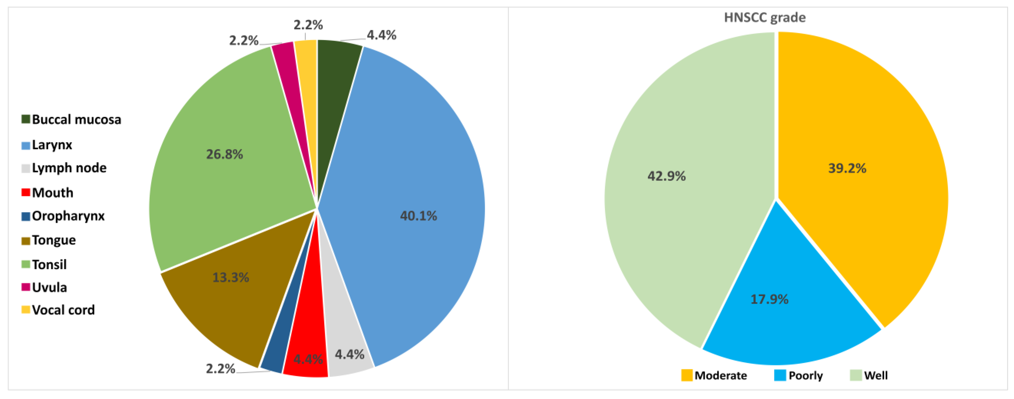
Histological sections were obtained from HPV16-positive HNSCC FFPE biopsies of 38 Greek patients aged between 45 and 82 years (median = 57 years). The histology grade and the tumor anatomic position are presented in Figure 2. The HPV genotyping was performed by using a commercially available real-time PCR assay kit (HPV Genotypes 14 Real-TM Quant, Sacace Biotechnologies, Como, Italy) after the DNA extraction with QIA amp DNA FFPE Tissue Kit (Qiagen GmbH) following the manufacturer’s instructions. The extracted DNA concentration was measured with QIAexpert technology (Qiagen, Heidelberg, Germany).
DNA was bisulfite converted with the application of EpiTect Bisulfite Kit (Qiagen, Heidelberg Germany) using the protocol for Sodium Bisulfite Conversion of Unmethylated Cytosines in DNA from Low-Concentration Solutions, following the manufacturer’s instructions and stored at −20 °C. For the quantification of the methylation of CpG sites (PyroMark Q24, Qiagen, Heidelberg, Germany), biotin-labeled primer sets, PCR conditions and sequencing primers were used in validated pyrosequencing assays already published [29,33]. Methylation investigation was conducted for 6UTR CpG sites (31, 37, 43, 52, 58, and 7862) and 6 CpG sites along the L1 gene (7034, 7091, 7136, 7145, 6367, 6457).
Subsequently, the studied samples were investigated for the physical status of HPV16 DNA. The episomal, mixed and integrated viral DNA forms were determined through E2/E6 DNA copy number ratios using the assay of quantitative Real Time—PCR. The main principle of the methodology is based on the fact that E2 and E6 genes are present in equal amounts in episomal viral DNA. On the other hand, HPV16 DNA integration leads to the disruption of the E2 gene, while the E6 gene remains intact and integrated into the host chromosome. To this end, two different plasmid constructs were designed as previously reported [15]. In particular, a plasmid containing a partial fragment of the GAPDH gene (pGAPDH) was constructed for the normalization of genomic DNA, whereas a plasmid containing the HPV16 portion from the E6 to E2 genes (pE6-E2) was constructed to normalize the RT-PCR assays. The copy numbers of E2 and E6 genes were measured considering the number of cells through the quantitative RT-PCR targeting the GAPDH gene. The results were expressed as E2, E6 copy number per 500 cells. Real-Time PCR conditions, primers and cut-off values were performed as previously described [15].
Data were recorded in Microsoft Excel 2016 (version 2308) spreadsheets (Microsoft Corporation, Redmond, Washington, DC, USA), with rows representing cases and columns variables. Statistical analysis was performed using the SAS for Windows, version 9.4 software platform (SAS Institute Inc., Cary, NC, USA). Descriptive values were expressed via the mean value and the standard deviation (SD) and, for completeness reasons, the median value, along with the quartile 1 (Q1) to quartile 3 (Q3) range, was also provided. For the categorical data, the relevant frequencies per category and percentages are provided. Comparisons among the groups of the methylations in the studied sites were performed using non parametric tests, since normality, as tested with the Shapiro Wilk test, was not always ensured. Specifically, the Kruskal–Wallis test was used to compare among more than two categories. In order to investigate for possible correlations between the methylation at the studied sites, the non-parametric Spearman correlation coefficient was used. Correlation coefficients less than 0.20 are characteristic of no correlation, between 0.20 and 0.39 are characterized as weak, between 0.40 and 0.59 as moderate, between 0.60 and 0.79 as strong and >0.80 as very strong. The significance level (p-value) for the study was set tο 0.05, and all tests were two-sided.

Author Contributions

Conceptualization, study design and supervision, C.K., D.T., Z.K. and M.Y.; conducted the experimental work, I.Z., D.L., S.P., G.K. and E.L.; analyzed the data and contributed to the interpretation of the data, A.P. (Abraham Pouliakis), A.B., I.S.P., M.S., A.D., A.P. (Amanda Psyrri), I.G.P. and M.Y.; writing—original draft preparation, C.K., D.T., I.Z., A.P. (Abraham Pouliakis) and I.S.P.; writing—review and editing, all authors. All authors have read and agreed to the published version of the manuscript.

Funding

This research received no external funding.

Institutional Review Board Statement

Not applicable.

Informed Consent Statement

Not applicable.

Data Availability Statement

Not applicable.

Conflicts of Interest

The authors declare no conflict of interest.

References

  1. Zandberg, D.P.; Bhargava, R.; Badin, S.; Cullen, K.J. The role of human papillomavirus in nongenital cancers. CA Cancer J. Clin. 2013, 63, 57–81. [Google Scholar] [CrossRef]
  2. Vos, T.; Abajobir, A.A.; Abate, K.H.; Abbafati, C.; Abbas, K.M.; Abd-Allah, F.; Abdulkader, R.S.; Abdulle, A.M.; Abebo, T.A.; Abera, S.F.; et al. Global, regional, and national incidence, prevalence, and years lived with disability for 328 diseases and injuries for 195 countries, 1990–2016: A systematic analysis for the Global Burden of Disease Study 2016. Lancet 2017, 390, 1211–1259. [Google Scholar] [CrossRef]
  3. Sturgis, E.M.; Ang, K.K. The epidemic of HPV-associated oropharyngeal cancer is here: Is it time to change our treatment paradigms? J. Natl. Compr. Cancer Netw. 2011, 9, 665–673. [Google Scholar] [CrossRef]
  4. Elrefaey, S.; Massaro, M.A.; Chiocca, S.; Chiesa, F.; Ansarin, M. HPV in oropharyngeal cancer: The basics to know in clinical practice. Acta Otorhinolaryngol. Ital. 2014, 34, 299–309. [Google Scholar]
  5. Suzich, J.A.; Ghim, S.J.; Palmer-Hill, F.J.; White, W.I.; Tamura, J.K.; Bell, J.A.; Newsome, J.A.; Jenson, A.B.; Schlegel, R. Systemic immunization with papillomavirus L1 protein completely prevents the development of viral mucosal papillomas. Proc. Natl. Acad. Sci. USA 1995, 92, 11553–11557. [Google Scholar] [CrossRef]
  6. Tommasino, M. The human papillomavirus family and its role in carcinogenesis. Semin. Cancer Biol. 2014, 26, 13–21. [Google Scholar] [CrossRef]
  7. de Sanjose, S.; Brotons, M.; Pavon, M.A. The natural history of human papillomavirus infection. Best Pract. Res. Clin. Obstet. Gynaecol. 2018, 47, 2–13. [Google Scholar] [CrossRef]
  8. Munoz, N.; Bosch, F.X.; de Sanjose, S.; Herrero, R.; Castellsague, X.; Shah, K.V.; Snijders, P.J.; Meijer, C.J.; International Agency for Research on Cancer Multicenter Cervical Cancer Study Group. Epidemiologic classification of human papillomavirus types associated with cervical cancer. N. Engl. J. Med. 2003, 348, 518–527. [Google Scholar] [CrossRef]
  9. Economopoulou, P.; Kotsantis, I.; Psyrri, A. Special Issue about Head and Neck Cancers: HPV Positive Cancers. Int. J. Mol. Sci. 2020, 21, 3388. [Google Scholar] [CrossRef]
  10. Näsman, A.; Du, J.; Dalianis, T. A global epidemic increase of an HPV-induced tonsil and tongue base cancer—Potential benefit from a pan-gender use of HPV vaccine. J. Intern. Med. 2020, 287, 134–152. [Google Scholar] [CrossRef]
  11. Kuss-Duerkop, S.K.; Westrich, J.A.; Pyeon, D. DNA Tumor Virus Regulation of Host DNA Methylation and Its Implications for Immune Evasion and Oncogenesis. Viruses 2018, 10, 82. [Google Scholar] [CrossRef]
  12. Dor, Y.; Cedar, H. Principles of DNA methylation and their implications for biology and medicine. Lancet 2018, 392, 777–786. [Google Scholar] [CrossRef]
  13. Schubeler, D. Function and information content of DNA methylation. Nature 2015, 517, 321–326. [Google Scholar] [CrossRef]
  14. Ekanayake Weeramange, C.; Tang, K.D.; Vasani, S.; Langton-Lockton, J.; Kenny, L.; Punyadeera, C. DNA Methylation Changes in Human Papillomavirus-Driven Head and Neck Cancers. Cells 2020, 9, 1359. [Google Scholar] [CrossRef]
  15. Tsakogiannis, D.; Kyriakopoulou, Z.; Ruether, I.G.A.; Amoutzias, G.D.; Dimitriou, T.G.; Diamantidou, V.; Kotsovassilis, C.; Markoulatos, P. Determination of human papillomavirus 16 physical status through E1/E6 and E2/E6 ratio analysis. J. Med. Microbiol. 2014, 63, 1716–1723. [Google Scholar] [CrossRef]
  16. Akagi, K.; Li, J.; Broutian, T.R.; Padilla-Nash, H.; Xiao, W.; Jiang, B.; Rocco, J.W.; Teknos, T.N.; Kumar, B.; Wangsa, D.; et al. Genome-wide analysis of HPV integration in human cancers reveals recurrent, focal genomic instability. Genome Res. 2014, 24, 185–199. [Google Scholar] [CrossRef]
  17. Balaji, H.; Demers, I.; Wuerdemann, N.; Schrijnder, J.; Kremer, B.; Klussmann, J.P.; Huebbers, C.U.; Speel, E.M. Causes and Consequences of HPV Integration in Head and Neck Squamous Cell Carcinomas: State of the Art. Cancers 2021, 13, 4089. [Google Scholar] [CrossRef]
  18. Reuschenbach, M.; Huebbers, C.U.; Prigge, E.S.; Bermejo, J.L.; Kalteis, M.S.; Preuss, S.F.; Seuthe, I.M.; Kolligs, J.; Speel, E.J.; Olthof, N.; et al. Methylation status of HPV16 E2-binding sites classifies subtypes of HPV-associated oropharyngeal cancers. Cancer 2015, 121, 1966–1976. [Google Scholar] [CrossRef]
  19. Giuliano, A.R.; Nedjai, B.; Lorincz, A.T.; Schell, M.J.; Rahman, S.; Banwait, R.; Boulware, D.; Sirak, B.; Martin-Gomez, L.; Abrahamsen, M.; et al. Methylation of HPV 16 and EPB41L3 in oral gargles: Associations with oropharyngeal cancer detection and tumor characteristics. Int. J. Cancer 2019, 146, 1018–1030. [Google Scholar] [CrossRef]
  20. Chaturvedi, A.K.; Engels, E.A.; Pfeiffer, R.M.; Hernandez, B.Y.; Xiao, W.; Kim, E.; Jiang, B.; Goodman, M.T.; Sibug-Saber, M.; Cozen, W.; et al. Human papillomavirus and rising oropharyngeal cancer incidence in the United States. J. Clin. Oncol. 2011, 29, 4294–4301. [Google Scholar] [CrossRef]
  21. Schache, A.G.; Powell, N.G.; Cuschieri, K.S.; Robinson, M.; Leary, S.; Mehanna, H.; Rapozo, D.; Long, A.; Cubie, H.; Junor, E.; et al. HPV-Related Oropharynx Cancer in the United Kingdom: An Evolution in the Understanding of Disease Etiology. Cancer Res. 2016, 76, 6598–6606. [Google Scholar] [CrossRef]
  22. Kim, K.S.; Park, S.A.; Ko, K.N.; Yi, S.; Cho, Y.J. Current status of human papillomavirus vaccines. Clin. Exp. Vaccine Res. 2014, 3, 168–175. [Google Scholar] [CrossRef]
  23. Kim, S.M. Human papilloma virus in oral cancer. J. Korean Assoc. Oral Maxillofac. Surg. 2016, 42, 327–336. [Google Scholar] [CrossRef]
  24. Pan, C.; Issaeva, N.; Yarbrough, W.G. HPV-driven oropharyngeal cancer: Current knowledge of molecular biology and mechanisms of carcinogenesis. Cancers Head Neck 2018, 3, 12. [Google Scholar] [CrossRef]
  25. Ang, K.K.; Harris, J.; Wheeler, R.; Weber, R.; Rosenthal, D.I.; Nguyen-Tan, P.F.; Westra, W.H.; Chung, C.H.; Jordan, R.C.; Lu, C.; et al. Human papillomavirus and survival of patients with oropharyngeal cancer. N. Engl. J. Med. 2010, 363, 24–35. [Google Scholar] [CrossRef]
  26. Cancer Genome Atlas, N. Comprehensive genomic characterization of head and neck squamous cell carcinomas. Nature 2015, 517, 576–582. [Google Scholar] [CrossRef]
  27. Nakagawa, T.; Kurokawa, T.; Mima, M.; Imamoto, S.; Mizokami, H.; Kondo, S.; Okamoto, Y.; Misawa, K.; Hanazawa, T.; Kaneda, A. DNA Methylation and HPV-Associated Head and Neck Cancer. Microorganisms 2021, 9, 801. [Google Scholar] [CrossRef]
  28. Camuzi, D.; Buexm, L.A.; Lourenco, S.Q.C.; Esposti, D.D.; Cuenin, C.; Lopes, M.S.A.; Manara, F.; Talukdar, F.R.; Herceg, Z.; Ribeiro Pinto, L.F.; et al. HPV Infection Leaves a DNA Methylation Signature in Oropharyngeal Cancer Affecting Both Coding Genes and Transposable Elements. Cancers 2021, 13, 3621. [Google Scholar] [CrossRef]
  29. Kottaridi, C.; Kyrgiou, M.; Pouliakis, A.; Magkana, M.; Aga, E.; Spathis, A.; Mitra, A.; Makris, G.; Chrelias, C.; Mpakou, V.; et al. Quantitative Measurement of L1 Human Papillomavirus Type 16 Methylation for the Prediction of Preinvasive and Invasive Cervical Disease. J. Infect. Dis. 2017, 215, 764–771. [Google Scholar] [CrossRef]
  30. Cheung, J.L.; Cheung, T.H.; Yu, M.Y.; Chan, P.K. Virological characteristics of cervical cancers carrying pure episomal form of HPV16 genome. Gynecol. Oncol. 2013, 131, 374–379. [Google Scholar] [CrossRef]
  31. Kim, K.; Garner-Hamrick, P.A.; Fisher, C.; Lee, D.; Lambert, P.F. Methylation patterns of papillomavirus DNA, its influence on E2 function, and implications in viral infection. J. Virol. 2003, 77, 12450–12459. [Google Scholar] [CrossRef]
  32. Thain, A.; Jenkins, O.; Clarke, A.R.; Gaston, K. CpG methylation directly inhibits binding of the human papillomavirus type 16 E2 protein to specific DNA sequences. J. Virol. 1996, 70, 7233–7235. [Google Scholar] [CrossRef]
  33. Kottaridi, C.; Leventakou, D.; Pouliakis, A.; Pergialiotis, V.; Chrelias, G.; Patsouri, E.; Zacharatou, A.; Panopoulou, E.; Damaskou, V.; Sioulas, V.; et al. Searching HPV genome for methylation sites involved in molecular progression to cervical precancer. J. Cancer 2019, 10, 4588–4595. [Google Scholar] [CrossRef]
  34. Tsakogiannis, D.; Gartzonika, C.; Levidiotou-Stefanou, S.; Markoulatos, P. Molecular approaches for HPV genotyping and HPV-DNA physical status. Expert Rev. Mol. Med. 2017, 19, e1. [Google Scholar] [CrossRef]
  35. Peitsaro, P.; Johansson, B.; Syrjanen, S. Integrated human papillomavirus type 16 is frequently found in cervical cancer precursors as demonstrated by a novel quantitative real-time PCR technique. J. Clin. Microbiol. 2002, 40, 886–891. [Google Scholar] [CrossRef]
  36. Koskinen, W.J.; Chen, R.W.; Leivo, I.; Makitie, A.; Back, L.; Kontio, R.; Suuronen, R.; Lindqvist, C.; Auvinen, E.; Molijn, A.; et al. Prevalence and physical status of human papillomavirus in squamous cell carcinomas of the head and neck. Int. J. Cancer 2003, 107, 401–406. [Google Scholar] [CrossRef]
  37. Parfenov, M.; Pedamallu, C.S.; Gehlenborg, N.; Freeman, S.S.; Danilova, L.; Bristow, C.A.; Lee, S.; Hadjipanayis, A.G.; Ivanova, E.V.; Wilkerson, M.D.; et al. Characterization of HPV and host genome interactions in primary head and neck cancers. Proc. Natl. Acad. Sci. USA 2014, 111, 15544–15549. [Google Scholar] [CrossRef]
  38. Chen, X.; Gao, L.; Sturgis, E.M.; Liang, Z.; Zhu, Y.; Xia, X.; Zhu, X.; Chen, X.; Li, G.; Gao, Z. HPV16 DNA and integration in normal and malignant epithelium: Implications for the etiology of laryngeal squamous cell carcinoma. Ann. Oncol. 2017, 28, 1105–1110. [Google Scholar] [CrossRef]
  39. Faust, H.; Eldenhed Alwan, E.; Roslin, A.; Wennerberg, J.; Forslund, O. Prevalence of human papillomavirus types, viral load and physical status of HPV16 in head and neck squamous cell carcinoma from the South Swedish Health Care Region. J. Gen. Virol. 2016, 97, 2949–2956. [Google Scholar] [CrossRef]
  40. Cricca, M.; Venturoli, S.; Leo, E.; Costa, S.; Musiani, M.; Zerbini, M. Disruption of HPV16 E1 and E2 genes in precancerous cervical lesions. J. Virol. Methods 2009, 158, 180–183. [Google Scholar] [CrossRef]
  41. Tsakogiannis, D.; Gortsilas, P.; Kyriakopoulou, Z.; Ruether, I.G.; Dimitriou, T.G.; Orfanoudakis, G.; Markoulatos, P. Sites of disruption within E1 and E2 genes of HPV16 and association with cervical dysplasia. J. Med. Virol. 2015, 87, 1973–1980. [Google Scholar] [CrossRef]
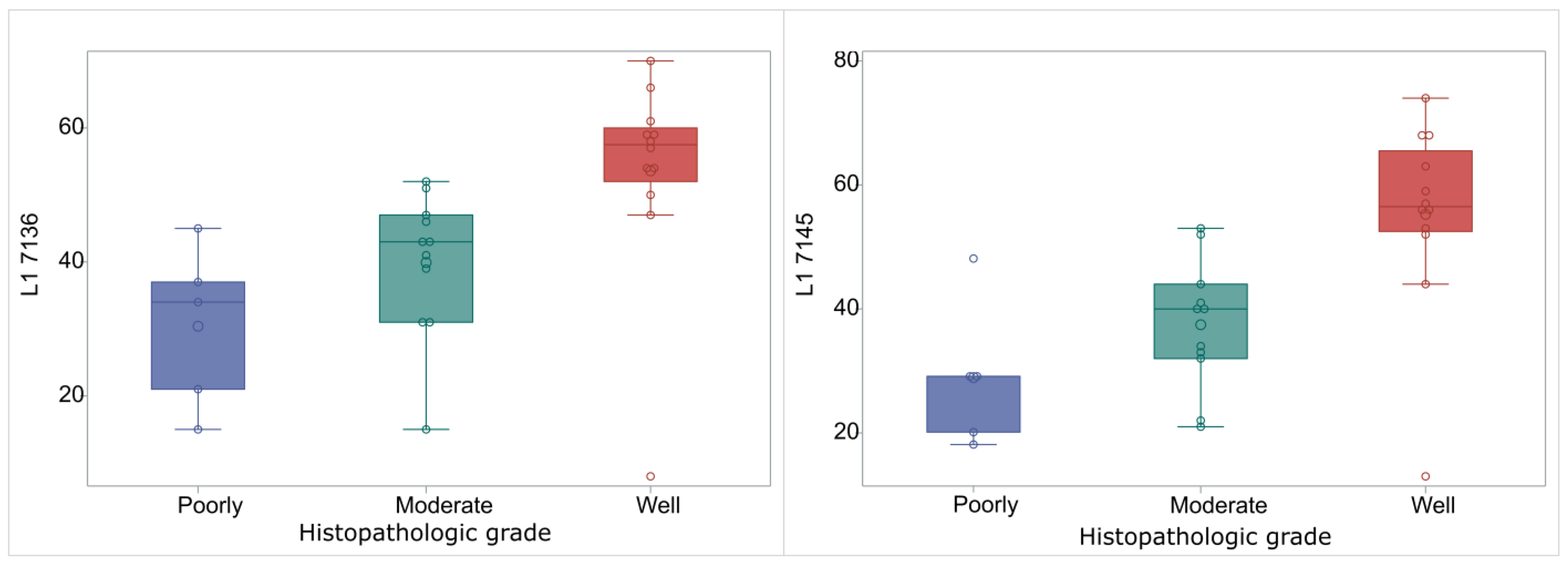
Figure 1. Box and whisker plot for L1 7136 and L1 7145 expression in relation to tumor grade. Box limits show Q1 and Q3 values. The lines within the boxes correspond to median values, the large circles indicate the median values and smaller circles correspond to measurements. Outliers are the circles outside the whisker limits, (left): L1 7136, (right): L1 7145.
Ijms 24 14593 g001
Figure 2. (Left): distribution of different anatomical sites where tumor derived; (Right): histology grading of studied samples.
Ijms 24 14593 g002
Table 1. Mean methylation and standard deviation for each CpG site studied.
CpG SiteMean Methylation ± SD
L1 636738.16 ± 12.60
L1 645737.11 ± 24.43
L1 703437.45 ± 24.65
L1 709141.47 ± 23.74
L1 713643.84 ± 16.69
L1 714543.11 ± 17.26
UTR 3124.74 ± 16.35
UTR 3731.00 ± 21.45
UTR 4331.42 ± 23.11
UTR 5232.55 ± 23.03
UTR 5828.37 ± 19.70
UTR 727012.37 ± 10.46
UTR 78629.71 ± 13.85
Table 2. Median methylation and Q1-Q3 range according to the histological grade and methylation quantification for each CpG site studied.
CpG SiteHistology Gradep-Value
Poorly ModerateWell
L1 636742 (39–49)37 (27–47)45 (40.5–50)0.2607
L1 645723 (19–25)35 (25–58)39 (11–61.5)0.1259
L1 703421 (16–51)44 (35–58)33 (15–61)0.4725
L1 709140 (35–45)39 (21–52)49.5 (25.5–75.5)0.5901
L1 713634 (21–37)43 (31–47)57.5 (52–60)0.0014
L1 714529 (20–29)40 (32–44)56.5 (52.5–65.5)0.0015
UTR 3110 (2–38)14 (10–32)32 (22–34)0.2312
UTR 3714 (13–41)18 (11–48)39 (28–45.5)0.3768
UTR 4318 (2–47)28 (7–47)38 (32–57)0.1573
UTR 5222 (11–47)16 (12–46)36 (25.5–51.5)0.3909
UTR 5815 (10–46)15 (9–37)37.5 (27–41)0.2920
UTR 727017 (8–21)6 (3–17)10.5 (3.5–18)0.6257
UTR 78628 (7–10)8 (5–12)6.5 (5–8)0.6219
Table 3. Median and Q1-Q3 range for the methylation sites and Spearman correlation coefficients. Values in square brackets indicate 95% confidence intervals. ***: p < 0.0001, **: p < 0.001, *: p < 0.05.
Median (Q1–Q3)UTR_37UTR_43UTR_52UTR_58UTR_7862UTR_7270sL1_7034L1_7091L1_7136L1_7145L1_6367L1_6457
UTR_3127 (11–35)0.92 [0.85–0.95] ***0.94 [0.89–0.96] ***0.93 [0.88–0.96] ***0.92 [0.85–0.95] ***0.3 [0–0.54] *0.12 [−0.18–0.4]0.13 [−0.18–0.4]0.31 [0.01–0.55] *0.59 [0.35–0.75] ***0.63 [0.41–0.78] ***0.38 [0.09–0.6] *0.49 [0.22–0.68] **
UTR_3731 (13–45) 0.91 [0.84–0.95] ***0.94 [0.89–0.97] ***0.92 [0.85–0.95] ***0.27 [−0.03–0.52]0.14 [−0.16–0.41]0.11 [−0.19–0.39]0.3 [0.01–0.55] *0.56 [0.32–0.73] ***0.59 [0.35–0.75] ***0.35 [0.05–0.58] *0.47 [0.19–0.67] *
UTR_4332 (10–47) 0.92 [0.86–0.96] ***0.92 [0.85–0.95] ***0.29 [−0.01–0.53]0.08 [−0.22–0.36]0.09 [−0.21–0.37]0.33 [0.03–0.56] *0.58 [0.34–0.74] ***0.65 [0.43–0.79] ***0.41 [0.13–0.62] *0.47 [0.19–0.67] *
UTR_5232 (13–47) 0.91 [0.83–0.95] ***0.31 [0.02–0.55] *0.14 [−0.16–0.42]0.11 [−0.19–0.39]0.35 [0.05–0.58] *0.59 [0.35–0.75] ***0.63 [0.4–0.77] ***0.4 [0.11–0.62] *0.42 [0.14–0.64] *
UTR_5824 (13–41) 0.27 [−0.03–0.52]0.1 [−0.2–0.38]0.1 [−0.2–0.38]0.33 [0.03–0.56] *0.51 [0.26–0.7] **0.59 [0.35–0.75] ***0.42 [0.14–0.63] *0.4 [0.12–0.62] *
UTR_78627 (5–10) 0.19 [−0.11–0.46]−0.03 [−0.32–0.27]−0.1 [−0.38–0.2]0.17 [−0.13–0.44]0.11 [−0.19–0.39]0.16 [−0.14–0.43]0.12 [−0.19–0.4]
UTR_727011 (4–21) 0.15 [−0.15–0.43]0.16 [−0.14–0.43]0.4 [0.11–0.61] *0.31 [0.01–0.55] *0.26 [−0.04–0.51]0.12 [−0.18–0.4]
L1_703435 (16–54) 0.51 [0.25–0.69] **0.26 [−0.04–0.51]0.19 [−0.11–0.46]0.3 [0–0.54] *0.07 [−0.24–0.35]
L1_709141 (24–53) 0.39 [0.1–0.61] *0.43 [0.15–0.64] *0.48 [0.21–0.68] **0.09 [−0.22–0.37]
L1_713647 (34–56) 0.9 [0.83–0.94] ***0.46 [0.19–0.66] *0.37 [0.07–0.59] *
L1_714544 (29–53) 0.46 [0.19–0.66] *0.41 [0.12–0.62] *
L1_636742 (35–49) 0.17 [−0.13–0.45]
L1_645727.5 (16–52.5) 1
Disclaimer/Publisher’s Note: The statements, opinions and data contained in all publications are solely those of the individual author(s) and contributor(s) and not of MDPI and/or the editor(s). MDPI and/or the editor(s) disclaim responsibility for any injury to people or property resulting from any ideas, methods, instructions or products referred to in the content.