Article

Transcriptomics of Canine Inflammatory Mammary Cancer Treated with Empty Cowpea Mosaic Virus Implicates Neutrophils in Anti-Tumor Immunity

by
Lucia Barreno
1,
Natalia Sevane
2,
Guillermo Valdivia
1,
Daniel Alonso-Miguel
1,
María Suarez-Redondo
1,
Angela Alonso-Diez
1,
Steven Fiering
3,4,*,
Veronique Beiss
5,
Nicole F. Steinmetz
5,6,7,8,9,10,11,
Maria Dolores Perez-Alenza
1 and
Laura Peña
1
1
Department of Animal Medicine, Surgery and Pathology, Mammary Oncology Unit, Veterinary Teaching Hospital, Veterinary Medicine School, Complutense University of Madrid, 28040 Madrid, Spain
2
Department of Animal Production, Complutense University of Madrid, 28040 Madrid, Spain
3
Department of Microbiology and Immunology, Geisel School of Medicine at Dartmouth, Lebanon, NH 03756, USA
4
Dartmouth Cancer Center, Geisel School of Medicine at Dartmouth, Lebanon, NH 03756, USA
5
Department of NanoEngineering, University of California San Diego, La Jolla, CA 92093, USA
6
Department of Radiology, University of California San Diego, 9500 Gilman Dr., La Jolla, CA 92093, USA
7
Department of Bioengineering, University of California San Diego, 9500 Gilman Dr., La Jolla, CA 92093, USA
8
Moores Cancer Center, University of California San Diego, 9500 Gilman Dr., La Jolla, CA 92093, USA
9
Center for Nano-ImmunoEngineering, University of California San Diego, 9500 Gilman Dr., La Jolla, CA 92093, USA
10
Institute for Materials Discovery and Design, University of California San Diego, 9500 Gilman Dr., La Jolla, CA 92093, USA
11
Center for Engineering in Cancer, Institute for Engineering in Medicine, University of California San Diego, 9500 Gilman Dr., La Jolla, CA 92093, USA
*
Author to whom correspondence should be addressed.
Int. J. Mol. Sci. 2023, 24(18), 14034; https://doi.org/10.3390/ijms241814034
Submission received: 24 August 2023 / Revised: 8 September 2023 / Accepted: 11 September 2023 / Published: 13 September 2023
(This article belongs to the Special Issue Molecular Immunology of Solid Tumors 2.0)

Abstract

:
Canine inflammatory mammary cancer (IMC) is a highly aggressive and lethal cancer in dogs serving as a valuable animal model for its human counterpart, inflammatory breast cancer (IBC), both lacking effective therapies. Intratumoral immunotherapy (IT-IT) with empty cowpea mosaic virus (eCPMV) nanoparticles has shown promising results, demonstrating a reduction in tumor size, longer survival rates, and improved quality of life. This study compares the transcriptomic profiles of tumor samples from female dogs with IMC receiving eCPMV IT-IT and medical therapy (MT) versus MT alone. Transcriptomic analyses, gene expression profiles, signaling pathways, and cell type profiling of immune cell populations in samples from four eCPMV-treated dogs with IMC and four dogs with IMC treated with MT were evaluated using NanoString Technologies using a canine immune-oncology panel. Comparative analyses revealed 34 differentially expressed genes between treated and untreated samples. Five genes (CXCL8, S100A9, CCL20, IL6, and PTGS2) involved in neutrophil recruitment and activation were upregulated in the treated samples, linked to the IL17-signaling pathway. Cell type profiling showed a significant increase in neutrophil populations in the tumor microenvironment after eCPMV treatment. These findings highlight the role of neutrophils in the anti-tumor response mediated by eCPMV IT-IT and suggest eCPMV as a novel therapeutic approach for IBC/IMC.

1. Introduction

Human inflammatory breast cancer (IBC) and canine inflammatory mammary cancer (IMC) are highly metastatic and deadly types of mammary cancer, commonly triple-negative, that lack effective therapies in both species [1,2,3]. IBC/IMC are an uncommon type of breast/mammary cancer with unusual specific genetic, pathogenic, and clinical characteristics [3,4,5]. The criterium for histological diagnosis of IBC/IMC is the neoplastic embolization of superficial dermal lymphatic vessels, which blocks lymphatic drainage and causes distinctive clinical features: mainly distinct diffuse inflammation and edema of the skin [3,4]. Intensive research efforts are ongoing to discover effective therapies for invasive and metastatic IBC [6,7]; however, optimal results have not been achieved [7]. The challenge in finding effective therapies for IBC is, in part, due to the lack of suitable experimental models to understand the aggressive behavior and mechanisms of IBC/IMC, and the limited knowledge of in vivo IBC/IMC biology. Most of our current understanding comes from studying cell lines and patient-derived models in mice lacking a functional immune system [8].
Dogs are genetically outbred animals with cancer prevalence, tumor type, and tissue origin that are similar to human cancer. In contrast to rodent cancer models, canine patients are outbred, long-lived, large patients that spontaneously develop cancers in a syngeneic environment in the presence of an intact immune system, thereby replicating the biology and heterogeneity of human disease [9,10,11]. Numerous epidemiologic, clinical, histopathological, and ultrastructural characteristics are shared by IBC and IMC, making IMC the only identified animal model that recapitulates the overall biology of IBC [12,13,14]. Canine IMC is a unique and excellent preclinical therapeutic model to evaluate the efficacy of new therapies for IBC [15].
Immunotherapy is a promising therapeutic strategy for cancer, given recent clinical advances in treating other human cancers, such as melanoma and lung cancer, that have demonstrated impressive clinical responses [16,17,18]. The activation of the immune system to recognize tumor antigens for cancer elimination [19] is an encouraging and novel therapeutic approach, although minimal progress has been made in the immunotherapy for breast cancer (BC) [20,21]. Immunotherapy with nanoparticles is an actively explored and promising area in oncology [22].
Our previous studies demonstrated the capacity of plant-based virus-like nanoparticles (VLPs) from empty cowpea mosaic virus (eCPMV) to stimulate anti-tumor immune responses and to enhance outcomes in different syngeneic murine BC tumor models [23,24] and in dogs with spontaneous IMC [25], with no side effects or toxicities. The immunotherapy involves the in situ administration of VLPs, which exhibit good tumor retention given the nanoparticle characteristics (measuring 30 nm in size) and are stable and non-toxic. CPMV can infect some plants but does not infect animal cells, while eCPMV lacks viral RNA, so is completely non-infectious [23]. Previous studies have demonstrated that the capsid of eCPMV is recognized by Toll-like receptors (TLR2 and TLR4) at the plasma membrane, triggering downstream signaling cascades involving the adaptor protein MyD88, ultimately activating the NF-κB pathway [26] in order to induce pro-inflammatory cytokines and anti-tumor efficacy. In this manner, the intratumor immunotherapy (IT-IT) strategy involves the direct manipulation of identified tumors to overcome the local tumor-mediated immunosuppression and, subsequently, stimulate systemic anti-tumor immunity [27].
The present study Is a continuation of a previous one conducted by our research group [25], in which eCPMV IT-IT stimulated a potent local leukocyte activation, induced a strong neutrophilic tumor infiltration, reduced tumor volume, and improved survival and quality of life in dogs with IMC, without adverse effects.
Transcriptomic analyses are powerful tools for detecting genes that influence the immune response. In BC, adaptive immune programs play diverse roles depending on the cellular infiltration found in each tumor [28], which is further complicated by heterogeneous genomic expression and genome instability [29,30]. There are no previous studies on the transcriptomic profile of dogs with IMC before or after treatment with eCPMV nanoparticles. Therefore, comparing the transcriptomic profiles in treated and untreated IMC samples can shed light on the complex gene networks involved and the improved clinical outcome of treated dogs with IMC.
In this study, an innovative NanoString nCounter Canine Immuno-oncology (IO) panel was used to evaluate RNA expression, active signaling pathways, and immune cell subtyping in tumors of female dogs diagnosed with IMC and treated with eCPMV IT-IT versus untreated IMC female dogs (only receiving medical therapy, MT) in order to characterize the immune response induced by the treatment. This advanced technology enables the analysis of numerous immune-related genes using RNA extracted from formalin-fixed paraffin-embedded (FFPE) tissues. These experiments also investigated the association between differential gene expression after eCPMV IT-IT and the clinical/cellular characterization obtained via flow cytometry, cytokine, histology, and immunohistochemistry assays in our previous study [25]. Our data show that eCPMV IT-IT induces strong transcriptional changes in the neoplastic tissue, which modify host immunity to promote potent neutrophil recruitment and activation against the tumor.

2. Results

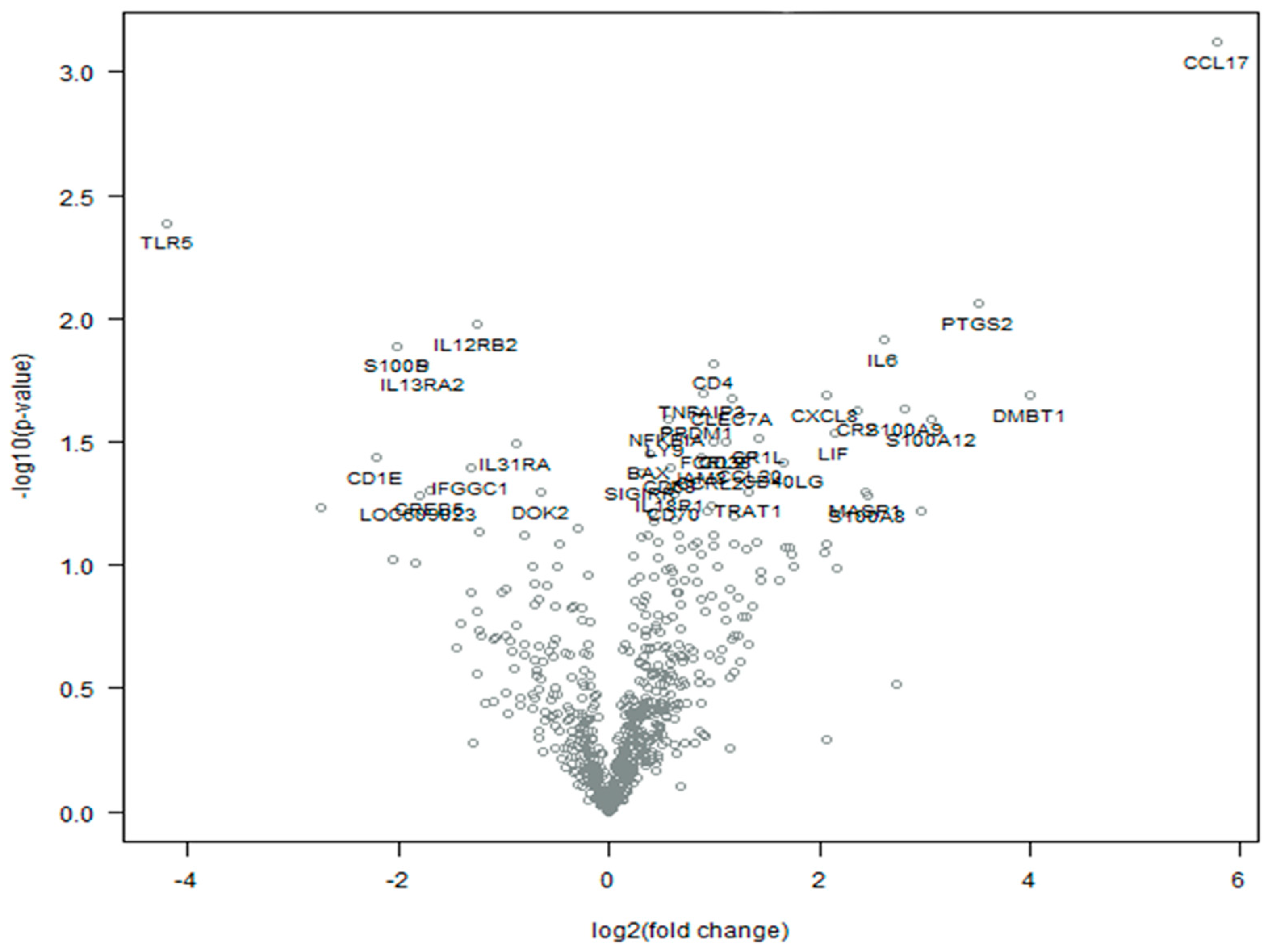
2.1. Differentially Expressed Genes in Tumor Samples after eCPMV Treatment

A total of 696 of the 780 immuno-oncology genes included in the NanoString nCounter technology passed QC and were included in the downstream analysis (Table S3). The comparison of gene expression profiles between the eCPMV-treated and -untreated samples revealed 34 significantly differentially expressed genes (DEGs) (p < 0.05), 26 of them upregulated (positive log2FC) and 8 downregulated (negative log2FC) in treated samples (Table 1; Figure 1).
Most of the DEGs observed in the IMC-treated samples were involved in chemokine and cytokine signaling pathways, including CCL17, PTGS2, IL12RB2, IL6, S100B, CD4, IL13RA2, CXCL8, S100A12, NFKBIA, LIF, IL31RA, CCL20, CD40LG, SIGIRR, and IL18R1. Among them, CCL17, S100B, and SIGIRR appeared to be specifically associated with the chemokine and cytokine signaling pathways. Furthermore, the expression of CCL17 mRNA showed the highest increase after treatment (log2FC = 5.77). On the other hand, the lowest downregulation value was displayed by TLR5 (log2FC = −4.21), a member of the Toll-like receptor family (TLR).

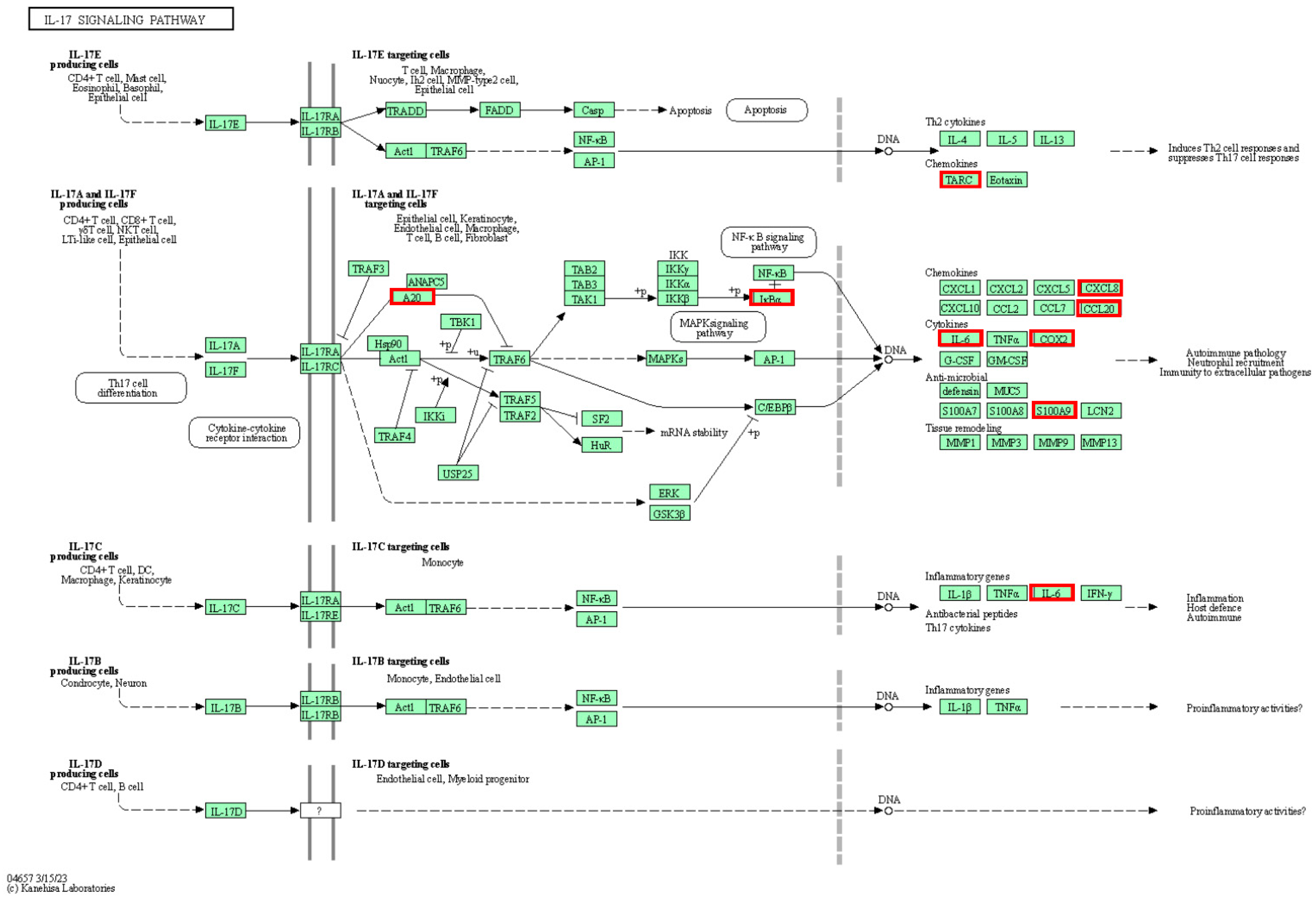
2.2. Pathway Analyses

The expression data were analyzed using the KEEG pathway analysis tool from DAVID. Of the 26 upregulated genes in the treated group, 25 were included in the analysis, resulting in a total of 37 pathways (Table S4). Among these pathways, the most relevant ones, which included five or more genes, were the interleukin 17 (IL-17) signaling pathway (NFKBIA, IL6, CXCL8, CCL20, TNFAIP3, PTGS2, CCL17, S100A9), and cytokine–cytokine receptor interaction (IL6, CD4, CD40LG, CXCL8, CCL20, LIF, CCL17, IL18R1), followed by the tumor necrosis factor (TNF) signaling pathway (NFKBIA, IL6, CCL20, LIF, TNFAIP3, PTGS2, IL18R1), viral protein interaction with cytokine and cytokine receptors (IL6, CXCL8, CCL20, CCL17, IL18R1), and the nuclear factor-kappaB (NF-κB) signaling pathway (NFKBIA, CD40LG, CXCL8, TNFAIP3, PTGS2). It is worth pointing out three other upregulated pathways, given their previous implication in key processes elicited after eCPMV IT-IT: the chemokine signaling pathway (NFKBIA, CXCL8, CCL20, CCL17), the Toll-like receptor signaling pathway (NFKBIA, IL6, CXCL8), and Th17 cell differentiation (NFKBIA, IL6, CD4). On the other hand, from the eight downregulated genes in the treated group, seven were included in the analysis and three pathways were retrieved (Table S4), with the decreased JAK-STAT signaling pathway (IL13RA2, IL12RB2) being the most interesting.

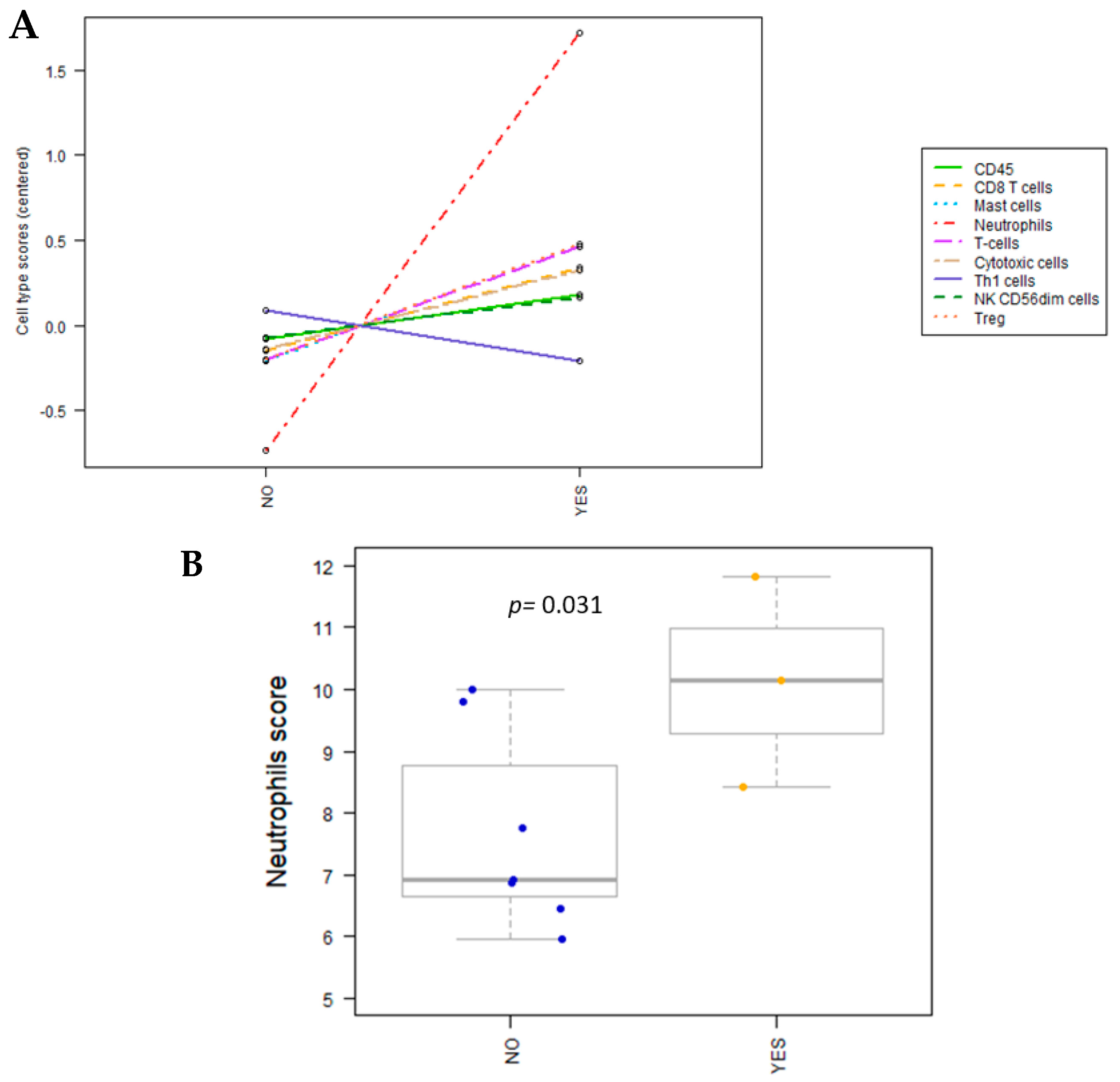
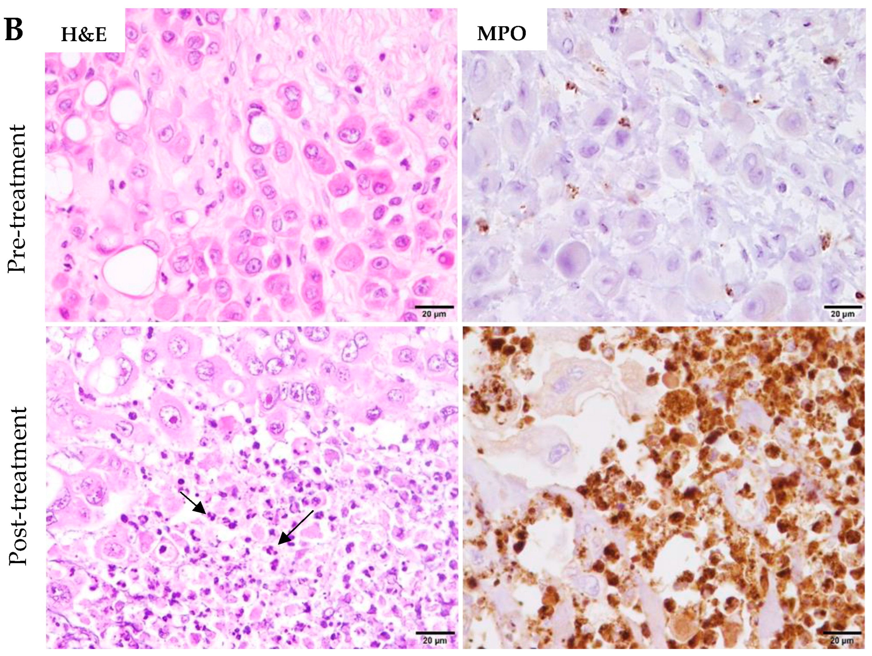
2.3. Cell Type Profiling
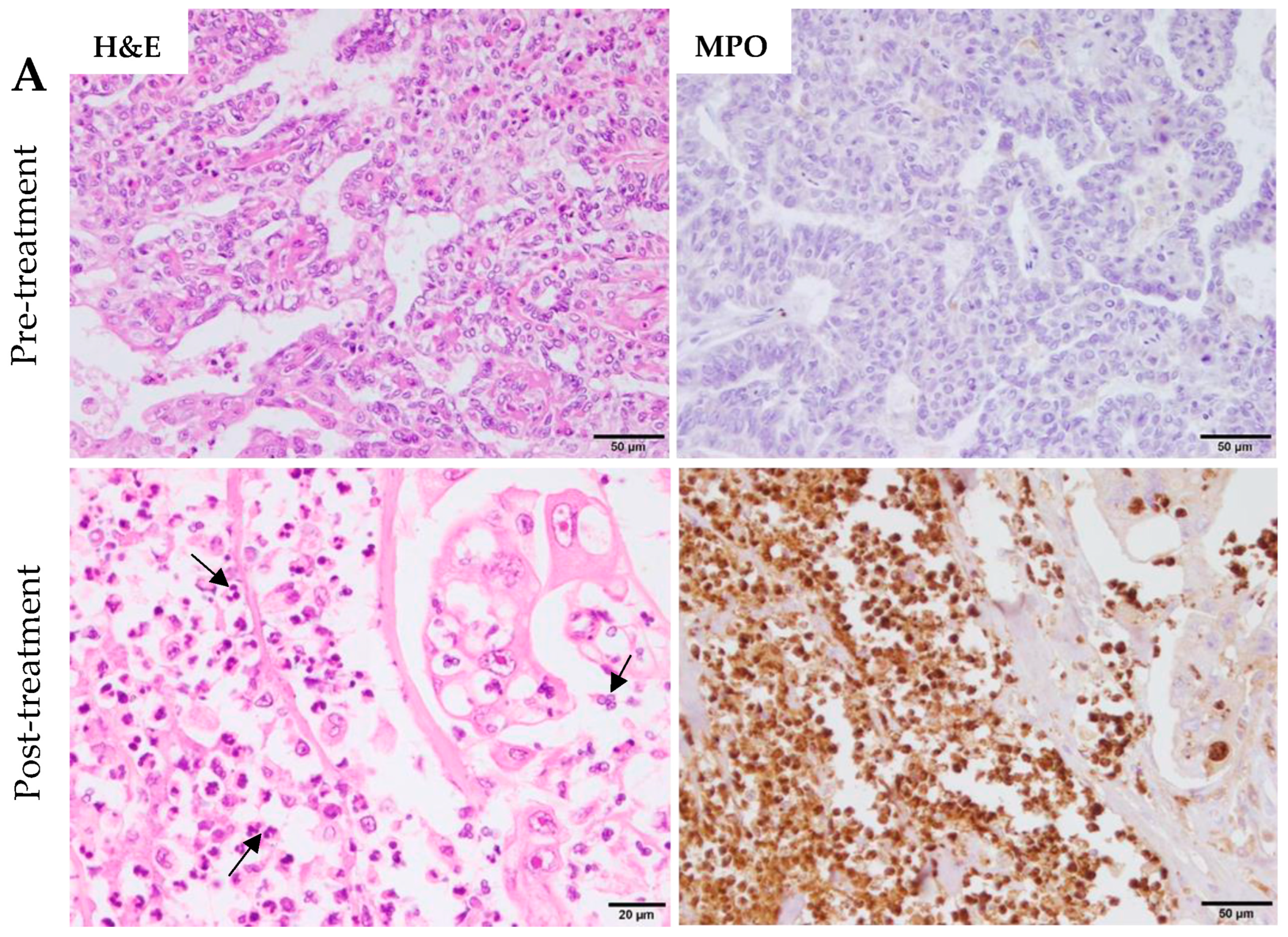
The nCounter software nSolver 4.0 used for canine NanoString analyses includes a specific cell type profiling analysis that can detect the abundance of immune cell populations by evaluating specific transcripts. This approach employs genes that have been previously identified as characteristic of various cell populations to measure the abundance of immune cellular populations in the TME. The IMC-treated samples showed a significantly higher proportion of neutrophils (p = 0.031) than the untreated samples (Figure 2A,B and Table 2). No other significant changes in immune cell populations were found with a p value < 0.05. However, there was a potential increase in T regulatory (Treg) cells and overall T cells, with p values < 0.1.

3. Discussion

In this study, we conducted a comprehensive evaluation of the transcriptomic profile and immune response in female dogs with IMC who received intratumoral eCPMV immunotherapy, compared to control untreated patients. By utilizing the NanoString nCounter Canine IO panel, we were able to analyze RNA expression and classify immune cell subtypes, providing valuable insights into the immune response following treatment. This approach allowed us to gain a deeper understanding of the signaling pathways involved in the anti-tumor immune response triggered by eCPMV nanoparticles, as well as identify potential candidate genes contributing to leukocyte activation. Our findings demonstrated significant transcriptional changes in the TME following eCPMV nanoparticle intratumoral immunotherapy. This is the first study in dogs with IMC to assess the impact of in situ eCPMV immunotherapy on the immune response using transcriptomics.
We identified 34 significant DEGs between treated and untreated IMC samples: 26 genes were upregulated and 8 genes downregulated in the treated group. The upregulated DEGs were primarily involved in signaling pathways related to chemokines and cytokines, indicating the activation of immune responses against the tumor. Among these, the IL-17 signaling pathway and cytokine–cytokine receptor interactions were particularly relevant, as they play crucial roles in immune responses and inflammation, suggesting their contribution to the anti-tumor effects triggered by eCPMV immunotherapy treatment.

3.1. The IL-17 Signaling Pathway’s Role in the Immune Response Triggered by eCPMV Immunotherapy

The IL-17 signaling pathway, which encompasses eight upregulated genes (NFKBIA, IL6, CXCL8, CCL20, TNFAIP3, PTGS2, CCL17, S100A9), includes five loci (CXCL8, S100A9, CCL20, PTGS2, and IL6) involved in the recruitment of immune cells, particularly neutrophils, to sites of inflammation (Figure 3). The CXCL8 gene encodes interleukin-8 (IL-8), which is a major cytokine that attracts and activates human neutrophils [31].
Intratumor CXCL8 expression is believed to play a critical role in regulating the recruitment of neutrophils into the TME [32]. In our previous study in dogs with IMC, we observed that eCPMV immunotherapy led to elevated blood levels of mature and immature neutrophils, transient increases in IL-8 levels, and a significant infiltration of neutrophils into the TME [25]. These findings suggest that eCPMV immunotherapy also has the potential to modulate neutrophil response in the TME at a transcriptomic level.
S100A9 gene, a member of the S100 calcium binding protein family, also known as calprotectin, is abundantly expressed in the cytosol of neutrophils and monocytes. Itcan induce degranulation of neutrophils, through the increased surface exposure of proteins found in secretory vesicles and gelatinase granules [33,34]. The increased expression of S100A9 in IMC-treated patients with eCPMV may contribute to neutrophil release of the enzyme myeloperoxidase (MPO) and corroborates the higher expression of this enzyme observed by immunohistochemistry (IHC) in our previous results [25]. In a recent study, it was shown that the S100A8/S100A9 complex acts as a damage-associated molecular pattern (DAMPs) molecule, activating TLR4 and initiating a signaling pathway that promotes the movement of the protein MyD88 from the cytoplasm to the cell membrane receptor complex, enhancing NF-κB-dependent transcriptional activity [35]. The present study identifies a substantial number of crucial genes implicated in the NF-κB signaling pathway, that play essential roles in the immune response, supporting the relevance of future development of anti-cancer therapies involving this pathway in combination with eCPMV. Furthermore, previous studies have demonstrated that the capsid of eCPMV is recognized by TLR2 and TLR4 at the plasma membrane, triggering downstream signaling cascades involving the adaptor protein MyD88, ultimately activating the NF-κB pathway [26]. This data supports the interpretation that S100A9 in IMC-treated animals may contribute to neutrophil degranulation, TLR4 activation, and enhanced NF-κB-dependent transcriptional activity through MyD88, and promotion of an anti-tumor immune response. Additionally, the upregulation of S100A12, another member of the S100 calcium binding protein family, after eCPMV treatment in this study suggests its regulatory effect on cytoskeletal components involved in various neutrophil activities and their migration [36].
These findings at transcriptional level agree with our previous studies which established a strong neutrophilic tumor infiltration associated with necrosis, upholding neutrophils as drivers of tumor cell death [25]. In Figure 4A,B demostrated the large neutrophilic infiltration and associated tumor cell death (necrosis) in post-treatment samples as compared with pre-treatment tumor samples in two patients by hematoxylin and eosin (H&E) and MPO staining. In our previous results published [25], MPO expression was significantly higher in post- treatment than in pre-treatment tumor samples.
In this study, the cell type profiling analysis showed a significant increased proportion of neutrophils in the treated versus untreated samples, corroborating their potential involvement in the anti-tumor immune response after eCPMV treatment. Although not reaching statistical significance, the observed changes in Treg lymphocytes and T cells highlight the overall modulation of immune cell populations following eCPMV IT-IT.
Recent studies on the role of tumor-associated neutrophils (TANs) in cancer biology indicate two TAN subpopulations (N1 and N2) having a dual role in tumor inhibition (N1) and progression (N2) [37,38]. The ability to distinguish between these neutrophil subpopulations is critical to understanding the pro- versus anti-tumor effects of TANs. TANs are influenced by signals in the TME such as the specific profile of chemokines, changing their phenotypic or functional plasticity between subpopulations [37,38,39,40]. The transcriptional profiles of the N1 (anti-tumor) and N2 (pro-tumor) TANs are currently under investigation [37,41]. The N1 subpopulation upregulates genes such as CCL2, CCL3, CXCL10, and CCL7 and downregulates CCL17, CXCL1, and CXCL14 [37]. Conversely, the N2 subpopulation upregulates a range of cytokines such as CCL17 and CCL5 [37,41].
Although the methodology of the present study does not allow for the separate detection of TAN subpopulations, it is worth noting that the upregulation of the S100A9 gene, as observed in our study, has been shown to enhance the chemotactic and enzymatic activity of the N1 (anti-tumor) subpopulation [42], which suggests that the upregulation of S100A9 could play an important role in the functional activity of N1 cells after eCPMV treatment. We have previously demonstrated that eCPMV treatment in dogs with IMC [25] dramatically increases TANs as the major driver of the anti-tumor immune response. Further studies are necessary to determine if the N1 subpopulation predominates among neutrophils in the TME after eCPMV treatment.
On the other hand, the high transcriptional overexpression of CCL17, also called TARC, seems to have a complex effect on tumor immunity. Some studies have associated CCL17 with the N2 tumor-promoting subtype due to its role in immune suppression mediated by regulatory T cells [37,43]. However, CCL17 is also expressed specifically by neutrophils and macrophages and plays a significant role in the alternative cross-priming of dendritic cells (DCs), enabling them to trigger CD8+ T lymphocyte responses against tumor antigens [44]. The response of CD8+ T cells involves the interaction between activated NK cells and DCs, leading to the production of CCL17, which attracts naïve CD8+ T lymphocytes. Increased expression of CD8+ granzyme B+ T cells has been correlated with higher transcript levels of CCL17, and higher serum levels of CCL17 were also associated with progression-free survival in advanced melanoma patients undergoing dendritic-cell-based immunotherapy [45]. Additionally, high serum levels of CCL17 have been associated with improved survival in human melanoma patients [46].
In a murine breast cancer model, the introduction of an adenoviral vector encoding CCL17 led to significant tumor regression and the generation of specific immunity in the TME [47], supporting a crucial role of this chemokine in the anti-tumor immune response.
At this point, it is interesting to remark that IBC/IMC is a rare and very special type of cancer with unique genetic, pathogenic, and clinical features [3,4,5]. In our study, the longest survival response to eCPMV therapy resulted in a patient (P1) that, interestingly, exhibited a remarkably high number of CCL17 RNA transcripts after treatment compared to the other eCPMV-treated patients. These results correlate with the significantly higher changes in the peripheral blood of CD8+ granzyme B+ T cells caused by eCPMV immunotherapy in our previous study of eCPMV treatment for dogs with IMC [25]. Further investigation is warranted to understand the clinical significance of this chemokine in the context of eCPMV immunotherapy.
After eCPMV treatment, the expressions of IL6 and CD4 genes were found to be significantly increased. These genes are involved in the differentiation of Th17 cells (Table S4), which produce IL17A and IL17F. Th17 cells play a crucial role in tumor immunity by attracting DCs and activating CD8+ T cells to fight against the tumor [48]. In our previous study, an increase in peripheral blood of CD8+ granzyme B+ T cells was observed in eCPMV-treated patients, which aligns with the role of Th17 cells in generating cytotoxic CD8+ T cells for tumor defense [25]. These findings highlight the potential importance of the IL17 signaling pathway in the immune response and its ability to modulate the anti-tumor immune response following treatment.
Interestingly, it has been recently shown that mature neutrophils could have an additional function as professional antigen-presenting cells (APCs) capable of inducing Th17 differentiation [49]. In our current study, genes associated with Th17 differentiation and IL17 signaling pathways, which may potentially recruit neutrophils (Table S4; Figure 3), were significantly upregulated following eCPMV IT-IT. This suggests that the increased proportion of neutrophils observed in IMC-treated patients (Figure 2) could shift their non-specific role to a specific immune action by behaving as APCs after nanoparticle inoculation. Consequently, this process could establish a cycle in which neutrophils participate in antigen presentation and induce Th17 cell differentiation, modulating the anti-tumor immune response after treatment.
The upregulation of CCL20 and PTGS2 after eCPMV treatment in our study has interesting implications. CCL20 can contribute to tumor progression by modulating the functions and phenotype of immune cells in the TME, particularly by inducing PD-L1 expression on neutrophils [50,51]. CCL20 is a potent chemoattractant of neutrophils to the TME [50,52]. In our study, increased expression of CCL20 in IMC-treated samples may be associated with the crucial involvement of the IL-17 signaling pathway in the immune response after eCPMV immunotherapy (Figure 3). It has been demonstrated that the IL-17 signaling pathway can increase CCL20 transcription [53]. Another upregulated gene, PTGS2 (Log2FC = 3.51), also known as cyclooxygenase-2 (COX-2), is the key enzyme in prostaglandin biosynthesis. This gene is highly expressed in the context of inflammatory responses and plays an important role in BC by influencing immune responses among other functions [54]. In this manner, the anti-tumor inflammatory response triggered by eCPMV treatment could potentially be the underlying cause of its overexpression. PTGS2 (COX-2) might shift its primary pro-tumor role to promote a potent immune response, thereby acquiring an anti-tumor effect. The pleiotropic nature of PTGS2 encompasses a wide range of effects, including the stimulation of angiogenesis and tumor cell proliferation [54]. Despite its role in tumor progression, a survival analysis revealed that BC patients with a high expression level of PTGS2 had a longer survival time after established treatments [55]. However, given that PTGS2 is generally known as a potent promoter of carcinogenesis, it is necessary to conduct further studies to determine its role in IMC patients following eCPMV IT-IT.

3.2. Other Upregulated Immune Pathways and Genes Triggered by eCPMV Immunotherapy

This study identifies crucial genes involved in the NF-κB signaling pathway, viral protein interactions with cytokine and cytokine receptors, the TNF signaling pathway, and the Toll-like receptor signaling pathway, all of which play essential roles in immune responses. Previous studies conducted on mice have demonstrated that the recognition of eCPMV viral capsids induces a downstream signaling cascade through TLRs 2 and 4, supporting the significance of these pathways [35].
In our analysis, we observed an upregulation of DMBT1 (Log2FC = 4), a tumor suppressor gene known to inhibit cell proliferation and survival in various cancers [56]. Recently, a reduction in DMBT1 expression has been observed in various cancer types, such as BC, prostate, and gallbladder cancers [57]. In accordance with our previous study, a decrease in the tumor Ki-67 proliferation index (PI) by IHC was observed after eCPMV immunotherapy, indicative of reduced tumor cell cycle proliferation [25].
Among the other upregulated DEGs that promote anti-tumor response, the following genes play important roles: CLEX7A, also known as dectin 1, is critical for NK-cell-dependent killing of tumor cells and might bolster anti-tumor immunity [58]; PRDM1, which is positively associated with both better cancer prognosis and immune infiltrates, with reduced expression linked to unfavorable prognoses in certain cancer types [59,60]; CD40LG, whose CD40/CD40L interaction leads to activation of DCs and contributes to their cytotoxicity against neoplastic cells [61]; CCRL2, which is a predictive indicator of potent anti-tumor T cell responses in human cancers [62]; and IL18R1, which has been associated with survival prediction in triple-negative breast cancer, primarily through the regulation of CD8+ T cells and various subtypes of CD4+ T cells [63].
JAM3, LIF, CD38, and SIGGIR, upregulated in our study, exert angiogenic and tumor-promoting effects and are linked to unfavorable cancer prognosis [64,65,66,67]. Why these poor prognosis factors are increased in our study needs to be elucidated, but it could be related to the special mechanisms found in IBC/IMC.

3.3. Downregulated Immune Pathways and Genes after eCPMV Immunotherapy

The JAK/STAT signaling pathway is involved in tumorigenesis, maintenance, and metastasis in BC. Its downregulation could potentially be a promising therapeutic approach in IBC [68].
In the present study, two genes of the JAK/STAT signaling pathway were significantly reduced after eCPMV treatment: IL13RA2 and IL12RB2. IL13RA2 plays an important role in cell migration, contributing to tumor progression [69], invasion, and metastasis in several cancers [70]. IL12RB2 was downregulated after eCPMV treatment. In a lung cancer transplant model assay, it was observed that mice lacking IL12RB2 developed lung adenocarcinoma [71]. Nevertheless, its role in BC remains unclear.
TLR5 was found to be the most significantly downregulated DEG compared to other genes (Log2FC = −4.2). This downregulation could be related to the activation of TLR2 and TLR4 by eCPMV treatment [35]. Overexpression of CD1 is indicative of a negative prognosis in hepatocellular carcinoma [72]. Therefore, the downregulation of this gene following treatment in our study may contribute to improved survival outcomes. Additionally, the downregulation of S100B, IL31RA, and CREB5 found here may also promote anti-tumor responses and reduce tumor progression. S100B, upregulated in malignant melanomas, has been shown to inhibit the function of the tumor suppressor protein p53 [73]. IL31RA has been implicated in BC progression and metastasis [74], while increased expression and activation of CREB are associated with tumor growth [75].
Considering the previously published benefits for dogs’ outcome and survival of eCPMV IT-IT, corroborated in the two additional patients of the present study, all these findings provide valuable insights into the immunological effects of in situ eCPMV immunotherapy in IMC. The modulation of immune cell profiles, enhanced anti-tumor immune responses, and improved overall survival collectively underscore the potential of eCPMV as an effective therapeutic strategy for a very-poor-outcome disease. This proof-of-concept study is limited by a small sample size and the genetically diverse nature of the patient groups, which, similar to the previous study, necessitated the comparison of untreated and treated samples not only from the same animal (pre-treatment versus post-treatment) and the absence of randomization. The small and genetically diverse nature of the animal patient groups are potential constraints. While diverse genetic backgrounds are often hailed for mirroring human diversity, it is important to recognize that such limited and diverse groups may pose challenges in terms of drawing unequivocal parallels to the human scenario. However, given the rarity of IMC, the significant results obtained in this study hold great value. Additionally, a more comprehensive transcriptional analysis of neutrophil subpopulations should be pursued to enhance our understanding of their role in the eCPMV therapeutic response.

4. Materials and Methods

4.1. Patients and Clinical Procedures

The present study is a continuation of our previous research study described in Alonso-Miguel et al. [25], where detailed information about patients, clinical procedures, and outcome are detailed. Here, two additional patients (one treated and one control) have been included, since the recruitment and treatment of animals continued after the publication of the previous study. In total, 12 patients were included in this study (6 eCPMV-treated and 6 eCPMV-untreated patients). Whenever possible, enrolled patients underwent a pretreatment incisional biopsy of the tumor, followed immediately by intratumoral eCPMV injections (Table 3). Briefly, eCPMV IT-IT resulted in tumor shrinkage in all patients by day 14 without adverse events. Although surgery is not recommended in cases of IMC [76], the reduction in tumor size from eCPMV treatment made surgical intervention possible in three of the IMC-treated patients (including the newly treated patient P6). Survival was significantly increased in eCPMV-treated dogs, including the newly enrolled dog P6.

4.2. RNA Extraction and NanoString nCounter Analyses

An assessment of tissue preservation and RNA viability was performed prior to RNA extraction from FFPE tissues. Some samples were excluded from further analysis either due to insufficient neoplastic tissue obtained from the incisional biopsy or because the owner did not opt to perform a necropsy (treated dogs P2 and P3; untreated dogs P10 and P11). Ultimately, RNA extraction was performed on 12 tumor samples from untreated patients (n = 8) and eCPMV-treated patients (n = 4). The identification of samples is provided in Table 4.
RNA was extracted from FFPE tissue blocks, where six 5 μm thin sections were taken, resulting in a total thickness of 30 μm scrolls. The samples were then treated with “Deparaffinization Solution” (Qiagen, Werfen, Barcelona, Spain) following the manufacturer’s instructions. Purification of total RNA from FFPE cores was performed using RNeasy FFPE Kit (Qiagen) following the manufacturer’s recommendations. The RNA concentration was quantified using a Nanodrop 2000 Spectrophotometer (Citogen Institution, Zaragoza, Spain).
DV200 (percentage of RNA fragments containing > 200 nucleotides) values were measured using a Bioanalyzer system. The capture and reporter probes contained 50 nucleotides (nt) complementary to the mRNA region of interest. Therefore, the RNA integrity number (RIN) values were not informative for assessing RNA quality for our assays. Instead, we used the DV200 value. Samples with a DV200 value > 30% were considered suitable for subsequent analysis. One IMC pre-treatment sample (P1) was excluded due to low RNA quantity (<20 ng/µL) and poor RNA quality, characterized by high fragmentation or degradation (DV200 < 30%; Supplemental File S1). A standard sample reference was included as an internal control test (Table S1).
The expression levels of immuno-oncology genes in the IMC-treated (n = 4) versus IMC-untreated (control dogs and pre-treatment) RNA samples were quantified using the canine IO panel NanoString nCounter (NanoString Technologies, Seattle, WA, USA). This panel analyzes the gene expression data of 780 genes that reflect cytokines, chemokines, interferon, and checkpoint signaling, complement cascades, immune cell abundance, tumor immunogenicity, inhibitory tumor mechanisms, and stromal factors (Table S1). Nineteen housekeeping genes were included as reference genes and showed homogeneity relative to the quantity/quality of the RNA (Table S2). The RNA was directly hybridized to the reporter and capture probes from the nCounter Canine IO panel. After hybridization, the samples were automatically processed on the nCounter Prep Station, and the captured transcripts were immobilized on the cartridge. The cartridge was then scanned using the nCounter Digital Analyzer to count the barcodes of the reporter probes.
After calculating the background or detection cut of the assay, an average of >700 genes were found to be significantly expressed in all samples (n = 11), except for a post-treatment sample (P2) that showed expression values below the mean (Table S1). This particular sample was taken at necropsy 24 h after the dog died, affecting its RNA quality, and therefore it was excluded from the analyses. A total of 10 samples were included in the downstream analyses.

4.3. Transcriptomic Data and Statistical Analyses

The raw data obtained from the NanoString nCounter analyses were normalized and underwent quality control using the nSolver Software v4.0 with the NanoString Advanced Analysis Module v2.0 plugin, following the manufacturer’s instructions. Only registered counts that passed the quality control (QC) parameters were used for subsequent analysis, and the normalized data were then scaled and transformed to log2. Functional analyses included differential gene expression, gene set analysis (GSA), and cell type profiling. Genes were deemed significantly differentially expressed when the p values were <0.05. Kyoto Encyclopedia of Genes and Genomes (KEGG) pathway analyses were also performed using the Database for Annotation, Visualization, and Integrated Discovery (DAVID) v6.8 [77] to map clusters of genes involved in common pathways and processes.
Unpaired Student’s t-test was used to compare the immune cellular subtyping population between IMC-treated versus IMC-untreated RNA samples. Two-tailed p values less than 0.05 were considered statistically significant. Statistical analyses were carried out using IBM SPSS Statistics program (V.25).

5. Conclusions

This study provides insights on the transcriptomic changes triggered by eCPMV immunotherapy and their potential influence on the immune response in IMC patients. The significant upregulation and downregulation of specific pathways and genes suggest that eCPMV nanoparticles induce changes in the TME that reflect an anti-tumor immune response, primarily driven by recruiting and activating neutrophils. The use of advanced transcriptomic analysis techniques, such as the Nanostring technology with the canine immune-oncology panel, will contribute to the future development of oncological immune studies in dogs from comparative and veterinary perspectives. These results improve the knowledge of the molecular mechanisms underlying the immune response to eCPMV therapy in IMC. The transcriptomic changes induced by eCPMV treatment and the subsequent amplification of the immune system response highlight the potential of this immunotherapy treatment as a promising therapeutic strategy for canine IMC and a potential future immunotherapy for human IBC patients.

Supplementary Materials

The following supporting information can be downloaded at https://www.mdpi.com/article/10.3390/ijms241814034/s1.

Author Contributions

Conception and design: L.P., M.D.P.-A., N.F.S. and S.F.; development of immunotherapy methodology: L.P., M.D.P.-A., N.F.S., N.S. and V.B.; acquisition of data (patient management, follow-up, and survival study) and transcriptomic analyses: M.D.P.-A., G.V., L.B., N.S., D.A.-M., A.A.-D., M.S.-R. and L.P.; interpretation of transcriptomic data: N.S., L.B., G.V. and L.P.; writing—review and/or revision of the paper: L.B., N.S., L.P. and rest of the authors. All authors have read and agreed to the published version of the manuscript.

Funding

This study was supported in part by the NCI (U01CA218292 and R01CA224605 to N.F.S. and S.F.); the Spanish Ministry of Science, Innovation and Technology (project PGC2018-094516-B-I00 to L.P. and M.D.P.-A.); the Spanish Ministry of Science, Innovation and Technology contract at Complutense University (PRE2019-089190 to L.B.); the ECVP specialization “Residency in Veterinary Pathology” grant from Complutense University (69/2018 to G.V.); a PhD grant from the Mexican Council for Science and Technology (CONACYT; 515916 to G.V.); and the PhD contract at Complutense University (7026349846-Y0SC001170 to D.A.-M.).

Institutional Review Board Statement

This prospective study was performed at the Mammary Oncology Unit of the Veterinary Teaching Hospital at Complutense University, Madrid, Spain, from October 2018 to November 2021 (approved by the institutional animal care and use committee and with owners consent.; Study #04/2018).

Informed Consent Statement

Not applicable.

Data Availability Statement

Data available on request from the authors.

Acknowledgments

We would like to thank all the patients involved in the study and their owners, who were very generous and supportive, and the personnel of the Complutense Veterinary Teaching Hospital for their help and support.

Conflicts of Interest

N.F.S. and S.F. are co-founders of, have equity in, and have a financial interest in Mosaic ImmunoEngineering Inc. (Novato, CA, USA). S.F. serves as scientific advisor and paid consultant for Mosaic; N.F.S. serves as Director, Board Member, Acting Chief Scientific Officer, and paid consultant for Mosaic.

Abbreviations

APCantigen-presenting cells
D0day of first immunotherapy dose
D7day of second immunotherapy dose
DAVIDDatabase for Annotation, Visualization, and Integrated Discovery
DAMPsdamage-associated molecular pattern
DCsdendritic cells
DEGsdifferentially expressed genes
eCPMVempty Cowpea Mosaic Virus
FFPEformalin-fixed paraffin-embedded
GSAgene set analysis
IBCinflammatory breast cancer
IHCimmunohistochemistry
IMCinflammatory mammary cancer
IL-8interleukin-8
IOimmuno-oncology
itRECISTintratumoral Response Evaluation Criteria in Solid Tumors
IT-ITIntratumoral immunotherapy
KEGGKyoto Encyclopedia of Genes and Genomes
MPOmieloperoxidase
MTmedical therapy
N1/N2neutrophil subpopulations
NF-κBnuclear factor-kappaB
NK cellsnatural killer cells
Ntnucleotide
PD-1programmed cell death 1
PIproliferation index
P1-12patients 1-12
QCquality control
QOLquality of life
RINRNA integrity number
TANstumor-associated neutrophils
TLR2/4Toll-like receptors 2/4
TMEtumor microenvironment
TNFtumor necrosis factor
Tregregulatory T cells
VLPsplant-based virus-like nanoparticles
DEGs Abbreviations
CCL17CC chemokine ligand 17
DMBT1deleted in malignant brain tumor 1
PTGS2prostaglandin-endoperoxide synthase 2
S100A12S100 Calcium Binding Protein A12
S100A9S100 calcium-binding protein A9
IL6interleukin 6
CR2complement receptor 2
LIFLeukemia Inhibitory Factor
CXCL8IL-8 or chemokine (C-X-C motif) ligand 8
CD40LGCD40 Ligand
CR1LComplement Receptor 1-Like
CCL20C-C Motif Chemokine Ligand 20
CLEC7AC-type lectin domain family 7 member A
CD38Cluster of Differentiation 38
CD4CD4 receptor, cluster of differentiation 4
FCRL2Fc Receptor-Like 2
CCRL2Chemokine C-C Motif Receptor-Like 2
TNFAIP3TNF Alpha Induced Protein 3
JAM3Junctional Adhesion Molecule 3
PRDM1PR domain zinc finger protein 1
IL18R1Interleukin 18 Receptor 1
CD68Cluster of Differentiation 68
NFKBIANuclear Factor Kappa B Inhibitor Alpha
LY9Lymphocyte Antigen 9
BAXBCL2 Associated X Protein
SIGIRRSingle Immunoglobulin and Toll-Interleukin 1 Receptor Domain-Containing Protein
TLR5Toll-like receptor 5
CD1ECluster of Differentiation 1E
S100BS100 calcium-binding protein B
IL13RA2Interleukin-13 receptor subunit alpha-2
CREB5cAMP Response Element-Binding Protein 5
IFGGC1Interferon Gamma-Inducible GTPase Candidate 1
IL12RB2interleukin 12 receptor, beta 2 subunit
IL31RAInterleukin 31 Receptor A

References

  1. van Uden, D.J.; van Laarhoven, H.W.; Westenberg, A.H.; de Wilt, J.H.; Blanken-Peeters, C.F. Inflammatory breast cancer: An overview. Crit. Rev. Oncol. Hematol. 2015, 93, 116–126. [Google Scholar] [CrossRef]
  2. Dabi, Y.; Darrigues, L.; Pons, K.; Mabille, M.; Abd Alsamad, I.; Mitri, R.; Skalli, D.; Haddad, B.; Touboul, C. Incidence of inflammatory breast cancer in patients with clinical inflammatory breast symptoms. PLoS ONE 2017, 12, e0189385. [Google Scholar] [CrossRef]
  3. Alenza MD, P.; Tabanera, E.; Peña, L. Inflammatory mammary carcinoma in dogs: 33 cases (1995–1999). J. Am. Vet. Med. Assoc. 2001, 219, 1110–1114. [Google Scholar] [CrossRef] [PubMed]
  4. Clemente, M.; Perez-Alenza, M.D.; Illera, J.C.; Pena, L. Histological, immunohistological, and ultrastructural description of vasculogenic mimicry in canine mammary cancer. Vet. Pathol. 2010, 47, 265–274. [Google Scholar] [CrossRef] [PubMed]
  5. Giordano, S.H. Update on Locally Advanced Breast Cancer. Oncologist 2003, 8, 521–530. [Google Scholar] [CrossRef] [PubMed]
  6. Low, J.A.; Berman, A.W.; Steinberg, S.M.; Danforth, D.N.; Lippman, M.E.; Swain, S.M. Long-term follow-up for locally advanced and inflammatory breast cancer patients treated with multimodality therapy. J. Clin. Oncol. 2004, 22, 4067–4074. [Google Scholar] [CrossRef]
  7. Ueno, N.T.; Buzdar, A.U.; Singletary, S.E.; Ames, F.C.; McNeese, M.D.; Holmes, F.A.; Theriault, R.L.; Strom, E.A.; Wasaff, B.J.; Asmar, L.; et al. Combined-modality treatment of inflammatory breast carcinoma: Twenty years of experience at MD Anderson Cancer Center. Cancer Chemother. Pharmacol. 1997, 40, 321–329. [Google Scholar] [CrossRef]
  8. Lim, B.; Woodward, W.A.; Wang, X.; Reuben, J.M.; Ueno, N.T. Inflammatory breast cancer biology: The tumour microenvironment is key. Nat. Rev. Cancer 2018, 18, 485–499. [Google Scholar] [CrossRef]
  9. Abdelmegeed, S.M.; Mohammed, S. Canine mammary tumors as a model for human disease. Oncol. Lett. 2018, 15, 8195–8205. [Google Scholar] [CrossRef]
  10. Schiffman, J.D.; Breen, M. Comparative oncology: What dogs and other species can teach us about humans with cancer. Philos. Trans. R. Soc. Lond. B Biol. Sci. 2015, 370, 20140231. [Google Scholar] [CrossRef]
  11. Dow, S. A Role for Dogs in Advancing Cancer Immunotherapy Research. Front. Immunol. 2019, 10, 2935. [Google Scholar] [CrossRef] [PubMed]
  12. Clemente, M.; Perez-Alenza, M.D.; Pena, L. Metastasis of canine inflammatory versus non-inflammatory mammary tumours. J. Comp. Pathol. 2010, 143, 157–163. [Google Scholar] [CrossRef] [PubMed]
  13. Clemente, M.; Sanchez-Archidona, A.R.; Sardon, D.; Diez, L.; Martin-Ruiz, A.; Caceres, S.; Sassi, F.; Dolores Perez-Alenza, M.; Illera, J.C.; Dunner, S.; et al. Different role of COX-2 and angiogenesis in canine inflammatory and non-inflammatory mammary cancer. Vet. J. 2013, 197, 427–432. [Google Scholar] [CrossRef] [PubMed]
  14. Barreno, L.; Caceres, S.; Alonso-Diez, A.; Vicente-Montana, A.; Garcia, M.L.; Clemente, M.; Illera, J.C.; Pena, L. Vasculogenic mimicry-associated ultrastructural findings in human and canine inflammatory breast cancer cell lines. BMC Cancer 2019, 19, 750. [Google Scholar] [CrossRef] [PubMed]
  15. Raposo, T.P.; Arias-Pulido, H.; Chaher, N.; Fiering, S.N.; Argyle, D.J.; Prada, J.; Pires, I.; Queiroga, F.L. Comparative aspects of canine and human inflammatory breast cancer. Semin. Oncol. 2017, 44, 288–300. [Google Scholar] [CrossRef]
  16. Hodi, F.S.; O’Day, S.J.; McDermott, D.F.; Weber, R.W.; Sosman, J.A.; Haanen, J.B.; Gonzalez, R.; Robert, C.; Schadendorf, D.; Hassel, J.C.; et al. Improved survival with ipilimumab in patients with metastatic melanoma. N. Engl. J. Med. 2010, 363, 711–723. [Google Scholar] [CrossRef] [PubMed]
  17. Garon, E.B.; Rizvi, N.A.; Hui, R.; Leighl, N.; Balmanoukian, A.S.; Eder, J.P.; Patnaik, A.; Aggarwal, C.; Gubens, M.; Horn, L.; et al. Pembrolizumab for the treatment of non-small-cell lung cancer. N. Engl. J. Med. 2015, 372, 2018–2028. [Google Scholar] [CrossRef]
  18. Makkouk, A.; Weiner, G.J. Cancer immunotherapy and breaking immune tolerance: New approaches to an old challenge. Cancer Res. 2015, 75, 5–10. [Google Scholar] [CrossRef]
  19. Leko, V.; Rosenberg, S.A. Identifying and Targeting Human Tumor Antigens for T Cell-Based Immunotherapy of Solid Tumors. Cancer Cell 2020, 38, 454–472. [Google Scholar] [CrossRef]
  20. Arab, A.; Behravan, N.; Razazn, A.; Barati, N.; Mosaffa, F.; Nicastro, J.; Slavcev, R.; Behravan, J. The viral approach to breast cancer immunotherapy. J. Cell Physiol. 2019, 234, 1257–1267. [Google Scholar] [CrossRef]
  21. van Rooijen, J.M.; Stutvoet, T.S.; Schröder, C.P.; de Vries, E.G.E. Immunotherapeutic options on the horizon in breast cancer treatment. Pharmacol. Ther. 2015, 156, 90–101. [Google Scholar] [CrossRef] [PubMed]
  22. Li, S.; Bennett, Z.T.; Sumer, B.D.; Gao, J. Nano-Immune-Engineering Approaches to Advance Cancer Immunotherapy: Lessons from Ultra-pH-Sensitive Nanoparticles. Acc. Chem. Res. 2020, 53, 2546–2557. [Google Scholar] [CrossRef] [PubMed]
  23. Lizotte, P.H.; Wen, A.M.; Sheen, M.R.; Fields, J.; Rojanasopondist, P.; Steinmetz, N.F.; Fiering, S. In Situ vaccination with cowpea mosaic virus nanoparticles suppresses metastatic cancer. Nat. Nanotechnol. 2016, 11, 295–303. [Google Scholar] [CrossRef] [PubMed]
  24. Cai, H.; Wang, C.; Shukla, S.; Steinmetz, N.F. Cowpea Mosaic Virus Immunotherapy Combined with Cyclophosphamide Reduces Breast Cancer Tumor Burden and Inhibits Lung Metastasis. Adv. Sci. 2019, 6, 1802281. [Google Scholar] [CrossRef] [PubMed]
  25. Alonso-Miguel, D.; Valdivia, G.; Guerrera, D.; Perez-Alenza, M.D.; Pantelyushin, S.; Alonso-Diez, A.; Beiss, V.; Fiering, S.; Steinmetz, N.F.; Suarez-Redondo, M.; et al. Neoadjuvant in situ vaccination with cowpea mosaic virus as a novel therapy against canine inflammatory mammary cancer. J. Immunother. Cancer 2022, 10, e004044. [Google Scholar] [CrossRef] [PubMed]
  26. Mao, C.; Beiss, V.; Fields, J.; Steinmetz, N.F.; Fiering, S. Cowpea mosaic virus stimulates anti-tumor immunity through recognition by multiple MYD88-dependent toll-like receptors. Biomaterials 2021, 275, 120914. [Google Scholar] [CrossRef]
  27. Sheen, M.R.; Fiering, S. In situ vaccination: Harvesting low hanging fruit on the cancer immunotherapy tree. Wiley Interdiscip. Rev. Nanomed. Nanobiotechnol. 2019, 11, e1524. [Google Scholar] [CrossRef]
  28. Varn, F.S.; Mullins, D.W.; Arias-Pulido, H.; Fiering, S.; Cheng, C. Adaptive immunity programmes in breast cancer. Immunology 2017, 150, 25–34. [Google Scholar] [CrossRef]
  29. Di Bonito, M.; Cantile, M.; Botti, G. Pathological and molecular characteristics of inflammatory breast cancer. Transl. Cancer Res. 2019, 8, S449–S456. [Google Scholar] [CrossRef]
  30. Ross, J.S.; Ali, S.M.; Wang, K.; Khaira, D.; Palma, N.A.; Chmielecki, J.; Palmer, G.A.; Morosini, D.; Elvin, J.A.; Fernandez, S.V.; et al. Comprehensive genomic profiling of inflammatory breast cancer cases reveals a high frequency of clinically relevant genomic alterations. Breast Cancer Res. Treat. 2015, 154, 155–162. [Google Scholar] [CrossRef]
  31. Metzemaekers, M.; Vandendriessche, S.; Berghmans, N.; Gouwy, M.; Proost, P. Truncation of CXCL8 to CXCL8(9-77) enhances actin polymerization and in vivo migration of neutrophils. J. Leukoc. Biol. 2020, 107, 1167–1173. [Google Scholar] [CrossRef] [PubMed]
  32. Russo, R.C.; Garcia, C.C.; Teixeira, M.M.; Amaral, F.A. The CXCL8/IL-8 chemokine family and its receptors in inflammatory diseases. Expert. Rev. Clin. Immunol. 2014, 10, 593–619. [Google Scholar] [CrossRef] [PubMed]
  33. Simard, J.C.; Girard, D.; Tessier, P.A. Induction of neutrophil degranulation by S100A9 via a MAPK-dependent mechanism. J. Leukoc. Biol. 2010, 87, 905–914. [Google Scholar] [CrossRef] [PubMed]
  34. Ryckman, C.; Vandal, K.; Rouleau, P.; Talbot, M.; Tessier, P.A. Proinflammatory activities of S100: Proteins S100A8, S100A9, and S100A8/A9 induce neutrophil chemotaxis and adhesion. J. Immunol. 2003, 170, 3233–3242. [Google Scholar] [CrossRef]
  35. Shabani, F.; Farasat, A.; Mahdavi, M.; Gheibi, N. Calprotectin (S100A8/S100A9): A key protein between inflammation and cancer. Inflamm. Res. 2018, 67, 801–812. [Google Scholar] [CrossRef]
  36. Gunaldi, M.; Okuturlar, Y.; Gedikbasi, A.; Akarsu, C.; Karabulut, M.; Kural, A. Diagnostic importance of S100A9 and S100A12 in breast cancer. Biomed. Pharmacother. 2015, 76, 52–56. [Google Scholar] [CrossRef]
  37. Shaul, M.E.; Levy, L.; Sun, J.; Mishalian, I.; Singhal, S.; Kapoor, V.; Horng, W.; Fridlender, G.; Albelda, S.M.; Fridlender, Z.G. Tumor-associated neutrophils display a distinct N1 profile following TGFbeta modulation: A transcriptomics analysis of pro- vs. antitumor TANs. Oncoimmunology 2016, 5, e1232221. [Google Scholar] [CrossRef]
  38. Fridlender, Z.G.; Sun, J.; Kim, S.; Kapoor, V.; Cheng, G.; Ling, L.; Worthen, G.S.; Albelda, S.M. Polarization of tumor-associated neutrophil phenotype by TGF-beta: “N1” versus “N2” TAN. Cancer Cell 2009, 16, 183–194. [Google Scholar] [CrossRef]
  39. Jaillon, S.; Ponzetta, A.; Di Mitri, D.; Santoni, A.; Bonecchi, R.; Mantovani, A. Neutrophil diversity and plasticity in tumour progression and therapy. Nat. Rev. Cancer 2020, 20, 485–503. [Google Scholar] [CrossRef]
  40. Furumaya, C.; Martinez-Sanz, P.; Bouti, P.; Kuijpers, T.W.; Matlung, H.L. Plasticity in Pro- and Anti-tumor Activity of Neutrophils: Shifting the Balance. Front. Immunol. 2020, 11, 2100. [Google Scholar] [CrossRef]
  41. Fridlender, Z.G.; Sun, J.; Mishalian, I.; Singhal, S.; Cheng, G.; Kapoor, V.; Horng, W.; Fridlender, G.; Bayuh, R.; Worthen, G.S.; et al. Transcriptomic analysis comparing tumor-associated neutrophils with granulocytic myeloid-derived suppressor cells and normal neutrophils. PLoS ONE 2012, 7, e31524. [Google Scholar] [CrossRef] [PubMed]
  42. Mihaila, A.C.; Ciortan, L.; Macarie, R.D.; Vadana, M.; Cecoltan, S.; Preda, M.B.; Hudita, A.; Gan, A.M.; Jakobsson, G.; Tucureanu, M.M.; et al. Transcriptional Profiling and Functional Analysis of N1/N2 Neutrophils Reveal an Immunomodulatory Effect of S100A9-Blockade on the Pro-Inflammatory N1 Subpopulation. Front. Immunol. 2021, 12, 708770. [Google Scholar] [CrossRef] [PubMed]
  43. Ralph, S.J.; Reynolds, M.J. Intratumoral pro-oxidants promote cancer immunotherapy by recruiting and reprogramming neutrophils to eliminate tumors. Cancer Immunol. Immunother. 2023, 72, 527–542. [Google Scholar] [CrossRef] [PubMed]
  44. Semmling, V.; Lukacs-Kornek, V.; Thaiss, C.A.; Quast, T.; Hochheiser, K.; Panzer, U.; Kurts, C. Alternative cross-priming through CCL17-CCR4-mediated attraction of CTLs toward NKT cell–licensed DCs. Nat. Immunol. 2010, 11, 313–320. [Google Scholar] [CrossRef] [PubMed]
  45. Cornforth, A.N.; Lee, G.J.; Fowler, A.W.; Carbonell, D.J.; Dillman, R.O. Increases in serum TARC/CCL17 levels are associated with progression-free survival in advanced melanoma patients in response to dendritic cell-based immunotherapy. J. Clin. Immunol. 2009, 29, 657–664. [Google Scholar] [CrossRef]
  46. Weide, B.; Allgaier, N.; Hector, A.; Forschner, A.; Leiter, U.; Eigentler, T.K.; Garbe, C.; Hartl, D. Increased CCL17 serum levels are associated with improved survival in advanced melanoma. Cancer Immunol. Immunother. 2015, 64, 1075–1082. [Google Scholar] [CrossRef]
  47. Bozic, T.; Sersa, G.; Kranjc Brezar, S.; Cemazar, M.; Markelc, B. Gene electrotransfer of proinflammatory chemokines CCL5 and CCL17 as a novel approach of modifying cytokine expression profile in the tumor microenvironment. Bioelectrochemistry 2021, 140, 107795. [Google Scholar] [CrossRef]
  48. Martin-Orozco, N.; Muranski, P.; Chung, Y.; Yang, X.O.; Yamazaki, T.; Lu, S.; Hwu, P.; Restifo, N.P.; Overwijk, W.W.; Dong, C. T helper 17 cells promote cytotoxic T cell activation in tumor immunity. Immunity 2009, 31, 787–798. [Google Scholar] [CrossRef]
  49. Abi Abdallah, D.S.; Egan, C.E.; Butcher, B.A.; Denkers, E.Y. Mouse neutrophils are professional antigen-presenting cells programmed to instruct Th1 and Th17 T-cell differentiation. Int. Immunol. 2011, 23, 317–326. [Google Scholar] [CrossRef]
  50. Kwantwi, L.B.; Wang, S.; Zhang, W.; Peng, W.; Cai, Z.; Sheng, Y.; Xiao, H.; Wang, X.; Wu, Q. Tumor-associated neutrophils activated by tumor-derived CCL20 (C-C motif chemokine ligand 20) promote T cell immunosuppression via programmed death-ligand 1 (PD-L1) in breast cancer. Bioengineered 2021, 12, 6996–7006. [Google Scholar] [CrossRef]
  51. Kadomoto, S.; Izumi, K.; Mizokami, A. The CCL20-CCR6 Axis in Cancer Progression. Int. J. Mol. Sci. 2020, 21, 5186. [Google Scholar] [CrossRef] [PubMed]
  52. Scapini, P.; Crepaldi, L.; Pinardi, C.; Calzetti, F.; Cassatella, M.A. CCL20/macrophage inflammatory protein-3alpha production in LPS-stimulated neutrophils is enhanced by the chemoattractant formyl-methionyl-leucyl-phenylalanine and IFN-gamma through independent mechanisms. Eur. J. Immunol. 2002, 32, 3515–3524. [Google Scholar] [CrossRef] [PubMed]
  53. Maddur, M.S.; Miossec, P.; Kaveri, S.V.; Bayry, J. Th17 cells: Biology, pathogenesis of autoimmune and inflammatory diseases, and therapeutic strategies. Am. J. Pathol. 2012, 181, 8–18. [Google Scholar] [CrossRef]
  54. Hashemi Goradel, N.; Najafi, M.; Salehi, E.; Farhood, B.; Mortezaee, K. Cyclooxygenase-2 in cancer: A review. J. Cell Physiol. 2019, 234, 5683–5699. [Google Scholar] [CrossRef] [PubMed]
  55. Wu, J.; Zhang, Y.; Li, M. Identification of genes and miRNAs in paclitaxel treatment for breast cancer. Gynecol. Endocrinol. 2021, 37, 65–71. [Google Scholar] [CrossRef] [PubMed]
  56. Ma, N.; Zhao, Y. DMBT1 suppresses cell proliferation, migration and invasion in ovarian cancer and enhances sensitivity to cisplatin through galectin-3/PI3k/Akt pathway. Cell Biochem. Funct. 2020, 38, 801–809. [Google Scholar] [CrossRef]
  57. Braidotti, P.; Nuciforo, P.G.; Mollenhauer, J.; Poustka, A.; Pellegrini, C.; Moro, A.; Bulfamante, G.; Coggi, G.; Bosari, S.; Pietra, G.G. DMBT1 expression is down-regulated in breast cancer. BMC Cancer 2004, 4, 46. [Google Scholar] [CrossRef]
  58. Kalia, N.; Singh, J.; Kaur, M. The role of dectin-1 in health and disease. Immunobiology 2021, 226, 152071. [Google Scholar] [CrossRef]
  59. Zhu, Z.; Wang, H.; Wei, Y.; Meng, F.; Liu, Z.; Zhang, Z. Downregulation of PRDM1 promotes cellular invasion and lung cancer metastasis. Tumour Biol. 2017, 39, 1010428317695929. [Google Scholar] [CrossRef]
  60. Shen, L.; Chen, Q.; Yang, C.; Wu, Y.; Yuan, H.; Chen, S.; Ou, S.; Jiang, Y.; Huang, T.; Ke, L.; et al. Role of PRDM1 in Tumor Immunity and Drug Response: A Pan-Cancer Analysis. Front. Pharmacol. 2020, 11, 593195. [Google Scholar] [CrossRef]
  61. Kuhn, N.F.; Purdon, T.J.; van Leeuwen, D.G.; Lopez, A.V.; Curran, K.J.; Daniyan, A.F.; Brentjens, R.J. CD40 Ligand-Modified Chimeric Antigen Receptor (CAR) T Cells Enhance Antitumor Function by Eliciting an Endogenous Antitumor Response. Cancer Cell. 2019, 35, 473–488. [Google Scholar] [CrossRef] [PubMed]
  62. Yin, W.; Li, Y.; Song, Y.; Zhang, J.; Wu, C.; Chen, Y.; Miao, Y.; Lin, C.; Lin, Y.; Yan, D.; et al. CCRL2 promotes antitumor T-cell immunity via amplifying TLR4-mediated immunostimulatory macrophage activation. Proc. Natl. Acad. Sci. USA 2021, 118, e2024171118. [Google Scholar] [CrossRef] [PubMed]
  63. Marchetti, P.; Antonov, A.; Anemona, L.; Vangapandou, C.; Montanaro, M.; Botticelli, A.; Mauriello, A.; Melino, G.; Catani, M.V. New immunological potential markers for triple negative breast cancer: IL18R1, CD53, TRIM, Jaw1, LTB, PTPRCAP. Discov. Oncol. 2021, 12, 6. [Google Scholar] [CrossRef]
  64. Segaert, P.; Lopes, M.B.; Casimiro, S.; Vinga, S.; Rousseeuw, P.J. Robust identification of target genes and outliers in triple-negative breast cancer data. Stat. Methods Med. Res. 2019, 28, 3042–3056. [Google Scholar] [CrossRef]
  65. Li, X.; Yang, Q.; Yu, H.; Wu, L.; Zhao, Y.; Zhang, C.; Hu, W. LIF promotes tumorigenesis and metastasis of breast cancer through the AKT-mTOR pathway. Oncotarget 2014, 5, 788. [Google Scholar] [CrossRef]
  66. Chen, L.; Diao, L.; Yang, Y.; Yi, X.; Rodriguez, B.L.; Li, Y.; Villalobos, P.A.; Cascone, T.; Liu, X.; Tan, L.; et al. CD38-Mediated Immunosuppression as a Mechanism of Tumor Cell Escape from PD-1/PD-L1 Blockade. Cancer Discov. 2018, 8, 1156–1175. [Google Scholar] [CrossRef] [PubMed]
  67. Campesato, L.F. High IL-1R8 expression in breast tumors promotes tumor growth and contributes to impaired antitumor immunity. Oncotarget 2017, 8, 49470–49483. [Google Scholar] [CrossRef]
  68. Stevens, L.E.; Peluffo, G.; Qiu, X.; Temko, D.; Fassl, A.; Li, Z.; Trinh, A.; Seehawer, M.; Jovanovic, B.; Aleckovic, M.; et al. JAK-STAT Signaling in Inflammatory Breast Cancer Enables Chemotherapy-Resistant Cell States. Cancer Res. 2023, 83, 264–284. [Google Scholar] [CrossRef]
  69. Marquez-Ortiz, R.A.; Contreras-Zarate, M.J.; Tesic, V.; Alvarez-Eraso, K.L.F.; Kwak, G.; Littrell, Z.; Costello, J.C.; Sreekanth, V.; Ormond, D.R.; Karam, S.D.; et al. IL13Ralpha2 Promotes Proliferation and Outgrowth of Breast Cancer Brain Metastases. Clin. Cancer Res. 2021, 27, 6209–6221. [Google Scholar] [CrossRef]
  70. Chong, S.T.; Tan, K.M.; Kok, C.Y.L.; Guan, S.P.; Lai, S.H.; Lim, C.; Hu, J.; Sturgis, C.; Eng, C.; Lam, P.Y.P.; et al. IL13RA2 Is Differentially Regulated in Papillary Thyroid Carcinoma vs Follicular Thyroid Carcinoma. J. Clin. Endocrinol. Metab. 2019, 104, 5573–5584. [Google Scholar] [CrossRef]
  71. Liu, Z.G.; Lei, Y.Y.; Li, W.W.; Chen, Z.G. The co-expression of ERbeta2 and IL-12Rbeta2 is better prognostic factor in non-small-cell lung cancer progression. Med. Oncol. 2013, 30, 592. [Google Scholar] [CrossRef] [PubMed]
  72. Fessas, P.; Spina, P.; Boldorini, R.L.; Pirisi, M.; Minisini, R.; Mauri, F.A.; Simpson, F.; Olivieri, P.; Gennari, A.; Wong, C.N.; et al. Phenotypic Characteristics of the Tumour Microenvironment in Primary and Secondary Hepatocellular Carcinoma. Cancers 2021, 13, 2137. [Google Scholar] [CrossRef] [PubMed]
  73. Harpio, R.; Einarsson, R. S100 proteins as cancer biomarkers with focus on S100B in malignant melanoma. Clin. Biochem. 2004, 37, 512–518. [Google Scholar] [CrossRef] [PubMed]
  74. He, Y.; Zhang, X.; Pan, W.; Tai, F.; Liang, L.; Shi, J. Interleukin-31 Receptor alpha Is Required for Basal-Like Breast Cancer Progression. Front. Oncol. 2020, 10, 816. [Google Scholar] [CrossRef]
  75. Ahmed, M.B.; Alghamdi, A.A.A.; Islam, S.U.; Lee, J.S.; Lee, Y.S. cAMP Signaling in Cancer: A PKA-CREB and EPAC-Centric Approach. Cells 2022, 11, 2020. [Google Scholar] [CrossRef]
  76. Marconato, L.; Romanelli, G.; Stefanello, D.; Giacoboni, C.; Bonfanti, U.; Bettini, G.; Finotello, R.; Verganti, S.; Valenti, P.; Ciaramella, L.; et al. Prognostic factors for dogs with mammary inflammatory carcinoma: 43 cases (2003–2008). J. Am. Vet. Med. Assoc. 2009, 235, 967–972. [Google Scholar] [CrossRef]
  77. Huang, D.W.; Sherman, B.T.; Lempicki, R.A. Systematic and integrative analysis of large gene lists using DAVID Bioinformatics Resources. Nat. Protoc. 2009, 4, 44–57. [Google Scholar] [CrossRef]
Figure 1. Volcano plot of differentially expressed genes between treated and untreated IMC samples with eCPMV immunotherapy. Thirty-four genes were significantly differentially expressed in IMC samples after eCPMV in situ immunotherapy versus untreated IMC samples (p value < 0.05), 26 upregulated with a positive log2FC and 8 downregulated with a negative log2FC.
Ijms 24 14034 g001
Figure 2. (A) Cell type profiling between IMC samples treated and untreated with eCPMV immunotherapy (NO: untreated IMC samples; YES: eCPMV-treated IMC samples). IMC-treated samples showed a remarkably higher neutrophil population than untreated samples among all the rest of the cell population. (B) Neutrophil subpopulations were significantly increased (p = 0.031) in treated IMC samples versus untreated IMC samples (p values estimated using Student’s t-test).
Ijms 24 14034 g002
Figure 3. IL-17 signaling pathway (cfa04657). Genes upregulated after treatment with eCPMV nanoparticles in IMC tumors are highlighted in red. KEGG IDs—official gene symbol equivalence: A20—TNFAIP3; IκBα—NFKBIA; COX2—PTGS2; TARC—CCL17.
Ijms 24 14034 g003
Figure 4. In situ eCPMV immunotherapy in P1 (A) and P6 (B) induced a high neutrophilic infiltration and associated tumor cell death (necrosis) in post-treatment as compared with pre-treatment tumor samples as indicated by H&E and myeloperoxidase (MPO) staining. Black arrows show multilobulated neutrophil subpopulation.
Ijms 24 14034 g004aIjms 24 14034 g004b
Table 1. Up- and downregulated DEGs in female dogs with IMC treated with eCPMV immunotherapy.
Gene SymbolLog2 Fold ChangeStd Error (log2)p-ValueNanoString nCounter Immuno-Oncology Gene Sets
Upregulated
CCL175.771.10.00076Chemokines, Cytokine and Chemokine Signaling
DMBT141.390.0204
PTGS23.511.020.0087Angiogenesis, Costimulatory Signaling, Cytokine and Chemokine Signaling, Cytokines, Hypoxia, Myeloid Compartment, NF-kB Signaling
S100A123.061.120.0257Cytokine and Chemokine Signaling, Myeloid Compartment
S100A92.810.0233Angiogenesis, Hypoxia
IL62.620.8140.0122Angiogenesis, Cytokine and Chemokine Signaling, Hypoxia, Interleukins, JAK-STAT Signaling, Metabolic Stress, PI3K-Akt
CR22.360.8470.0235B-Cell Functions, Complement System
LIF2.140.8050.0291Angiogenesis, Cell Functions, Cytokine and Chemokine Signaling, Cytotoxicity, JAK-STAT Signaling, Myeloid Compartment
CXCL82.070.720.0206Chemokines, Cytokine and Chemokine Signaling, Cytokines, Interleukins, Pathogen Defense, Regulation
CD40LG1.660.6690.0382Costimulatory Signaling, Cytokine and Chemokine Signaling, Immune Cell Adhesion and Migration, Lymphoid Compartment, NF-kB Signaling, Regulation
CR1L1.430.5450.0304Complement System
CCL201.350.5380.036Chemokines, Cytokine and Chemokine Signaling, Myeloid Compartment
CLEC7A1.170.4110.0212Immune Cell Adhesion and Migration, Myeloid Compartment
CD381.110.4260.0316B-Cell Functions, Hypoxia, Lymphoid Compartment, Regulation, T Cell Functions
CD40.9930.3230.0152Antigen Presentation, Costimulatory Signaling, Cytokine and Chemokine Signaling, Immune Cell Adhesion and Migration
FCRL20.9890.380.0316
CCRL20.9720.3940.0389Chemokines
TNFAIP30.8910.3080.02Angiogenesis, Hypoxia, NF-kB Signaling, TNF Superfamily
JAM30.8830.3520.0365Angiogenesis, Immune Cell Adhesion and Migration
PRDM10.8290.2990.0242Cell Functions, Epigenetic Regulation
IL18R10.5850.250.0478Costimulatory Signaling, Cytokine and Chemokine Signaling, Lymphoid Compartment, NK Cell Functions, T Cell Functions
CD680.5780.2360.0401Cell Functions
NFKBIA0.5730.210.0257Apoptosis, Costimulatory Signaling, Cytokine and Chemokine Signaling, NF-kB Signaling
LY90.5340.20.0284Costimulatory Signaling, Lymphoid Compartment
BAX0.3840.1520.0353Apoptosis, Cell Cycle, Regulation
SIGIRR0.3140.130.0423Cytokine and Chemokine Signaling
Downregulated
TLR5−4.211.060.0041TLR
CD1E−2.210.8820.0365Antigen Processing
S100B−2.020.6350.0129Cytokine and Chemokine Signaling
IL13RA2−1.770.5770.0153Chemokines, Cytokine and Chemokine Signaling, JAK-STAT Signaling, T Cell Functions
CREB5−1.70.7360.0494
IFGGC1−1.320.540.0406Macrophage Functions, Myeloid Compartment
IL12RB2−1.250.3780.0106Cytokine and Chemokine Signaling, Cytokines, Cytotoxicity, JAK-STAT Signaling, Lymphoid Compartment, NK Cell Functions, T Cell Functions
IL31RA−0.8840.3420.0323Cytokine and Chemokine Signaling, Cytokines, Regulation
Table 2. Leukocyte subpopulations between IMC samples treated and untreated with eCPMV immunotherapy.
Cell TypeUntreated IMC DogsTreated IMC Dogsp-Value *
CD4510.8 ± 0.411.0 ± 0.20.192
CD8+ T cells7.0 ± 0.97.5 ± 0.40.208
Mast cells6.8 ± 1.47.5 ± 1.20.248
Neutrophils7.7 ± 1.610.1 ± 1.70.031 *
T cells7.6 ± 0.78.3 ± 0.60.09
Cytotoxic cells6.1 ± 0.96.6 ± 0.70.227
Th1 cells6.8 ± 0.66.5 ± 0.30.226
NK CD56dim cells8.0 ± 0.58.2 ± 0.70.276
Treg4.9 ± 0.65.6 ± 0.60.069
CD45, leucocyte population; CD8+ T cells, cytotoxic T cell population; mast cells, mastocytic cell population; neutrophils, neutrophilic population; cytotoxic cells, cytotoxic cell population; T cells, lymphocyte cell population; Th1 cells, T helper cell population; NK CD56dim cells, CD56+ Natural killer (NK) cells; Treg, regulatory T cells; ± denotes standard error; * denotes p-value below 0.05.
Table 3. Epidemiological and clinicopathological characteristics of eCPMV-treated and -untreated IMC patients.
PATIENTAge
y
Weight kg BreedTypeHisto GradeHisto TypesdLVILNITreatments
Target Tumor
TherapyOS Days
eCPMV-treated IMC patients
P1 1110.3MixedPrimaryIIISpecial typeYesYes8FCT+174
P2 13.525.6MixedSecondaryIIISimpleYesYes7FCT+156
P310.78.2PoodleSecondaryIIISimpleYesYes2FCT+109
P4 10.717Kerry Blue
Terrier
SecondaryIIISimpleYesYes3FCT+165
P5 11.92.7Bichon FriseSecondaryIIISimpleYesYes2FCT+67
P6 1322.3German
Sheperd
SecondaryIIISimpleYesYes2FCT+104
Untreated IMC patients
P7 1326.2MixedSecondaryIIIDAYesYes FCT27
P8 14.27.6MalteseSecondaryIIISimpleYesYes FCT40
P9 9.710.3MixedSecondaryIIISimpleYesYes FCT132
P10139.3Miniature
Schnauzer
SecondaryIIISimpleYesYes FCT63
P118.97.7PoodleSecondaryIIISimpleYesYes FCT73
P12 8.326German
Sheperd
SecondaryIIISimpleYesYes FCT14
Age y, age at diagnosis in years; FCT, firocoxib+cyclophosphamide+toceranib; FTC+ indicates that eCPMV-treated dogs received FCT therapy starting after second eCPMV injection until surgery or death; DA, ductal associated; eCPMV, empty cowpea mosaic virus; Histo, histologic; IMC, inflammatory mammary cancer; sdLVI, superficial dermal lymphovascular invasion; LNI, regional lymph node involvement; OS days, overall survival time.
Table 4. RNA-IMC samples included in the study.
PatientGroupD0Post—TT
P1IMC—eCPMVRNA-T1a *RNA-T1b
P2IMC—eCPMVRNA-T2aRNA-T2b *
P5IMC—eCPMVRNA-T3aRNA-T3b
P6IMC—eCPMVRNA-T4aRNA-T4b
P7IMC—controlRNA-C1
P8IMC—controlRNA-C2
P9IMC—controlRNA-C3
P12IMC—controlRNA-C4
Legend. eCPMV: empty cowpea mosaic virus; IMC: inflammatory mammary cancer; RNA-T: treatment group sample; RNA-C: untreated control group sample; D0: biopsy sample before eCPMV inoculation; Post-TT: biopsy sample after eCPMV inoculation. Four samples (n = 4) from untreated control IMC patients (RNA-C1, RNA-C2, RNA-C3, RNA-C4) and samples from IMC-treated patients (RNA-T1, RNA-T2, RNA-T3, RNA-T4) at two different time points: before eCPMV inoculation on day 0 (n = 4) (RNA-T1a, RNA-T2a, RNA-T3a, RNA-T4a) and after treatment (n = 4) (RNA-T1b, RNA-T2b, RNA-T3b, RNA-T4b). * These samples were excluded from the analyses due to poor RNA quality.
Disclaimer/Publisher’s Note: The statements, opinions and data contained in all publications are solely those of the individual author(s) and contributor(s) and not of MDPI and/or the editor(s). MDPI and/or the editor(s) disclaim responsibility for any injury to people or property resulting from any ideas, methods, instructions or products referred to in the content.

