Article

Genomic and Phenotypic Characterization of Mastitis-Causing Staphylococci and Probiotic Lactic Acid Bacteria Isolated from Raw Sheep’s Milk

Department of Dairy Research, Institution of Technology of Agricultural Products, Hellenic Agricultural Organization “DIMITRA”, 3rd Ethnikis Antistaseos St., 45221 Ioannina, Greece
*
Author to whom correspondence should be addressed.
Int. J. Mol. Sci. 2023, 24(18), 13883; https://doi.org/10.3390/ijms241813883
Submission received: 20 August 2023 / Revised: 5 September 2023 / Accepted: 7 September 2023 / Published: 9 September 2023

Abstract

:
Dairy products play a crucial role in human nutrition as they provide essential nutrients. However, the presence of diverse microorganisms in these products can pose challenges to food safety and quality. Here, we provide a comprehensive molecular characterization of a diverse collection of lactic acid bacteria (LAB) and staphylococci isolated from raw sheep’s milk. Whole-genome sequencing, phenotypic characterization, and bioinformatics were employed to gain insight into the genetic composition and functional attributes of these bacteria. Bioinformatics analysis revealed the presence of various genetic elements. Important toxin-related genes in staphylococci that contribute to their pathogenic potential were identified and confirmed using phenotypic assays, while adherence-related genes, which are essential for attachment to host tissues, surfaces in the dairy environment, and the creation of biofilms, were also present. Interestingly, the Staphylococcus aureus isolates belonged to sequence type 5, which largely consists of methicillin-susceptible isolates that have been involved in severe nosocomial infections. Although genes encoding methicillin resistance were not identified, multiple resistance genes (RGs) conferring resistance to aminoglycosides, macrolides, and fluroquinolones were found. In contrast, LAB had few inherently present RGs and no virulence genes, suggesting their likely safe status as food additives in dairy products. LAB were also richer in bacteriocins and carbohydrate-active enzymes, indicating their potential to suppress pathogens and effectively utilize carbohydrate substrates, respectively. Additionally, mobile genetic elements, present in both LAB and staphylococci, may facilitate the acquisition and dissemination of genetic traits, including RGs, virulence genes, and metabolic factors, with implications for food quality and public health. The molecular and phenotypic characterization presented herein contributes to the effort to mitigate risks and infections (e.g., mastitis) and enhance the safety and quality of milk and products thereof.

1. Introduction

Dairy food products play a key role in the human diet as they provide a necessary source of high-value nutrients, including proteins, vitamins, and minerals [1]. Their safety and quality are largely affected by the various microorganisms present. Two of the major groups of bacteria found in dairy products are of great interest due to their diverse metabolic activities and potential impact on dairy product characteristics and safety.
The first group are lactic acid bacteria (LAB), which are Gram-positive, non-sporulating bacteria that are widely distributed in various ecological niches [2]. They contribute to milk fermentation, ensuring desirable flavor and texture in fermented milk-based products, and thus play a key role in the dairy industry [3]. Moreover, they produce lactic acid as a major catabolic product from lactose, which reduces the pH and thereby inhibits the growth of pathogenic bacteria [3]. Furthermore, studies show that LAB produce antimicrobial substances than can further contribute to the preservation of dairy foods [4].
The second group are staphylococci (coagulase-negative and -positive strains), which are Gram-positive, non-motile cocci found in abundance on the skin and mucosal surfaces of humans and animals. Staphylococci impact both animal health and the quality of dairy foods, and they are often isolated from dairy environments. Although some species are used as starter cultures for the production of fermented products (e.g., Staphylococcus xylosus), others can cause infections and foodborne diseases and therefore pose risks to consumer health and livestock [5]. Staphylococcus aureus, for example, causes intramammary, skin, and respiratory infections as well as food poisoning [6]. Hence, it is of critical importance to understand the genetic traits of staphylococci in dairy environments to ensure animal health and food safety.
The development of molecular techniques such as whole-genome sequencing and bioinformatics has advanced our understanding of microbial populations, providing valuable insight into the genetic structure and functional potential of microorganisms present in complex environments. Multiple studies have investigated various aspects of LAB and staphylococci in dairy foods (e.g., [4,5,7]), providing insight into the functional diversity within each species.
We previously investigated the functional traits and safety status of various LAB [8,9] and enterococci [10] isolated from artisanal cheeses. The aim of the present study was to expand the existing body of knowledge by performing a comprehensive molecular characterization of the LAB and coagulase-negative and -positive staphylococcal strains isolated from raw sheep’s milk. Genetic characterization of the strains was accompanied with phenotypic assays to confirm their genetic potential. Our main goal was to elucidate the presence and diversity of antimicrobial resistance and virulence genes, the phylogenetic relationships within the LAB and staphylococcal populations, as well as the functional diversity and potential metabolic capabilities of these groups.

2. Results and Discussion

2.1. Assembly Statistics

Table 1 presents information regarding the species identification, isolation source, and assembly statistics for each genome. Statistical analysis was conducted to assess the fundamental characteristics of the LAB genomes. The average size of the genomes in this collection was 2.31 Mbp, with Staphylococcus aureus strains having the largest average genome of 2.78 Mbp. The genomes of S. aureus strains were significantly larger (p ≤ 0.05) than those of Lactococcus lactis (+0.48 Mbp, on average), Staphylococcus simulans (+0.54 Mbp), Lactococcus petauri (+0.83 Mbp), Lactobacillus delbrueckii subsp. lactis (+ 0.88 Mbp), and Limosilactobacillus fermentum (+0.94 Mbp). In terms of GC content, the average GC content was 39.94%, with Lmb. fermentum (52.67%) and Staphylococcus (Mammaliicoccus) lentus (31.79%) having the significantly largest and smallest GC contents (p ≤ 0.05), respectively. Moreover, the average number of coding sequences (CDSs) was 2194.

2.2. Lactic Acid Bacteria

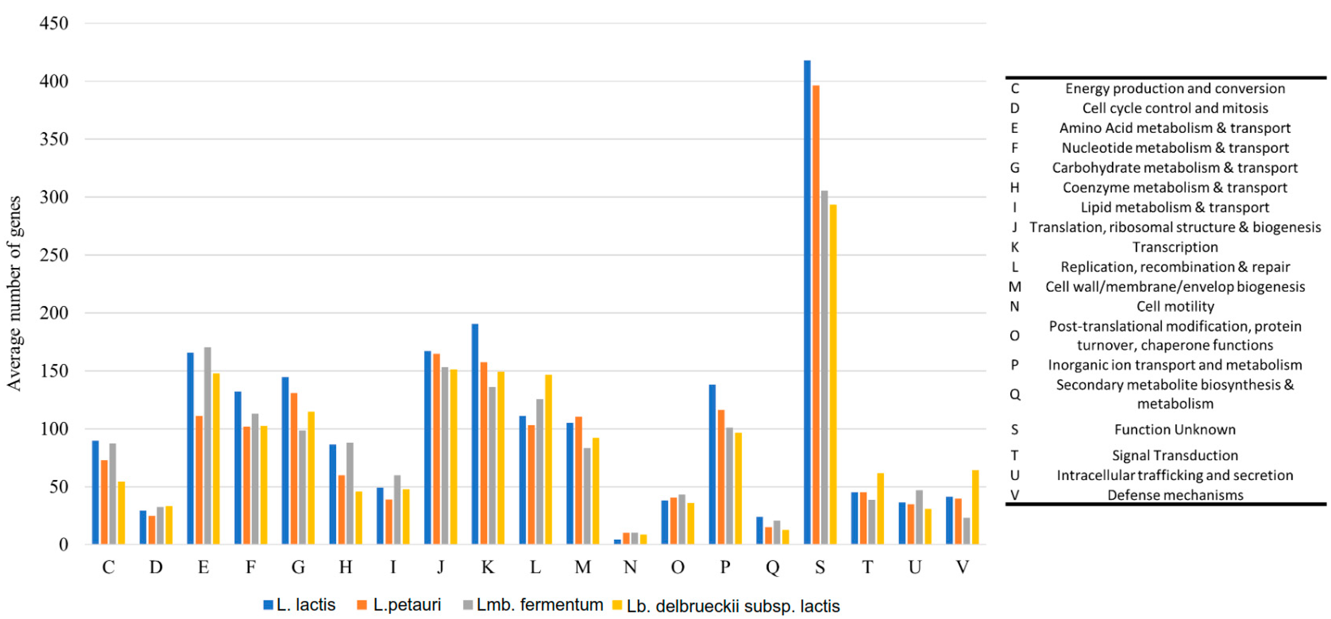
2.2.1. Phylogenetic Comparison and Functional Analysis

The phylogenetic tree of LAB is shown in Figure 1A. Overall, the combined gene pool of the LAB genomes contained 8103 clusters of orthologous genes (COGs), while the fundamental set of genes common to ≥90% of all LAB isolates (core genome) consisted of 21 COGs. By constructing a phylogenetic tree via alignment of the core genes, we were able to discern the genomic similarities among the investigated LAB isolates (Figure 1A). The isolates formed distinct clusters based on their assigned species, with no instances of cluster overlap observed. The calculated average nucleotide identity (ANI) values (Figure 1B) supported the isolate clustering using a threshold value of 95–96% for species delineation.
A subsystem comprises a group of protein-coding sequences (CDSs) that collectively perform a distinct biological function or form a structural complex [11]. Analysis of the subsystems in the LAB genomes identified the presence of 19 COG categories that exhibited enrichment (Figure 2). Among these categories, the COG designated as unknown function (S) displayed the highest degree of enrichment, with an average of 353 CDSs observed across the LAB genomes (19.6% of all COGs). Notably, L. lactis demonstrated significantly greater enrichment in this category, as well as in 5 additional categories out of the 19 examined, compared to the other LAB genomes (Figure 2). These categories were inorganic ion transport and metabolism (P, 6.3% of COGs), transcription (K, 8.8%), ribosomal structure and biogenesis (J, 8.8%), and carbohydrate metabolism and transport (G, 6.8%). These findings suggested that L. lactis possessed a highly functional genome while also harboring a substantial portion of CDSs with yet undetermined functions, which warrants further investigation and characterization [12].

2.2.2. Virulence and Antimicrobial Resistance Determinants

Antimicrobial resistance genes (RGs) were generally infrequent in LAB. The chromosomally located ABC efflux pump encoded by the lmrD gene, which provides resistance to lincosamides in Streptomyces lincolnensis and L. lactis and its deletion renders the bacteria susceptible to toxic compounds [3], was found in all (n = 8) strains of L. lactis. Moreover, the mdtA gene was found in all strains of L. petauri (n = 6). This gene encodes a membrane fusion protein of the mdtABC resistance–nodulation–cell division (RND) multidrug efflux complex, which is a prevalent resistance mechanism in Escherichia and Shigella spp. [13]. Given that the other two components of the system (mdtB and mdtC) were absent, it is likely that the efflux pump was inactive in the L. petauri strains of our collection. Lastly, we did not identify any virulence genes (VGs) in the LAB.

2.2.3. Bacteriocins and Carbohydrate-Active Enzymes (CAZymes)

In the in silico typing of LAB, we identified five genes encoding for bacteriocins in the genomes of L. delbrueckii subsp. lactis and L. lactis (n = 22). More specifically, in eight strains we found bovicin 255, a bacteriocin initially isolated from Streptococcus bovis inhabiting the rumen of herbivores that exhibits antimicrobial activity against a range of pathogens, such as Enterococcus faecium and Clostridium perfringens [14]. Moreover, enterolysin A was identified in eight strains. This bacteriocin, typically produced by Enterococcus faecalis, disrupts the cell membrane integrity of target bacteria (other enterococci), leading to cell lysis. Enterolysin A has potential applications in controlling enterococcal infections and combating antibiotic-resistant strains [15]. Finally, we found carnolysin and lactococcin B in two strains each. Carnolysin is a bacteriocin produced by Carnobacterium maltaromaticum, an LAB, which acts by disrupting the cell membrane integrity of target bacteria, leading to cell death. It has potent antimicrobial activity against Listeria monocytogenes, making it a potential natural alternative for controlling this foodborne pathogen [16]. Lactococcin B exhibits broad-spectrum antimicrobial activity against various Gram-positive bacteria, including Listeria monocytogenes [17]. These findings highlighted the potential of the analyzed LAB strains to produce antimicrobial substances that can contribute to their ecological fitness and potential applications in food preservation and probiotic formulations. Moreover, bacteriocin production by LAB may exert inhibitory effects on staphylococci, which could influence microbial competition and enhance the safety and quality of dairy products. Further experimental investigations on the identified bacteriocins are warranted to explore these potential interactions and their practical applications in dairy food production [18].
We found 23 distinct CAZymes clustered in five groups. Glycosyltransferases (GTs) were the most abundant class, making up 59% of all identified CAZymes. GTs are enzymes responsible for the transfer of sugar moieties; hence, they play an essential role in the biosynthesis of carbohydrates. Moreover, GTs participate in various biological processes, including the synthesis of cell wall polysaccharides, exopolysaccharides, as well as other complex carbohydrates [19]. Glycoside hydrolases (GHs) were also prevalent (29% of all CAZymes). These enzymes are involved in the breakdown of glycosidic bonds and catabolism of carbohydrates, such as starch, cellulose, and chitin. The abundance of GHs suggested that LAB possessed the enzymatic machinery to hydrolyze and utilize a broad spectrum of complex carbohydrates [20]. Lastly, carbohydrate-binding modules (CBMs) were found less frequently (8% of CAZymes). However, their presence indicated the potential for LAB to interact with and adhere to specific carbohydrate substrates, potentially facilitating their utilization and metabolism [21].

2.2.4. Mobile Genetic Elements and Prophages

Although no plasmids were identified in the LAB genomes, we found eight insertion sequences (ISs). ISs are small, self-transferable mobile genetic elements that can move across bacterial genomes, playing a significant role in genome plasticity and adaptation to the environment [22]. ISLll1, the predominant IS identified in the LAB genomes, was found in two strains of L. lactis and four strains of L. petauri. According to the ISfinder database, this IS was first detected in 1992 by Durmaz et al. [23]; however, we found only one recent publication reporting its presence in a L. petauri strain from Montenegrin brine cheeses [24]. ISLll1 belongs to the IS982 family, members of which are prevalent in LAB, and it is implicated in the acquisition and dissemination of RGs [25]. Notably, in the same IS family, we identified the representative element IS982 (n = 3). Moreover, ISLL6 (IS3 family), the second most prevalent IS (n = 4), is the most abundant IS element found in L. garvieae and has been associated with the rearrangement of LAB genomes through recombination events [26].
Seven prophages with intact genomes were identified in LAB, with Lactococcus phage 63301 (accession: NC_031017.1) being the most prevalent (n = 5 LAB genomes), followed by Lactococcus phage bIL310 (accession: AF323671.1) (n = 4). Overall, 16 out of 20 LAB strains had at least one intact prophage integrated in their genome. Specific information regarding these prophages is scarce; however, their presence in LAB from dairy products is significant for several reasons. From a safety and quality standpoint, prophages are contaminants, since they can be induced during fermentation under certain conditions, leading to cell lysis, which negatively impacts the final product [27]. On the other hand, studies have shown that prophages significantly enhance the genetic diversity and phenotypic plasticity of LAB and may enable their adaptation to various environmental conditions [28].

2.2.5. Probiotic Status of the Isolated LAB Strains

LAB probiotic status was assessed using the probiotic predictor tool iProbiotics, which comprises three different models based on a support vector machine (SVM) algorithm and different datasets, namely Probiotic Predictor (model 1), Probiotic Lactobacillus, Bifidobacterium, and others Predictor (model 2), and Probiotic Lactobacillus Predictor (model 3). The LAB genomes were run with all available models and the results are presented in Figure 3.
The first generic model (Figure 3A) predicted that all LAB species had probiotic properties (probability > 95%) except L. petauri, which was a non-probiotic species (low probability). The other two models (Figure 3B,C) were more specific and showed that eventually only Lb. delbrueckii subsp. bulgaricus and L. lactis could be classified as probiotic cultures and considered as potential candidates for fighting mastitis-causing pathogens.
One important parameter for preventing mastitis-causing pathogens is the adhesion capability of bacteria in order to successfully colonize the udder. Cell surface hydrophobicity (CSH) and auto-aggregation (AA) are the two cell surface properties that demonstrate the microbial adhesion ability [29]. Auto-aggregation increased with time for all strains (exception Lmb. fermentum, which showed a peak at 4 h, AA = 16.64%). Lb. delbrueckii subsp. bulgaricus presented the highest AA value (30.72 ± 0.94%) after 5 h, followed by L. lactis (23.51 ± 1.23%), while L. petauri and Lmb. fermentum showed the lowest AA values (10.98 ± 0.56% and 12.92 ± 1.75%, respectively) (Figure 4). A similar trend was observed for CSH. L. lactis had the optimal CSH value (83.49 ± 4.94%), followed by Lb. delbrueckii subsp. bulgaricus (54.53 ± 2.32%). Again, the CSH values of L. petauri and Lmb. fermentum were significantly lower (26.72 ± 4.83% and 19.39 ± 5.30%, respectively) than those of the other two strains. These results supported the predictions about the probiotic status of the LAB strains. Previously, it was found that the Lb. delbrueckii subsp. bulgaricus and L. lactis genomes contained genes encoding bacteriocins (antagonistic activity against pathogens) and genes related to probiotic traits (Table 2). In addition, these two microorganisms showed the best CSH and AA values, which aligned with the adhesion-related genes found in their genomes (Table 2). Therefore, the two species are suitable for potential application as protective probiotic cultures.

2.3. Staphylococci

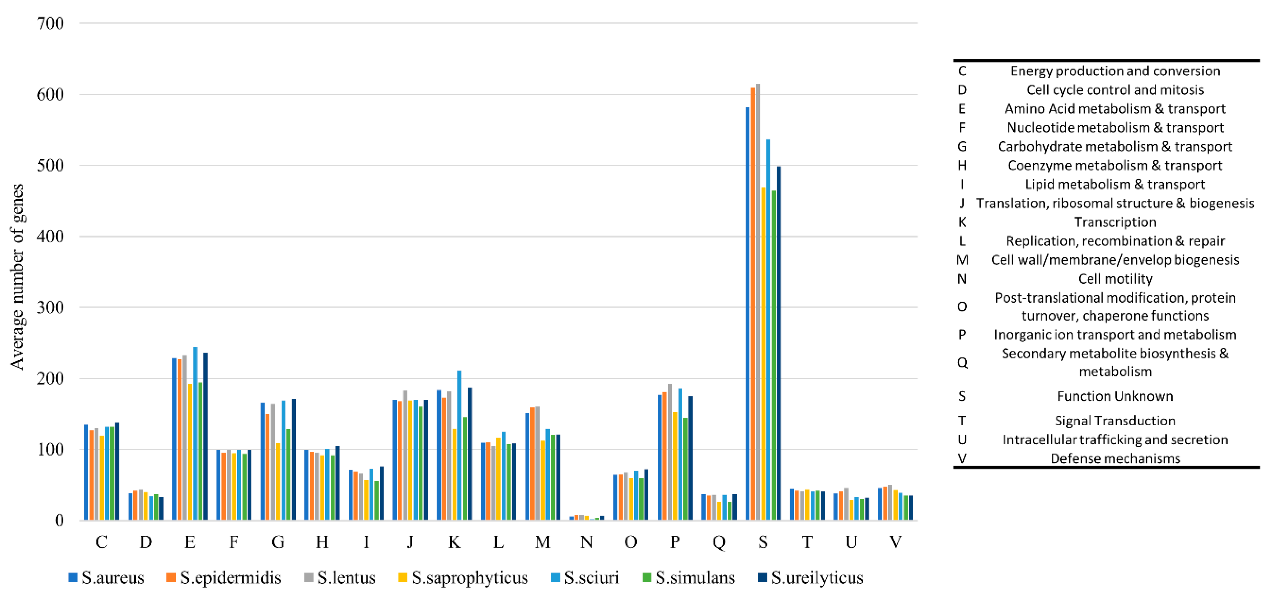
2.3.1. Phylogenetic Analysis and Comparative Genomics

The phylogenetic comparison of staphylococci is shown in Figure 5A. Overall, the combined gene pool of the staphylococcal genomes contained 10,717 COGs, while the core genome (at a 90% threshold) had 323 COGs. By constructing a phylogenetic tree via core genome alignment, we identified the genomic similarities among staphylococci (Figure 5A). As with the phylogenetic tree of the LAB strains, the Staphylococcus spp. isolates formed distinct clusters based on their assigned species, while no cluster overlap was observed. Similarly, the estimated ANI values (Figure 5B) confirmed the clustering of staphylococci into different species (species delineation threshold value equal to 95–96%).
The mecA/mecC genes were not detected in all S. aureus and coagulase-negative staphylococcal (CNS) strains; thus, they were designated as methicillin-susceptible (MSSA) microorganisms. All S. aureus strains were predicted to be human pathogens (probability 98.4%), as they carried the vSa-gamma (vSaγ) pathogenicity island (SaPI) and other mobile genetic elements (MGEs), with genes related to adherence, antiphagocytosis, exoenzyme, hostimm, immune evasion, iron uptake, plasminogen activation, the secretion system, and toxin production. The CNS strains were also predicted to be human pathogens (probability ranged from 63.8% to 98.5%). S. simulans displayed a much lower probability of being a human pathogen (82% on average) than other staphylococci, such as S. aureus (98.4%), S. saprophyticus (97.5%), S. epidermidis (95.4%), and S. ureilyticus (98.5%). Indeed, although S. simulans can cause persistent intramammary infections as well as increased somatic cell counts to values comparable to those of S. aureus, the infection and incurred symptoms are less severe (e.g., no clinical mastitis) compared to other mastitis-causing staphylococci (e.g., S. aureus) [30,31]. Additionally, the S. aureus strains belonged to sequence type (ST) 5 (clonal complex 5) and spa type t306. Notably, this ST has been implicated in severe nosocomial infections and is usually recovered from human blood isolates; however, it seems to largely consist of methicillin-susceptible isolates [32]. It is worth mentioning that the MLST analysis detected a point mutation in the glpF gene at the base pair position 438 (A instead of T), which may indicate a novel ST close to ST5, but further experimental confirmation is required, e.g., sequencing of this specific gene fragment using the Sanger technique.
Analysis of the subsystems present in the Staphylococcus spp. genomes revealed the presence of 19 COG categories that exhibited enrichment (Figure 6). The CDSs with unknown function (S) were the most enriched, with an average of 540 CDSs observed across the staphylococcal genomes (24% of all COGs). Other enriched COG categories were amino acid metabolism and transport (E) with 222 CDSs (9.5%); transcription (K) and inorganic ion transport/metabolism (P), each with 173 CDSs (7.4%); as well as ribosomal structure and biogenesis with 170 CDSs (7.3%). The rest of the COG categories together made less than half (45.2%) of all COGs (Figure 6). Notably, our statistical analysis showed a significant difference in the enrichment of only one COG, secondary metabolite biosynthesis (Q), for which S. aureus was significantly more enriched than S. simulans (11 more CDSs, on average).

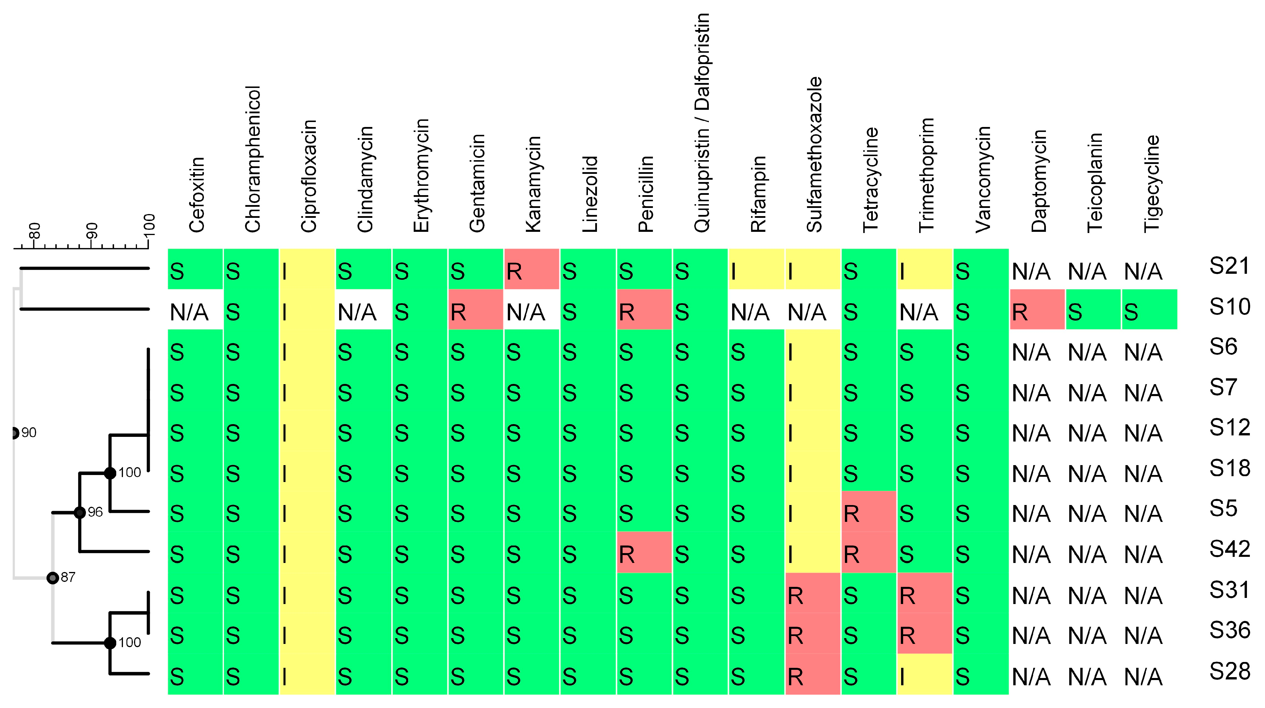
2.3.2. Virulence and Antimicrobial Resistance Determinants

Our analysis revealed the presence of 108 RGs in staphylococci (Figure 7). Most RGs (81.5%) were found in S. aureus, followed by S. simulans (7.4%) and Staphylococcus epidermidis (6.5%). Resistance to drugs and biocides was the most prevalent type of resistance (27.8% of all RGs), primarily represented by the norA and norB genes (5.6%, each). norA encodes a well-studied efflux system in S. aureus and serves as a model for understanding efflux-mediated resistance. Additionally, it contributes to resistance against fluoroquinolones, antiseptics, and disinfectants [33]. Similarly, norB encodes a multidrug efflux pump providing resistance to fluroquinolones that is, however, independent of norA [34]. Furthermore, the arlR-arlS locus, which regulates the (over)expression and multidrug resistance (MDR) phenotype of norA-carrying S. aureus strains [35], was identified in six strains (11.2%) of this species. Another gene found in the MDR class of resistance was the regulator mgrA, which controls norA, norB, as well as tet38 [36].
In the macrolides, lincosamides, and streptogramines (MLS) category (13.9% of all RGs), we found ribosomal large-subunit methyltransferase H (rlmH, also known as orfX [37]) in all strains of S. aureus (n = 6) and S. simulans (n = 8). Except from its action in preventing the activity of the MLS group of antibiotics, the rlmH gene, found in all staphylococci, remains unaffected even after insertion of the staphylococcal chromosome cassette mec (SCCmec). This cassette carries the mecA gene, which is responsible for encoding resistance to β-lactam antibiotics, and it is inserted at the C terminus of the rlmH gene [37]. Notably, we did not detect any mec genes (mecA, mecB, mecC) in our collection of staphylococci.
Genes conferring resistance to aminoglycosides were also found (11.1% of RGs). This category includes N-acetyltransferase aac(3′) and O-phosphotransferase aph(3′). Other resistance classes were less common (Figure 7). We identified tet38, which encodes a tetracycline efflux pump, in the strains of S. aureus (n = 6), whereas blaZ, which encodes β-lactamase (penicillinase), was found in one strain each of S. lentus and S. epidermidis.
All identified RGs are listed in Supplementary Table S1.
The antimicrobial resistance of selected staphylococci was examined phenotypically (Figure 8). Some discrepancies in antimicrobial resistance (AMR) between the detection of ARGs and the actual phenotype were observed among strains, suggesting that AMR predictions relying on the in silico analysis of a sequenced genome should be always accompanied with AMR testing to reliably conclude the AMR of the microorganism [38]. The discrepancies were mainly related to the expression or not of the detected gene(s) during the AMR examination. For instance, the tet38 gene, which encodes a tetracycline efflux pump, was detected in all S. aureus strains, but only the S5 strain showed resistance to the tetracycline antibiotic class. In addition, although genes conferring resistance to different antibiotic classes were detected in S. aureus, no strain presented an MDR phenotype. Staphylococcus (Mammaliicoccus) sciuri (S10) displayed an MDR pattern (resistant to multiple, three or more, different classes of antibiotics), which may have been attributed to the presence of the MDR phenotype, as predicted previously. Finally, S. epidermidis (S42) was also resistant to three different antibiotic categories (tetracycline-tetracyclines, penicillin-beta-lactams, and streptomycin-aminoglycosides), which aligned with the detected ARGs. The results for streptomycin are not shown in Figure 8 because there was no minimum inhibitory concentration (MIC) available for staphylococci; however, notably, S. epidermidis had higher streptomycin resistance (>32 mg/L) than the other tested strains (≤4 mg/L).
We identified 62 VGs in the strains of S. aureus (n = 6) and two strains of S. simulans. Exopolysaccharide synthesis VGs were the most common (Figure 9). This category exclusively comprised type 8 capsular polysaccharide (cap8) operon genes (cap8Acap8G and cap8Lcap8P). Type 8 is the most prevalent capsule type found in clinical S. aureus isolates, yet its exact role in virulence remains unclear and it most likely acts as a antiphagocytic factor [39].
Several genes encoding toxins were identified, shedding light on the potential virulence of staphylococci in dairy foods. The hlb and hld genes, which respectively encode beta- and delta-hemolysin, were found in six strains. This finding indicated that the staphylococcal strains analyzed in this study had the potential to cause hemolysis, which may have implications for host tissue damage and pathogenesis [40]. Moreover, we identified the hlgA, hlgB, and hlgC genes, which encode components of the gamma-hemolysin AB complex, as well as the alpha-hemolysin (hla) gene. Gamma- and alpha-hemolysins are associated with cytotoxicity and hemolytic activity and their presence in staphylococcal strains further highlighted their virulence potential and pathogenicity [41,42]. Furthermore, the lukF-PV gene (Panton–Valentine leukocidin F subunit) was detected. Panton–Valentine leukocidin (PVL) is a potent toxin that causes the lysis of leukocytes, while its presence has been strongly associated with the virulence of S. aureus strains [42]. Lastly, the sea gene, which encodes a heat-stable enterotoxin responsible for staphylococcal food poisoning, was found [43] in all S. aureus strains except the S12 strain. Interestingly, the sak gene, which advances microbial dissemination, was not found in the S12 strain either. The locus encoding staphylokinase (sak) usually contains additional virulence-related genes like enterotoxin A (sea) [44]. Phenotype testing confirmed that strain S12 was enterotoxin A-negative (Figure 10). The first screening test examined the production of enterotoxins A, B, C, D, or E and showed that only strains S6, S7, and S18 were able to produce staphylococcal enterotoxin (SE). The second specific test supported the previously obtained results specifying the SE production by the S6, S7, and S18 strains as enterotoxin A. The concurrent detection of enterotoxins A and E could be attributed to the occurrence of a cross-reaction that occurs between the antibody and toxin. However, the see gene (enterotoxin E) was not found in any of the S. aureus strains. Other enterotoxin-encoding genes detected in the S. aureus genomes were seg, sei, sem, sen, seo, and seu. Moreover, we were able to identify genes encoding adherence proteins. Among the identified adherence-related genes, clfAB, ebp, fnbAB, sdrCD, and sdrEB were of particular interest. These genes play a pivotal role in the regulation of staphylococcal adhesion to host tissues. The presence of these genes and the exoenzyme aur gene (encoding the metalloprotease aureolysin or protease III) suggested the ability of staphylococci to interact with host cells (immune evasion and dissemination) and likely contributes to their pathogenicity and persistence (survival) in various environmental niches [44,45].
Finally, it is worth mentioning the identification of genes related to biofilm formation. More specifically, we found multiple genes of the ica operon (icaABCD and icaR). This operon regulates the synthesis of polysaccharide intercellular adhesin (PIA), which is an essential component of biofilms formed by staphylococci, enabling adherence to surfaces as well as resistance to host immune responses [46].
The presence of virulence genes raises concerns regarding food safety and pathogenicity. Further investigation is needed to establish their expression levels and function along with their impact on the quality of dairy products.

2.3.3. Bacteriocins (Auto-Inducing Peptides)

We found only one category of genes encoding bacteriocin-like molecules in staphylococci. Auto-inducing peptides (AIPs) I, II, and IV were identified in 13 strains of S. aureus and S. simulans. AIPs are a specific class of signaling molecules called quorum-sensing molecules. In staphylococci, AIPs play a crucial role in regulating various physiological processes, including VG and RG production and biofilm formation [47,48]. Moreover, AIPs seem to play a role in interspecies communication, thus enabling staphylococci to have interactions within microbial communities. These interactions can have implications for microbial pathogenesis, host–microbe interactions, as well as the overall balance of the microbiota [47].

2.3.4. Mobile Genetic Elements and Prophages

Four plasmids were discovered in only two strains of our staphylococcal collection. The largest plasmid (29.2 Kbp), harbored by S. aureus strain S8, was a large, non-mobilizable plasmid with a GC content of 33.8%. The plasmid had high nucleotide identity with plasmid pSALNT106 (accession: CP042118), which was recently reported in S. aureus isolated from retail meat [49]. Notably, the authors identified phage proteins in pSALNT106, suggesting these plasmids may be implicated in the transmission of virulence factors [49]. The other three plasmids were all found in S. lentus strain S74. These were small, non-mobilizable plasmids (average size of 2.3 Kbp) that also had high nucleotide identity with plasmids in the aforementioned study, suggesting that a shared mobilome, largely encoding for phage proteins, exists between the staphylococcal strains of dairy and meat origin identified in the two studies.
MGEs were carried by S. lentus, S. aureus, S. epidermidis, and Staphylococcus ureilyticus, specifically MGEs ISSep3, Tn559, and ISSau4. ISSep3 and ISSau4 are ISs that are frequently found in S. epidermidis and S. aureus, respectively, and are highly associated with the transfer of RGs [50,51]. Lastly, we identified Tn559, a transposon that often carries the dfrK gene (trimethoprim resistance) in methicillin-resistant S. aureus [52]. As previously discussed, none of these genes (dfrK or mec-like) were present in our collection.
In the staphylococcal genomes, we confirmed the integration of 10 intact prophage genomes. Staphylococcus phage SA7 (accession: NC_048658.1) (found in n = 6 S. simulans genomes) was predominant, followed by Staphylococcus phage PT1028 (accession: AY954948.1) (n = 5). Prophages can harbor diverse genes that may promote staphylococcal virulence and antibiotic resistance [53]. Moreover, the integration of prophages can result in genomic rearrangements that further contribute to the genetic diversity of staphylococci. The presence of prophages in pathogens such as S. aureus can potentially enhance their pathogenicity or survival under different environmental conditions during the production of dairy products [54].

3. Materials and Methods

3.1. Microbial Strains and Culture Conditions

A collection of 39 microorganisms was used in this study, comprising two groups of LAB and staphylococci, which were all retrieved from raw sheep’s milk. The LAB species included L. lactis (n = 8), L. petauri (n = 6), Lb. delbrueckii subsp. lactis (n = 4), Lmb. fermentum (n = 2). The CNS comprised S. simulans (n = 8), S. lentus) (n = 1), S. sciuri (n = 1), S. epidermidis (n = 1), S. saprophyticus (n = 1), and S. ureilyticus (n = 1). Additionally, six strains of coagulase-positive S. aureus (n = 6) were also isolated.
The LAB and staphylococci strains were stored at –80 °C in de Man, Rogosa and Sharpe (bacilli) (MRS, Madrid, Condalab, Spain, 1215) or M17 (cocci) broth (Himedia, Einhausen, Germany, M1029) and Brain Heart Infusion broth (BHI, Condalab, Madrid, Spain, 1400), respectively, with 30% glycerol (Penta Chemicals, Prague, Czech Rebublic, 14530–11000PE) as a cryoprotectant. Before use, the strains were resuscitated twice in the respective media for 24–48 h at 30 °C or 37 °C.

3.2. Whole Genome Sequencing, Assembly, and Quality Control

Bacterial DNA extraction and sequencing (Novaseq 6000 platform, Illumina, San Diego, CA, USA, 2 × 150 bp) was performed by Novogene Genomics Service (Novogene Co., Cambridge UK), as described in Syrokou et al. (2020) [55]. The quality of the adapter-free raw reads was checked using FastQC v.0.12.1 software available in the KBase platform [56,57]. Polishing and de novo assembling of the raw reads into contigs was performed using the Unicycler assembler and Pilon, respectively, provided by the BV-BRC v3.30.19a web platform [58,59,60]. Multi-Draft based Scaffolder (MeDuSa) v1.6 [61] was used to organize the contigs into scaffolds. Scaffolds were ordered and oriented based on the complete reference genomes present in the NCBI database (https://www.ncbi.nlm.nih.gov/, accessed on 10 January 2023); Lactococcus lactis LAC460, Lactococcus petauri B1726, Lactobacillus delbrueckii subsp. jakobsenii ZN7a-9 = DSM 26046, Limosilactobacillus fermentum SCB0035, Staphylococcus aureus NCTC 8325, and Staphylococcus simulans IVB6189. The CheckM tool v1.0.18 [62] of the KBase system was employed for quality evaluation of the scaffolds to ensure that the assembled genomes were of high quality, i.e., completeness (≥95%) and contamination (≤5%). Only one S. aureus genome had a completeness of 92.75% and contamination of 6.21%, but the genome was included in the downstream analysis because it was still of acceptable quality (i.e., completeness ≥ 90% and contamination ≤ 10%). Possible misassemblies after scaffolding were assessed by the mean of the Skew Index Test (SkweIT) v1.0 [63].

3.3. Genotyping and Comparative Genomics

The quality of genome assembly was evaluated using QUAST v5.2.0 [64]. Identification of species was carried out using Kraken2 v2.1.3 [65] and TYGS (https://tygs.dsmz.de/, accessed on 23 June 2023) [66]. ANI values between the strains’ genomes were estimated using OrthANI (https://www.ezbiocloud.net/, accessed on 23 June 2023) [67] and a heatmap of the resulted ANI values was constructed using MORPHEUS (https://software.broadinstitute.org/morpheus/, accessed on 09 August 2023). The annotation of genomes was performed using PROKKA v1.14.5 [68], whereas the functional annotation of open reading frames and analysis of subsystems were performed using eggnog-mapper v2.1.12 [69]. Additionally, prophages were identified using the PHASTEST API service (https://phastest.ca/, accessed on 27 June 2023) [70]. Abricate v1.0.0 [71] along with the reference databases VFDB v1.0 [72], MobileElementFinder v1.1.2 [73], Resfinder v2.1 [74], and PlasmidFinder v2.2 [75] were used to establish the occurrence of VGs, MGEs, RGs, and plasmid replicons, respectively. Furthermore, bacteriocins were detected using the BAGEL4 webserver (http://bagel5.molgenrug.nl/, accessed on 27 June 2023), whereas CAZymes were identified using the dbCAN2 server (https://bcb.unl.edu/dbCAN2/, accessed on 27 June 2023) [76]. ST classification and spa typing for S. aureus strains were carried out using Center for Genomic Epidemiology (CGE) services (http://www.genomicepidemiology.org/services/, accessed on 27 June 2023), i.e., MLST 2.0 [77] and spaTyper 1.0 [78], respectively. Finally, resistance to methicillin (detection of Staphylococcal Cassette Chromosome Elements—SCC mec) and pathogenicity of staphylococci were evaluated using CGE services SCCmecFinder 1.2 [79] and PathogenFinder 1.1 [80], respectively.
Roary v3.11.2 [81] software was utilized to conduct pangenome analysis and core genome alignment of both LAB and staphylococci. Proteins were classified into the same family if their amino acid sequence similarity was equal to or greater than 75%. For a gene to be considered a core gene, it needed to be present in at least 90% of the isolates. To visualize the phylogenetic relationship, the FastTree v2.1 algorithm [82] was employed, and the resulting tree was annotated and visualized using iTOL v6 [83]. The European public Galaxy server (https://usegalaxy.eu/, accessed on 27 June 2023) [84] was used for some of the aforementioned analyses. Finally, predictions regarding LAB probiotic status were made using the iProbiotics webtool (http://bioinfor.imu.edu.cn/iprobiotics/public/Home, accessed on 9 August 2023).

3.4. Phenotype Testing

3.4.1. Antimicrobial Resistance

Selected strains of staphylococci were tested for AMR using the Sensititre MIC (Thermo Fisher Scientific, Waltham, MA, USA) technique employing the broth microdilution MIC approach, according to manufacturer’s instructions. The AMR plates used were the Sensititre EU Surveillance Staphylococcus EUST AST Plate and Sensititre EU Surveillance Enterococcus EUVENC AST Plate (Thermo Fisher Scientific). The results were presented in a dendrogram using Bionumerics v8.1 software and the antibiotic susceptibility plugin (bioMérieux, Sint-Martens-Latem, Belgium). The strains were classified as susceptible (S), resistant (R) or intermediate (I) based on the European Committee on Antimicrobial Susceptibility Testing (EUCAST) Breakpoint Table v13.0 for Staphylococcus spp. (https://www.eucast.org/clinical_breakpoints/, accessed on 10 January 2023).

3.4.2. Staphylococcal Enterotoxin Production

Selected staphylococci strains were studied for the production of enterotoxin(s) (A, B, C, D, or E) using the Ridascreen Set Total assay kit (R-Biopharm, Darmstadt, Germany, R4105) (screening test) and the Ridascreen Set A, B, C, D, E assay kit (R-Biopharm, Darmstadt, Germany, R4101) (specific test), according to manufacturer’s instructions. A heatmap was constructed with the obtained results using ImageGP software (https://www.bic.ac.cn/ImageGP/, accessed on 9 August 2023) [85].

3.4.3. Probiotic Properties

LAB (L. petauri, L. lactis, Lb. delbrueckii subsp. bulgaricus, and Lmb. fermentum) microbial cells were harvested at 3350 × g for 15 min at 4 °C (Centrifuge 5804 R, Eppendorf, Oslo, Norway), washed twice with sterile Ringer solution (Sigma-Aldrich, Taufkirchen, Germany, 96724), and resuspended using the same dilutor until an optical density (OD) of 0.6–0.7 was achieved at 600 nm (BioTek Instruments – Agilent Technologies, Epoch, Santa Clara, CA, USA) (A0). Afterwards, two milliliters of the bacterial suspension were mixed with two milliners of xylene (Fisher Scientific, Loughborough, Leicestershire, UK, X/0250/17) and homogenized with vortexing. Aliquots of 150 μL of each bacterial suspension were transferred to a 96-well microplate (Thermo Fisher Scientific, Lillerød, Denmark, 167008) and left for 0.5 h at room temperature (A). The cell surface hydrophobicity was calculated using the following equation [86]:
c e l l   s u r f a c e   h y d r o p h o b i c i t y   % = A 0 A A 0 × 100
where A0 and A are the initial (0 h) and final (after 0.5 h) OD600, respectively.
For the auto-aggregation assay, samples in the 96-well microplate were incubated at 37 °C for 5 h and the OD600 was measured every 1 h (At). The auto-aggregation was estimated as follows [76]:
a u t o - a g g r e g a t i o n   % = 1 A t A 0 × 100
where At is the measured OD600 at each time interval (1, 2, 3, 4, and 5 h).
Both assays were carried out in quadruplicate. Statistical comparisons were performed using GraphPad Prism v9.5.1 software (GraphPad Software, Boston, MA, USA) using the unpaired multiple parametric t-test with Welch’s correction, i.e., no assumption was made about consistent SDs between species (columns) for each cell surface assay (row), and the false discovery rate (FDR) approach (two-stage linear step-up method of Benjamini, Krieger, and Yekutieli), i.e., adjusted p-value (q-value = 0.05) for multiple comparisons.

4. Conclusions

Τhe in silico characterization of staphylococcal and LAB strains isolated from raw sheep’s milk revealed the presence of various genetic elements, notably MGEs and specific genes associated with AMR, virulence, and metabolic capabilities. These findings hold practical implications for dairy product safety and handling.
The presence of MGEs, including plasmids, transposons, and IS elements, underscores the importance of safe dairy production practices. Since raw sheep’s milk seems to serve as a reservoir for these genetic elements, the necessity for tight measures to ensure the safety of dairy products should be emphasized. Therefore, implementation of rigorous pasteurization and quality control procedures is crucial to mitigate the risk of transmitting AMR and potential foodborne pathogens to consumers. Moreover, concerns arise regarding the potential transfer of RGs and VGs from staphylococci to LAB. While LAB are generally recognized as safe (GRAS), it is essential to continuously monitor the genetic dynamics between these bacterial groups. Further research is warranted to investigate gene transfer events in order to ensure that LAB maintain their safe status and can continue to be used for their probiotic properties. This knowledge can guide the development of safer and more effective probiotics for dairy product applications.

Supplementary Materials

The following supporting information can be downloaded at: https://www.mdpi.com/article/10.3390/ijms241813883/s1.

Author Contributions

Conceptualization, M.M.; methodology, I.A., T.S., K.V., A.G. and M.M.; software, I.A. and M.M.; formal analysis, I.A., L.B. and M.M.; investigation, I.A., T.S., K.V. and A.G.; resources, M.M.; data curation, I.A., L.B. and M.M.; writing—original draft preparation, I.A., T.S., K.V. and A.G.; writing—review and editing, L.B. and M.M.; supervision, M.M.; project administration, M.M.; funding acquisition, M.M. All authors have read and agreed to the published version of the manuscript.

Funding

This study was supported by European Union and Greek national funds through the operational program “Competitiveness, Entrepreneurship and Innovation” under the action “Reinforcement of the Research and Innovation Infrastructure” (MIS 5045647).

Institutional Review Board Statement

Not applicable.

Informed Consent Statement

Not applicable.

Data Availability Statement

The whole-genome sequencing data have been deposited at GenBank (NCBI) under accession (BioProject) number PRJNA1007360 (https://www.ncbi.nlm.nih.gov/).

Acknowledgments

We acknowledge support of this work by the project “Research Infrastructure “Milk Quality” in Agri-food: Control of mastitis in small dairy ruminants and improvement of the quality of raw milk and dairy products by applying advanced molecular and statistical methods” (MIS 5045647), which was implemented under the action “Reinforcement of the Research and Innovation Infrastructure” funded by the operational program "Competitiveness, Entrepreneurship and Innovation" (NSRF 2014-2020) and co-financed by Greece and the European Union (European Regional Development Fund).

Conflicts of Interest

The authors declare no conflict of interest.

References

  1. Romero-Velarde, E.; Delgado-Franco, D.; García-Gutiérrez, M.; Gurrola-Díaz, C.; Larrosa-Haro, A.; Montijo-Barrios, E.; Muskiet, F.A.J.; Vargas-Guerrero, B.; Geurts, J. The Importance of Lactose in the Human Diet: Outcomes of a Mexican Consensus Meeting. Nutrients 2019, 11, 2737. [Google Scholar] [CrossRef] [PubMed]
  2. König, H.; Unden, G.; Fröhlich, J. (Eds.) Biology of Microorganisms on Grapes, in Must and in Wine; Springer International Publishing: Cham, Switzerland, 2017; ISBN 978-3-319-60020-8. [Google Scholar]
  3. Ayivi, R.D.; Gyawali, R.; Krastanov, A.; Aljaloud, S.O.; Worku, M.; Tahergorabi, R.; da Silva, R.C.; Ibrahim, S.A. Lactic Acid Bacteria: Food Safety and Human Health Applications. Dairy 2020, 1, 202–232. [Google Scholar] [CrossRef]
  4. Zapaśnik, A.; Sokołowska, B.; Bryła, M. Role of Lactic Acid Bacteria in Food Preservation and Safety. Foods 2022, 11, 1283. [Google Scholar] [CrossRef]
  5. Irlinger, F. Safety Assessment of Dairy Microorganisms: Coagulase-Negative Staphylococci. Int. J. Food Microbiol. 2008, 126, 302–310. [Google Scholar] [CrossRef]
  6. Chen, Y.-L.; Chou, C.-C. Factors Affecting the Susceptibility of Staphylococcus aureus CCRC 12657 to Water Soluble Lactose Chitosan Derivative. Food Microbiol. 2005, 22, 29–35. [Google Scholar] [CrossRef]
  7. Vernozy-Rozand, C.; Mazuy, C.; Meugnier, H.; Bes, M.; Lasne, Y.; Fiedler, F.; Etienne, J.; Freney, J. Staphylococcus Fleurettii Sp. Nov., Isolated from Goat’s Milk Cheeses. Int. J. Syst. Evol. Microbiol. 2000, 50, 1521–1527. [Google Scholar] [CrossRef] [PubMed]
  8. Apostolakos, I.; Paramithiotis, S.; Mataragas, M. Comparative Genomic Analysis Reveals the Functional Traits and Safety Status of Lactic Acid Bacteria Retrieved from Artisanal Cheeses and Raw Sheep Milk. Foods 2023, 12, 599. [Google Scholar] [CrossRef]
  9. Apostolakos, I.; Paramithiotis, S.; Mataragas, M. Functional and Safety Characterization of Weissella Paramesenteroides Strains Isolated from Dairy Products through Whole-Genome Sequencing and Comparative Genomics. Dairy 2022, 3, 799–813. [Google Scholar] [CrossRef]
  10. Apostolakos, I.; Tsigkrimani, M.; Paramithiotis, S.; Mataragas, M. Whole-Genome Sequencing and Comparative Genomic Analysis of Enterococcus Spp. Isolated from Dairy Products: Genomic Diversity, Functional Characteristics, and Pathogenic Potential. Appl. Sci. 2022, 12, 9620. [Google Scholar] [CrossRef]
  11. Olson, R.D.; Assaf, R.; Brettin, T.; Conrad, N.; Cucinell, C.; Davis, J.J.; Dempsey, D.M.; Dickerman, A.; Dietrich, E.M.; Kenyon, R.W.; et al. ntroducing the Bacterial and Viral Bioinformatics Resource Center (BV-BRC): A resource combining PATRIC, IRD and ViPR. Nucleic Acids Res. 2019, 51, D678–D689. [Google Scholar] [CrossRef]
  12. Wels, M.; Siezen, R.; van Hijum, S.; Kelly, W.J.; Bachmann, H. Comparative Genome Analysis of Lactococcus Lactis Indicates Niche Adaptation and Resolves Genotype/Phenotype Disparity. Front. Microbiol. 2019, 10, 4. [Google Scholar] [CrossRef] [PubMed]
  13. Lubelski, J.; de Jong, A.; van Merkerk, R.; Agustiandari, H.; Kuipers, O.P.; Kok, J.; Driessen, A.J.M. LmrCD Is a Major Multidrug Resistance Transporter in Lactococcus Lactis. Mol. Microbiol. 2006, 61, 771–781. [Google Scholar] [CrossRef]
  14. Whitford, M.F.; McPherson, M.A.; Forster, R.J.; Teather, R.M. Identification of Bacteriocin-like Inhibitors from Rumen Streptococcus Spp. and Isolation and Characterization of Bovicin 255. Appl. Environ. Microbiol. 2001, 67, 569–574. [Google Scholar] [CrossRef] [PubMed]
  15. Zhang, T.; Pan, Y.; Li, B.; Ou, J.; Zhang, J.; Chen, Y.; Peng, X.; Chen, L. Molecular Cloning and Antimicrobial Activity of Enterolysin A and Helveticin J of Bacteriolysins from Metagenome of Chinese Traditional Fermented Foods. Food Control 2013, 31, 499–507. [Google Scholar] [CrossRef]
  16. Tulini, F.L.; Lohans, C.T.; Bordon, K.C.F.; Zheng, J.; Arantes, E.C.; Vederas, J.C.; De Martinis, E.C.P. Purification and Characterization of Antimicrobial Peptides from Fish Isolate Carnobacterium Maltaromaticum C2: Carnobacteriocin X and Carnolysins A1 and A2. Int. J. Food Microbiol. 2014, 173, 81–88. [Google Scholar] [CrossRef]
  17. Yıldırım, Z.; Yerlikaya, S.; Öncül, N.; Sakin, T. Inhibitory Effect of Lactococcin BZ Against Listeria Innocua and Indigenous Microbiota of Fresh Beef. Food Technol. Biotechnol. 2016, 54, 317–323. [Google Scholar] [CrossRef]
  18. Sheoran, P.; Tiwari, S.K. Anti-Staphylococcal Activity of Bacteriocins of Food Isolates Enterococcus Hirae LD3 and Lactobacillus Plantarum LD4 in Pasteurized Milk. 3 Biotech 2019, 9, 8. [Google Scholar] [CrossRef]
  19. Lairson, L.L.; Henrissat, B.; Davies, G.J.; Withers, S.G. Glycosyltransferases: Structures, Functions, and Mechanisms. Annu. Rev. Biochem. 2008, 77, 521–555. [Google Scholar] [CrossRef]
  20. Coombs, J.; Brenchley, J.E. Characterization of Two New Glycosyl Hydrolases from the Lactic Acid Bacterium Carnobacterium Piscicola Strain BA. Appl. Environ. Microbiol. 2001, 67, 5094–5099. [Google Scholar] [CrossRef]
  21. Sidar, A.; Albuquerque, E.D.; Voshol, G.P.; Ram, A.F.J.; Vijgenboom, E.; Punt, P.J. Carbohydrate Binding Modules: Diversity of Domain Architecture in Amylases and Cellulases From Filamentous Microorganisms. Front. Bioeng. Biotechnol. 2020, 8, 871. [Google Scholar] [CrossRef]
  22. Nicoloff, H.; Bringel, F. IS Lpl1 Is a Functional IS 30 -Related Insertion Element in Lactobacillus Plantarum That Is Also Found in Other Lactic Acid Bacteria. Appl. Environ. Microbiol. 2003, 69, 6032–6040. [Google Scholar] [CrossRef] [PubMed]
  23. Durmaz, E.; Higgins, D.L.; Klaenhammer, T.R. Molecular Characterization of a Second Abortive Phage Resistance Gene Present in Lactococcus Lactis Subsp. Lactis ME2. J. Bacteriol. 1992, 174, 7463–7469. [Google Scholar] [CrossRef]
  24. Martinovic, A.; Cabal, A.; Nisic, A.; Sucher, J.; Stöger, A.; Allerberger, F.; Ruppitsch, W. Genome Sequences of Lactococcus Garvieae and Lactococcus Petauri Strains Isolated from Traditional Montenegrin Brine Cheeses. Microbiol. Resour. Announc. 2021, 10, e0054621. [Google Scholar] [CrossRef]
  25. Fayad, N.; Kallassy Awad, M.; Mahillon, J. IS982 and Kin: New Insights into an Old IS Family. Mob. DNA 2020, 11, 24. [Google Scholar] [CrossRef] [PubMed]
  26. Eraclio, G.; Ricci, G.; Fortina, M.G. Insertion Sequence Elements in Lactococcus Garvieae. Gene 2015, 555, 291–296. [Google Scholar] [CrossRef] [PubMed]
  27. Garneau, J.E.; Moineau, S. Bacteriophages of Lactic Acid Bacteria and Their Impact on Milk Fermentations. Microb. Cell Fact. 2011, 10, S20. [Google Scholar] [CrossRef]
  28. Aucouturier, A.; Chain, F.; Langella, P.; Bidnenko, E. Characterization of a Prophage-Free Derivative Strain of Lactococcus Lactis Ssp. Lactis IL1403 Reveals the Importance of Prophages for Phenotypic Plasticity of the Host. Front. Microbiol. 2018, 9, 2032. [Google Scholar] [CrossRef]
  29. Riaz Rajoka, M.S.; Mehwish, H.M.; Siddiq, M.; Haobin, Z.; Zhu, J.; Yan, L.; Shao, D.; Xu, X.; Shi, J. Identification, Characterization, and Probiotic Potential of Lactobacillus Rhamnosus Isolated from Human Milk. LWT 2017, 84, 271–280. [Google Scholar] [CrossRef]
  30. De Buck, J.; Ha, V.; Naushad, S.; Nobrega, D.B.; Luby, C.; Middleton, J.R.; De Vliegher, S.; Barkema, H.W. Non-Aureus Staphylococci and Bovine Udder Health: Current Understanding and Knowledge Gaps. Front. Vet. Sci. 2021, 8, 658031. [Google Scholar] [CrossRef]
  31. Supré, K.; Haesebrouck, F.; Zadoks, R.N.; Vaneechoutte, M.; Piepers, S.; De Vliegher, S. Some Coagulase-Negative Staphylococcus Species Affect Udder Health More than Others. J. Dairy Sci. 2011, 94, 2329–2340. [Google Scholar] [CrossRef]
  32. Jian, Y.; Zhao, L.; Zhao, N.; Lv, H.-Y.; Liu, Y.; He, L.; Liu, Q.; Li, M. Increasing Prevalence of Hypervirulent ST5 Methicillin Susceptible Staphylococcus aureus Subtype Poses a Serious Clinical Threat. Emerg. Microbes Infect. 2021, 10, 109–122. [Google Scholar] [CrossRef] [PubMed]
  33. Costa, S.S.; Sobkowiak, B.; Parreira, R.; Edgeworth, J.D.; Viveiros, M.; Clark, T.G.; Couto, I. Genetic Diversity of NorA, Coding for a Main Efflux Pump of Staphylococcus aureus. Front. Genet. 2019, 9, 710. [Google Scholar] [CrossRef]
  34. Kwak, Y.G.; Truong-Bolduc, Q.C.; Bin Kim, H.; Song, K.-H.; Kim, E.S.; Hooper, D.C. Association of NorB Overexpression and Fluoroquinolone Resistance in Clinical Isolates of Staphylococcus aureus from Korea. J. Antimicrob. Chemother. 2013, 68, 2766–2772. [Google Scholar] [CrossRef] [PubMed]
  35. Fournier, B.; Aras, R.; Hooper, D.C. Expression of the Multidrug Resistance Transporter NorA from Staphylococcus aureus Is Modified by a Two-Component Regulatory System. J. Bacteriol. 2000, 182, 664–671. [Google Scholar] [CrossRef] [PubMed]
  36. Truong-Bolduc, Q.C.; Dunman, P.M.; Strahilevitz, J.; Projan, S.J.; Hooper, D.C. MgrA Is a Multiple Regulator of Two New Efflux Pumps in Staphylococcus aureus. J. Bacteriol. 2005, 187, 2395–2405. [Google Scholar] [CrossRef]
  37. Boundy, S.; Safo, M.K.; Wang, L.; Musayev, F.N.; O’Farrell, H.C.; Rife, J.P.; Archer, G.L. Characterization of the Staphylococcus aureus RRNA Methyltransferase Encoded by OrfX, the Gene Containing the Staphylococcal Chromosome Cassette Mec (SCCmec) Insertion Site. J. Biol. Chem. 2013, 288, 132–140. [Google Scholar] [CrossRef]
  38. Apostolakos, I.; Mughini-Gras, L.; Fasolato, L.; Piccirillo, A. Impact of Selective and Non-Selective Media on Prevalence and Genetic Makeup of ESBL/PAmpC-Producing Escherichia Coli in the Broiler Production Pyramid. Vet. Microbiol. 2020, 240, 108536. [Google Scholar] [CrossRef]
  39. Luong, T.T.; Lee, C.Y. Overproduction of Type 8 Capsular Polysaccharide Augments Staphylococcus aureus Virulence. Infect. Immun. 2002, 70, 3389–3395. [Google Scholar] [CrossRef]
  40. Schmidt, T.; Kock, M.M.; Ehlers, M.M. Molecular Characterization of Staphylococcus aureus Isolated from Bovine Mastitis and Close Human Contacts in South African Dairy Herds: Genetic Diversity and Inter-Species Host Transmission. Front. Microbiol. 2017, 8, 511. [Google Scholar] [CrossRef]
  41. Xiao, M.; Zhao, R.; Zhang, Q.; Fan, X.; O’Sullivan, M.V.N.; Li, D.-F.; Wang, X.-Y.; Wu, H.-L.; Kong, F.; Xu, Y.-C. Genotypic Diversity of Staphylococcus aureus α-Hemolysin Gene (Hla) and Its Association with Clonal Background: Implications for Vaccine Development. PLoS ONE 2016, 11, e0149112. [Google Scholar] [CrossRef]
  42. Spaan, A.N.; Vrieling, M.; Wallet, P.; Badiou, C.; Reyes-Robles, T.; Ohneck, E.A.; Benito, Y.; de Haas, C.J.C.; Day, C.J.; Jennings, M.P.; et al. The Staphylococcal Toxins γ-Haemolysin AB and CB Differentially Target Phagocytes by Employing Specific Chemokine Receptors. Nat. Commun. 2014, 5, 5438. [Google Scholar] [CrossRef] [PubMed]
  43. Karimzadeh, R.; Ghassab, R.K. Identification of Nuc Nuclease and Sea Enterotoxin Genes in Staphylococcus aureus Isolates from Nasal Mucosa of Burn Hospital Staff: A Cross-Sectional Study. New Microbes New Infect. 2022, 47, 100992. [Google Scholar] [CrossRef]
  44. Tam, K.; Torres, V.J. Staphylococcus aureus Secreted Toxins and Extracellular Enzymes. Microbiol. Spectr. 2019, 7. [Google Scholar] [CrossRef] [PubMed]
  45. Speziale, P.; Pietrocola, G.; Rindi, S.; Provenzano, M.; Provenza, G.; Di Poto, A.; Visai, L.; Arciola, C.R. Structural and Functional Role of Staphylococcus aureus Surface Components Recognizing Adhesive Matrix Molecules of the Host. Future Microbiol. 2009, 4, 1337–1352. [Google Scholar] [CrossRef]
  46. O’Gara, J.P. Ica and beyond: Biofilm Mechanisms and Regulation in Staphylococcus Epidermidis and Staphylococcus aureus. FEMS Microbiol. Lett. 2007, 270, 179–188. [Google Scholar] [CrossRef] [PubMed]
  47. Vasquez, J.K.; Blackwell, H.E. Simplified Autoinducing Peptide Mimetics with Single-Nanomolar Activity Against the Staphylococcus aureus AgrC Quorum Sensing Receptor. ACS Infect. Dis. 2019, 5, 484–492. [Google Scholar] [CrossRef]
  48. Kim, M.K.; Zhao, A.; Wang, A.; Brown, Z.Z.; Muir, T.W.; Stone, H.A.; Bassler, B.L. Surface-Attached Molecules Control Staphylococcus aureus Quorum Sensing and Biofilm Development. Nat. Microbiol. 2017, 2, 17080. [Google Scholar] [CrossRef] [PubMed]
  49. Karki, A.B.; Neyaz, L.; Fakhr, M.K. Comparative Genomics of Plasmid-Bearing Staphylococcus aureus Strains Isolated From Various Retail Meats. Front. Microbiol. 2020, 11, 574923. [Google Scholar] [CrossRef]
  50. Malachowa, N.; DeLeo, F.R. Mobile Genetic Elements of Staphylococcus aureus. Cell. Mol. Life Sci. 2010, 67, 3057–3071. [Google Scholar] [CrossRef]
  51. Raue, S.; Fan, S.-H.; Rosenstein, R.; Zabel, S.; Luqman, A.; Nieselt, K.; Götz, F. The Genome of Staphylococcus Epidermidis O47. Front. Microbiol. 2020, 11, 2061. [Google Scholar] [CrossRef]
  52. Schouls, L.M.; Veldman, K.; Brouwer, M.S.M.; Dierikx, C.; Witteveen, S.; van Santen-Verheuvel, M.; Hendrickx, A.P.A.; Landman, F.; Hengeveld, P.; Wullings, B.; et al. Cfr and FexA Genes in Methicillin-Resistant Staphylococcus aureus from Humans and Livestock in the Netherlands. Commun. Med. 2022, 2, 135. [Google Scholar] [CrossRef] [PubMed]
  53. Dini, M.; Shokoohizadeh, L.; Jalilian, F.A.; Moradi, A.; Arabestani, M.R. Genotyping and Characterization of Prophage Patterns in Clinical Isolates of Staphylococcus aureus. BMC Res. Notes 2019, 12, 669. [Google Scholar] [CrossRef] [PubMed]
  54. Ko, D.-S.; Seong, W.-J.; Kim, D.; Kim, E.-K.; Kim, N.-H.; Lee, C.-Y.; Kim, J.-H.; Kwon, H.-J. Molecular Prophage Typing of Staphylococcus aureus Isolates from Bovine Mastitis. J. Vet. Sci. 2018, 19, 771–781. [Google Scholar] [CrossRef]
  55. Syrokou, M.K.; Themeli, C.; Paramithiotis, S.; Mataragas, M.; Bosnea, L.; Argyri, A.A.; Chorianopoulos, N.G.; Skandamis, P.N.; Drosinos, E.H. Microbial Ecology of Greek Wheat Sourdoughs, Identified by a Culture-Dependent and a Culture-Independent Approach. Foods 2020, 9, 1603. [Google Scholar] [CrossRef] [PubMed]
  56. Andrews, S. FastQC: A Quality Control Tool for High Throughput Sequence Data. Available online: http://www.bioinformatics.babraham.ac.uk/projects/fastqc (accessed on 10 January 2023).
  57. Arkin, A.P.; Cottingham, R.W.; Henry, C.S.; Harris, N.L.; Stevens, R.L.; Maslov, S.; Dehal, P.; Ware, D.; Perez, F.; Canon, S.; et al. KBase: The United States Department of Energy Systems Biology Knowledgebase. Nat. Biotechnol. 2018, 36, 566–569. [Google Scholar] [CrossRef]
  58. Wick, R.R.; Judd, L.M.; Gorrie, C.L.; Holt, K.E. Completing Bacterial Genome Assemblies with Multiplex MinION Sequencing. Microb. Genom. 2017, 3, e000132. [Google Scholar] [CrossRef]
  59. Walker, B.J.; Abeel, T.; Shea, T.; Priest, M.; Abouelliel, A.; Sakthikumar, S.; Cuomo, C.A.; Zeng, Q.; Wortman, J.; Young, S.K.; et al. Pilon: An Integrated Tool for Comprehensive Microbial Variant Detection and Genome Assembly Improvement. PLoS ONE 2014, 9, e112963. [Google Scholar] [CrossRef]
  60. Davis, J.J.; Wattam, A.R.; Aziz, R.K.; Brettin, T.; Butler, R.; Butler, R.M.; Chlenski, P.; Conrad, N.; Dickerman, A.; Dietrich, E.M.; et al. The PATRIC Bioinformatics Resource Center: Expanding Data and Analysis Capabilities. Nucleic Acids Res. 2019, 48, D606–D612. [Google Scholar] [CrossRef]
  61. Bosi, E.; Donati, B.; Galardini, M.; Brunetti, S.; Sagot, M.F.; Lió, P.; Crescenzi, P.; Fani, R.; Fondi, M. MeDuSa: A Multi-Draft Based Scaffolder. Bioinformatics 2015, 31, 2443–2451. [Google Scholar] [CrossRef]
  62. Parks, D.H.; Imelfort, M.; Skennerton, C.T.; Hugenholtz, P.; Tyson, G.W. CheckM: Assessing the Quality of Microbial Genomes Recovered from Isolates, Single Cells, and Metagenomes. Genome Res. 2015, 25, 1043–1055. [Google Scholar] [CrossRef]
  63. Lu, J.; Salzberg, S.L. SkewIT: The Skew Index Test for Large-Scale GC Skew Analysis of Bacterial Genomes. PLoS Comput. Biol. 2020, 16, e1008439. [Google Scholar] [CrossRef] [PubMed]
  64. Gurevich, A.; Saveliev, V.; Vyahhi, N.; Tesler, G. QUAST: Quality Assessment Tool for Genome Assemblies. Bioinformatics 2013, 29, 1072–1075. [Google Scholar] [CrossRef] [PubMed]
  65. Wood, D.E.; Lu, J.; Langmead, B. Improved Metagenomic Analysis with Kraken 2. Genome Biol. 2019, 20, 257. [Google Scholar] [CrossRef]
  66. Meier-Kolthoff, J.P.; Göker, M. TYGS Is an Automated High-Throughput Platform for State-of-the-Art Genome-Based Taxonomy. Nat. Commun. 2019, 10, 2182. [Google Scholar] [CrossRef] [PubMed]
  67. Lee, I.; Ouk Kim, Y.; Park, S.-C.; Chun, J. OrthoANI: An Improved Algorithm and Software for Calculating Average Nucleotide Identity. Int. J. Syst. Evol. Microbiol. 2016, 66, 1100–1103. [Google Scholar] [CrossRef]
  68. Seemann, T. Prokka: Rapid Prokaryotic Genome Annotation. Bioinformatics 2014, 30, 2068–2069. [Google Scholar] [CrossRef]
  69. Cantalapiedra, C.P.; Hernández-Plaza, A.; Letunic, I.; Bork, P.; Huerta-Cepas, J. EggNOG-Mapper v2: Functional Annotation, Orthology Assignments, and Domain Prediction at the Metagenomic Scale. Mol. Biol. Evol. 2021, 38, 5825–5829. [Google Scholar] [CrossRef]
  70. Arndt, D.; Marcu, A.; Liang, Y.; Wishart, D.S. PHAST, PHASTER and PHASTEST: Tools for Finding Prophage in Bacterial Genomes. Brief. Bioinform. 2019, 20, 1560–1567. [Google Scholar] [CrossRef]
  71. Seemann, T. Abricate, Github 2020. Available online: https://github.com/tseemann/abricate (accessed on 27 June 2023).
  72. Chen, L.; Zheng, D.; Liu, B.; Yang, J.; Jin, Q. VFDB 2016: Hierarchical and Refined Dataset for Big Data Analysis—10 Years On. Nucleic Acids Res. 2016, 44, D694–D697. [Google Scholar] [CrossRef]
  73. Johansson, M.H.K.; Bortolaia, V.; Tansirichaiya, S.; Aarestrup, F.M.; Roberts, A.P.; Petersen, T.N. Detection of Mobile Genetic Elements Associated with Antibiotic Resistance in Salmonella Enterica Using a Newly Developed Web Tool: MobileElementFinder. J. Antimicrob. Chemother. 2021, 76, 101–109. [Google Scholar] [CrossRef]
  74. Zankari, E.; Hasman, H.; Cosentino, S.; Vestergaard, M.; Rasmussen, S.; Lund, O.; Aarestrup, F.M.; Larsen, M.V. Identification of Acquired Antimicrobial Resistance Genes. J. Antimicrob. Chemother. 2012, 67, 2640–2644. [Google Scholar] [CrossRef]
  75. Carattoli, A.; Zankari, E.; Garciá-Fernández, A.; Larsen, M.V.; Lund, O.; Villa, L.; Aarestrup, F.M.; Hasman, H. In Silico Detection and Typing of Plasmids Using Plasmidfinder and Plasmid Multilocus Sequence Typing. Antimicrob. Agents Chemother. 2014, 58, 3895–3903. [Google Scholar] [CrossRef]
  76. Zhang, H.; Yohe, T.; Huang, L.; Entwistle, S.; Wu, P.; Yang, Z.; Busk, P.K.; Xu, Y.; Yin, Y. DbCAN2: A Meta Server for Automated Carbohydrate-Active Enzyme Annotation. Nucleic Acids Res. 2018, 46, W95–W101. [Google Scholar] [CrossRef] [PubMed]
  77. Larsen, M.V.; Cosentino, S.; Rasmussen, S.; Friis, C.; Hasman, H.; Marvig, R.L.; Jelsbak, L.; Sicheritz-Pontén, T.; Ussery, D.W.; Aarestrup, F.M.; et al. Multilocus Sequence Typing of Total-Genome-Sequenced Bacteria. J. Clin. Microbiol. 2012, 50, 1355–1361. [Google Scholar] [CrossRef] [PubMed]
  78. Bartels, M.D.; Petersen, A.; Worning, P.; Nielsen, J.B.; Larner-Svensson, H.; Johansen, H.K.; Andersen, L.P.; Jarløv, J.O.; Boye, K.; Larsen, A.R.; et al. Comparing Whole-Genome Sequencing with Sanger Sequencing for Spa Typing of Methicillin-Resistant Staphylococcus aureus. J. Clin. Microbiol. 2014, 52, 4305–4308. [Google Scholar] [CrossRef] [PubMed]
  79. International Working Group on the Classification of Staphylococcal Cassette Chromosome Elements (IWG-SCC). Classification of Staphylococcal Cassette Chromosome Mec (SCCmec): Guidelines for Reporting Novel SCCmec Elements. Antimicrob. Agents Chemother. 2009, 53, 4961–4967. [Google Scholar] [CrossRef]
  80. Cosentino, S.; Voldby Larsen, M.; Møller Aarestrup, F.; Lund, O. PathogenFinder—Distinguishing Friend from Foe Using Bacterial Whole Genome Sequence Data. PLoS ONE 2013, 8, e77302. [Google Scholar] [CrossRef]
  81. Page, A.J.; Cummins, C.A.; Hunt, M.; Wong, V.K.; Reuter, S.; Holden, M.T.G.; Fookes, M.; Falush, D.; Keane, J.A.; Parkhill, J. Roary: Rapid Large-Scale Prokaryote Pan Genome Analysis. Bioinformatics 2015, 31, 3691–3693. [Google Scholar] [CrossRef]
  82. Price, M.N.; Dehal, P.S.; Arkin, A.P. FastTree: Computing Large Minimum Evolution Trees with Profiles Instead of a Distance Matrix. Mol. Biol. Evol. 2009, 26, 1641–1650. [Google Scholar] [CrossRef]
  83. Letunic, I.; Bork, P. Interactive Tree Of Life (ITOL) v5: An Online Tool for Phylogenetic Tree Display and Annotation. Nucleic Acids Res. 2021, 49, W293–W296. [Google Scholar] [CrossRef]
  84. Afgan, E.; Baker, D.; Batut, B.; van den Beek, M.; Bouvier, D.; Cech, M.; Chilton, J.; Clements, D.; Coraor, N.; Grüning, B.A.; et al. The Galaxy Platform for Accessible, Reproducible and Collaborative Biomedical Analyses: 2018 Update. Nucleic Acids Res. 2018, 46, W537–W544. [Google Scholar] [CrossRef] [PubMed]
  85. Chen, T.; Liu, Y.; Huang, L. ImageGP: An Easy-to-use Data Visualization Web Server for Scientific Researchers. iMeta 2022, 1, e5. [Google Scholar] [CrossRef]
  86. Huang, L.; Goda, H.A.; Abdel-Hamid, M.; Renye, J.A., Jr.; Yang, P.; Huang, Z.; Zeng, Q.-K.; Li, L. Partial Characterization of Probiotic Lactic Acid Bacteria Isolated from Chinese Dairy Products. Int. J. Food Prop. 2021, 24, 446–456. [Google Scholar] [CrossRef]
Figure 1. Phylogenetic tree (A) and heatmap of ANI values (B) of the 20 LAB isolates. The colored ring designates the species of the isolates according to the legend, while the colored squares designate the microorganisms’ relatedness based on their ANI values (red color > the threshold value of 95–96%, dark and light blue < the threshold value of 95–96%).
Ijms 24 13883 g001
Figure 2. Distribution of COG functional categories among LAB.
Ijms 24 13883 g002
Figure 3. Stacked bar chart regarding LAB probiotic status as predicted by the iProbiotics webtool using the three available predictive SVM models: Probiotic Predictor (model 1) (A), Probiotic Lactobacillus, Bifidobacterium and other Predictor (model 2) (B), and Probiotic Lactobacillus Predictor (model 3) (C). LDB, Lactobacillus delbrueckii subsp. bulgaricus; LL, Lactococcus lactis; LP, Lactococcus petauri; LF, Limosilactobacillus fermentum.
Ijms 24 13883 g003
Figure 4. Percentage and statistical comparison of LAB species for cell surface hydrophobicity after 0.5 h and auto-aggregation after 5 h. ns, not significant, * q < 0.05, ** q < 0.01, *** q < 0.001, and **** q < 0.0001. LP, Lactococcus petauri; LL, Lactococcus lactis; LDB, Lactobacillus delbrueckii subsp. bulgaricus; LF, Limosilactobacillus fermentum.
Ijms 24 13883 g004
Figure 5. Phylogenetic tree (A) and heatmap of the average nucleotide identity (ANI) values (B) of the 19 staphylococcal isolates. The colored ring designates the species of the isolates according to the legend, while the colored squares designate the microorganisms’ relatedness based on their ANI values (red color > the threshold value of 95–96%, dark and light blue < the threshold value of 95–96%).
Ijms 24 13883 g005
Figure 6. Distribution of COG functional categories among staphylococci.
Ijms 24 13883 g006
Figure 7. Distribution of antimicrobial resistance genes (ARGs) in seven species of staphylococci. The type of resistance is colored according to the legend. Data labels in the center of bars indicate the number of genes found in the corresponding species and type of resistance.
Ijms 24 13883 g007
Figure 8. Antimicrobial resistance of selected strains of staphylococci. The type of resistance (S: sensitive, I: intermediate, and R: resistant) is colored according to the legend (green for S, yellow for I, and red for R). Hierarchical clustering was performed using the UPGMA method and the numbers indicate the calculated branch quality. N/A, this antibiotic was not tested for the corresponding strain. S5, S6, S7, S12, and S18: S. aureus; S21, S28, S31, and S36: S. simulans; S10: S. sciuri; S42: S. epidermidis.
Ijms 24 13883 g008
Figure 9. Distribution of virulence genes identified in staphylococci into functional categories.
Ijms 24 13883 g009
Figure 10. Staphylococcal enterotoxin production by selected species of staphylococci. Screening refers to application of the first test for enterotoxin A, B, C, D, or E production; SE-A, SE-B, SE-C, SE-D, and SE-E refer to application of the second test for specifying the production of the respective staphylococcal enterotoxin (SE), i.e., A, B, C, D, or E; red, enterotoxin-positive strains; blue, enterotoxin-negative strains; S6, S7, S12, and S18: S. aureus; S13, S21, S28, S29, S31, S34, and S36: S. simulans; S42: S. epidermidis.
Ijms 24 13883 g010
Table 1. Identification of species and assembly statistics for all genomes of this study. All strains were isolated from raw sheep’s milk.
Strain IDSpeciesGenome Size (Mbp)GC Content (%)No. of ScaffoldsN50 (Mbp)No. of CDSs
S87Limosilactobacillus fermentum1.86 52.7131.52 1765
S94Limosilactobacillus fermentum1.83 52.6321.57 1746
S54Lactobacillus delbrueckii subsp. lactis1.92 49.6930.60 1794
S70Lactobacillus delbrueckii subsp. lactis1.91 49.7121.91 1791
S88Lactobacillus delbrueckii subsp. lactis1.85 49.9421.85 1725
S96Lactobacillus delbrueckii subsp. lactis1.91 49.740.66 1790
S43Lactococcus lactis2.27 35.0612.27 2052
S44Lactococcus lactis2.27 35.0612.27 2051
S49Lactococcus lactis2.25 35.1212.25 2053
S58Lactococcus lactis2.25 35.1212.25 2053
S71Lactococcus lactis2.35 3522.27 2103
S73Lactococcus lactis2.45 34.9931.28 2232
S76Lactococcus lactis2.32 34.9322.20 2098
S9Lactococcus lactis2.27 35.0612.27 2052
S27Lactococcus petauri1.96 38.1921.77 1864
S46Lactococcus petauri1.95 38.1921.76 1848
S77Lactococcus petauri1.95 38.2321.76 1839
S78Lactococcus petauri1.96 38.221.77 1856
S82Lactococcus petauri1.92 38.2621.70 1820
S84Lactococcus petauri1.96 38.221.77 1855
S12Staphylococcus aureus2.75 32.7821.50 2565
S18Staphylococcus aureus2.75 32.7821.39 2379
S5Staphylococcus aureus2.80 32.7432.76 2536
S6Staphylococcus aureus2.75 32.7812.75 2534
S7Staphylococcus aureus2.75 32.7932.64 2579
S8Staphylococcus aureus2.87 32.9332.42 2531
S42Staphylococcus epidermidis2.32 32.0512.32 2534
S74Staphylococcus lentus2.58 31.79480.17 2594
S85Staphylococcus saprophyticus2.56 33.0212.56 2153
S10Staphylococcus sciuri2.40 32.4122.17 2510
S13Staphylococcus simulans2.68 35.8972.65 2531
S21Staphylococcus simulans2.66 35.87110.66 2582
S28Staphylococcus simulans2.36 35.9612.36 2278
S29Staphylococcus simulans2.57 36.03100.55 2425
S31Staphylococcus simulans2.23 36.1212.23 2141
S34Staphylococcus simulans2.57 36.0521.33 2396
S36Staphylococcus simulans2.57 36.0512.57 2396
S98Staphylococcus simulans2.57 36.0412.57 2388
S99Staphylococcus ureilyticus2.53 32.5122.49 2379
Abbreviations: Mbp; megabase pair, GC content; guanine-cytosine content, N50; 50% of the entire assembly is contained in contigs equal to or larger than this value, CDSs; coding sequences.
Table 2. Probiotic marker genes identified in the 20 LAB strains of this study.
FunctionGeneNumber of LAB Strains
Acid stressatpABCDEFGH20/20
gadB14/20
nhaC12/20
Adhesioneno, lspA, srtA, tpiA, tuf20/20
epsB6/20
mapA16/20
pgi18/20
Antioxidantfnr3/20
mntH, nrdH16/20
msrAB, trxAB20/20
ndh14/20
nox1/20
npr2/20
poxL8/20
tpx10/20
Bile tolerancecfa16/20
ppaC20/20
Cold stresscspA18/20
Heat stressclpB14/20
clpCEPX, dnaJK, grpE, hrcA, hslO, lon20/20
clpL, hslUV4/20
ctsR16/20
htpX6/20
ImmunomodulationdltABCD20/20
Disclaimer/Publisher’s Note: The statements, opinions and data contained in all publications are solely those of the individual author(s) and contributor(s) and not of MDPI and/or the editor(s). MDPI and/or the editor(s) disclaim responsibility for any injury to people or property resulting from any ideas, methods, instructions or products referred to in the content.

