Article

Detection and Monitoring of Tumor-Derived Mutations in Circulating Tumor DNA Using the UltraSEEK Lung Panel on the MassARRAY System in Metastatic Non-Small Cell Lung Cancer Patients

by
Paul van der Leest
1,†,
Melanie Janning
2,3,4,5,6,†,
Naomi Rifaela
1,
Maria L. Aguirre Azpurua
1,
Jolanthe Kropidlowski
6,
Sonja Loges
3,4,5,
Nicolas Lozano
7,
Alexander Sartori
8,
Darryl Irwin
9,
Pierre-Jean Lamy
7,10,
T. Jeroen N. Hiltermann
11,
Harry J. M. Groen
11,
Klaus Pantel
6,
Léon C. van Kempen
1,
Harriet Wikman
6,‡ and
Ed Schuuring
1,*,‡
1
Department of Pathology (EA10), University Medical Center Groningen, University of Groningen, 9700 RB Groningen, The Netherlands
2
German Cancer Research Center (DKFZ)-Hector Cancer Institute, University Medical Center Mannheim, 68167 Mannheim, Germany
3
Division of Personalized Medical Oncology (A420), German Cancer Research Center (DKFZ), 69120 Heidelberg, Germany
4
Department of Personalized Oncology, University Hospital Mannheim, Medical Faculty Mannheim, University of Heidelberg, 68167 Mannheim, Germany
5
Department of Oncology, Hematology and Bone Marrow Transplantation with Section Pneumology, University Medical Center Hamburg-Eppendorf, 20251 Hamburg, Germany
6
Department of Tumor Biology, University Medical Center Hamburg-Eppendorf, 20251 Hamburg, Germany
7
Institut d’Analyse Génomique Imagenome, Labosud, 34070 Montpellier, France
8
Agena Bioscience GmbH, 22761 Hamburg, Germany
9
Agena Bioscience, Brisbane 4006, Australia
10
Department of Clinical Research, Clinique BeauSoleil, 34070 Montpellier, France
11
Department of Pulmonary Medicine, University Medical Center Groningen, University of Groningen, 9700 RB Groningen, The Netherlands
*
Author to whom correspondence should be addressed.
These authors contributed equally to this work.
Int. J. Mol. Sci. 2023, 24(17), 13390; https://doi.org/10.3390/ijms241713390
Submission received: 29 July 2023 / Revised: 24 August 2023 / Accepted: 25 August 2023 / Published: 29 August 2023
(This article belongs to the Special Issue Mass Spectrometry in Molecular Biology)

Abstract

:
Analysis of circulating tumor DNA (ctDNA) is a potential minimally invasive molecular tool to guide treatment decision-making and disease monitoring. A suitable diagnostic-grade platform is required for the detection of tumor-specific mutations with high sensitivity in the circulating cell-free DNA (ccfDNA) of cancer patients. In this multicenter study, the ccfDNA of 72 patients treated for advanced-stage non-small cell lung cancer (NSCLC) was evaluated using the UltraSEEK® Lung Panel on the MassARRAY® System, covering 73 hotspot mutations in EGFR, KRAS, BRAF, ERBB2, and PIK3CA against mutation-specific droplet digital PCR (ddPCR) and routine tumor tissue NGS. Variant detection accuracy at primary diagnosis and during disease progression, and ctDNA dynamics as a marker of treatment efficacy, were analyzed. A multicenter evaluation using reference material demonstrated an overall detection rate of over 90% for variant allele frequencies (VAFs) > 0.5%, irrespective of ccfDNA input. A comparison of UltraSEEK® and ddPCR analyses revealed a 90% concordance. An 80% concordance between therapeutically targetable mutations detected in tumor tissue NGS and ccfDNA UltraSEEK® analysis at baseline was observed. Nine of 84 (11%) tumor tissue mutations were not covered by UltraSEEK®. A decrease in ctDNA levels at 4–6 weeks after treatment initiation detected with UltraSEEK® correlated with prolonged median PFS (46 vs. 6 weeks; p < 0.05) and OS (145 vs. 30 weeks; p < 0.01). Using plasma-derived ccfDNA, the UltraSEEK® Lung Panel with a mid-density set of the most common predictive markers for NSCLC is an alternative tool to detect mutations both at diagnosis and during disease progression and to monitor treatment response.

1. Introduction

Liquid biopsy approaches using biological fluids for the detection of circulating tumor cells or tumor cell-derived genomic material (i.e., DNA or RNA) offer a suitable alternative to overcome the limitations of conventional tumor tissue-based diagnostics. The analysis of circulating cell-free DNA (ccfDNA) from blood plasma has shown promising clinical value for the identification of targetable mutations and monitoring tumor response to therapy, being minimally invasive and having short turnaround times (TAT) [1,2]. Current clinical applications of ccfDNA-based analysis encompass treatment guidance in non-small cell lung cancer (NSCLC) through identification of, e.g., EGFR, BRAF, and KRAS mutations both at primary diagnosis [3] and progression upon treatment resistance [4]. Previous studies showed that monitoring serial ccfDNA samples during treatment has predictive value for both early tumor response and durable clinical benefit, which is important for accurate treatment decision-making [5,6,7,8].
Currently, many diagnostic laboratories use molecular tumor tissue profiling in their routine to detect therapeutically targetable mutations. Accurate detection of molecular predictive profiles in pretreatment tumor tissue biopsies and acquired resistance mutations in recurrent disease is crucial for suitable patient care. Despite the advances in reliable tumor tissue-based molecular profiling, the invasive nature of collecting tumor biopsies hinders repetitive sample collection and, thereby, optimal disease monitoring and early identification of resistance mechanisms. In addition, in daily practice, molecular predictive testing in tumor tissue is regularly not feasible due to insufficient or inadequate material, making liquid biopsy testing relevant [9]. Liquid biopsy approaches show great promise for accurate variant calling in circulating tumor DNA (ctDNA) with sufficient analytical potential to surmount the shortcomings of tumor tissue-based testing [10]. Early detection of treatment resistance mutations enables therapy adjustments to halt progressive disease and extend survival. Particularly in EGFR-mutated NSCLC, treatment with first-line tyrosine kinase inhibitors (TKI) frequently results in the development of primary and secondary resistance mutations (e.g., EGFR p.(T790M) and EGFR p.(C797S), respectively) that might act as targets for other treatment options [11,12].
Accumulating reports support the applicability of the ctDNA fraction of ccfDNA for molecular tumor profiling both at primary diagnosis and at progression [1,9,13]. In general, recurrent challenges of tissue biopsy, such as intra- and inter-tumor heterogeneity and tumor accessibility, can be resolved using ccfDNA-based molecular tumor profiling [9,14]. However, plasma ctDNA represents a low percentage (<1%) of the total ccfDNA [1]. Due to recent advances in blood-based mutation-specific analysis techniques based on polymerase chain reaction (PCR) and next-generation sequencing (NGS), tumor-derived genetic aberrations are detectable with increasing sensitivity and specificity when optimal preanalytical conditions of plasma and ccfDNA processing are guaranteed [15,16,17]. Droplet digital PCR (ddPCR) is one of the most sensitive and least expensive methods for the analysis of low copies of mutant ctDNA [18]. However, each ddPCR assay presently allows the detection of a single or few mutations. Therefore, ddPCR is not the preferred method for routine testing of numerous mutations but holds promise for monitoring and predicting treatment response with tumor-informed selected variants [7,19]. For predictive testing in tumor tissue, broader NGS panels that cover the most common predictive markers are used in current routine clinical practice [20]. Since guidelines recommend elaborate molecular tumor profiling to identify actionable mutations, ccfDNA-based versions of these NGS panels with sufficient analytical sensitivity have significant clinical value [21,22,23]. However, on-site NGS of large gene panels on plasma-derived ccfDNA (e.g., Avenio ctDNA Expanded Kit) or commercial centralized testing (i.e., FoundationOne Liquid CDx, Guardant360 CDx) is cost-prohibitive and not reimbursed in most countries, with a long TAT (a minimum of 5–10 days) and the need for expertise to perform complex sequence analysis and proper variant calling [14,23,24]. Analysis of smaller NGS panels (i.e., Oncomine Lung cfDNA Assay and QIAseq Human Actionable Solid Tumor Panel) reduces the TAT and facilitates clinical interpretation; however, they remain cost-prohibitive as long as sequencing costs are not reduced. Targeted panels covering the majority of current actionable mutations therefore have clinical value as cost-effective alternatives with short processing times [23,25]. PCR-based assays, generally encompassing small panels targeting hotspot mutations in a single gene, have been developed as well. To date, only two PCR-based liquid biopsy tests have FDA approval as companion diagnostic assays for selecting TKI treatment in EGFR-mutated NSCLC (i.e., Cobas® EGFR Mutation Test v2 and Therascreen® EGFR RGQ PCR) [26]. Targeted treatment options are not limited to mutated EGFR; therefore, broader plasma mutation screening panels are required.
The UltraSEEK® Lung Panel on the MassARRAY® System is an assay specifically designed for the detection of multiple clinically relevant NSCLC-associated mutations in plasma. A recent comparison study showed comparable sensitivity and specificity in EGFR mutation detection between the PCR-based Cobas® EGFR Mutation Test v2 and UltraSEEK® Lung Panel, especially with an optimal ccfDNA input for UltraSEEK® (≥10 ng) [27]. Furthermore, it was demonstrated previously that clinically relevant variants beyond EGFR could be identified as well [27,28], including in patients with limited oligo–brain metastatic disease [29]. Within the framework of the Innovative Medicines Initiative (IMI) program CANCER-ID (http://www.cancer-id.eu, accessed on 14 June 2023), the UltraSEEK® Lung Panel on the MassARRAY® System to detect tumor-derived driver mutations in ccfDNA of NSCLC patients at diagnosis and at progression was evaluated, as well as its ability to quantify changes in ctDNA levels during treatment as a monitoring tool.

2. Results

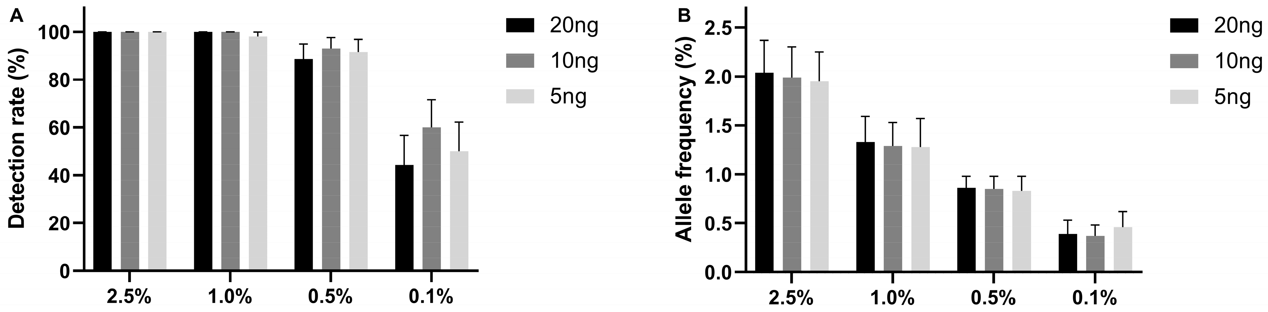
2.1. The Limit of Detection of the UltraSEEK® Lung Panel Using Reference Material

An international multicenter evaluation by three laboratories participating in the CANCER-ID consortium evaluated the limit of detection of the UltraSEEK® Lung Panel using Seraseq® ctDNA Complete™ Mutation Mix reference material. Reference samples contain ten clinically relevant mutations at different VAFs (0.1, 0.5, 1.0, and 2.5%) covered by the UltraSEEK® Lung Panel. Different DNA input amounts (5, 10, and 20 ng) were analyzed. At a VAF of ≥1%, an overall detection rate of over 99% was observed for all DNA input amounts (Figure 1A; Supplemental Table S2). The detection rate drops to >90% for 0.5% VAF and >50% for 0.1%, irrespective of ctDNA input. VAFs measured by UltraSEEK® were comparable with the expected VAFs for all DNA inputs (Figure 1B). Twenty-two false-positive calls were identified in a total of 5022 variant positions, resulting in a variant level specificity of >99.5% (Supplemental Table S2). False-positive variants were observed across eleven samples (88% sample-level specificity), of which seven had a DNA input of 5 ng.

2.2. Comparison of Detection of Mutant ctDNA in Patient-Derived Plasma between UltraSEEK® and ddPCR

In total, 131 variants were detected across 45 patients at any timepoint using UltraSEEK®. Sixty-one (47%) were identified in EGFR, 38 (29%) in KRAS, 21 (16%) in BRAF, 10 (8%) in PIK3CA, and 1 (1%) in ERBB2. An independent mutant ctDNA detection method (i.e., mutation-specific ddPCR analysis) was performed on the same plasma for 157 samples to determine the agreement between both assays. Twelve discordant samples were observed that were either negative with UltraSEEK® (n = 5) or ddPCR (n = 7), resulting in an overall concordance of 92% (145/157) with a PPA of 93% and an NPA of 91% (Supplemental Table S3).

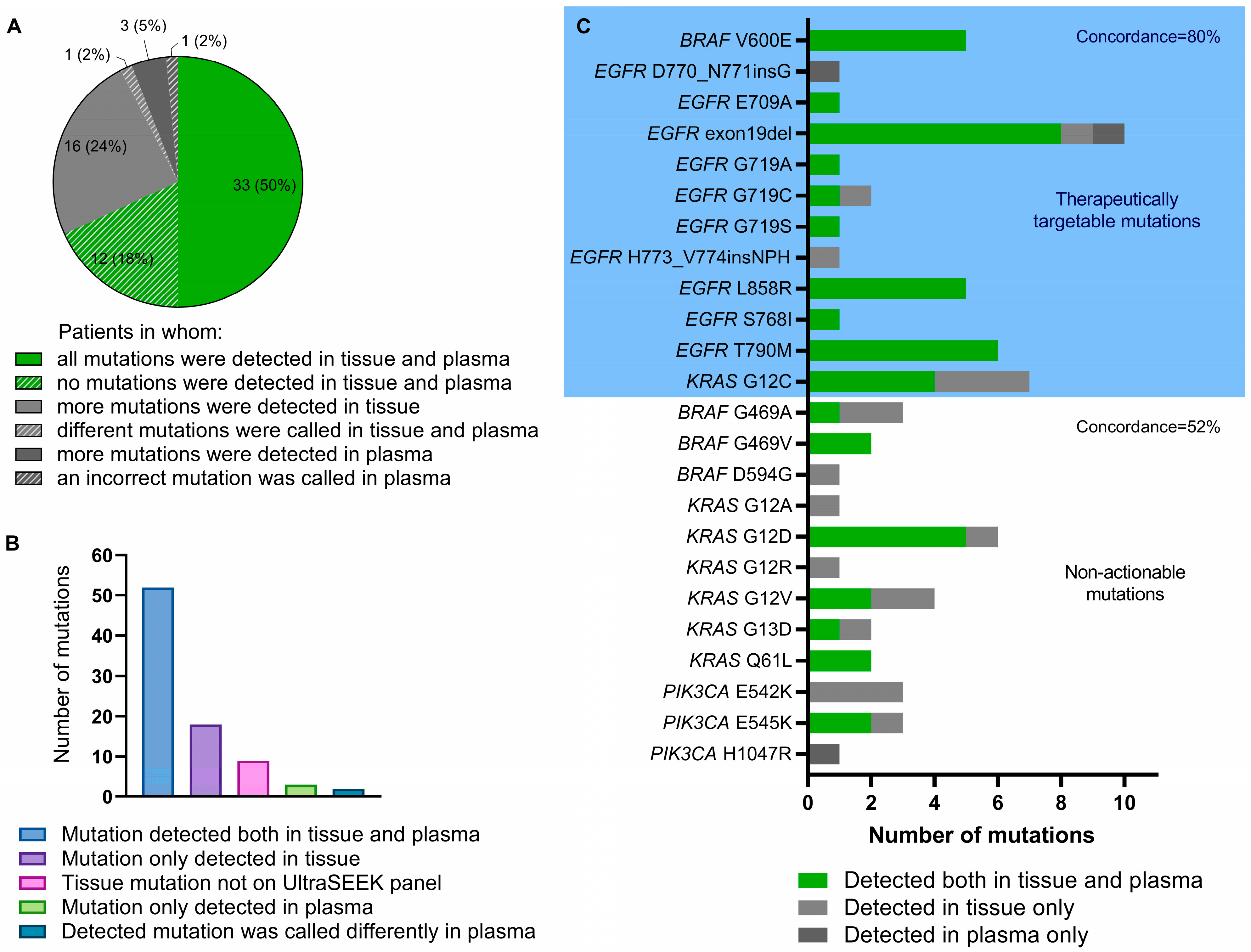
2.3. Detection of Tumor Tissue-Derived Variants in the Baseline Plasma Sample Using the UltraSEEK® Lung Panel

Data on mutational profiles from routine diagnostic NGS analysis on the pretreatment tissue biopsy was available for 66 matched tumor tissue samples (92%). Of the 77 diagnostically or clinically relevant variants in BRAF, EGFR, ERBB2, KRAS, and PIK3CA reported in these pretreatment tissue samples, 53 variants were detected in plasma at baseline using UltraSEEK® (Supplemental Table S4). Eighteen (23%) tumor tissue mutations were not detected in the plasma, and three (4%) of the mutations detected in the plasma were not present in the tumor specimen. CcfDNA UltraSEEK® analysis showed concordant results with tumor tissue NGS analysis in 68% of the patients for the mutations covered by the UltraSEEK® Lung Panel (Figure 2A). Nine variants reported in the tumor tissue across EGFR, KRAS, BRAF, and PIK3CA were not covered by the UltraSEEK® Lung Panel (Figure 2B). Restricting the comparison to therapeutically targetable mutations only covered by the UltraSEEK® Lung Panel, an 80% concordance between tumor tissue NGS and UltraSEEK® was observed (Figure 2C). A considerably lower concordance was found for non-actionable mutations in NSCLC (52%). One mutation (KRAS c.34_35delinsTT; p.(G12F)) called in the tumor affected the same genomic position in plasma but with a different annotation (KRAS c.34G>T; p.(G12C)), most probably due to the inability of the platform to differentiate between covered variants and certain complex variants at similar coding DNA sequences (Supplemental Table S5).
In eleven patients, none of the variants previously reported in tissue samples were identified with UltraSEEK® ccfDNA analyses in the baseline plasma sample. To determine whether these were truly non-shedding tumors, the same plasma was reevaluated with ddPCR as an independent analytically sensitive mutant ctDNA detection assay (Supplemental Table S5). Twelve of fourteen variants in 9/11 patients were not detected with ddPCR as well, whereas two cases harboring KRAS mutations were detected (Supplemental Table S5). Two out of 69 variants reported in the tumor tissue and detected with UltraSEEK® were incorrectly missed in the baseline plasma sample, leading to a 3% false-negative rate at baseline.

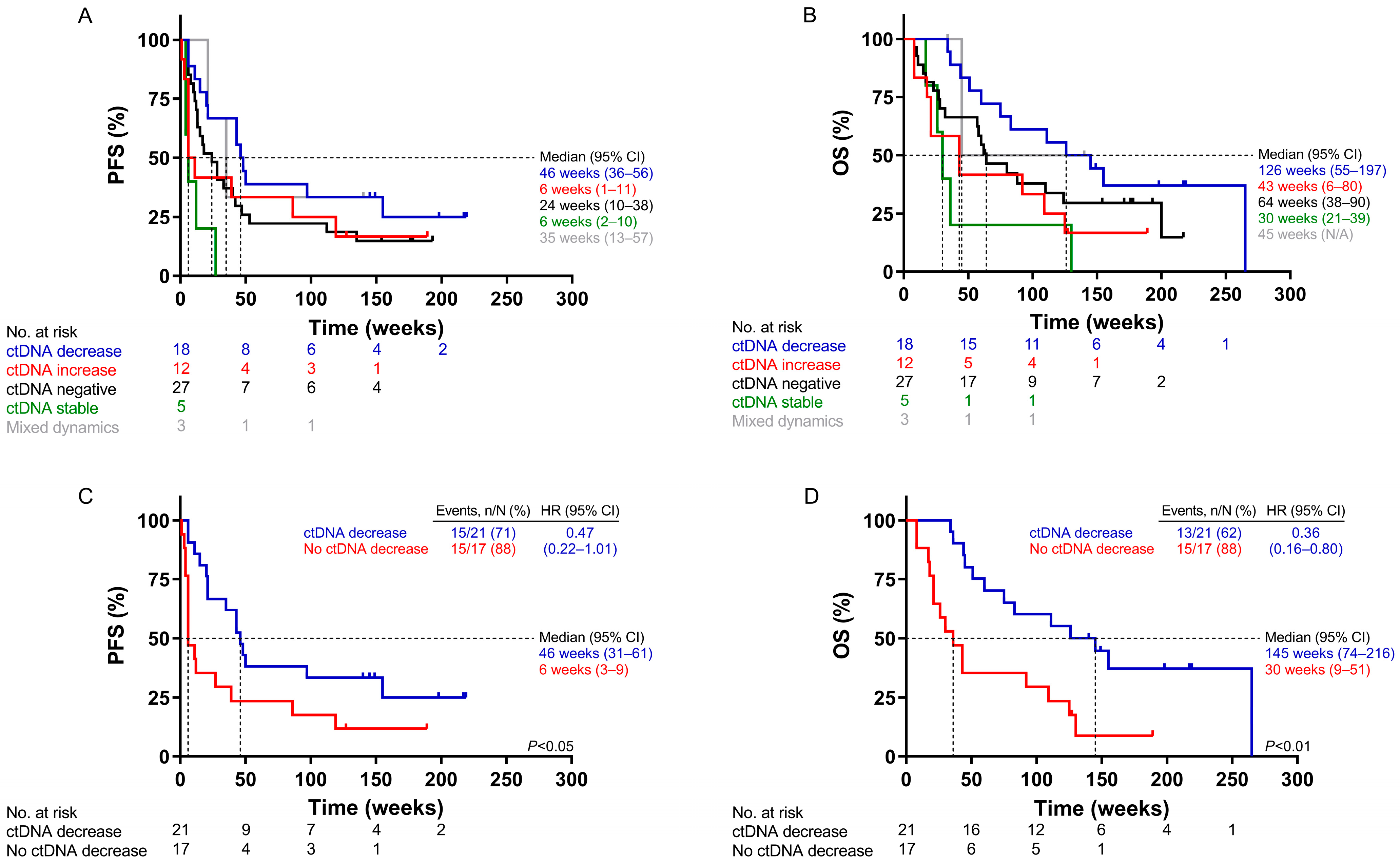
2.4. Survival Analysis According to ctDNA Dynamics Using UltraSEEK®

When comparing the mutant molecule levels at start of treatment (t0) and first response evaluation (t1), patients were categorized as ctDNA decrease (a ≥15% lower VAF at t1, n = 18), ctDNA increase (a ≥ 15% higher VAF at t1, n = 12), ctDNA negative (no detectable ctDNA at both timepoints, n = 27), or ctDNA stable (change in VAF < 15% at t1, n = 5); decreased ctDNA levels were associated with improved clinical outcome (Figure 3A,B). Comparing all patients with increasing or stable ctDNA levels (n = 17) against those with decreased ctDNA levels (n = 21), a decrease was associated significantly with longer median survival and a reduced hazard ratio (HR): PFS (46 versus 6 weeks; HR: 0.47; p < 0.05) and OS (145 versus 30 weeks; HR: 0.36; p < 0.01) (Figure 3C,D).
For 41 patients, an observed change in VAF (UltraSEEK®) was compared with the change in mutant molecule levels (ddPCR) between t0 and t1. Stepwise Cox regression analysis resulted in significantly prolonged PFS and OS upon decreased ctDNA levels, but with lower hazard ratios for PFS with ddPCR (0.32) than UltraSEEK® (0.48) (Supplemental Table S6). Concordant dynamics were observed in 85% of the cases (Supplemental Figure S1; Supplemental Table S7). In six discordant cases, the number of mutant molecules measured with ddPCR was very low in all cases (less than 55 copies/mL of plasma, equivalent to <10 mutant droplets), of which either the UltraSEEK® or ddPCR was negative at both timepoints in plasma in four cases (Supplemental Table S7).

2.5. Plasma-Based Determination of Disease Progression and Acquired Treatment Resistance

In our cohort of 72 patients, twelve of thirteen were progressive on TKI and 48 of 59 on other treatment modalities. Plasma samples at progression were available for 40 patients. Mutations detected in ccfDNA at progression only and associated with treatment resistance were identified in four patients with an activating EGFR mutation in the pretreatment tumor biopsy and treated with first-line EGFR-TKI (Supplemental Table S8). Two patients developed the EGFR p.(T790M) mutation following afatinib treatment, and two patients acquired the EGFR p.(C797S) mutation during osimertinib therapy. In all four patients, these mutations were not detected in any of the earlier plasma samples.

3. Discussion

In the absence of tumor tissue biopsy specimens for predictive testing or during treatment response monitoring, there is a need to accurately detect tumor-derived mutations in a blood sample with high sensitivity. Advancements in ultrasensitive ctDNA detection offer new opportunities for applications of mutation detection in cell-free plasma for personalized treatment management [1]. Targeted panel analyses such as UltraSEEK® are a cost-efficient alternative to detecting most of the current actionable mutations. Previous comparative analysis revealed that targetable mutation screening for an equal number of NSCLC patients reduced the total costs by approximately two-thirds compared to NGS [25]. Therefore, the clinical utility of the UltraSEEK® test for the detection of mutations in plasma from patients with metastatic NSCLC was assessed in this multicenter evaluation.
UltraSEEK® analysis of reference material (Seraseq® ctDNA Complete™ Mutation Mix) demonstrated an overall high detection rate for ten different mutations, irrespective of ctDNA input, with 22 false-positive calls, a variant level specificity of >99.5%, and a sample level specificity of 88%. False-positive results, however, were predominantly observed in low-input samples (5 ng), supporting the use of the recommended ≥10 ng input for accurate results. The analysis of 125 plasma samples from 37 NSCLC patients revealed an overall concordance of 90% with a PPA of 91% and an NPA of 88% between UltraSEEK® and ddPCR. Similar studies with several types of cancer identified an 88% and 80% concordance between these two platforms [30,31].
The availability of tumor tissue remains today’s best clinical practice and is essential for the initial molecular characterization of cancer. However, when tissue is not available or adequate for NGS analysis, tumor-derived mutation testing in plasma serves as an alternative method. Our results demonstrated concordant results in 68% of the patients between routine diagnostic NGS analysis on the pretreatment tissue biopsy and UltraSEEK® analysis in 66 matched baseline plasma samples, comparable to previous data [28,32]. In 23% of the patients with a known driver mutation in the tumor tissue, this variant could not be detected in the matched baseline plasma sample. This discrepancy is in line with previous reports [13,28]. Increased clearance, a short half-life, or a lack of shedding of tumor-specific DNA into the circulation were suggested causes for the absence of ctDNA in pretreatment plasma samples [1]. Reevaluation of negative plasma samples with highly sensitive ddPCR analysis revealed that nine of eleven ctDNA-negative patients were true negatives. A lack of sufficient analytical sensitivity of UltraSEEK® and ddPCR to detect ctDNA levels below 0.5% VAF cannot be excluded.
Elaborate molecular tumor profiling using an NGS panel could elevate the number of ctDNA-positive patients by analyzing more targets simultaneously [6]. However, it does not necessarily identify more actionable targets for patients with advanced-stage NSCLC. Recently, Aggarwal et al. reported on a ccfDNA-based NGS assay with over 74 cancer-related genes and identified 113 variants in 323 patients with metastatic NSCLC. Only 42 patients were treated with targeted therapy as a result of the detection of mutations in BRCA1 (n = 1), ALK (n = 1 fusion), MET exon14 skipping (n = 4), BRAF p.(V600E) (n = 2), and EGFR (n = 34) (20). Theoretically, mutations in 36 of these 42 patients (85%) would have been detected with UltraSEEK® as well. When restricting the data of the present study to therapeutically targetable mutations covered by the panel only, the concordance with tumor tissue NGS at baseline was 80%. Recent studies comparing NGS of tumor tissue biopsies and matched plasma at baseline reported a comparable overall concordance of 81% [6,21] and up to 91% for actionable variants only [33]. In our cohort, nine diagnostically relevant variants reported in tumor tissue were not covered by the platform. These data imply that, in cases of inadequate tumor tissue, UltraSEEK® enables sensitive genotyping and covers >80% of the common predictive markers detected in tumor tissue of advanced NSCLC used in routine diagnostic settings. Furthermore, complementary ccfDNA molecular profiling might display the actual mutational spectrum of patients, which may provide insights on predictive mutations missed during molecular tumor profiling, identify acquired low copy resistance mechanisms (i.e., secondary mutations), or show inter- or intratumor heterogeneity in patients with discordant clinical responses [23]. When the analysis renders the result negative, however, subsequent testing for actionable mutations and fusions is required to cover all demanded targets in accordance with guidelines. Prospective studies to prove the clinical utility of complementary ccfDNA molecular testing using targeted panels are urgently needed.
Cancer progression as a result of resistance to targeted therapy is often accompanied by the detection of novel mutations or clonal expansion of non-responsive tumor cells [34,35]. For instance, on- and off-target resistance mutations after treatment with the EGFR inhibitor osimertinib include de novo EGFR p.(C797S), BRAF p.(V600E), PIK3CA p.(H1047), and KRAS mutations [35,36,37]. Despite advances in treatment guidance, ctDNA-based companion testing for resistance mutations remains limited to EGFR testing in routine diagnostics.
The analytical sensitivity to detect EGFR mutations in the cell-free plasma of NSCLC patients with methods such as Cobas® [38], ddPCR [9], and ARMS [39] revealed a pooled sensitivity of 67% and a specificity of 94% [40]. A high concordance of 86% between UltraSEEK® and Cobas® for the detection of EGFR mutations in a cohort of 137 patients who progressed under EGFR-TKI treatment was reported recently [27]. Importantly, resistance mechanisms that cannot be detected with the Cobas® test, including KRAS mutations and the EGFR p.(C797S) mutation, were detected in eighteen cases [27]. In the present study, in four out of twelve patients who progressed under EGFR-TKI, on-target resistance mutations were detected with UltraSEEK®. UltraSEEK® enables sensitive genotyping for the detection of the most common EGFR-TKI resistance mutations in plasma with the advantages of ease of use, low costs, and a short TAT. As such, ccfDNA molecular profiling using UltraSEEK® is an alternative to the FDA-approved Cobas® EGFR Mutation Test v2 assay. However, the current panel is not useful for the detection of resistance toward and immune checkpoint inhibitor therapy (representing 75% of the entire cohort) since mutations in KEAP1 and STK11 are not covered [41], as well as TKI other than EGFR-TKI (e.g., ALK) [42].
Longitudinal analysis of ccfDNA has shown clinical utility in the identification of early progression and durable responses using single [7,43] and multiple target approaches [6,8,44]. Here, a decrease in ctDNA over time measured with UltraSEEK® was similarly associated with prolonged PFS and OS. However, quantitative ddPCR analysis, regarded as a robust early response assessment tool [7,45], better predicts PFS, resulting in a lower HR. Nonetheless, semiquantitative UltraSEEK® analysis successfully identified patients with durable treatment responses, illustrating its potential as a monitoring tool (Supplemental Table S6).
In conclusion, ideal blood-based tests to primarily diagnose cancer patients and monitor the clonal evolution of their tumors would identify tumor-derived biomarkers with ultra-sensitivity, be fast, and be cost-efficient. Cost-efficiency is mainly determined by testing a limited number of clinically relevant variants during the follow-up of patients, optimized for a specific cancer type. Such an approach covers the known on-target and off-target resistance variants and must be flexible to incorporate future novel variants. The MassARRAY®-based UltraSEEK® Lung Panel enables rapid, sensitive, and semiquantitative genotyping for 73 relevant mutations in BRAF, EGFR, ERBB2, KRAS, and PIK3CA. Although a tumor tissue biopsy remains essential for initial cancer diagnosis, the UltraSEEK® Lung Panel serves as a molecular profiling tool to detect the most common actionable targets in the absence of adequate tumor tissue DNA. In the event that UltraSEEK® results turn out negative, subsequent molecular profiling—including genomic rearrangements, e.g., ALK and MET exon14 skipping—is necessary to cover all actionable targets. Future prospective studies are required to explore the clinical utility of UltraSEEK® versus comparable ctDNA detection assays at diagnosis and disease progression.

4. Materials and Methods

4.1. Sample Collection

For patient inclusion, a cell-free plasma sample should at least have been available at the start of treatment (t0), preferably at the first response evaluation (t1, 4–6 weeks after the start of treatment), and, when applicable, at the presentation of disease progression (tp). In addition, the inclusion of patients with NSCLC who were tested for the presence of predictive mutations in the pretreatment tissue biopsy was preferred. Patients were treated for NSCLC at either the University Medical Center Groningen (UMCG cohort) or the University Medical Center Hamburg-Eppendorf (UKE cohort). In total, 177 cell-free plasma samples from 72 NSCLC patients were included, of which 72 were at t0, 65 at t1, and 40 at tp. The clinical and pathological characteristics are summarized in Table 1. The majority of patients (n = 54) were treated with immunotherapy as no targetable mutations were detected in the pretreatment tumor biopsy, while most of the EGFR-mutated tumors were treated with TKIs (n = 13). Other patients received chemotherapy either alone or in combination with immunotherapy. Plasma sample processing was performed as described in the Supplemental Material and Methods.

4.2. Molecular Analysis

CcfDNA was extracted from blood plasma as described previously [7,17,46] (see Supplemental Materials and Methods). Tumor tissue NGS results were retrieved from the Dutch nationwide pathology registry (PALGA) for relevant predictive variants in BRAF, EGFR, ERBB2, KRAS, and PIK3CA. The UltraSEEK® Lung Panel covers 73 hotspot mutations in these five genes on the matrix-assisted laser desorption ionization time-of-flight (MALDI-TOF)-based MassARRAY® System (Supplemental Table S1). Molecular profiling was performed at t0, t1, and tp using the UltraSEEK® Lung Panel on the MassARRAY® System [24,29,31,47,48] (Agena Bioscience, San Diego, CA, USA) and ddPCR [7,17] (Bio-Rad Laboratories, Pleasanton, CA, USA) according to the manufacturer’s recommendations (for details, see Supplemental Materials and Methods; Supplemental Table S9). With respect to the dynamics of ctDNA levels, changes in variant allele frequency (VAF) equal to or greater than 15% (UltraSEEK®) and changes in mutant molecule levels equal to or greater than 31% (ddPCR) between t0 and t1 were considered an increase or decrease (for details, see Supplemental Materials and Methods).

4.3. Statistical Analysis

Descriptive statistics were used for patient and tumor characteristics. Clopper–Pearson binomial confidence intervals were calculated for the limit of detection. For statistical assessment between different ccfDNA inputs, the Kruskal–Wallis test was performed, followed by Dunn’s multiple comparison test. Agreement between UltraSEEK® analysis and either tumor tissue NGS or ddPCR analysis on ccfDNA was expressed as concordance, positive percent agreement (PPA), or negative percent agreement (NPA). Progression-free survival (PFS) and overall survival (OS) were defined as the period between the date of the start of therapy and the date of progressive disease or death, respectively. The data were censored at the date of the last follow-up in the absence of an event. Kaplan–Meier survival data were stratified for mutant ctDNA data and compared with the log-rank test. Radiological reports and liquid biopsy test results were assessed independently. GraphPad Prism 8.4.2 software was used for all statistical analyses. A p-value < 0.05 was considered significant.

Supplementary Materials

The following supporting information can be downloaded at: https://www.mdpi.com/article/10.3390/ijms241713390/s1.

Author Contributions

Conceptualization, P.v.d.L., M.J., H.W. and E.S.; methodology, P.v.d.L., M.J., P.-J.L., H.W. and E.S.; software, P.v.d.L., A.S. and D.I.; validation, P.v.d.L., M.J., P.-J.L., H.W. and E.S.; formal Analysis, P.v.d.L. and M.J.; investigation, P.v.d.L., N.R., M.L.A.A., J.K. and N.L.; resources, S.L., A.S., D.I., P.-J.L., T.J.N.H., H.J.M.G., K.P., H.W. and E.S.; data curation, A.S., D.I., P.-J.L., L.C.v.K., H.W. and E.S.; writing—original draft preparation, P.v.d.L. and M.J.; writing—review and editing, S.L., P.-J.L., T.J.N.H., H.J.M.G., K.P., L.C.v.K., H.W. and E.S.; visualization, P.v.d.L., M.J., H.W. and E.S.; supervision, P.-J.L., K.P., L.C.v.K., H.W. and E.S.; project administration, H.W. and E.S.; funding acquisition, P.-J.L., H.W. and E.S. All authors have read and agreed to the published version of the manuscript.

Funding

This work was supported by Agena Bioscience (Hamburg, Germany) within the scope of the CANCER-ID consortium, which has received support from the Innovative Medicines Initiative (IMI) Joint Undertaking under grant agreement no. 115749. Additional funding was received for Bio-Rad (Digital Biology Center, Pleasanton, CA, USA) by E.S. The funding had no role in the design of the study (other than the multicenter evaluation study), in the collection, analyses, or interpretation of data, in the writing of the manuscript, or in the decision to publish the results.

Institutional Review Board Statement

The study was conducted according to the guidelines of the Declaration of Helsinki and approved by the Medical Ethics Committee (METc) of the University Medical Hospital Groningen (2010/109).

Informed Consent Statements

Informed consent was obtained from all subjects involved in the study.

Data Availability Statement

Data is available upon reasonable request.

Acknowledgments

The authors would like to thank SeraCare for their in-kind provision of the Seraseq® ctDNA Complete™ Mutation Mixes and Seraseq® ctDNA Complete™ Mutation Mix WT reagents in the scope of the CANCER-ID consortium.

Conflicts of Interest

M.J. has received honoraria for advisory boards and talks from Amgen, Pfizer, BMS, Roche, and AstraZeneca and is funded by the Hector Stiftung II and the Margarete Clemens Stiftung. N.R. is employed by the Dutch Cancer Society (KWF, Alpe D’HuZes research grant RUG 2013-6355). S.L. has received commercial research grants from BerGenBio AS, BMS, Eli Lilly, Roche Pharma, and ADC Therapeutics; has received speaker and/or advisory board honoraria from the Speakers Bureau of BerGenBio AS, BMS, Boehringer Ingelheim, Eli Lilly, Roche Pharma, Medac GmbH, Sanofi Aventis, Novartis, AstraZeneca, Pfizer, Takeda, Amgen, Bayer, and Janssen-Cilag; and is funded by the Hector Stiftung II and the Margarete Clemens Stiftung; P.J.L. is a consultant on advisory boards for Agena Bioscience, Amgen, AstraZeneca, BMS, Janssen-Cilag, Eli Lilly, Novartis, and Roche; and has received research grants from Agena Bioscience and AstraZeneca. T.J.N.H. is a consultant on advisory boards for AstraZeneca, Roche, and BMS and has received research grants from AstraZeneca, BMS, and Roche. K.P. has received honoraria as a speaker from Agena Bioscience, BMS, Novartis, Roche, MSD/Merck, and is a member of the Scientific Advisory Board of Menarini/Silicon Biosystems and BMS. L.C.v.K. has received grants and non-financial support from Roche, Invitae, Biocartis, and nanoString and is a consultant on advisory boards for AstraZeneca, Janssen-Cilag, and Bayer. E.S. has performed lectures for Bio-Rad, Novartis, Roche, Biocartis, Illumina, Pfizer, AstraZeneca, and Agena Bioscience; is a consultant on advisory boards for AstraZeneca, Roche, Pfizer, Novartis, Bayer, BMS, Amgen, BioCartis, Illumina, Agena Bioscience, and MSD/Merck; and has received research grants from Pfizer, Biocartis, Agena Bioscience, BMS, Bio-Rad, Roche, and Boehringer Ingelheim. Other authors declare no conflict of interest.

References

  1. Heitzer, E.; Haque, I.S.; Roberts, C.E.S.; Speicher, M.R. Current and future perspectives of liquid biopsies in genomics-driven oncology. Nat. Rev. Genet. 2019, 20, 71–88. [Google Scholar] [CrossRef] [PubMed]
  2. Boonstra, P.A.; Wind, T.T.; van Kruchten, M.; Schuuring, E.; Hospers, G.A.P.; van der Wekken, A.J.; de Groot, D.-J.; Schröder, C.P.; Fehrmann, R.S.N.; Reyners, A.K.L. Clinical utility of circulating tumor DNA as a response and follow-up marker in cancer therapy. Cancer Metastasis Rev. 2020, 39, 999–1013. [Google Scholar] [CrossRef] [PubMed]
  3. NCCN Clinical Practice Guidelines in Oncology: Non-Small Cell Lung Cancer. Available online: https://www.nccn.org/login?ReturnURL=https://www.nccn.org/professionals/physician_gls/PDF/nscl.pdf (accessed on 14 June 2023).
  4. Planchard, D.; Popat, S.; Kerr, K.; Novello, S.; Smit, E.F.; Faivre-Finn, C.; Mok, T.S.; Reck, M.; Van Schil, P.E.; Hellmann, M.D.; et al. Metastatic non-small cell lung cancer: ESMO Clinical Practice Guidelines for diagnosis, treatment and follow-up. Ann. Oncol. Off. J. Eur. Soc. Med. Oncol. 2018, 29 (Suppl. S4), iv192–iv237. [Google Scholar] [CrossRef] [PubMed]
  5. Thompson, J.C.; Carpenter, E.L.; Silva, B.A.; Rosenstein, J.; Chien, A.L.; Quinn, K.; Espenschied, C.R.; Mak, A.; Kiedrowski, L.A.; Lefterova, M.; et al. Serial Monitoring of Circulating Tumor DNA by Next-Generation Gene Sequencing as a Biomarker of Response and Survival in Patients with Advanced NSCLC Receiving Pembrolizumab-Based Therapy. JCO Precis. Oncol. 2021, 5, 510–524. [Google Scholar] [CrossRef] [PubMed]
  6. Goldberg, S.B.; Narayan, A.; Kole, A.J.; Decker, R.H.; Teysir, J.; Carriero, N.J.; Lee, A.; Nemati, R.; Nath, S.K.; Mane, S.M.; et al. Early assessment of lung cancer immunotherapy response via circulating tumor DNA. Clin. Cancer Res. 2018, 24, 1872–1880. [Google Scholar] [CrossRef] [PubMed]
  7. Leest, P.v.d.; Hiddinga, B.; Miedema, A.; Aguirre Azpurua, M.L.; Rifaela, N.; Elst, A.t.; Timens, W.; Groen, H.J.M.; Kempen, L.C.v.; Hiltermann, T.J.N.; et al. Circulating tumor DNA as a biomarker for monitoring early treatment responses of patients with advanced lung adenocarcinoma receiving immune checkpoint inhibitors. Mol. Oncol. 2021, 15, 2910–2922. [Google Scholar] [CrossRef]
  8. Weber, S.; van der Leest, P.; Donker, H.C.; Schlange, T.; Timens, W.; Tamminga, M.; Hasenleithner, S.O.; Graf, R.; Moser, T.; Spiegl, B.; et al. Dynamic Changes of Circulating Tumor DNA Predict Clinical Outcome in Patients with Advanced Non–Small-Cell Lung Cancer Treated with Immune Checkpoint Inhibitors. JCO Precis. Oncol. 2021, 5, 1540–1553. [Google Scholar] [CrossRef] [PubMed]
  9. Oxnard, G.R.; Paweletz, C.P.; Kuang, Y.; Mach, S.L.; O’Connell, A.; Messineo, M.M.; Luke, J.J.; Butaney, M.; Kirschmeier, P.; Jackman, D.M.; et al. Noninvasive detection of response and resistance in EGFR-mutant lung cancer using quantitative next-generation genotyping of cell-free plasma DNA. Clin. Cancer Res. 2014, 20, 1698–1705. [Google Scholar] [CrossRef]
  10. Heitzer, E.; Ulz, P.; Geigl, J.B. Circulating tumor DNA as a liquid biopsy for cancer. Clin. Chem. 2015, 61, 112–123. [Google Scholar] [CrossRef]
  11. Rangachari, D.; To, C.; Shpilsky, J.E.; VanderLaan, P.A.; Kobayashi, S.S.; Mushajiang, M.; Lau, C.J.; Paweletz, C.P.; Oxnard, G.R.; Jänne, P.A.; et al. EGFR-Mutated Lung Cancers Resistant to Osimertinib through EGFR C797S Respond to First-Generation Reversible EGFR Inhibitors but Eventually Acquire EGFR T790M/C797S in Preclinical Models and Clinical Samples. J. Thorac. Oncol. 2019, 14, 1995–2002. [Google Scholar] [CrossRef]
  12. Mok, T.S.; Wu, Y.-L.; Ahn, M.-J.; Garassino, M.C.; Kim, H.R.; Ramalingam, S.S.; Shepherd, F.A.; He, Y.; Akamatsu, H.; Theelen, W.S.M.E.; et al. Osimertinib or Platinum-Pemetrexed in EGFR T790M-Positive Lung Cancer. N. Engl. J. Med. 2017, 376, 629–640. [Google Scholar] [CrossRef]
  13. Merker, J.D.; Oxnard, G.R.; Compton, C.; Diehn, M.; Hurley, P.; Lazar, A.J.; Lindeman, N.; Lockwood, C.M.; Rai, A.J.; Schilsky, R.L.; et al. Circulating Tumor DNA Analysis in Patients with Cancer: American Society of Clinical Oncology and College of American Pathologists Joint Review. J. Clin. Oncol. 2018, 36, 1631–1641. [Google Scholar] [CrossRef] [PubMed]
  14. Lampignano, R.; Neumann, M.H.D.; Weber, S.; Kloten, V.; Herdean, A.; Voss, T.; Groelz, D.; Babayan, A.; Tibbesma, M.; Schlumpberger, M.; et al. Multicenter Evaluation of Circulating Cell-Free DNA Extraction and Downstream Analyses for the Development of Standardized (Pre)analytical Work Flows. Clin. Chem. 2020, 66, 149–160. [Google Scholar] [CrossRef]
  15. Wan, J.C.M.; Massie, C.; Garcia-Corbacho, J.; Mouliere, F.; Brenton, J.D.; Caldas, C.; Pacey, S.; Baird, R.; Rosenfeld, N. Liquid biopsies come of age: Towards implementation of circulating tumour DNA. Nat. Rev. Cancer 2017, 17, 223–238. [Google Scholar] [CrossRef] [PubMed]
  16. Leest, P.v.d.; Ketelaar, E.M.; Noesel, C.J.M.v.; Broek, D.v.d.; Boerdonk, R.A.A.v.; Deiman, B.; Rifaela, N.; Geize, R.v.d.; Huijsmans, C.J.J.; Speel, E.J.M.; et al. Dutch National Round Robin Trial on Plasma-Derived Circulating Cell-Free DNA Extraction Methods Routinely Used in Clinical Pathology for Molecular Tumor Profiling. Clin. Chem. 2022, 68, 963–972. [Google Scholar] [CrossRef] [PubMed]
  17. van der Leest, P.; Boonstra, P.A.; ter Elst, A.; van Kempen, L.C.; Tibbesma, M.; Koopmans, J.; Miedema, A.; Tamminga, M.; Groen, H.J.M.; Reyners, A.K.L.; et al. Comparison of Circulating Cell-Free DNA Extraction Methods for Downstream Analysis in Cancer Patients. Cancers 2020, 12, 1222. [Google Scholar] [CrossRef]
  18. Lamy, P.-J.; Castan, F.; Lozano, N.; Montélion, C.; Audran, P.; Bibeau, F.; Roques, S.; Montels, F.; Laberenne, A.-C. Next-Generation Genotyping by Digital PCR to Detect and Quantify the BRAF V600E Mutation in Melanoma Biopsies. J. Mol. Diagn. 2015, 17, 366–373. [Google Scholar] [CrossRef]
  19. Lee, J.H.; Menzies, A.M.; Carlino, M.S.; McEvoy, A.C.; Sandhu, S.; Weppler, A.M.; Diefenbach, R.J.; Dawson, S.-J.; Kefford, R.F.; Millward, M.J.; et al. Longitudinal Monitoring of ctDNA in Patients with Melanoma and Brain Metastases Treated with Immune Checkpoint Inhibitors. Clin. Cancer Res. 2020, 26, 4064–4071. [Google Scholar] [CrossRef]
  20. Steeghs, E.M.P.; Kroeze, L.I.; Tops, B.B.J.; Van Kempen, L.C.; Ter Elst, A.; Raaij, A.W.M.K.-V.; Hendriks-Cornelissen, S.J.B.; Hermsen, M.J.W.; Jansen, E.A.M.; Nederlof, P.M.; et al. Comprehensive routine diagnostic screening to identify predictive mutations, gene amplifications, and microsatellite instability in FFPE tumor material. BMC Cancer 2020, 20, 291. [Google Scholar] [CrossRef]
  21. Aggarwal, C.; Thompson, J.C.; Black, T.A.; Katz, S.I.; Fan, R.; Yee, S.S.; Chien, A.L.; Evans, T.L.; Bauml, J.M.; Alley, E.W.; et al. Clinical Implications of Plasma-Based Genotyping with the Delivery of Personalized Therapy in Metastatic Non-Small Cell Lung Cancer. JAMA Oncol. 2019, 5, 173–180. [Google Scholar] [CrossRef]
  22. Ignatiadis, M.; Sledge, G.W.; Jeffrey, S.S. Liquid biopsy enters the clinic—implementation issues and future challenges. Nat. Rev. Clin. Oncol. 2021, 18, 297–312. [Google Scholar] [CrossRef]
  23. Heitzer, E.; Broek, D.v.D.; Denis, M.; Hofman, P.; Hubank, M.; Mouliere, F.; Paz-Ares, L.; Schuuring, E.; Sültmann, H.; Vainer, G.; et al. Recommendations for a practical implementation of circulating tumor DNA mutation testing in metastatic non-small-cell lung cancer. ESMO Open 2022, 7, 100399. [Google Scholar] [CrossRef]
  24. Weber, S.; Spiegl, B.; Perakis, S.O.; Ulz, C.M.; Abuja, P.M.; Kashofer, K.; van der Leest, P.; Azpurua, M.A.; Tamminga, M.; Brudzewsky, D.; et al. Technical Evaluation of Commercial Mutation Analysis Platforms and Reference Materials for Liquid Biopsy Profiling. Cancers 2020, 12, 1588. [Google Scholar] [CrossRef]
  25. Kramer, A.; Schuuring, E.; Vessies, D.C.; van der Leest, P.; Geerlings, M.J.; Rozendal, P.; Lanfermeijer, M.; Linders, T.C.; van Kempen, L.C.; Fijneman, R.J.A.; et al. A Micro-Costing Framework for Circulating Tumor DNA Testing in Dutch Clinical Practice. J. Mol. Diagn. 2023, 25, 36–45. [Google Scholar] [CrossRef]
  26. List of Cleared or Approved Companion Diagnostic Devices (In Vitro and Imaging Tools). Available online: https://www.fda.gov/medical-devices/in-vitro-diagnostics/list-cleared-or-approved-companion-diagnostic-devices-in-vitro-and-imaging-tools (accessed on 14 June 2023).
  27. Lamy, P.-J.; van der Leest, P.; Lozano, N.; Becht, C.; Duboeuf, F.; Groen, H.J.M.; Hilgers, W.; Pourel, N.; Rifaela, N.; Schuuring, E.; et al. Mass Spectrometry as a Highly Sensitive Method for Specific Circulating Tumor DNA Analysis in NSCLC: A Comparison Study. Cancers 2020, 12, 3002. [Google Scholar] [CrossRef]
  28. Kulasinghe, A.; O’Leary, C.; Monkman, J.; Bharti, V.; Irwin, D.; Dutta, S.; Richard, D.J.; Hughes, B.; Ladwa, R.; O’Byrne, K. The identification of circulating tumour DNA using MassARRAY technology in non-small-cell lung cancer (NSCLC). Lung Cancer 2021, 160, 73–77. [Google Scholar] [CrossRef]
  29. Belloum, Y.; Janning, M.; Mohme, M.; Simon, R.; Kropidlowski, J.; Sartori, A.; Irwin, D.; Westphal, M.; Lamszus, K.; Loges, S.; et al. Discovery of Targetable Genetic Alterations in NSCLC Patients with Different Metastatic Patterns Using a MassARRAY-Based Circulating Tumor DNA Assay. Cells 2020, 9, 2337. [Google Scholar] [CrossRef] [PubMed]
  30. Gray, E.S.; Witkowski, T.; Pereira, M.; Calapre, L.; Herron, K.; Irwin, D.; Chapman, B.; Khattak, M.A.; Raleigh, J.; Hatzimihalis, A.; et al. Genomic Analysis of Circulating Tumor DNA Using a Melanoma-Specific UltraSEEK Oncogene Panel. J. Mol. Diagn. 2019, 21, 418–426. [Google Scholar] [CrossRef] [PubMed]
  31. Mehrotra, M.; Singh, R.R.; Loghavi, S.; Duose, D.Y.; Barkoh, B.A.; Behrens, C.; Patel, K.P.; Routbort, M.J.; Kopetz, S.; Broaddus, R.R.; et al. Detection of somatic mutations in cell-free DNA in plasma and correlation with overall survival in patients with solid tumors. Oncotarget 2018, 9, 10259–10271. [Google Scholar] [CrossRef]
  32. Kapeleris, J.; Müller Bark, J.; Ranjit, S.; Irwin, D.; Hartel, G.; Warkiani, M.E.; Leo, P.; O’Leary, C.; Ladwa, R.; O’Byrne, K.; et al. Prognostic value of integrating circulating tumour cells and cell-free DNA in non-small cell lung cancer. Heliyon 2022, 8, e09971. [Google Scholar] [CrossRef] [PubMed]
  33. Remon, J.; Lacroix, L.; Jovelet, C.; Caramella, C.; Howarth, K.; Plagnol, V.; Rosenfeld, N.; Morris, C.; Mezquita, L.; Pannet, C.; et al. Real-World Utility of an Amplicon-Based Next-Generation Sequencing Liquid Biopsy for Broad Molecular Profiling in Patients with Advanced Non–Small-Cell Lung Cancer. JCO Precis. Oncol. 2019, 3, 1–14. [Google Scholar] [CrossRef]
  34. Nagano, T.; Tachihara, M.; Nishimura, Y. Mechanism of Resistance to Epidermal Growth Factor Receptor-Tyrosine Kinase Inhibitors and a Potential Treatment Strategy. Cells 2018, 7, 212. [Google Scholar] [CrossRef]
  35. Schmid, S.; Li, J.J.N.; Leighl, N.B. Mechanisms of osimertinib resistance and emerging treatment options. Lung Cancer 2020, 147, 123–129. [Google Scholar] [CrossRef]
  36. Paik, P.K.; Arcila, M.E.; Fara, M.; Sima, C.S.; Miller, V.A.; Kris, M.G.; Ladanyi, M.; Riely, G.J. Clinical characteristics of patients with lung adenocarcinomas harboring BRAF mutations. J. Clin. Oncol. 2011, 29, 2046–2051. [Google Scholar] [CrossRef]
  37. Anguera, G.; Majem, M. BRAF inhibitors in metastatic non-small cell lung cancer. J. Thorac. Dis. 2018, 10, 589–592. [Google Scholar] [CrossRef] [PubMed]
  38. Weber, B.; Meldgaard, P.; Hager, H.; Wu, L.; Wei, W.; Tsai, J.; Khalil, A.; Nexo, E.; Sorensen, B.S. Detection of EGFR mutations in plasma and biopsies from non-small cell lung cancer patients by allele-specific PCR assays. BMC Cancer 2014, 14, 294. [Google Scholar] [CrossRef] [PubMed]
  39. Syed, Y.Y. therascreen ® EGFR RGQ PCR Kit: A Companion Diagnostic for Afatinib and Gefitinib in Non-Small Cell Lung Cancer. Mol. Diagn. Ther. 2016, 20, 191–198. [Google Scholar] [CrossRef] [PubMed]
  40. Luo, J.; Shen, L.; Zheng, D. Diagnostic value of circulating free DNA for the detection of EGFR mutation status in NSCLC: A systematic review and meta-analysis. Sci. Rep. 2014, 4, 6269. [Google Scholar] [CrossRef]
  41. Passaro, A.; Brahmer, J.; Antonia, S.; Mok, T.; Peters, S. Managing Resistance to Immune Checkpoint Inhibitors in Lung Cancer: Treatment and Novel Strategies. J. Clin. Oncol. 2022, 40, 598–610. [Google Scholar] [CrossRef]
  42. Du, X.; Shao, Y.; Qin, H.-F.; Tai, Y.-H.; Gao, H.-J. ALK-rearrangement in non-small-cell lung cancer (NSCLC). Thorac. Cancer 2018, 9, 423–430. [Google Scholar] [CrossRef]
  43. Paz-Ares, L.; Vicente, D.; Tafreshi, A.; Robinson, A.; Soto Parra, H.; Mazières, J.; Hermes, B.; Cicin, I.; Medgyasszay, B.; Rodríguez-Cid, J.; et al. A Randomized, Placebo-Controlled Trial of Pembrolizumab Plus Chemotherapy in Patients with Metastatic Squamous NSCLC: Protocol-Specified Final Analysis of KEYNOTE-407. J. Thorac. Oncol. 2020, 15, 1657–1669. [Google Scholar] [CrossRef] [PubMed]
  44. Guibert, N.; Jones, G.; Beeler, J.F.; Plagnol, V.; Morris, C.; Mourlanette, J.; Delaunay, M.; Keller, L.; Rouquette, I.; Favre, G.; et al. Targeted sequencing of plasma cell-free DNA to predict response to PD1 inhibitors in advanced non-small cell lung cancer. Lung Cancer 2019, 137, 1–6. [Google Scholar] [CrossRef] [PubMed]
  45. Zulato, E.; Attili, I.; Pavan, A.; Nardo, G.; Del Bianco, P.; Boscolo Bragadin, A.; Verza, M.; Pasqualini, L.; Pasello, G.; Fassan, M.; et al. Early assessment of KRAS mutation in cfDNA correlates with risk of progression and death in advanced non-small-cell lung cancer. Br. J. Cancer 2020, 123, 81–91. [Google Scholar] [CrossRef] [PubMed]
  46. Boonstra, P.A.; Elst, A.t.; Tibbesma, M.; Bosman, L.J.; Mathijssen, R.; Atrafi, F.; Coevorden, F.v.; Steeghs, N.; Farag, S.; Gelderblom, H.; et al. A single digital droplet PCR assay to detect multiple KIT exon 11 mutations in tumor and plasma from patients with gastrointestinal stromal tumors. Oncotarget 2018, 9, 13870–13883. [Google Scholar] [CrossRef]
  47. Gray, J.E.; Okamoto, I.; Sriuranpong, V.; Vansteenkiste, J.; Imamura, F.; Lee, J.S.; Pang, Y.-K.; Cobo, M.; Kasahara, K.; Cheng, Y.; et al. Tissue and Plasma EGFR Mutation Analysis in the FLAURA Trial: Osimertinib versus Comparator EGFR Tyrosine Kinase Inhibitor as First-Line Treatment in Patients with EGFR-Mutated Advanced Non-Small Cell Lung Cancer. Clin. Cancer Res. 2019, 25, 6644–6652. [Google Scholar] [CrossRef]
  48. Mosko, M.J.; Nakorchevsky, A.A.; Flores, E.; Metzler, H.; Ehrich, M.; van den Boom, D.J.; Sherwood, J.L.; Nygren, A.O.H. Ultrasensitive Detection of Multiplexed Somatic Mutations Using MALDI-TOF Mass Spectrometry. J. Mol. Diagn. 2016, 18, 23–31. [Google Scholar] [CrossRef]
Figure 1. Detection of hotspot mutations common in NSCLC using UltraSEEK® Lung Panel. (A) Detection rate and (B) quantification of Seraseq® ctDNA Complete™ Mutation Mix reference material at variant allele frequencies (VAFs) of 2.5%, 1.0%, 0.5%, and 0.1% were analyzed using 20 ng (black), 10 ng (dark grey), and 5 ng (light grey) ccfDNA input. Error bars represent the confidence intervals calculated from the detection rates per mutation; detailed results of ten hotspot mutations in EGFR, KRAS, BRAF, ERBB2, and PIK3CA are presented in Supplemental Table S1. Kruskal–Wallis test was performed, followed by Dunn’s multiple comparison test; all comparisons were not significant.
Ijms 24 13390 g001
Figure 2. Concordance between mutations detected in tumor tissue and plasma-derived ccfDNA in EGFR, KRAS, BRAF, ERBB2, and PIK3CA. (A) Pie chart representing the patients in whom mutation detection using tissue NGS and plasma UltraSEEK® Lung Panel analyses was considered concordant (green) and discordant (grey) for all mutations covered by the UltraSEEK® Lung Panel. (B) Bar chart representing the distribution of all mutations detected using tumor tissue NGS reported in the pathology archives and plasma UltraSEEK® Lung Panel analysis. (C) Stacked bar chart displaying the distribution of individual mutations detected both with tissue NGS and plasma UltraSEEK® analysis (green), only with tissue NGS (light grey), and only with plasma UltraSEEK® (dark grey). Clinically relevant mutations (light blue box) were separated from currently non-actionable mutations in non-small cell lung cancer (white box). Individual patient results and mutations not covered by the UltraSEEK® Lung Panel are depicted in Supplemental Table S3.
Ijms 24 13390 g002
Figure 3. Survival analysis according to ctDNA dynamics using the UltraSEEK® Lung Panel. Kaplan–Meier plot displaying the (A) progression-free survival (PFS) and (B) overall survival (OS) of patients with decreasing (blue), negative (black), stable (green), or increasing (red) ctDNA levels. (C) PFS and (D) OS of patients with decreasing ctDNA levels (blue) or no decrease in ctDNA (red). No decrease in ctDNA encompasses all patients without a >15% decrease in ctDNA. Horizontal dashed lines indicate the median survival; vertical lines indicate the moment at which the median survival was reached. HR, hazard ratio; CI, confidence interval. Log-rank test, p-value < 0.05, was considered significant.
Ijms 24 13390 g003
Table 1. Clinical and pathological characteristics.
Total CohortUMCG CohortUKE Cohort
Patients726012
Median age65 (38–85)63 (38–85)68 (57–79)
Sex
Male37 (51%)29 (48%)8 (67%)
Female35 (49%)31 (52%)4 (33%)
ECOG PS
023 (32%)21 (35%)2 (17%)
144 (61%)37 (62%)7 (58%)
25 (7%)2 (3%)3 (25%)
Histology
Adenocarcinoma64 (89%)56 (93%)8 (67%)
Squamous cell carcinoma8 (11%)4 (7%)4 (33%)
Smoking status
Active smoker33 (46%)29 (48%)4 (33%)
Former smoker27 (38%)23 (38%)4 (33%)
Never smoker11 (15%)8 (13%)3 (25%)
Unknown1 (1%) 1 (8%)
Current treatment
Chemotherapy3 (4%)1 (2%)2 (17%)
Carboplatine/pemetrexed1 (1%) 1 (8%)
Cisplatine/pemetrexed1 (1%)1 (2%)
Docetaxel/ramucirumab1 (1%) 1 (8%)
Chemo-immunotherapy2 (3%)1 (2%)1 (8%)
Carboplatine/paclitaxel/bevacizumab1 (1%)1 (2%)
Carboplatine/pemetrexed/pembrolizumab1 (1%) 1 (8%)
Immunotherapy54 (75%)45 (75%)9 (75%)
Atezolizumab1 (1%)1 (2%)
Nivolumab40 (56%)37 (62%)3 (25%)
Pembrolizumab13 (18%)7 (12%)6 (50%)
Targeted therapy13 (18%)13 (22%)
Afatinib7 (10%)7 (12%)
Gefitinib1 (1%)1 (2%)
Osimertinib5 (7%)5 (8%)
Previous lines of therapies
019 (26%)11 (18%)8 (67%)
132 (44%)30 (50%)2 (17%)
215 (21%)15 (25%)
≥36 (8%)4 (7%)2 (17%)
ECOG PS, Eastern Cooperative Oncology Group performance-status score.
Disclaimer/Publisher’s Note: The statements, opinions and data contained in all publications are solely those of the individual author(s) and contributor(s) and not of MDPI and/or the editor(s). MDPI and/or the editor(s) disclaim responsibility for any injury to people or property resulting from any ideas, methods, instructions or products referred to in the content.

