
Advances in Preparation and Properties of Regenerated Silk Fibroin


Abstract
1. Introduction
2. Silk Fibroin
3. SF Degumming
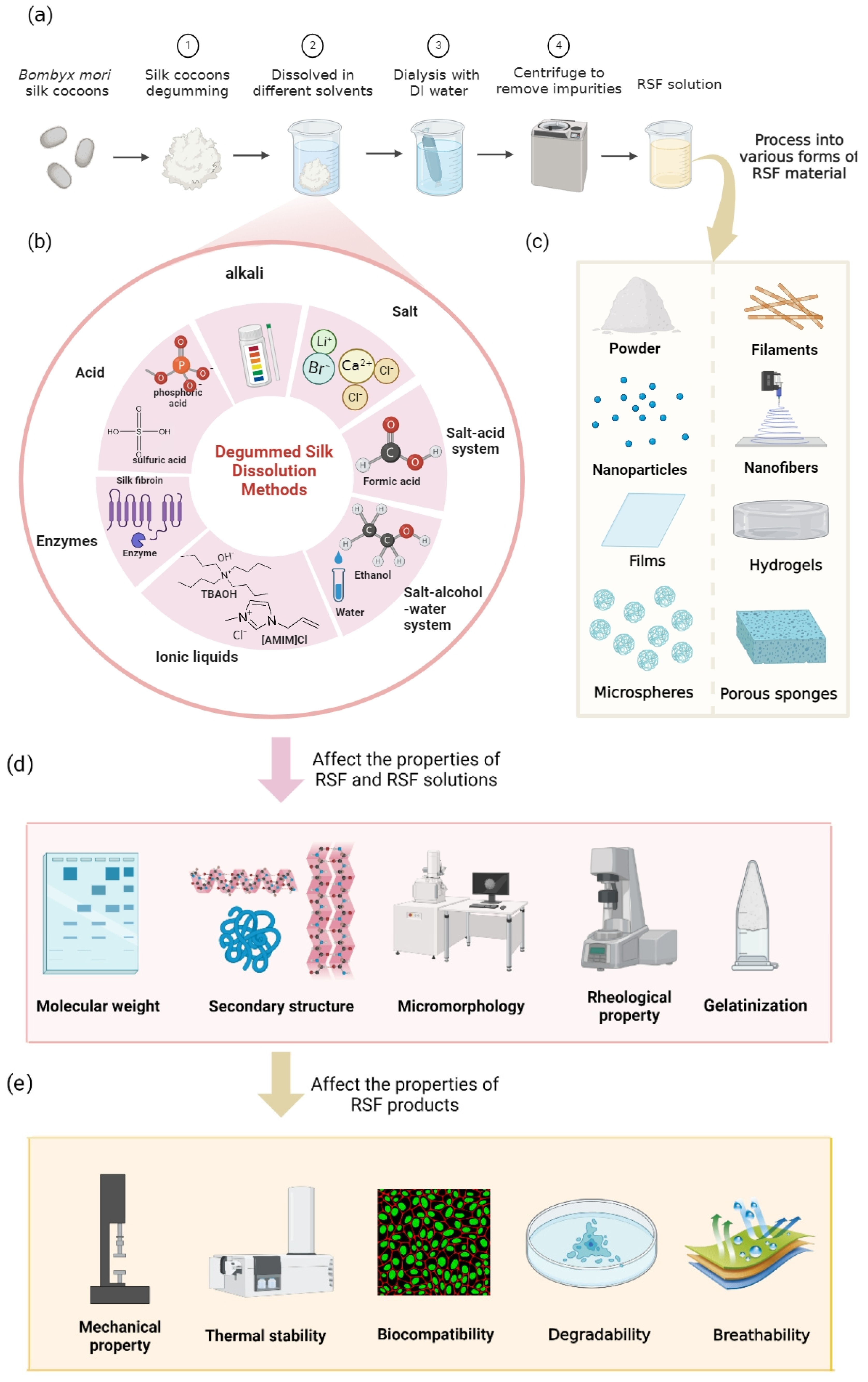
4. SF Dissolution Processes and Their Influence on RSF Characters
4.1. Methods and Properties of RSF by Acid Dissolution
4.2. Methods and Properties of RSF by Alkali Dissolution
4.3. Preparation Methods and Properties of RSF by Salt and Salt Complex System Dissolution
4.3.1. Salt
4.3.2. Salt–Acid Complex System
4.3.3. Salt–Alcohol–Water System
4.4. Preparation Methods and Properties of RSF by Ionic Liquid Dissolution
4.5. Preparation Methods and Properties of RSF by Enzyme Dissolution
5. Perspectives
Author Contributions
Funding
Institutional Review Board Statement
Informed Consent Statement
Data Availability Statement
Acknowledgments
Conflicts of Interest
References
- Chen, S.; Liu, M.; Huang, H.; Cheng, L.; Zhao, H. Mechanical properties of Bombyx mori silkworm silk fibre and its corresponding silk fibroin filament: A comparative study. Mater. Des. 2019, 181, 108077. [Google Scholar] [CrossRef]
- Koh, L.; Cheng, Y.; Teng, C.; Khin, Y.; Loh, X.; Tee, S.; Low, M.; Ye, E.; Yu, H.; Zhang, Y.; et al. Structures, mechanical properties and applications of silk fibroin materials. Prog. Polym. Sci. 2015, 46, 86–110. [Google Scholar] [CrossRef]
- Cao, Y.; Wang, B. Biodegradation of silk biomaterials. Int. J. Mol. Sci. 2009, 10, 1514–1524. [Google Scholar] [CrossRef]
- You, R.; Xu, Y.; Liu, G.; Liu, Y.; Li, X.; Li, M. Regulating the degradation rate of silk fibroin films through changing the genipin crosslinking degree. Polym. Degrad. Stab. 2014, 109, 226–232. [Google Scholar] [CrossRef]
- Lu, S.; Tang, X.; Lu, Q.; Huang, J.; You, X.; Zhang, F. In vitro and in vivo degradation of silk fibers degummed with various sodium carbonate concentrations. Mater. Today Commun. 2021, 27, 102369. [Google Scholar] [CrossRef]
- Liu, L.; Zhang, S.; Huang, J. Progress in modification of silk fibroin fiber. Sci. China Technol. Sci. 2019, 62, 919–930. [Google Scholar] [CrossRef]
- Charu, V.; David, L.K. Silk as a biomaterial. Prog. Polym. Sci. 2007, 32, 991–1007. [Google Scholar]
- Yu, B.; Li, Y.; Lin, Y.; Zhu, Y.; Hao, T.; Wu, Y.; Sun, Z.; Yang, X.; Xu, H. Research progress of natural silk fibroin and the application for drug delivery in chemotherapies. Front. Pharmacol. 2022, 13, 1071868. [Google Scholar] [CrossRef]
- Patil, P.P.; Reagan, M.R.; Bohara, R.A. Silk fibroin and silk-based biomaterial derivatives for ideal wound dressings. Int. J. Biol. Macromol. 2020, 164, 4613–4627. [Google Scholar] [CrossRef]
- Smeets, R.; Tauer, N.; Vollkommer, T.; Gosau, M.; Henningsen, A.; Hartjen, P.; Fruh, L.; Beikler, T.; Sturmer, E.; Rutkowski, R.; et al. Tissue adhesives in reconstructive and aesthetic surgery-application of silk fibroin-based biomaterials. Int. J. Mol. Sci. 2022, 23, 7687. [Google Scholar] [CrossRef]
- Ealla, K.; Veeraraghavan, V.P.; Ravula, N.R.; Durga, C.S.; Ramani, P.; Sahu, V.; Poola, P.K.; Patil, S.; Panta, P. Silk hydrogel for tissue engineering: A review. J. Contemp. Dent. Pract. 2022, 23, 467–477. [Google Scholar]
- Sun, W.; Gregory, D.A.; Tomeh, M.A.; Zhao, X. Silk fibroin as a functional biomaterial for tissue engineering. Int. J. Mol. Sci. 2021, 22, 1499. [Google Scholar] [CrossRef] [PubMed]
- Sultan, M.T.; Lee, O.J.; Lee, J.S.; Park, C.H. Three-dimensional digital light-processing bioprinting using silk fibroin-based bio-ink: Recent advancements in biomedical applications. Biomedicines 2022, 10, 3224. [Google Scholar] [CrossRef]
- Tu, J.; Liu, G.H.; Yan, W. Effects of Four Common Stabilizers Combined with Silk Fibroin on Viscosity and Water-Holding Capacity of Stirred Yogurt. Food Sci. 2012, 33, 136–140. [Google Scholar]
- Low, J.T.; Yusoff, N.; Othman, N.; Wong, T.W.; Wahit, M.U. Silk fibroin-based films in food packaging applications: A review. Compr. Rev. Food Sci. Food Saf. 2022, 21, 2253–2273. [Google Scholar] [CrossRef] [PubMed]
- Ling, S.; Qin, Z.; Huang, W.; Cao, S.; Kaplan, D.L. Design and function of biomimetic multilayer water purification membranes. Sci. Adv. 2017, 3, e1601939. [Google Scholar] [CrossRef] [PubMed]
- Huang, W.; Ling, S.; Li, C.; Omenetto, F.G.; Kaplan, D.L. Silkworm silk-based materials and devices generated using bio-nanotechnology. Chem. Soc. Rev. 2018, 47, 6486–6504. [Google Scholar] [CrossRef]
- Wu, R.; Ma, L.; Liu, X.Y. From mesoscopic functionalization of silk fibroin to smart fiber devices for textile electronics and photonics. Adv. Sci. 2022, 9, e2103981. [Google Scholar] [CrossRef]
- Zhou, L.; Hou, J.Y.; Chen, Y.N.; Li, S.C.; Zou, B.X. Porous carbon composite generated from silk fibroins and graphene for supercapacitors. ACS Omega 2022, 7, 28284–28292. [Google Scholar] [CrossRef]
- He, H.; Zhang, Y.; Zhang, W.; Li, Y.; Wang, Y.; Wang, P. Dual metal-loaded porous carbon materials derived from silk fibroin as bifunctional electrocatalysts for hydrogen evolution reaction and oxygen evolution reaction. ACS Appl. Mater. Interfaces 2021, 13, 30678–30692. [Google Scholar] [CrossRef]
- Sarkar, L.; Sushma, M.V.; Yalagala, B.P.; Rengan, A.K.; Singh, S.G.; Vanjari, S. ZnO nanoparticles embedded silk fibroin-a piezoelectric composite for nanogenerator applications. Nanotechnology 2022, 33, 265403. [Google Scholar] [CrossRef] [PubMed]
- Shen, S.; Yi, J.; Sun, Z.; Guo, Z.; He, T.; Ma, L.; Li, H.; Fu, J.; Lee, C.; Wang, Z.L. Human machine interface with wearable electronics using biodegradable triboelectric films for calligraphy practice and correction. Nanomicro Lett. 2022, 14, 225. [Google Scholar] [CrossRef]
- Li, J.; Yang, F.; Liu, D.; Han, S.; Li, J.; Sui, G. Graphene composite paper synergized with micro/nanocellulose-fiber and silk fibroin for flexible strain sensor. Int. J. Biol. Macromol. 2023, 240, 124439. [Google Scholar] [CrossRef]
- Wang, Q.; Chen, Q.; Yang, Y.; Shao, Z. Effect of various dissolution systems on the molecular weight of regenerated silk fibroin. Biomacromolecules 2013, 14, 284–285. [Google Scholar] [CrossRef] [PubMed]
- Yao, X.; Zou, S.; Fan, S.; Niu, Q.; Zhang, Y. Bioinspired silk fibroin materials: From silk building blocks extraction and reconstruction to advanced biomedical applications. Mater. Today Bio 2022, 16, 100381. [Google Scholar] [CrossRef]
- Zhao, Y.; Zhu, Z.S.; Guan, J.; Wu, S.J. Processing, mechanical properties and bio-applications of silk fibroin-based high-strength hydrogels. Acta Biomater. 2021, 125, 57–71. [Google Scholar] [CrossRef] [PubMed]
- Aznar-Cervantes, S.D.; Vicente-Cervantes, D.; Meseguer-Olmo, L.; Cenis, J.L.; Lozano-Perez, A.A. Influence of the protocol used for fibroin extraction on the mechanical properties and fiber sizes of electrospun silk mats. Mater. Sci. Eng. C Mater. Biol. Appl. 2013, 33, 1945–1950. [Google Scholar] [CrossRef]
- Aoki, M.; Masuda, Y.; Ishikawa, K.; Tamada, Y. Fractionation of regenerated silk fibroin and characterization of the fractions. Molecules 2021, 26, 6317. [Google Scholar] [CrossRef]
- Debari, M.K.; Abbott, R.D. Microscopic considerations for optimizing silk biomaterials. Wiley Interdiscip. Rev. Nanomed. Nanobiotechnol. 2019, 11, e1534. [Google Scholar] [CrossRef]
- Wang, Y.; Rudym, D.D.; Walsh, A.; Abrahamsen, L.; Kim, H.J.; Kim, H.S.; Kirker-Head, C.; Kaplan, D.L. In vivo degradation of three-dimensional silk fibroin scaffolds. Biomaterials 2008, 29, 3415–3428. [Google Scholar] [CrossRef]
- Zhang, X.; Zhu, D.; Cheng, Y.; Zhang, X.; Guo, X.; Lin, N.; Zuo, B. Preparation and biocompatibility characterization of regenerated silk fibroin films. J. Macromol. Sci. Part B 2021, 60, 603–615. [Google Scholar] [CrossRef]
- Nadgorny, M.; Gentekos, D.T.; Xiao, Z.; Singleton, S.P.; Fors, B.P.; Connal, L.A. Manipulation of molecular weight distribution shape as a new strategy to control processing parameters. Macromol. Rapid Commun. 2017, 38. [Google Scholar] [CrossRef] [PubMed]
- Hai-Yan, W.; Yu-Qing, Z. Effect of regeneration of liquid silk fibroin on its structure and characterization. Soft Matter. 2013, 9, 138–145. [Google Scholar]
- Rockwood, D.N.; Preda, R.C.; Yucel, T.; Wang, X.; Lovett, M.L.; Kaplan, D.L. Materials fabrication from Bombyx mori silk fibroin. Nat. Protoc. 2011, 6, 1612–1631. [Google Scholar] [CrossRef] [PubMed]
- Jin, H.J.; Kaplan, D.L. Mechanism of silk processing in insects and spiders. Nature 2003, 424, 1057–1061. [Google Scholar] [CrossRef] [PubMed]
- Zhou, C.Z.; Confalonieri, F.; Jacquet, M.; Perasso, R.; Li, Z.G.; Janin, J. Silk fibroin: Structural implications of a remarkable amino acid sequence. Proteins 2001, 44, 119–122. [Google Scholar] [CrossRef]
- Masuhiro, T.; Yohko, G.; Norihiko, M. Characterization of the regenerated silk fibroin from Bombyx mori. J. Sericultural Sci. Jpn. 1990, 59, 325–330. [Google Scholar]
- Sridhara, S.; Prudhomme, J.C.; Daillie, J. Studies on silk fibroin of the silkworm Bombyx mori. Arch. Biochem. Biophys. 1973, 156, 168–175. [Google Scholar] [CrossRef]
- Ajisawa, A. Dissolution of silk fibroin with calciumchloride/ethanol aqueous solution. Jpn. J. Silkworm Sci. 1998, 67, 91–94. [Google Scholar]
- Kiyoichi, M.; Hiroyuki, U. Regenerated protein fibers. I. Research and development of a novel solvent for silk fibroin. J. Polym. Sci. Part. A Polym. Chem. 1997, 35, 1949–1954. [Google Scholar]
- Wang, H.Y.; Zhang, Y.Q.; Wei, Z.G. Dissolution and processing of silk fibroin for materials science. Crit. Rev. Biotechnol. 2021, 41, 406–424. [Google Scholar] [CrossRef] [PubMed]
- Xu, Z.; Gao, W.; Bai, H. Silk-based bioinspired structural and functional materials. iScience 2022, 25, 103940. [Google Scholar] [CrossRef] [PubMed]
- Xing, X.; Han, Y.; Cheng, H. Biomedical applications of chitosan/silk fibroin composites: A review. Int. J. Biol. Macromol. 2023, 240, 124407. [Google Scholar] [CrossRef] [PubMed]
- Sabarees, G.; Tamilarasi, G.P.; Velmurugan, V.; Alagarsamy, V.; Sibuh, B.Z.; Sikarwar, M.; Taneja, P.; Kumar, A.; Gupta, P.K. Emerging trends in silk fibroin based nanofibers for impaired wound healing. J. Drug. Deliv. Sci. Technol. 2023, 79, 103994. [Google Scholar] [CrossRef]
- Sahoo, J.K.; Hasturk, O.; Falcucci, T.; Kaplan, D.L. Silk chemistry and biomedical material designs. Nat. Rev. Chem. 2023, 7, 302–318. [Google Scholar]
- Liu, B.; Song, Y.W.; Jin, L.; Wang, Z.J.; Pu, D.Y.; Lin, S.Q.; Zhou, C.; You, H.J.; Ma, Y.; Li, J.M.; et al. Silk structure and degradation. Colloids Surf. B. Biointerfaces 2015, 131, 122–128. [Google Scholar] [CrossRef]
- Gregory, H.A.; Frank, D.; Caroline, J.; Tara, C.; Rebecca, L.H.; Jingsong, C.; Helen, L.; John, R.; David, L.K. Silk-based biomaterials. Biomaterials 2003, 24, 401–416. [Google Scholar]
- Cao, T.T.; Zhang, Y.Q. Processing and characterization of silk sericin from Bombyx mori and its application in biomaterials and biomedicines. Mater. Sci. Eng. C Mater. Biol. Appl. 2016, 61, 940–952. [Google Scholar] [CrossRef]
- Inoue, S.; Tanaka, K.; Arisaka, F.; Kimura, S.; Ohtomo, K.; Mizuno, S. Silk fibroin of Bombyx mori is secreted, assembling a high molecular mass elementary unit consisting of h-chain, l-chain, and p25, with a 6:6:1 molar ratio. J. Biol. Chem. 2000, 275, 40517–40528. [Google Scholar] [CrossRef]
- Feng, P. The study of structure and edidility of silk protein. Food Res. Dev. 2004, 25, 51–54. [Google Scholar]
- Zhou, C.Z.; Confalonieri, F.; Medina, N.; Zivanovic, Y.; Esnault, C.; Yang, T.; Jacquet, M.; Janin, J.; Duguet, M.; Perasso, R.; et al. Fine organization of Bombyx mori fibroin heavy chain gene. Nucleic Acids Res. 2000, 28, 2413–2419. [Google Scholar] [CrossRef] [PubMed]
- Ha, S.W.; Gracz, H.S.; Tonelli, A.E.; Hudson, S.M. Structural study of irregular amino acid sequences in the heavy chain of Bombyx mori silk fibroin. Biomacromolecules 2005, 6, 2563–2569. [Google Scholar] [CrossRef]
- Mita, K.; Ichimura, S.; James, T.C. Highly repetitive structure and its organization of the silk fibroin gene. J. Mol. Evol. 1994, 38, 583–592. [Google Scholar] [CrossRef] [PubMed]
- Kazunori, T.; Satoshi, I.; Shigeki, M. Hydrophobic interaction of p25, containing asn-linked oligosaccharide chains, with the h-l complex of silk fibroin produced by Bombyx mori. Insect Biochem. Mol. Biol. 1999, 29, 269–276. [Google Scholar]
- Asakura, T.; Kuzuhara, A.; Tabeta, R.; Saito, H. Conformational characterization of Bombyx mori silk fibroin in the solid state by high-frequency carbon-13 cross polarization-magic angle spinning nmr, X-ray diffraction, and infrared spectroscopy. Macromolecules 1985, 18, 1841–1845. [Google Scholar] [CrossRef]
- Asakura, T. Structure of silk I (Bombyx mori silk fibroin before spinning) -typeII β-turn, not α-helix. Molecules 2021, 26, 3706. [Google Scholar] [CrossRef]
- Asakura, T.; Aoki, A.; Komatsu, K.; Ito, C.; Suzuki, I.; Naito, A.; Kaji, H. Lamellar structure in alanine-glycine copolypeptides studied by solid-state nmr spectroscopy: A model for the crystalline domain of Bombyx mori silk fibroin in silkIIform. Biomacromolecules 2020, 21, 3102–3111. [Google Scholar] [CrossRef]
- Bucciarelli, A.; Greco, G.; Corridori, I.; Pugno, N.M.; Motta, A. A design of experiment rational optimization of the degumming process and its impact on the silk fibroin properties. ACS Biomater. Sci. Eng. 2021, 7, 1374–1393. [Google Scholar] [CrossRef]
- Luping, W.; Zuwei, L.; Qiang, Z.; Yupin, G.; Junyi, C.; Renchuan, Y.; Xiufang, L. Effect of degumming methods on the degradation behavior of silk fibroin biomaterials. Fibers Polym. 2019, 20, 45–50. [Google Scholar]
- Gulrajanid, M.L.; Sheetal, S.; Sanjay, G. Some studies in degumming of silk with organic acids. Color. Technol. 1992, 108, 79–86. [Google Scholar] [CrossRef]
- Ninpetch, W.P.A.P. Pilot-scale protease production by bacillussp.c4 for silk degumming processes. J. Nat. Fibers 2022, 19, 1055–1068. [Google Scholar] [CrossRef]
- Wuchao, W.; Yi, P.; Kang, G.; Qi, Z.; Tonghua, Z.; Qing, L. A comparative study of ultrasonic degumming of silk sericin using citric acid, sodium carbonate and papain. Color. Technol. 2019, 135, 195–201. [Google Scholar]
- Wang, Y.; Liang, Y.; Huang, J.; Gao, Y.; Xu, Z.; Ni, X.; Yang, Y.; Yang, X.; Zhao, Y. Proteomic analysis of silk fibroin reveals diverse biological function of different degumming processing from different origin. Front. Bioeng. Biotechnol. 2022, 9, 77732. [Google Scholar] [CrossRef]
- Guo, X.; Lin, N.; Lu, S.; Zhang, F.; Zuo, B. Preparation and biocompatibility characterization of silk fibroin 3d scaffolds. ACS Appl. Bio. Mater. 2021, 4, 1369–1380. [Google Scholar] [CrossRef] [PubMed]
- Carissimi, G.; Lozano-Pérez, A.A.; Montalbán, M.G.; Aznar-Cervantes, S.D.; Cenis, J.L.; Víllora, G. Revealing the influence of the degumming process in the properties of silk fibroin nanoparticles. Polymers 2019, 11, 2045. [Google Scholar] [CrossRef] [PubMed]
- Hao, D.; Baoqi, Z. Effect of sodium carbonate concentrations on the degumming and regeneration process of silk fibroin. J. Text. Inst. 2015, 106, 311–319. [Google Scholar]
- Partlow, B.P.; Tabatabai, A.P.; Leisk, G.G.; Cebe, P.; Blair, D.L.; Kaplan, D.L. Silk fibroin degradation related to rheological and mechanical properties. Macromol. Biosci. 2016, 16, 666–675. [Google Scholar] [CrossRef] [PubMed]
- Fei, W.; Ting-Ting, C.; Yu-Qing, Z. Effect of silk protein surfactant on silk degumming and its properties. Mater. Sci. Eng. C 2015, 55, 131–136. [Google Scholar]
- Ko, J.S.; Yoon, K.; Ki, C.S.; Kim, H.J.; Bae, D.G.; Lee, K.H.; Park, Y.H.; Um, I.C. Effect of degumming condition on the solution properties and electrospinnablity of regenerated silk solution. Int. J. Biol. Macromol. 2013, 55, 161–168. [Google Scholar] [CrossRef]
- Kira, N.; Oliver, G. Silk fibroin degumming affects scaffold structure and release of macromolecular drugs. Eur. J. Pharm. Sci. 2017, 106, 254–261. [Google Scholar]
- Jiang, P.; Liu, H.; Wang, C.; Wu, L.; Huang, J.; Guo, C. Tensile behavior and morphology of differently degummed silkworm (Bombyx mori) cocoon silk fibres. Mater. Lett. 2006, 60, 919–925. [Google Scholar] [CrossRef]
- Shao, Z.; Fritz, V. The effect of solvents on the contraction and mechanical properties of spider silk. Polymer 1999, 40, 1799–1806. [Google Scholar] [CrossRef]
- Hyun, J.K.; Moo, K.K.; Ki, H.L.; Si, K.N.; Myung, S.H.; In, C.U. Effect of degumming methods on structural characteristics and properties of regenerated silk. Int. J. Biol. Macromol. 2017, 104, 294–302. [Google Scholar]
- Wang, Z.; Yang, H.; Li, W.; Li, C. Effect of silk degumming on the structure and properties of silk fibroin. J. Text. Inst. 2019, 110, 134–140. [Google Scholar] [CrossRef]
- Yang, H.; Wang, Z.; Wang, M.; Li, C. Structure and properties of silk fibroin aerogels prepared by non-alkali degumming process. Polymer 2020, 192, 122298. [Google Scholar] [CrossRef]
- Shweta, K.V.; Sanjeev, R.S. Comparative study of degumming of silk varieties by different techniques. J. Text. Inst. 2016, 107, 191–199. [Google Scholar]
- Khan, M.R.; Tsukada, M.; Gotoh, Y.; Morikawa, H.; Freddi, G.; Shiozaki, H. Physical properties and dyeability of silk fibers degummed with citric acid. Bioresour. Technol. 2010, 101, 8439–8445. [Google Scholar] [CrossRef]
- Zhu, L.; Lin, J.; Pei, L.; Luo, Y.; Li, D.; Huang, Z. Recent advances in environmentally friendly and green degumming processes of silk for textile and non-textile applications. Polymers 2022, 14, 659. [Google Scholar] [CrossRef] [PubMed]
- Niyaz, M.M.; Mokhtar, A.; Firoozmehr, M.; Shahram, R. Degradation of sericin (degumming) of persian silk by ultrasound and enzymes as a cleaner and environmentally friendly process. J. Clean. Prod. 2009, 18, 146–151. [Google Scholar]
- Suwannaphan, S.; Fufeungsombut, E.; Promboon, A.; Chim-Anage, P. A serine protease from newly isolated bacillus sp. For efficient silk degumming, sericin degrading and colour bleaching activities. Int. Biodeterior. Biodegrad. 2017, 117, 141–149. [Google Scholar] [CrossRef]
- Ishizaka, H.; Watanabe, Y.; Ishida, K.; Fukumoto, O. Regenerated silk prepared from ortho phosphoric acid solution of fibroin. Nippon. Sanshigaku Zasshi 1989, 58, 87–95. [Google Scholar]
- Schurz, J. Silk–phosphoric acid solutions. Nature 1954, 4411, 952–953. [Google Scholar] [CrossRef]
- Xie, R.; Li, M.; Lu, S.; Huang, X.; Pan, T. Preparation of Silk Fibroin Peptide. J. Silk 2008, 526, 18–20. [Google Scholar] [CrossRef]
- Dong, K.X. Studies on Preparation and Physical & Chemical Properties of Fibroin Power; Zhejiang University: Hangzhou, China, 2001. [Google Scholar]
- Ki, C.S.; Lee, K.H.; Baek, D.H.; Hattori, M.; Um, I.C.; Ihm, D.W.; Park, Y.H. Dissolution and wet spinning of silk fibroin using phosphoric acid/formic acid mixture solvent system. J. Appl. Polym. Sci. 2007, 105, 1605–1610. [Google Scholar] [CrossRef]
- Sun, F.; Xiao, D.; Su, H.; Chen, Z.; Wang, B.; Feng, X.; Mao, Z.; Sui, X. Highly stretchable porous regenerated silk fibroin film for enhanced wound healing. J. Mater. Chem. B 2023, 11, 1486–1494. [Google Scholar] [CrossRef]
- Sun, S.; Deng, Y.; Sun, F.; Mao, Z.; Feng, X.; Sui, X.; Liu, F.; Zhou, X.; Wang, B. Engineering regenerated nanosilk to efficiently stabilize pickering emulsions. Colloids Surf. A Physicochem. Eng. Asp. 2022, 635, 128065. [Google Scholar] [CrossRef]
- Li, L.J.; Wu, Q.T.; Yang, F.; Huang, Y.C.; Li, Y.Q. Research on of silk fibroin peptide with microwave-assisted acid hydrolysis. J. Anhui Agric. Sci. 2010, 38, 6528–6530. [Google Scholar]
- Hu, Y.; Yu, J.; Liu, L.; Fan, Y. Preparation of natural amphoteric silk nanofibers by acid hydrolysis. J. Mater. Chem. B. 2019, 7, 1450–1459. [Google Scholar] [CrossRef]
- Lin, M.; Xie, W.; Cheng, X.; Yang, Y.; Sonamuthu, J.; Zhou, Y.; Yang, X.; Cai, Y. Fabrication of silk fibroin film enhanced by acid hydrolyzed silk fibroin nanowhiskers to improve bacterial inhibition and biocompatibility efficacy. J. Biomater. Sci. Polym. Ed. 2022, 33, 1308–1323. [Google Scholar] [CrossRef]
- Yang, S.F.; Han, J.H.; Huang, Y.B.; Sun, J.H.; Liu, J.Y. The health function of fibroin power and its development on food. Food Res. Dev. 2006, 27, 154–156. [Google Scholar]
- Earland, C.; Raven, D.J. A new solvent for silk. Nature 1954, 174, 461. [Google Scholar] [CrossRef]
- In, C.U.; Haeyong, K.; Young, H.P.; Sam, H. Structural characteristics and properties of the regenerated silk fibroin prepared from formic acid. Int. J. Biol. Macromol. 2001, 29, 91–97. [Google Scholar]
- Yao, Y.; Allardyce, B.J.; Rajkhowa, R.; Hegh, D.; Qin, S.; Usman, K.A.; Motasantiago, P.; Zhang, J.; Lynch, P.; Wang, X.; et al. Toughening wet-spun silk fibers by silk nanofiber templating. Macromol. Rapid Commun. 2021, 43, 2100891. [Google Scholar] [CrossRef]
- Muhammad, A.N.; Emel, Y.; Iskender, Y. Electrospun polycaprolactone/silk fibroin nanofibrous bioactive scaffolds for tissue engineering applications. Polymer 2019, 168, 86–94. [Google Scholar]
- Hai-Yan, W.; Yun-Yun, C.; Yu-Qing, Z. Processing and characterization of powdered silk micro- and nanofibers by ultrasonication. Mater. Sci. Eng. C. 2015, 48, 444–452. [Google Scholar]
- Huiying, W.; Guiying, W.; Ping, Y.; Yan, Z.; Feng, Z.; Baoqi, Z. Preparation and property of wet-spun silk fibroin filaments with fibrils regenerated by dissolving in CaCl2-formic acid. Polym. Korea 2018, 42, 721–728. [Google Scholar] [CrossRef]
- Liu, Z.; Wan, Y.; Dou, H.; He, J. Effect of Na2CO3 degumming concentration on LiBr-formic acid-silk fibroin solution properties. Therm. Sci. 2016, 20, 985–991. [Google Scholar] [CrossRef]
- Wang, P.; Zuo, B.Q. Preparation of silk fibroin films in different salt/formic acid dissolution system and its property characterization. J. Silk 2016, 53, 18–22. [Google Scholar]
- Kgomo, H.; Ncube, S.; Mhuka, V.; Kebede, T.G.; Dube, S.; Nindi, M.M. A comparative study on the dissolution of argema mimosae silk fibroin and fabrication of films and nanofibers. Polymers 2021, 13, 549. [Google Scholar] [CrossRef]
- Um, I.C.; Kweon, H.Y.; Lee, K.G.; Park, Y.H. The role of formic acid in solution stability and crystallization of silk protein polymer. Int. J. Biol. Macromol. 2003, 33, 203–213. [Google Scholar] [CrossRef]
- Hirano, A.; Wada, M.; Sato, T.K.; Kameda, T. The solubility of n-acetyl amino acid amides in organic acid and alcohol solutions: Mechanistic insight into structural protein solubilization. Int. J. Biol. Macromol. 2021, 178, 607–615. [Google Scholar] [CrossRef]
- Qiong, W.; Qi, H. Rerview of the development and comprehensive application of silk protein. Mod. Silk Sci. Technol. 2012, 27, 111–113. [Google Scholar]
- Meng, Q.; Zhao, L.; Geng, Y.; Yin, P.; Mao, Z.; Sui, X.; Zhao, M.; Benetti, E.; Feng, X. A one-pot approach to prepare stretchable and conductive regenerated silk fibroin/cnt films as multifunctional sensors. Nanoscale 2023, 15, 9403–9412. [Google Scholar] [CrossRef]
- Niu, Q.; Peng, Q.; Lu, L.; Fan, S.; Shao, H.; Zhang, H.; Wu, R.; Hsiao, B.S.; Zhang, Y. Single molecular layer of silk nanoribbon as potential basic building block of silk materials. ACS Nano 2018, 12, 11860–11870. [Google Scholar] [CrossRef] [PubMed]
- Mandal, B.B.; Grinberg, A.; Gil, E.S.; Panilaitis, B.; Kaplan, D.L. High-strength silk protein scaffolds for bone repair. Proc. Natl. Acad. Sci. USA 2012, 109, 7699–7704. [Google Scholar] [CrossRef] [PubMed]
- Tao, J.Y.; Shao, J.H.; Zhang, H.P.; Deng, L.X.; Zhu, L.J. Exploration of the Preparation Conditions for Silk Protein. Bull. Seric. 2012, 43, 18–21. [Google Scholar]
- Biswas, S.; Bhunia, B.K.; Janani, G.; Mandal, B.B. Silk fibroin based formulations as potential hemostatic agents. ACS Biomater. Sci. Eng. 2022, 8, 2654–2663. [Google Scholar] [CrossRef]
- Xiao, W.; Tan, Y.; Li, J.; Gu, C.; Li, H.; Li, B.; Liao, X. Fabrication and characterization of silk microfiber-reinforced methacrylated gelatin hydrogel with turnable properties. J. Biomater. Sci.-Polym. Ed. 2018, 29, 2068–2082. [Google Scholar] [CrossRef]
- Wei, Z.K.; Ding, S.M.; Lu, M.X.; Lin, H.T.; Ning, W. Preparation methods of silk protein short fibers. J. Silk 2019, 56, 28–33. [Google Scholar]
- Zhao, S.; Gu, W.J.; Li, Z.Y.; Zhang, B.; Xin, X.D.; Zhang, L.; Gui, Z.Z. Study on Dissolving Process of Silk Protein with NaOH-Urea and Its Skin Care Characteristics. Acta Sericologica Sin. 2019, 45, 932–936. [Google Scholar]
- Hee, J.C.; Chang, S.K.; Hanjin, O.; Ki, H.L.; In, C.U. Molecular weight distribution and solution properties of silk fibroins with different dissolution conditions. Int. J. Biol. Macromol. 2012, 51, 336–341. [Google Scholar]
- Banani, K.; Nicholas, E.K.; Vamsi, K.Y.; Subhas, C.K. Isolation and processing of silk proteins for biomedical applications. Int. J. Biol. Macromol. 2014, 70, 70–77. [Google Scholar]
- Cho, H.J.; Chul, U.I. The effect of dissolution condition on the yield, molecular weight, and wet- and electro-spinnability of regenerated silk fibroins prepared by LiBr aqueous solution. Int. J. Ind. Entomol. 2010, 20, 99–105. [Google Scholar]
- Hiromi, Y.; Hiroshi, N.; Yoko, T.; Kozo, T. Preparation of undegraded native molecular fibroin solution from silkworm cocoons. Mater. Sci. Eng. C. 2001, 14, 41–46. [Google Scholar]
- Bhat, N.V.; Ahirrao, S.M. Investigation of the structure of silk film regenerated with lithium thiocyanate solution. J. Polym. Sci. Polym. Chem. Ed. 1983, 21, 1273–1280. [Google Scholar] [CrossRef]
- Yuji, M.; Jianen, H.U. Physicochemical properties of silk fibroin after solubilization using calcium chloride with or without ethanol. Food Sci. Technol. Res. 2005, 11, 37–42. [Google Scholar]
- Kazuko, H.; Yuriko, K.; Kiharu, I. Foaming properties of fibroin solution prepared from silk yarn, and utilization of foam for making sponge cake. J. Jpn. Inst. Food Sci. Eng. 1998, 45, 692–699. [Google Scholar]
- Wang, X.; Wang, J.M.; Zhou, Y.N. Extraction process of silk fibroin based on calcium alcohol system. China Dye. Finish. 2022, 48, 36–40. [Google Scholar]
- Satoshi Kishi, A.S.J.O. Synthesis and conformational study of silk model polypeptides [Ala-Gly]12 by solid-state NMR. J. Mol. Struct. 2003, 1–2, 155–167. [Google Scholar] [CrossRef]
- Andreas, K.; Peter, R.L.; Chris, H. The influence of metal ions on native silk rheology. Acta Biomater. 2020, 117, 204–212. [Google Scholar]
- Chen, X.; Knight, D.P.; Shao, Z.; Vollrath, F. Regenerated bombyx silk solutions studied with rheometry and FTIR. Polymer 2001, 42, 9969–9974. [Google Scholar] [CrossRef]
- Reizabal, A.; Costa, C.M.; Saiz, P.G.; Gonzalez, B.; Perez-Alvarez, L.; Fernandez, D.L.R.; Garcia, A.; Vilas-Vilela, J.L.; Lanceros-Mendez, S. Processing strategies to obtain highly porous silk fibroin structures with tailored microstructure and molecular characteristics and their applicability in water remediation. J. Hazard. Mater. 2021, 403, 123675. [Google Scholar] [CrossRef] [PubMed]
- Cheng, G.; Wang, X.; Tao, S.; Xia, J.; Xu, S. Differences in regenerated silk fibroin prepared with different solvent systems: From structures to conformational changes. J. Appl. Polym. Sci. 2015, 132. [Google Scholar] [CrossRef]
- Wu, Z.W.; Feng, X.X.; Zhu, H.L.; Liu, N.; Sun, B.; Wu, B.W.; Chen, J.Y. Effects of Different Solvent Systems on Molecular Mass of Silk Fibroin and Properties of the Regenerated Silk Fibroin Membranes. Acta Sericologica Sin. 2010, 36, 707–712. [Google Scholar]
- Ye, X.; Dai, X.; Wang, X.; Wu, M.; Zhao, S.; Ruan, J.; Zhong, B. Mechanism of silk secretion revealed by proteomic analysis of silkworm cocoons with fibroin light chain mutations. J. Proteom. 2022, 265, 104649. [Google Scholar] [CrossRef]
- Saitoh, H.; Ohshima, K.; Tsubouchi, K.; Takasu, Y.; Yamada, H. X-ray structural study of noncrystalline regenerated Bombyx mori silk fibroin. Int. J. Biol. Macromol. 2004, 34, 317–323. [Google Scholar] [CrossRef]
- Rajkhowa, R.; Levin, B.; Redmond, S.L.; Li, L.H.; Wang, L.; Kanwar, J.R.; Atlas, M.D.; Wang, X. Structure and properties of biomedical films prepared from aqueous and acidic silk fibroin solutions. J. Biomed. Mater. Res. A. 2011, 97, 37–45. [Google Scholar] [CrossRef]
- Du, S.; Zhang, J.; Zhou, W.T.; Greene, G.W.; Zhu, H.J.; Li, J.L.; Wang, X.G. Interactions between fibroin and sericin proteins from Antheraea pernyi and Bombyx mori silk fibers. J. Colloid. Interface Sci. 2016, 478, 316–323. [Google Scholar] [CrossRef]
- Tao, W.; Li, M.; Zhao, C. Structure and properties of regenerated Antheraea pernyi silk fibroin in aqueous solution. Int. J. Biol. Macromol. 2007, 40, 472–478. [Google Scholar] [CrossRef]
- Du, S.; Wei, Y.; Tan, Y.; Li, Y.; Li, J.; Wu, T.; Han, L.; Wang, M.; Ahmed, S.; Zhou, W.; et al. Self-assembled mesoporous particles of Antheraea pernyi silk fibroin for encapsulation and sustained release of 5-fluorouracil. Colloids Surf. A Physicochem. Eng. Asp. 2023, 673, 131772. [Google Scholar] [CrossRef]
- Anshu Bagga Mathur, A.T.T.R. The dissolution and characterization of Bombyx mori silk fibroin in calcium nitrate-methanol solution and the regeneration of films. Pept. Sci. 1997, 1, 61–74. [Google Scholar] [CrossRef]
- Xin, W.; Yi, L.; Kang, X.; Qiying, Y.; Quanmei, C.; Xiaohuan, W.; Hong, S.; Qingyou, X.; Ping, Z. Ca2+ and endoplasmic reticulum Ca2+-atpase regulate the formation of silk fibers with favorable mechanical properties. J. Insect Physiol. 2015, 73, 53–59. [Google Scholar]
- Cao, X.Z.; Li, L.L.; Liu, J. Study on Preparation of Silk Fibroin Solution. Food Ind. 2015, 36, 55–59. [Google Scholar]
- Yohko, G.; Masuhiro, T.; Norihiko, M. Molecular cleavage of silk fibroin by an aqueoui solution of lithium thiocyanate. J. Sericultural Sci. Jpn. 1990, 59, 402–409. [Google Scholar]
- Li, H.; Tian, J.; Wu, A.; Wang, J.; Ge, C.; Sun, Z. Self-assembled silk fibroin nanoparticles loaded with binary drugs in the treatment of breast carcinoma. Int. J. Nanomed. 2016, 11, 4373–4380. [Google Scholar] [CrossRef]
- Yin, Z.; Liu, H.; Lin, M.; Xie, W.; Yang, X.; Cai, Y. Controllable performance of a dopamine-modified silk fibroin-based bio-adhesive by doping metal ions. Biomed. Mater. 2021, 16, 045025. [Google Scholar] [CrossRef]
- Mitropoulos, A.N.; Marelli, B.; Ghezzi, C.E.; Applegate, M.B.; Partlow, B.P.; Kaplan, D.L.; Omenetto, F.G. Transparent, nanostructured silk fibroin hydrogels with tunable mechanical properties. ACS Biomater. Sci. Eng. 2015, 1, 964–970. [Google Scholar] [CrossRef]
- Abeer, M.A.; Atef, A.I.; Amira, M.E.; Mona, T.A. Inclusion complex of clove oil with chitosan/β-cyclodextrin citrate/oxidized nanocellulose biocomposite for active food packaging. Food Packag. Shelf Life 2019, 20, 100307. [Google Scholar]
- Dong, S.; Zhang, Y.; Li, B.; Ren, J.; Ling, S.; Cao, L. Self-adhesive and contractile silk fibroin/graphene nano-ionotronic skin for strain sensing of irregular surfaces. Nanotechnology 2021, 32, 475505. [Google Scholar] [CrossRef]
- Macleod, J.; Rosei, F. Photonic crystals: Sustainable sensors from silk. Nat. Mater. 2013, 12, 98–100. [Google Scholar] [CrossRef]
- Li, X.R. Preparation and properties of silk fibroin nano-fiber membranes under calcium chloride/formic acid dissolution system. J. Text. Sci. Eng. 2019, 36, 62–67. [Google Scholar]
- Zhang, F.; You, X.; Dou, H.; Liu, Z.; Zuo, B.; Zhang, X. Facile fabrication of robust silk nanofibril films via direct dissolution of silk in CaCl2-formic acid solution. ACS Appl. Mater. Interfaces 2015, 7, 3352–3361. [Google Scholar] [CrossRef] [PubMed]
- Sasithorn, N.; Martinová, L. Fabrication of silk nanofibres with needle and roller electrospinning methods. J. Nanomater. 2014, 2014, 140. [Google Scholar] [CrossRef]
- Park, B.K.; Um, I.C. Effect of molecular weight on electro-spinning performance of regenerated silk. Int. J. Biol. Macromol. 2018, 106, 1166–1172. [Google Scholar] [CrossRef] [PubMed]
- Vu, N.D.; Tung, T.T.; Ha, L.B.T. Fabrication of turmeric powder-loaded silk fibroin film for wound healing application. J. Biomim. Biomater. Biomed. Eng. 2020, 44, 63–72. [Google Scholar]
- Zhu, J.; Wu, H.; Wang, D.; Ma, Y.; Jia, L. A facile strategy for fabrication lysozyme-loaded mesoporous silica nanotubes from electrospun silk fibroin nanofiber templates. Molecules 2021, 26, 1073. [Google Scholar] [CrossRef]
- Shuang, F.; Wang, C.; Zhu, W.; Chen, T.; Yao, X.; Zhang, D.; Zhao, W. Preparation of a robust silk fibroin scaffold with a reinforced concrete structure constructed with silk nanofibers as the skeleton based on a CaCl2-formic acid solution and freeze-drying method. Polym. Test. 2022, 111, 107599. [Google Scholar] [CrossRef]
- Xiaoxiao Yue, F.Z.H.W. A novel route to prepare dry-spun silk fibers from CaCl2–formic acid solution. Mater. Lett. 2014, 128, 175–178. [Google Scholar]
- Zhang, F.; Lu, Q.; Yue, X.; Zuo, B.; Qin, M.; Li, F.; Kaplan, D.L.; Zhang, X. Regeneration of high-quality silk fibroin fiber by wet spinning from CaCl2-formic acid solvent. Acta Biomater. 2015, 12, 139–145. [Google Scholar] [CrossRef]
- Wang, F.; Yu, H.; Gu, Z.; Si, L.; Liu, Q.; Hu, X. Impact of calcium chloride concentration on structure and thermal property of thai silk fibroin films. J. Therm. Anal. Calorim. 2017, 130, 851–859. [Google Scholar] [CrossRef]
- Liu, Q.; Wang, F.; Gu, Z.; Ma, Q.; Hu, X. Exploring the structural transformation mechanism of chinese and thailand silk fibroin fibers and formic-acid fabricated silk films. Int. J. Mol. Sci. 2018, 19, 3309. [Google Scholar] [CrossRef]
- Dong, X.; Zhao, Q.; Xiao, L.; Lu, Q.; Kaplan, D.L. Amorphous silk nanofiber solutions for fabricating silk-based functional materials. Biomacromolecules 2016, 17, 3000–3006. [Google Scholar] [CrossRef] [PubMed]
- Zhang, F.; Lu, Q.; Ming, J.; Dou, H.; Liu, Z.; Zuo, B.; Qin, M.; Li, F.; Kaplan, D.L.; Zhang, X. Silk dissolution and regeneration at the nanofibril scale. J. Mater. Chem. B. 2014, 2, 3879–3885. [Google Scholar] [CrossRef] [PubMed]
- Ye, X.; Fang, W.; Maria, T.; Samuel, L.; Xiao, H. Formic acid regenerated mori, tussah, eri, thai, and muga silk materials: Mechanism of self-assembly. ACS Biomater. Sci. Eng. 2019, 5, 6361–6373. [Google Scholar]
- Yang, C.; Shang, S.; Shou, D.; Ran, L.; Lan, G.; Hu, E. Transforming natural silk nonwovens into robust bioadhesives for in vivo tissue amendment. J. Clean. Prod. 2021, 314, 127996. [Google Scholar] [CrossRef]
- Chiesa, I.; De Maria, C.; Tonin, R.; Ripanti, F.; Ceccarini, M.R.; Salvatori, C.; Mussolin, L.; Paciaroni, A.; Petrillo, C.; Cesprini, E.; et al. Biocompatible and printable ionotronic sensing materials based on silk fibroin and soluble plant-derived polyphenols. ACS Omega 2022, 7, 43729–43737. [Google Scholar] [CrossRef]
- Zhang, X.; Hang, Y.; Ding, Z.; Xiao, L.; Cheng, W.; Lu, Q. Macroporous silk nanofiber cryogels with tunable properties. Biomacromolecules 2022, 23, 2160–2169. [Google Scholar] [CrossRef]
- Sun, Y.; Zhang, P.; Zhang, F.; Pu, M.; Zhong, W.; Zhang, Y.; Shen, Y.; Zuo, B. Injectable peg-induced silk nanofiber hydrogel for vancomycin delivery. J. Drug. Deliv. Sci. Technol. 2022, 75, 103596. [Google Scholar] [CrossRef]
- John, M.G.; Mark, W.D.; DeMont, M.E. Spider silk as rubber. Nature 1984, 309. [Google Scholar]
- Zheng, Z.; Guo, S.; Liu, Y.; Wu, J.; Li, G.; Liu, M.; Wang, X.; Kaplan, D. Lithium-free processing of silk fibroin. J. Biomater. Appl. 2016, 31, 450–463. [Google Scholar] [CrossRef]
- Yang, W.; Yan, Z.; Hongjing, Z.; Dexiang, L.; Zengxun, L.; Xiaojing, C.; Yi, L.; Jichao, J.; Jintong, L.; Fang, P. The toxic effect of lithium ion on neurons (pc12 cells) and aβ42 molecules. Biol. Trace Elem. Res. 2014, 159, 410–415. [Google Scholar] [CrossRef]
- Cheng, G.; Wang, X.; Wu, M.; Wu, S.; Cheng, L.; Zhang, X.; Dai, F. Insignificant difference in biocompatibility of regenerated silk fibroin prepared with ternary reagent compared with regenerated silk fibroin prepared with lithium bromide. Polymers 2022, 14, 3903. [Google Scholar] [CrossRef]
- Chen, J.H.; Jiang, H.R.; Yu, L.; Wang, C.H.; Zhao, M.M.; Liu, X.L. Dissolution Behavior and Structural Changes of Silk Fibroin in Calcium Chloride-Ethanol-Water Solvent Systems. Mod. Food Sci. Technol. 2017, 33, 37–45. [Google Scholar]
- Zhang, H.; Li, L.L.; Dai, F.Y.; Zhang, H.H.; Ni, B.; Zhou, W.; Yang, X.; Wu, Y.Z. Preparation and characterization of silk fibroin as a biomaterial with potential for drug delivery. J. Transl. Med. 2012, 10, 117. [Google Scholar] [CrossRef] [PubMed]
- Li, L.L.; Zhou, W.; Dai, F.Y.; Mei, X.X.; Fan, C.Q.; Yang, X.; Wu, Y.Z. Preparation and Characterization of Silk Fibroin Treated with Different Calcium-alcohol Solution. China Biotechnol. 2012, 32, 28–32. [Google Scholar]
- Ha, S.W.; Park, Y.H.; Hudson, S.M. Dissolution of Bombyx mori silk fibroin in the calcium nitrate tetrahydrate-methanol system and aspects of wet spinning of fibroin solution. Biomacromolecules 2003, 4, 488–496. [Google Scholar] [CrossRef] [PubMed]
- Ha, S.W.; Tonelli, A.E.; Hudson, S.M. Structural studies of Bombyx mori silk fibroin during regeneration from solutions and wet fiber spinning. Biomacromolecules 2005, 6, 1722–1731. [Google Scholar] [CrossRef] [PubMed]
- Kiyoichi, M.; Hiroyuki, U.; Yoshiyuki, S.; Hiroshi, S. Regenerated protein fibers. Ii. Viscoelastic behavior of silk fibroin solutions. J. Polym. Sci. Part A Polym. Chem. 1997, 35, 1955–1959. [Google Scholar]
- Ma, L.; He, W.R.; Huang, A.M.; Li, L.S.; Tong, Z.F.; Wei, Q.N.; Huang, Z.L. A theoretical study on ion solvation of CaCl2/methanol solution. Spectrosc. Spectr. Anal. 2010, 30, 3047–3051. [Google Scholar]
- Canetti, M.; Seves, A.; Secundo, F.; Vecchio, G. Cd and small-angle X-ray scattering of silk fibroin in solution. Biopolymers 1989, 28, 1613–1624. [Google Scholar] [CrossRef]
- Lu, Q.; Zhu, H.; Zhang, C.; Zhang, F.; Zhang, B.; Kaplan, D.L. Silk self-assembly mechanisms and control from thermodynamics to kinetics. Biomacromolecules 2012, 13, 826–832. [Google Scholar] [CrossRef]
- Kim, U.J.; Park, J.; Li, C.; Jin, H.J.; Valluzzi, R.; Kaplan, D.L. Structure and properties of silk hydrogels. Biomacromolecules 2004, 5, 786–792. [Google Scholar] [CrossRef] [PubMed]
- Cho, H.J.; Um, I.C. The effect of molecular weight on the gelation behavior of regenerated silk solutions. Int. J. Ind. Entomol. 2011, 23, 21–23. [Google Scholar] [CrossRef][Green Version]
- Xiao, L.; Yang, X.C.; Yu, W.H.; Wang, J.L.; Tang, S.C. Properties of Regenerated Silk Fibroin from Mulberry/Tussah Silk in Different Solvent Systems. Prog. Text. Sci. Technol. 2017, 202, 13–16. [Google Scholar]
- Zeng, D.M.; Pan, J.J.; Wang, Q.; Liu, X.F.; Wang, H.; Zhang, K.Q. Controlling silk fibroin microspheres via molecular weight distribution. Mater. Sci. Eng. C. Mater. Biol. Appl. 2015, 50, 226–233. [Google Scholar] [CrossRef] [PubMed]
- Yan, H.; Zhang, Y.; Ma, Y.; Zhou, L. Biosynthesis of insulin-silk fibroin nanoparticles conjugates and in vitro evaluation of a drug delivery system. J. Nanoparticle Res. 2009, 11, 1937–1946. [Google Scholar] [CrossRef]
- Yu, K.; Lu, F.; Li, Q.; Zou, Y.; Xiao, Y.; Lu, B.; Liu, J.; Dai, F.; Wu, D.; Lan, G. Accelerated wound-healing capabilities of a dressing fabricated from silkworm cocoon. Int. J. Biol. Macromol. 2017, 102, 901–913. [Google Scholar] [CrossRef]
- Pandey, A.; Yang, T.S.; Cheng, S.L.; Huang, C.S.; Brangule, A.; Kareiva, A.; Yang, J.C. A novel one-pot synthesis and characterization of silk fibroin/alpha-calcium sulfate hemihydrate for bone regeneration. Polymers 2021, 13, 1996. [Google Scholar] [CrossRef]
- Kaewpirom, S.; Boonsang, S. Influence of alcohol treatments on properties of silk-fibroin-based films for highly optically transparent coating applications. RSC Adv. 2020, 10, 15913–15923. [Google Scholar] [CrossRef]
- Dyakonov, T.; Yang, C.H.; Bush, D.; Gosangari, S.; Majuru, S.; Fatmi, A. Design and characterization of a silk-fibroin-based drug delivery platform using naproxen as a model drug. J. Drug. Deliv. 2012, 2012, 490514. [Google Scholar] [CrossRef]
- Kiyoichi, M.; Hiroyuki, U.; Tatsuo, I.; Yoshiyuki, S.; Hiroshi, S. Studies on regenerated protein fibers. Iii. Production of regenerated silk fibroin fiber by the self-dialyzing wet spinning method. J. Appl. Polym. Sci. 1996, 60, 503–511. [Google Scholar]
- Welton, T. Room-temperature ionic liquids. Solvents for synthesis and catalysis. Chem. Rev. 1999, 99, 2071–2084. [Google Scholar] [CrossRef] [PubMed]
- Rogers, R.D.; Seddon, K.R. Ionic liquids as green solvents: Progress and prospects. J. Am. Chem. Soc. 2003, 125, 121–133. [Google Scholar]
- Phillips, D.M.; Drummy, L.F.; Conrady, D.G.; Fox, D.M.; Naik, R.R.; Stone, M.O.; Trulove, P.C.; De Long, H.C.; Mantz, R.A. Dissolution and regeneration of Bombyx mori silk fibroin using ionic liquids. J. Am. Chem. Soc. 2004, 126, 14350–14351. [Google Scholar] [CrossRef]
- Benlian, L.; Airong, X.; Jianji, W. Cation does matter: How cationic structure affects the dissolution of cellulose in ionic liquids. Green. Chem. 2014, 16, 1326–1335. [Google Scholar]
- Heng, H.; Deng, Q.; Yang, Y.; Wang, F. Recent research progress of ionic liquid dissolving silks for biomedicine and tissue engineering applications. Int. J. Mol. Sci. 2022, 23, 8706. [Google Scholar] [CrossRef]
- Schindl, A.; Hagen, M.; Muzammal, S.; Gunasekera, H.A.D.; Croft, A.K. Proteins in ionic liquids: Reactions, applications, and futures. Front. Chem. 2019, 7, 347. [Google Scholar] [CrossRef]
- Zhang, C.; Chen, X.; Shao, Z. Sol-gel transition of regenerated silk fibroins in ionic liquid/water mixtures. ACS Biomater. Sci. Eng. 2016, 2, 12–18. [Google Scholar] [CrossRef]
- Carissimi, G.; Baronio, C.M.; Montalban, M.G.; Villora, G.; Barth, A. On the secondary structure of silk fibroin nanoparticles obtained using ionic liquids: An infrared spectroscopy study. Polymers 2020, 12, 1294. [Google Scholar] [CrossRef]
- Gupta, M.K.; Khokhar, S.K.; Phillips, D.M.; Sowards, L.A.; Drummy, L.F.; Kadakia, M.P.; Naik, R.R. Patterned silk films cast from ionic liquid solubilized fibroin as scaffolds for cell growth. Langmuir 2007, 23, 1315–1319. [Google Scholar] [CrossRef]
- Samie, M.; Muhammad, N.; Khalid, H.; Khan, A.F. Method for Extraction of Powdered Silk Fibroin from Bombyx mori Cocoons Using Aqueous Solution of Basic Ionic Liquid. U.S. Patent No. 11,512,109, 29 November 2022. [Google Scholar]
- Zhu, H.L.; Feng, X.X.; Chen, J.Y.; Guo, Y.H.; Zhang, H.P. Dissolution and regeneration of silk fibroin in room-temperature ionic liquids. J. Funct. Mater. 2008, 1710–1713. [Google Scholar]
- Abel Lozano-Perez, A.; Garcia Montalban, M.; David Aznar-Cervantes, S.; Cragnolini, F.; Luis Cenis, J.; Villora, G. Production of silk fibroin nanoparticles using ionic liquids and high-power ultrasounds. J. Appl. Polym. Sci. 2015, 132. [Google Scholar] [CrossRef]
- Fuster, M.G.; Carissimi, G.; Montalban, M.G.; Villora, G. Improving anticancer therapy with naringenin-loaded silk fibroin nanoparticles. Nanomaterials 2020, 10, 718. [Google Scholar] [CrossRef]
- Wang, Q.; Yang, Y.; Chen, X.; Shao, Z. Investigation of rheological properties and conformation of silk fibroin in the solution of amimcl. Biomacromolecules 2012, 13, 1875–1881. [Google Scholar] [CrossRef]
- Xu, Y.; Zhang, Y.; Shao, H.; Hu, X. Solubility and rheological behavior of silk fibroin (Bombyx mori) in n-methyl morpholine n-oxide. Int. J. Biol. Macromol. 2005, 35, 155–161. [Google Scholar] [CrossRef] [PubMed]
- Samie, M.; Muhammad, N.; Yameen, M.A.; Chaudhry, A.A.; Khalid, H.; Khan, A.F. Aqueous solution of a basic ionic liquid: A perspective solvent for extraction and regeneration of silk powder from Bombyx mori silk cocoons. J. Polym. Environ. 2020, 28, 657–667. [Google Scholar] [CrossRef]
- Kundu, J.; Chung, Y.I.; Kim, Y.H.; Tae, G.; Kundu, S.C. Silk fibroin nanoparticles for cellular uptake and control release. Int. J. Pharm. 2010, 388, 242–250. [Google Scholar] [CrossRef] [PubMed]
- Medronho, B.; Filipe, A.; Napso, S.; Khalfin, R.L.; Pereira, R.; de Zea, B.V.; Romano, A.; Cohen, Y. Silk fibroin dissolution in tetrabutylammonium hydroxide aqueous solution. Biomacromolecules 2019, 20, 4107–4116. [Google Scholar] [CrossRef]
- Mruthunjayappa, M.H.; Kotrappanavar, N.S.; Mondal, D. Bioinspired engineering protein nanofibrils-based multilayered self-cleaning membranes for universal water purification. J. Hazard. Mater. 2022, 424, 127561. [Google Scholar] [CrossRef] [PubMed]
- Montalban, M.G.; Coburn, J.M.; Lozano-Perez, A.A.; Cenis, J.L.; Villora, G.; Kaplan, D.L. Production of curcumin-loaded silk fibroin nanoparticles for cancer therapy. Nanomaterials 2018, 8, 126. [Google Scholar] [CrossRef] [PubMed]
- Cheng, N.; Wang, Y.; Zhang, Y.; Shi, B. The osteogenic potential of mesoporous bioglasses/silk and non-mesoporous bioglasses/silk scaffolds in ovariectomized rats: In vitro and in vivo evaluation. PLoS ONE 2013, 8, e81014. [Google Scholar] [CrossRef]
- Moreira, I.P.; Esteves, C.; Palma, S.; Ramou, E.; Carvalho, A.; Roque, A. Synergy between silk fibroin and ionic liquids for active gas-sensing materials. Mater. Today Bio. 2022, 15, 100290. [Google Scholar] [CrossRef] [PubMed]
- Fox, D.M.; Fylstra, P.; Hanley, M.; Henderson, W.; Trulove, P.C.; Bellayer, S.; Gilman, J.W.; De Long, H.C. The Preparation and Characterization of Bombyx mori Silk Nanocomposites Using Ionic Liquids; IOP Science: Cancun, Mexico, 2006; pp. 11–20. [Google Scholar]
- Phillips, D.M.; Drummy, L.F.; Naik, R.R.; De Long, H.C.; Fox, D.M.; Trulove, P.C.; Mantz, R.A. Regenerated silk fiber wet spinning from an ionic liquid solution. J. Mater. Chem. 2005, 15, 4206–4208. [Google Scholar] [CrossRef]
- Schroeder, W.A.; Kay, L.M.; Lewis, B.; Munger, N. The amino acid composition of Bombyx mori silk fibroin and of tussah silk fibroin. J. Am. Chem. Soc. 1955, 77, 3908–3913. [Google Scholar] [CrossRef]
- Kyung-Min, P.; Sang-Hyun, P.; Toshinori, S. Innovative strategies and emerging technologies for food safety. J. Chem. 2019, 2019. [Google Scholar] [CrossRef]
- Guo, C.; Li, C.; Kaplan, D.L. Enzymatic degradation of Bombyx mori silk materials: A review. Biomacromolecules 2020, 21, 1678–1686. [Google Scholar] [CrossRef]
- Li, Y.M.; Lin, H.T.; Li, L.B.; Zhao, X.Y.; Zheng, Z.F. Preparation of Silk Fibroin Peptide by Enzymatic Method and Test of Its Structure and Properties. China Condiment 2022, 47, 26–31. [Google Scholar]
- Zhang, K.; Song, X.Y.; Xie, K.L. Preparation of silk fibroin peptide and study on its free radical scavenging property. J. Silk 2015, 52, 1–6. [Google Scholar]
- Min, L.; Liu, M.; Liu, L.; Rao, Z.; Zhu, C.; Fan, L. Enzymatic synthesis of quaternary ammonium chitosan-silk fibroin peptide copolymer and its characterization. Int. J. Biol. Macromol. 2018, 109, 1125–1131. [Google Scholar] [CrossRef]
- Eom, S.J.; Lee, N.H.; Kang, M.C.; Kim, Y.H.; Lim, T.G.; Song, K.M. Silk peptide production from whole silkworm cocoon using ultrasound and enzymatic treatment and its suppression of solar ultraviolet-induced skin inflammation. Ultrason. Sonochem 2020, 61, 104803. [Google Scholar] [CrossRef] [PubMed]
- Luo, Q.; Chen, Z.; Hao, X.; Zhu, Q.; Zhou, Y. Preparation and properties of nanometer silk fibroin peptide/polyvinyl alcohol blend films for cell growth. Int. J. Biol. Macromol. 2013, 61, 135–141. [Google Scholar] [CrossRef]
- Wongpinyochit, T.; Johnston, B.F.; Seib, F.P. Degradation behavior of silk nanoparticles-enzyme responsiveness. ACS Biomater. Sci. Eng. 2018, 4, 942–951. [Google Scholar] [CrossRef] [PubMed]
- Eatemadi, A.; Aiyelabegan, H.T.; Negahdari, B.; Mazlomi, M.A.; Daraee, H.; Daraee, N.; Eatemadi, R.; Sadroddiny, E. Role of protease and protease inhibitors in cancer pathogenesis and treatment. Biomed. Pharm. 2017, 86, 221–231. [Google Scholar] [CrossRef]
- Chen, R.; Zhou, L.; Yang, H.; Zheng, H.; Zhou, Y.; Hu, Z.; Wang, B. Degradation behavior and immunological detection of silk fibroin exposure to enzymes. Anal. Sci. 2019, 35, 1243–1249. [Google Scholar] [CrossRef] [PubMed]
- Changli, H.; Jianyun, C.; Fazheng, R.; Cheng, P. Enzyme hydrolysis of silk fibroin and the anti-diabetic activity of the hydrolysates. Int. J. Food Eng. 2008, 4. [Google Scholar] [CrossRef]
- Lan, J.J.; Liu, X.H.; Wu, S.G.; Liao, D.K. Process study on enzymatic hydrolysis of inhibitory cholesterol peptides from silk fibroin hypocholesterolemic peptide. J. Anhui Agric. Sci. 2011, 39, 13119–13121. [Google Scholar]
- Ha, J.; Jae, J.S.; Song, J.J.; Lee, S.G. Preparation of soluble silk peptides by food-grade proteinases. Korean J. Microbiol. Biotechnol. 2006, 34, 115–120. [Google Scholar]
- Wang, L.L.; Lei, C.H.; Zhu, H.L.; Chen, J.Y. Morphological Structure and Hemostatic Property of Silk Fibroin Peptides Prepared by Enzymatic Digestion. Acta Sericologica Sin. 2016, 42, 1099–1105. [Google Scholar]
- Dai, J.J.; Fan, T.; Zhang, Y.P.; Wu, C.H.; Liu, J.; Zhang, L.L.; Chen, M. Study on the Degradation of Papain on Silk Fibroin. N. Seric. 2015, 36, 11–13. [Google Scholar]
- Meiqi, L. Enzymatic Preparation and Physiological Activities of Silk Fibroin Peptide; South China University of Technology: Guangzhou, China, 2012. [Google Scholar]
- Ding, S.M.; Lin, H.T.; Liu, S.Q.; Qu, H.G. Exploration of mulberry cocoon filament fibrillation enzymatic digestion and dietary fibrillation application. Light. Text. Ind. Technol. 2021, 50, 1–3. [Google Scholar]


| Degumming Reagents/Types | Concentration | Temperature, °C | Time, min | Cocoon-to-Liquor Ratio | Effects on SF | References |
|---|---|---|---|---|---|---|
| Na2CO3 | 1 g/L Na2CO3 | 90 | 60 | 1:40 | SF fibers display a clean fiber surface with only a few deposits and a light coating of the residual sericin. | [62] |
| 0.2 mol/L Na2CO3 | 90 | 30 | - | The light and heavy chains of SF are slightly degraded, and the structure is relatively complete. | [70] | |
| 0.5 wt% Na2CO3 | 100 | 30 | 1:40 | Various rod-like deposits instead of individual fibrils are visible on the surface of the degummed SF. | [66] | |
| Urea | 8 mol/L Urea | 90 | 180 | 1:30 | SF fibers have an average particle diameter of 221.1 nm with little or no silk sericin residue on the surface. | [75] |
| Citric acid | 15 wt% Citric acid | 98 | 30 | 1:20 | SF fiber surface is highly smooth, showing very fine longitudinal striation. The molecular conformation does not change and shows β-sheet and random coil. | [77] |
| Serine protease | crude enzyme | 50 | 120 | - | SF fibers are bleached, and the surface is extremely smooth without silk sericin residue. | [80] |
| Ultrasonic treatment | - | 60 | 30 | 1:200 | SF fibers have sericin residue, and mechanical strength decreases. | [65] |
| Solvent | H3PO4 | H2SO4 | HCl | H3PO4–HCOOH |
|---|---|---|---|---|
| Dissolution Method | 11.5 wt% degummed SF dissolves in 85% H3PO4 solution at room temperature for 30 min [81] | Degummed SF dissolves in a 20 wt% H2SO4 acid solution at 120 °C for 6 h [83] | Degummed SF dissolves in 4 mol/L HCl solution at 98 °C in a water bath for 40 h [84] | Degummed SF dissolves in H3PO4–HCOOH solution at room temperature for 2 h [85] |
| Color | Light brown | Dark brown | Yellow | Light brown |
| Molecular weight | - | 25–100 kDa | 10–100 kDa | - |
| Secondary structure | Mainly random coil with small amount of β-sheet | Mainly random coil | Mainly random coil/β-sheet | Mainly random coil |
| Rheological properties | Shear thinning | - | - | Shear thinning |
| Gelation | E 1 | E | E | E |
| Film-forming properties | G 2 | - | - | G |
| RSF material properties | RSF films have excellent tensile strength and ductility with 143% breaking strain | Silk peptides are very water-soluble and are easily absorbed by digestive organs and skin. | Silk powder has good water solubility and poor thermal stability with a degradation temperature of 241.5 °C. | RSF filament has excellent mechanical properties with the tenacity and breaking strain of 2.3 gf/d and 18%. |
| Processing forms | Filament, porous film, peptide | Natural amphoteric nanofiber, peptide | Powder, peptide | Filament, film, powder, peptide |
| Applications | Wound healing [86], spinning [81], food, cosmetic [87], biosensing [104], waste cocoon recycling [88] | Biomedicine [89], food processing [84] | Food processing [91] | Tissue engineering [95], spinning [85] |
| Solvent | LiBr | LiSCN | CaCl2 | Ca(NO3)2 |
|---|---|---|---|---|
| Dissolution Method | 2 wt% degummed SF dissolves in 9 mol/L LiBr solution at 80 °C [122,124] | Degummed SF dissolves in 9 mol/L saturated LiSCN solution at 30 °C for 35 min [135] | 2 wt% degummed SF dissolves in 50 wt% CaCl2 solution at 80 °C for 4 h under vigorous magnetic stirring [124] | 2 wt% degummed SF dissolves in 50 wt% Ca(NO3)2 solution of at 70 °C for 6 h [119] |
| Color | Light yellow | Slightly milky | White | Light yellow |
| Molecular weight | 25 kDa and 60–100 kDa | 20 kDa or 30 kDa–200 kDa or more | 25 kDa and 66.2–100 kDa | - |
| Secondary structure | Mainly random coil with small amount of β-sheet | Random coil | Random coil | Random coil |
| Rheological properties | Shear thinning at low shear rate, shear thickening at high shear rate | - | - | - |
| Gelation | E 1 | E | E | E |
| Film-forming properties | G 2 | G | G | G |
| RSF material properties | RSF film has poor stability with thermal degradation temperature of 278 °C and water solubility. RSF fibers have good electrospinnability, outstanding stiffness with tensile strength of 210 MPa, and poor ductility with elongation at break of 11%. | RSF films are mainly amorphous and have high thermal stability with a thermal decomposition temperature of 286 °C. | RSF film conformation is between the structure of Silk I and Silk II, with a thermal decomposition temperature of 288 °C. | RSF film is Silk I crystalline conformation. |
| Processing forms | Film, gel, fiber, nanoparticle | Film, powder | Filament, film, powder, gel | Film |
| Applications | Drug delivery [136], tissue adhesion [137], tissue engineering [138], food packaging [139], electrochemistry [140], biosensing [141] | Biomedicine, tissue engineering [128], protein analysis [126] | Biomaterials, waste cocoon recycling [134] | Biomedicine [119] |
| Solvent | CaCl2–HCOOH | CaBr2–HCOOH | LiBr–HCOOH |
|---|---|---|---|
| Dissolution Method | Degummed SF dissolves in 4% (w/v) CaCl2–HCOOH solution with stirring at room temperature for 4 h [99,154] | Degummed SF dissolves in 4% (w/v) CaBr2–FA solution with stirring at room temperature for 2 h [99] | Degummed silk dissolves in 2% (w/v) LiBr–FA at room temperature for 3 h [145,154] |
| Color | Light yellow | Dark yellow | Yellow |
| Molecular weight | - | - | - |
| Secondary structure | Mainly random coil | Random coil | Random coil/α-helix |
| Rheological properties | First shear thickening, then shear thinning | Shear thinning | Shear thinning |
| Gelation | D 1 | D | D |
| Film-forming properties | G 2 | G | G |
| RSF material properties | The filament film is a β-sheet structure with excellent strength, ductility, biocompatibility, and biodegradability. Electrospun nanofibers have good degradability. | SF film surface is smooth and dense. | The silk fibroin film has a dense surface and β-sheet structure, and its modulus, strength, and ductility are significantly improved. |
| Processing forms | Filament, film, gel, nanofiber | Film | Film, nanofiber, electrostatic spinning fiber mat, SF coating |
| Applications | Tissue engineering [148], wound healing [146], drug delivery [147], tissue adhesion [156], electrochemistry [157], spinning [144] | - | Tissue engineering [158], drug release [159], spinning [145] |
| Solvent | CaCl2–EtOH 1–H2O | CaCl2–MeOH 2–H2O | |||
| Dissolution Method | 5 wt% degummed SF dissolves in CaCl2–EtOH–H2O (1:2:8 molar ratio) solution at 58 °C for 2 h [166] | 5 wt% degummed SF dissolves in CaCl2–EtOH–H2O (1:2:8 molar ratio) solution at 65 °C in water bath for 1 h [165] | 2 wt% degummed SF dissolves in CaCl2–EtOH–H2O (1:2:8 molar ratio) solution at 80 °C in water bath [124] | 5 wt% degummed SF dissolves in CaCl2–MeOH–H2O (1:2:8 molar ratio) solution in a water bath at 58 °C for 2 h [166] | 5 wt% degummed SF dissolves in CaCl2–MeOH–H2O (1:2:8 molar ratio) solution in a water bath at 65 °C for 1 h [165] |
| Color | Yellow | - | White and highly opaque | Yellow | Yellow and opaque |
| Molecular weight | 25 kDa–200 kDa or more | 100–300 kDa | 25 kDa and 60–100 kDa | 25 kDa–200 kDa or more | 140–200 kDa |
| Secondary structure | Between random coil and β-sheet | More α-helix, type II β-turns, a few β-sheets | Mainly random coil with a small portion of β-sheet | Between random coil and β-sheet | Mainly β-sheet with partial random coil |
| Rheological properties | Shear thinning at low shear rates and Newtonian fluid at high shear rates | - | |||
| Gelation | E 3 | E | |||
| Film-forming properties | G 4 | G | |||
| RSF material properties | RSF film has strong toughness with a fracture strain of 215.1% and high thermal stability with a thermal decomposition temperature of 284 °C. | Freeze-dried SF powder contains more Silk II structures versus fewer Silk I structures. | |||
| Processing forms | Film, gel, nanoparticle, nanofiber mesh, microsphere | Film, fiber, hydrogel, nanoparticle | |||
| Applications | Drug delivery [177], wound dressings [178], tissue engineering [179], optics [180] | Biomaterials [166] | |||
| Solvent | Ca(NO3)2–EtOH–H2O | Ca(NO3)2–MeOH–H2O | LiBr–EtOH–H2O | ||
| Dissolution Method | Degummed SF dissolves in Ca(NO3)2–4H2O:EtOH (1:2 molar ratio) solution in water bath at 68 °C for 2 h [166] | 5 wt% degummed SF dissolves in Ca(NO3)2–4H2O:EtOH (1:3 molar ratio) solution with stirring at 80 °C for 30 min [124] | 5 wt% degummed SF dissolves in Ca(NO3)2–4H2O:ethanol (1:2 molar ratio) solution at 65 °C in water bath for 1 h [165,168] | 2 wt% degummed SF dissolves in LiBr–EtOH–H2O (45:44:11 weight ratio) solution at 80 °C [122,124] | |
| Color | Yellow and opaque | Yellow | Yellow | Light yellow | |
| Molecular weight | 25–150 kDa | 25 kDa and 100 kDa | 95 kDa–170 kDa or more | 25 kDa and 50–110 kDa | |
| Secondary structure | Between random coil and β-sheet | Mainly β-sheet | Mainly random coil with a small portion of ordered structure | Mainly random coil with a small portion of β-sheet | |
| Rheological properties | - | Linear shear thinning at low shear rates and Newtonian fluid at high shear rates | Rapid shear thinning at low shear rates and Newtonian fluid at high shear rates | ||
| Gelation | E | E | E | ||
| Film-forming properties | G | G | G | ||
| RSF material properties | RSF film is a Silk II structure and has excellent thermal stability with a thermal degradation temperature of 288 °C. | RSF fiber conformation is random coil and β-sheet, with good elasticity but reduced strength. | RSF film is a Silk II structure and has good thermal stability with a thermal degradation temperature of 288 °C. | ||
| Processing forms | Film, fiber, hydrogel, nanoparticle | Film, nanofiber, powder | Hydrogel, porous sponge, nanoparticle | ||
| Applications | Biomaterials [124] | Drug delivery [181], spinning [168] | Biomaterials, spinning [182] | ||
| Solvent | [AMIM]Cl 1 | 46 wt% Choline Hydroxide Aqueous Solution | TBAOH 2 |
|---|---|---|---|
| Dissolution Method | 90 °C oil bath 1.5 h [196] | Stir in 46 wt% choline hydroxide aqueous solution at 200 rpm for 2 h at 50 °C [198] | Stir in 40 wt% TBAOH aqueous solution at room temperature for 30 min [200] |
| Color | Amber | - | Light yellow |
| Molecular weight | 144 kDa | - | 321 kDa |
| Secondary structure | Random coil | random coil/β-sheet | Nearly random coil with little ordered structure |
| Rheological properties | Newtonian flow at a low shear rate and shear thinning at a high shear rate | - | Shear thinning |
| Gelation | D 3 | D | D |
| Film-forming properties | G 4 | - | - |
| RSF material properties | RSF film is β-sheet with a thermal degradation temperature of 310 °C. | RSF powder crystallinity decreases, and the thermal degradation temperature is 320 °C. | - |
| Processing forms | Powder, film, sponge, fiber | Nanoparticle, sponge, fiber, gel | - |
| Applications | Drug delivery [202],tissue bone engineering [203], electrochemistry [204], spinning [205,206] | Tissue engineering [198] | Effluent treatment [201] |
| Solvent | Alkaline Protease | Papain | Trypsin |
|---|---|---|---|
| Dissolution Method | 4% SF concentration, enzyme to substrate ratio 2%, and reaction at 55 °C, pH 8 for 6 h [211] | 5% SF concentration, enzyme amount of 1000 U/g, and reaction at 50 °C, pH 7.5 for 240 min [222,223] | 5% SF concentration, enzyme amount of 1000 U/g, and reaction at 37 °C, pH 8 for 240 min [223] |
| Molecular weight | 5–14 kDa | 14.1–20.1 kDa | <20 kDa |
| Secondary structure | Random coil/β-turns (type II) | Random coil/α-helix/β-sheet | Random coil/α-helix/β-sheet |
| Gelation | D 1 | D | D |
| Film-forming properties | P 2 | N 3 | N |
| RSF material properties | RSF powder has excellent water solubility and significant procoagulant properties in vitro. | - | - |
| Processing forms | Film, powder, silk peptide | Powder, silk peptide | Powder, silk peptide |
| Applications | Wound dressings [212,221], skin care [211], food processing [224] | Food materials, skin care [213] | Food processing, biomedicine [214] |
Disclaimer/Publisher’s Note: The statements, opinions and data contained in all publications are solely those of the individual author(s) and contributor(s) and not of MDPI and/or the editor(s). MDPI and/or the editor(s) disclaim responsibility for any injury to people or property resulting from any ideas, methods, instructions or products referred to in the content. |
© 2023 by the authors. Licensee MDPI, Basel, Switzerland. This article is an open access article distributed under the terms and conditions of the Creative Commons Attribution (CC BY) license (https://creativecommons.org/licenses/by/4.0/).
Share and Cite
Huang, L.; Shi, J.; Zhou, W.; Zhang, Q. Advances in Preparation and Properties of Regenerated Silk Fibroin. Int. J. Mol. Sci. 2023, 24, 13153. https://doi.org/10.3390/ijms241713153
Huang L, Shi J, Zhou W, Zhang Q. Advances in Preparation and Properties of Regenerated Silk Fibroin. International Journal of Molecular Sciences. 2023; 24(17):13153. https://doi.org/10.3390/ijms241713153
Chicago/Turabian StyleHuang, Linlin, Jifeng Shi, Wei Zhou, and Qing Zhang. 2023. "Advances in Preparation and Properties of Regenerated Silk Fibroin" International Journal of Molecular Sciences 24, no. 17: 13153. https://doi.org/10.3390/ijms241713153
APA StyleHuang, L., Shi, J., Zhou, W., & Zhang, Q. (2023). Advances in Preparation and Properties of Regenerated Silk Fibroin. International Journal of Molecular Sciences, 24(17), 13153. https://doi.org/10.3390/ijms241713153