Article

Predictive Genetic Biomarkers for the Development of Peritoneal Metastases in Colorectal Cancer

by
Danique J. I. Heuvelings
1,2,*,
Anne G. W. E. Wintjens
1,2,
Laura Moonen
3,4,
Sanne M. E. Engelen
2,
Ignace H. J. T. de Hingh
3,5,
Liselot B. Valkenburg-van Iersel
3,6,
Marcel den Dulk
1,2,7,
Jan Beckervordersandforth
4,
Sharon G. M. Thijssen
4,
Daphne J. G. Leunissen
3,4,
Laurents P. S. Stassen
1,2,
Daniel Keszthelyi
8,
Zlatan Mujagic
8,
Ernst-Jan M. Speel
3,4 and
Nicole D. Bouvy
1,3
1
NUTRIM School of Nutrition and Translational Research in Metabolism, Maastricht University, 6229 ER Maastricht, The Netherlands
2
Department of General Surgery, Maastricht University Medical Center (MUMC+), 6229 HX Maastricht, The Netherlands
3
GROW School for Oncology and Reproduction, Maastricht University, 6229 ER Maastricht, The Netherlands
4
Department of Pathology, Maastricht University Medical Center (MUMC+), 6229 HX Maastricht, The Netherlands
5
Department of General Surgery, Catharina Ziekenhuis, 5623 EJ Eindhoven, The Netherlands
6
Department of Internal Medicine, Division of Medical Oncology, Maastricht University Medical Center (MUMC+), 6229 HX Maastricht, The Netherlands
7
Department of Surgery and Transplantation, University Hospital RWTH Aachen, 52074 Aachen, Germany
8
Division of Gastroenterology and Hepatology, Maastricht University Medical Center (MUMC+), 6229 HX Maastricht, The Netherlands
*
Author to whom correspondence should be addressed.
Int. J. Mol. Sci. 2023, 24(16), 12830; https://doi.org/10.3390/ijms241612830
Submission received: 25 July 2023 / Revised: 4 August 2023 / Accepted: 11 August 2023 / Published: 15 August 2023
(This article belongs to the Section Molecular Oncology)

Abstract

:
Metastatic colorectal cancer (CRC) is a common cause of cancer-related mortality, of which peritoneal metastases (PMs) have the worse outcome. Metastasis-specific markers may help predict the spread of tumor cells and select patients for preventive strategies. This exploratory pilot study aimed to gain more insight into genetic alterations in primary CRC tumors, which might be a predictive factor for the development of PM. Forty patients with T3 stage CRC were retrospectively divided in three groups: without metachronous metastases during 5-year follow-up (M0, n = 20), with metachronous liver metastases (LM, n = 10) and with metachronous PM (PM, n = 10). Patients with synchronous metastases were excluded. Primary formalin-fixed paraffin-embedded tumor samples were analyzed via comprehensive genome sequencing (TSO500 analysis) to identify DNA alterations and RNA fusion transcripts in 523 genes and 55 genes, respectively. Thirty-eight samples were included for final analysis. Four M0 tumors and one PM tumor were microsatellite instable. BRAF mutations were uniquely identified in three microsatellite-stable (MSS) PM tumors (37.5%, p = 0.010). RNA analysis showed an additional FAM198A-RAF1 fusion in one PM sample. BRAF p.V600E mutations were only present in PM patients with MSS tumors. Greater attention should be paid to BRAF-mutated tumors in relation to the development of metachronous PM.

1. Introduction

Metastatic colorectal cancer (CRC) is a common cause of cancer-related mortality. At initial diagnoses, almost one-fourth of CRC patients present with metastases [1,2]. Liver metastases (LMs) occur most frequently, followed by peritoneal metastases (PMs) [2,3]. PMs are characterized by the development of solid tumor deposits on the peritoneal surface [4]. It is suggested that PMs develop through the shedding of tumor cells from the primary tumor, leading to intraperitoneal seeding [1]. Synchronous PMs are found in approximately 5–15% of patients with colorectal cancer at primary surgery [2,3,4,5], but PM may also develop metachronously after curative-intent treatment of the primary tumor. In clinical studies, these metachronous PMs are reported in 4–12% of patients following curative resection for colon cancer and in 2–19% of patients following curative resection for rectal cancer [5]. Routine imaging techniques frequently fail to detect PMs due to their small size along with the inherently low contrast resolution of the soft tissue in which they occur, resulting in an underestimation of their true incidence [2,5,6].
Since colorectal PMs occur less frequently than liver and lymph node metastases, they are considered less important from a prognostic perspective [7,8]. Nonetheless, the consequences of PMs are significant. Without treatment, the average life expectancy is six to twelve months after diagnosis [4,9,10]. Currently, the only potential treatment to improve the survival of patients with colorectal PM is the surgical removal of all visible tumor deposits (cytoreductive surgery, CRS) followed by the application of heated chemotherapy, called hyperthermic intraperitoneal chemotherapy (HIPEC). Inquiries emerged concerning the requisite of adjuvant HIPEC subsequent to CRS, as CRS alone resulted in a survival advantage of over 40 months in the PRODIGE-7 trial [11].
Unfortunately, only a selection of physically fit patients with limited colorectal PM (peritoneal cancer index (PCI) below 20) are eligible for this therapy [2,9,10,12]. With the changing perspective of this disease, many aspects of the biological and clinical understanding of this challenging disease process remain to be better understood [13].
In patients with synchronous PMs, genetic alterations are interesting as a biomarker to determine prognosis or to predict response to therapy [14,15,16]. In addition, genetic alterations in the primary tumor may also be useful for the prediction of PM occurrence. Several pathogenic mutations occur during adenoma-to-carcinoma transformation in CRC. Important oncogenes are adenomatous Polyposis Coli (APC), tumor suppressor gene TP53, KRAS, transforming growth factor beta (TGF-β), phosphatidylinositol-4,5-bisphosphate 3-kinase catalytic subunit alpha (PIK3CA), and loss of the chromosome arm 18q [17]. Additionally, some genetic alterations are described in relation to a specific metastatic site. For example, differences in APC, BRAF, KRAS, and NRAS are associated with the location of the primary tumor, whereby mutations in KRAS and BRAF seem to result in worse overall survival and the recurrence site in patients with PM [17,18].
The aim of this study was to identify genomic changes in primary CRC that are associated with development of PMs, which would allow early detection and personal and early patient treatment. Such a study has not been reported yet [17,19], despite the growing attention and possibilities for the genomic analysis of cancer using, for example, next-generation sequencing (NGS) techniques with broad gene panels investigating DNA and RNA alternations.
In this explorative study, we identified specific DNA/RNA alterations (via TruSight Oncology (TSO) 500 analysis) in primary colorectal T3 tumors to predict metachronous PMs after curative resection.

2. Results

2.1. Study Cohort

Initially, 40 cases were selected according to predefined in- and exclusion criteria. After revision of the CT, one of the patients was diagnosed with a synchronous metastatic lesion in the lung and excluded from further analysis. All patient characteristics and clinicopathological variables are summarized in Table 1. Most patients were males (64%), with a median age of 69 years (61.00–74.00) at the time of diagnosis of the primary colorectal tumor. There was an overall significant difference for differentiation grade (p value < 0.001) and neoadjuvant treatment (p value = 0.039). After pairwise comparison, a significant difference was found in the differentiation grade when primary tumors of patients with metachronous PM were compared to patients without metachronous metastases (M0) and with metachronous liver metastases (LM) (p value < 0.001 and 0.015, respectively). Patients in the LM group were more often treated with neoadjuvant therapy compared to the M0 group (p value = 0.030), which did not remain significant after Bonferroni correction.

2.2. DNA Sequencing

In one LM sample, no (likely) pathogenic mutations were found, most probably due to the low residual tumor area after neo-adjuvant treatment. This outcome was considered unreliable, and the sample was excluded from further DNA analysis. The final study cohort thus consisted of 38 patients (Supplementary Section S1, Figure S1 and Table S1).
Microsatellite instability (MSI) analysis showed that a total of 5/38 samples (four M0 [20%] and one PM [11%]) were MSI with a median of 53.91% unstable MSI sites (Q1 32.55–68.11; Supplementary Section S1, Figure S2). These samples also showed a significant TMB with a median of 64.3 mut/Mb (Q1 49.45–Q3 180.60). All significant MSI and TMB patients had a right-sided primary tumor with poor or poor/moderate differentiation grade. The occurrence of MSI and TMB was not significantly different between the three groups. One of the MSI samples harbored a nonsense mutation in MSH6 (i.e., c.3772C>T p.(Q1258*)), a DNA mismatch repair protein, which could explain the instability of the sample. All other four samples showed MLH1 promotor hypermethylation.
Mutational signatures from each sample were individually analyzed. Base substitution of C>T and T>C were the most common ones in all samples. No specific profile was identified when comparing the three subgroups. A general overview of all variant type frequencies and amplifications is displayed in Supplementary Section S1, Table S2 and of all tumor mutations and amplifications in Table S3. Analysis of the total cohort did not identify significant gene mutations in the PM group nor other subgroups. As MSI samples showed a lot of passenger genes that were influencing analysis outcomes, all MSI samples were excluded for a separate analysis with only microsatellite stable (MSS) tumors. The analysis of the total cohort (MSI + MSS samples, n = 38) can be found in Supplementary Section S2 (Figures S4 and S5, Table S5).

2.2.1. MSS Samples Analysis

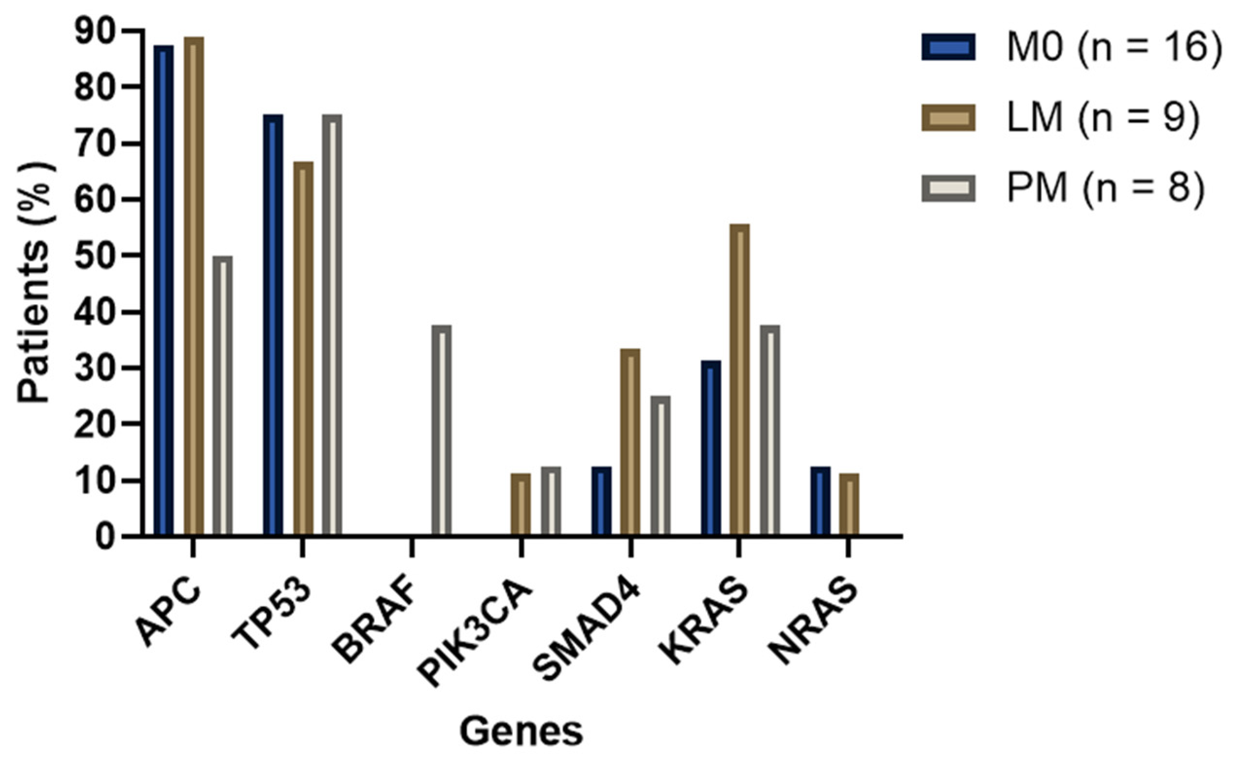
All MSI tumors were excluded for a separate analysis. This resulted in a study population of 33 patients with MSS tumors (M0 N = 16, LM N = 9, and PM N = 8). A total of 164 (likely) pathogenic genetic alterations were detected in 78 genes (Figure 1). Missense, frameshift, and nonsense mutations were most commonly detected. When comparing the occurrence of all variant types, no significant differences were found. The distribution among cancer genes related to CRC was investigated (Figure 2). APC mutations occurred most frequently; in 4/8 (50%) of the PM cases and 8/9 (89, 89%) LM and 14/16 (87, 50%) M0 patients (not significant). BRAF (c.1799T>A p.(V600E) exon 15) mutations were only present in PM patients in this cohort (3/8 = 37.5%, p value = 0.010). None of the M0 samples were carrying PIK3CA mutations after MSI exclusion, and none of the PM samples were carrying NRAS mutations, although these findings were not significantly different. A detailed overview of all MSS subgroup comparisons with statistical p values can be found in the Supplementary Section S1, Table S4.

2.2.2. Additional Analyses

Pathways, molecular functions, and biological processes were not significantly different between the three CRC subgroups. Also, after the additional inclusion of all identified variants of uncertain significance (VUSs), no significant differences were found between the subgroups. A detailed overview of all additional data analyses can be requested via the corresponding author.

2.3. RNA Sequencing

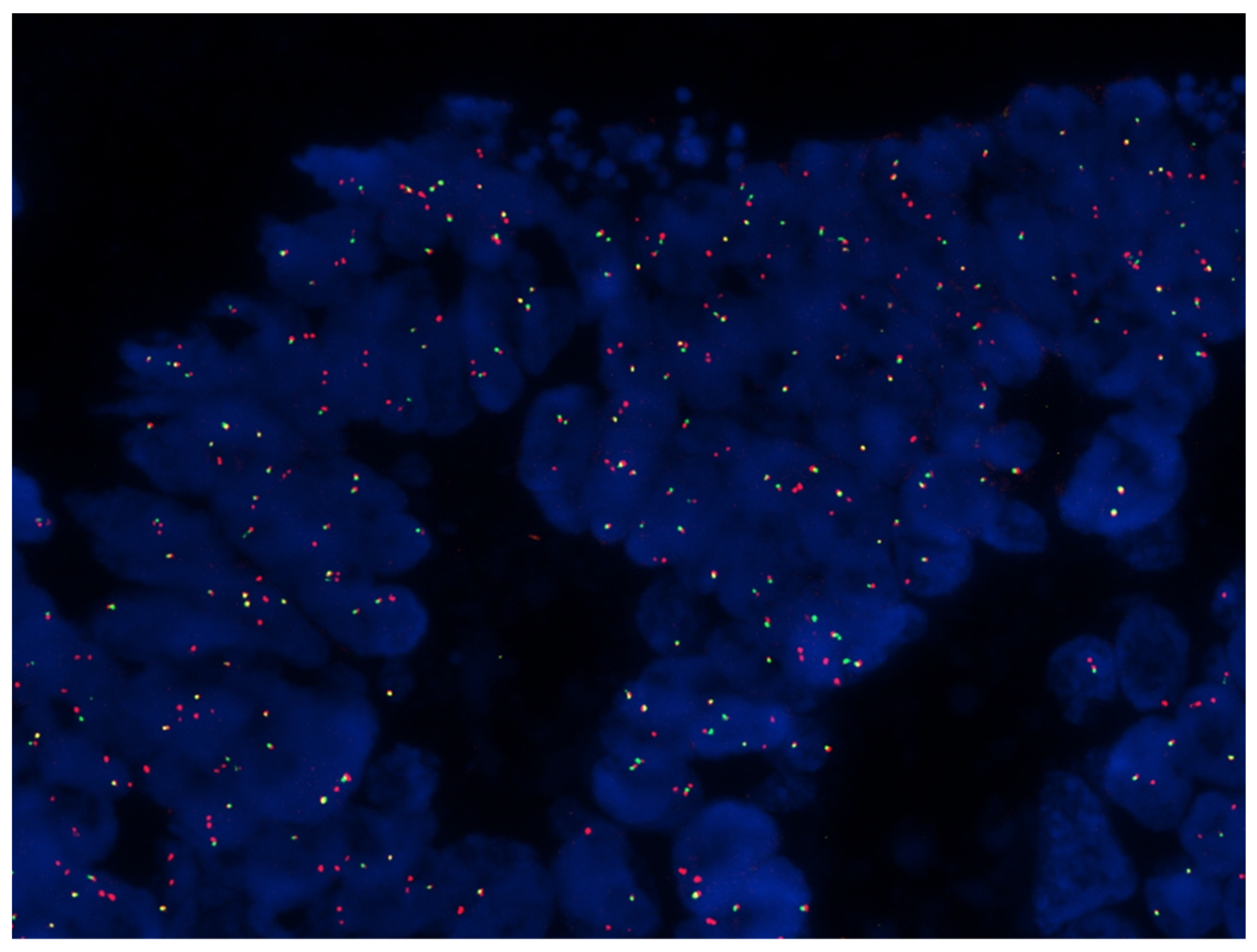
RNA sequencing was performed on 28 samples, divided as follows: M0 (n = 10), LM (n = 9), and PM (n = 9). Data analysis revealed no splice variants for the genes in the panel, whereas three samples (one M0 and two PM samples) showed gene fusion transcripts, which are summarized in Table 2. Interestingly, two gene fusions were identified which can be considered driving mutations, i.e., FAM198A-RAF1 and TARSL2-NTRK3. The NTRK3 fusion was confirmed via fluorescence in situ hybridization (FISH), using an NTRK3 break-apart probe (Figure 3).

3. Discussion

In this study, we performed an integrated pan-cancer oncology enrichment next-generation sequencing assay (TSO500 analysis) to assess DNA and RNA alterations in 523 and 55 genes, respectively, in primary colorectal adenocarcinomas with or without metachronous PM or LM. Our cohort showed a significant difference in differentiation grade when PM samples were compared to LM and M0 samples, and in the LM group for neoadjuvant treatment. Genetic analysis of all MSS tumors revealed that pathogenic BRAF exon 15 p.(V600E) mutations were exclusively identified in three RAS wildtype tumors with metachronous PM (37.5%, p value = 0.010). RNA sequencing identified a FAM198A-RAF1 fusion in an additional tumor with PM, as well as a TARSL2-NTRK3 fusion in a M0 sample.

3.1. Patient Characteristics and Clinicopathological Variables

We identified two clinicopathological characteristics that were significantly different between the three tumor groups. First, the PM group contained more poor/moderately differentiated tumors, while M0 and LM tumors were more often moderately differentiated. The latter was also shown in an extensive analysis of the association between metachronous PM and clinicopathological characteristics by Zhang et al. [20]. Tumor location is not mentioned in this analysis, although another study reports that right-sided primary colorectal tumors are associated with PM [2]. Only 22% of the PM tumors in our cohort were right sided. Second, the lowest tumor cell percentages were observed in the LM group, which may be explained by the fact that in this group, more patients received neoadjuvant treatment via chemoradiation because of a low rectal primary origin. Other previously described clinicopathological risk factors for the development of metachronous PMs are advanced tumor stage, infiltrative or ulcero-infiltrative tumors, a history of perforation, and obstruction [1,4,21]. A clinical trial investigating the potential of adjuvant HIPEC in high-risk PM patients, based on these clinicopathological risk factors, showed that adjuvant HIPEC did not improve survival as compared to patients receiving systemic adjuvant chemotherapy [22]. In contrast, Arjona-Sánchez et al. concluded that adjuvant HIPEC therapy might be useful in patients with T4 tumors [23]. These outcomes suggest that specific biomarkers identified in the primary tumor might be helpful to further estimate the risk of metastatic spread and the need for preventive adjuvant treatments. As our study population has a semi-advanced tumor stage (T3) without (ulcero-)infiltrative or obstructing tumors, we exclude any influence of these possible clinical–pathological risk factors in our current study.

3.2. DNA and RNA Sequencing

The most frequently mutated cancer genes found in our study include APC, TP53, KRAS, SMAD, NRAS, BRAF, PIK3CA, and SOX9. These genes are well known to be involved in the tumorigenesis of CRC [17]. Prevalence data in the literature on these well-known oncogenes are in line with our findings [15,24,25,26,27,28,29,30,31,32,33,34,35,36,37,38,39,40,41,42,43,44,45]. In addition, 12.5% of our tumors contained MSI, in four cases associated with MLH1 promoter hypermethylation, and in one case with an inactivating MSH6 mutation. This finding is in accordance with the literature, as was the finding that these tumors are often right-sided [46,47,48]. MSI results from the inactivation of the mismatch repair genes (MMR), which leads to the accumulation of somatic mutations, genomic instability, and cancer-associated alterations [31]. TMB represents the total number of mutations per Mb found in the DNA of tumor cells and is therefore often significantly higher in MSI tumors. In this study, the five tumors with MSI all had a high TMB (IQR 49.45–Q3 180.60).
It has been suggested that MSI status may be useful as a predictor of the risk of developing metachronous CRC, because it can cause a further increase in metastatic potential [24,46]. However, we did not observe a higher incidence of MSI tumors in our CRC cohort that developed metastases. Interestingly, BRAF p.V600E mutations were found to be exclusively present in PM patients with RAS wildtype MSS tumors (37.5%, p value = 0.010). Approximately 10–14% of all CRC cases have BRAF-activating mutations [24,31,49]. BRAF encodes a serine/threonine protein kinase, which plays an important role in the mitogen-activated protein kinase (MAPK) pathway. This pathway drives cell proliferation, differentiation, migration, survival, and angiogenesis, and therefore, changes in this pathway are associated with tumorigenesis [49]. The BRAF p.V600 mutation, caused by a transversion in exon 15 resulting in a valine amino acid substitution [50], accounts for more than 90–95% of BRAF mutations [31,49] and is associated with poor overall survival [24]. In addition, we identified a FAM198A-RAF1 fusion in one PM sample. Both RAF1 and BRAF belong to the RAF family of protein kinases playing a role in MAPK signaling. Previous studies suggested that BRAF p.V600 mutant tumors are more likely to develop PM [15,32,35,36,37,51]. Therefore, we and some authors recommend analyzing BRAF mutation for its prognostic value in primary T3 CRC [31,34].
The clinical significance of NTRK3 fusion identified in our study, in the setting of CRC, as well as the possibility for targeted treatments should be explored in the future.
Prior to our explorative study, we performed a systematic review to summarize the current knowledge on genetics and genomics in CRC-PM [19]. An NGS analysis with 409 cancer genes showed several additional genetic mutations, i.e., ARID1A, PKHD1, UBR5, PAX5, TP53, ASXL1, and AR, presumably associated with PM [52]. In our TSO500 NGS panel, ARID1A, PAX5, TP53, ASXL1, and AR were included as well. AR and ARID1A mutations occurred in one PM (11%) and one M0 sample (5%). Only one PAX5 mutation was found in one M0 patient, and ASXL1 mutations were not detected. Thereby, the suggested genes related to PM by Lee et al. are not confirmed in our paper. The latter may be explained by the difference in study population; Lee et al. included patients with small obstructing adenocarcinomas (≤3 cm) with synchronous or metachronous PM and compared them with large non-obstructing tumors without PM. Another explanation could be our small sample size. Other authors describe NEK2, MACC1, REG1A, KIF18A, RET, and TIP60 as possible PM-related cancer genes [53,54,55,56,57,58]. In our TSO500 panel, only RET was investigated. In contrast to the suggestion of Yang et al. concerning the association of RET mutations and PM, we did not identify any mutation in this gene in our cohort [57].
Another factor that can contribute to the difficulty of finding biomarkers is the genetic differences between the primary tumor and metastatic lesions. Studies investigating the differences between peritoneal lesions and their primary tumors reported some small unique differences [59], whilst other studies report high concordance [60,61,62]. A very recent study by Lenos et al. showed that peritoneal lesions seemed to have much more similarity to their primary tumor compared to other metastases, and these lesions seemed to retain both clonal heterogeneity and transcriptional profile [61].
A new way to look at CRC tumors is through dividing them into subtypes, for example, the previously described four consensus molecular subtypes (CMS 1–4). These subtypes aid in prognostication as well as in determining treatment strategies for individual patients based on the mutations, activated pathways, and phenotypic characteristics and responses to treatment of other tumors with similar signatures [63]. The majority of PMs in their study are of the CMS4 subtype, known as the mesenchymal subgroup [64,65]. CMS4 is presented in 23% of CRC cases, which are most often distal tumors with poor relapse-free and overall survival and harbor prominent transforming growth factor β activation, stromal infiltration, and angiogenesis [66,67]. CMS4 tumors have extremely low levels of hypermutation, MSS, and very high somatic copy number alteration counts [63]. The latter was also seen in our cohort. Unfortunately, we were not able to examine all of these characteristics in our study due to the limited content of our RNA NGS gene panel. Therefore, the translation to CMS subgroups was not possible in our study.

3.3. Treatment Options and Future Perspectives

BRAF mutations can be considered as an independent negative prognostic factor in early stage MSS tumors and as a negative predictive factor for therapeutic approaches [49]. The therapeutic approach to treat BRAF-mutated tumors is not straightforward due to its resistance to standard therapies [49]. Research into anti-epidermal growth factor receptor (EGFR) and anti-vascular endothelial growth factor (VEGF) antibodies has not shown statistical benefits in BRAF-mutated patients [49,68]. BRAF inhibitors (iBRAF) have revolutionized the treatment of BRAF V600E metastatic melanoma, but so far, results in CRC patients are disappointing due to resistance [34,49,69]. Studies are currently ongoing with dual or triple drug therapy to blockade the MAPK pathway [49,69]. Until now, partial activity of different combinations has been shown, but this is far from the promising results in melanoma patients. Ongoing research will hopefully demonstrate that combination strategies with iBRAF and other drugs can overcome the lack of efficacy [49]. As survival is about half as long as that of BRAF wildtype patients [68], there is an urgency to unravel new treatments that improve BRAF-mutant CRC patients’ outcomes.
In current clinical practice, the classification of the MSI status is the only genetic test that is routinely performed in CRC patients to decide adjuvant therapy decisions [70]. Other genetic tests, such as BRAF mutation status, are only evaluated in metastatic tumors. Based on the results of this paper, we believe greater attention should be paid to BRAF-mutated tumors in relation to the development of metachronous PM in CRC patients without metastases. Standard clinical screening for BRAF mutations might feel too early as it does not offer any new treatment options, but a stricter follow-up in this population may be clinically beneficial. Based on new international guidelines, the first follow-up CT scan is not performed until 12 months after primary surgery. However, in a BRAF-mutated population, earlier follow-up imaging and more clinical monitoring for PM development may be warranted. Of course, future prospective research (e.g., with liquid biopsies) into the validation of BRAF mutations in relation to the development of metachronous PM is needed to substantiate this proposition.

3.4. Strengths and Limitations

A very homogenous group of tumors was selected for genetic analysis. To our knowledge, this is the first study investigating T3 tumors in relation to metachronous CRC metastases. Previous studies focused on T4 tumors with mostly synchronous PMs and had no other metastases group (LM) as a comparator. While PMs may develop from different cancer types, we specifically examined the colorectal origin and excluded appendiceal origin as it is known that gene expression from appendiceal tumors is distinct from CRC [59]. Due to refinements in DNA and RNA extraction techniques from formalin-fixed paraffin-embedded (FFPE) tissue material, the sensitivity of DNA and RNA testing has been increased. Our targeted TSO500 NGS technique accurately measures TMB, microsatellite instability, single-nucleotide variants, indels, copy-number/structural variation, and gene fusions in a single assay using relatively small amounts of DNA and RNA as input. Combining DNA and RNA hybrid-capture with sophisticated informatics reduces errors and yields high-quality data, even from FFPE samples.
We did not perform an extensive sample size calculation due to the predictive and explorative character of this study. Despite the efforts made to create as much homogeneity between the three groups as possible, the number of patients in our cohort is small. A larger-scale study should be conducted to confirm the mutation differences in relation to PMs. Thereby, being a retrospective study, there is a likelihood of selection bias and information bias. Additionally, we performed a very broad cancer gene analysis with our TSO500 panel, although the method does not cover all genes. Through performing whole exome or genome sequencing (WES or WGS), potential candidate genes that can act as a predictive PM biomarker that are not included in the TSO500 panel may be identified. Unfortunately, WES and/or WGS are more expensive and have additional logistic limitations.

4. Materials and Methods

This study was conducted in a collaboration between the Maastricht University Medical Centre (MUMC+) and Catharina Hospital Eindhoven (CZE). The study was approved by the Institutional Medical Ethics Committee from MUMC+ (nr. 2021-2888) and CZE (nr. 2021-089) and conducted according to the Declaration of Helsinki.

4.1. Patients

The medical records of patients who underwent curative resection between 1 January 2012 and 31 December 2021 for colorectal adenocarcinoma were retrospectively reviewed. The research team deliberately chose to include a maximum of 40 patients in this pilot study, based on clinical prediction modeling which states at least 10 persons with the event (development of PM or LM) and 10 persons without the event (no metastases within 5 years) per included variable in the prediction model to obtain sufficient power [71]. Patients with T3 tumors were classified into three groups: those who had developed metachronous PMs (n = 10); those who had developed metachronous liver metastases (LM, n = 10); and those who never developed metastatic disease within 5 years after primary surgery (M0, n = 20). Patients with metachronous PMs were not allowed to be diagnosed with metachronous LMs and vice versa. As T4 tumors penetrate the surface of the visceral peritoneum and directly invade other organs or structures, the risk of spread into the peritoneal cavity is higher. Therefore, we only included T3 tumors and deliberately excluded T1 and T2 tumors to create a homogenous population. Patients with synchronous disease were excluded. Patients in the LM and PM group had no signs of metastases during resection of the primary tumor but were diagnosed with PM or LM during follow-up, at least 6 months after initial surgery. Patients in the M0 group did not develop any type of metastases during the follow-up period of at least 5 years. All in- and exclusion criteria are summarized in Table 3. Patient record files were screened, and the first 40 patients who met inclusion criteria were contacted. Informed consent was obtained from all participants. Demographics, pre-operative, operative, and follow-up data of all patients were retrospectively retrieved from medical records.

4.2. Tumor Samples

Primary tumor FFPE tissue samples were obtained from MUMC+ and CZE. From each FFPE tissue specimen, 10 paraffin sections of 5 μm were cut. Hematoxylin and eosin (H&E) staining was performed. An experienced pathologist (J.B.) marked the tumor circumflex and estimated the tumor cell percentage under the microscope. Only samples with ≥10% tumor cell percentage were considered eligible for further analysis. Subsequently, microdissection with a pointed surgical blade was performed.
DNA and RNA were extracted and isolated using a Maxwell RSC® System for Genomic DNA or RNA Extraction with a FFPE AS1450 kit and FFPE AS1440 kit, respectively (Promega, Madison, WI, USA). A blank control sample was analyzed in parallel to each set of samples. A minimum amount of 40 ng DNA or RNA was necessary for further analysis. DNA samples were stored at 4 °C and RNA samples at −80 °C. Fragment analysis of both DNA and RNA samples was performed as quality control. For DNA, a PCR was performed to visualize all DNA fragments. For RNA, the samples were assessed using a 4150 TapeStation system, which separates nucleic acids through electrophoresis. All fragments needed to be at least 200 bp in length.

4.3. TruSight Oncology 500 Analysis

TSO500 is an NGS assay that enables the comprehensive genomic profiling of tumor samples. The TSO500 panel (20028216; Illumina, Hayward, San Diego, CA, USA) was used to detect mutations and identify other relative pan-cancer genes in the tumor samples, as previously described by Verkouteren et al. [72]. The analysis includes 523 genes for mutations (all for single-nucleotide variants (SNVs)) and 59 for copy number variations (CNVs) (amplifications, insertions, and deletions). In addition, the assay allows for the identification of MSI and TMB. Besides DNA analysis, 55 genes are screened for fusion and splice variants on the RNA level. All genes included in the TSO 500 panel can be found in Supplementary Section S1, Figure S3. DNA and RNA processing and the generation of library preparations were performed according to the manufacturers’ instructions. Data analysis was performed using the TSO500 Local App (Illumina, Hayward, San Diego, CA, USA). For DNA analysis, additional thresholds were maintained. First, for variant allele frequency, a percentage of ≥5% was maintained. Second, for classification as an amplification, a fold change of ≥3 was maintained. Third, the threshold for classification as MSI-high was ≥20% of microsatellite sites being unstable. Fourth, a threshold of ≥15 mutations per megabase (mut/Mb) was used to define high TMB. Variants were classified subsequently using the inline Varsome application (access via https://varsome.com). Only pathogenic and likely pathogenic variants were included for further analysis; variants of uncertain significance (VUSs) were excluded.

4.4. Statistical Analysis

Gene mutation frequencies and associations between the found mutations and pathological patient characteristics were estimated. Analysis of both the total cohort (with MSI samples) as well as MSS samples only were performed. Numerical variables were presented as medians with interquartile range (IQR) as appropriate. For categorical variables, the number of patients and percentage were used. To evaluate the statistical significance of numerical variable differences observed between groups, non-parametric tests (Kruskal–Wallis and Mann–Whitney U-tests) were applied. Differences in categorical variables were tested using the Fisher–Freeman–Halton test and Fisher’s exact test. Bonferroni correction for multiple comparison was applied to significant outcomes. All tests were two-sided, and differences were considered significant when the p value was <0.05. All the statistical analyses were performed with SPSS (IBM SPSS Statistics for Apple, Version 27, Armonk, NY, USA). In addition, an analysis with protein analysis through evolutionary relationships (PANTHER) was performed [73]. The latter was performed for Gene Ontology molecular functions and biological processes [74,75], for PANTHER pathways, and for Reactome pathways [76]. For each sample, the significantly enriched terms were extracted for subsequent analyses using R (R core team, version 4.2.0., Vienna, Austria). Analysis and visualization of the genetic outcomes were carried out with Python (Matplotlib v3.7.0, Salt Lake, UT, USA) and GraphPad Prism (GraphPad software for Apple, version 8.0.0, San Diego, CA, USA).

5. Conclusions

Over the last decade, the genetic analysis of CRC has evolved enormously, resulting in better tumor classifications, improved treatment decisions, and finally enabling personalized treatment options. Specific genetic changes and mutations that could predict PM remain largely unknown. In our cohort, we identified genes that have not been described in relation to metachronous PMs, or metastases in general, before. The clinical significance of this finding remains unknown due to the small sample size. BRAF V600E mutations were only present in PM patients with MSS tumors. We believe greater attention should be paid to BRAF-mutated tumors in relation to the development of metachronous PMs. Future prospective research into and validation of the molecular players identified here, specifically within non-synchronous tumors, might influence the efficacy of existing and future diagnostic (biomarker identification), prognostic (patient grouping and recurrence), and therapeutic (molecular) actions.

Supplementary Materials

The supporting information can be downloaded at: https://www.mdpi.com/article/10.3390/ijms241612830/s1.

Author Contributions

Conceptualization, D.J.I.H., A.G.W.E.W., L.M., S.M.E.E., I.H.J.T.d.H., E.-J.M.S. and N.D.B.; data curation, D.J.I.H.; formal analysis, D.J.I.H., S.G.M.T. and D.J.G.L.; funding acquisition, D.J.I.H., A.G.W.E.W., S.M.E.E., I.H.J.T.d.H., L.P.S.S., D.K., Z.M., E.-J.M.S. and N.D.B.; methodology, D.J.I.H., A.G.W.E.W., L.M. and E.-J.M.S.; project administration, N.D.B.; software, D.J.G.L. and E.-J.M.S.; supervision, E.-J.M.S. and N.D.B.; validation, J.B.; visualization, D.J.I.H.; writing—original draft, D.J.I.H. and A.G.W.E.W.; writing—review and editing, L.M., S.M.E.E., I.H.J.T.d.H., L.B.V.-v.I., M.d.D., J.B., S.G.M.T., D.J.G.L., L.P.S.S., D.K., Z.M., E.-J.M.S. and N.D.B. All authors have read and agreed to the published version of the manuscript.

Funding

This work was supported by the Stichting Jules Coenegracht Sr. RSIN 8148.99.110.

Institutional Review Board Statement

The study was conducted according to the guidelines of the Declaration of Helsinki and was approved by the Institutional Medical Ethics Committee from MUMC+ (nr. 2021-2888) and CZE (nr. 2021-089).

Informed Consent Statement

Informed consent was obtained from all subjects involved in the study via an informed consent form signed before participation.

Data Availability Statement

Data supporting this study are included within Supporting Materials. More information can be gained through contacting the corresponding author.

Acknowledgments

Bjorn Winkens was involved as a professional statistician to ensure an appropriate statistical analysis. We would like to thank all analysts from the pathology department for contributing to this project.

Conflicts of Interest

The authors have no conflict of interest to declare related to the current study. D.K. has received research funding from Grunenthal, Allergan, Will Pharma, UEG, MLDS, Rome Foundation, ZonMw, Horizon 2020, and has received speaker’s fees (paid to host institution) from Falk. Z.M. reports grants from Niels Stensen Fellowship, MLDS, ZonMW, and Galapagos and consulting fees from Johnson and Johnson and Pfizer, all outside the submitted work.

References

  1. van Gestel, Y.R.; Thomassen, I.; Lemmens, V.E.; Pruijt, J.F.; van Herk-Sukel, M.P.; Rutten, H.J.; Creemers, G.J.; de Hingh, I.H. Metachronous peritoneal carcinomatosis after curative treatment of colorectal cancer. Eur. J. Surg. Oncol. 2014, 40, 963–969. [Google Scholar] [CrossRef]
  2. Simkens, G.A.; Wintjens, A.; Rovers, K.P.; Nienhuijs, S.W.; de Hingh, I.H. Effective Strategies to Predict Survival of Colorectal Peritoneal Metastases Patients Eligible for Cytoreductive Surgery and HIPEC. Cancer Manag. Res. 2021, 13, 5239–5249. [Google Scholar] [CrossRef]
  3. Lurvink, R.J.; Bakkers, C.; Rijken, A.; van Erning, F.N.; Nienhuijs, S.W.; Burger, J.W.; Creemers, G.J.; Verhoef, C.; Lemmens, V.E.; De Hingh, I.H. Increase in the incidence of synchronous and metachronous peritoneal metastases in patients with colorectal cancer: A nationwide study. Eur. J. Surg. Oncol. 2021, 47, 1026–1033. [Google Scholar] [CrossRef] [PubMed]
  4. Koppe, M.J.; Boerman, O.C.; Oyen, W.J.; Bleichrodt, R.P. Peritoneal carcinomatosis of colorectal origin: Incidence and current treatment strategies. Ann. Surg. 2006, 243, 212–222. [Google Scholar] [CrossRef] [PubMed]
  5. Klaver, Y.L.; Lemmens, V.E.; Nienhuijs, S.W.; Luyer, M.D.; de Hingh, I.H. Peritoneal carcinomatosis of colorectal origin: Incidence, prognosis and treatment options. World J. Gastroenterol. 2012, 18, 5489–5494. [Google Scholar] [CrossRef] [PubMed]
  6. Kranenburg, O.; van der Speeten, K.; de Hingh, I. Peritoneal Metastases from Colorectal Cancer: Defining and Addressing the Challenges. Front. Oncol. 2021, 11, 650098. [Google Scholar] [CrossRef]
  7. Kamiyama, H.; Noda, H.; Konishi, F.; Rikiyama, T. Molecular biomarkers for the detection of metastatic colorectal cancer cells. World J. Gastroenterol. 2014, 20, 8928–8938. [Google Scholar]
  8. Sadahiro, S.; Suzuki, T.; Ishikawa, K.; Nakamura, T.; Tanaka, Y.; Masuda, T.; Mukoyama, S.; Yasuda, S.; Tajima, T.; Makuuchi, H.; et al. Recurrence patterns after curative resection of colorectal cancer in patients followed for a minimum of ten years. Hepatogastroenterology 2003, 50, 1362–1366. [Google Scholar]
  9. Maggiori, L.; Elias, D. Curative treatment of colorectal peritoneal carcinomatosis: Current status and future trends. Eur. J. Surg. Oncol. 2010, 36, 599–603. [Google Scholar] [CrossRef] [Green Version]
  10. Jayne, D.G.; Fook, S.; Loi, C.; Seow-Choen, F. Peritoneal carcinomatosis from colorectal cancer. Br. J. Surg. 2002, 89, 1545–1550. [Google Scholar] [CrossRef]
  11. Quénet, F.; Elias, D.; Roca, L.; Goéré, D.; Ghouti, L.; Pocard, M.; Facy, O.; Arvieux, C.; Lorimier, G.; Pezet, D.; et al. Cytoreductive surgery plus hyperthermic intraperitoneal chemotherapy versus cytoreductive surgery alone for colorectal peritoneal metastases (PRODIGE 7): A multicentre, randomised, open-label, phase 3 trial. Lancet Oncol. 2021, 22, 256–266. [Google Scholar] [CrossRef] [PubMed]
  12. Bakkers, C.; Lurvink, R.J.; Rijken, A.; Nienhuijs, S.W.; Kok, N.F.; Creemers, G.J.; Verhoef, C.; Lemmens, V.E.; van Erning, F.N.; De Hingh, I.H. Treatment Strategies and Prognosis of Patients with Synchronous or Metachronous Colorectal Peritoneal Metastases: A Population-Based Study. Ann. Surg. Oncol. 2021, 28, 9073–9083. [Google Scholar] [CrossRef]
  13. Xue, L.; Hyman, N.H.; Turaga, K.K.; Eng, O.S. Peritoneal Metastases in Colorectal Cancer: Biology and Barriers. J. Gastrointest. Surg. 2020, 24, 720–727. [Google Scholar] [CrossRef]
  14. Sorich, M.J.; Wiese, M.D.; Rowland, A.; Kichenadasse, G.; McKinnon, R.A.; Karapetis, C.S. Extended RAS mutations and anti-EGFR monoclonal antibody survival benefit in metastatic colorectal cancer: A meta-analysis of randomized, controlled trials. Ann. Oncol. 2015, 26, 13–21. [Google Scholar] [CrossRef] [PubMed]
  15. Taniguchi, H.; Uehara, K.; Nakayama, G.; Nakayama, H.; Aiba, T.; Hattori, N.; Kataoka, M.; Nakano, Y.; Kawase, Y.; Okochi, O.; et al. Tumor Location Is Associated with the Prevalence of Braf And Pik3ca Mutations in Patients with Wild-Type Ras Colorectal Cancer: A Prospective Multi-Center Cohort Study in Japan. Transl. Oncol. 2020, 13, 100786. [Google Scholar] [CrossRef]
  16. Graf, W.; Cashin, P.H.; Ghanipour, L.; Enblad, M.; Botling, J.; Terman, A.; Birgisson, H. Prognostic Impact of BRAF and KRAS Mutation in Patients with Colorectal and Appendiceal Peritoneal Metastases Scheduled for CRS and HIPEC. Ann. Surg. Oncol. 2020, 27, 293–300. [Google Scholar] [CrossRef] [PubMed] [Green Version]
  17. Roth, L.; Russo, L.; Ulugoel, S.; Freire Dos Santos, R.; Breuer, E.; Gupta, A.; Lehmann, K. Peritoneal Metastasis: Current Status and Treatment Options. Cancers 2021, 14, 60. [Google Scholar] [CrossRef]
  18. Schneider, M.A.; Eden, J.; Pache, B.; Laminger, F.; Lopez-Lopez, V.; Steffen, T.; Hübner, M.; Kober, F.; Roka, S.; Campos, P.C.; et al. Mutations of RAS/RAF Proto-oncogenes Impair Survival after Cytoreductive Surgery and HIPEC for Peritoneal Metastasis of Colorectal Origin. Ann. Surg. 2018, 268, 845–853. [Google Scholar] [CrossRef] [Green Version]
  19. Heuvelings, D.J.I.; Wintjens, A.; Luyten, J.; Wilmink, G.; Moonen, L.; Speel, E.M.; de Hingh, I.; Bouvy, N.D.; Peeters, A. DNA and RNA Alterations Associated with Colorectal Peritoneal Metastases: A Systematic Review. Cancers 2023, 15, 549. [Google Scholar] [CrossRef]
  20. Zhang, Y.; Qin, X.; Chen, W.; Liu, D.; Luo, J.; Wang, H.; Wang, H. Risk factors for developing peritoneal metastases after curative surgery for colorectal cancer: A systematic review and meta-analysis. Color. Dis. 2021, 23, 2846–2858. [Google Scholar] [CrossRef]
  21. Segelman, J.; Granath, F.; Holm, T.; Machado, M.; Mahteme, H.; Martling, A. Incidence, prevalence and risk factors for peritoneal carcinomatosis from colorectal cancer. Br. J. Surg. 2012, 99, 699–705. [Google Scholar] [CrossRef] [PubMed]
  22. Klaver, C.E.L.; Wisselink, D.D.; Punt, C.J.A.; Snaebjornsson, P.; Crezee, J.; Aalbers, A.G.J.; Brandt, A.; Bremers, A.J.A.; Burger, J.W.A.; Fabry, H.F.J.; et al. Adjuvant hyperthermic intraperitoneal chemotherapy in patients with locally advanced colon cancer (COLOPEC): A multicentre, open-label, randomised trial. Lancet Gastroenterol. Hepatol. 2019, 4, 761–770. [Google Scholar] [CrossRef] [PubMed]
  23. Arjona-Sánchez, A.; Barrios, P.; Boldo-Roda, E.; Camps, B.; Carrasco-Campos, J.; Concepción Martín, V.; García-Fadrique, A.; Gutiérrez-Calvo, A.; Morales, R.; Ortega-Pérez, G.; et al. HIPECT4: Multicentre, randomized clinical trial to evaluate safety and efficacy of Hyperthermic intra-peritoneal chemotherapy (HIPEC) with Mitomycin C used during surgery for treatment of locally advanced colorectal carcinoma. BMC Cancer 2018, 18, 183. [Google Scholar] [CrossRef] [PubMed]
  24. Karunasena, E.; Sham, J.; McMahon, K.W.; Ahuja, N. Genomics of Peritoneal Malignancies. Surg. Oncol. Clin. N. Am. 2018, 27, 463–475. [Google Scholar] [CrossRef]
  25. Fodde, R. The APC gene in colorectal cancer. Eur. J. Cancer 2002, 38, 867–871. [Google Scholar] [CrossRef]
  26. Zhang, L.; Shay, J.W. Multiple Roles of APC and Its Therapeutic Implications in Colorectal Cancer. JNCI J. Natl. Cancer Inst. 2017, 109, djw332. [Google Scholar] [CrossRef] [Green Version]
  27. Lipsyc, M.; Yaeger, R. Impact of somatic mutations on patterns of metastasis in colorectal cancer. J. Gastrointest. Oncol. 2015, 6, 645–649. [Google Scholar] [CrossRef]
  28. Lan, Y.T.; Jen-Kou, L.; Lin, C.H.; Yang, S.H.; Lin, C.C.; Wang, H.S.; Chen, W.S.; Lin, T.C.; Jiang, J.K.; Chang, S.C. Mutations in the RAS and PI3K pathways are associated with metastatic location in colorectal cancers. J. Surg. Oncol. 2015, 111, 905–910. [Google Scholar] [CrossRef]
  29. He, K.; Wang, Y.; Zhong, Y.; Pan, X.; Si, L.; Lu, J. KRAS Codon 12 Mutation is Associated with More Aggressive Invasiveness in Synchronous Metastatic Colorectal Cancer (mCRC): Retrospective Research. OncoTargets Ther. 2020, 13, 12601–12613. [Google Scholar] [CrossRef]
  30. Zihui Yong, Z.; Ching, G.T.H.; Ching, M.T.C. Metastatic Profile of Colorectal Cancer: Interplay between Primary Tumor Location and KRAS Status. J. Surg. Res. 2020, 246, 325–334. [Google Scholar] [CrossRef]
  31. Oh, H.H.; Joo, Y.E. Novel biomarkers for the diagnosis and prognosis of colorectal cancer. Intest. Res. 2020, 18, 168–183. [Google Scholar] [CrossRef] [PubMed]
  32. Christensen, T.D.; Palshof, J.A.; Larsen, F.O.; Poulsen, T.S.; Høgdall, E.; Pfeiffer, P.; Jensen, B.V.; Yilmaz, M.K.; Nielsen, D. Associations between primary tumor RAS, BRAF and PIK3CA mutation status and metastatic site in patients with chemo-resistant metastatic colorectal cancer. Acta Oncol. 2018, 57, 1057–1062. [Google Scholar] [CrossRef] [PubMed] [Green Version]
  33. Smith, C.G.; Fisher, D.; Claes, B.; Maughan, T.S.; Idziaszczyk, S.; Peuteman, G.; Harris, R.; James, M.D.; Meade, A.; Jasani, B.; et al. Somatic profiling of the epidermal growth factor receptor pathway in tumors from patients with advanced colorectal cancer treated with chemotherapy ± cetuximab. Clin. Cancer Res. 2013, 19, 4104–4113. [Google Scholar] [CrossRef] [PubMed] [Green Version]
  34. Sepulveda, A.R.; Hamilton, S.R.; Allegra, C.J.; Grody, W.; Cushman-Vokoun, A.M.; Funkhouser, W.K.; Kopetz, S.E.; Lieu, C.; Lindor, N.M.; Minsky, B.D.; et al. Molecular Biomarkers for the Evaluation of Colorectal Cancer: Guideline from the American Society for Clinical Pathology, College of American Pathologists, Association for Molecular Pathology, and American Society of Clinical Oncology. Arch. Pathol. Lab. Med. 2017, 141, 625–657. [Google Scholar] [CrossRef] [Green Version]
  35. Yokota, T.; Ura, T.; Shibata, N.; Takahari, D.; Shitara, K.; Nomura, M.; Kondo, C.; Mizota, A.; Utsunomiya, S.; Muro, K.; et al. BRAF mutation is a powerful prognostic factor in advanced and recurrent colorectal cancer. Br. J. Cancer 2011, 104, 856–862. [Google Scholar] [CrossRef] [Green Version]
  36. Cheng, H.H.; Lin, J.K.; Chen, W.S.; Jiang, J.K.; Yang, S.H.; Chang, S.C. Clinical significance of the BRAFV600E mutation in Asian patients with colorectal cancer. Int. J. Color. Dis. 2018, 33, 1173–1181. [Google Scholar] [CrossRef]
  37. Sayagués, J.M.; Del Carmen, S.; Del Mar Abad, M.; Corchete, L.A.; Bengoechea, O.; Anduaga, M.F.; Baldeón, M.J.; Cruz, J.J.; Alcazar, J.A.; Angoso, M.; et al. Combined assessment of the TNM stage and BRAF mutational status at diagnosis in sporadic colorectal cancer patients. Oncotarget 2018, 9, 24081–24096. [Google Scholar] [CrossRef] [Green Version]
  38. Sanz-Pamplona, R.; Lopez-Doriga, A.; Paré-Brunet, L.; Lázaro, K.; Bellido, F.; Alonso, M.H.; Aussó, S.; Guinó, E.; Beltrán, S.; Castro-Giner, F.; et al. Exome Sequencing Reveals AMER1 as a Frequently Mutated Gene in Colorectal Cancer. Clin. Cancer Res. 2015, 21, 4709–4718. [Google Scholar] [CrossRef] [Green Version]
  39. Fang, L.; Ford-Roshon, D.; Russo, M.; O’Brien, C.; Xiong, X.; Gurjao, C.; Grandclaudon, M.; Raghavan, S.; Corsello, S.M.; Carr, S.A.; et al. RNF43 G659fs is an oncogenic colorectal cancer mutation and sensitizes tumor cells to PI3K/mTOR inhibition. Nat. Commun. 2022, 13, 3181. [Google Scholar] [CrossRef]
  40. Giannakis, M.; Hodis, E.; Jasmine Mu, X.; Yamauchi, M.; Rosenbluh, J.; Cibulskis, K.; Saksena, G.; Lawrence, M.S.; Qian, Z.R.; Nishihara, R.; et al. RNF43 is frequently mutated in colorectal and endometrial cancers. Nat. Genet. 2014, 46, 1264–1266. [Google Scholar] [CrossRef] [PubMed]
  41. Randon, G.; Fucà, G.; Rossini, D.; Raimondi, A.; Pagani, F.; Perrone, F.; Tamborini, E.; Busico, A.; Peverelli, G.; Morano, F.; et al. Prognostic impact of ATM mutations in patients with metastatic colorectal cancer. Sci. Rep. 2019, 9, 2858. [Google Scholar] [CrossRef] [PubMed] [Green Version]
  42. Li, X.; Oh, S.; Song, H.; Shin, S.; Zhang, B.; Freeman, W.M.; Janknecht, R. A potential common role of the Jumonji C domain-containing 1A histone demethylase and chromatin remodeler ATRX in promoting colon cancer. Oncol. Lett. 2018, 16, 6652–6662. [Google Scholar] [CrossRef] [PubMed] [Green Version]
  43. AACR Project GENIE Consortium. AACR Project GENIE: Powering Precision Medicine through an International Consortium. Cancer Discov. 2017, 7, 818–831. [Google Scholar] [CrossRef] [Green Version]
  44. Poulogiannis, G.; McIntyre, R.E.; Dimitriadi, M.; Apps, J.R.; Wilson, C.H.; Ichimura, K.; Luo, F.; Cantley, L.C.; Wyllie, A.H.; Adams, D.J.; et al. PARK2 deletions occur frequently in sporadic colorectal cancer and accelerate adenoma development in Apc mutant mice. Proc. Natl. Acad. Sci. USA 2010, 107, 15145–15150. [Google Scholar] [CrossRef]
  45. Bhat, Z.I.; Kumar, B.; Bansal, S.; Naseem, A.; Tiwari, R.R.; Wahabi, K.; Sharma, G.D.; Alam Rizvi, M.M. Association of PARK2 promoter polymorphisms and methylation with colorectal cancer in North Indian population. Gene 2019, 682, 25–32. [Google Scholar] [CrossRef]
  46. Gonzalez-Pons, M.; Cruz-Correa, M. Colorectal Cancer Biomarkers: Where Are We Now? Biomed. Res. Int. 2015, 2015, 149014. [Google Scholar] [CrossRef] [PubMed] [Green Version]
  47. Baran, B.; Mert Ozupek, N.; Yerli Tetik, N.; Acar, E.; Bekcioglu, O.; Baskin, Y. Difference between Left-Sided and Right-Sided Colorectal Cancer: A Focused Review of Literature. Gastroenterol. Res. 2018, 11, 264–273. [Google Scholar] [CrossRef] [PubMed] [Green Version]
  48. Akce, M.; Zakka, K.; Jiang, R.; Williamson, S.; Alese, O.; Shaib, W.; Wu, C.; Behera, M.; El-Rayes, B. Impact of Tumor Side on Clinical Outcomes in Stage II and III Colon Cancer with Known Microsatellite Instability Status. Front. Oncol. 2021, 11, 592351. [Google Scholar] [CrossRef] [PubMed]
  49. Caputo, F.; Santini, C.; Bardasi, C.; Cerma, K.; Casadei-Gardini, A.; Spallanzani, A.; Andrikou, K.; Cascinu, S.; Gelsomino, F. BRAF-Mutated Colorectal Cancer: Clinical and Molecular Insights. Int. J. Mol. Sci. 2019, 20, 5369. [Google Scholar] [CrossRef] [Green Version]
  50. Cantwell-Dorris, E.R.; O’Leary, J.J.; Sheils, O.M. BRAFV600E: Implications for carcinogenesis and molecular therapy. Mol. Cancer Ther. 2011, 10, 385–394. [Google Scholar] [CrossRef] [Green Version]
  51. Tran, B.; Kopetz, S.; Tie, J.; Gibbs, P.; Jiang, Z.Q.; Lieu, C.H.; Agarwal, A.; Maru, D.M.; Sieber, O.; Desai, J. Impact of BRAF mutation and microsatellite instability on the pattern of metastatic spread and prognosis in metastatic colorectal cancer. Cancer 2011, 117, 4623–4632. [Google Scholar] [CrossRef] [PubMed] [Green Version]
  52. Lee, J.H.; Ahn, B.K.; Baik, S.S.; Lee, K.H. Comprehensive Analysis of Somatic Mutations in Colorectal Cancer with Peritoneal Metastasis. In Vivo 2019, 33, 447–452. [Google Scholar] [CrossRef] [PubMed] [Green Version]
  53. Astrosini, C.; Roeefzaad, C.; Dai, Y.Y.; Dieckgraefe, B.K.; Jöns, T.; Kemmner, W. REG1A expression is a prognostic marker in colorectal cancer and associated with peritoneal carcinomatosis. Int. J. Cancer 2008, 123, 409–413. [Google Scholar] [CrossRef] [PubMed]
  54. Takahashi, Y.; Iwaya, T.; Sawada, G.; Kurashige, J.; Matsumura, T.; Uchi, R.; Ueo, H.; Takano, Y.; Eguchi, H.; Sudo, T.; et al. Up-regulation of NEK2 by microRNA-128 methylation is associated with poor prognosis in colorectal cancer. Ann. Surg. Oncol. 2014, 21, 205–212. [Google Scholar] [CrossRef] [PubMed]
  55. Shirahata, A.; Shinmura, K.; Kitamura, Y.; Sakuraba, K.; Yokomizo, K.; Goto, T.; Mizukami, H.; Saito, M.; Ishibashi, K.; Kigawa, G.; et al. MACC1 as a marker for advanced colorectal carcinoma. Anticancer Res. 2010, 30, 2689–2692. [Google Scholar] [PubMed]
  56. Nagahara, M.; Nishida, N.; Iwatsuki, M.; Ishimaru, S.; Mimori, K.; Tanaka, F.; Nakagawa, T.; Sato, T.; Sugihara, K.; Hoon, D.S.; et al. Kinesin 18A expression: Clinical relevance to colorectal cancer progression. Int. J. Cancer 2011, 129, 2543–2552. [Google Scholar] [CrossRef]
  57. Yang, Y.Z.; Hu, W.M.; Xia, L.P.; He, W.Z. Association between somatic RET mutations and clinical and genetic characteristics in patients with metastatic colorectal cancer. Cancer Med. 2021, 10, 8876–8882. [Google Scholar] [CrossRef]
  58. Sakuraba, K.; Yasuda, T.; Sakata, M.; Kitamura, Y.H.; Shirahata, A.; Goto, T.; Mizukami, H.; Saito, M.; Ishibashi, K.; Kigawa, G.; et al. Down-regulation of Tip60 gene as a potential marker for the malignancy of colorectal cancer. Anticancer Res. 2009, 29, 3953–3955. [Google Scholar]
  59. Stein, M.K.; Williard, F.W.; Xiu, J.; Tsao, M.W.; Martin, M.G.; Deschner, B.W.; Dickson, P.V.; Glazer, E.S.; Yakoub, D.; Shibata, D.; et al. Comprehensive tumor profiling reveals unique molecular differences between peritoneal metastases and primary colorectal adenocarcinoma. J. Surg. Oncol. 2020, 121, 1320–1328. [Google Scholar] [CrossRef]
  60. Brannon, A.R.; Vakiani, E.; Sylvester, B.E.; Scott, S.N.; McDermott, G.; Shah, R.H.; Kania, K.; Viale, A.; Oschwald, D.M.; Vacic, V.; et al. Comparative sequencing analysis reveals high genomic concordance between matched primary and metastatic colorectal cancer lesions. Genome Biol. 2014, 15, 454. [Google Scholar] [CrossRef]
  61. Lenos, K.J.; Bach, S.; Ferreira Moreno, L.; Ten Hoorn, S.; Sluiter, N.R.; Bootsma, S.; Vieira Braga, F.A.; Nijman, L.E.; van den Bosch, T.; Miedema, D.M.; et al. Molecular characterization of colorectal cancer related peritoneal metastatic disease. Nat. Commun. 2022, 13, 4443. [Google Scholar] [CrossRef] [PubMed]
  62. Priestley, P.; Baber, J.; Lolkema, M.P.; Steeghs, N.; de Bruijn, E.; Shale, C.; Duyvesteyn, K.; Haidari, S.; van Hoeck, A.; Onstenk, W.; et al. Pan-cancer whole-genome analyses of metastatic solid tumours. Nature 2019, 575, 210–216. [Google Scholar] [CrossRef] [PubMed] [Green Version]
  63. Thanki, K.; Nicholls, M.E.; Gajjar, A.; Senagore, A.J.; Qiu, S.; Szabo, C.; Hellmich, M.R.; Chao, C. Consensus Molecular Subtypes of Colorectal Cancer and their Clinical Implications. Int. Biol. Biomed. J. 2017, 3, 105–111. [Google Scholar]
  64. Ten Hoorn, S.; de Back, T.R.; Sommeijer, D.W.; Vermeulen, L. Clinical Value of Consensus Molecular Subtypes in Colorectal Cancer: A Systematic Review and Meta-Analysis. JNCI J. Natl. Cancer Inst. 2021, 114, 503–516. [Google Scholar] [CrossRef]
  65. Valenzuela, G.; Canepa, J.; Simonetti, C.; Solo de Zaldívar, L.; Marcelain, K.; González-Montero, J. Consensus molecular subtypes of colorectal cancer in clinical practice: A translational approach. World J. Clin. Oncol. 2021, 12, 1000–1008. [Google Scholar] [CrossRef]
  66. Rebersek, M. Consensus molecular subtypes (CMS) in metastatic colorectal cancer—Personalized medicine decision. Radiol. Oncol. 2020, 54, 272–277. [Google Scholar] [CrossRef] [PubMed]
  67. Ubink, I.; van Eden, W.J.; Snaebjornsson, P.; Kok, N.F.M.; van Kuik, J.; van Grevenstein, W.M.U.; Laclé, M.M.; Sanders, J.; Fijneman, R.J.A.; Elias, S.G.; et al. Histopathological and molecular classification of colorectal cancer and corresponding peritoneal metastases. Br. J. Surg. 2018, 105, e204–e211. [Google Scholar] [CrossRef]
  68. Ros, J.; Baraibar, I.; Sardo, E.; Mulet, N.; Salvà, F.; Argilés, G.; Martini, G.; Ciardiello, D.; Cuadra, J.L.; Tabernero, J.; et al. BRAF, MEK and EGFR inhibition as treatment strategies in BRAF V600E metastatic colorectal cancer. Ther. Adv. Med. Oncol. 2021, 13, 1758835921992974. [Google Scholar] [CrossRef]
  69. Korphaisarn, K.; Kopetz, S. BRAF-Directed Therapy in Metastatic Colorectal Cancer. Cancer J. 2016, 22, 175–178. [Google Scholar] [CrossRef] [Green Version]
  70. Argilés, G.; Tabernero, J.; Labianca, R.; Hochhauser, D.; Salazar, R.; Iveson, T.; Laurent-Puig, P.; Quirke, P.; Yoshino, T.; Taieb, J.; et al. Localised colon cancer: ESMO Clinical Practice Guidelines for diagnosis, treatment and follow-up. Ann. Oncol. 2020, 31, 1291–1305. [Google Scholar] [CrossRef]
  71. Riley, R.D.; Ensor, J.; Snell, K.I.E.; Harrell, F.E.; Martin, G.P.; Reitsma, J.B.; Moons, K.G.M.; Collins, G.; van Smeden, M. Calculating the sample size required for developing a clinical prediction model. BMJ 2020, 368, m441. [Google Scholar] [CrossRef] [Green Version]
  72. Verkouteren, B.J.; Roemen, G.M.; Schuurs-Hoeijmakers, J.H.; Abdul Hamid, M.; van Geel, M.; Speel, E.M.; Mosterd, K. Molecular mechanism of extracutaneous tumours in patients with basal cell nevus syndrome. J. Clin. Pathol. 2022, 76, 345–348. [Google Scholar] [CrossRef] [PubMed]
  73. Mi, H.; Ebert, D.; Muruganujan, A.; Mills, C.; Albou, L.-P.; Mushayamaha, T.; Thomas, P.D. PANTHER version 16: A revised family classification, tree-based classification tool, enhancer regions and extensive API. Nucleic Acids Res. 2020, 49, D394–D403. [Google Scholar] [CrossRef]
  74. Gene Ontology Consortium. The Gene Ontology resource: Enriching a GOld mine. Nucleic Acids Res. 2021, 49, D325–D334. [Google Scholar] [CrossRef]
  75. Ashburner, M.; Ball, C.A.; Blake, J.A.; Botstein, D.; Butler, H.; Cherry, J.M.; Davis, A.P.; Dolinski, K.; Dwight, S.S.; Eppig, J.T.; et al. Gene Ontology: Tool for the unification of biology. Nat. Genet. 2000, 25, 25–29. [Google Scholar] [CrossRef] [PubMed] [Green Version]
  76. Jassal, B.; Matthews, L.; Viteri, G.; Gong, C.; Lorente, P.; Fabregat, A.; Sidiropoulos, K.; Cook, J.; Gillespie, M.; Haw, R. The reactome pathway knowledgebase. Nucleic Acids Res. 2020, 48, D498–D503. [Google Scholar] [CrossRef] [PubMed]
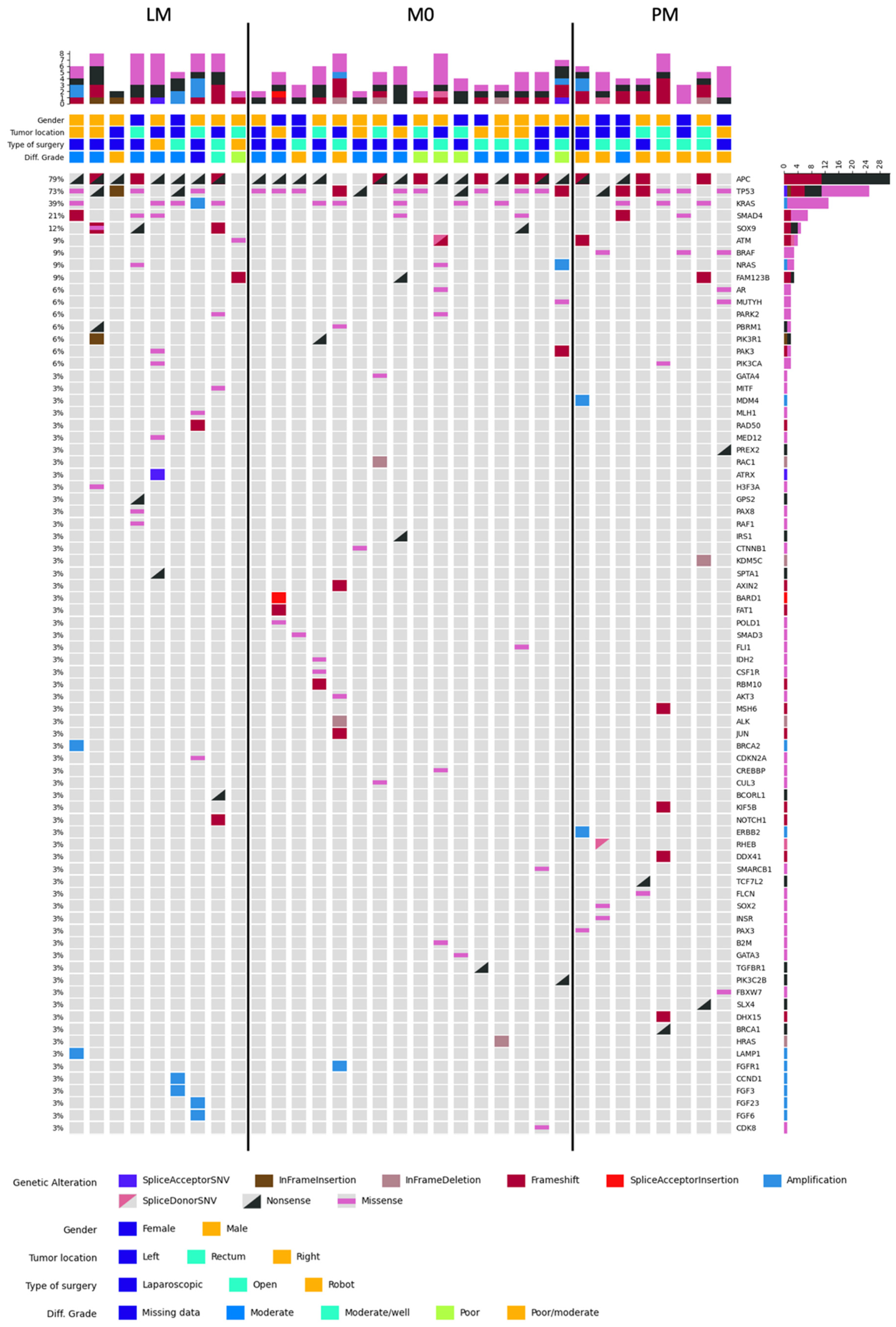
Figure 1. Oncoplot of variants across MSS samples. Genes on y-axis; samples on x-axis.
Ijms 24 12830 g001
Figure 2. Distribution of well-known cancer genes related to MSS CRC.
Ijms 24 12830 g002
Figure 3. FISH analysis of the M0 sample harboring the TARSL2—NTRK3 fusion, showing isolated green and red signals confirming an NTRK3 gene rearrangement.
Ijms 24 12830 g003
Table 1. Comparison of patient characteristics and clinicopathological variables in the relation to the development of metastases.
VariableM0 (N = 20)LM (N = 10)PM (N = 9)p Value
Age at time of diagnosis (years)—median (Q1–Q3)69.0069.0068.000.801 a
(62.00–74.90)(63.75–74.25)(58.00–74.00)
Gendern (%) 0.514 b
Male 12 (60)8 (80)5 (55.6)
Female 8 (40)2 (20)4 (44.4)
Primary tumor location n (%) 0.433 b
Right colon 10 (50)2 (20)2 (22.2)
Left colon 7 (35)5 (50)4 (44.4)
Rectum 3 (15)3 (30)3 (33.3)
Tumor size (cm)
—median (Q1–Q3)
4.10 2.25 3.00 0.061 a
(3.28–5.38)(1.80–5.43)(2.40–3.50)
Differentiation graden (%) * <0.001 b
Poor 4 (20)0 (0)0 (0)
Poor/moderate2 (10)2 (20)8 (88.9)
Moderate14 (70)6 (60)1 (11.1)
Moderate/well0 (0)1 (10)0 (0)
Type of surgeryn (%) 0.153 b
Open10 (50)2 (20)5 (55.6)
Laparoscopic 10 (50)6 (60)4 (44.4)
Robot assisted 0 (0)2 (20)0 (0)
Positive lymph nodesn (%) 0.389 b
No 11 (55)8 (80)5 (55.6)
Yes 9 (45)2 (20)4 (44.4)
Neoadjuvant treatmentn (%) 0.039 b
No 17 (85)4 (40)7 (77.8)
Yes 3 (15)6 (60)2 (22.2)
Adjuvant treatmentn (%) * 0.247 b
No 9 (45)7 (70)4 (44.4)
Yes 11 (55)2 (20)5 (55.6)
Oncological historyn (%) 0.882 b
No 18 (90)8 (80)8 (88.9)
Yes2 (10)2 (20)1 (11.1)
Oncological family historyn (%) * 1.000 b
No 6 (30)3 (30)0 (0)
Yes 12 (60)5 (50)1 (11.1)
Time between surgery and metastases (months)—median (Q1–Q3)N/A18.09 16.420.744 c
(7.77–28.95)(9.71–25.05)
PCI score—median (Q1–Q3)N/AN/A3.50 N/A
(3.00–4.00)
a Kruskal–Wallis Test; b Fisher–Freeman–Halton Exact Test; c Mann–Whitney test. Right-sided = from caecum to transverse colon; left-sided = from the splenic flexure to sigmoid. * Missing data in differentiation grade (LM = 1), adjuvant treatment (LM = 1), and oncological family history (M0 = 2, LM = 2 and PM = 8). M0, no metastases; LM, liver metastases; PM, peritoneal metastases; Q1–Q3, quartile 1–quartile 3; N/A, not applicable; PCI, peritoneal cancer index.
Table 2. Detailed output of RNA analysis.
M GroupGene PairBreakpoint 1Breakpoint 2Fusion Supporting Reads
M0TARSL2-NTRK3Exon 18
chr15:102197123
Exon 14
chr15:88576274
19
PMFAM198A-RAF1Exon not found
chr3:43101459
Exon 3
chr3:12653448
85
PMRPS6KB1-HSF5Exon 1
chr17:57970685
Exon 3
chr17:56544340
21
M0, no metastases; PM, peritoneal metastases.
Table 3. In- and exclusion criteria for patient selection.
Inclusion CriteriaExclusion Criteria
  • Tumor histological type defined as an adenocarcinoma
  • Pathology report confirmed a radical resection with >15 lymph nodes
  • Pathological T3 classified according to the TNM classification
  • M0 group: Follow-up of 5 years without development of metastases
  • Acute colorectal surgery with blow-out or proven perforation
  • Anastomotic leak after surgery
  • Patients with hereditary CRC
  • LM and PM group: Metachronous metastases > 6 months after primary surgery
M0: no metastases; LM, liver metastases; PM, peritoneal metastases.
Disclaimer/Publisher’s Note: The statements, opinions and data contained in all publications are solely those of the individual author(s) and contributor(s) and not of MDPI and/or the editor(s). MDPI and/or the editor(s) disclaim responsibility for any injury to people or property resulting from any ideas, methods, instructions or products referred to in the content.