Article

CRK12: A Key Player in Regulating the Phaseolus vulgaris-Rhizobium tropici Symbiotic Interaction

by
Antonino M. Lecona
1,
Kalpana Nanjareddy
1,
Lourdes Blanco
2,
Valeria Piazza
3,
José Antonio Vera-Núñez
4,
Miguel Lara
2 and
Manoj-Kumar Arthikala
1,*
1
Ciencias Agrogenómicas, Escuela Nacional de Estudios Superiores Unidad León, Universidad Nacional Autónoma de México (UNAM), León 37689, GTO, Mexico
2
Departamento de Biología Molecular de Plantas, Instituto de Biotecnología, Universidad Nacional Autónoma de México (UNAM), Cuernavaca 62210, MOR, Mexico
3
Centro de Investigaciones en Óptica A. C., Loma del Bosque 115, León 37150, GTO, Mexico
4
Departamento Biotecnología, Centro de Investigación y de Estudios Avanzados, Unidad Irapuato, Irapuato 36821, GTO, Mexico
*
Author to whom correspondence should be addressed.
Int. J. Mol. Sci. 2023, 24(14), 11720; https://doi.org/10.3390/ijms241411720
Submission received: 16 June 2023 / Revised: 16 July 2023 / Accepted: 18 July 2023 / Published: 21 July 2023
(This article belongs to the Special Issue Molecular Research on Plant-Associated Nitrogen-Fixing Bacteria)

Abstract

:
Cysteine-rich receptor-like kinases (CRKs) are a type of receptor-like kinases (RLKs) that are important for pathogen resistance, extracellular reactive oxygen species (ROS) signaling, and programmed cell death in plants. In a previous study, we identified 46 CRK family members in the Phaseolus vulgaris genome and found that CRK12 was highly upregulated under root nodule symbiotic conditions. To better understand the role of CRK12 in the PhaseolusRhizobia symbiotic interaction, we functionally characterized this gene by overexpressing (CRK12-OE) and silencing (CRK12-RNAi) it in a P. vulgaris hairy root system. We found that the constitutive expression of CRK12 led to an increase in root hair length and the expression of root hair regulatory genes, while silencing the gene had the opposite effect. During symbiosis, CRK12-RNAi resulted in a significant reduction in nodule numbers, while CRK12-OE roots showed a dramatic increase in rhizobial infection threads and the number of nodules. Nodule cross sections revealed that silenced nodules had very few infected cells, while CRK12-OE nodules had enlarged infected cells, whose numbers had increased compared to controls. As expected, CRK12-RNAi negatively affected nitrogen fixation, while CRK12-OE nodules fixed 1.5 times more nitrogen than controls. Expression levels of genes involved in symbiosis and ROS signaling, as well as nitrogen export genes, supported the nodule phenotypes. Moreover, nodule senescence was prolonged in CRK12-overexpressing roots. Subcellular localization assays showed that the PvCRK12 protein localized to the plasma membrane, and the spatiotemporal expression patterns of the CRK12-promoter::GUS-GFP analysis revealed a symbiosis-specific expression of CRK12 during the early stages of rhizobial infection and in the development of nodules. Our findings suggest that CRK12, a membrane RLK, is a novel regulator of Phaseolus vulgaris-Rhizobium tropici symbiosis.

1. Introduction

Receptor-like kinases (RLKs) are abundant proteins located in the plasma membrane of various eukaryotic organisms, as evidenced by several studies [1,2,3]. Within the RLK family, cysteine-rich receptor-like kinases (CRKs) or domains of unknown function (DUF26) proteins represent a significant subgroup. CRKs consist of distinct domains, including an extracellular domain responsible for perceiving signals, a transmembrane domain, and an intracellular serine/threonine (Ser/Thr) protein kinase domain that facilitates signal transduction [4]. The classification of CRK proteins is based on domain types, such as DUF26 and Ginkbilobin-2 (Gnk2), or the presence of antifungal domain motifs in the extracellular region [5].
DUF26 domains possess a unique structure comprising of two α-helices and a five-stranded β-sheet, along with three conserved cysteine residues arranged in a C-X(8)-C-X(2)-C configuration, capable of forming two cysteine bridges. The cysteine bridges in DUF26 domains allow CRKs to sense reactive oxygen species (ROS)/redox signals (similar to other cysteine-rich domains) before transmitting the signal through the cytoplasmic kinase domain [6]. Plant CRKs exhibit considerable diversity, with examples such as 30 members in Gossypium [7], over 40 members in Oryza [8], 44 in Arabidopsis [4,9], 46 in Phaseolus [10], and 91 in Glycine [11].
CRKs have been implicated in the regulation of various physiological processes in plants, including stomatal dynamics and density, organogenesis, root length and density, cell death, differentiation of vascular tissue, and seed germination [12,13,14].
Plant-microbe interactions are characterized by their dynamic and continuous nature, encompassing both pathogenic and mutualistic relationships. These interactions involve the exchange of signals through distinct molecules produced by the host plant, microbes, or both. Membrane-bound receptor-like kinases play a critical role as receptors in these interactions, facilitating microbe-specific responses through signal transduction. In the context of pathogen infection, the host plant activates defense responses to counteract the invading pathogens. Substantial evidence suggests the involvement of CRKs in plant-pathogen interactions.
Previous studies have demonstrated the significant impact of overexpressing certain cysteine-rich receptor-like kinases on plant resistance against specific pathogens. For instance, Arabidopsis CRK5, CRK6, CRK36, and CRK45 [9,15,16] have been shown to enhance resistance to Pseudomonas syringae by rapidly inducing the expression of defense genes and promoting the production of reactive oxygen species (ROS) [17,18,19]. In Triticum aestivum, the overexpression of TaCRK2 was found to slow down the penetration and intercellular growth of Zymoseptoria tritici [20], and TaCRK2 also exhibited resistance against Puccinia triticina infection while positively regulating the hypersensitive reaction (HR) cell death process induced by the pathogen [21].
Furthermore, TaCRK-7A was shown to directly inhibit the growth of Fusarium pseudograminearum and confer Fusarium Crown Rot (FCR) resistance in wheat by promoting the expression of defense genes associated with the jasmonate pathway [22]. Manipulation of the CaCRK5 expression in Nicotiana benthamiana and Capsicum annum was found to modulate resistance against Ralstonia solanacearum [16], while GbCRK18 provided resistance to Verticillium wilt in Gossypium barbadense [7]. The molecular evidence and crystal structure of Gnk2 revealed the importance of fungal mannose binding to specific residues (asparagine-11, arginine-93, and glutamate-104) in Gnk2 for its antifungal activity [5]. These studies collectively highlight the role of CRKs in enhancing plant defense mechanisms against various pathogens and provide insights into their molecular interactions.
The interaction between legumes and rhizobia initiates in the rhizosphere through the exchange of molecular signals between the host’s root hairs and the bacteria. The recognition process during this symbiotic relationship involves crucial molecules, namely, plant-derived isoflavonoids and bacterial-derived Nod factors. These signals play a role in suppressing plant defenses and enabling bacterial access to the epidermal root hairs and cortical cells of the host. This access is facilitated by the formation of a tubular infection thread, guiding the rhizobia towards the division of cortical cells. As a result, a nodule primordium is formed, which eventually gives rise to nodules containing symbiosomes-specialized cell organelles inclosing nitrogen-fixing bacteroids [23,24].
The establishment of symbiosis involves the temporary suppression of defense responses, which is crucial for symbiosome development and bacterial differentiation. Through the study of legume mutants, researchers have identified several host genes contributing to this suppression, including Medicago SymCRK, regulator of symbiosome differentiation (RSD), defective in nitrogen fixation 2 (DNF2), and nodules with activated defense 1 (NAD1) [25,26,27,28]. It has been demonstrated that DNF2 and SymCRK affect defense signaling through the ethylene pathway, while NAD1 regulates immune responses in nodules via the CDPK-Rboh signaling axis [26,29]. Additionally, a recent genetic study conducted on Aeschynomene evenia revealed the requirement of AeCRK for triggering both root and stem nodulation [30].
In the context of Phaseolus vulgaris L. (common bean), our previous transcriptomic analysis identified several upregulated CRK genes in the roots colonized by rhizobia. Among the nine CRK genes identified, five were common genes expressed under both mycorrhizal and rhizobial symbiosis conditions, while the remaining four genes CRK8, CRK12, CRK20, and CRK42 were unique genes expressed exclusively under nodulated conditions. Notably, the upregulation of the CRK12 gene was particularly significant, as observed in the study by Quezada et al. [10].
Our objective in this study was to conduct a comprehensive functional analysis of the CRK12 gene in the grain legume Phaseolus vulgaris. To achieve this, we employed RNA interference (RNAi) to downregulate and overexpress the CRK12 gene in transgenic hairy roots of P. vulgaris, aiming to investigate its impact on the symbiotic interaction with Rhizobium. As a result, the overexpression of CRK12 genes led to notable changes in root morphology, including increased lateral root and root hair density, as well as longer root hairs. In contrast, silencing of the CRK12 gene produced contradictory results. During the process of rhizobial colonization, we observed the activity of the CRK12 promoter in the early stages of symbiosis, specifically at the sites of rhizobia infection units, infection threads, and dividing cortical cells. Quantitative analysis revealed that the overexpression of CRK12 significantly increased the number of rhizobial infection units and nodule primordia. Moreover, at later stages, these roots exhibited a hypernodulation phenotype compared to the control lines. Conversely, CRK12-RNAi roots displayed a phenotype that was contrary to the overexpression lines. Additionally, the ectopic expression of CRK12 resulted in delayed nodule senescence. Taken together, our findings suggest that CRK12, a membrane receptor kinase, is a novel regulator of Phaseolus vulgaris-Rhizobium tropici symbiosis.

2. Results

2.1. Structure of CRK12 Gene and Its Expression

The Phaseolus CRK12 gene sequence was identified in Phytozome 13 database for gene structure studies. The CRK12 gene is located on chromosome 6, and its gene structure revealed 5 exons and 4 introns in the CDS region and one intron in the 3′ UTR. The mature transcript length was 1729 bp, the CDS was 1242 bp, the 5′ UTR was 4 bp, and the 3′ UTR was 483 bp (Figure 1A). The CRK12 encodes 413 amino acids with 2 DUF26 domains composed of 2 alpha-helices and a five-stranded beta-sheet, which forms a compact single-domain architecture with an alpha + beta-fold. The DUF26 domain contains a C-X(8)-C-X(2)-C motif, and its structure predicted by the ExPASy PROSITE tool shows cysteine residues form three intramolecular disulfide bridges: C1-C5, C2-C3, and C4-C6 (Figure 1B,C) [31].
We next sought to determine the temporal expression of CRK12 in wild-type P. vulgaris roots. We inoculated the roots with R. tropici and monitored CRK12 expression at different time points: during early signaling (3 dpi); in nodule primordia (7 dpi); and in young, mature, and senescent nodules (14, 21, and 31 dpi, respectively). A significant surge in CRK12 transcript abundance was observed in all R. tropic inoculated root tissues compared to uninoculated root tissues at all the measured time points (Figure 1D). CRK12 expression was strongly expressed at 3 and 7 dpi, and maximum expression was observed at 14 dpi in the roots. These results indicate that the CRK12 gene is temporally expressed from the establishment of the nodule to senescence in P. vulgaris.

2.2. Spatiotemporal Promoter Expression Analysis and Protein Subcellular Localization CRK12

To demonstrate spaciotemporal expression of CRK12 promoter activity in P. vulgaris roots under rhizobial symbiosis conditions, we identified and isolated the sequence 1044 bp upstream of the CRK12 start codon. This isolated promoter fragment was cloned and ligated to drive the expression of two reporters, GUS and GFP, within the vector pBGWFS7.0 [32]. The resultant pBGWFS7.0/pCRK12::GUS-GFP binary vector was transformed into the Agrobacterium rhizogenes strain K599, which in turn was utilized to generate transgenic hairy roots in P. vulgaris. We observed that the transgenic roots at 3 days post-inoculation (dpi) with R. tropici (strain CIAT899-RFP) showed CRK12-driven GFP expression at the site of rhizobia infection in root hair cells (Figure 2A–C). No such GFP expression was seen in the root hair cells of uninoculated transgenic roots (Supplementary Figure S1). GFP expression intensified during nodule primordium formation specifically at the site of the Rhizobium infection and in the dividing cortical cells of the nodule (Figure 2D–F). Transgenic roots containing 14-, 21-, and 28-day-old nodules were assayed for the histochemical localization of GUS. Fourteen-day-old young nodules presented an intense GUS color (Figure 2G). The sections show that CRK12 was most active in the inner cortical cells and vasculature of the 21-day-old mature nodules (Figure 2H). Upon the onset of nodule senescence (28 days old), GUS activity was decreased in the nodules (Figure 2I). Together, our results showed that at the early stages of nodule development, the CRK12 promoter was active in ITs, dividing cortical cells of nodule primordia. Strong CRK12 promoter activity was observed in the young nodules, whereas in the mature nodules, the activity was restricted to the inner cortical cells and vasculature of the mature and senescent nodules of P. vulgaris.
To investigate the subcellular localization of the CRK12 protein, a pEarleyGate104 vector was used for a transient expression of the CRK12 protein fused to yellow fluorescent protein (YFP). The confocal images of the P. vulgaris hairy roots expressing YFP-CRK12 showed that CRK12 was localized to the plasma membrane of the root hair cells (Figure 3B), and hairy roots expressing non-fused YFP were used as controls. As predicted, unfused YFP was observed in both the cytoplasm and nuclei of the root hair cells (Figure 3A). NaCl (250 mM)-induced plasmolysis further confirmed the association of fluorescence with the plasma membrane (Figure 3D). In contrast, plasmolysis of control root hairs showed that YFP fluorescence remained in the cytoplasm (Figure 3C). Simultaneously, we analyzed the subcellular localization using an in silico tool. For this, the full protein sequence of CRK12 was submitted to the protein subcellular localization prediction tool WoLF PSORT (https://www.genscript.com/wolf-psort.html?src=leftbar, accessed on 15 March 2022). As anticipated, at the subcellular level, the CRK12 protein was targeted to the plasma membrane.

2.3. CRK12 Alter Root and Root Hair Morphology

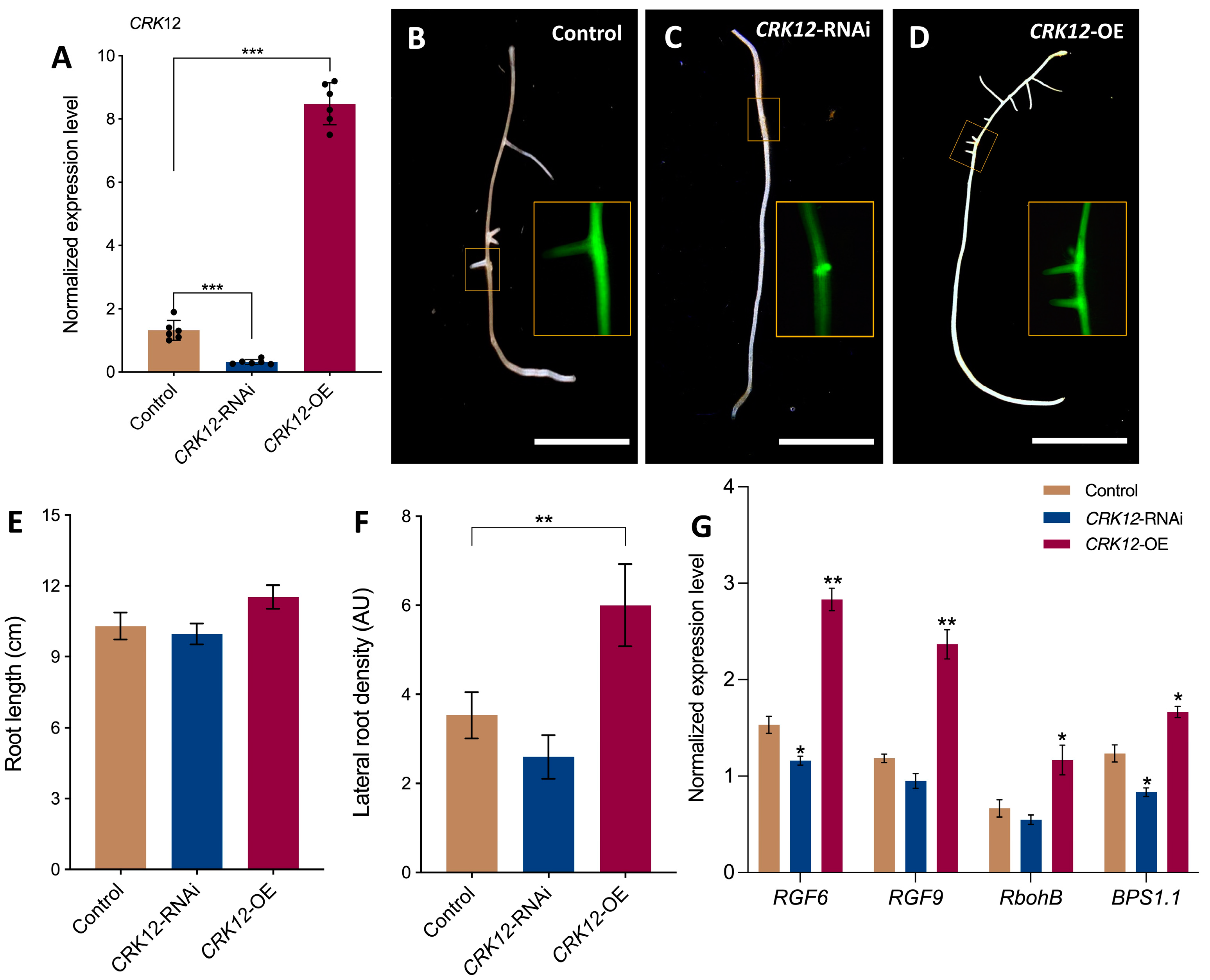
To investigate the function of CRK12, we generated P. vulgaris with transgenic hairy roots expressing CRK12-RNAi and CRK12-OE to observe the root and nodule phenotypes under symbiotic conditions. The non-conserved sequence of CRK12 or complete coding sequence of CRK12 was isolated from fresh P. vulgaris cDNA and cloned into a pK7GWIWG2D(II) and pH7WG2D.1 binary vector downstream of the constitutive 35S promoter, respectively. Agrobacterium rhizogenes K599 harboring a 35S-promoter::CRK12-RNAi (CRK12-RNAi) or 35S-promoter::CRK12 (CRK12-OE) was used to generate the transgenic hairy roots. The quantitative RT–PCR results showed (Figure 4A) a 0.76-fold lesser and 5.44-fold greater CRK12 transcript abundance in the CRK12-RNAi and CRK12-OE roots, respectively, compared to the control roots (those expressing the empty pH7WG2D.1 vector), demonstrating transcript downregulation and overexpression in respective roots of the P. vulgaris transgenic roots.
The phenotypes of the CRK12-RNAi, CRK12-OE transgenic and control hairy roots were analyzed at 7 days post-emergence. A pK7GWIWG2D(II)-RNAi vector and pH7WG2D.1 vectors expressing a visible marker, eGFP (Figure 4C,D), were used to select the transgenic roots of EV control and CRK12-RNAi and CRK12-OE composite transgenic plants (which had transgenic hairy roots but wild-type shoots). The primary root length had marginally decreased in CRK12-RNAi and slightly, but not significantly, increased in CRK12-OE plants compared to controls (Figure 4C–E). However, the density of lateral roots was found marginally decreased in CRK12-RNAi and significantly increased in the CRK12-OE plants relative to the controls (Figure 4F). Quantitative RT–PCR analysis of these roots showed a significant surge in the abundance of transcripts of root meristem regulatory genes such as the root meristem growth factor-like6 (RGF6) and RGF9, and respiratory burst oxidase homologues, RbohB and BPS1.1 (Bypass 1.1), in the transgenic roots, in which CRK12 was overexpressed and a transcript downregulation in CRK12 silenced roots was recorded compared to the control roots (Figure 4G). These results indicated that the overexpression of CRK12 increased the lateral root numbers, and which could be justified by the abundance of transcripts of genes related to lateral root development in P. vulgaris.
Next, the root hair morphology of the transgenic roots at 10 days post-emergence was analyzed. Observations via light microscopy revealed a decrease in the density of root hairs both in root hair elongation (Figure 5A–C) and maturation zone (Figure 5D–F) of CRK12-RNAi roots and the contrary was true for the CRK12-OE plants compared to the controls. In the elongation and mature zones of CRK12-OE roots, the root hairs exhibited a range of lengths, from 279 µm to 594 µm. In comparison, the control group had root hairs measuring 174 µm in the elongation zone and 307 µm in the mature zone, which were comparable to the measurements of CRK12-RNAi root hairs, specifically 195 µm and 325 µm, respectively (Figure 5G,H). However, the root hairs were slightly denser (but not statistically different) on the CRK12-OE roots than control roots. In contrast, the density of CRK12-RNAi root hairs was significantly lower than that of the control (Figure 5I). Subsequently, several genes, viz., auxin response factor 5 and 7 (ARF5, ARF7), RHD6 (root hair defective 6)-like 2 (RSL2), YUCCA and CAPRICE, which regulate the growth and elongation of root hairs, were analyzed. The subsequent qPCR results showed that ARF7, RSL2, YUCCA and CAPRICE transcripts increased significantly in the CRK12-OE roots relative to the CRK12-RNAi and control roots (Figure 5J). Together, these results suggested that the overexpression of CRK12 increased root hair length and the expression of root hair regulatory genes.

2.4. CRK12 Regulates Nodule Numbers and Infection Units in P. vulgaris

To assess the CRK12 overexpression and down-regulation effect on nodulation, we first inoculated the composite transgenic plants with R. tropici CIAT 899 expressing a GUS reporter [33]. Periodically, the roots were assayed for GUS and analyzed to determine the symbiosis phenotype. After one week of CIAT 899 inoculation, the frequency of infection events on the CRK12-RNAi revealed to be lower (3.8/plant) and CRK12-OE roots was higher (29/plant) than that on the control roots (7.4/plant; Figure 6A–C,J). Light microscopy observations in CRK12-OE and CRK12-RNAi revealed that the phenotype of early nodule development processes, such as the root hair infection thread progression and cortical cell division of nodule primordium, were like those of the control roots (Figure 6C–F).
Next, we compared the number of nodule primordia in the CRK12-RNAi and CRK12-overexpression to that of control roots. Significantly higher number of nodule primordia (108/plant) were observed in the CRK12-OE roots than in the control roots (24/plant) at 10 days post-inoculation in the contrary, CRK12 downregulation led to a highly significant reduction in nodule primordia (5/plant; Figure 6K). All the young nodules of the control and CRK12-OE roots were colonized successfully with rhizobia, whereas the CRK12-RNAi show poor rhizobia density (Figure 6G–I).
To determine whether this phenotype is associated with changes in the expression of genes involved in early rhizobial signaling, we measured the expression levels of some of the key early signaling genes, such as SymRK (symbiosis receptor kinase), CCaMK (calcium-calmodulin kinase), NIN (nodule inception), Nsp2 (nodule signaling pathway 2), Enod40 (early nodulin 40) and RACK1 (receptor for activated C kinase 1). Based on the quantitative RT–PCR results, CCaMK, NIN, Nsp2 and Enod40 showed a significant surge in transcript levels in the CRK12-OE transgenic roots compared with the CRK12-RNAi and control roots, whereas SymRK and RACK1 showed slightly higher expression in the CRK12-OE roots relative to controls (Figure 6L). Together, our data indicate that CRK12 functions during the early stages of nodule formation and development, which is reflected in terms of increased rhizobial infection units, nodule primordial numbers and increased expression of early signaling genes in P. vulgaris.

2.5. CRK12 Overexpression Results in Hypernodulation in P. vulgaris Transgenic Roots

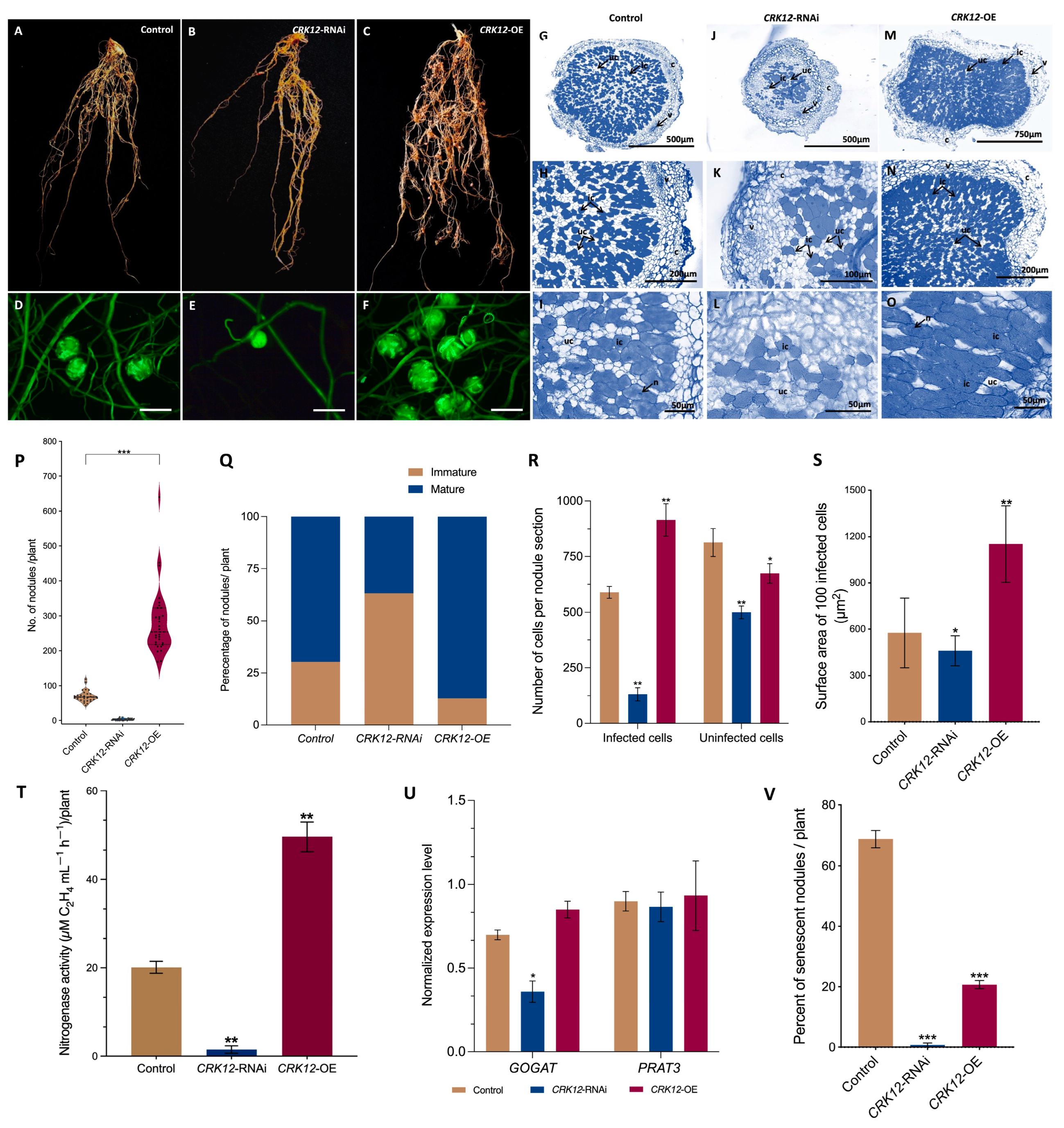
While we were trying to identify the impact of the CRK12 transcript down-regulation on root nodule symbiosis, at 21-day post inoculation we found that the nodule numbers remained critically low. The CRK12-RNAi transgenic roots exhibited fewer number of nodules and were remained to be juvenile/primordial implying their failure to reach to mature nodule stage. In addition, the transgenic CRK12-OE roots shows increased nodule numbers compared to control transgenic roots (Figure 7A–F). Furthermore, the quantitative data revealed a 4-fold higher number of nodules in the CRK12-OE roots than in the control roots. The average number of nodules was 71.7 per control plant, 4 per RNAi and 289.3 per CRK12-OE plant (Figure 7P). Among them, 87.1 percent of the nodules were mature pink at 21 dpi, and the remaining were immature white nodules in the CRK12-OE roots. In control 69.7 percent of the nodules were pink, and 30.3 percent were white. Nevertheless, the CRK12-RNAi roots show 36.8 and 63.2 percent pink and white nodules, respectively (Figure 7Q).
The transverse section of mature control nodules (Figure 7G–I) depicted typical histological characteristics of a determinate nodule, such as the outer and inner cortex, nodule vasculature, and nodule core [34]. The nodule core tissue was comprised of infected cells harboring R. tropici CIAT 899 and uninfected cells (Figure 7I). On the other hand, the CRK12-RNAi nodules were smaller in size. However, the nodule structural details remained comparable to controls (Figure 7J–O). The most interesting detail was in the highly reduced number of infected cells in the CRK12-RNAi nodules (Figure 7L). The CRK12-OE nodules displayed characteristics similar to control (Figure 7G,H,M,N) except the nodule core showing a significant increase in infected cell density compared to the control and CRK12-RNAi (Figure 7R). On the contrary, the uninfected cells numbers were fewer than the control. Additionally, the infected cell area in the CRK12 overexpressed nodules were significantly larger (1151 µm2/100 cells) than the control (577 µm2/100 cells) and CRK12-RNAi (460 µm2/100 cells) (Figure 7S).
Subsequently, at 21 dpi, we estimated the nitrogen fixation rate in control, CRK12-RNAi and CRK12-OE transgenic roots and results showed a significant increase of nitrogen fixation in the CRK12-OE nodules compared to controls; at the same time, CRK12-RNAi demonstrated highly reduced abilities to fix nitrogen (Figure 7T). Next, we measured key organic nitrogen export-related genes, such as Gln synthetase and glutamate synthase (GOGAT), and glutamine phosphoribosyl pyrophosphate amidotransferase 3 (PRAT3) in the mature nodules. The quantitative RT–PCR results showed similar expression patterns for both GOGAT and PRAT3 in the CRK12-OE and control nodules at 21 dpi, in CRK12-RNAi roots the GOGAT significantly decreased compared to control (Figure 7U). At 35 dpi, the nodule morphology showed that 70% of control root nodules were senescent, and only 21% of the CRK12-overexpressing nodules were senescent. These results indicate the prolonged nitrogen fixing capabilities of the CRK12-overexpressing root nodules (Figure 7V).
Together, these data suggest that the transgenic roots that expressed the CRK12-RNAi vector severely affected root nodule numbers and their nitrogen fixing abilities. On the contrary, overexpression of CRK12 showed a phenotype with increased nodules numbers and infected cell density and size. Furthermore, these overexpressed nodules fixed more nitrogen and the presence of key nitrogen export genes in these nodules confirmed the function of these nodules.

3. Discussion

Receptor-like kinases (RLKs) possess distinct extracellular domains that enable the recognition of different ligands and facilitate the transduction of various extracellular signals, including those involved in symbiosis [35,36]. Symbiosis is initiated by legumes and rhizobia exchanging signaling molecules as part of a bidirectional communication process. In response to host-secreted flavones or isoflavones, rhizobia synthesize and discharge nod factors, also known as lipo-chitooligosaccharides (LCOs) [37]. These LCOs are perceived by LysM-type plasma membrane receptors such as NFR1, NFR5, and NFRe in Lotus japonicus; among them, both NFR1 and NFR5 were essential for the nod factor signaling [38,39], whereas NFRe may increase signaling in root epidermal cells [40]. Downstream of LysM-type receptors, a cascade of symbiotic signaling genes and transcription factors (TFs) function in triggering infection thread formation, nodule organogenesis and other processes in legume roots [41].
The largest group of plant RLKs consists of cysteine-rich receptor kinases or proteins that possess the DUF26 domain. However, the biological functions of these RLKs in plant symbiotic interactions have been relatively understudied. Earlier investigations in Medicago truncatula have demonstrated that mutations in symCRK result in the formation of nodules exhibiting defense-like reactions, bacterial death, and ultimately an inability to fix nitrogen [26,42,43]. In a previous study focused on P. vulgaris, it was indicated that potential PvCRK genes may play a role in regulating these diverse symbiotic interactions [10]. Additionally, more recent research had discovered that AeCRK is crucial for initiating root and stem nodulation in Aeschynomene evenia [30].
The main outcome of our preliminary investigations in P. vulgaris led us to perform a functional characterization of CRK12 in the current study. This investigation aimed to elucidate the specific role of CRK12 in the interactions between P. vulgaris and Rhizobium tropici. CRK12 in Phaseolus is characterized by two DUF26 domains that contain a C-X(8)-C-X(2)-C motif similar to other CRKs [10]. Although the precise function of this domain remained unknown, previous reports suggested its potential involvement in redox regulation and protein–protein interactions [44,45]. Interestingly, the temporal expression patterns of CRK12 in plants inoculated with Rhizobium symbionts showed an increase in transcript levels at all stages of symbiosis. The analysis of cis-elements in the regulatory region of CRK12 indicated the absence of symbiosis-specific transcription factors [10]. However, an abundance of transcription factors involved in phytohormone regulation was identified, suggesting the presence of a potential signaling mechanism [46,47] (Lin et al., 2020; Li et al., 2022). To further investigate this, we examined the expression of the CRK12 gene promoter, which yielded intriguing results. The CRK12 promoter exhibited expression in Rhizobium infection units, infection threads, dividing cortical cells, inner cortex, and vasculature of mature nodules.
The roles of CRKs in growth and developmental aspects of plants have been previously characterized [6]. Herein, the overexpression of CRK12 resulted in an increased density of lateral roots as well as root hairs, and root hairs grew longer both in the root hair elongation and in the maturation zones in comparison to the controls. Conversely, when CRK12 expression was suppressed using RNA interference (RNAi), we observed a contrasting phenotype in the roots and root hairs, thereby reinforcing the significance of this gene in the development of roots and root hairs. Interestingly, our findings differ from previous studies on Arabidopsis CRK28, CRK29, and CRK42 mutants, where mutations in CRK genes resulted in longer primary roots and denser lateral roots [6,13].
To gain insights into the underlying mechanism behind the altered root phenotype, we conducted transcript analysis of key genes involved in regulating root hair length, including auxin responsive factors (ARF5, ARF7) [48] and the auxin biosynthesis gene YUCCA [49]. We found significantly higher expression levels of these genes in CRK12-overexpressing (CRK12-OE) roots compared to the CRK12-RNAi and control roots. Additionally, transcripts related to root growth regulation, such as RGF6, RGF9 [50], RbohB [51], and BPS1.1 [52], were also induced in CRK12-OE roots. These findings suggested a potential mechanism underlying the observed root and root hair phenotypes. It is important to note that root hairs serve as entry points for rhizobia, and an increased density of root hairs could enhance the opportunity for symbiotic interactions with these microorganisms.
Previous studies have demonstrated the involvement of CRKs in immune responses during plant–pathogen interactions [17,53]. These CRKs perceive signals through extracellular, transmembrane, and intracellular domains, leading to the activation of MAPK pathways and subsequent gene transcription [54]. In Medicago truncatula, the participation of SymCRK, a cysteine-rich receptor-like kinase, has been reported in mutualistic interactions, such as symbiosis [43]. In this study, we focused on investigating the effects of the silencing and overexpression of CRK12 on rhizobial nodule symbiosis (RNS). In Phaseolus plants, overexpression of CRK12 resulted in a remarkable rise in the occurrence of infection events, with 108 events per plant, which was significantly higher compared to the controls with only 24 events per plant. This increase was also observed in the number of nodules, as CRK12-OE led to 289.3 nodules in the roots, representing a fourfold increase compared to the control group which had 71.7 nodules. Conversely, the silencing of CRK12 resulted in a notable decrease in both infection events (5 events per plant) and nodule numbers (4 nodules per plant). Furthermore, the few nodules observed in the CRK12-RNAi plants displayed similar anatomical characteristics to the control but had very few infected cells. In contrast, histological observations of CRK12-overexpressing (CRK12-OE) nodules revealed an increase in both the number and size of infected cells. These findings are consistent with the nitrogen-fixing abilities exhibited by both CRK-RNAi and CRK-OE nodules. Previous reports involving SymCRK in M. truncatula mutants did not show a significant change in nodule numbers however, most of the nodules developed were nonfunctional necrotic nodules [26]. Further, the investigation of Aeschynomene evenia’s stem and root nodule development indicated that AeCRK, in conjunction with other symbiotic pathway genes, was essential for the process.
In Medicago, the overexpression of a lectin-like receptor kinase (LecRK), a potential rhizobial lipochitooligosaccharide-binding RLK, has been shown to increase nodule numbers [55]. In the present study, the observed increase in infection events and nodule numbers upon overexpression of CRK12, as well as the contrasting effect when CRK12 transcript was downregulated, suggest a potential role of CRK12 as a receptor for rhizobial nod factors. Furthermore, in Phaseolus plants, the overexpression of CRK12 led to the upregulation of respiratory burst oxidative homologue B (RbohB), resulting in increased levels of reactive oxygen species. Previous studies have reported the involvement of RbohB in maintaining symbiosome number, bacteroid size, and nitrogen fixation in Phaseolus nodules [56]. Notably, CRKs have been implicated in direct ROS sensing due to the redox regulation possibilities within their extracellular protein domain [6]. Hence, it is plausible to propose that the crosstalk between CRK12 and ROS signaling may contribute to the observed increase in nodule numbers.
In conclusion, our investigations provide compelling evidence of the significant influence exerted by CRK12 on the development of root hairs and root nodules, as well as nitrogen fixation in P. vulgaris. These findings underscore the undeniable role played by CRK12 in governing the mutualistic association between R. tropici and P. vulgaris, as downregulation or overexpression of CRK12 transcripts directly impact these processes. Finally, we suggest that harnessing symbiosis-specific gene(s) like CRKs in breeding programs for genetic modification presents exciting opportunities to enhance legume crops, leading to improved nitrogen fixation and supporting more sustainable and productive agricultural practices.

4. Materials and Methods

4.1. Plant Material and Rhizobium Inoculation

Seeds of Phaseolus vulgaris cv. Negro Jamapa obtained from the Instituto de Biotecnología, UNAM, Mexico, were used in this study. The seeds were surface disinfested and germinated, as described previously by Nanjareddy and colleagues [57]. The seedlings were then transplanted into sterile vermiculite in the greenhouse under a 16 h photoperiod at 28 ± 1 °C. The plants were irrigated with Broughton and Dilworth (B&D) [58] nutrient media with limited amounts of nitrogen (2 mM KNO3) to promote root nodulation. For the induction of nodules, 1 mL of Rhizobium tropici (wild-type strain CIAT899 or that expressing RFP or a GUS reporter) at an OD600 dilution of 0.6 was inoculated. Root or nodule tissues were collected at various time points, and the samples were immediately immersed in liquid nitrogen and stored at −80 °C.

4.2. Gene and Protein Structure

The exon–intron structure was determined using the genomic sequence of the CRK12 gene in Phytozome 13. Phaseolus CRK12 protein domains were predicted by PROSITE (https://prosite.expasy.org/, accessed on 29 April 2022), and their secondary structure was predicted using the Swiss model (http://swissmodel.expasy.org, accessed on 29 April 2022).

4.3. Promoter Construction and Composite Plant Production

To analyze the spatiotemporal expression patterns of the CRK12 promoter during nodulation, a 1044 bp promoter fragment was amplified from P. vulgaris genomic DNA using specific primers (Supplementary Table S1) and cloned into a pENTR/D-TOPO vector (Invitrogen, Carlsbad, CA, USA). The Gateway LR reaction was performed between the entry vector pENTR/D-TOPO-pCRK12 and the destination vector pBGWSF7.0 according to the manufacturer’s (Invitrogen) instructions. The sequence of the resultant pBGWSF7.0-pCRK12::GUS-GFP was verified.
To generate the P. vulgaris composite transgenic plants (whose hairy roots expressed the recombinant plasmid vector), 2-day-old germinated seedlings were infected with Agrobacterium rhizogenes K599 strains carrying the pBGWSF7.0-pCRK12::GUS-GFP construct. The empty pBGWSF7.0 vector was used as a control. The plasmid constructs were introduced into A. rhizogenes strain K599 separately. All the composite transgenic plants were generated as described by Nanjareddy et al. [57], after which they were transplanted into sterile vermiculite and inoculated with the wild-type Rhizobium tropici CIAT 899 or that expressing RFP markers, as previously described by Nanjareddy & colleagues [57].

4.4. Subcellular Localization Analysis of CRK12

The open reading frames of CRK12 were amplified from cDNA freshly prepared from 10-day-old P. vulgaris seedlings using the appropriate oligonucleotides (Supplementary Table S1). The amplicon was cloned into a pENTR/D-TOPO vector. The Gateway LR reaction was performed between the entry vector pENTR/D-TOPO-CRK12 and the destination vector pEarleyGate104 according to the manufacturer’s (Invitrogen) instructions. The N-terminus of the resulting binary vector consisted of fusion of CRK12 and yellow fluorescent protein (YFP). A pEarleyGate104 vector expressing YFP was used as a control.

4.5. Cloning of CRK12 Overexpression and Silencing Constructs

The primer pair used (Supplementary Table S1) was designed to amplify the open reading frame of the CRK12 gene (Phvul.006G006800) from freshly prepared P. vulgaris seedling cDNA. The amplified fragment of 1271 bp (1242 bp ORF + 29 bp 3′UTR) was cloned into a pENTR/D-TOPO vector. The Gateway LR reaction was performed between the entry vector pENTR/D-TOPO-CRK12 and the destination binary vector pH7WG2D.1, which was under the control of the constitutive 35S promoter [32] according to the manufacturer’s instructions (Invitrogen). The sequence of the resultant pH7WG2D.1-CRK12-OE was verified. The empty pH7WG2D.1 vector was used as a control in the experiments.
The CRK12-RNAi silencing construct was created through the cloning of a non-conserved 280 bp fragment from the 3′UTR of CRK12. This fragment was amplified using common bean root cDNA as a template and specific primers (Supplementary Table S1). Subsequently, the fragment was inserted into the pENTR/D-TOPO vector, and the resulting construct was combined with the binary vector pK7GWIWG2D(II) using Gateway Technology (Invitrogen Gateway cloning technology). To ensure the correct orientation of the inserted fragments in the CRK12-RNAi construct, PCR and sequencing were conducted for verification. The empty pK7GWIWG2D(II) vector was used as a control in the experiments.
The composite transgenic plants were generated as described above. After removing the wild-type primary root from the composite transgenic plants, we selected the hairy roots under an epifluorescence microscope with a GFP filter with an excitation of 488 nm and an emission fluorescence ranging from 510 to 540 nm. At the same time, the non-transformed roots were removed, transplanted into sterile vermiculite and then inoculated with the R. tropici CIAT 899 strain.

4.6. Expression Analysis

Total RNA was extracted from frozen tissues using a Spectrum™ Plant Total RNA Kit (Merck KGaA, Darmstadt, Germany) following the manufacturer’s instructions. Contaminant DNA among the RNA samples was eliminated by incubating the samples with RNase-free DNase (1 U µL−1) at 37 °C for 15 min. The RNA integrity and concentration were verified via 1% agarose gel electrophoresis and via spectrometry with a NanoDropTM 2000 instrument, respectively.
Quantitative PCR was performed using an iScriptTM One-Step RT–PCR Kit with SYBR® Green following the manufacturer’s (Bio-Rad, Hercules, CA, USA) recommendations in a Bio-Rad iQTM 5 real-time PCR detection system. Each reaction had included 40 ng of RNA as a template. A control sample without reverse transcriptase was included to confirm the absence of contaminant DNA. Relative gene expression levels were calculated using the formula 2–ΔCT, where the cycle threshold value (ΔCT) is the CT of the gene of interest minus the CT of the reference gene. The relative expression values, normalized to those of two reference genes (Phaseolus EF1a—‘elongation factor 1-alpha’ and IDE—insulysin’), were calculated according to the methods of Vandesompele et al. [59]. The gene-specific oligonucleotides used in the present study are listed in Supplementary Table S1.

4.7. Root Hair Measurements

The number of root hairs was determined in 1 mm long sections within the root hair elongation zone and root hair mature zone of the control, CRK12-OE and CRK12-RNAi transgenic hairy roots at 10 days post emergence. The root hair length was measured using the LAS EZ 3.4.0.272 suite, and the density was calculated according to the methods of Ma et al. [60].

4.8. Nodule Phenotype and Microscopy

The uninoculated and R. tropici CIAT899 (wild type)-inoculated transgenic root samples in which pCRK12::GUS-GFP or CRK12 was overexpressed or CRK12 silenced were inoculated with R. tropici CIAT899 expressing GUS (harbouring pGUS 32) [61] or CIAT899 expressing RFP. The GUS assay was performed as described by Jefferson [62]. Root and nodule tissues were transferred to a 3 cm diameter Petri dishes containing a GUS staining solution and incubated at 37 °C in the dark until blue or magenta spots were visible (approximately 8–12 h). The GUS-stained roots were clarified using 0.5% sodium hypochlorite for 8 h and then examined for nodule symbiosis phenotype, viz., infection threads, nodule primordia, and nodules, under a light microscope (Leica, DMLB bright-field microscope, Buffalo Grove, IL, USA). The GUS-stained mature nodules were sectioned using a razor blade. The sections were mounted in 10% glycerol and observed under a light microscope. The GUS-stained transgenic roots expressing the pCRK12::GUS-GFP promoter were observed under a stereomicroscope (Leica), and images of young and mature nodules were obtained.
The transgenic roots that expressed subcellular localization construct or the CRK12 promoter construct was observed under a Zeiss LSM 710-NLO confocal microscope (Centro de Investigaciones en Optica A.C., León, Mexico) with a 25X 0.8 NA oil immersion objective. GFP was excited with an argon laser (488 nm), and the fluorescence from 510 to 540 nm was recorded. YFP was excited with the 514 nm line of the argon laser, and the fluorescence from 519 to 620 nm was recorded. RFP was excited with a solid-state laser (561 nm), and the fluorescence was filtered using a 640-/650-nm bandpass filter. The nodule cross sections were obtained by paraffin embedding and semithin sectioning following the protocol by Chen et al. [63]. The tissues were fixed in FAA fixative containing 10% formalin, 5% glacial acetic acid, 50% ethanol and 35% deionized water. Post fixation, the tissues were processed through a dehydration series of ethanol solutions (from 30% to 100%). The paraffin embedded tissues were sectioned using the Leica RM2125 RTS microtome and stained with 0.1% Toluidine blue. Acetylene reduction assay was performed following the method described by Burris [64]. 21 dpi nodulated transgenic roots of control, CRK12-RNAi and CRK12-OE were incubated in acetylene gas for 30, 60 and 90 min, and the ethylene production was measured by gas chromatography in a Hewlett Packard 5890 Series II (Wilmington, DE, USA).

4.9. Statistical Analysis

All the statistical data were analyzed by Student’s t test using Prism 9.0 software (GraphPad Software Inc., Boston, MA, USA). In the figures, single, double, and triple asterisks were used indicate differences that are significant (p < 0.05 or p < 0.01) or highly significant (p < 0.001), respectively.

Supplementary Materials

The following supporting information can be downloaded at: https://www.mdpi.com/article/10.3390/ijms241411720/s1.

Author Contributions

M.-K.A. conceived the research, designed the experiments and acquired the funding. A.M.L. performed all the experiments. K.N. assisted in methodology and bacterial cultures. L.B. isolated and cloned the overexpression construct. V.P. performed confocal microscopy. J.A.V.-N. did nitrogenase activity. A.M.L., M.L. and K.N. analyzed the results and wrote the manuscript. All authors have read and agreed to the published version of the manuscript.

Funding

The authors wish to acknowledge the CONACyT: CF-MI-20191017134234199-316538 to M.K.A for funding this work, and was partially supported by the DGAPA/PAPIIT-UNAM grant no. IN216321 to K.N and IN213221 to M.K.A.

Institutional Review Board Statement

Not applicable.

Informed Consent Statement

Not applicable.

Data Availability Statement

Data are contained within the article and Supplementary Materials.

Acknowledgments

We are grateful to the SGC-LII certified lab (ISO 9001: 2015), ENES-Unidad Leon for providing facilities during this work. We thank Gabriel X. Garcia for cloning the promoter construct piolet experiments. We thank Govindappa Melappa, Davangere University, India for the statistical analysis.

Conflicts of Interest

The authors declare no conflict of interest.

References

  1. Santoni, V.; Vinh, J.; Pflieger, D.; Sommerer, N.; Maurel, C. A proteomic study reveals novel insights into the diversity of aquaporin forms expressed in the plasma membrane of plant roots. Biochem. J. 2003, 373, 289–296. [Google Scholar] [CrossRef] [PubMed]
  2. Alexandersson, E.; Saalbach, G.; Larsson, C.; Kjellbom, P. Arabidopsis Plasma Membrane Proteomics Identifies Components of Transport, Signal Transduction and Membrane Trafficking. Plant Cell Physiol. 2004, 45, 1543–1556. [Google Scholar] [CrossRef] [Green Version]
  3. Marmagne, A.; Rouet, M.-A.; Ferro, M.; Rolland, N.; Alcon, C.; Joyard, J.; Garin, J.; Barbier-Brygoo, H.; Ephritikhine, G. Identification of New Intrinsic Proteins in Arabidopsis Plasma Membrane Proteome. Mol. Cell. Proteom. 2004, 3, 675–691. [Google Scholar] [CrossRef] [PubMed] [Green Version]
  4. Chen, Z. A Superfamily of Proteins with Novel Cysteine-Rich Repeats. Plant Physiol. 2001, 126, 473–476. [Google Scholar] [CrossRef] [PubMed] [Green Version]
  5. Miyakawa, T.; Hatano, K.-I.; Miyauchi, Y.; Suwa, Y.-I.; Sawano, Y.; Tanokura, M. A Secreted Protein with Plant-Specific Cysteine-Rich Motif Functions as a Mannose-Binding Lectin That Exhibits Antifungal Activity. Plant Physiol. 2014, 166, 766–778. [Google Scholar] [CrossRef] [Green Version]
  6. Bourdais, G.; Burdiak, P.; Gauthier, A.; Nitsch, L.; Salojärvi, J.; Rayapuram, C.; Idänheimo, N.; Hunter, K.; Kimura, S.; Merilo, E.; et al. Large-Scale Phenomics Identifies Primary and Fine-Tuning Roles for CRKs in Responses Related to Oxidative Stress. PLOS Genet. 2015, 11, e1005373. [Google Scholar] [CrossRef] [Green Version]
  7. Li, T.-G.; Zhang, D.-D.; Zhou, L.; Kong, Z.-Q.; Hussaini, A.S.; Wang, D.; Li, J.-J.; Short, D.P.G.; Dhar, N.; Klosterman, S.J.; et al. Genome-Wide Identification and Functional Analyses of the CRK Gene Family in Cotton Reveals GbCRK18 Confers Verticillium Wilt Resistance in Gossypium barbadense. Front. Plant Sci. 2018, 9, 1266. [Google Scholar] [CrossRef] [Green Version]
  8. Chern, M.; Xu, Q.; Bart, R.S.; Bai, W.; Ruan, D.; Sze-To, W.H.; Canlas, P.E.; Jain, R.; Chen, X.; Ronald, P.C. A Genetic Screen Identifies a Requirement for Cysteine-Rich–Receptor-Like Kinases in Rice NH1 (OsNPR1)-Mediated Immunity. PLoS Genet. 2016, 12, e1006049. [Google Scholar] [CrossRef] [Green Version]
  9. Burdiak, P.; Rusaczonek, A.; Witoń, D.; Głów, D.; Karpiński, S. Cysteine-rich receptor-like kinase CRK5 as a regulator of growth, development, and ultraviolet radiation responses in Arabidopsis thaliana. J. Exp. Bot. 2015, 66, 3325–3337. [Google Scholar] [CrossRef] [Green Version]
  10. Quezada, E.-H.; García, G.-X.; Arthikala, M.-K.; Melappa, G.; Lara, M.; Nanjareddy, K. Cysteine-Rich Receptor-Like Kinase Gene Family Identification in the Phaseolus Genome and Comparative Analysis of Their Expression Profiles Specific to Mycorrhizal and Rhizobial Symbiosis. Genes 2019, 10, 59. [Google Scholar] [CrossRef] [Green Version]
  11. Delgado-Cerrone, L.; Alvarez, A.; Mena, E.; De León, I.P.; Montesano, M. Genome-wide analysis of the soybean CRK-family and transcriptional regulation by biotic stress signals triggering plant immunity. PLoS ONE 2018, 13, e0207438. [Google Scholar] [CrossRef]
  12. Yadeta, K.A.; Elmore, J.M.; Creer, A.Y.; Feng, B.; Franco, J.Y.; Rufian, J.S.; He, P.; Phinney, B.; Coaker, G. A Cysteine-Rich Protein Kinase Associates with a Membrane Immune Complex and the Cysteine Residues Are Required for Cell Death. Plant Physiol. 2016, 173, 771–787. [Google Scholar] [CrossRef] [Green Version]
  13. Pelagio-Flores, R.; Muñoz-Parra, E.; Barrera-Ortiz, S.; Ortiz-Castro, R.; Saenz-Mata, J.; Ortega-Amaro, M.A.; Jiménez-Bremont, J.F.; López-Bucio, J. The cysteine-rich receptor-like protein kinase CRK28 modulates Arabidopsis growth and development and influences abscisic acid responses. Planta 2019, 251, 2. [Google Scholar] [CrossRef] [PubMed]
  14. Arellano-Villagómez, F.C.; Guevara-Olvera, L.; Zuñiga-Mayo, V.M.; Cerbantez-Bueno, V.E.; Verdugo-Perales, M.; Medina, H.R.; De Folter, S.; Acosta-García, G. Arabidopsis cysteine-rich receptor-like protein kinase CRK33 affects stomatal density and drought tolerance. Plant Signal. Behav. 2021, 16, 1905335. [Google Scholar] [CrossRef] [PubMed]
  15. Lee, D.S.; Kim, Y.C.; Kwon, S.J.; Ryu, C.-M.; Park, O.K. The Arabidopsis Cysteine-Rich Receptor-Like Kinase CRK36 Regulates Immunity through Interaction with the Cytoplasmic Kinase BIK1. Front. Plant Sci. 2017, 8, 1856. [Google Scholar] [CrossRef] [Green Version]
  16. Mou, S.; Meng, Q.; Gao, F.; Zhang, T.; He, W.; Guan, D.; He, S. A cysteine-rich receptor-like protein kinase CaCKR5 modulates immune response against Ralstonia solanacearum infection in pepper. BMC Plant Biol. 2021, 21, 382. [Google Scholar] [CrossRef]
  17. Chen, K.; Du, L.; Chen, Z. Sensitization of defense responses and activation of programmed cell death by a pathogen-induced receptor-like protein kinase in Arabidopsis. Plant Mol. Biol. 2003, 53, 61–74. [Google Scholar] [CrossRef] [PubMed]
  18. Zhang, X.; Han, X.; Shi, R.; Yang, G.; Qi, L.; Wang, R.; Li, G. Arabidopsis cysteine-rich receptor-like kinase 45 positively regulates disease resistance to Pseudomonas syringae. Plant Physiol. Biochem. 2013, 73, 383–391. [Google Scholar] [CrossRef]
  19. Yeh, Y.-H.; Chang, Y.-H.; Huang, P.-Y.; Huang, J.-B.; Zimmerli, L. Enhanced Arabidopsis pattern-triggered immunity by overexpression of cysteine-rich receptor-like kinases. Front. Plant Sci. 2015, 6, 322. [Google Scholar] [CrossRef] [Green Version]
  20. Saintenac, C.; Cambon, F.; Aouini, L.; Verstappen, E.; Ghaffary, S.M.T.; Poucet, T.; Marande, W.; Berges, H.; Xu, S.; Jaouannet, M.; et al. A wheat cysteine-rich receptor-like kinase confers broad-spectrum resistance against Septoria tritici blotch. Nat. Commun. 2021, 12, 433. [Google Scholar] [CrossRef]
  21. Gu, J.; Sun, J.; Liu, N.; Sun, X.; Liu, C.; Wu, L.; Liu, G.; Zeng, F.; Hou, C.; Han, S.; et al. A novel cysteine-rich receptor-like kinase gene, TaCRK2, contributes to leaf rust resistance in wheat. Mol. Plant Pathol. 2020, 21, 732–746. [Google Scholar] [CrossRef] [PubMed] [Green Version]
  22. Wu, T.; Guo, F.; Xu, G.; Yu, J.; Zhang, L.; Wei, X.; Zhu, X.; Zhang, Z. The Receptor-like Kinase TaCRK-7A Inhibits Fusarium pseudograminearum Growth and Mediates Resistance to Fusarium Crown Rot in Wheat. Biology 2021, 10, 1122. [Google Scholar] [CrossRef] [PubMed]
  23. Peters, N.K.; Frost, J.W.; Long, S.R. A Plant Flavone, Luteolin, Induces Expression of Rhizobium meliloti Nodulation Genes. Science 1986, 233, 977–980. [Google Scholar] [CrossRef]
  24. Redmond, J.W.; Batley, M.; Djordjevic, M.A.; Innes, R.W.; Kuempel, P.L.; Rolfe, B.G. Flavones induce expression of nodulation genes in Rhizobium. Nature 1986, 323, 632–635. [Google Scholar] [CrossRef]
  25. Bourcy, M.; Brocard, L.; Pislariu, C.I.; Cosson, V.; Mergaert, P.; Tadege, M.; Mysore, K.S.; Udvardi, M.K.; Gourion, B.; Ratet, P. Medicago truncatulaDNF2 is aPI-PLC-XD-containing protein required for bacteroid persistence and prevention of nodule early senescence and defense-like reactions. New Phytol. 2012, 197, 1250–1261. [Google Scholar] [CrossRef]
  26. Berrabah, F.; Bourcy, M.; Eschstruth, A.; Cayrel, A.; Guefrachi, I.; Mergaert, P.; Wen, J.; Jean, V.; Mysore, K.; Gourion, B.; et al. A non RD receptor-like kinase prevents nodule early senescence and defense-like reactions during symbiosis. New Phytol. 2014, 203, 1305–1314. [Google Scholar] [CrossRef] [PubMed]
  27. Wang, C.; Yu, H.; Luo, L.; Duan, L.; Cai, L.; He, X.; Wen, J.; Mysore, K.S.; Li, G.; Xiao, A.; et al. NODULES WITH ACTIVATED DEFENSE 1 is required for maintenance of rhizobial endosymbiosis in Medicago truncatula. New Phytol. 2016, 212, 176–191. [Google Scholar] [CrossRef] [Green Version]
  28. Domonkos, Á.; Kovács, S.; Gombár, A.; Kiss, E.; Horváth, B.; Kováts, G.Z.; Farkas, A.; Tóth, M.T.; Ayaydin, F.; Bóka, K.; et al. NAD1 Controls Defense-Like Responses in Medicago truncatula Symbiotic Nitrogen Fixing Nodules Following Rhizobial Colonization in a BacA-Independent Manner. Genes 2017, 8, 387. [Google Scholar] [CrossRef] [Green Version]
  29. Yu, H.; Xiao, A.; Dong, R.; Fan, Y.; Zhang, X.; Liu, C.; Wang, C.; Zhu, H.; Duanmu, D.; Cao, Y.; et al. Suppression of innate immunity mediated by the CDPK-Rboh complex is required for rhizobial colonization in Medicago truncatula nodules. New Phytol. 2018, 220, 425–434. [Google Scholar] [CrossRef] [Green Version]
  30. Quilbé, J.; Lamy, L.; Brottier, L.; Leleux, P.; Fardoux, J.; Rivallan, R.; Benichou, T.; Guyonnet, R.; Becana, M.; Villar, I.; et al. Genetics of nodulation in Aeschynomene evenia uncovers mechanisms of the rhizobium–legume symbiosis. Nat. Commun. 2021, 12, 829. [Google Scholar] [CrossRef]
  31. Miyakawa, T.; Miyazono, K.-I.; Sawano, Y.; Hatano, K.-I.; Tanokura, M. Crystal structure of ginkbilobin-2 with homology to the extracellular domain of plant cysteine-rich receptor-like kinases. Proteins: Struct. Funct. Bioinform. 2009, 77, 247–251. [Google Scholar] [CrossRef] [PubMed]
  32. Karimi, M.; Inzé, D.; Depicker, A. GATEWAY™ vectors for Agrobacterium-mediated plant transformation. Trends Plant Sci. 2002, 7, 193–195. [Google Scholar] [CrossRef] [PubMed]
  33. Vinuesa, P.; Neumann-Silkow, F.; Pacios-Bras, C.; Spaink, H.P.; Martínez-Romero, E.; Werner, D. Genetic Analysis of a pH-Regulated Operon from Rhizobium tropici CIAT899 Involved in Acid Tolerance and Nodulation Competitiveness. Mol. Plant-Microbe Interactions 2003, 16, 159–168. [Google Scholar] [CrossRef] [Green Version]
  34. Cermola, M.; Fedorova, E.; Riccio, A.; Favre, R.; Patriarca, E.J.; Cermola, E.F.M.; Sujkowska-Rybkowska, M.; Ważny, R.; Via, V.D.; Traubenik, S.; et al. Nodule Invasion and Symbiosome Differentiation During Rhizobium etli-Phaseolus vulgaris Symbiosis. Mol. Plant-Microbe Interact. 2000, 13, 733–741. [Google Scholar] [CrossRef] [PubMed] [Green Version]
  35. Considine, M.J.; Foyer, C.H.; Zaffagnini, M.; Fermani, S.; Marchand, C.H.; Costa, A.; Sparla, F.; Rouhier, N.; Geigenberger, P.; Lemaire, S.D.; et al. Redox regulation of plant development. Antioxidants Redox Signal. 2014, 21, 1305–1326. [Google Scholar] [CrossRef] [Green Version]
  36. He, Y.; Zhou, J.; Shan, L.; Meng, X. Plant cell surface receptor-mediated signaling—A common theme amid diversity. J. Cell Sci. 2018, 131, jcs209353. [Google Scholar] [CrossRef] [Green Version]
  37. Mulligan, J.T.; Long, S.R. Induction of Rhizobium meliloti nodC expression by plant exudate requires nodD. Proc. Natl. Acad. Sci. USA 1985, 82, 6609–6613. [Google Scholar] [CrossRef]
  38. Madsen, E.B.; Madsen, L.H.; Radutoiu, S.; Olbryt, M.; Rakwalska, M.; Szczyglowski, K.; Sato, S.; Kaneko, T.; Tabata, S.; Sandal, N.; et al. A receptor kinase gene of the LysM type is involved in legumeperception of rhizobial signals. Nature 2003, 425, 637–640. [Google Scholar] [CrossRef]
  39. Radutoiu, S.; Madsen, L.H.; Madsen, E.B.; Felle, H.H.; Umehara, Y.; Grønlund, M.; Sato, S.; Nakamura, Y.; Tabata, S.; Sandal, N.; et al. Plant recognition of symbiotic bacteria requires two LysM receptor-like kinases. Nature 2003, 425, 585–592. [Google Scholar] [CrossRef]
  40. Murakami, E.; Cheng, J.; Gysel, K.; Bozsoki, Z.; Kawaharada, Y.; Hjuler, C.T.; Sørensen, K.K.; Tao, K.; Kelly, S.; Venice, F.; et al. Epidermal LysM receptor ensures robust symbiotic signalling in Lotus japonicus. Elife 2018, 7, e33506. [Google Scholar] [CrossRef]
  41. Roy, S.; Liu, W.; Nandety, R.S.; Crook, A.D.; Mysore, K.S.; Pislariu, C.I.; Frugoli, J.A.; Dickstein, R.; Udvardi, M.K. Celebrating 20 Years of Genetic Discoveries in Legume Nodulation and Symbiotic Nitrogen Fixation. Plant Cell 2019, 32, 15–41. [Google Scholar] [CrossRef] [PubMed]
  42. Berrabah, F.; Ratet, P.; Gourion, B. Multiple steps control immunity during the intracellular accommodation of rhizobia. J. Exp. Bot. 2015, 66, 1977–1985. [Google Scholar] [CrossRef] [PubMed] [Green Version]
  43. Berrabah, F.; Balliau, T.; Aït-Salem, E.H.; George, J.; Zivy, M.; Ratet, P.; Gourion, B. Control of the ethylene signaling pathway prevents plant defenses during intracellular accommodation of the rhizobia. New Phytol. 2018, 219, 310–323. [Google Scholar] [CrossRef] [PubMed]
  44. Chen, K.; Fan, B.; Du, L.; Chen, Z. Activation of hypersensitive cell death by pathogen-induced receptor-like protein kinases from Arabidopsis. Plant Mol. Biol. 2004, 56, 271–283. [Google Scholar] [CrossRef]
  45. Wrzaczek, M.; Brosché, M.; Kangasjärvi, J. ROS signaling loops—Production, perception, regulation. Curr. Opin. Plant Biol. 2013, 16, 575–582. [Google Scholar] [CrossRef] [PubMed]
  46. Lin, J.; Frank, M.; Reid, D. No Home without Hormones: How Plant Hormones Control Legume Nodule Organogenesis. Plant Commun. 2020, 1, 100104. [Google Scholar] [CrossRef]
  47. Li, M.; Zhu, Y.; Li, S.; Zhang, W.; Yin, C.; Lin, Y. Regulation of Phytohormones on the Growth and Development of Plant Root Hair. Front. Plant Sci. 2022, 13, 865302. [Google Scholar] [CrossRef]
  48. Mangano, S.; Denita-Juarez, S.P.; Choi, H.-S.; Marzol, E.; Hwang, Y.; Ranocha, P.; Velasquez, S.M.; Borassi, C.; Barberini, M.L.; Aptekmann, A.A.; et al. Molecular link between auxin and ROS-mediated polar growth. Proc. Natl. Acad. Sci. USA 2017, 114, 5289–5294. [Google Scholar] [CrossRef]
  49. Yamamoto, Y.; Kamiya, N.; Morinaka, Y.; Matsuoka, M.; Sazuka, T. Auxin Biosynthesis by the YUCCA Genes in Rice. Plant Physiol. 2007, 143, 1362–1371. [Google Scholar] [CrossRef] [Green Version]
  50. Delay, C.; Imin, N.; Djordjevic, M.A. Regulation of Arabidopsis root development by small signaling peptides. Front Plant Sci. 2013, 6, 352. [Google Scholar] [CrossRef] [Green Version]
  51. Montiel, J.; Nava, N.; Cárdenas, L.; Sánchez-López, R.; Arthikala, M.-K.; Santana, O.; Sánchez, F.; Quinto, C. A Phaseolus vulgaris NADPH Oxidase Gene is Required for Root Infection by Rhizobia. Plant Cell Physiol. 2012, 53, 1751–1767. [Google Scholar] [CrossRef] [PubMed] [Green Version]
  52. Arthikala, M.-K.; Nanjareddy, K.; Lara, M. In BPS1 Downregulated Roots, the BYPASS1 Signal Disrupts the Induction of Cortical Cell Divisions in Bean-Rhizobium Symbiosis. Genes 2018, 9, 11. [Google Scholar] [CrossRef] [PubMed] [Green Version]
  53. Zhou, H.; Li, S.; Deng, Z.; Wang, X.; Chen, T.; Zhang, J.; Chen, S.; Ling, H.; Zhang, A.; Wang, D.; et al. Molecular analysis of three new receptor-like kinase genes from hexaploid wheat and evidence for their participation in the wheat hypersensitive response to stripe rust fungus infection. Plant J. 2007, 52, 420–434. [Google Scholar] [CrossRef] [PubMed]
  54. Smakowska-Luzan, E.; Mott, G.A.; Parys, K.; Stegmann, M.; Howton, T.C.; Layeghifard, M.; Neuhold, J.; Lehner, A.; Kong, J.; Grünwald, K.; et al. An extracellular network of Arabidopsis leucine-rich repeat receptor kinases. Nature 2018, 553, 342–346. [Google Scholar] [CrossRef] [PubMed]
  55. Navarro-Gochicoa, M.-T.; Camut, S.; Timmers, A.C.; Niebel, A.; Hervé, C.; Boutet, E.; Bono, J.-J.; Imberty, A.; Cullimore, J.V. Characterization of Four Lectin-Like Receptor Kinases Expressed in Roots of Medicago truncatula. Structure, Location, Regulation of Expression, and Potential Role in the Symbiosis with Sinorhizobium meliloti. Plant Physiol. 2003, 133, 1893–1910. [Google Scholar] [CrossRef] [PubMed] [Green Version]
  56. Arthikala, M.; Sánchez-López, R.; Nava, N.; Santana, O.; Cárdenas, L.; Quinto, C. RbohB, a Phaseolus vulgaris NADPH oxidase gene, enhances symbiosome number, bacteroid size, and nitrogen fixation in nodules and impairs mycorrhizal colonization. New Phytol. 2014, 202, 886–900. [Google Scholar] [CrossRef]
  57. Nanjareddy, K.; Arthikala, M.-K.; Aguirre, A.-L.; Gómez, B.-M.; Lara, M. Plant Promoter Analysis: Identification and Characterization of Root Nodule Specific Promoter in the Common Bean. J. Vis. Exp. 2017, 130, e56140. [Google Scholar] [CrossRef]
  58. Broughton, W.J.; Dilworth, M.J. Control of leghaemoglobin synthesis in snake beans. Biochem. J. 1971, 125, 1075–1080. [Google Scholar] [CrossRef] [Green Version]
  59. Vandesompele, J.; De Preter, K.; Pattyn, F.; Poppe, B.; Van Roy, N.; De Paepe, A.; Speleman, F. Accurate normalization of real-time quantitative RT-PCR data by geometric averaging of multiple internal control genes. Genome Biol. 2002, 3, RESEARCH0034. [Google Scholar] [CrossRef] [Green Version]
  60. Ma, Z.; Bielenberg, D.G.; Brown, K.M.; Lynch, J.P. Regulation of root hair density by phosphorus availability in Arabidopsis thaliana. Plant Cell Environ. 2001, 24, 459–467. [Google Scholar] [CrossRef] [Green Version]
  61. Mercante, F.M.; Franco, A.A. Expression of nod genes in Rhizobium tropici, R. etli, R. leguminosarum bv. phaseoli and bean nodulation in the presence of Mimosa flocculosa and Leucaena leucocephala seed exudates. Rev. Bras. Ciênc. Solo 2000, 24, 301–310. [Google Scholar] [CrossRef] [Green Version]
  62. Jefferson, R.A. Assaying chimeric genes in plants: The GUS gene fusion system. Plant Mol. Biol. Rep. 1987, 5, 387–405. [Google Scholar] [CrossRef]
  63. Chen, T.-K.; Yang, H.-T.; Fang, S.-C.; Lien, Y.-C.; Yang, T.-T.; Ko, S.-S. Hybrid-Cut: An Improved Sectioning Method for Recalcitrant Plant Tissue Samples. J. Vis. Exp. 2016, 117, e54754. [Google Scholar] [CrossRef] [Green Version]
  64. Burris, R.H. Methodology. In Biology of Nitrogen Fixation; Quispel, A., Ed.; North-Holland Publishing Co.: Amsterdam, The Netherlands, 1974; pp. 3–42. [Google Scholar]
Figure 1. Structure and expression of the Phaseolus CRK12 gene. (A) The gene structure was retrieved from the Phytozome Phaseolus vulgaris v2.1 genome database. (B) The domain structure was determined based on the ExPASy PROSITE online tool. (C) Three-dimensional (3D) protein structure alignment of CRK12 constructed by a homology-modelling server. (D) Quantitative RT–PCR analysis of roots of P. vulgaris inoculated with R. tropici. Statistical significance was determined using an unpaired two-tailed Student’s t test (**, p < 0.01; ***, p < 0.001), and the data are presented as the means ± SDs. The data shown were obtained from three biological replications (n > 9).
Ijms 24 11720 g001
Figure 2. Spatiotemporal expression patterns of the CRK12 promoter in P. vulgaris roots. Rhizobium tropici CIAT 899 expressing the RFP marker was inoculated into transgenic hairy roots expressing a pCRK12::GUS-GFP construct, and observations were recorded using a Confocal fluorescence microscope at 3 dpi. (A) Root hair showing R. tropici (RFP marker) infection (B) pCRK12 activity detected as GFP fluorescence and (C) overlay. Image of a transgenic root at 6 dpi showing nodule primordia with (D) RFP fluorescence at the site of ‘riu’, (E) GFP expression at ‘riu’ and ‘ccd’, and (F) overlay. The transgenic roots were inoculated with the wild-type R. tropici CIAT 899 and assessed to understand the CRK12 promoter activity in developing nodules using a GUS assay. Representative image of (G) a 14-day-old young nodule, nodule sections showing GUS in (H) a 21-day-old mature nodule and (I) a 28-day-old senescent nodule. dpi, days post-inoculation; rh, root hai; riu, Rhizobium infection unit; ccd, cortical cell division; c, cortex; v, vasculature; ic, infected cell. Bars: (AF) 20 µm; (G) 500 µm; and (H,I) 1 mm.
Ijms 24 11720 g002
Figure 3. Subcellular localization of Phaseolus CRK12. The ORF of PvCRK12 was cloned into pEarleyGate104 to construct an N-terminal YFP, which was fused and transformed into P. vulgaris hairy roots to determine the subcellular localization of the protein. The images were obtained with a confocal microscope equipped with a digital camera. (A) The nonfused 35S-YFP control construct localizes to the cytoplasm and nuclei. (B) The YFP-CRK12 construct exhibits plasma membrane localization in growing root hairs. Plasmolysis was induced in P. vulgaris root hair cells by treatment with 150 mM NaCl for 12 min before imaging; (C) control, and (D) YFP-CRK12. Bars = 10 µm. n, nucleus.
Ijms 24 11720 g003
Figure 4. Quantitative RT–PCR of CRK12 and phenotypes of transgenic roots. (A) Transcript abundance of CRK12 in the CRK12-RNAi, CRK12-OE and control hairy roots at one-week post-emergence (wpe), as measured using quantitative RT–PCR. Phenotypes of the length and lateral root numbers of (B) control, (C) CRK12-RNAi, and (D) CRK12-OE transgenic hairy roots at one wpe. The inset images show the transgenic nature of hairy roots expressing a visible marker, eGFP. Quantitative analysis of (E) primary root length and (F) lateral root density in CRK12-RNAi, CRK12-OE and control hairy roots. (G) Expression levels of root meristem regulatory genes, viz., RGF6, RGF9, RbohB and BPS1.1, in CRK12-RNAi, CRK12-OE and control hairy roots, as assessed by quantitative RT–PCR. The statistical significance of differences between the control group and CRK12-RNAi or CRK12-OE group was determined using an unpaired two-tailed Student’s t test (* p < 0.05; ** p < 0.01; *** p < 0.001). The error bars represent the means ± standard errors of the means (SEM). The data shown were obtained from three biological replications (n > 6 for (A); n > 27 for (D,E); n > 9 for (F)). Bars: (BD) 1 cm.
Ijms 24 11720 g004
Figure 5. Qualitative and quantitative analysis of root hair morphology of transgenic roots. Root hair morphology in the (AC) RH elongation zone and (DF) RH mature zone in the control, CRK12-RNAi and CRK12-OE transgenic hairy roots at 10 dpe. Quantitative analysis of root hair length at the (G) RH elongation zone and (H) RH mature zone in CRK12-OE, CRK12-RNAi and control hairy roots. (I) Quantitative analysis of root hair density in the RH mature zone. (J) Expression levels of genes that regulate the growth and elongation of root hairs, viz., ARF5, ARF7, RSL2, YUCCA and CAPRICE, in CRK12-RNAi, CRK12-OE and control hairy roots, as measured by quantitative RT–PCR. The statistical significance of differences between control group and CRK12-RNAi or CRK12-OE group was determined using an unpaired two-tailed Student’s t test (* p < 0.05; ** p < 0.01; *** p < 0.001). The error bars represent the means ± standard errors of the means (SEM). The data shown were obtained from three biological replications (n > 27 for (EG); n > 9 for (H)). Bars = 500 µm.
Ijms 24 11720 g005
Figure 6. Response to rhizobia infection of CRK12 transgenic roots. The control, CRK12-RNAi and CRK12-OE transgenic roots were inoculated with Rhizobium tropici CIAT 899 expressing the GUS reporter, and observations were recorded. GUS-assayed roots showing rhizobial infection units at 7 dpi on (A) control, (B) CRK12-RNAi and (C) CRK12-OE transgenic roots. The roots showing characteristic (DF) infection threads and dividing cortical cells in control and CRK12-OE transgenic roots. Similarly, GUS-stained young nodules in (G) control, (H) CRK12-RNAi and (I) CRK12-OE transgenic roots. The data depicted in the violin plot shows (J) infection thread numbers per transgenic root and (K) the average number of nodule primordia per plant. (L) Expression levels of key early signaling genes viz., SymRK, CCaMK, NIN, Nsp2, Enod40 and RACK1, in CRK12-RNAi, CRK12-OE and control hairy roots, as measured by quantitative RT–PCR. The statistical significance of differences between control group and CRK12-RNAi or CRK12-OE group was determined using an unpaired two-tailed Student’s t test (* p < 0.05; *** p < 0.001). The error bars represent the means ± standard errors of the means (SEM). The data shown were obtained from three biological replications (n > 30). dpi, days post-inoculation; riu, rhizobium infection unit; rh, root hair; IT, infection thread; ccd, cortical cell division. Bars: (AD) 20 µm; (E,F) 50 µm; and (GI) 200 µm.
Ijms 24 11720 g006
Figure 7. Nodule phenotype and RT–qPCR analysis of CRK12 transgenic roots. Representative images of transgenic whole root system of (A) control, (B) CRK12-RNAi and (C) CRK12-OE showing nodules numbers at 21 days post inoculation with Rhizobium tropici CIAT899. GFP expression of the nodulated transgenic roots of the (D) control, (E) CRK12-RNAi and (F) CRK12-OE under a fluorescence stereomicroscope. Toluidine blue-stained transverse sections of (GI) control and (JL) CRK12-RNAi, (MO) CRK12-OE nodules revealing structural features as shown in light micrographs. (P) Quantitative analysis showing the average number of mature nodules per plant. (Q) Percentage of nodules per control, CRK12-RNAi and CRK12-OE composite plants at 21 days post inoculation. (R) Average number of infected and uninfected cells, and (S) Infected cell-surface area of transgenic nodules. (T) Nitrogenase activity in control, CRK12-RNAi and CRK12-OE nodulated transgenic roots inoculated with at 21 dpi, as determined by an acetylene reduction assay. (U) Expression levels of GOGAT and PRAT3 genes in the control and CRK12 transgenic nodules. Quantitative RT–PCR was performed using freshly isolated RNA from 21-day old mature nodules of control and CRK12-OE. Error bars represent the means ± standard error of the mean (SEM). (V) Percentage of senescent nodules per control and CRK12-OE composite plants at 35 days post inoculation. The statistical significance of differences between control group and CRK12-RNAi or CRK12-OE group was determined using an unpaired two-tailed Student’s t test (* p < 0.05; ** p < 0.01; *** p < 0.001). The error bars represent the means ± standard errors of the means (SEM). The data shown were obtained from three biological replicates (n > 30). dpi, days post-inoculation; uc, uninfected cell; ic, infected cell; c, cortex; n, nucleus; v, vascular bundles. Bars: (DF) 2 mm.
Ijms 24 11720 g007
Disclaimer/Publisher’s Note: The statements, opinions and data contained in all publications are solely those of the individual author(s) and contributor(s) and not of MDPI and/or the editor(s). MDPI and/or the editor(s) disclaim responsibility for any injury to people or property resulting from any ideas, methods, instructions or products referred to in the content.