Article

Synergistic Effects of Vitis vinifera L. and Centella asiatica against CCl4-Induced Liver Injury in Mice

1
Research Institute of Clinical Medicine of Jeonbuk National University, Biomedical Research Institute, Jeonbuk National University Hospital, Jeonju 54907, Republic of Korea
2
Alka Hospital Private Limited, Jwalakhel, Kathmandu 446010, Nepal
3
Department of Biochemistry, School of Medical Sciences, Kathmandu University, Dhulikhel 45200, Nepal
4
Department of Pathology, Jeonbuk National University Hospital, Jeonju 54907, Republic of Korea
5
Department of Pharmacology and New Drug Development Research Institute, Jeonbuk National Hospital, Jeonju 54907, Republic of Korea
6
Department of Surgery, Jeonbuk National University Hospital, Jeonju 54907, Republic of Korea
*
Author to whom correspondence should be addressed.
These authors contributed equally to this work.
Int. J. Mol. Sci. 2023, 24(14), 11255; https://doi.org/10.3390/ijms241411255
Submission received: 20 June 2023 / Revised: 6 July 2023 / Accepted: 7 July 2023 / Published: 9 July 2023
(This article belongs to the Special Issue Oxidative Stress and Antioxidants in Human Diseases)

Abstract

:
Liver injury can be acute or chronic, resulting from a variety of factors, including viral hepatitis, drug overdose, idiosyncratic drug reaction, or toxins, while the progression of pathogenesis in the liver rises due to the involvement of numerous cytokines and growth factor mediators. Thus, the identification of more effective biomarker-based active phytochemicals isolated from medicinal plants is a promising strategy to protect against CCl4-induced liver injury. Vitis vinifera L. (VE) and Centella asiatica (CE) are well-known medicinal plants that possess anti-inflammatory and antioxidant properties. However, synergism between the two has not previously been studied. Here, we investigated the synergistic effects of a V. vinifera L. (VE) leaf, C. asiatica (CE) extract combination (VCEC) against CCl4-induced liver injury. Acute liver injury was induced by a single intraperitoneal administration of CCl4 (1 mL/kg). VCEC was administered orally for three consecutive days at various concentrations (100 and 200 mg/kg) prior to CCl4 injection. The extent of liver injury and the protective effects of VCEC were evaluated by biochemical analysis and histopathological studies. Oxidative stress was evaluated by measuring malondialdehyde (MDA) and glutathione (GSH) levels and Western blotting. VCEC treatment significantly reduced serum transaminase levels (AST and ALT), tumor necrosis factor-α (TNF-α), and reactive oxygen species (ROS). CCl4- induced apoptosis was inhibited by VCEC treatment by reducing cleaved caspase-3 and Bcl2-associated X protein (Bax). VCEC-treated mice significantly restored cytochrome P450 2E1, nuclear factor erythroid 2-related factor 2 (Nrf2), and heme oxygenase-1 (HO-1) expression in CCl4-treated mice. In addition, VCEC downregulated overexpression of proinflammatory cytokines and hepatic nuclear factor kappa B (NF-κB) and inhibited CCl4-mediated apoptosis. Collectively, VCEC exhibited synergistic protective effects against liver injury through its antioxidant, anti-inflammatory, and antiapoptotic ability against oxidative stress, inflammation, and apoptosis. Therefore, VCEC appears promising as a potential therapeutic agent for CCl4-induced acute liver injury in mice.

1. Introduction

The liver is a vital organ that plays a crucial role in performing various bodily functions such as storage, production, metabolism, and detoxification of harmful substances within the body [1,2]. Nevertheless, the oxidative stress and lipid peroxidation that occur as a result of repeated exposure to toxic chemicals promotes liver damage [3]. Among these chemicals, carbon tetrachloride (CCl4) is a highly toxic compound widely used in experimental research to study liver injury [4]. In the liver, cytochrome P450 enzymes convert CCl4 into free radicals such as trichloromethyl radical (•CCl3) and peroxy trichloromethyl radical (•OOCCl3), which are responsible for the induction of oxidative stress, inflammation, and cellular damage [4]. Thus, a better understanding of this mechanism will be useful in developing potential therapeutic strategies to prevent or treat CCl4-induced liver injury.
Oxidative stress and inflammation are two major pathophysiological mechanisms that play a crucial role in the progression of liver injury by triggering the production of reactive oxygen species (ROS) and the antioxidant defense system [4,5,6,7]. The liver has a variety of antioxidant defense mechanisms, including both enzymatic and nonenzymatic antioxidants such as superoxide dismutase (SOD), catalase (CAT), glutathione peroxidase (GPx), and glutathione (GSH), that help to maintain the proper balance against oxidative stress and protect the liver from damage [8,9,10]. Therefore, interventions that target both oxidative stress and inflammation may be effective in preventing or treating acute liver injury.
Current treatments for liver disease with conventional and newer synthetic drugs can be challenging due to poor management and serious side effects. Thus, a need exists to explore traditional and herbal medicines as potential therapies for liver disease is required [2]. Vitis vinifera L. (VE) is a species of grapevine plant used mainly for herbal medicine as a food supplement, whereas Centella asiatica L. (CE), also known as Gotu Kola, is a herbaceous plant that has been used in traditional medicine to treat skin conditions, respiratory problems, and anxiety [11,12]. Both VE and CE possess antioxidant and anti-inflammatory properties and have been studied for their prophylactic effects against various diseases [13,14]. Earlier studies have shown that VE is hepatoprotective against CCl4-induced acute liver injury and alcohol-induced oxidative damage in rats [11,15]. The synergistic effects of these phytochemicals have generated increasing interest as investigators aim to develop more effective therapies for treating various diseases, including cancer, cardiovascular disease, diabetes, and neurodegenerative disorders. However, the protective mechanism behind combinatorial effects is likely to be complex and multifactorial, and further research is needed to fully understand how different protective agents can work together to enhance their overall protective effect. Here, we investigate the synergistic effects of a standardized herbal combination of VE and CE extract against CCl4-induced acute liver injury in mice.

2. Results

2.1. VE and CE in Combination Acted Synergistically and Demonstrated the Highest Antioxidant Activities

The antioxidant properties of VE, CE, and VCEC were determined by DPPH (2,2-diphenyl-1-picrylhydrazyl) radical scavenging activity. As shown in Figure 1, VCEC exhibited notably more antioxidant activity than VE and CE alone in concentration-dependent order. VCEC exhibited a level of radical-scavenging activity (RSA) that was comparable to vitamin C. Hence, VCEC demonstrated greater antioxidant properties than either VE or CE alone and did so synergistically.

2.2. VCEC ameliorates CCl4-Induced Liver Injury

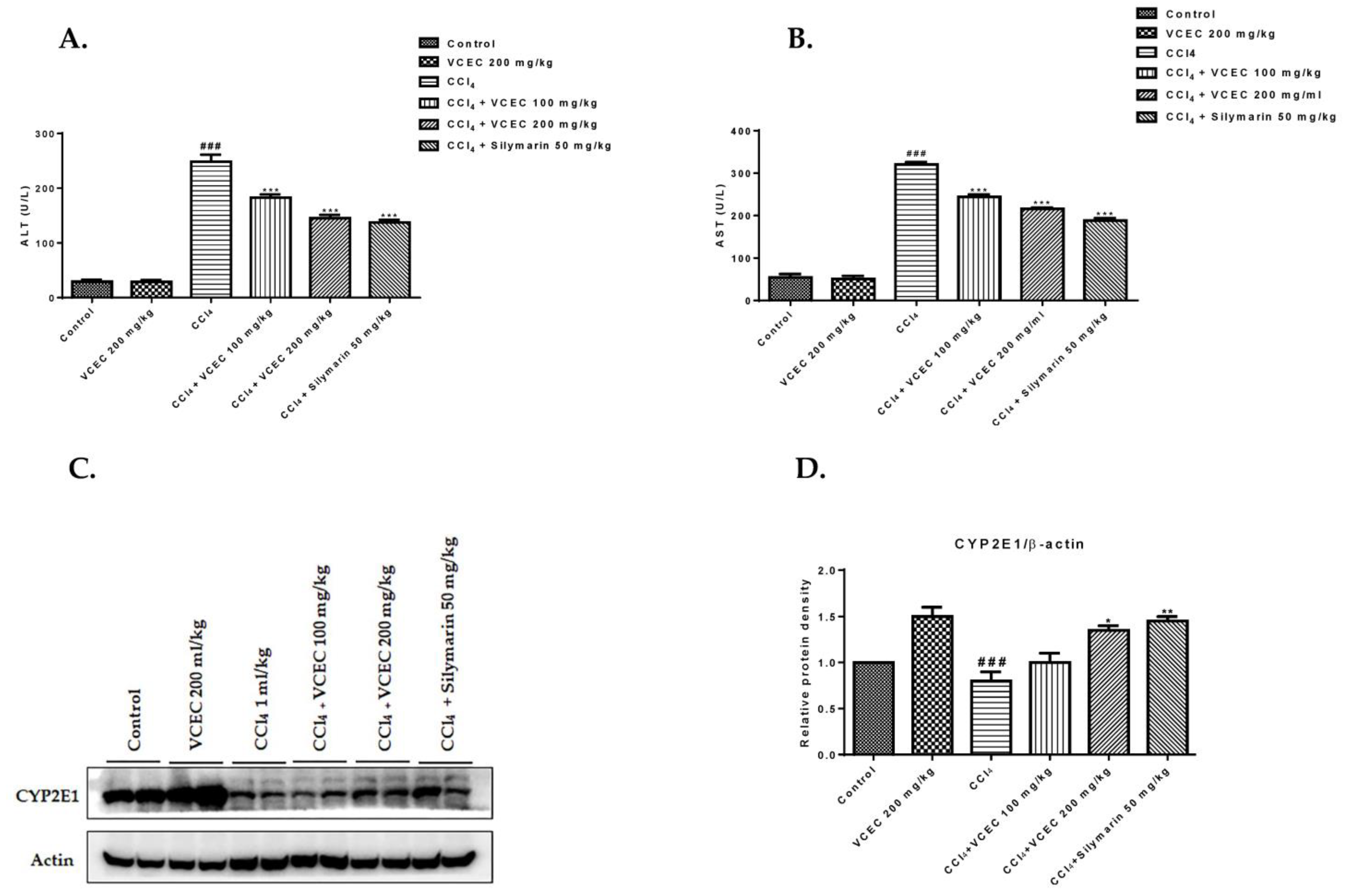
To determine the protective role of VCEC, a mouse model of CCl4-induced acute liver injury was established. To determine liver function, serum ALT, AST, and protein expression of CYP2E1 were evaluated, as shown in Figure 2. The results demonstrate that the CCl4-treated mice had significantly higher levels of serum ALT and AST and dramatically reduced CYP2E1 expression compared to the control group Figure 2A,B. However, pretreatment with VCEC effectively mitigated the elevation in serum ALT and AST activities and restored CYP2E1 expression Figure 2C. This suggests that VCEC possesses hepatoprotective effects against CCl4-induced acute liver injury.

2.3. VCEC Reduces Hepatic Histopathological Damage Caused by CCl4

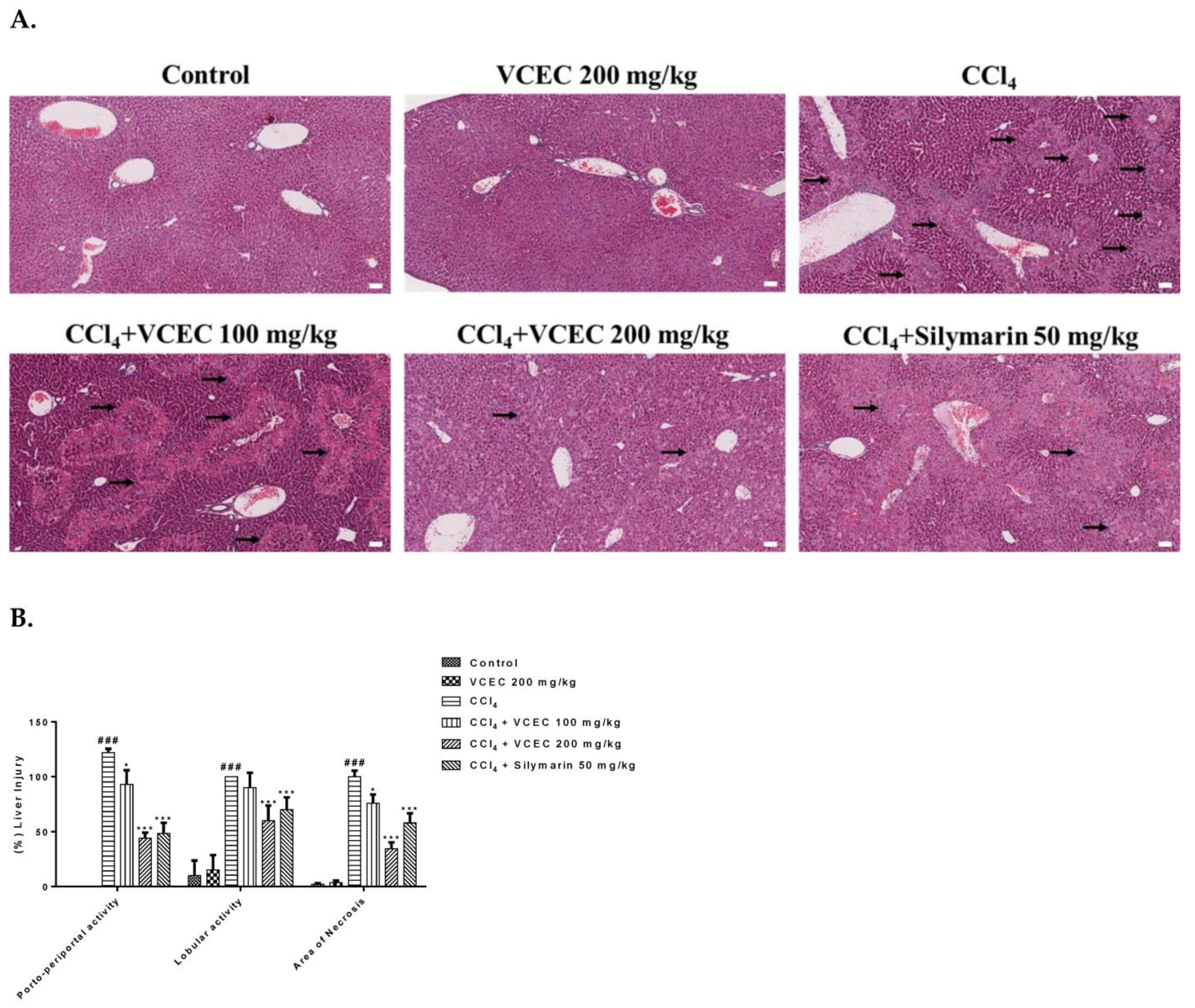
To determine the level of histopathological damage caused by CCl4 and to evaluate the protective role of VCEC, H&E staining of liver sections was performed, as shown in Figure 3. Histopathology caused by CCl4 was manifested by loss of liver structure, hepatocyte necrosis, inflammatory cell infiltration, and increased intercellular gaps and was significantly attenuated by pretreatment with VCEC (100 mg/kg and 200 mg/kg) and silymarin (50 mg/kg), in a dose-dependent manner. The extent of liver injury scoring was performed on the basis of previous studies [16].

2.4. Antioxidant Properties of VCEC against CCl4-Induced Oxidative Stress

The use of antioxidants to protect against CCl4-induced liver injury by reducing lipid peroxidation, inhibiting the production of proinflammatory mediators, and restoring normal antioxidant enzyme levels has gained significant attention in recent years [17,18]. In the present study, we investigated the effects of VCEC on the antioxidant status of the liver.
First, the antioxidant potential of phytochemical extracts on LPS-induced hepatotoxicity was in, as shown in Figure S1. AML12 cells were either left untreated or pretreated with various concentrations of individual extracts VE (4.7, 9.4, 18.8, and 37.8 µg/mL), CE (1.6, 3.2, 6.2, and 12.5 µg/mL), and VCEC (2.5, 6.3, 12.5, 37.5, and 50 µg/mL). Following LPS treatment (5 µg/mL), protein levels of antioxidant-related markers were significantly reduced. However, pretreatment with VCEC remarkably increased the expression of antioxidant-related protein markers such as GPx4, Nrf2, and HO-1 in a concentration-dependent manner. VCEC was much more potent than either VE or CE alone, as shown in Figure S1.
On the basis of preliminary studies, we next established a mouse model of CCl4-induced acute liver injury to investigate the antioxidant mechanism of VCEC. As shown in Figure 4A, pretreatment with VCEC significantly improved the endogenous levels of reduced GSH. Additionally, Western blot analysis was used to measure the levels of GPx4, Cu/Zn SOD, Nrf2, and its target genes, as shown in Figure 4B, VCEC pretreatment significantly restored protein levels of Nrf2, HO-1, GPx4, and Cu/Zn SOD in compared with the CCl4 only treated group. These results suggest that the protective effects of VCEC against CCl4-induced oxidative injury are mediated by enhancing antioxidant defense mechanisms through Nrf2/HO-1 activation and reducing oxidative stress.

2.5. VCEC Alleviated Lipid Peroxidation, Inflammation, and ROS Production in Response to CCl4 Treatment

Measuring inflammatory responses, ROS production, and lipid peroxidation, is important for defining the extent of pathogenesis related to liver injury. Therefore, we first explored the effects of VCEC on ROS production and inflammatory responses. As shown in Figures S2 and S3, LPS treatment dramatically upregulated the levels of intracellular ROS and inflammatory cytokines in AML12 cells; however, VCEC pretreatment inhibited the overproduction of ROS and reduced the expression of inflammatory markers compared to either VE or CE alone.
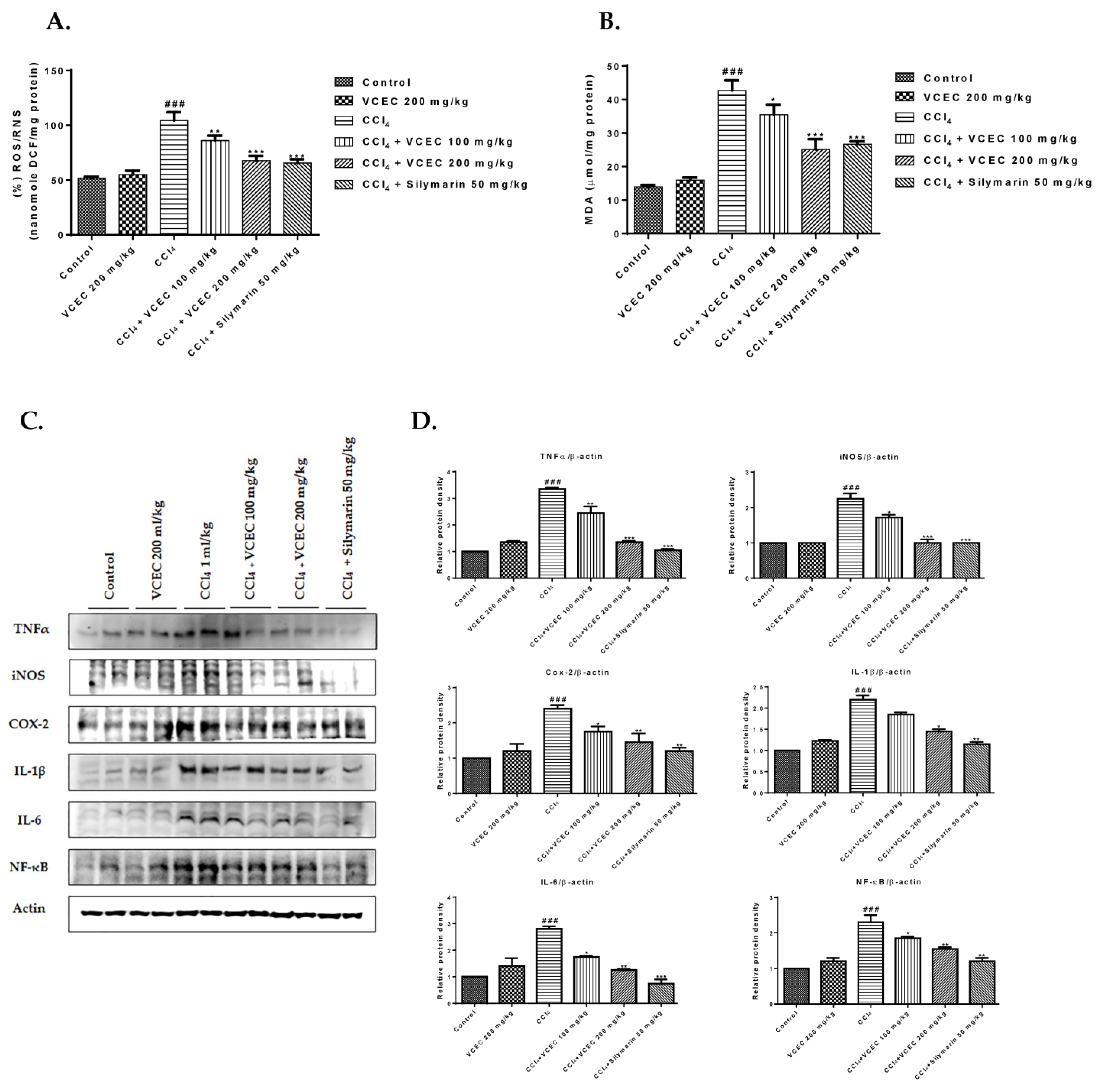
Furthermore, we evaluated the protective effects of VCEC against CCl4-induced acute liver injury in an in vivo model. Specifically, we measured malondialdehyde (MDA), ROS/RNS levels, and inflammatory cytokine protein expression. As shown in Figure 5A,B, VCEC significantly attenuated the levels of MDA and ROS/RNS in liver tissues compared with the CCl4-only-treated group. Similarly, activities of inflammatory cytokines were increased after CCl4 treatment; however, pretreatment with VCEC significantly reduced these (Figure 5C).

2.6. VCEC Inhibits CCl4-Induced Hepatocyte Apoptosis in Mice

Apoptosis serves as a mechanism to eliminate damaged or injured hepatocytes, thus contributing to the progression of liver injury [19]. To evaluate the hepatoprotective effect of VCEC against CCl4-induced apoptosis, we performed immunoblotting. The results, as depicted in Figure 6, demonstrate that CCl4 administration led to a significant decrease in the expression of the antiapoptotic protein Bcl2, along with an increase in the expression of the proapoptotic protein Bax. However, pretreatment with VCEC reversed these effects by restoring Bcl2 levels and reducing Bax. Additionally, VCEC pretreatment reduced CCl4-induced apoptotic responses by ameliorating the expression of cleaved caspase-3, 9, and parp compared with the CCl4-only group. These findings suggest that VCEC exerts a protective effect against CCl4-induced liver injury by suppressing the apoptotic pathway.

3. Discussion

The liver plays a pivotal role in various metabolic processes, including detoxification, protein synthesis, and bile production [1]. Damage in the liver results in the impairment of liver function, causing serious health problems. Among several causes, acute liver injury is a drug-induced liver injury caused by exposure to toxins such as CCl4. CCl4 is a potent liver toxin that can cause severe damage in both animals and humans and is characterized by hepatocellular necrosis, inflammation, and fibrosis [20,21]. Earlier work has shown the core mechanism of CCl4 through the formation of highly reactive free radicals that, in turn, cause oxidative stress, lipid peroxidation, and reactive oxygen species generation (ROS) in hepatocytes [22,23]. The extent of liver injury can be evaluated by measuring biochemical markers such as serum ALT and AST, as well as histological analysis of liver tissue [24]. Therefore, in this study, we evaluated the extent of liver damage in our CCl4 model by use of various biomarkers, such as serum ALT and AST, the level of MDA as a marker for lipid peroxidation, the carbonyl protein group as an indicator of protein oxidation, and TNF-α as an indicator of inflammation. Collectively, the use of these markers can provide a comprehensive assessment of liver damage and oxidative stress, which can help to shed light on the underlying mechanisms behind liver disease and assist in developing new treatments.
Due to potentially serious side effects, toxicity, drug interactions, and resistance, the use of conventional and synthetic drugs for the treatment of liver disease remains controversial. Therefore, the concept of developing plant-derived natural therapeutics is becoming increasingly popular. CE, also known as Gotu Kola, and VE, commonly known as grapevine, are two herbal medicines with antioxidant and anti-inflammatory qualities that can protect cells from oxidative stress and prevent cellular damage [25,26,27]. There is growing evidence to suggest that different herbs may have complementary or synergistic effects when combined, resulting in more potent and effective treatments [28,29]. Previous studies have noted that the antioxidant capacity of VCEC is substantially higher than that of VE or CE alone [25]. Consistent with other studies, we found that the radical scavenging activity of VCEC was significantly greater and exhibited higher, dose-dependent, free radical scavenging capacity than antioxidant positive control vitamin C and VE or CE alone. Several studies have found that VE and CE have potential hepatoprotective activity against CCl4 and ethanol-induced liver toxicity [11,30,31,32,33]. However, more research is needed to fully understand why the combination of the two extracts has synergistic effects in reducing liver damage compared to using either extract alone. In the present study, we attempted to explore the hepatoprotective effects of combining extracts VCEC against CCl4-induced acute liver injury in mice and evaluate its ability to enhance antioxidative effects and reduce both inflammation and ROS.
Oxidative stress and inflammation are two important factors that play a critical role in the pathophysiology of CCl4-induced liver injury [2,4,34]. Antioxidant response elements (AREs) such as Nrf2 and enzymes such as superoxide dismutase (SOD), MDA, catalase, glutathione peroxidase (GPx), heme oxygenase (HO-1), and glutathione reductase (GR), are crucial in mitigating CCl4-induced inflammation in liver injury by scavenging ROS and maintaining the cellular redox balance [35,36]. Accumulating evidence has revealed that parameters such as MDA, SOD, CAT, and reduced glutathione can be used to monitor oxidative stress in mouse models of liver injury. In this study, we showed that VCEC significantly inhibited the production of MDA, restored the levels of reduced GSH, and also enhanced the expression of antioxidant response elements Nrf2 and enzymes HO-1 in the CCl4-damaged liver. Furthermore, scavenging DPPH radicals is a major antioxidation mechanism that inhibits the chain reaction leading to lipid peroxidation [37,38]. The results of VCEC DPPH assays indicate that VCEC had a potent radical scavenging capability, which might be helpful in counteracting the pathological changes induced by the free radical CCl3, which is induced by CCl4. These findings suggest that VCEC effectively protects against CCl4-induced hepatic lipid peroxidation by preventing the downregulation of antioxidant enzyme activities, thereby mitigating oxidative damage to the liver.
Oxidative stress can activate inflammatory pathways, while conversely, inflammation can increase oxidative stress by promoting the production of ROS [39,40]. ROS causes oxidative damage, lipid peroxidation, and DNA damage while activating NF-κB signaling and proinflammatory cytokine production [5,41]. Increased oxidative stress and inflammatory mediators, such as TNF-α, IL-1β, and IL-6, are produced as a result of Kupffer cell activation and further stimulate infiltrating macrophages and neutrophils, whereas inflammation is also associated with the production of inflammatory cytokines, iNOS, and COX-2 that contribute to the pathogenesis of liver injury [2,4,42]. The combination of oxidative stress and the release of inflammatory mediators leads to tissue damage, hepatocyte apoptosis, and the development of liver fibrosis [43]. Prior research has revealed that various plant extracts, including VE and CE, have the ability to induce antioxidant enzymes, regulate oxidative stress pathways, and attenuate the production of inflammatory cytokines, leading to liver prophylaxis [25,44,45]. In our study, CCl4 administration significantly upregulated the expression of TNF-α, IL-1β, IL-6, iNOS, and COX-2, as well as increased the levels of NF-κB. However, VCEC pretreatment dramatically reduced the upregulation of those proinflammatory cytokines as well as transcription factors, suggesting that VCEC can alleviate CCl4-induced liver injury by suppressing the inflammatory response.
Apoptosis and necrosis both contribute to the progression of liver injury [2,4,46]. However, the dominant mode of cell death in CCl4-induced liver injury is still unclear [4,47]. Previous studies have reported contradictory findings, with one suggesting necrotic cell death as the primary mechanism [48], while another proposed the involvement of hepatic cell apoptosis [49]. In this study, we focused on the apoptotic pathway, which involves caspase activation. The significant increases we observed in caspases 3 and 9 indicated induction of apoptosis in response to the CCl4 administration. However, pretreatment with VCEC downregulated caspase expression, indicating its antiapoptotic activity. Furthermore, apoptosis has been associated with ROS production in mitochondria. Various proapoptotic and antiapoptotic proteins, such as Bax and Bcl2, play a role in cellular apoptosis [50,51]. In the present study, VCEC reversed CCl4-induced Bax expression and restored the antiapoptotic protein Bcl2. This is the first time coadministration of these two potent natural extracts has shown a synergistic effect on the apoptosis pathway induced by CCl4 in mice. These findings provide valuable insights into the therapeutic potential of VCEC for the treatment or prevention of liver injury.
In summary, our study demonstrated that the combination of VE and CE extracts had synergistic effects in protecting the liver against CCl4-induced ROS, oxidative stress, and inflammation. Compared with VE or CE alone, VCEC exhibits stronger antioxidant and anti-inflammatory properties, making it a promising approach for managing liver diseases. However, additional research is needed to investigate the absorption mechanisms of VCEC and its bioavailability. Understanding how VCEC is absorbed in the body and finding strategies to increase its availability will contribute to optimizing its therapeutic potential against CCl4-induced liver injury.

4. Materials and Methods

4.1. Chemicals and Reagents

CCl4 was purchased from Sigma-Aldrich Chemical Co., St. Louis, MO, USA, while serum alanine transaminase (ALT, Cat. # AM 102- K) and aspartate transaminase (AST, Cat. # AM 103-K) assay kits were obtained from Asam Pharm. Co. Ltd., South Korea. Reduced glutathione (GSH, Cat. # DIGT-250) assay kits were purchased from BioAssay Systems, Hayward, CA, USA, while malondialdehyde (MDA Cat. # STA-330) and In vitro ROS/RNS (Cat. # STA-347) assay kits were purchased from Cell Biolabs Inc., San Diego, CA, USA.

4.2. Preparation of V. vinifera L. leaf and C. asiatica Extracts

The V. vinifera L. leaf (VE) and C. asiatica (CE) extracts were obtained from the herbal market in Danyan, China. The extracts were authenticated by the Korea Institute of Oriental Medicine and were prepared as described previously [25,52]. Each extract was mixed at a ratio of 1:3 (1 part of VE and 3 part of CE) to prepare the combination drug VCEC.

4.3. DDPH Radical Scavenging Assays

The free radical scavenging activities of VE, CE, and VCEC were evaluated using 2,2-diphenyl-1-picryl-hydrazyl (DDPH). The method was performed as described previously [53]. Briefly, the extracts (2.5 mL) at various concentrations (10, 20, 40, 60, 80, and 100 µg/mL) were added to 1 mL of freshly prepared 0.3 mM alcoholic solution of DDPH. Similarly, standard ascorbic acid solutions of various concentrations were prepared using the same procedure. Control solutions were prepared by replacing the extract with ethanol and 1 mL of DDPH solution. A blank solution was prepared with 3 mL of 95% ethanol. The tube containing the blank, control, and sample was mixed vigorously and allowed to incubate for 30 min in the dark at room temperature. Changes in the absorbance were measured at 518 nm using a spectrophotometer. The percentage of DDPH scavenging activity (% SA) was calculated using the following equation:
(% SA) = (A0 − A1/A0) × 100,
where % SA = percentage scavenging activity;
A0 = absorbance of control;
A1 = absorbance of sample.

4.4. Animals

Specific pathogen-free male C57BL6 mice weighing 20 ± 2 g and aged 7 ~ 8 weeks were purchased from Koatech, Pyeongtake, Korea. Throughout the experiment, mice were provided with a standard diet and had access to water ad libitum. This research adhered to ethical guidelines, and the experimental protocols were approved by the Institutional Animal Care and Use Committee (IACUC) of Jeonbuk National University, Jeonju, South Korea, Approval no: JBUH-IACUC-2023-2.

4.5. Animal Model and Drug Treatment

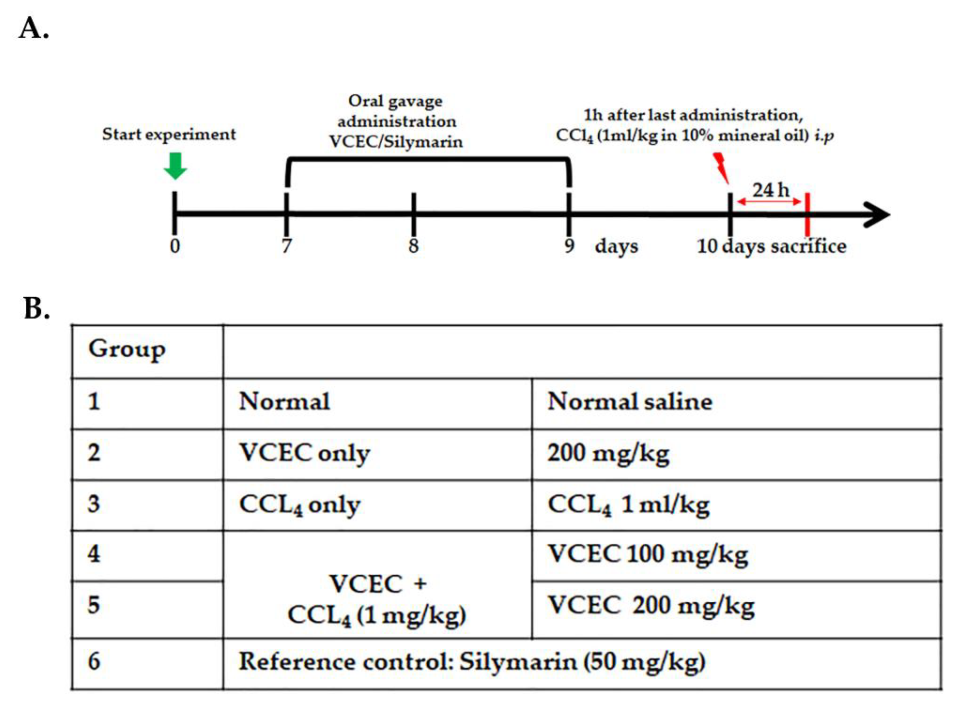
A total of 42 mice were divided into six groups, with each group consisting of seven mice. In the control group (Group I) and negative control group (Group III), mice were treated intraperitoneally with normal saline for three consecutive days. Group II mice received an oral administration of 200 mg/kg VCEC only, while Group VI served as the positive control group and received oral administration of 50 mg/kg silymarin for three days. Groups IV and V, known as the CCl4 + VCEC groups, were orally administered VCEC at doses of 100 and 200 mg/kg for three consecutive days. On the third day, one hour after the final administration of VCEC, all groups except Groups I and II were injected with a single dose of CCl4 (1mg/mL in 10% mineral oil) intraperitoneally. At 24 h after the CCl4 injection, mice from each group were anesthetized, blood samples were collected, and then they were sacrificed. Serum from blood and liver tissues were collected from the mice for further analysis. The experimental timeline and details are noted in Figure 7.

4.6. Determination of Biochemical Parameters

After blood samples were collected, serum was obtained as described previously [2,4]. The enzymatic activities of serum alanine transaminase (ALT) and aspartate aminotransferase (AST) were determined using commercially available detection kits in accordance with the manufacturer’s instructions.

4.7. Liver Histopathological Examination

Liver tissues were fixed in a 4% paraformaldehyde solution and embedded in paraffin. Tissue sections of 5 µm thickness were prepared and subsequently deparaffinized using xylene. The sections were stained with hematoxylin and eosin (H&E) and observed under a light microscope at a magnification of 100× to determine the tissue morphology. To quantify the degree of liver injury, five randomly selected areas were analyzed using image analysis software. The software enabled the assessment of necrotic foci in liver tissue.

4.8. Measurements of Reduced Glutathione (GSH) and Malondialdehyde (MDA)

To determine the amount of reduced GSH and MDA, liver tissues were homogenized and centrifuged at 12,000 rpm for 15 min at 4 °C. Then the resultant supernatant was collected to measure the levels of reduced GSH and MDA using commercially available assay kits from BioAssay Systems (Hayward, CA, USA) and Cell Biolabs, Inc. (San Diego, CA, USA), respectively, following the manufacturer’s instructions. The measurements of reduced GSH were performed spectrophotometrically at a wavelength of 405 nm, while the amount of MDA was determined by measuring the absorbance of the colored complex at a wavelength of 532 nm by kinetic spectrophotometer.

4.9. Determination of ROS Formation

In order to assess the intracellular levels of reactive oxygen species (ROS) in AML12 cells, the DCFH-DA staining technique was used. After treatment with desired drugs for 24-h, the cells were exposed to serum-free media containing 10 µmol/L DCFH-DA for 45 min at 37 °C. The specific absorbance of the fluorescent product DCF was quantified using flow cytometry, following the instructions provided by the manufacturer. Furthermore, OxiSelectTM In Vitro ROS/RNS assay kit Cell Biolabs, Inc. (San Diego, CA, USA) was used to quantify the levels of ROS in mice with CCl4-induced liver injury. Liver tissue lysates were homogenized and centrifuged at 10,000× g for 5 min at 4 °C. The supernatants from homogenized lysates were incubated with DCF-DA solution for 15–45 min at room temperature, and then the fluorescence was observed at 480 nm excitation/530 nm emission wavelengths using a SpectraMax Gemini XS fluorometer as described by manufacturer’s instructions.

4.10. Immunoblotting

Cell lysates and liver tissue homogenates were prepared and quantified as described previously [2,4]. Proteins were separated using SDS-PAGE gels and electrotransferred onto PVDF membranes. The membranes were then blocked for 1 h using 5% skim milk and subsequently incubated with specific primary antibodies and horseradish peroxidase-conjugated secondary antibodies. The primary and corresponding secondary antibodies used in this study are listed in Table 1. After incubation with respective primary and secondary antibodies, the protein signals were enhanced and detected using a chemiluminescence detection device.

4.11. Statistical Analysis

The findings presented in this study are derived from a minimum of three repeated experiments and are reported as mean values with corresponding standard deviations. Statistical analysis was conducted using GraphPad Prism software (Graph Pad v5 Software, San Diego, CA, USA). The significance of the data was assessed using one-way ANOVA, followed by Tukey’s post hoc test. A p-value of less than 0.05 (p < 0.05) was considered statistically significant.

5. Conclusions

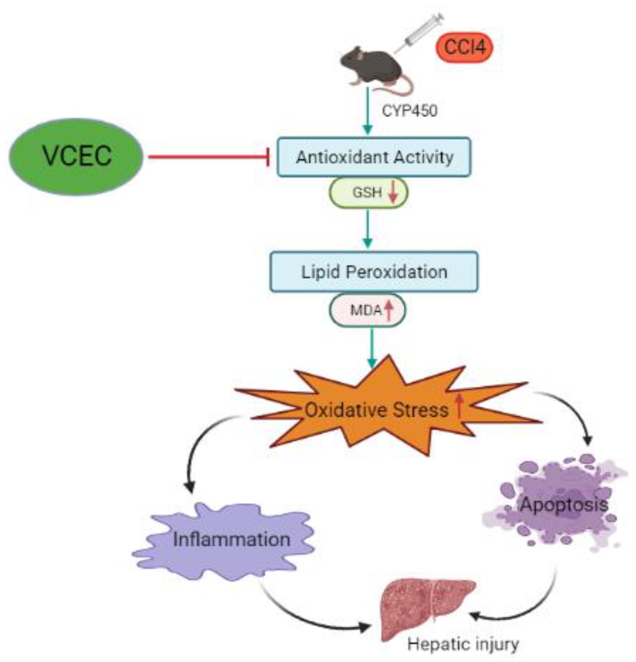
We evaluated the hepato-protective and synergistic effects of VCEC on CCl4-induced acute liver injury in mice and explored the underlying mechanisms. The results indicate that VCEC mitigated CCl4-induced damage, as shown by reduced liver histopathology and serum ALT and AST levels. The combined phytochemical extracts that comprise VCEC primarily ameliorate liver injury by inhibiting the NF-κB pathway, with subsequent suppression of inflammatory responses. In addition, VCEC alleviated hepatic oxidative stress by activating antioxidant response elements. These distinct mechanisms of action highlight the potential of VCEC as a therapeutic agent for liver injury and suggest that the effects may be attributed to different pathways that target inflammation and oxidative stress, respectively (Figure 8).

Supplementary Materials

The following supporting information can be downloaded at: https://www.mdpi.com/article/10.3390/ijms241411255/s1, Figure S1: VCEC regulates antioxidant activity in LPS-induced hepatoxicity; Figure S2: VCEC protects against CCl4-induced ROS production; Figure S3: VCEC protects against LPS-induced inflammation in hepatocytes.

Author Contributions

Conceptualization, S.M. and Y.J.J.; methodology, S.M. and P.R.; software, S.M. and Y.G.; validation, S.M. and Y.J.J.; formal analysis, S.M., J.S., H.S.P., G.-H.L., and Y.G.; investigation, S.M.; resources, S.M. and Y.J.J.; data curation, S.M., J.S., Y.G., and H.S.P.; writing—original draft preparation, S.M.; writing—review and editing, S.M., Y.G., and Y.J.J.; visualization, S.M. and Y.J.J.; supervision, Y.J.J.; project administration, S.M. and Y.J.J. All authors have read and agreed to the published version of the manuscript.

Funding

This study was supported by funding from the Biomedical Research Institute, Jeonbuk National University Hospital.

Institutional Review Board Statement

Not applicable.

Informed Consent Statement

Not applicable.

Data Availability Statement

Not applicable.

Conflicts of Interest

The authors have no conflicts of interest to disclose. The funders had no role in the design of the study, data collection, data analysis, data interpretation, writing of the manuscript, or the decision to publish this study.

References

  1. Ozougwu, J.C. Physiology of the liver. Int. J. Res. Pharm. Biosci. 2017, 4, 13–24. [Google Scholar]
  2. Munakarmi, S.; Gurau, Y.; Shrestha, J.; Risal, P.; Park, H.S.; Shin, H.B.; Jeong, Y.J. Hepatoprotective Effects of a Natural Flavanol 3,3&prime;-Diindolylmethane against CCl4-Induced Chronic Liver Injury in Mice and TGF&beta;1-Induced EMT in Mouse Hepatocytes via Activation of Nrf2 Cascade. Int. J. Mol. Sci. 2022, 23, 11407. [Google Scholar]
  3. Cichoż-Lach, H.; Michalak, A. Oxidative stress as a crucial factor in liver diseases. World J. Gastroenterol. WJG 2014, 20, 8082. [Google Scholar] [CrossRef]
  4. Munakarmi, S.; Chand, L.; Shin, H.B.; Jang, K.Y.; Jeong, Y.J. Indole-3-Carbinol Derivative DIM Mitigates Carbon Tetrachloride-Induced Acute Liver Injury in Mice by Inhibiting Inflammatory Response, Apoptosis and Regulating Oxidative Stress. Int. J. Mol. Sci. 2020, 21, 2048. [Google Scholar] [CrossRef] [Green Version]
  5. Xu, D.; Xu, M.; Jeong, S.; Qian, Y.; Wu, H.; Xia, Q.; Kong, X. The role of Nrf2 in liver disease: Novel molecular mechanisms and therapeutic approaches. Front. Pharmacol. 2019, 9, 1428. [Google Scholar] [CrossRef] [Green Version]
  6. Koruk, M.; Taysi, S.; Savas, M.C.; Yilmaz, O.; Akcay, F.; Karakok, M. Oxidative stress and enzymatic antioxidant status in patients with nonalcoholic steatohepatitis. Ann. Clin. Lab. Sci. 2004, 34, 57–62. [Google Scholar] [PubMed]
  7. De Minicis, S.; Brenner, D.A. Oxidative stress in alcoholic liver disease: Role of NADPH oxidase complex. J. Gastroenterol. Hepatol. 2008, 23, S98–S103. [Google Scholar] [CrossRef] [PubMed]
  8. Florescu, I.E.; Georgescu, S.E.; Dudu, A.; Balaș, M.; Voicu, S.; Grecu, I.; Dediu, L.; Dinischiotu, A.; Costache, M. Oxidative stress and antioxidant defense mechanisms in response to starvation and refeeding in the intestine of stellate sturgeon (Acipenser stellatus) juveniles from aquaculture. Animals 2021, 11, 76. [Google Scholar] [CrossRef] [PubMed]
  9. Azab, K.S.; Mostafa, A.-H.A.; Ali, E.M.; Abdel-Aziz, M.A. Cinnamon extract ameliorates ionizing radiation-induced cellular injury in rats. Ecotoxicol. Environ. Saf. 2011, 74, 2324–2329. [Google Scholar] [CrossRef]
  10. Mallikarjuna, K.; Shanmugam, K.; Nishanth, K.; Wu, M.-C.; Hou, C.-W.; Kuo, C.-H.; Reddy, K.S. Alcohol-induced deterioration in primary antioxidant and glutathione family enzymes reversed by exercise training in the liver of old rats. Alcohol 2010, 44, 523–529. [Google Scholar] [CrossRef]
  11. Orhan, D.D.; Orhan, N.; Ergun, E.; Ergun, F. Hepatoprotective effect of Vitis vinifera L. leaves on carbon tetrachloride-induced acute liver damage in rats. J. Ethnopharmacol. 2007, 112, 145–151. [Google Scholar] [CrossRef]
  12. Fong, L.Y.; Ng, C.T.; Cheok, Z.L.; Moklas, M.A.M.; Hakim, M.N.; Ahmad, Z. Barrier protective effect of asiatic acid in TNF-α-induced activation of human aortic endothelial cells. Phytomedicine 2016, 23, 191–199. [Google Scholar] [CrossRef] [PubMed]
  13. Puglisi, R.; Severgnini, A.; Tava, A.; Montedoro, M. In vitro assessment of the antioxidant properties of aqueous byproduct extracts of Vitis vinifera. Food Technol. Biotechnol. 2019, 57, 119–125. [Google Scholar] [CrossRef] [PubMed]
  14. Somchit, M.; Sulaiman, M.; Zuraini, A.; Samsuddin, L.; Somchit, N.; Israf, D.; Moin, S. Antinociceptive and antiinflammatory effects of Centella asiatica. Indian J. Pharmacol. 2004, 36, 377. [Google Scholar]
  15. Pari, L.; Suresh, A. Effect of grape (Vitis vinifera L.) leaf extract on alcohol induced oxidative stress in rats. Food Chem. Toxicol. 2008, 46, 1627–1634. [Google Scholar] [CrossRef]
  16. Park, Y.-N.; Kim, H.-G.; Chon, C.-Y.; Park, J.-B.; Sohn, J.-H.; Yang, S.-H.; Yu, E.-S.; Lee, M.-S.; Jang, J.-J.; Chang, H.-K. Histological grading and staging of chronic hepatitis standardized guideline proposed by the Korean Study Group for the Pathology of Digestive Diseases. Korean J. Pathol. 1999, 33, 337–346. [Google Scholar]
  17. Patel, M.; Mehta, P. Bamboo a supplement to human health: A comprehensive review on its ethnopharmacology, phytochemistry, and pharmacological activity. Nat. Prod. J. 2021, 11, 140–168. [Google Scholar] [CrossRef]
  18. Paul Kamdem, J.; Olalekan Abolaji, A.; Olusola Elekofehinti, O.; Olaposi Omotuyi, I.; Ibrahim, M.; Hassan, W.; Vargas Barbosa, N.; Onofre Souza, D.; Batista Teixeira da Rocha, J. Therapeutic potential of plant extracts and phytochemicals against brain ischemia-reperfusion injury: A review. Nat. Prod. J. 2016, 6, 250–284. [Google Scholar] [CrossRef]
  19. Chen, L.; Zhen, X.; Jiang, X. Activatable Optical Probes for Fluorescence and Photoacoustic Imaging of Drug-Induced Liver Injury. Adv. NanoBiomed Res. 2022, 2, 2200097. [Google Scholar] [CrossRef]
  20. McGill, M.R.; Jaeschke, H. Animal models of drug-induced liver injury. Biochim. Biophys. Acta (BBA)-Mol. Basis Dis. 2019, 1865, 1031–1039. [Google Scholar] [CrossRef]
  21. El-Hadary, A.E.; Ramadan Hassanien, M.F. Hepatoprotective effect of cold-pressed Syzygium aromaticum oil against carbon tetrachloride (CCl4)-induced hepatotoxicity in rats. Pharm. Biol. 2016, 54, 1364–1372. [Google Scholar] [CrossRef] [Green Version]
  22. Xu, G.; Han, X.; Yuan, G.; An, L.; Du, P. Screening for the protective effect target of deproteinized extract of calf blood and its mechanisms in mice with CCl4-induced acute liver injury. PLoS ONE 2017, 12, e0180899. [Google Scholar] [CrossRef] [PubMed] [Green Version]
  23. Clawson, G.A. Mechanisms of carbon tetrachloride hepatotoxicity. Pathol. Immunopathol. Res. 1989, 8, 104–112. [Google Scholar] [CrossRef] [PubMed]
  24. Ozer, J.; Ratner, M.; Shaw, M.; Bailey, W.; Schomaker, S. The current state of serum biomarkers of hepatotoxicity. Toxicology 2008, 245, 194–205. [Google Scholar] [CrossRef]
  25. Jeon, S.Y.; Kim, M.R.; Yu, S.H.; Kim, M.J.; Shim, K.-S.; Shin, E.; Lee, J.J.; Lee, Y.C. Combined extract of Vitis vinifera L. and Centella asiatica synergistically attenuates oxidative damage induced by hydrogen peroxide in human umbilical vein endothelial cells. Prev. Nutr. Food Sci. 2020, 25, 173. [Google Scholar] [CrossRef]
  26. Gonçalves, P.B.; Romeiro, N.C. Multi-target natural products as alternatives against oxidative stress in Chronic Obstructive Pulmonary Disease (COPD). Eur. J. Med. Chem. 2019, 163, 911–931. [Google Scholar] [CrossRef] [PubMed]
  27. Mukherjee, P.K.; Maity, N.; Nema, N.K.; Sarkar, B.K. Bioactive compounds from natural resources against skin aging. Phytomedicine 2011, 19, 64–73. [Google Scholar] [CrossRef]
  28. Rizeq, B.; Gupta, I.; Ilesanmi, J.; AlSafran, M.; Rahman, M.; Ouhtit, A. The power of phytochemicals combination in cancer chemoprevention. J. Cancer 2020, 11, 4521–4533. [Google Scholar] [CrossRef]
  29. Che, C.-T.; Wang, Z.J.; Chow, M.S.S.; Lam, C.W.K. Herb-herb combination for therapeutic enhancement and advancement: Theory, practice and future perspectives. Molecules 2013, 18, 5125–5141. [Google Scholar] [CrossRef] [Green Version]
  30. Liu, T.; Zhao, J.; Ma, L.; Ding, Y.; Su, D. Hepatoprotective effects of total triterpenoids and total flavonoids from Vitis vinifera L against immunological liver injury in mice. Evid.-Based Complement. Altern. Med. 2012, 2012, 969386. [Google Scholar] [CrossRef] [Green Version]
  31. Giribabu, N.; Karim, K.; Kilari, E.K.; Kassim, N.M.; Salleh, N. Anti-inflammatory, antiapoptotic and proproliferative effects of vitis vinifera seed ethanolic extract in the liver of streptozotocin-nicotinamide-induced type 2 diabetes in male rats. Can. J. Diabetes 2018, 42, 138–149. [Google Scholar] [CrossRef]
  32. Antony, B.; Santhakumari, G.; Merina, B.; Sheeba, V.; Mukkadan, J. Hepatoprotective effect of Centella asiatica (L) in carbon tetrachloride-induced liver injury in rats. Indian J. Pharm. Sci. 2006, 68, 772. [Google Scholar] [CrossRef] [Green Version]
  33. Park, D.W.; Jeon, H.; Kwon, J.E.; Lee, Y.G.; So, R.; Choe, T.H.; Jeong, Y.J.; Kang, S.C. Hepatoprotective effect of Centella asiatica 50% ethanol extract against acetaminophen-induced acute liver injury in BALB/c mice. Toxicol. Res. 2021, 37, 261–275. [Google Scholar] [CrossRef]
  34. Ben Hsouna, A.; Hfaiedh, M.; Ben Slima, S.; Romdhane, W.B.; Akacha, B.B.; Bouterra, M.T.; Dhifi, W.; Mnif, W.; Brini, F.; Ben Saad, R. Antioxidant and hepatoprotective effects of novel heteropolysaccharide isolated from Lobularia maritima on CCl4-induced liver injury in rats. Food Sci. Nutr. 2022, 10, 2271–2284. [Google Scholar] [CrossRef] [PubMed]
  35. Zhao, Z.-W.; Chang, J.-C.; Lin, L.-W.; Tsai, F.-H.; Chang, H.-C.; Wu, C.-R. Comparison of the hepatoprotective effects of four endemic Cirsium species extracts from Taiwan on CCl4-induced acute liver damage in C57BL/6 mice. Int. J. Mol. Sci. 2018, 19, 1329. [Google Scholar] [CrossRef] [Green Version]
  36. Ponnusam, Y.; Louis, T.; Madhavachandran, V.; Kumar, S.; Thoprani, N.; Hamblin, M.R.; Lakshmanan, S. Antioxidant activity of the ancient herb, Holy Basil in CCl4-induced liver injury in rats. Ayurvedic 2015, 2, 34. [Google Scholar] [CrossRef] [PubMed] [Green Version]
  37. Rekka, E.; Kourounakis, P.N. Effect of hydroxyethyl rutosides and related compounds on lipid peroxidation and free radical scavenging activity. Some structural aspects. J. Pharm. Pharmacol. 1991, 43, 486–491. [Google Scholar] [CrossRef] [PubMed]
  38. Yang, J.; Li, Y.; Wang, F.; Wu, C. Hepatoprotective effects of apple polyphenols on CCl4-induced acute liver damage in mice. J. Agric. Food Chem. 2010, 58, 6525–6531. [Google Scholar] [CrossRef]
  39. Yuan, T.; Yang, T.; Chen, H.; Fu, D.; Hu, Y.; Wang, J.; Yuan, Q.; Yu, H.; Xu, W.; Xie, X. New insights into oxidative stress and inflammation during diabetes mellitus-accelerated atherosclerosis. Redox Biol. 2019, 20, 247–260. [Google Scholar] [CrossRef]
  40. Chaudhari, N.; Talwar, P.; Parimisetty, A.; Lefebvre d’Hellencourt, C.; Ravanan, P. A molecular web: Endoplasmic reticulum stress, inflammation, and oxidative stress. Front. Cell. Neurosci. 2014, 8, 213. [Google Scholar] [CrossRef] [Green Version]
  41. Ma, J.-Q.; Ding, J.; Xiao, Z.-H.; Liu, C.-M. Ursolic acid ameliorates carbon tetrachloride-induced oxidative DNA damage and inflammation in mouse kidney by inhibiting the STAT3 and NF-κB activities. Int. Immunopharmacol. 2014, 21, 389–395. [Google Scholar] [CrossRef]
  42. Bilzer, M.; Roggel, F.; Gerbes, A.L. Role of Kupffer cells in host defense and liver disease. Liver Int. 2006, 26, 1175–1186. [Google Scholar] [CrossRef] [PubMed]
  43. Farzanegi, P.; Dana, A.; Ebrahimpoor, Z.; Asadi, M.; Azarbayjani, M.A. Mechanisms of beneficial effects of exercise training on non-alcoholic fatty liver disease (NAFLD): Roles of oxidative stress and inflammation. Eur. J. Sport Sci. 2019, 19, 994–1003. [Google Scholar] [CrossRef] [PubMed]
  44. Martín, M.Á.; Serrano, A.B.G.; Ramos, S.; Pulido, M.I.; Bravo, L.; Goya, L. Cocoa flavonoids up-regulate antioxidant enzyme activity via the ERK1/2 pathway to protect against oxidative stress-induced apoptosis in HepG2 cells. J. Nutr. Biochem. 2010, 21, 196–205. [Google Scholar] [CrossRef] [PubMed]
  45. Giribabu, N.; Karim, K.; Kilari, E.K.; Nelli, S.R.; Salleh, N. Oral administration of Centella asiatica (L.) Urb leave aqueous extract ameliorates cerebral oxidative stress, inflammation, and apoptosis in male rats with type-2 diabetes. Inflammopharmacology 2020, 28, 1599–1622. [Google Scholar] [CrossRef] [PubMed]
  46. Alkhouri, N.; Carter-Kent, C.; Feldstein, A.E. Apoptosis in nonalcoholic fatty liver disease: Diagnostic and therapeutic implications. Expert Rev. Gastroenterol. Hepatol. 2011, 5, 201–212. [Google Scholar] [CrossRef] [PubMed] [Green Version]
  47. Ma, X.; Zhou, Y.; Qiao, B.; Jiang, S.; Shen, Q.; Han, Y.; Liu, A.; Chen, X.; Wei, L.; Zhou, L. Androgen aggravates liver fibrosis by activation of NLRP3 inflammasome in CCl4-induced liver injury mouse model. Am. J. Physiol.-Endocrinol. Metab. 2020, 318, E817–E829. [Google Scholar] [CrossRef] [PubMed]
  48. Adelusi, O.B.; Ramachandran, A.; Lemasters, J.J.; Jaeschke, H. The role of Iron in lipid peroxidation and protein nitration during acetaminophen-induced liver injury in mice. Toxicol. Appl. Pharmacol. 2022, 445, 116043. [Google Scholar] [CrossRef]
  49. Tong, J.; Lan, X.-T.; Zhang, Z.; Liu, Y.; Sun, D.-Y.; Wang, X.-J.; Ou-Yang, S.-X.; Zhuang, C.-L.; Shen, F.-M.; Wang, P. Ferroptosis inhibitor liproxstatin-1 alleviates metabolic dysfunction-associated fatty liver disease in mice: Potential involvement of PANoptosis. Acta Pharmacol. Sin. 2023, 44, 1014–1028. [Google Scholar] [CrossRef]
  50. Chota, A.; George, B.P.; Abrahamse, H. Interactions of multidomain pro-apoptotic and anti-apoptotic proteins in cancer cell death. Oncotarget 2021, 12, 1615. [Google Scholar] [CrossRef]
  51. Tsujimoto, Y. Cell death regulation by the Bcl-2 protein family in the mitochondria. J. Cell. Physiol. 2003, 195, 158–167. [Google Scholar] [CrossRef] [PubMed]
  52. Seo, M.-G.; Jo, M.-J.; Hong, N.I.; Kim, M.J.; Shim, K.S.; Shin, E.; Lee, J.J.; Park, S.-J. Anti-inflammatory and anti-vascular leakage effects by combination of Centella asiatica and Vitis vinifera L. Leaf extracts. Evid.-Based Complement. Altern. Med. 2021, 2021, 7381620. [Google Scholar] [CrossRef] [PubMed]
  53. Cai, L.; Zou, S.; Liang, D.; Luan, L. Structural characterization, antioxidant and hepatoprotective activities of polysaccharides from Sophorae tonkinensis Radix. Carbohydr. Polym. 2018, 184, 354–365. [Google Scholar] [CrossRef] [PubMed]
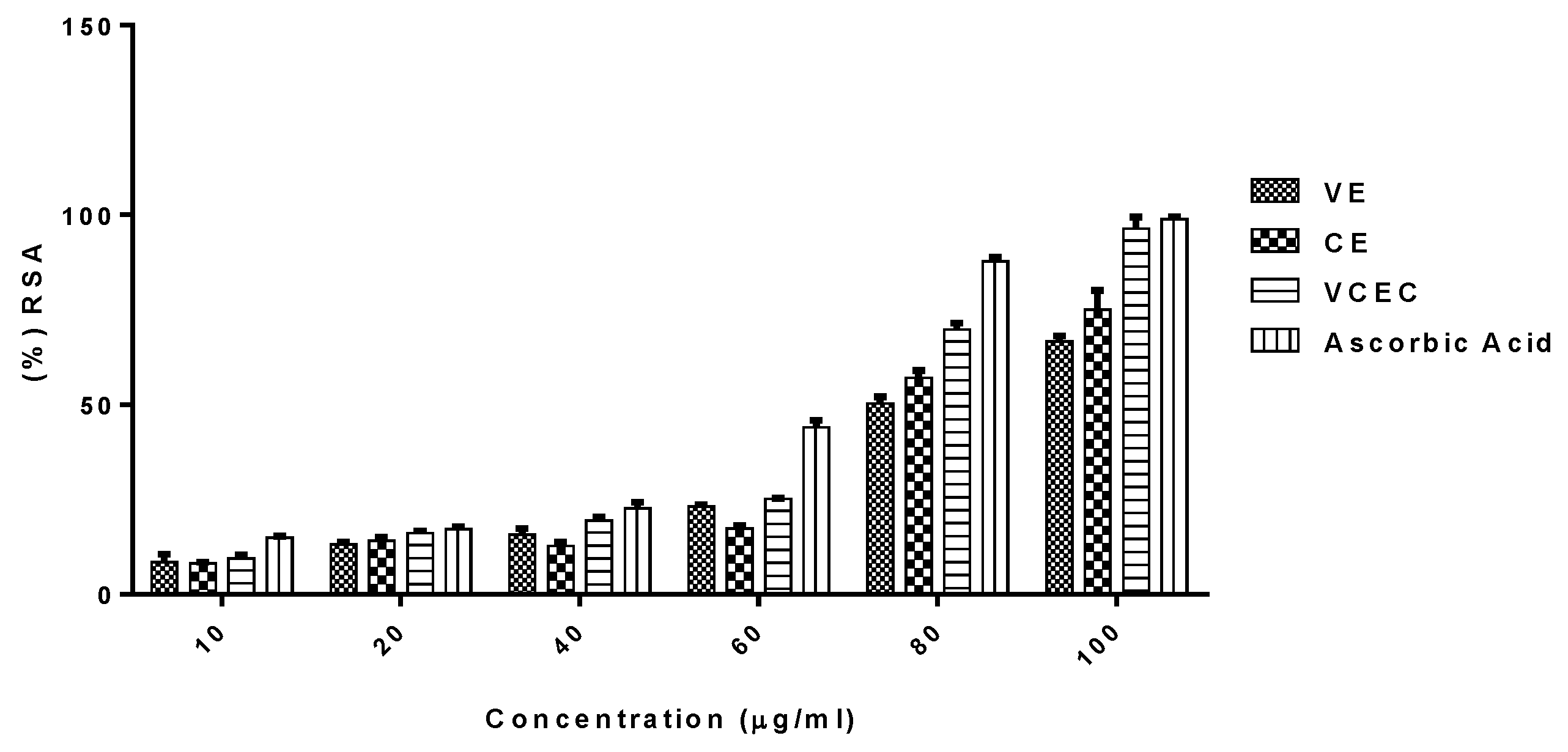
Figure 1. Potential antioxidant properties of VE, CE, and VCEC in comparison with vitamin C. The antioxidant activities of VE, CE, and VCEC were evaluated by DPPH radical scavenging assay.
Ijms 24 11255 g001
Figure 2. VCEC pretreatment ameliorates CCl4-induced acute liver injury in mice. (A) Serum levels of alanine aminotransferase (ALT) and (B) aspartate aminotransferase (AST) were analyzed 24 h after CCl4 injection. Values are presented as mean ± SD (n = 7). (C) Protein expression of cytochrome P450 2E1 (CYP2E1) 24 h after CCl4 injection. (D) Quantification of relative protein expression normalized to β-actin. Data are expressed as mean ± SD (n = 3). ### p < 0.001 denotes a significant difference compared to the control group; * p < 0.05, ** p < 0.01, and *** p < 0.001 indicates significant differences compared to the CCl4-treated group.
Ijms 24 11255 g002
Figure 3. VCEC reduces the histopathological changes caused by CCl4 in liver tissues. (A) Representative H&E-stained liver sections from the indicated groups with necrotic areas and liver damage as indicated by black arrows. (B) Quantitative assessment of (%) area score of liver damage in the indicated groups. The H&E-stained tissues were observed under light microscope at magnification of ×100. The white bar in figure represents the scale bar of 50 µmm, respectively. The values presented are expressed as the mean ± SD (n = 7). ### p < 0.001 denotes significancy compared with the control group; * p < 0.05 and *** p < 0.001 indicates significant differences compared with the CCl4-treated group.
Ijms 24 11255 g003
Figure 4. VCEC enhanced antioxidant activity by regulating the Nrf2/HO-1 pathway and inhibiting oxidative stress in CCl4-induced acute liver injury in mice. (A) Reduced glutathione (GSH) levels. Data presented are expressed as mean ± SD (n = 7). (B) Western blot analysis determining the protein expression of antioxidant-related markers GPx4, Nrf2, HO-1, and Cu/Zn SOD. (C) Quantification of relative protein expression normalized to β-actin. The values presented are expressed as mean ± SD (n = 3). ### p < 0.001 denotes significancy compared with the control group; * p < 0.05, ** p < 0.01, and *** p < 0.001 indicates significant differences compared with the CCl4-treated group.
Ijms 24 11255 g004
Figure 5. Effects of VCEC extracts on levels of MDA, ROS/RNS, and protein expression level of inflammatory cytokine in CCl4-induced acute liver injury. (A) Lipid peroxidation as determined by MDA content; (B) ROS/RNS level. Data presented are expressed as mean ± SD (n = 7). (C) Protein expression level of inflammatory cytokines. (D) Quantification of relative protein expression normalized to β-actin. Data are expressed as mean ± SD (n = 3). ### p < 0.001 denotes significancy compared with the control group; * p < 0.05, ** p < 0.01, and *** p < 0.001 indicates significant differences compared with the CCl4-treated group.
Ijms 24 11255 g005
Figure 6. VCEC pretreatment ameliorates the CCl4-induced apoptotic response. (A) Immunoblot analyses showing the levels of apoptotic proteins (cleaved caspsase-3, 9, and cleaved parp), proapoptotic protein Bax, and antiapoptotic protein Bcl2. (B) Quantification of relative protein expression normalized to β-actin. The values represent the mean ± SD (n = 3). ### p < 0.001 denotes significancy compared with the control group; * p < 0.05, ** p < 0.01, and *** p < 0.001 indicates significant differences compared with the CCl4-treated group.
Ijms 24 11255 g006
Figure 7. An experimental procedure involved the establishment of a CCl4-induced acute liver injury model and subsequent treatment. (A) A diagram illustrates the drug administration route and time points at which the animals were sacrificed following CCl4 administration. (B) A table outlining the different groups along with their corresponding doses of CCl4.
Ijms 24 11255 g007
Figure 8. A schematic diagram of the proposed mechanism by which VCEC exerts its effects on CCl4-induced acute liver injury through multiple pathways. CCl4 exposure leads to a decrease in antioxidant capacity, represented by reduced levels of SOD, GSH, and Nrf2, indicated by downward red arrows. Additionally, CCl4 administration increases inflammatory responses, oxidative stress, and apoptotic protein levels, depicted by upward red arrows. However, VCEC pretreatment counteracts these effects.
Ijms 24 11255 g008
Table 1. List of specific primary and secondary antibodies used for immunoblotting.
S.NTargetBlocking SolutionDilutionSecondaryManufacturerCatalogue Number
1β-actin5% Skim Milk1:3000Mouse IgGSigma AldrichA5441
2COX-25% BSA1:1000Rabbit IgGCell Signaling4842S
3C-Caspase-95% BSA1:1000Mouse IgGCell Signaling9508S
4Parp5% Skim Milk1:3000Rabbit IgGSanta CruzSc-7150
5Keap-15% Skim Milk1:3000Mouse IgGSanta CruzSc-365626
6Nrf-25% BSA1:1000Rabbit IgGGene TexGTX103322
7NQO15% Skim Milk1:3000Mouse IgGSanta CruzSc-32793
8HO-15% Skim Milk1:3000Mouse IgGSanta CruzSc-136960
9GPX45% Skim Milk1:3000Mouse IgGSanta CruzSc-166570
10Cu/Zn SOD5% BSA1:1000Rabbit IgGEnzoADI-SOD-100
11Mn SOD5% BSA1:3000Rabbit IgGEnzoADI-SOD-110
12iNOS5% BSA1:1500Mouse IgGR&D SystemMAB9502
13NF-κB5% Skim Milk1:3000Rabbit IgGSanta CruzSc-8008
14IL-1β5% Skim Milk1:3000Rabbit IgGSanta CruzSc-7884
15IL-65% BSA1:1000Rabbit IgGBio worldBS-6419
16TNF-α5% Skim Milk1:3000Hamster IgGSanta CruzSc-12744
17CYP2E15% BSA1:1000Rabbit IgGAbcamab28146
18Bax5% Skim Milk1:3000Mouse IgGSanta CruzSc-7480
19Bcl25% Skim Milk1:3000Mouse IgGSanta CruzSc-7382
20Caspase-35% Skim Milk1:3000Rabbit IgGSanta CruzSc-7148
21C-Caspase-35% BSA1:1000Rabbit IgGCell Signaling#9664
Disclaimer/Publisher’s Note: The statements, opinions and data contained in all publications are solely those of the individual author(s) and contributor(s) and not of MDPI and/or the editor(s). MDPI and/or the editor(s) disclaim responsibility for any injury to people or property resulting from any ideas, methods, instructions or products referred to in the content.