
Insight into Unusual Supramolecular Self-Assemblies of Terthiophenes Directed by Weak Hydrogen Bonding


Abstract

1. Introduction
2. Results
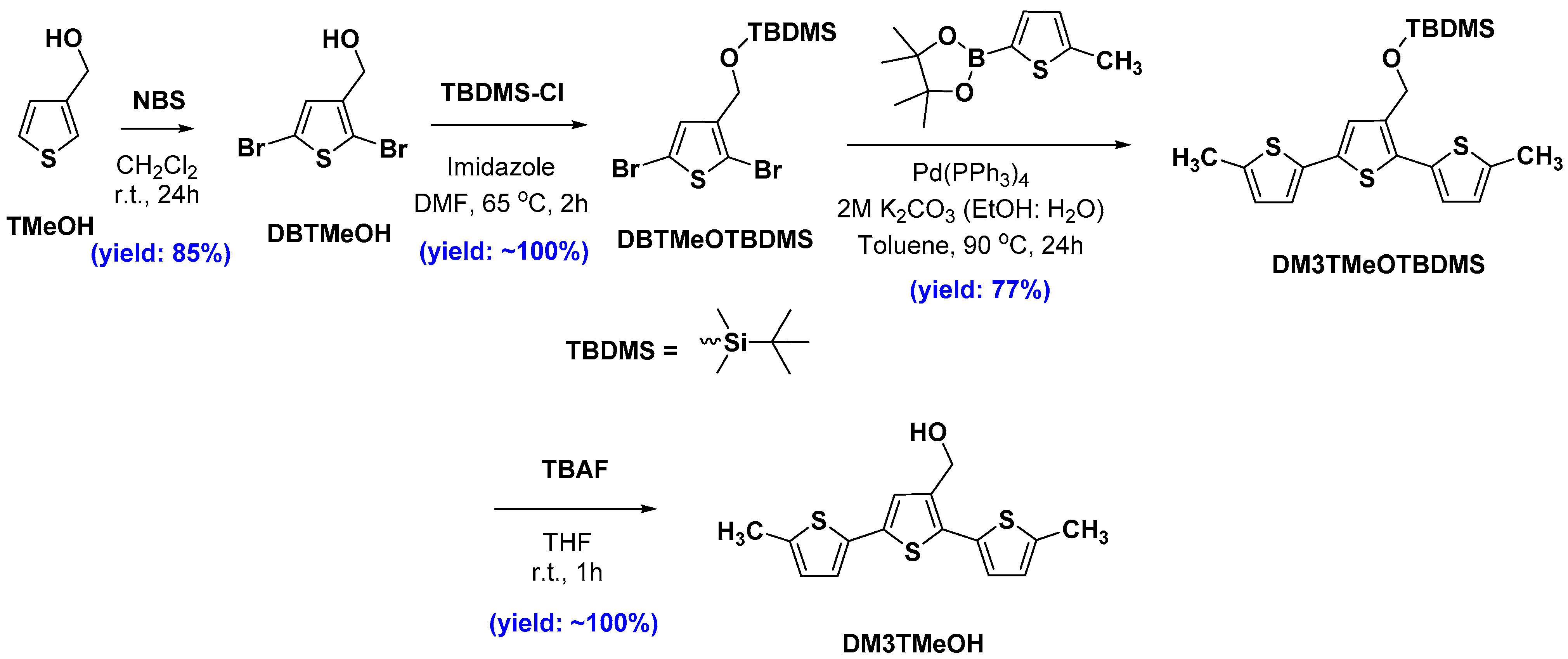
2.1. Synthesis
2.2. Crystal Growth
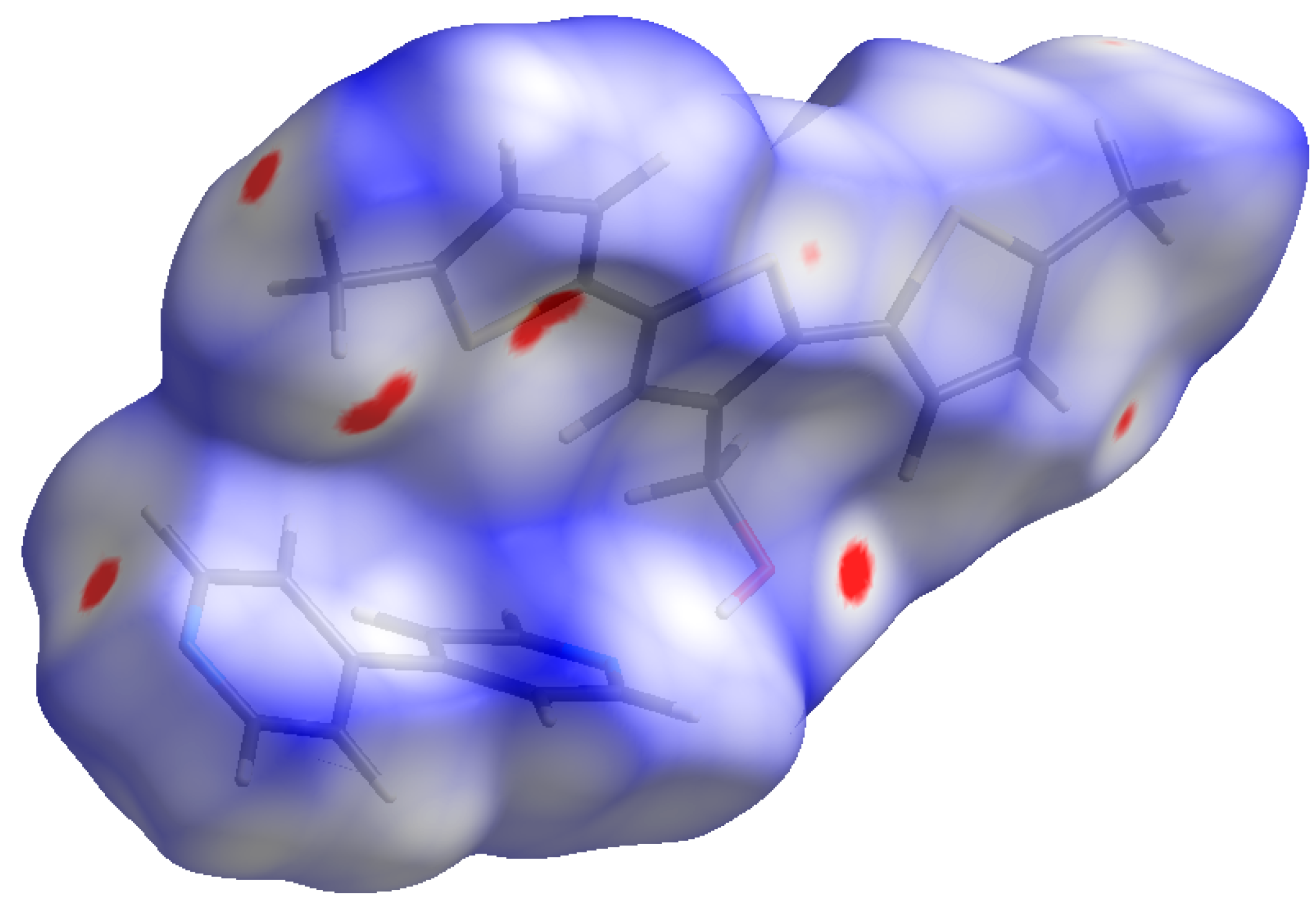
2.3. Single-Crystal X-ray Diffraction Analysis
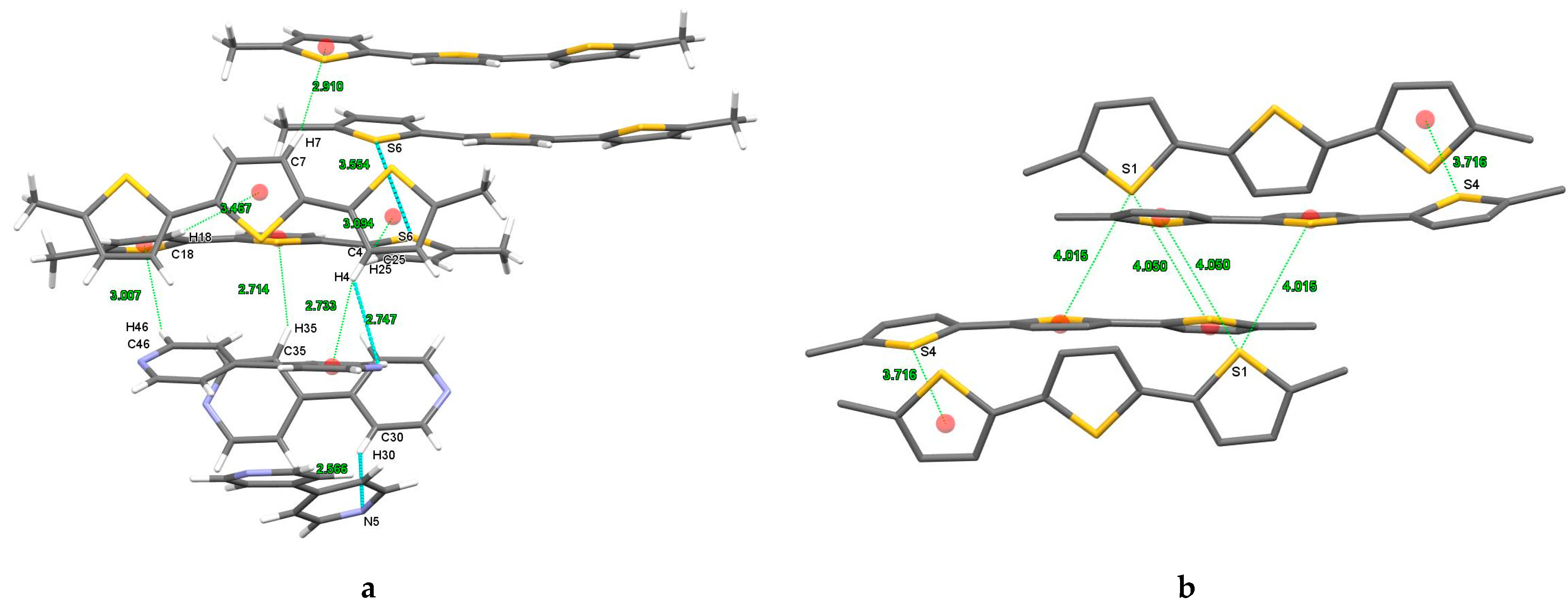
2.3.1. DM3T
2.3.2. DM3T–44BiPy
2.3.3. DM3TMeOH–44BiPy
2.4. Photophysical Properties
3. Discussion
4. Materials and Methods
5. Conclusions
Supplementary Materials
Author Contributions
Funding
Institutional Review Board Statement
Informed Consent Statement
Data Availability Statement
Acknowledgments
Conflicts of Interest
References
- Köhler, A.; Bässler, H. Electronic Processes in Organic Semiconductors: An Introduction; Wiley-VCH Verlag GmbH & Co. KGaA: Weinheim, Germany, 2015. [Google Scholar] [CrossRef]
- Geffroy, B.; Le Roy, P.; Prat, C. Organic light-emitting diode (OLED) technology: Materials, devices and display technologies. Polym. Int. 2006, 55, 572–582. [Google Scholar] [CrossRef]
- Hoppe, H.; Sariciftci, N.S. Organic solar cells: An overview. J. Mater. Res. 2004, 19, 1924–1945. [Google Scholar] [CrossRef]
- Horowitz, G. Organic field-effect transistors. Adv. Mater. 1998, 10, 365–377. [Google Scholar] [CrossRef]
- Samuel, I.D.W.; Turnbull, G.A. Organic semiconductor lasers. Chem. Rev. 2007, 107, 1272–1295. [Google Scholar] [CrossRef] [PubMed]
- Rahman, M.A.; Kumar, P.; Park, D.-S.; Shim, Y.-B. Electrochemical sensors based on organic conjugated polymers. Sensors 2008, 8, 118–141. [Google Scholar] [CrossRef] [PubMed]
- Thompson, R.B. Fluorescence Sensors and Biosensors; CRC Press: Boca Raton, FL, USA, 2005. [Google Scholar] [CrossRef]
- Fichou, D. Handbook of Oligo-and Polythiophenes; Wiley-VCH: Weinheim, Germany, 1999. [Google Scholar]
- Meyer, F. Fluorinated conjugated polymers in organic bulk heterojunction photovoltaic solar cells. Prog. Polym. Sci. 2015, 47, 70–91. [Google Scholar] [CrossRef]
- Birnbaum, D.; Kohler, B.E. Lowest energy excited singlet state of 2,2′:5′,2″-terthiophene, an oligomer of polythiophene. J. Chem. Phys. 1989, 90, 3506–3510. [Google Scholar] [CrossRef]
- Rossi, R.; Ciofalo, M.; Carpita, A.; Ponterini, G. Singlet—Triplet intersystem crossing in 2, 2′: 5′, 2″-terthiophene and some of its derivatives. J. Photochem. Photobiol. A 1993, 70, 59–67. [Google Scholar] [CrossRef]
- Hernandez, V.; Hotta, S.; López Navarrete, J. Vibrational spectroscopic study of 5, 5 ″-bis (dicyanomethylene)-5, 5 ″-dihydro-Δ 2, 2′: 5′, 2″-terthiophene bearing a heteroquinonoid structure as a model of doped polythiophene. J. Chem. Phys. 1998, 109, 2543–2548. [Google Scholar] [CrossRef]
- Rubio, M.; Ortí, E.; Pou-Amérigo, R.; Merchán, M. Electronic Spectra of 2, 2 ′-Bithiophene and 2, 2′: 5′, 2″-Terthiophene Radical Cations: A Theoretical Analysis. J. Phys. Chem. A 2001, 105, 9788–9794. [Google Scholar] [CrossRef]
- Pappenfus, T.M.; Raff, J.D.; Hukkanen, E.J.; Burney, J.R.; Casado, J.; Drew, S.M.; Miller, L.L.; Mann, K.R. Dinitro and quinodimethane derivatives of terthiophene that can be both oxidized and reduced. Crystal structures, spectra, and a method for analyzing quinoid contributions to structure. J. Org. Chem. 2002, 67, 6015–6024. [Google Scholar] [CrossRef] [PubMed]
- Casado, J.; Pappenfus, T.M.; Mann, K.R.; Ortí, E.; Viruela, P.M.; Milián, B.; Hernández, V.; López Navarrete, J.T. Spectroscopic and Theoretical Study of the Molecular and Electronic Structures of a Terthiophene-Based Quinodimethane. ChemPhysChem 2004, 5, 529–539. [Google Scholar] [CrossRef] [PubMed]
- Rubio, M.; Merchán, M.; Ortí, E. A Theoretical Study on the Low-Lying Excited States of 2, 2′: 5′, 2″-Terthiophene and 2, 2′: 5′, 2″: 5″, 2‴-Quaterthiophene. ChemPhysChem 2005, 6, 1357–1368. [Google Scholar] [CrossRef] [PubMed]
- Sang, L.; Matz, D.L.; Pemberton, J.E. Understanding the Reaction Chemistry of 2, 2′: 5′, 2 ″-Terthiophene Films with Vapor-Deposited Ag, Al, and Ca. J. Phys. Chem. C 2015, 119, 24290–24298. [Google Scholar] [CrossRef]
- Clément, S.; Meyer, F.; De Winter, J.; Coulembier, O.; Vande Velde, C.M.L.; Zeller, M.; Gerbaux, P.; Balandier, J.-Y.; Sergeyev, S.; Lazzaroni, R.; et al. Synthesis and Supramolecular Organization of Regioregular Polythiophene Block Oligomers. J. Org. Chem. 2010, 75, 1561–1568. [Google Scholar] [CrossRef]
- Clement, S.; Coulembier, O.; Meyer, F.; Zeller, M.; Vande Velde, C.M. (E)-3-(2,3,4,5,6-Penta-fluoro-styr-yl)thio-phene. Acta Crystallogr. E 2010, 66 Pt 4, o896–o897. [Google Scholar] [CrossRef]
- Beny, J.P.; Dhawan, S.N.; Kagan, J.; Sundlass, S. Synthesis of 3,2′:5′,3″-terthiophene and other terthiophenes by the thiophenecarboxaldehyde. fwdarw. ethynylthiophene. fwdarw. dithienylbutadiyne route. J. Org. Chem. 1982, 47, 2201–2204. [Google Scholar] [CrossRef]
- Miller, L.L.; Yu, Y. Synthesis of β-Methoxy, Methyl-Capped α-Oligothiophenes. J. Org. Chem. 1995, 60, 6813–6819. [Google Scholar] [CrossRef]
- Berger, G.; Frangville, P.; Meyer, F. Halogen bonding for molecular recognition: New developments in materials and biological sciences. Chem. Commun. 2020, 56, 4970–4981. [Google Scholar] [CrossRef]
- Berger, G.; Soubhye, J.; Van der Lee, A.; Vande Velde, C.; Wintjens, R.; Dubois, P.; Clément, S.; Meyer, F. Interplay between Halogen Bonding and Lone Pair–π Interactions: A Computational and Crystal Packing Study. ChemPlusChem 2014, 79, 552–558. [Google Scholar] [CrossRef]
- Kumar, S.; Body, C.; Leyssens, T.; Van Hecke, K.; Berger, G.; Van der Lee, A.; Laurencin, D.; Richeter, S.; Clément, S.; Meyer, F. Halogen-Bonded Thiophene Derivatives Prepared by Solution and/or Mechanochemical Synthesis. Evidence of N···S Chalcogen Bonds in Homo- and Cocrystals. Cryst. Growth Des. 2023, 23, 2442–2454. [Google Scholar] [CrossRef]
- Miyaura, N.; Suzuki, A. Palladium-Catalyzed Cross-Coupling Reactions of Organoboron Compounds. Chem. Rev. 1995, 95, 2457–2483. [Google Scholar] [CrossRef]
- Bondi, A. van der Waals Volumes and Radii. J. Phys. Chem. 1964, 68, 441–451. [Google Scholar] [CrossRef]
- Cheng, J.; Liang, X.; Cao, Y.; Guo, K.; Wong, W.-Y. Aldehyde end-capped terthiophene with aggregation-induced emission characteristics. Tetrahedron 2015, 71, 5634–5639. [Google Scholar] [CrossRef]
- Becker, R.S.; Seixas de Melo, J.; Macanita, A.L.; Elisei, F. Comprehensive evaluation of the absorption, photophysical, energy transfer, structural, and theoretical properties of α-oligothiophenes with one to seven rings. J. Phys. Chem. 1996, 100, 18683–18695. [Google Scholar] [CrossRef]
- Becker, R.S.; de Melo, J.S.; Macanita, A.L.; Elisei, F. Comprehensive investigation of the solution photophysics and theoretical aspects of oligothiophenes of 1–7 rings. Pure Appl. Chem. 1995, 67, 9–16. [Google Scholar] [CrossRef]
- Waragai, K.; Hotta, S. Structural and spectroscopic studies of charge-transfer complexes based on oligothiophenes. Synth. Met. 1991, 41, 519–522. [Google Scholar] [CrossRef]
- Periasamy, N.; Danieli, R.; Ruani, G.; Zamboni, R.; Taliani, C. Location of the low-energy 1 A g state in a polythiophene oligomer by two-photon absorption spectroscopy: α-sexithienyl. Phys. Rev. Lett. 1992, 68, 919. [Google Scholar] [CrossRef]
- Ma, X.; Sun, R.; Cheng, J.; Liu, J.; Gou, F.; Xiang, H.; Zhou, X. Fluorescence Aggregation-Caused Quenching versus Aggregation-Induced Emission: A Visual Teaching Technology for Undergraduate Chemistry Students. J. Chem. Educ. 2016, 93, 345–350. [Google Scholar] [CrossRef]
- Lee, E.; Hammer, B.; Kim, J.-K.; Page, Z.; Emrick, T.; Hayward, R.C. Hierarchical Helical Assembly of Conjugated Poly(3-hexylthiophene)-block-poly(3-triethylene glycol thiophene) Diblock Copolymers. J. Am. Chem. Soc. 2011, 133, 10390–10393. [Google Scholar] [CrossRef]
- Nakamura, T.; Asano, M.; Sekiguchi, Y.; Mizuno, Y.; Tamaki, K.; Kimura, T.; Nara, F.; Kawase, Y.; Shimozato, T.; Doi, H.; et al. Discovery of CS-2100, a potent, orally active and S1P3-sparing S1P1 agonist. Bioorg. Med. Chem. 2012, 22, 1788–1792. [Google Scholar] [CrossRef] [PubMed]
- Rigaku Corporation. Rigaku Oxford Diffraction, CrysAlisPro Software System; Rigaku Corporation: Oxford, UK, 2019. [Google Scholar]
- Dolomanov, O.V.; Bourhis, L.J.; Gildea, R.J.; Howard, J.A.; Puschmann, H. OLEX2: A complete structure solution, refinement and analysis program. J. Appl. Crystallogr. 2009, 42, 339–341. [Google Scholar] [CrossRef]
- Sheldrick, G.M. SHELXT–Integrated space-group and crystal-structure determination. Acta Crystallogr. A 2015, 71, 3–8. [Google Scholar] [CrossRef]
- Sheldrick, G.M. Crystal structure refinement with SHELXL. Acta Crystallogr. C 2015, 71, 3–8. [Google Scholar] [CrossRef] [PubMed]


| Hydrogen Bonding | |||
|---|---|---|---|
| D–H···A | d(H···A) Å | d(D···A) Å | Θ(D–H···A) deg |
| C21–H21···S1 (intermolecular) | 2.963 | 3.825(1) | 147.51 |
| C11–H11···S2 (intramolecular) | 2.835 | 3.142(1) | 100.02 |
| C–H···π interactions | |||
| C–H···π | d(H···π) a Å | d(C···π) b Å | Θ(C–H···π) c deg |
| C3–H3···π | 3.152 | 3.843 | 131.00 |
| C8–H8···π | 2.779 | 3.491 | 132.39 |
| C24–H24···π | 3.214 | 3.996 | 140.85 |
| C28–H28···π | 2.723 | 3.489 | 138.20 |
| Chalcogen bonding | |||
| X···π | d(X···π) d Å | ||
| S23···π | 3.868 | ||
| Chalcogen-Chalcogen contacts | |||
| X···X | d(X···X) Å | ||
| S1···S21 | 3.690(3) | ||
| Hydrogen Bonding | |||
|---|---|---|---|
| D–H···A | d(H···A) Å | d(D···A) Å | Θ(D-H···A) deg |
| C4–H4···N3 | 2.747 | 3.438(4) | 130.27 |
| C30–H30···N5 | 2.566 | 3.389(4) | 145.14 |
| C50–H50···N1 | 2.590 | 3.529(4) | 170.38 |
| C58–H58···N1 | 2.662 | 3.575(4) | 161.27 |
| C37–H37···N2 | 2.676 | 3.524(4) | 149.08 |
| C56–H56···N2 | 2.640 | 3.555(5) | 161.84 |
| C31–H31···N3 | 2.561 | 3.395(4) | 146.75 |
| C55–H55···N4 | 2.565 | 3.511(4) | 173.74 |
| C57–H57···N5 | 2.576 | 3.395(4) | 144.59 |
| C38–H38···N6 | 2.588 | 3.429(4) | 147.79 |
| C52–H52···N6 | 2.551 | 3.391(4) | 147.60 |
| C15–H15A···S4 | 3.048 | 3.917(4) | 148.37 |
| C32–H32···S6 | 3.058 | 3.626(4) | 119.92 |
| C–H···π interactions | |||
| C–H···π | d(H···π) a Å | d(C···π) b Å | Θ(C-H···π) c deg |
| C4–H4···π | 2.733 | 3.606 | 153.02 |
| C7–H7···π | 2.910 | 3.655 | 136.17 |
| C25–H25···π | 3.094 | 3.647 | 118.71 |
| C46–H46···π | 3.007 | 3.807 | 142.73 |
| C35–H35···π | 2.714 | 3.542 | 146.04 |
| C18–H18···π | 3.467 | 4.386 | 163.45 |
| Chalcogen bonding | |||
| S···π | d(S···π) d Å | ||
| S1···π | 4.015 | ||
| S1···π | 4.050 | ||
| S4···π | 3.716 | ||
| Chalcogen–chalcogen contacts | |||
| X···X | d(X···X) Å | ||
| S6···S6 | 3.534 | ||
| Hydrogen Bonding | |||
|---|---|---|---|
| D–H···A | d(H···A) Å | d(D···A) Å | Θ(D–H···A) deg |
| O1–H1···N2 | 2.053 | 2.841(2) | 164.95 |
| C7–H7···O1 (intramolecular) | 2.389 | 3.183(2) | 140.90 |
| C15–H15B···N1 | 2.557 | 3.362(2) | 139.48 |
| C8–H8···O1 | 2.616 | 3.477(2) | 150.95 |
| C25–H25···O1 | 2.567 | 3.487(2) | 163.13 |
| C15–H15A···S1 | 2.950 | 3.839(2) | 151.41 |
| C–H···π interactions | |||
| C–H···π | d(H···π) a Å | d(C···π) b Å | Θ(C-H···π) c deg |
| C10–H10A···π | 3.051 | 3.602 | 116.93 |
| C15–H15A···π | 2.638 | 3.553 | 155.28 |
Disclaimer/Publisher’s Note: The statements, opinions and data contained in all publications are solely those of the individual author(s) and contributor(s) and not of MDPI and/or the editor(s). MDPI and/or the editor(s) disclaim responsibility for any injury to people or property resulting from any ideas, methods, instructions or products referred to in the content. |
© 2023 by the authors. Licensee MDPI, Basel, Switzerland. This article is an open access article distributed under the terms and conditions of the Creative Commons Attribution (CC BY) license (https://creativecommons.org/licenses/by/4.0/).
Share and Cite
Kumar, S.; Van Hecke, K.; Meyer, F. Insight into Unusual Supramolecular Self-Assemblies of Terthiophenes Directed by Weak Hydrogen Bonding. Int. J. Mol. Sci. 2023, 24, 11127. https://doi.org/10.3390/ijms241311127
Kumar S, Van Hecke K, Meyer F. Insight into Unusual Supramolecular Self-Assemblies of Terthiophenes Directed by Weak Hydrogen Bonding. International Journal of Molecular Sciences. 2023; 24(13):11127. https://doi.org/10.3390/ijms241311127
Chicago/Turabian StyleKumar, Shiv, Kristof Van Hecke, and Franck Meyer. 2023. "Insight into Unusual Supramolecular Self-Assemblies of Terthiophenes Directed by Weak Hydrogen Bonding" International Journal of Molecular Sciences 24, no. 13: 11127. https://doi.org/10.3390/ijms241311127
APA StyleKumar, S., Van Hecke, K., & Meyer, F. (2023). Insight into Unusual Supramolecular Self-Assemblies of Terthiophenes Directed by Weak Hydrogen Bonding. International Journal of Molecular Sciences, 24(13), 11127. https://doi.org/10.3390/ijms241311127