Article

Exploring the Therapeutic Potential of trans-Chalcone: Modulation of MicroRNAs Linked to Breast Cancer Progression in MCF-7 Cells

by
Tatiana Takahasi Komoto
1,2,
Felipe Garcia Nishimura
1,
Adriane Feijó Evangelista
3,
Ana Julia Aguiar de Freitas
2,
Gabriel da Silva
1,
Wilson Araujo Silva
4,
Kamila Peronni
4,
Marcia Maria Chiquitelli Marques
2,
Mozart Marins
1 and
Ana Lucia Fachin
1,*
1
Biotechnology Unit, University of Ribeirão Preto, SP, Av. Costábile Romano, 2201, Ribeirão Preto 14096-900, Brazil
2
Molecular Oncology Research Center, Barretos Cancer Hospital, Teaching and Research Institute, Barretos 14784-400, Brazil
3
Sergio Arouca National School of Public Health, Oswaldo Cruz Foundation, Manguinhos, Rio de Janeiro 21040-361, Brazil
4
Center for Medical Genomics at the Clinics Hospital of Ribeirão Preto Medical School, University of São Paulo, Ribeirão Preto 14049-900, Brazil
*
Author to whom correspondence should be addressed.
Int. J. Mol. Sci. 2023, 24(13), 10785; https://doi.org/10.3390/ijms241310785
Submission received: 5 May 2023 / Revised: 10 June 2023 / Accepted: 16 June 2023 / Published: 28 June 2023
(This article belongs to the Special Issue Therapeutic Effects of Plant Based Bioactive Compounds on Cancer 2.0)

Abstract

:
Breast cancer is responsible for 25% of all cancers that affect women. Due to its high heterogeneity pattern in clinical diagnosis and its molecular profile differences, researchers have been seeking new targets and therapies, with more specificity and fewer side effects. Thus, one compound that has garnered our attention is trans-chalcone, which is naturally occurring in various plants and possesses promising biological properties, including antitumor effects. MiRNA is an extensive class of non-coding small, endogenous, and single-stranded RNAs, and it is involved in post-translational gene regulation. Therefore, the objective of this study was to investigate the effects of TChal on miRNAs expression and its relationship with anticancer activity against MCF-7. Initially, the trans-chalcone IC50 value was established by MTT assay for MCF-7and HaCat (non-cancer cell), in which we found out that it was 53.73 and 44.18 μ M, respectively. Subsequently, we treated MCF-7 cells with trans-chalcone at its IC50 concentration and performed Mi-seq analysis, which unveiled 23 differentially expressed miRNAs. From this set, we selected five miRNAs (miR-25-5p, miR-27a-3p, miR-891a, miR-449a, and miR-4485) for further validation using qRT-PCR, guided by in silico analysis and their known association with tumorigenesis. In conclusion, our research provides valuable insights into the potential use of TChal to reveal MicroRNAs molecular targets that can be applied in breast cancer therapy.

1. Introduction

Breast cancer is the most common type of cancer and accounts for 25% of all cancers affecting women. It is a heterogeneous disease at a molecular level [1], allowing it to be classified into five molecular subtypes: Luminal A, Luminal B HER-2 negative, Luminal B HER-2 positive, HER2 and basal-like tumors (basal-like or triple-negative) [2,3]. The complex nature of breast cancer, characterized by variations in clinical diagnosis and molecular profiles [4] has prompted researchers to explore novel targets and therapies that offer greater specificity and reduced side effects.
Flavonoid group has been outstanding in the research community due to its biological activities, for example, antioxidant effects, anti-aging properties, anti-inflammatory activity, immunomodulatory activity, cardioprotective effects, antiviral, antimicrobial, antifungal activity, and anti-cancer properties [5,6,7,8]. Among this group, trans-chalcone (TChal) has caught our attention, firstly because it is widely distributed in plants and secondly due to its biological properties. Our group has been dedicated to unraveling the mechanisms underlying the action of TChal. Our investigations have demonstrated the efficacy of TChal in combating breast cancer and osteosarcoma by targeting crucial genes involved in cancer development, such as p53, Bcl2, AURKA, and MDR1 [9,10,11]. Furthermore, we have observed that TChal possesses the ability to suppress tumor growth mediated by Heme-Oxygenase 1 [12]. These findings highlight the potential of TChal as a promising therapeutic agent in the fight against cancer.
MiRNA is an extensive class of non-coding small RNAs that are endogenous, single-stranded, and 19–25 nucleotides in length. MiRNAs play a pivotal role in post-translational gene regulation, orchestrating a wide range of essential biological functions including proliferation, apoptosis, tumorigenesis, cellular differentiation, metastasis, and chemoresistance [13]. Because miRNAs are related to a variety of biological processes, they could be used as strategies in disease treatment and diagnosis as biomarkers or new targets for therapies [14,15].
Therefore, the present study aimed to investigate the TChal effects in miRNAs expression and its relationship with anticancer activity against breast cancer cell lines.

2. Results

2.1. trans-Chalcone Cytotoxicity

The cytotoxicity of TChal was evaluated against MCF-7, MDA-MB-231, and HaCaT (keratinocytes non-cancerous cell line) after 24 h exposure (Table 1). Using the established IC50 from MCF-7, the total RNA was extracted from MCF-7, treated and non-treated (0.1% DMSO) in three independent experiments. The bioanalyzer was then used to guarantee the RNA integrity (RIN > 7 for all samples - MCF-7 treated and non-treated with Tchal). Next, a sequencing experiment was performed only on MCF-7 cell line. Thereafter, the RNA from MCF-7, MDA-MB-231, and HaCaT were extracted and used to validate the sequencing data via qRT-PCR.

2.2. MicroRNAs Analysis of MCF-7 Cell Line in Response to trans-Chalcone

Following the sequencing assay, we conducted an analysis of the data, which took into account the quality of the sequencing, normalization data, and the microRNAs that were differentially expressed in MCF-7 cells treated with and without Tchal. The analysis identified 23 microRNAs that were modulated in MCF-7 cells following Tchal treatment. Of these, seventeen miRNAs were up-regulated and six miRNAs were down-regulated (Figure 1).

2.3. Validation by qRT-PCR

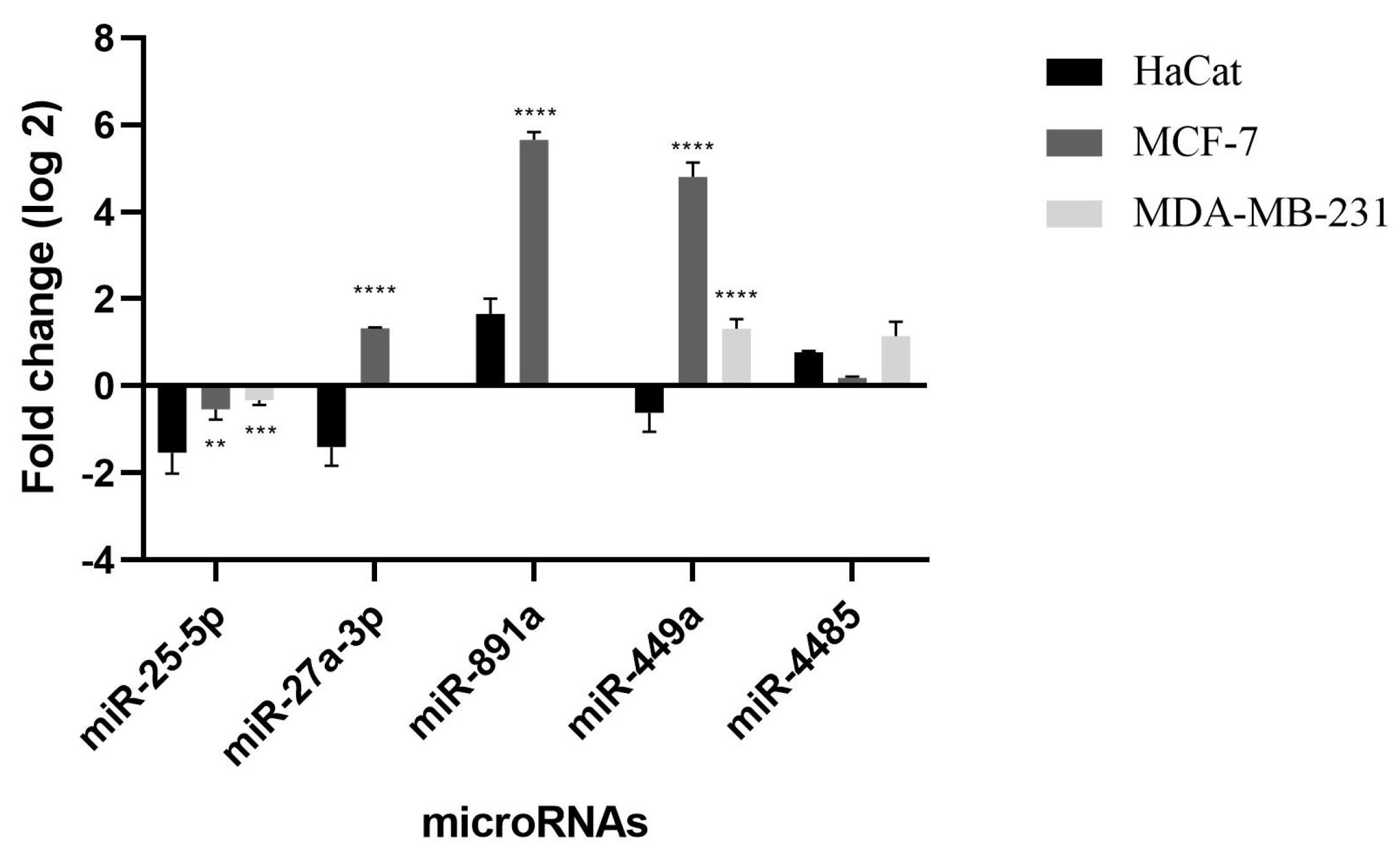
Five differently expressed miRNAs in MCF-7 cell line were selected for validation via qRT-PCR, two miRNAs oncogenic (miR-27a-3p and miR-891a-5p) and three miRNAs tumor suppressors (miR-25-5p, miR-449a and miR-4485-3p). In order to analyze whether some modulation could present a different pattern between cancer cell and non-cancer cell lines, we decided to perform validation of MicroRNAs selected using a normal keratinocytes cell line to compare the expression profile (Figure 2). Subsequently, when compared to HaCaT cells, all miRNAs exhibited statistically significant differences. Notably, miR-449a displayed sustained repression even after treatment with TChal, indicating its potential as a target for novel drug therapies. These findings suggest that targeting miR-449a could hold promise for developing effective treatments. Gene modulation values obtained by qRT-PCR demonstrate the reproducibility and accuracy of the sequencing.

2.4. Functional Categorization of Differentially Expressed microRNAs

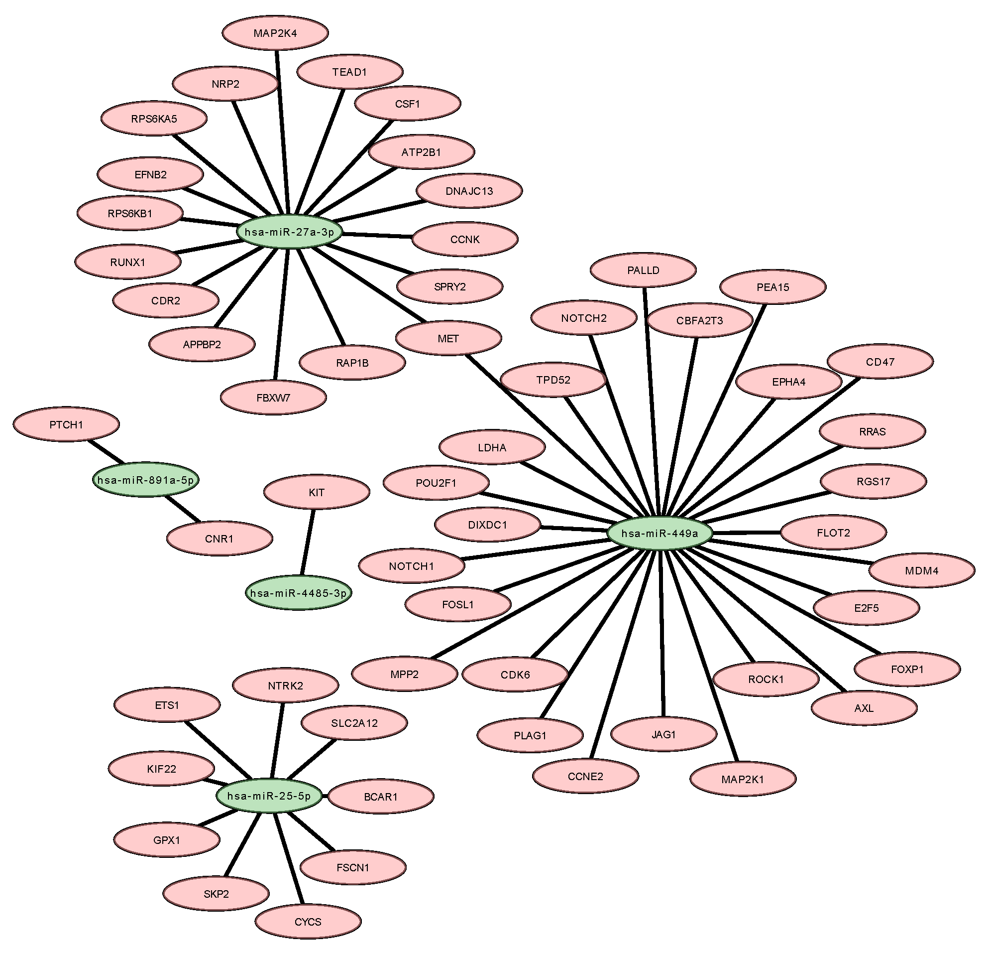
Among the five miRNAs identified and validated in this study, all of them were found to be associated with genes that have been previously described in breast tumors. In this context, we identified fifty-six potential genes that could be targets of those miRNA. Then, a target miRNA–mRNA interaction network was assessed on Cytoscape, from which we can observe the interaction networks between the miRNAs and their predicted target genes (Figure 3).

2.5. Enrichment Analysis Results

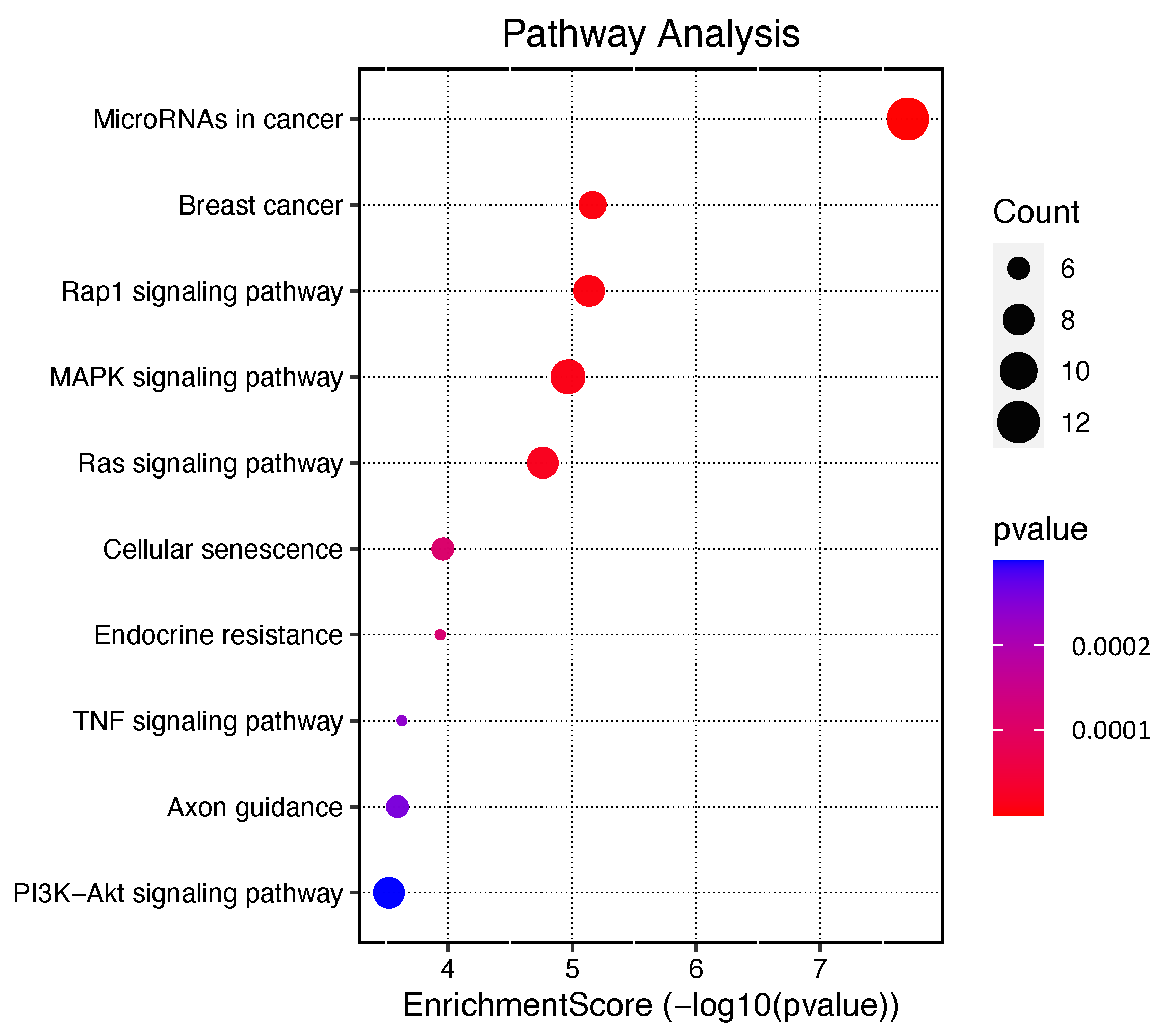
The enriched signaling pathways are highlighted in Figure 4 and in the Supplementary Materials (Table S1). Among the top ten significant signaling pathways are pathways involved in ‘MicroRNAs in cancer’ with twelve of the fifty-six genes involved, ‘MAPK signaling pathway’ with nine genes, ‘Ras signaling pathway’, ‘PI3K-Akt signaling pathway’ both with eight genes, ‘Breast cancer’ with seven genes, and ‘Cellular senescence’ with six genes (Figure 4).

2.6. GO Enrichment Analysis Results

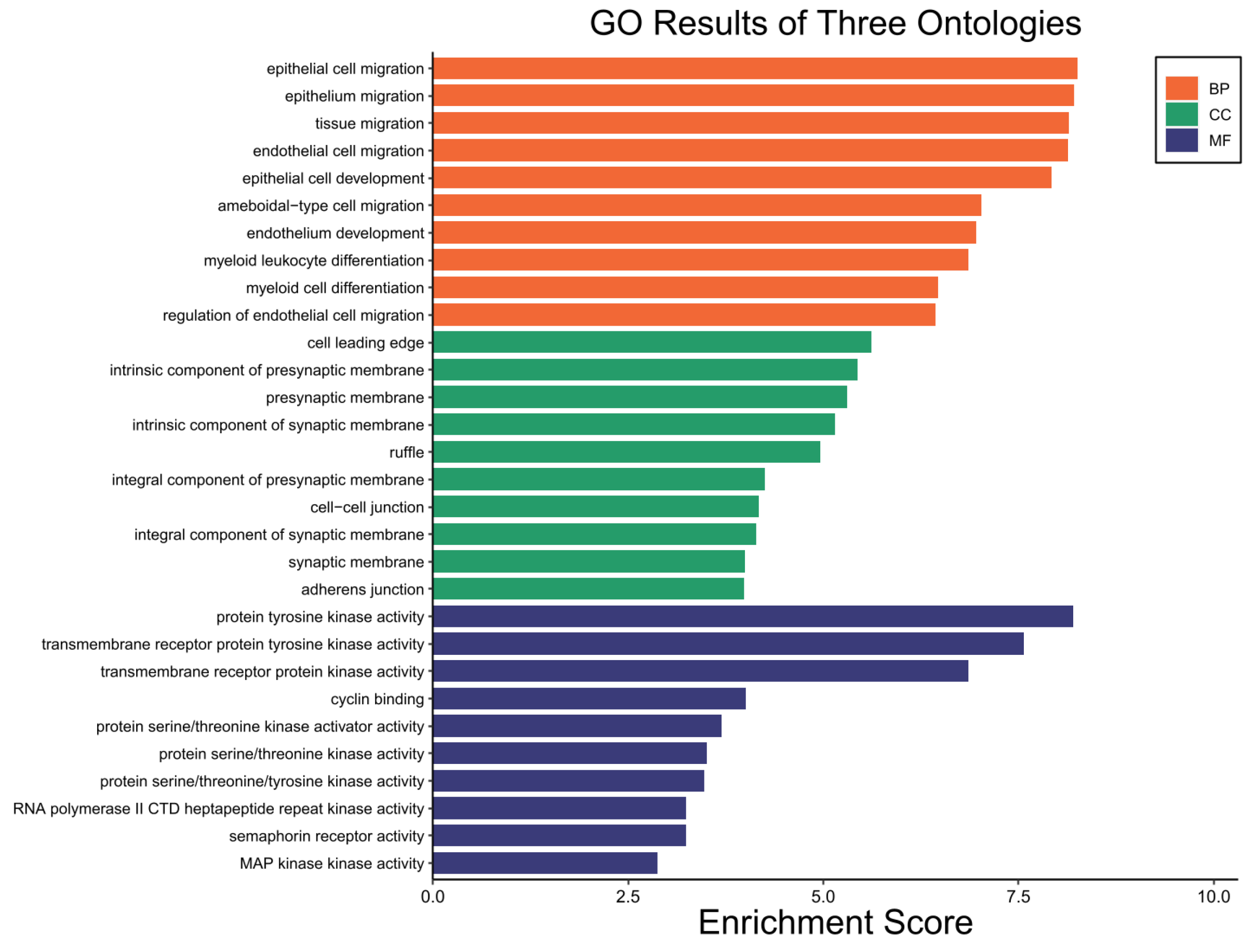
GO enrichment analysis was further employed to explore the enriched biological process (BP), molecular function (MF) and cellular component (CC). The top 10 that were significantly enriched according to an adj p < 0.05 identified are shown in Figure 5. Of the main BPs identified, ‘epithelial cell migration’, ‘epithelium migration’, and ‘tissue migration’ were among the more important ones according to the number of genes involved in these processes. For MFs, ‘protein tyrosine kinase activity’, ‘transmembrane receptor protein tyrosine kinase activity’ and ‘transmembrane receptor protein kinase activity’ were the main ones. For CCs, we highlight ‘cell leading edge’, ‘synaptic membrane’, ‘presynaptic membrane’ and ‘cell-cell junction’ (Figure 5).

2.7. In Silico Analysis of Gene Expression and Overall Survival

The expression levels of the CSF1 and KIT genes were compared between breast cancer and adjacent non-tumor tissues from patients, using original published data available in the GEPIA database. The results showed that the expression of both CSF1 and KIT was significantly downregulated in breast cancer tissues compared to non-tumor samples (Figure 6A,C). The association between the CSF1 and KIT genes with patient OS was analyzed using the Kaplan–Meier plotter database. Based on the median expression of CSF1 and KIT, patients were divided into high- and low-expression groups, and the OS of patients with low expression of both genes was significantly longer than that of patients with high expression of CSF1 and KIT (Figure 6B,D).

3. Discussion

Our research group has been studying the antitumor effects of TChal in order to understand its mechanism and reveal its molecular targets. In some of these studies, TChal was able to act on the cell cycle, apoptosis, and metastasis [9,10,11]. Molecularly, it has been observed that microRNAs are important to many cellular processes and signaling pathways, including cell development, differentiation, proliferation, and apoptosis [16,17]. Also, the dysregulation of a single miRNA is able to bring many consequences to cells in terms of cellular outcomes and it can be associated with disease development, such as cardiovascular diseases, neurodevelopmental diseases, autoimmune disorders, bone diseases, and human cancers like breast cancer [16].
MiRNA sequencing of the MCF-7 cell line following TChal treatment revealed 23 miRNAs that were differentially expressed. Enrichment analysis revealed that all of the identified pathways were consistent with those previously identified by our research group, validating their relevance in cancer development. Additionally, the analysis also uncovered novel pathways that play a crucial role in cancer, including the MicroRNAs in cancer, breast cancer, MAPK, RAS signaling, TNF signaling, and PI3K-Akt pathways as shown in Figure 4. Based on their function in tumorigenesis and expression level, we selected five miRNAs (including oncogenic and tumor suppressor miRNAs) for validation and proceeded with the in silico analysis.
Two isoforms (miR-27a-3p and 5p) of miR-27a were identified. MiR-27a-3p isoform was selected as it is more frequently modulated in tumor cells [18]. The function of MiR-27a-3p in tumor development has been controversial because it can be both oncogenic and a tumor suppressor. However, in general, miR-27a-3p is associated with various aspects of tumor progression, including apoptosis regulation, cell migration, invasion, and proliferation [19]. During our in silico analysis, we identified several key genes associated with tumorigenesis such as FBXW7, APPBP2, RUNX1, SPRY2, MAP2K4, NRP2, DNAJC13, CDR2, EFNB2, RAP1B, TEAD1, CCNK, RPS6KA5, MET, ATP2B1, RPS6KB1, and CSF1. MiR-27a-3p has been implicated in tumorigenesis and suggests that the miR-23a/24-2/27a cluster may have a causal role in breast tumorigenesis by functioning as an oncogene that promotes cell invasion and metastasis. This occurs through the targeting of Sprouty2 (SPRY2) and subsequent activation of the p44/42 MAPK signaling pathway in breast cancer [20]. Sprouty2 (SPRY2) belongs to the highly conserved family of signal modulatory proteins that inhibit the Ras/MAPK pathway. It has been reported to be dysregulated in various cancer types, such as breast, liver, and prostate cancer, suggesting its potential role as a modulator of cancer progression. Specifically, SPRY2 has been implicated in regulating key pathways involved in cancer cell growth, migration, and invasion [20,21]. Another gene targeted by miRNA is colony-stimulating factor 1 (CSF1), which is responsible for attracting and differentiating macrophages into the M2 phenotype. In the development of breast cancer, the use of inhibitors targeting the colony-stimulating factor 1 receptor (CSF1-R) can eliminate TAMs, leading to effective inhibition of metastasis and angiogenesis, as well as reduction in tumor invasiveness [22]. Tumor-associated macrophages (TAMs) are known to secrete cellular cytokines and surface receptors that play a critical role in promoting breast cancer metastasis. One of the key factors is the high expression of epidermal growth factor (EGF) in TAMs, which activates epidermal growth factor receptors (EGFRs) in cancer cells, leading to metastasis and increased secretion of colony-stimulating factor 1 (CSF-1). CSF-1, in turn, recruits and activates TAMs to further secrete EGF, indicating the presence of an EGF/CSF-1 positive feedback loop between TAMs and cancer cells. The EGF signaling pathway also induces the infiltration of breast cancer cells into blood vessels, thereby promoting blood vessel metastasis. These findings highlight the importance of TAMs in breast cancer progression and provide potential therapeutic targets for inhibiting metastasis [23,24]. Interestingly, the EGFR inhibition by TChal on MCF-7 and BT-20 cell lines had previously been proved by our research group [10]. Another study by Li et al. demonstrated that overexpression of miR-27a-3p led to EGFR inhibition, which is related to tumor growth, and induced changes in the cellular process such as those stated above [25]. Then, we suggested that the up-regulation of miR-27a-3p could inhibit the EGFR gene and CSF1. Moreover, when the OS patients were analyzed, the one who presented the lowest expression of the CSF1 gene was significantly longer than that of patients with high expression with a HR = 0.81.
Likewise, in vitro and in vivo assays have shown that modulation of miR-27a-3p can significantly inhibit the proliferation and invasion of ESCC cells in nude mice [26]. Furthermore, it is also involved in the regulation of the ZBTB10 gene, which is related to up-regulating Sps proteins important to tumor survival and angiogenesis [27]. Moreover, we have demonstrated that Sp1 was downregulated by TChal [11]. Additionally, the Heme-Oxygenase 1 (HO-1) is up-regulated at a protein level by TChal, and this is able to reduce breast cancer in xenograft assays with nude mice [12].
MiR-449a is often repressed in breast cancer and is associated with improved cell proliferation, migration, invasion, and metastasis in cancers such as breast cancer, hepatocarcinoma, gastric cancer, prostate cancer, and endometrial cancer [28,29]. In addition, the suppression of this microRNA is negatively associated with aggression and progression in breast cancer [30]. It occurs due to the modulation of genes that lead to the activation of resistance (ADAM22), the cell cycle (CDC25), proliferation and metastasis (PLAGL2 and TPD52) [28,29,31,32]. Hence, these data suggest that drugs that can increase the expression of miR-449a could lead to a decrease in tumor progression. Thus, TChal has been shown to be an alternative in the treatment of breast cancer, as it significantly induced the expression of this miRNA in MCF-7, whereas, in the HaCat, normal cell, this miRNA remained repressed. One of the gene targets of this microRNA is MDM4. Previously, our group observed that the MDM4 gene was induced in the MCF-7 cell line after treatment with both trans-chalcone and licochalcone A; however, in the BT-20 cell line, this gene was induced only by Licochalcona A [10]. The MDM2 and MDM4 genes are p53 inhibitors and are frequently induced in cancers [33]. Although some tumors have high levels of MDM4 modulation and mutations in the TP53 gene, this mutation may confer a cooperativity of both genes in tumor development. Approximately 14% of invasive breast tumors have MDM4 amplification and 3% have TP53 gene mutation [34]. In addition, authors have suggested that MDM4 is able to act independently of p53, due to its interaction with other proteins such as p21, 14-3-3gamma, ARF, HAUSP and Nbs1, these interactions could cause genetic instability and increase cell transformation [34,35,36,37]. Therefore, our research group has proposed that TChal exerts its effects by inducing the expression of TP53, a tumor suppressor gene, while suppressing p21, an oncogenic gene [11]. These findings suggest that the regulation of the MDM4 gene, which is known to interact with TP53, may occur independently from TP53 itself. Considering that the relationship of modulation of microRNAs to their target genes is usually inversely proportional, we suggest that miR-25-5p repression may positively modulate the MDM4 gene.
During the in silico analysis, two miRNAs, miR-27a-3p and miR-449a, were found to modulate the MET gene. The MET receptor tyrosine kinase is known to activate various cellular functions that are crucial to organ development and cancer progression upon binding with its ligand, hepatocyte growth factor (HGF). Abnormal c-Met signaling has been observed in different types of cancer, making the receptor a promising therapeutic target [38]. Although a high number of MET copies did not independently predict recurrence-free survival, the researchers did observe lower recurrence-free survival rates in the group with amplified MET during univariate analysis. Furthermore, a positive correlation was found between the number of MET copies and triple-negative status [39].
The in silico analysis revealed that miR-25-5p targets KIF22, GPX1, BCAR1, SKP2, SLC2A12, FSCN1, CYCS, NTRK2, and ETS1 genes. MiR-25-5p is known to promote cancer cell proliferation, anti-apoptosis, cell cycle progression, and cell motility, and is considered an oncogenic miRNA [40,41]. It has been shown that miR-25-5p is induced by the WNT/ β -catenin pathway and promotes epithelial–mesenchymal transition (EMT) and metastasis by regulating the RhoGDI1 gene [42]. Moreover, the interaction between mutant TP53 and BCAR1 activates downstream signaling pathways that contribute to cancer cell invasion [43]. Therefore, inhibiting miR-25-5p with drugs like TChal could be a potential therapeutic strategy for breast cancer.
miR-4485 is related to mitochondrial functions, although it is synthesized in the cell nucleus, it is transported to the mitochondria. This miRNA is involved in ATP production processes, ROS level, cell cycle, activation of caspases 3/7, and apoptosis [44,45]. In addition, Sripada et al. 2017 proved the influence of miR-4485 on tumor development by performing miR-4485 mimic transfection, leading to glycolytic pathway regulation and reduced chlorogenic ability of MDA-MB-231 cells, and they also observed a reduction of tumorigenicity in vivo [45]. Otherwise, the in silico data demonstrated that the gene KIT is a target by the identified miRNA. The c-Kit gene has been shown to be important in breast cancer, particularly in triple-negative breast cancer (TNBC), which lacks targeted therapies. Studies have shown that c-Kit is involved in promoting the migration and invasion of TNBC cells, and that inhibiting c-Kit with tyrosine kinase inhibitors may be a promising treatment option. Further research is needed to fully understand the role of c-Kit in breast cancer and to develop effective targeted therapies [46,47,48]. Moreover, when the OS patients was analyzed the one whose presented the lowest expression of KIT gene was significantly longer than that of patients with high expression with a HR = 0.69. These data indicate that miR-4485 could negatively affect tumor development, suggesting that this gene is a tumor suppressor in breast cancer.
Finally, miR-891a-5p has been identified as a potential biomarker for breast cancer as it is highly modulated by TChal. This microRNA is located on chromosome Xq27.3 along with 7 other microRNAs (miR-892, miR-890, miR-888, miR-892b, miR-891b and miR-891a) and has been identified as proto-oncogenic. The target gene of miR-891a-5p is TIMP2, which is associated with proliferation, migration, and tumor formation in prostate cancer [49]. In addition, our in silico analysis showed that it can modulate CNR1 and PTCH1 genes. Zhang et al. showed that miR-891a-5p inhibited the expression of ADAM10 by targeting its 3’UTR. This inhibition resulted in the suppression of proliferation and migration of breast cancer cells, particularly those that are Hormone receptor-positive [50]. The miR-891a-5p overexpression can negatively regulate the HOXA-5 gene (Homeobox A5), known as a tumor suppressor, related to the regulation of TP53 [51].

4. Materials and Methods

4.1. Materials

The trans-Chalcone (TChal) and some of the main reagents including trypsin, doxorubicin hydrochloride, dimethyl sulfoxide (DMSO), DMEM, penicillin, kanamycin, streptomycin, and Fetal bovine serum were purchased from Sigma-Aldrich (St. Louis, MO, USA). All the materials used for sequencing as Taqman microRNA Reverse Transcription kit and Taqman Small RNA Assays were both purchased from Applied Biosystems and MiSeq v2 Reagent kit (Illumina, San Diego, CA, USA). Finally, the materials to gene expression assay TaqManTM MicroRNA Assay and TaqManTM Universal Master Mix II, no UNG were from Thermo Fisher, Waltham, MA, USA.

4.2. Cell Culture

The breast cancer cell lines MCF-7 (Luminal A, estrogen receptor (ER) positive), MDA-MB-231 (triple-negative breast cancer), and HaCat (human keratinocyte cell line) were grown in DMEM medium supplemented with 10% fetal bovine serum, 100 U/mL penicillin, 100 μ g/mL streptomycin, and 100 μ g/mL kanamycin. The cultures were maintained in a humidified atmosphere with 5% CO 2 at 37 °C until 90% confluence.

4.3. Viability Assay Conditions

The inhibition of viability cells induced by TChal was evaluated by MTT assay on MCF-7, MDA-MB-231, and HaCat (non-cancerous cell line). This test was performed as described by Komoto et al. [8] with modifications. Briefly, each cell line was seeded at a density of 2 × 10³ cells/well in 96-well plates and incubated for 24 h. The cells were treated with 7 different concentrations of TChal (100, 50, 25, 12.5, 6.25 μ M) dissolved in 0.1% DMSO for 24 h. Next, 20 μ L MTT solution (5 mg/mL) was added to each well, and the plates were incubated for an additional 3 h. The formazan dye was diluted in 200 μ L DMSO. Absorbance was verified at 550 nm in a microplate reader MultiSkan FC. The assays were carried out in three independent experiments performed in triplicate. Doxorubicin was used at 2.5 μ g/mL (4.31 μ M) as the positive control for this test. The percentage of inhibition of cell viability and the IC50 calculation was based on Komoto et al. [10].

4.4. cDNA Library Construction and Mi-seq Assay

The total RNA from both MCF-7 cells treated with TChal and MCF-7 control treated with DMSO were extracted using the miRNeasy Isolation (Qiagen, Germantown, MD, USA) and the preparation of the miRNA library used the TruSeq Small RNA Library (Illumina, San Diego, CA, USA) kit, and both assays were done following the manufacturer’s instructions. The integrity of each miRNA library was analyzed by a bioanalyzer (Agilent Technologies, Santa Clara, CA, USA) as per the manufacturer’s instructions. For the library construction, the total RNA sample was used to the manufacturer’s instructions, and adaptors were added to the small RNAs. The small RNAs were converted into cDNAs by PCR, then to obtain the mature miRNA, electrophoresis was performed using the polyacrylamide gel (12%), and the mature miRNAs were between 145–160 pb, so the fragments were cut off the gel. Afterward, the fragments were purified, and the cDNA library was quantified by KAPA library quantification (Illumina sequencing platforms). Lastly, the samples were sequenced using a MiSeq v2 Reagent kit (Illumina, San Diego, CA, USA) on a Mi-seq system.

4.5. Validation Data

The validation data were performed by qRT-PCR using primes TaqMan™ MicroRNA Assay (Thermo Fisher, Waltham, MA, USA) following the manufacturer’s instruction, the selected miRNAs were: miR-25-5p, miR-27a-3p, miR-891a-5p, miR-449a, and miR-4485-3p, with RNU44 used as a housekeeping microRNA gene. RNU44 was chosen because it is present in more than 219 breast cancer types [52]. The entire experiment was carried out in three independent experiments using the TaqMan™ Universal Master Mix II, no UNG kit (Thermo Fisher, Waltham, MA, USA) and the machine Mx3300 qPCR System (Stratagene, San Diego, CA, USA) for MCF-7 (breast cancer) and normal cell line HaCaT (keratinocyte non-cancerous cell) after treatment with TChal at IC50 pre-established concentrations.

4.6. Bioinformatic Analysis

Raw miRNA sequences were initially submitted to a quality filter (q < 28) using Trimmomatic v.0.36 and small RNA sequencing adapters were removed using CutAdapt v.1.4.1. Alignments were performed using Bowtie2 v.2.1.0 and the counts of reads mapped to the human miRNA dataset in miRBase, Release 20 were obtained according to HT-Seq v. 0.7.2. Differential expression analysis (DEA) was performed using the DeSeq2 package. Differentially expressed miRNAs were considered with a false discovery rate (FDR) cut-off of 0.1 (10% FDR) and fold-change ≥ 2.0. The comparisons of the miRNA profiles were visualized according to heatmaps and constructed using the Complex Heatmaps package.

4.7. Prediction of microRNA Targets

To evaluate the biological role of miRNAs, bioinformatics analysis was initially applied to predict their targets using the free and online program miRDIP (http://ophid.utoronto.ca/mirDIP/, accessed on 13 April 2023), with a focus on the Top 1% predicted target genes (score class very high). This program integrates dozens of miRNA target prediction tools, each using a specific algorithm, thereby increasing prediction accuracy. From the generated lists of genes, targets predicted by at least three of the following prediction algorithms were selected: DIANA, microrna.org, RNA22, RNAHybrid, and TargetScan.

4.8. Enrichment Analysis

To assess the potential cooperative effects of identified miRNAs on biological processes and pathways associated with breast cancer, we performed pathway enrichment analyses using Reactome and its Cytoscape plugin that provides curated regulatory interaction networks and biological pathways derived from Reactome and other databases. We selected genes associated with breast cancer, according to the Cancer Gene Index Annotations (provided by the National Cancer Institute, NCI), by applying the Load Cancer Index function available in Reactome. We only considered pathways with FDR values ≤0.001 or less and at least three genes involved to increase the accuracy of the analysis. Enrichment analysis maps were obtained from an online data analysis website (http://www.bioinformatics.com.cn/, accessed on 13 April 2023).

4.9. Gene Expression Profiling and Interactive Analysis (GEPIA2)

Gene Expression Profiling and Interactive Analysis (GEPIA2) is an online tool for gene expression analysis (http://gepia2.cancer-pku.cn/index, accessed on 13 April 2023). This tool allows performing differential analyses with the gene expression profile of the tumor in relation to normal TCGA or GTEx data. We used this server to analyze target gene expression for the miRNAs identified in our study. GEPIA2 performs differential expression analysis and returns the result in a boxplot [53].

4.10. Overall Survival (OS) Analysis

An overall survival (OS) analysis was performed using the Kaplan–Meier plotter database (http://kmplot.com/analysis/, accessed on 13 April 2023) to evaluate the OS of target genes in breast cancer. Kaplan–Meier Plotter is an online database containing survival and gene expression information for various types of cancer. We selected only data available for breast cancer. The analysis compares high and low gene expression, and the results were shown by log-rank and p-value.

4.11. Statistical Analysis

All experimental data were submitted to analysis of variance (ANOVA) followed by Bonferroni test. The level of statistical significance was set at p ≤ 0.05.

5. Conclusions

In conclusion, this research has provided valuable insights into the therapeutic potential of TChal for breast cancer by shedding light on its potential molecular targets. The miRNA sequencing and enrichment analysis employed in this study have identified several crucial pathways involved in tumorigenesis, including miRNA in cancer, breast cancer, MAPK, RAS signaling, TNF signaling, and PI3K-Akt pathways. These findings underscore the importance of TChal in breast cancer treatment. Furthermore, the in silico analysis conducted in this research has revealed the targeting of several significant genes associated with tumorigenesis, such as CSF-1 and KIT. Importantly, it was observed that lower expression of these genes correlates with better overall survival. These findings highlight the potential of TChal to inhibit tumor progression and metastasis in breast cancer. Overall, this research contributes to our understanding of TChal’s therapeutic potential and provides a foundation for further investigations and the development of targeted therapies for breast cancer. By elucidating the molecular mechanisms and pathways affected by TChal, this study paves the way for future studies and clinical trials aimed at harnessing the benefits of TChal in breast cancer treatment.

Supplementary Materials

The following are available online at https://www.mdpi.com/article/10.3390/ijms241310785/s1.

Author Contributions

T.T.K. performed the laboratory experiments and wrote the manuscript. F.G.N. developed qRT-PCR experiments. A.F.E. and A.J.A.d.F. performed the bioinformatics analysis. K.P. and W.A.S. constructed the libraries and performed the sequencing. G.d.S. Contributed to all experiments and discussed the manuscript. M.M.C.M. contributed to the discussion in this manuscript. M.M. supervised the research and contributed reagents and materials. A.L.F. designed the project, supervised the research, contributed reagents/materials/analysis tools, and wrote the manuscript. All authors read and approved the final version of the manuscript.

Funding

This study was supported by grants from Fundação de Amparo à Pesquisa do Estado de São Paulo (FAPESP 2019/10514-8), CAPES fellowships granted to T.T.K., F.G.N., and G.S.

Institutional Review Board Statement

Not applicable.

Informed Consent Statement

Not applicable.

Data Availability Statement

The data presented in this study are available on request from the corresponding author.

Conflicts of Interest

The authors declare no conflict of interest.

References

  1. Harbeck, N.; Penault-Llorca, F.; Cortes, J.; Gnant, M.; Houssami, N.; Poortmans, P.; Ruddy, K.; Tsang, J.; Cardoso, F. Breast cancer. Nat. Rev. Dis. Prim. 2019, 5, 66. [Google Scholar] [CrossRef] [PubMed]
  2. Barzaman, K.; Karami, J.; Zarei, Z.; Hosseinzadeh, A.; Kazemi, H.M.; Moradi-Kalbolandi, S.; Safari, E.; Farahmand, L. Breast cancer: Biology, biomarkers, and treatments. Int. Immunopharmacol. 2020, 84. [Google Scholar] [CrossRef] [PubMed]
  3. Huszno, J.; Kolosza, Z. Molecular characteristics of breast cancer according to clinicopathological factors. Mol. Clin. Oncol. 2019, 11, 192–200. [Google Scholar] [CrossRef] [PubMed] [Green Version]
  4. Anderson, W.F.; Rosenberg, P.S.; Prat, A.; Perou, C.M.; Sherman, M.E. How many etiological subtypes of breast cancer: Two, three, four, or more? J. Natl. Cancer Inst. 2014, 106, dju165. [Google Scholar] [CrossRef] [PubMed] [Green Version]
  5. Bitencourt, T.A.; Macedo, C.; Franco, M.E.; Assis, A.F.; Komoto, T.T.; Stehling, E.G.; Beleboni, R.O.; Malavazi, I.; Marins, M.; Fachin, A.L. Transcription profile of Trichophyton rubrum conidia grown on keratin reveals the induction of an adhesin-like protein gene with a tandem repeat pattern. BMC Genom. 2016, 17, 249. [Google Scholar] [CrossRef] [Green Version]
  6. Bitencourt, T.A.; Macedo, C.; Franco, M.E.; Rocha, M.C.; Moreli, I.S.; Cantelli, B.A.M.; Sanches, P.R.; Beleboni, R.O.; Malavazi, I.; Passos, G.A.; et al. Trans-chalcone activity against Trichophyton rubrum relies on an interplay between signaling pathways related to cell wall integrity and fatty acid metabolism. BMC Genom. 2019, 20, 411. [Google Scholar] [CrossRef]
  7. Jucá, M.M.; Cysne Filho, F.M.S.; de Almeida, J.C.; Mesquita, D.d.S.; Barriga, J.R.d.M.; Dias, K.C.F.; Barbosa, T.M.; Vasconcelos, L.C.; Leal, L.K.A.M.; Ribeiro, J.E.; et al. Flavonoids: Biological activities and therapeutic potential. Nat. Prod. Res. 2020, 34, 692–705. [Google Scholar] [CrossRef]
  8. Komoto, T.T.; Bitencourt, T.A.; Silva, G.; Beleboni, R.O.; Marins, M.; Fachin, A.L. Gene Expression Response of Trichophyton rubrum during Coculture on Keratinocytes Exposed to Antifungal Agents. Evid. Based Complement. Altern. Med. 2015, 2015, 180535. [Google Scholar] [CrossRef] [Green Version]
  9. Bortolotto, L.F.B.; Barbosa, F.R.; Silva, G.; Bitencourt, T.A.; Beleboni, R.O.; Baek, S.J.; Marins, M.; Fachin, A.L. Cytotoxicity of trans-chalcone and licochalcone A against breast cancer cells is due to apoptosis induction and cell cycle arrest. Biomed. Pharmacother. 2017, 85, 425–433. [Google Scholar] [CrossRef]
  10. Komoto, T.T.; Bernardes, T.M.; Mesquita, T.B.; Bortolotto, L.F.B.; Silva, G.; Bitencourt, T.A.; Baek, S.J.; Marins, M.; Fachin, A.L. Chalcones Repressed the AURKA and MDR Proteins Involved in Metastasis and Multiple Drug Resistance in Breast Cancer Cell Lines. Molecules 2018, 23, 2018. [Google Scholar] [CrossRef] [Green Version]
  11. Silva, G.; Marins, M.; Fachin, A.L.; Lee, S.H.; Baek, S.J. Anti-cancer activity of trans-chalcone in osteosarcoma: Involvement of Sp1 and p53. Mol. Carcinog. 2016, 55, 1438–1448. [Google Scholar] [CrossRef]
  12. Komoto, T.T.; Lee, J.; Lertpatipanpong, P.; Ryu, J.; Mozart Marins, a.A.L.F.; Baek, S.J. Trans- chalcone suppresses tumor growth mediated at least in part by the induction of heme oxygenase-1 in breast cancer. Toxicol. Res. 2021, 37, 485–493. [Google Scholar] [CrossRef]
  13. Ha, M.; Kim, V.N. Regulation of microRNA biogenesis. Nat. Rev. Mol. Cell Biol. 2014, 15, 509–524. [Google Scholar] [CrossRef] [PubMed]
  14. Filipów, S.; Łaczmański, Ł. Blood Circulating miRNAs as Cancer Biomarkers for Diagnosis and Surgical Treatment Response. Front. Genet. 2019, 10, 169. [Google Scholar] [CrossRef] [PubMed]
  15. Salehi, M.; Sharifi, M. Exosomal miRNAs as novel cancer biomarkers: Challenges and opportunities. J. Cell. Physiol. 2018, 233, 6370–6380. [Google Scholar] [CrossRef]
  16. Loh, H.Y.; Norman, B.P.; Lai, K.S.; Rahman, N.M.A.N.A.; Alitheen, N.B.M.; Osman, M.A. The Regulatory Role of MicroRNAs in Breast Cancer. Int. J. Mol. Sci. 2019, 20, 4940. [Google Scholar] [CrossRef] [Green Version]
  17. Rupaimoole, R.; Slack, F.J. MicroRNA therapeutics: Towards a new era for the management of cancer and other diseases. Nat. Rev. Drug Discov. 2017, 16, 203–222. [Google Scholar] [CrossRef]
  18. Zhou, L.; Liang, X.; Zhang, L.; Yang, L.; Nagao, N.; Wu, H.; Liu, C.; Lin, S.; Cai, G.; Liu, J. MiR-27a-3p functions as an oncogene in gastric cancer by targeting BTG2. Oncotarget 2016, 7, 51943–51954. [Google Scholar] [CrossRef] [PubMed] [Green Version]
  19. Zhang, J.; Cao, Z.; Yang, G.; You, L.; Zhang, T.; Zhao, Y. MicroRNA-27a (miR-27a) in Solid Tumors: A Review Based on Mechanisms and Clinical Observations. Front. Oncol. 2019, 9. [Google Scholar] [CrossRef] [PubMed] [Green Version]
  20. Li, X.; Liu, X.; Xu, W.; Zhou, P.; Gao, P.; Jiang, S.; Lobie, P.E.; Zhu, T. c-MYC-regulated miR-23a/24-2/27a cluster promotes mammary carcinoma cell invasion and hepatic metastasis by targeting Sprouty2. J. Biol. Chem. 2013, 288, 18121–18133. [Google Scholar] [CrossRef] [Green Version]
  21. Hanafusa, H.; Torii, S.; Yasunaga, T.; Nishida, E. Sprouty1 and Sprouty2 provide a control mechanism for the Ras/MAPK signalling pathway. Nat. Cell Biol. 2002, 4, 850–858. [Google Scholar] [CrossRef] [PubMed]
  22. Xu, X.; Gong, X.; Wang, Y.; Li, J.; Wang, H.; Wang, J.; Sha, X.; Li, Y.; Zhang, Z. Reprogramming tumor associated macrophages toward M1 phenotypes with nanomedicine for anticancer immunotherapy. Adv. Ther. 2020, 3, 1900181. [Google Scholar] [CrossRef]
  23. Huang, X.; Cao, J.; Zu, X. Tumor-associated macrophages: An important player in breast cancer progression. Thorac. Cancer 2022, 13, 269–276. [Google Scholar] [CrossRef] [PubMed]
  24. Condeelis, J.; Pollard, J.W. Macrophages: Obligate partners for tumor cell migration, invasion, and metastasis. Cell 2006, 124, 263–266. [Google Scholar] [CrossRef] [PubMed] [Green Version]
  25. Li, Y.; Li, J.; Sun, X.; Chen, J.; Sun, X.; Zheng, J.; Chen, R. MicroRNA-27a functions as a tumor suppressor in renal cell carcinoma by targeting epidermal growth factor receptor. Oncol. Lett. 2016, 11, 4217–4223. [Google Scholar] [CrossRef] [Green Version]
  26. Jiang, Y.; Duan, Y.; Zhou, H. MicroRNA-27a directly targets KRAS to inhibit cell proliferation in esophageal squamous cell carcinoma. Oncol. Lett. 2015, 9, 471–477. [Google Scholar] [CrossRef] [PubMed] [Green Version]
  27. Mertens-Talcott, S.U.; Chintharlapalli, S.; Li, X.; Safe, S. The oncogenic microRNA-27a targets genes that regulate specificity protein transcription factors and the G2-M checkpoint in MDA-MB-231 breast cancer cells. Cancer Res. 2007, 67, 11001–11011. [Google Scholar] [CrossRef] [Green Version]
  28. Li, J.; Lu, M.; Jin, J.; Lu, X.; Xu, T.; Jin, S. miR-449a Suppresses Tamoxifen Resistance in Human Breast Cancer Cells by Targeting ADAM22. Cell. Physiol. Biochem. Int. J. Exp. Cell. Physiol. Biochem. Pharmacol. 2018, 50, 136–149. [Google Scholar] [CrossRef]
  29. Zhang, Z.; Wang, J.; Gao, R.; Yang, X.; Zhang, Y.; Li, J.; Zhang, J.; Zhao, X.; Xi, C.; Lu, X. Downregulation of MicroRNA-449 Promotes Migration and Invasion of Breast Cancer Cells by Targeting Tumor Protein D52 (TPD52). Oncol. Res. 2017, 25, 753–761. [Google Scholar] [CrossRef]
  30. Chikh, A.; Ferro, R.; Abbott, J.J.; Piñeiro, R.; Buus, R.; Iezzi, M.; Ricci, F.; Bergamaschi, D.; Ostano, P.; Chiorino, G.; et al. Class II phosphoinositide 3-kinase C2β regulates a novel signaling pathway involved in breast cancer progression. Oncotarget 2016, 7, 18325–18345. [Google Scholar] [CrossRef] [Green Version]
  31. Xu, B.; Zhang, X.; Wang, S.; Shi, B. MiR-449a suppresses cell migration and invasion by targeting PLAGL2 in breast cancer. Pathol. Res. Pract. 2018, 214, 790–795. [Google Scholar] [CrossRef]
  32. Ye, W.; Xue, J.; Zhang, Q.; Li, F.; Zhang, W.; Chen, H.; Huang, Y.; Zheng, F. MiR-449a functions as a tumor suppressor in endometrial cancer by targeting CDC25A. Oncol. Rep. 2014, 32, 1193–1199. [Google Scholar] [CrossRef] [PubMed] [Green Version]
  33. Wasylishen, A.R.; Lozano, G. Attenuating the p53 pathway in human cancers: Many means to the same end. Cold Spring Harb. Perspect. Med. 2016, 6, a026211. [Google Scholar] [CrossRef]
  34. Xiong, S.; Pant, V.; Zhang, Y.; Aryal, N.K.; You, M.J.; Kusewitt, D.; Lozano, G. The p53 inhibitor Mdm4 cooperates with multiple genetic lesions in tumourigenesis. J. Pathol. 2017, 241, 501–510. [Google Scholar] [CrossRef] [PubMed]
  35. Carrillo, A.M.; Bouska, A.; Arrate, M.P.; Eischen, C.M. Mdmx promotes genomic instability independent of p53 and Mdm2. Oncogene 2015, 34, 846–856. [Google Scholar] [CrossRef] [PubMed] [Green Version]
  36. Jackson, M.W.; Lindströ, M.S.; Berberich, S.J. MdmX binding to ARF affects Mdm2 protein stability and p53 transactivation. J. Biol. Chem. 2001, 276, 25336–25341. [Google Scholar] [CrossRef] [PubMed] [Green Version]
  37. Lee, J.H.; Lu, H. 14-3-3γ inhibition of MDMX-mediated p21 turnover independent of p53. J. Biol. Chem. 2011, 286, 5136–5142. [Google Scholar] [CrossRef] [Green Version]
  38. Ho-Yen, C.M.; Jones, J.L.; Kermorgant, S. The clinical and functional significance of c-Met in breast cancer: A review. Breast Cancer Res. 2015, 17, 1–11. [Google Scholar] [CrossRef] [Green Version]
  39. Gonzalez-Angulo, A.M.; Chen, H.; Karuturi, M.S.; Chavez-MacGregor, M.; Tsavachidis, S.; Meric-Bernstam, F.; Do, K.A.; Hortobagyi, G.N.; Thompson, P.A.; Mills, G.B.; et al. Frequency of mesenchymal-epithelial transition factor gene (MET) and the catalytic subunit of phosphoinositide-3-kinase (PIK3CA) copy number elevation and correlation with outcome in patients with early stage breast cancer. Cancer 2013, 119, 7–15. [Google Scholar] [CrossRef] [Green Version]
  40. Liu, H.; Chen, W.; Zhi, X.; Chen, E.J.; Wei, T.; Zhang, J.; Shen, J.; Hu, L.Q.; Zhao, B.; Feng, X.H.; et al. Tumor-derived exosomes promote tumor self-seeding in hepatocellular carcinoma by transferring miRNA-25-5p to enhance cell motility. Oncogene 2018, 37, 4964–4978. [Google Scholar] [CrossRef]
  41. Zhang, H.; Zuo, Z.; Lu, X.; Wang, L.; Wang, H.; Zhu, Z. MiR-25 regulates apoptosis by targeting Bim in human ovarian cancer. Oncol. Rep. 2012, 27, 594–598. [Google Scholar] [CrossRef]
  42. Wang, C.; Wang, X.; Su, Z.; Fei, H.; Liu, X.; Pan, Q. MiR-25 promotes hepatocellular carcinoma cell growth, migration and invasion by inhibiting RhoGDI1. Oncotarget 2015, 6, 36231–36244. [Google Scholar] [CrossRef] [PubMed] [Green Version]
  43. Guo, A.K.; Itahana, Y.; Seshachalam, V.P.; Chow, H.Y.; Ghosh, S.; Itahana, K. Mutant TP53 interacts with BCAR1 to contribute to cancer cell invasion. Br. J. Cancer 2021, 124, 299–312. [Google Scholar] [CrossRef] [PubMed]
  44. Fitzpatrick, C.; Bendek, M.F.; Briones, M.; Farfán, N.; Silva, V.A.; Nardocci, G.; Montecino, M.; Boland, A.; Deleuze, J.F.; Villegas, J.; et al. Mitochondrial ncRNA targeting induces cell cycle arrest and tumor growth inhibition of MDA-MB-231 breast cancer cells through reduction of key cell cycle progression factors. Cell Death Dis. 2019, 10, 423. [Google Scholar] [CrossRef] [Green Version]
  45. Sripada, L.; Singh, K.; Lipatova, A.V.; Singh, A.; Prajapati, P.; Tomar, D.; Bhatelia, K.; Roy, M.; Singh, R.; Godbole, M.M.; et al. hsa-miR-4485 regulates mitochondrial functions and inhibits the tumorigenicity of breast cancer cells. J. Mol. Med. 2017, 95, 641–651. [Google Scholar] [CrossRef] [PubMed]
  46. López-Mejía, J.A.; Tallabs-Utrilla, L.F.; Salazar-Sojo, P.; Mantilla-Ollarves, J.C.; Sánchez-Carballido, M.A.; Rocha-Zavaleta, L. c-Kit Induces Migration of Triple-Negative Breast Cancer Cells and Is a Promising Target for Tyrosine Kinase Inhibitor Treatment. Int. J. Mol. Sci. 2022, 23, 8702. [Google Scholar] [CrossRef] [PubMed]
  47. Ashman, L.K.; Griffith, R. Therapeutic targeting of c-KIT in cancer. Expert Opin. Investig. Drugs 2013, 22, 103–115. [Google Scholar] [CrossRef]
  48. Da Silva, J.L.; Nunes, N.C.C.; Izetti, P.; de Mesquita, G.G.; de Melo, A.C. Triple negative breast cancer: A thorough review of biomarkers. Crit. Rev. Oncol. 2020, 145, 102855. [Google Scholar] [CrossRef]
  49. Hasegawa, T.; Glavich, G.J.; Pahuski, M.; Short, A.; Semmes, O.J.; Yang, L.; Galkin, V.; Drake, R.; Esquela-Kerscher, A. Characterization and Evidence of the miR-888 Cluster as a Novel Cancer Network in Prostate. Mol. Cancer Res. MCR 2018, 16, 669–681. [Google Scholar] [CrossRef] [Green Version]
  50. Zhang, Z.; Xu, L.; He, L.; Wang, J.; Shi, X.; Li, Z.; Shi, S.; Hou, K.; Teng, Y.; Qu, X. MiR-891a-5p as a prognostic marker and therapeutic target for hormone receptor-positive breast cancer. J. Cancer 2020, 11, 3771–3782. [Google Scholar] [CrossRef]
  51. Lee, H.Y.; Han, S.S.; Rhee, H.; Park, J.H.; Lee, J.S.; Oh, Y.M.; Choi, S.S.; Shin, S.H.; Kim, W.J. Differential expression of microRNAs and their target genes in non-small-cell lung cancer. Mol. Med. Rep. 2015, 11, 2034–2040. [Google Scholar] [CrossRef] [Green Version]
  52. Gee, H.E.; Buffa, F.M.; Camps, C.; Ramachandran, A.; Leek, R.; Taylor, M.; Patil, M.; Sheldon, H.; Betts, G.; Homer, J.; et al. The small-nucleolar RNAs commonly used for microRNA normalisation correlate with tumour pathology and prognosis. Br. J. Cancer 2011, 104, 1168–1177. [Google Scholar] [CrossRef] [Green Version]
  53. Tang, Z.; Kang, B.; Li, C.; Chen, T.; Zhang, Z. GEPIA2: An enhanced web server for large-scale expression profiling and interactive analysis. Nucleic Acids Res. 2019, 47, W556–W560. [Google Scholar] [CrossRef] [Green Version]
Figure 1. Differentially expressed microRNAs of MCF-7 treated and non-treated with Tchal. The IC50 was used for this experiment.
Ijms 24 10785 g001
Figure 2. Validation of five miRNAs differentially expressed after treatment with TChal at IC50 in MCF-7, MDA-MB-231 and HaCat cell lines. Considering ** p < 0.01, *** p < 0.001, **** p < 0.0001.
Ijms 24 10785 g002
Figure 3. Based on in silico, an interaction network between miRNA and their respective gene target was designed, as it demonstrated the gene MET could be regulated by two microRNAs.
Ijms 24 10785 g003
Figure 4. Dotplot-enriched signaling pathways were used to demonstrate the primary biological actions of key potential targets. Bubble size indicates the number of deferentially expressed genes in the corresponding pathway. The color indicates value −log10(lowest p); the more it shifts to red, the more significant the pathway is.
Ijms 24 10785 g004
Figure 5. Enrichment analysis considering biological processes (BP), cellular components (CC), and molecular functions (MF) of target genes from the pre-selected miRNA.
Ijms 24 10785 g005
Figure 6. In silico analysis. (A) Analysis based on the GEPIA 2 database showed low expression of CSF1 in breast cancer tissues (BRCA). (B) The association between CSF1 expression and overall survival of breast cancer patients was analyzed using the Kaplan–Meier plotter database. A log-rank test was performed to compare the groups. n = 1879; p = 0.031. (C) The GEPIA 2 database analysis revealed high expression of KIT in breast cancer tissues (BRCA). (D) The Kaplan–Meier plotter database was used to analyze the association between KIT expression and the overall survival of patients with breast cancer. A log-rank test was performed to compare the groups. n = 1879; p < 0.00016. For both genes, CSF1 and KIT, the boxplot analysis revealed statistical significance with * p < 0.05.
Ijms 24 10785 g006
Table 1. The IC50 ( μ M) of trans-chalcone against breast cancer and normal cell lines.
Cell LinesConcentration ( μ M)
MCF-753.73
MDA-MB-23129.06
HaCat44.18
Disclaimer/Publisher’s Note: The statements, opinions and data contained in all publications are solely those of the individual author(s) and contributor(s) and not of MDPI and/or the editor(s). MDPI and/or the editor(s) disclaim responsibility for any injury to people or property resulting from any ideas, methods, instructions or products referred to in the content.