
Nicotine and Microvascular Responses in Skeletal Muscle from Acute Exposure to Cigarettes and Vaping

 , ,
, , 
Abstract
1. Introduction
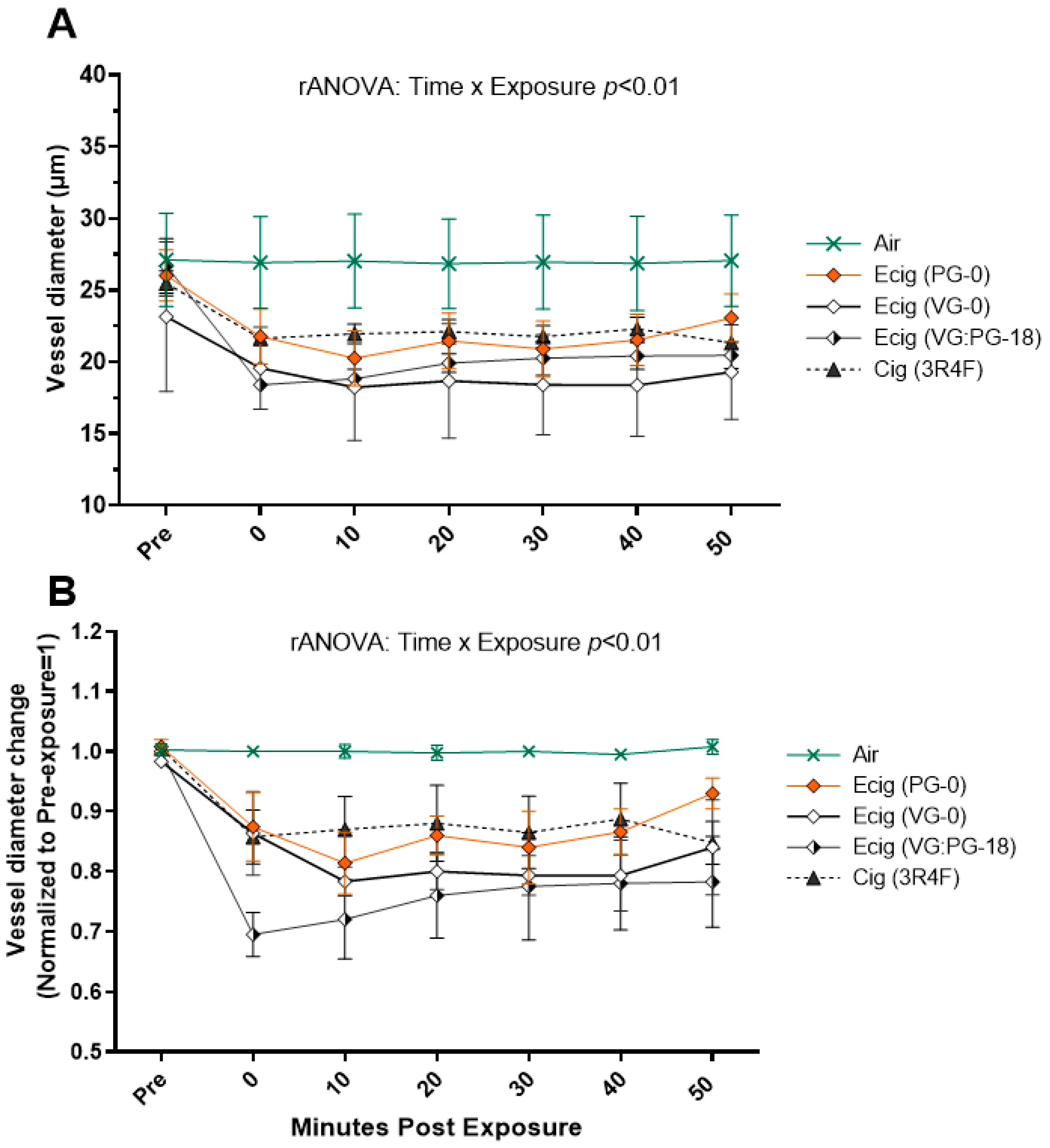
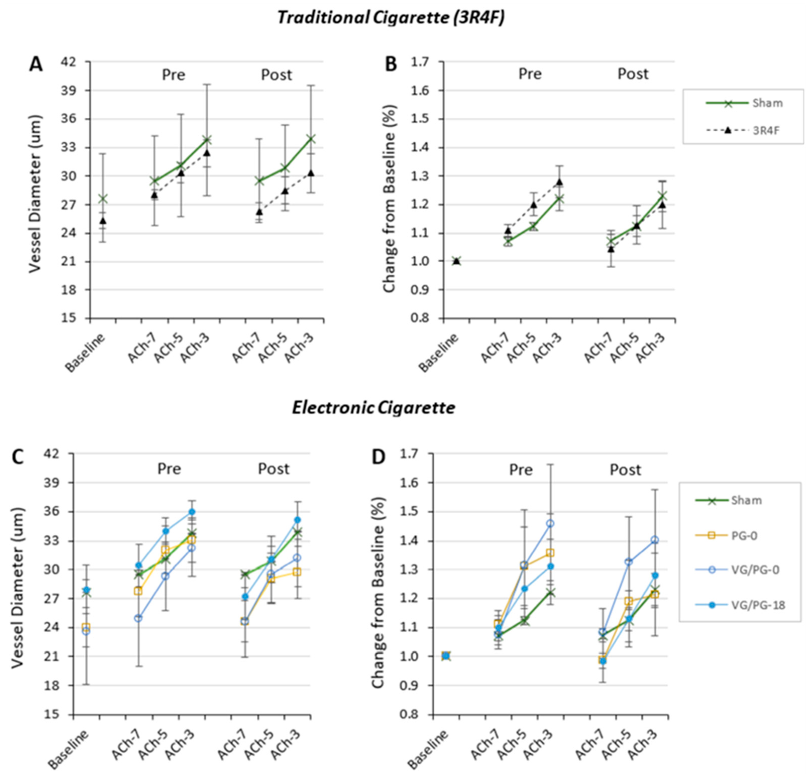
2. Results
3. Discussion
3.1. Nicotine
3.2. E-Liquid Base Solution
3.3. Flavors
3.4. Study Limitations
4. Materials and Methods
4.1. Animals
4.2. Intravital Microscopy
4.3. Inhalation Exposure
4.4. Vascular Assessments
4.5. Statistical Analysis
5. Conclusions
Author Contributions
Funding
Institutional Review Board Statement
Informed Consent Statement
Data Availability Statement
Acknowledgments
Conflicts of Interest
References
- Jani, B.; Rajkumar, C. Ageing and vascular ageing. Postgrad. Med. J. 2006, 82, 357–362. [Google Scholar] [CrossRef]
- Jin, K. A Microcirculatory Theory of Aging. Aging Dis. 2019, 10, 676–683. [Google Scholar] [CrossRef]
- McGill, H.C. The cardiovascular pathology of smoking. Am. Heart J. 1988, 115 Pt 2, 250–257. [Google Scholar] [CrossRef] [PubMed]
- Taylor, B.V.; Oudit, G.Y.; Kalman, P.G.; Liu, P. Clinical and pathophysiological effects of active and passive smoking on the cardiovascular system. Can. J. Cardiol. 1998, 14, 1129–1139. [Google Scholar] [PubMed]
- US-DHHS. The Health Consequences of Smoking—50 Years of Progress: A Report of the Surgeon General; Centers for Disease Control and Prevention: Atlanta, GA, USA, 2014. [Google Scholar]
- Berg, C.J.; Melena, A.; Wittman, F.D.; Robles, T.; Henriksen, L. The Reshaping of the E-Cigarette Retail Environment: Its Evolution and Public Health Concerns. Int. J. Environ. Res. Public Health 2022, 19, 8518. [Google Scholar] [CrossRef]
- Lyu, J.C.; Huang, P.; Jiang, N.; Ling, P.M. A Systematic Review of E-Cigarette Marketing Communication: Messages, Communication Channels, and Strategies. Int. J. Environ. Res. Public Health 2022, 19, 9263. [Google Scholar] [CrossRef] [PubMed]
- Margham, J.; McAdam, K.; Cunningham, A.; Porter, A.; Fiebelkorn, S.; Mariner, D.; Digard, H.; Proctor, C. The Chemical Complexity of e-Cigarette Aerosols Compared With the Smoke From a Tobacco Burning Cigarette. Front. Chem. 2021, 9, 743060. [Google Scholar] [CrossRef]
- Grandjean, P. Paracelsus Revisited: The Dose Concept in a Complex World. Basic Clin. Pharmacol. Toxicol. 2016, 119, 126–132. [Google Scholar] [CrossRef] [PubMed]
- Ooi, B.G.; Dutta, D.; Kazipeta, K.; Chong, N.S. Influence of the E-Cigarette Emission Profile by the Ratio of Glycerol to Propylene Glycol in E-Liquid Composition. ACS Omega 2019, 4, 13338–13348. [Google Scholar] [CrossRef] [PubMed]
- Ikonomidis, I.; Vlastos, D.; Kourea, K.; Kostelli, G.; Varoudi, M.; Pavlidis, G.; Efentakis, P.; Triantafyllidi, H.; Parissis, J.; Andreadou, I.; et al. Electronic Cigarette Smoking Increases Arterial Stiffness and Oxidative Stress to a Lesser Extent Than a Single Conventional Cigarette: An Acute and Chronic Study. Circulation 2018, 137, 303–306. [Google Scholar] [CrossRef]
- Kosmider, L.; Sobczak, A.; Fik, M.; Knysak, J.; Zaciera, M.; Kurek, J.; Goniewicz, M.L. Carbonyl Compounds in Electronic Cigarette Vapors: Effects of Nicotine Solvent and Battery Output Voltage. Nicotine Tob. Res. 2014, 16, 1319–1326. [Google Scholar] [CrossRef] [PubMed]
- Goniewicz, M.L.; Knysak, J.; Gawron, M.; Kosmider, L.; Sobczak, A.; Kurek, J.; Prokopowicz, A.; Jablonska-Czapla, M.; Rosik-Dulewska, C.; Havel, C.; et al. Levels of selected carcinogens and toxicants in vapour from electronic cigarettes. Tob. Control 2014, 23, 133–139. [Google Scholar] [CrossRef] [PubMed]
- West, J.W.; Guzman, S.V.; Bellett, S. Cardiac effects of intracoronary arterial injections of nicotine. Circ. Res. 1958, 6, 389–395. [Google Scholar] [CrossRef] [PubMed]
- Farsalinos, K.E.; Gillman, G. Carbonyl Emissions in E-cigarette Aerosol: A Systematic Review and Methodological Considerations. Front. Physiol. 2017, 8, 1119. [Google Scholar] [CrossRef]
- Khlystov, A.; Samburova, V. Flavoring Compounds Dominate Toxic Aldehyde Production during E-Cigarette Vaping. Environ. Sci. Technol. 2016, 50, 13080–13085. [Google Scholar] [CrossRef]
- Behar, R.Z.; Davis, B.; Wang, Y.; Bahl, V.; Lin, S.; Talbot, P. Identification of toxicants in cinnamon-flavored electronic cigarette refill fluids. Toxicol. In Vitro 2014, 28, 198–208. [Google Scholar] [CrossRef]
- Hess, C.A.; Olmedo, P.; Navas-Acien, A.; Goessler, W.; Cohen, J.E.; Rule, A.M. E-cigarettes as a source of toxic and potentially carcinogenic metals. Environ. Res. 2017, 152, 221–225. [Google Scholar] [CrossRef]
- Olfert, I.M.; DeVallance, E.; Hoskinson, H.; Branyan, K.W.; Clayton, S.; Pitzer, C.R.; Sullivan, D.P.; Breit, M.J.; Wu, Z.; Klinkhachorn, P.; et al. Chronic exposure to electronic cigarettes results in impaired cardiovascular function in mice. J. Appl. Physiol. (1985) 2018, 124, 573–582. [Google Scholar] [CrossRef]
- Kuntic, M.; Oelze, M.; Steven, S.; Kröller-Schön, S.; Stamm, P.; Kalinovic, S.; Frenis, K.; Vujacic-Mirski, K.; Bayo Jimenez, M.T.; Kvandova, M.; et al. Short-term e-cigarette vapour exposure causes vascular oxidative stress and dysfunction: Evidence for a close connection to brain damage and a key role of the phagocytic NADPH oxidase (NOX-2). Eur. Heart J. 2020, 41, 2472–2483. [Google Scholar] [CrossRef]
- Carnevale, R.; Sciarretta, S.; Violi, F.; Nocella, C.; Loffredo, L.; Perri, L.; Peruzzi, M.; Marullo, A.G.; De Falco, E.; Chimenti, I.; et al. Acute Impact of Tobacco vs Electronic Cigarette Smoking on Oxidative Stress and Vascular Function. Chest 2016, 150, 606–612. [Google Scholar] [CrossRef]
- Caporale, A.; Langham, M.C.; Guo, W.; Johncola, A.; Chatterjee, S.; Wehrli, F.W. Acute Effects of Electronic Cigarette Aerosol Inhalation on Vascular Function Detected at Quantitative MRI. Radiology 2019, 293, 97–106. [Google Scholar] [CrossRef]
- Ogunwale, M.A.; Li, M.; Ramakrishnam Raju, M.V.; Chen, Y.; Nantz, M.H.; Conklin, D.J.; Fu, X.A. Aldehyde Detection in Electronic Cigarette Aerosols. ACS Omega 2017, 2, 1207–1214. [Google Scholar] [CrossRef] [PubMed]
- Conklin, D.J.; Malovichko, M.V.; Zeller, I.; Das, T.P.; Krivokhizhina, T.V.; Lynch, B.H.; Lorkiewicz, P.; Agarwal, A.; Wickramasinghe, N.; Haberzettl, P.; et al. Biomarkers of Chronic Acrolein Inhalation Exposure in Mice: Implications for Tobacco Product-Induced Toxicity. Toxicol. Sci. 2017, 158, 263–274. [Google Scholar] [CrossRef]
- Conklin, D.J.; Ogunwale, M.A.; Chen, Y.; Theis, W.S.; Nantz, M.H.; Fu, X.A.; Chen, L.C.; Riggs, D.W.; Lorkiewicz, P.; Bhatnagar, A.; et al. Electronic cigarette-generated aldehydes: The contribution of e-liquid components to their formation and the use of urinary aldehyde metabolites as biomarkers of exposure. Aerosol Sci. Technol. 2018, 52, 1219–1232. [Google Scholar] [CrossRef] [PubMed]
- Li, Y.; Burns, A.E.; Tran, L.N.; Abellar, K.A.; Poindexter, M.; Li, X.; Madl, A.K.; Pinkerton, K.E.; Nguyen, T.B. Impact of e-Liquid Composition, Coil Temperature, and Puff Topography on the Aerosol Chemistry of Electronic Cigarettes. Chem. Res. Toxicol. 2021, 34, 1640–1654. [Google Scholar] [CrossRef] [PubMed]
- Jin, L.; Lynch, J.; Richardson, A.; Lorkiewicz, P.; Srivastava, S.; Theis, W.; Shirk, G.; Hand, A.; Bhatnagar, A.; Srivastava, S.; et al. Electronic cigarette solvents, pulmonary irritation, and endothelial dysfunction: Role of acetaldehyde and formaldehyde. Am. J. Physiol. Heart Circ. Physiol. 2021, 320, H1510–H1525. [Google Scholar] [CrossRef]
- Alarabi, A.B.; Lozano, P.A.; Khasawneh, F.T.; Alshbool, F.Z. The effect of emerging tobacco related products and their toxic constituents on thrombosis. Life Sci. 2022, 290, 120255. [Google Scholar] [CrossRef]
- Richter, P.; Faroon, O.; Pappas, R.S. Cadmium and Cadmium/Zinc Ratios and Tobacco-Related Morbidities. Int. J. Environ. Res. Public Health 2017, 14, 1154. [Google Scholar] [CrossRef]
- Rastian, B.; Wilbur, C.; Curtis, D.B. Transfer of Metals to the Aerosol Generated by an Electronic Cigarette: Influence of Number of Puffs and Power. Int. J. Environ. Res. Public Health 2022, 19, 9334. [Google Scholar] [CrossRef]
- Pappas, R.S.; Gray, N.; Halstead, M.; Valentin-Blasini, L.; Watson, C. Toxic Metal-Containing Particles in Aerosols from Pod-Type Electronic Cigarettes. J. Anal. Toxicol. 2021, 45, 337–347. [Google Scholar] [CrossRef]
- Cuevas, A.K.; Liberda, E.N.; Gillespie, P.A.; Allina, J.; Chen, L.C. Inhaled nickel nanoparticles alter vascular reactivity in C57BL/6 mice. Inhal. Toxicol. 2010, 22 (Suppl. S2), 100–106. [Google Scholar] [CrossRef]
- Whitehead, A.K.; Erwin, A.P.; Yue, X. Nicotine and vascular dysfunction. Acta Physiol. 2021, 231, e13631. [Google Scholar] [CrossRef]
- Dominiak, P.; Fuchs, G.; von Toth, S.; Grobecker, H. Effects of nicotine and its major metabolites on blood pressure in anaesthetized rats. Klin. Wochenschr. 1985, 63, 90–92. [Google Scholar] [CrossRef]
- De Aquino, J.P.; MacLean, R.R.; Gueorguieva, R.; DeVito, E.E.; Eid, T.; Sofuoglu, M. Impact of delivery rate on the acute response to intravenous nicotine: A human laboratory study with implications for regulatory science. Addict. Biol. 2022, 27, e13161. [Google Scholar] [CrossRef] [PubMed]
- Tzivoni, D.; Keren, A.; Meyler, S.; Khoury, Z.; Lerer, T.; Brunel, P. Cardiovascular safety of transdermal nicotine patches in patients with coronary artery disease who try to quit smoking. Cardiovasc. Drugs Ther. 1998, 12, 239–244. [Google Scholar] [CrossRef] [PubMed]
- Joseph, A.M.; Norman, S.M.; Ferry, L.H.; Prochazka, A.V.; Westman, E.C.; Steele, B.G.; Sherman, S.E.; Cleveland, M.; Antonuccio, D.O.; Hartman, N.; et al. The safety of transdermal nicotine as an aid to smoking cessation in patients with cardiac disease. N. Engl. J. Med. 1996, 335, 1792–1798. [Google Scholar] [CrossRef]
- Chalon, S.; Moreno, H.; Benowitz, N.L.; Hoffman, B.B.; Blaschke, T.F. Nicotine impairs endothelium-dependent dilatation in human veins in vivo. Clin. Pharmacol. Ther. 2000, 67, 391–397. [Google Scholar] [CrossRef]
- Galanzha, E.L.; Chowhury, P.; Tuchim, V.V.; Zharov, V.P. Monitoring of nicotine impact in microlymphatics of rat mesentery with time-resolved microscopy. Lymphology 2005, 38, 181–192. [Google Scholar] [PubMed]
- Burrage, E.N.; Aboaziza, E.; Hare, L.; Reppert, S.; Moore, J.; Goldsmith, W.T.; Kelley, E.E.; Mills, A.; Dakhlallah, D.; Chantler, P.D.; et al. Long-term cerebrovascular dysfunction in the offspring from maternal electronic cigarette use during pregnancy. Am. J. Physiol. Heart Circ. Physiol. 2021, 321, H339–H352. [Google Scholar] [CrossRef]
- Aboaziza, E.; Feaster, K.; Hare, L.; Chantler, P.D.; Olfert, I.M. Maternal electronic cigarette use during pregnancy affects long-term arterial function in offspring. J. Appl. Physiol. (1985) 2023, 134, 59–71. [Google Scholar] [CrossRef]
- Anderson, C.; Majeste, A.; Hanus, J.; Wang, S. E-Cigarette Aerosol Exposure Induces Reactive Oxygen Species, DNA Damage, and Cell Death in Vascular Endothelial Cells. Toxicol. Sci. 2016, 154, 332–340. [Google Scholar] [CrossRef]
- Taniyama, Y.; Griendling, K.K. Reactive Oxygen Species in the Vasculature: Molecular and Cellular Mechanisms. Hypertension 2003, 42, 1075–1081. [Google Scholar] [CrossRef] [PubMed]
- Majid, S.; Weisbrod, R.M.; Fetterman, J.L.; Keith, R.J.; Rizvi, S.H.M.; Zhou, Y.; Behrooz, L.; Robertson, R.M.; Bhatnagar, A.; Conklin, D.J.; et al. Pod-based e-liquids impair human vascular endothelial cell function. PLoS ONE 2023, 18, e0280674. [Google Scholar] [CrossRef] [PubMed]
- Nabavizadeh, P.; Liu, J.; Rao, P.; Ibrahim, S.; Han, D.D.; Derakhshandeh, R.; Qiu, H.; Wang, X.; Glantz, S.A.; Schick, S.F.; et al. Impairment of Endothelial Function by Cigarette Smoke Is Not Caused by a Specific Smoke Constituent, but by Vagal Input From the Airway. Arterioscler. Thromb. Vasc. Biol. 2022, 42, 1324–1332. [Google Scholar] [PubMed]
- Rao, P.; Liu, J.; Springer, M.L. JUUL and Combusted Cigarettes Comparably Impair Endothelial Function. Tob. Regul. Sci. 2020, 6, 30–37. [Google Scholar] [CrossRef]
- Benowitz, N.L.; Gourlay, S.G. Cardiovascular Toxicity of Nicotine: Implications for Nicotine Replacement Therapy 1. J. Am. Coll. Cardiol. 1997, 29, 1422–1431. [Google Scholar] [CrossRef]
- Fried, N.D.; Oakes, J.M.; Whitehead, A.K.; Lazartigues, E.; Yue, X.; Gardner, J.D. Nicotine and novel tobacco products drive adverse cardiac remodeling and dysfunction in preclinical studies. Front. Cardiovasc. Med. 2022, 9, 993617. [Google Scholar] [CrossRef]
- Oakes, J.M.; Xu, J.; Morris, T.M.; Fried, N.D.; Pearson, C.S.; Lobell, T.D.; Gilpin, N.W.; Lazartigues, E.; Gardner, J.D.; Yue, X. Effects of Chronic Nicotine Inhalation on Systemic and Pulmonary Blood Pressure and Right Ventricular Remodeling in Mice. Hypertension 2020, 75, 1305–1314. [Google Scholar] [CrossRef]
- Palpant, N.J.; Hofsteen, P.; Pabon, L.; Reinecke, H.; Murry, C.E. Cardiac development in zebrafish and human embryonic stem cells is inhibited by exposure to tobacco cigarettes and e-cigarettes. PLoS ONE 2015, 10, e0126259. [Google Scholar] [CrossRef]
- Li, Q.; Zhan, Y.; Wang, L.; Leischow, S.J.; Zeng, D.D. Analysis of symptoms and their potential associations with e-liquids’ components: A social media study. BMC Public Health 2016, 16, 674. [Google Scholar] [CrossRef]
- Harvanko, A.; Kryscio, R.; Martin, C.; Kelly, T. Stimulus effects of propylene glycol and vegetable glycerin in electronic cigarette liquids. Drug Alcohol Depend. 2019, 194, 326–329. [Google Scholar] [CrossRef]
- Varughese, S.; Teschke, K.; Brauer, M.; Chow, Y.; van Netten, C.; Kennedy, S.M. Effects of theatrical smokes and fogs on respiratory health in the entertainment industry. Am. J. Ind. Med. 2005, 47, 411–418. [Google Scholar] [CrossRef]
- Rao, P.; Han, D.D.; Tan, K.; Mohammadi, L.; Derakhshandeh, R.; Navabzadeh, M.; Goyal, N.; Springer, M.L. Comparable Impairment of Vascular Endothelial Function by a Wide Range of Electronic Nicotine Delivery Devices. Nicotine Tob. Res. 2022, 24, 1055–1062. [Google Scholar] [CrossRef]
- Nabavizadeh, P.; Liu, J.; Havel, C.M.; Ibrahim, S.; Derakhshandeh, R.; Jacob Iii, P.; Springer, M.L. Vascular endothelial function is impaired by aerosol from a single IQOS HeatStick to the same extent as by cigarette smoke. Tob. Control 2018, 27 (Suppl. S1), s13–s19. [Google Scholar] [CrossRef]
- Poredos, P.; Orehek, M.; Tratnik, E. Smoking is associated with dose-related increase of intima-media thickness and endothelial dysfunction. Angiology 1999, 50, 201–208. [Google Scholar] [CrossRef]
- Allen, J.G.; Flanigan, S.S.; LeBlanc, M.; Vallarino, J.; MacNaughton, P.; Stewart, J.H.; Christiani, D.C. Flavoring Chemicals in E-Cigarettes: Diacetyl, 2,3-Pentanedione, and Acetoin in a Sample of 51 Products, Including Fruit-, Candy-, and Cocktail-Flavored E-Cigarettes. Environ. Health Perspect. 2016, 124, 733–739. [Google Scholar] [CrossRef]
- Bitzer, Z.T.; Goel, R.; Reilly, S.M.; Elias, R.J.; Silakov, A.; Foulds, J.; Muscat, J.; Richie, J.P., Jr. Effect of flavoring chemicals on free radical formation in electronic cigarette aerosols. Free Radic. Biol. Med. 2018, 120, 72–79. [Google Scholar] [CrossRef] [PubMed]
- Omaiye, E.E.; McWhirter, K.J.; Luo, W.; Pankow, J.F.; Talbot, P. High-Nicotine Electronic Cigarette Products: Toxicity of JUUL Fluids and Aerosols Correlates Strongly with Nicotine and Some Flavor Chemical Concentrations. Chem. Res. Toxicol. 2019, 32, 1058–1069. [Google Scholar] [CrossRef]
- Fetterman, J.L.; Weisbrod, R.M.; Feng, B.; Bastin, R.; Tuttle, S.T.; Holbrook, M.; Baker, G.; Robertson, R.M.; Conklin, D.J.; Bhatnagar, A.; et al. Flavorings in Tobacco Products Induce Endothelial Cell Dysfunction. Arterioscler. Thromb. Vasc. Biol. 2018, 38, 1607–1615. [Google Scholar] [CrossRef] [PubMed]
- Farsalinos, K.E.; Romagna, G.; Voudris, V. Cytotoxicity of cinnamon-flavored electronic cigarette refills: Are the results truly applicable to electronic cigarette use? Toxicol. In Vitro 2014, 28, 1016–1017. [Google Scholar] [CrossRef] [PubMed]
- Kerasioti, E.; Veskoukis, A.S.; Skaperda, Z.; Zacharias, A.; Poulas, K.; Lazopoulos, G.; Kouretas, D. The flavoring and not the nicotine content is a decisive factor for the effects of refill liquids of electronic cigarette on the redox status of endothelial cells. Toxicol. Rep. 2020, 7, 1095–1102. [Google Scholar] [CrossRef]
- University of Kentucky Center for Tobacco Reference Product—Reference Cigarettes—3R4F. Available online: https://ctrp.uky.edu/products/gallery/Reference%20Cigarettes (accessed on 17 March 2023).
- United Kingdom—Tobacco Advisory Group of the Royal College of Physicians: Nicotine without smoke: Tobacco harm reduction. R. Coll. Physician Policy Public Health. 2016. Available online: https://www.rcplondon.ac.uk/projects/outputs/nicotine-without-smoke-tobacco-harm-reduction-0 (accessed on 20 November 2017).
- Chaumont, M.; de Becker, B.; Zaher, W.; Culie, A.; Deprez, G.; Melot, C.; Reye, F.; Van Antwerpen, P.; Delporte, C.; Debbas, N.; et al. Differential Effects of E-Cigarette on Microvascular Endothelial Function, Arterial Stiffness and Oxidative Stress: A Randomized Crossover Trial. Sci. Rep. 2018, 8, 10378. [Google Scholar] [CrossRef] [PubMed]
- Farsalinos, K.E.; Tsiapras, D.; Kyrzopoulos, S.; Savvopoulou, M.; Voudris, V. Acute effects of using an electronic nicotine-delivery device (electronic cigarette) on myocardial function: Comparison with the effects of regular cigarettes. BMC Cardiovasc. Disord. 2014, 14, 78. [Google Scholar] [CrossRef] [PubMed]
- Moheimani, R.S.; Bhetraratana, M.; Peters, K.M.; Yang, B.K.; Yin, F.; Gornbein, J.; Araujo, J.A.; Middlekauff, H.R. Sympathomimetic Effects of Acute E-Cigarette Use: Role of Nicotine and Non-Nicotine Constituents. J. Am. Heart Assoc. 2017, 6, e006579. [Google Scholar] [CrossRef] [PubMed]


Disclaimer/Publisher’s Note: The statements, opinions and data contained in all publications are solely those of the individual author(s) and contributor(s) and not of MDPI and/or the editor(s). MDPI and/or the editor(s) disclaim responsibility for any injury to people or property resulting from any ideas, methods, instructions or products referred to in the content. |
© 2023 by the authors. Licensee MDPI, Basel, Switzerland. This article is an open access article distributed under the terms and conditions of the Creative Commons Attribution (CC BY) license (https://creativecommons.org/licenses/by/4.0/).
Share and Cite
Pitzer, C.R.; Aboaziza, E.A.; O’Reilly, J.M.; Mandler, W.K.; Olfert, I.M. Nicotine and Microvascular Responses in Skeletal Muscle from Acute Exposure to Cigarettes and Vaping. Int. J. Mol. Sci. 2023, 24, 10208. https://doi.org/10.3390/ijms241210208
Pitzer CR, Aboaziza EA, O’Reilly JM, Mandler WK, Olfert IM. Nicotine and Microvascular Responses in Skeletal Muscle from Acute Exposure to Cigarettes and Vaping. International Journal of Molecular Sciences. 2023; 24(12):10208. https://doi.org/10.3390/ijms241210208
Chicago/Turabian StylePitzer, Christopher R., Eiman A. Aboaziza, Juliana M. O’Reilly, W. Kyle Mandler, and I. Mark Olfert. 2023. "Nicotine and Microvascular Responses in Skeletal Muscle from Acute Exposure to Cigarettes and Vaping" International Journal of Molecular Sciences 24, no. 12: 10208. https://doi.org/10.3390/ijms241210208
APA StylePitzer, C. R., Aboaziza, E. A., O’Reilly, J. M., Mandler, W. K., & Olfert, I. M. (2023). Nicotine and Microvascular Responses in Skeletal Muscle from Acute Exposure to Cigarettes and Vaping. International Journal of Molecular Sciences, 24(12), 10208. https://doi.org/10.3390/ijms241210208