Abstract
MicroRNAs (miRNAs) are endogenous small non-coding RNAs that play crucial regulatory roles in many biological processes, including the growth and development of skeletal muscle. miRNA-100-5p is often associated with tumor cell proliferation and migration. This study aimed to uncover the regulatory mechanism of miRNA-100-5p in myogenesis. In our study, we found that the miRNA-100-5p expression level was significantly higher in muscle tissue than in other tissues in pigs. Functionally, this study shows that miR-100-5p overexpression significantly promotes the proliferation and inhibits the differentiation of C2C12 myoblasts, whereas miR-100-5p inhibition results in the opposite effects. Bioinformatic analysis predicted that Trib2 has potential binding sites for miR-100-5p at the 3′UTR region. A dual-luciferase assay, qRT-qPCR, and Western blot confirmed that Trib2 is a target gene of miR-100-5p. We further explored the function of Trib2 in myogenesis and found that Trib2 knockdown markedly facilitated proliferation but suppressed the differentiation of C2C12 myoblasts, which is contrary to the effects of miR-100-5p. In addition, co-transfection experiments demonstrated that Trib2 knockdown could attenuate the effects of miR-100-5p inhibition on C2C12 myoblasts differentiation. In terms of the molecular mechanism, miR-100-5p suppressed C2C12 myoblasts differentiation by inactivating the mTOR/S6K signaling pathway. Taken together, our study results indicate that miR-100-5p regulates skeletal muscle myogenesis through the Trib2/mTOR/S6K signaling pathway.
1. Introduction
Skeletal muscle is one of the most dynamic and plastic tissues in mammals; it accounts for about 40–50% of body mass in the adult stage; and plays a vital role in movement, respiration, and metabolism [1]. Skeletal muscle myogenesis is a strict and complex process through which mononuclear myoblasts undergo proliferation and differentiation, further fuse into multinuclear myotubes, and ultimately form muscle fibers with contractile traits [2]. This process is regulated by the cascade control of multiple transcription factors [3,4,5,6]. For example, the differentiation of myoblasts is activated by myogenic differentiation (MyoD), myogenin (MyoG), and myocyte enhancer factor 2 (MEF2). Non-coding RNAs also play crucial roles in the myogenesis process [7].
To date, emerging studies have suggested that non-coding RNAs affect skeletal muscle development by regulating gene expression, including microRNAs (miRNAs) [8], circular RNAs [9], and long non-coding RNAs (IncRNAs) [10]. Notably, miRNAs are a class of conserved small single-stranded RNAs that do not encode protein [11]. These miRNAs generally act as negative regulators of protein translation by influencing messenger RNA (mRNA) stability [12]. An increasing number of studies have suggested that miRNAs play a critical regulatory role in the process of skeletal muscle myogenesis. For example, miR-325-3p [13], miR-2400 [14], and microRNA-24-3p [15] were found to regulate skeletal growth and development. miR-100-5p is an important member of the miR-99 family (miR-99a, miR-99b, and miR-100) with the same seed region sequence [16]. Previous studies have reported that the miR-99 family plays a regulatory role in many kinds of cancer cells by targeting different genes, such as IGF1R, AKT1, and mTOR [17,18,19]. Notably, members of the miR-99 family are also involved in the regulation of myogenesis. For example, in a previous study, miR-99a-5p promoted the proliferation and inhibited the differentiation of skeletal muscle satellite cells by targeting MTMR3 in chickens [20]. miR-100 overexpression could inhibit differentiation and promote intramuscular lipid deposition by modulating IGF1R in fetal bovine muscle satellite cells [17]. However, the effect of miR-100-5p in skeletal muscle myoblasts and its molecular regulatory mechanism remains unclear.
Previous studies have reported that miR-100 can bind directly to tribbles homolog 2 (TRIB2) 3′UTR in pulmonary fibroblasts [21]. TRIB2 is a member of the tribbles family that functions as scaffold molecules (MAPK and Akt signaling pathways) in different cellular processes [22,23,24]. Additionally, as a cancer-associated pseudokinase, TRIB2 can interact with E3 ubiquitin ligases and regulate the stability of downstream effectors, which impact various cellular processes such as proliferation, differentiation, migration, and cell death [25,26,27]. Recent studies have reported that TRIB2 plays a critical role in vascular smooth muscle cell proliferation by modulating ERK activity [28] and that TRIB2 is also involved in visceral fat accumulation [29]. However, to date, it remains unknown whether TRIB2 plays a role in the growth and development of skeletal muscle.
The mammalian target of rapamycin (mTOR) is an evolutionarily conserved serine/threonine kinase complex that plays a critical role in regulating the development of various cells [30]. The mTOR complex has two protein complexes, mTOR complex 1 (mTORC1) and mTOR complex 2 (mTORC2). mTORC1 plays a dominant role in skeletal muscle development by activating the downstream ribosomal protein S6kin-1 (S6k1) and elF4-E binding protein 1 (4E-BP1), which affect protein synthesis [31]. During myoblast differentiation, the mTOR signaling pathway is activated by upstream IGF1R/PI3K/Akt signaling, resulting in increased phosphorylation of S6K and promoting mRNA translation and protein synthesis [32]. Therefore, activation of the mTOR signaling pathway is essential for skeletal muscle differentiation and regeneration [33].
In this study, we aimed to systematically investigate the molecular mechanism of miR-100-5p regulating skeletal muscle myogenesis. A series of experiments demonstrated that miR-100-5p promotes C2C12 myoblast proliferation by targeting Trib2 and inhibits C2C12 myoblast differentiation through the Trib2/mTOR/S6K signaling pathway. This finding will contribute to expanding our understanding of muscle growth regulation.
2. Results
2.1. The Expression of miR-100-5p Is Associated with Myogenesis
Sequence alignment analysis revealed that miR-100-5p is highly conserved, with almost no differences between different species (Figure 1A). For the expression patterns of miR-100-5p in different pig tissues, qRT–PCR results showed that miR-100-5p is highly expressed in muscle tissue and spleen in piglets (Figure 1B). In addition, the expression trend of miR-100-5p in the four developmental stages of the Longissimus dorsi muscle was found to be dynamic (Figure 1C). These results suggest that miR-100-5p may also be expressed in myoblasts and exert certain influences. To explore the influence of miR-100-5p on skeletal muscle myogenesis, we selected mouse skeletal muscle C2C12 myoblasts because the mature sequences of miR-100-5p are highly conserved between pigs and mice. First, we detected the expression patterns of miR-100-5p in myoblasts using qTR-PCR. The results showed that the expression levels of the proliferation marker gene Pcna and miR-100-5p increased gradually with cell proliferation (Figure 1D,E). The expression levels of the differentiation marker gene Myhc gradually increased, while miR-100-5p gradually decreased with cell differentiation (Figure 1F,G). To investigate the function of miR-100-5p in myoblast proliferation and differentiation, miR-100-5p inhibitors, and mimics were transfected into myoblasts to inhibit and promote their expression levels, respectively (Figure 1H,I).
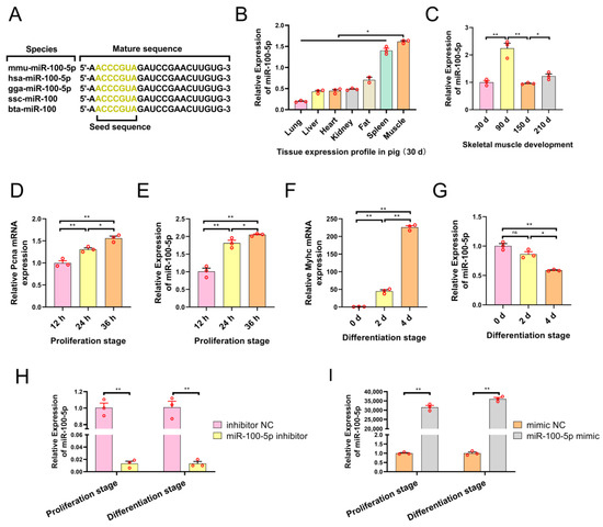
Figure 1.
The expression of miR-100-5p is associated with myogenesis. (A) Conservation of the miR-100-5p sequence in different species. (B) Expression levels of miR-100-5p in different pig tissues. (C) Expression levels of miR-100-5p in Longissimus dorsi muscles at four developmental stages in pigs. (D,E) Expression levels of the proliferation marker gene Pcna and miR-100-5p in the C2C12 myoblast proliferation stage. (F,G) Expression levels of the differentiation marker gene Myhc and miR-100-5p in the C2C12 myoblast differentiation stage. (H) The transfection efficiency of miR-100-5p inhibition. (I) The transfection efficiency of miR-100-5p overexpression. The results are presented as the mean ± SEM (n = 3). * p < 0.05; ** p < 0.01; p ≥ 0.05: ns (Not significant).
2.2. miR-100-5p Promotes Myoblast Proliferation and Inhibits Myoblast Differentiation
To investigate the effects of miR-100-5p on myoblast proliferation, we conducted miR-100-5p overexpression and inhibition experiments to detect the proliferation abilities of myoblasts. The EdU incorporation assay showed that the anti-miR-100-5p group significantly inhibited myoblast proliferation (Figure 2A,C), whereas miR-100-5p overexpression significantly promoted myoblast proliferation (Figure 2A,B). The CCK-8 assay found that cell proliferation in the miR-100-5p silence group was markedly inhibited (Figure 2D). In contrast, miR-100-5p overexpression promoted myoblast proliferation (Figure 2E). Next, the result of flow cytometry suggested that miR-100-5p knockdown increased the population of cells in the G1 phase and decreased the population of cells in the G2 phase and S phase (Figure 2F). After transfection with the miR-100-5p mimic, the number of cells in the G1 phase decreased while those in the G2 and S phases increased significantly (Figure 2G). Results of the qRT–PCR and Western blot showed that miR-100-5p knockdown or overexpression could regulate the mRNA and protein expression of proliferation-related genes (Pcna and Cdk4) (Figure 2H–J). Taken together, these results indicate that miR-100-5p promotes myoblast proliferation.
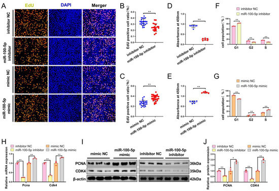
Figure 2.
miR-100-5p promotes myoblast proliferation. (A–C) EdU staining detected the proliferation of myoblasts transfected with the miR-100-5p inhibitor and mimic. Cells in the DNA replication phase were stained with EdU (green), and nuclei were stained with DAPI (blue). Scale bar = 50 μm. The proportion of EdU-positive cells among the total cells was calculated. (D,E) CCK-8 assay for myoblasts after miR-100-5p overexpression and inhibition. (F,G) Cell-cycle analysis, using propidium iodide staining, of myoblasts transfected with the miR-100-5p inhibitor and mimic. (H–J) qRT–PCR and Western-blot-detected expression of the proliferation-related gene after transfection with the miR-100-5p inhibitor and mimic. The results are presented as the mean ± SEM (n = 3). * p < 0.05; ** p < 0.01.
To further confirm miR-100-5p’s role in myoblast differentiation, we measured the expression of myogenic differentiation genes and fusion marker genes using qRT–PCR and Western blot. The qRT–PCR suggested that the mRNA expression levels of Myod and MyoG significantly increased after transfection with miR-100-5p inhibitor at 2 d, 4 d, and 6 d of differentiation (Figure 3A,B). Conversely, miR-100-5p overexpression significantly decreased mRNA expression levels of Myod and Myog (Figure 3A,B). Western blot analysis showed that the protein expression of MyoG and MyHC significantly increased following miR-100-5p knockdown (Figure 3C,D), whereas the protein expression of MyoG and MyHC decreased markedly after miR-100-5p overexpression (Figure 3E,F). Furthermore, qRT–PCR results showed that miR-100-5p knockdown remarkably promoted the mRNA expression level of Myomaker and Myomixer (Figure 3G), which are marker genes of muscle fusion, whereas miR-100-5p overexpression significantly decreased their mRNA expression levels (Figure 3H). These results suggest that miR-100-5p inhibits myoblast differentiation.
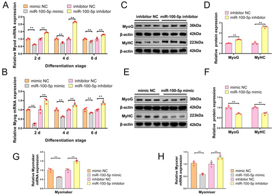
Figure 3.
miR-100-5p inhibits myoblast differentiation. (A,B) qRT–PCR-detected mRNA expression levels of myogenic differentiation (Myod) and Myogenin (Myog) after overexpression and inhibition of miR-100-5p in the differentiation stage. (C–F) Western-blot-detected protein expression of Myogenin (MyoG) and Myosin heavy chain (MyHC) in myoblasts after transfection with the miR-100-5p inhibitor and miR-100-5p mimic, respectively. (G,H) The mRNA expression levels of muscle fusion marker genes (Myomaker and Myomixer) were detected by qRT–PCR in myoblasts after miR-100-5p overexpression and inhibition. The results are presented as the mean ± SEM (n = 3). ** p < 0.01.
2.3. Target Gene Screening Revealed That miR-100-5p Directly Targeted Trib2
To explore the molecular mechanism of miR-100-5p’s impact on skeletal muscle myoblast development, potential target genes were predicted using the miRDB, Targetsan, StarBase, and miRcode (Figure 4A). Venn analysis was applied to obtain eight candidate target genes of miR-100-5p (Figure 4B). To determine which of these target genes were negatively regulated by miR-100-5p, qRT–PCR was used to detect mRNA expression levels of eight genes at the myoblast proliferation and differentiation stages. Muscleblind-like splicing regulator 1 (Mbnl1) and ribonucleoprotein PTB-binding 2 (Raver2) were not detected by qRT–PCR because they featured low expression in the myoblasts, while the other six genes could be detected. At the proliferation stage, qRT–PCR analysis showed that miR-100-5p overexpression significantly increased the mRNA expression level of Cdk4 (proliferation marker gene) and decreased the expression levels of tribbles pseudokinase 2 (Trib2), myotubularin-related protein (Mtmr3), and adaptor-related protein-complex-1-associated regulatory protein (Aplar), while miR-100-5p knockdown caused the opposite results (Figure 4C). At the differentiation stage, miR-100-5p overexpression significantly decreased the mRNA expression level of Myhc (differentiation marker gene) and Trib2, whereas miR-100-5p inhibition caused the opposite results (Figure 4D). Taking these qRT–PCR results together, Mtmr3 and Trib2 were selected as candidate target genes for further study because their expression was negatively regulated by miR-100-5p in both the proliferation and differentiation stages. Notably, miR-100-5p had a stronger effect on Trib2 than on Mtmr3.
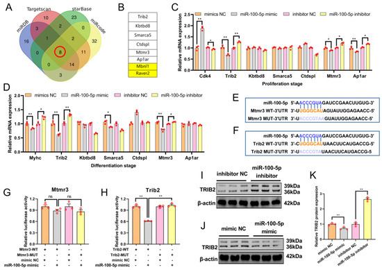
Figure 4.
Target gene screening revealed that miR-100-5p directly targeted Trib2. (A) The target genes of miR-100-5p were predicted using miRDB, Targetsan, StarBase and miRcode. (B) Eight candidate target genes of miR-100-5p were obtained via Venn analysis. (C) The mRNA expression levels of proliferation marker gene Cdk4 and six candidate target genes were detected by qRT–PCR after miR-100-5p overexpression and inhibition at the proliferation stage in myoblasts. (D) The mRNA expression levels of the differentiation marker gene Myhc and six candidate target genes were detected by qRT–PCR in myoblasts after miR-100-5p overexpression and inhibition at the differentiation stage. (E,F) Wild-type binding sites (WT) and mutation binding sites (MUT) of miR-100-5p in the Mtmr3 3′UTR and Trib2 3′UTR regions. (G,H) In the dual-luciferase reporter assay, the pmirGLO–Mtmr3/Trib2–WT and pmirGLO–Mtmr3/Trib2–MUT report vectors were co–transfected with the miR-100-5p mimic or mimic NC into HEK293T cells. (I–K) Western-blot-detected protein expression of TRIB2 in myoblasts after transfection with miR-100-5p inhibitor and miR-100-5p mimic. The results are presented as the mean ± SEM (n = 3). * p < 0.05; ** p < 0.01; p ≥ 0.05: ns (Not significant).
To verify whether miR-100-5p can target the Mtmr3 and Trib2 genes, the putative sequences of the miR-100-5p binding site (WT) in Mtmr3/Trib2 3′UTR and the corresponding mutated sequences (MUT) were cloned into a pmirGLO Dual-Luciferase miRNA Target Expression Vector (Figure 4E,F). Then, the pmirGLO–Mtmr3/Trib2–WT and pmirGLO–Mtmr3/Trib2–MUT report vectors were co–transfected with miR-100-5p mimic or mimic NC into HEK293T cells. Our results confirmed that the miR-100-5p mimic failed to decrease the luciferase activity of pmirGLO–Mtmr3–MUT (Figure 4G). Conversely, miR-100-5p overexpression decreased the luciferase activity of pmirGLO–Trib2–MUT (Figure 4H). Therefore, Trib2 was further identified as a possible target gene of miRNA-100-5p. Next, Western blot analysis showed that miR-100-5p could negatively regulate the protein expression of TRIB2 after miR-100-5p overexpression and knockdown at the myoblast differentiation stage (Figure 4I–K). These results indicated that Trib2 is the direct target gene of miR-100-5p.
2.4. Trib2 Knockdown Promotes Myoblast Proliferation and Inhibits Myoblast Differentiation
We further examined the function of Trib2 using specific RNA knockdown. We used the RNA-seq results from a previous study [34] and found that TRIB2 expression was gradually up-regulated with chicken myoblast differentiation (Figure 5A). In addition, the result of qRT–PCR showed that the mRNA expression levels of Myhc and Trib2 increased gradually with mouse C2C12 myoblast differentiation (Figure 5B,C). Immunofluorescence staining showed that TRIB2 was localized to the nucleus and cytoplasm in differentiated C2C12 myoblasts (Figure 5D). To investigate the effects of Trib2 on myoblast proliferation, Western blot analysis, and CCK-8 were performed. Western blot analysis showed that Trib2 knockdown significantly decreased the protein expression of TRIB2 while increasing the protein expression of PCNA (Figure 5E,F). The CCK-8 assay revealed that Trib2 silencing significantly promoted myoblast proliferation 24 h, 48 h, and 72 h after transfection (Figure 5G). We also detected the influence of Trib2 knockdown on myoblast differentiation. Trib2 knockdown decreased the mRNA expression levels of Trib2 and differentiation marker genes (MyoG and MyHC) (Figure 5H–J) and decreased the protein expression of MyoG and MyHC (Figure 5K,L). These results indicate that Trib2 knockdown promotes myoblast proliferation and suppresses myoblast differentiation.
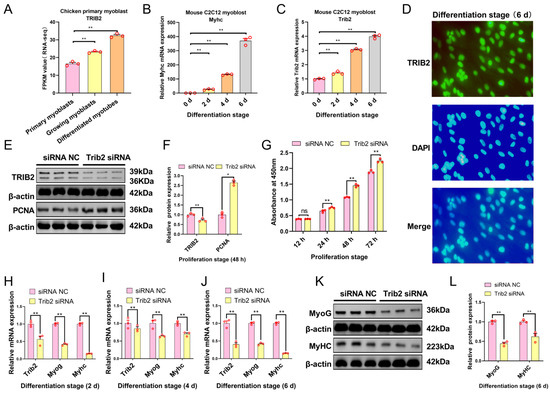
Figure 5.
Trib2 knockdown promotes myoblast proliferation and inhibits differentiation. (A) TRIB2 expression was gradually up-regulated during chicken myoblast differentiation. (B,C) qRT–PCR detected the mRNA expression levels of Myhc and Trib2 during C2C12 myoblast differentiation. (D) Differentiated mouse C2C12 myoblasts were stained with the TRIB2 antibody (green) and DAPI (blue); scale bar = 50 μm. (E,F) Western-blot-detected protein expression of TRIB2 and proliferation marker gene (PCNA) after transfection with Trib2 siRNA in the myoblasts. (G) Cell counting detected by CCK-8 during myoblast proliferation. (H–J) The mRNA expression levels of Trib2 and muscle differentiation maker genes (Myog and Myhc) were detected via qRT–PCR in myoblasts after being transfected with Trib2 siRNA. (K,L) Western-blot-detected protein expression of differentiation marker genes (MyoG and MyHC) in myoblasts after transfection with Trib2 siRNA. The results are presented as the mean ± SEM (n = 3). * p < 0.05; ** p < 0.01; p ≥ 0.05: ns (Not significant).
2.5. miR-100-5p Attenuates Activation of the mTOR Signaling Pathway during Myoblast Differentiation
Previous studies have reported that the activation of PI3K/Akt/mTOR signaling is indispensable for myogenic differentiation. Thus, we hypothesized that miR-100-5p might negatively regulate myoblast differentiation through the mTOR signaling pathway. To verify this hypothesis, we examined the protein expression of phosphor-mTOR (p-mTOR) and phosphor-S6K (p-S6K) after transfection with miR-100-5p inhibition, Trib2 knockdown, and miR-100-5p overexpression in myoblasts using Western blot. The results showed that miR-100-5p knockdown significantly increased the protein expression of p-mTOR and p-S6K (Figure 6A,B), while Trib2 knockdown and miR-100-5p overexpression markedly decreased the protein expression of p-mTOR and p-S6K (Figure 6C,D). Additionally, Trib2 knockdown attenuated the effects of miR-100-5p inhibition on the protein expression of TRIB2, p-mTOR, p-S6K, and differentiation-related genes MyoG and MyHC (Figure 6E,F). To clarify the direct relationship between the mTOR signaling pathway and myoblast differentiation, an mTOR-specific inhibitors (Rapamycin) and its control group, dimethyl sulfoxide (DMSO), were used to treat myoblasts that were differentiated for four days. The results showed that Rapamycin significantly inhibited the protein expression of p-mTOR, p-S6K, TRIB2, and differentiation-related genes MyoG and MyHC (Figure 6G,H). Collectively, these data demonstrate that miR-100-5p is a negative regulator that inhibits myoblast differentiation and inactivates the mTOR signaling pathway through the downregulation of Trib2 expression.
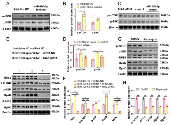
Figure 6.
miR-100-5pmiR-100-5p inactivates the mTOR signaling pathway through downregulation of Trib2 expression during myoblast differentiation. (A,B) Effect of miR-100-5p inhibition on the protein expression of phosphor-mTOR (p-mTOR) and phosphor-S6K (p-S6K) during myoblast differentiation. (C,D) Effects of miR-100-5p overexpression and Trib2 knockdown on the protein expression of phosphor-mTOR (p-mTOR) and phosphor-S6K (p-S6K) during myoblast differentiation. (E,F) miR-100-5p inhibitor and Trib2 siRNA were co-transfected into myoblasts. Western-blot-detected the protein expression of TRIB2, phosphor-mTOR (p-mTOR), phosphor-S6K (p-S6K), and differentiation-related genes (MyoG and MyHC). (G,H) Western-blot-detected the protein expression of phosphor-mTOR (p-mTOR), phosphor-S6K (p-S6K), TRIB2, and differentiation-related genes (MyoG and MyHC) in differentiated myoblasts that were treated with mTOR-pathway-specific inhibitors (Rapamycin) and the corresponding control group treated with dimethyl sulfoxide (DMSO). The results are presented as the mean ± SEM (n = 3). * p < 0.05; ** p < 0.01.
3. Discussion
In the orderly arranged myogenesis process, myoblasts undergo proliferation. Then, they exit the cell cycle and begin to differentiate. Finally, they fuse to form multinuclear fibers [35]. This process is regulated by many transcription factors, signaling pathways, and non-coding RNAs [5,36,37]. Currently, most studies on the miR-99 family focus on the development of cancer cells [16]. Recently, researchers have found that miR-99a-5p promotes proliferation and inhibits the differentiation of chicken skeletal muscle satellite cells [20]. Notably, miR-99a-5p and miR-100-5p belong to the miR-99 family, whose members have the same maturation sequences. Therefore, we speculated that miR-100-5p may have an effect on skeletal muscle myogenesis. In our study, we aimed to investigate the function and mechanism of miR-100-5p in myogenesis. First, the expression profile of miR-100-5p in pig tissues was detected. The results showed that miR-100-5p expression was the highest in muscle tissues and was dynamically expressed in the Longissimus dorsi muscle development stages. Hence, miR-100-5p was found to be a broad-spectrum miRNA, not a muscle-specific miRNA. However, this miRNA was highly expressed in muscle tissue, consistent with previous studies [38]. Then, the correlation between miR-100-5p and C2C12 myoblast proliferation and differentiation was further verified. The results indicated that the miR-100-5p expression level increased gradually with myoblast proliferation and decreased gradually with myoblast differentiation. This result suggests that the role of miR-100-5p may be different in the proliferation and differentiation stages. Additionally, miR-100-5p has a highly conservative sequence among different species (mouse, human, chicken, pig, and cattle). These results suggest that miR-100-5p might be a muscle-associated miRNA, similar to previously reported muscle-related miRNAs [39,40,41].
miR-100-5p was previously identified as a cancer-related miRNA that plays a crucial role in different cancer cells [42,43,44]. It was also reported that miR-100-5p is significantly correlated with fat formation and is a candidate regulator of intramuscular fat deposition [45]. Functional studies showed that miR-100 could inhibit 3T3-L1 cell differentiation by decreasing the expression of mTOR and IGF1R [46]. In this study, EdU staining and CCK-8 assays showed that miR-100-5p inhibition and overexpression, respectively, suppressed and promoted C2C12 myoblast proliferation. Flow cytometry analysis showed that miR-100-5p knockdown caused proliferating myoblasts to remain at the G1 phase, whereas the number of myoblasts in the S phase was significantly increased under miR-100-5p overexpression compared to the negative control group. The mRNA and protein expression of proliferation marker genes were also significantly changed when miR-100-5p was inhibited or overexpressed. Further, we found that miR-100-5p repressed C2C12 myoblast differentiation by detecting key regulators of muscle differentiation and fusion. This result is consistent with previous results where miR-100 inhibited the differentiation of bovine satellite cells [17]. In the myogenesis process, myoblasts exit the cell cycle and begin to differentiate [5]. Hence, the relationship between proliferation and differentiation is usually the opposite. Taken together, these findings suggest that miR-100-5p promotes the proliferation and inhibits the differentiation of C2C12 myoblast.
Most studies on miRNAs have concentrated on their capacity to regulate cellular processes by targeting different genes [11]. Therefore, the identification of miR-100-5p targets is vital for a comprehensive understanding of miRNA-mediated gene regulation [12]. Previous reports have shown that miR-100-5p regulates cell biological processes by targeting different genes. In this study, eight candidate target genes of miR-100-5p were identified via bioinformatics analysis (miRDB, starBase, miRcode, and Targetscan). Among these genes, the mRNA expression of Trib2 and Mtmr3 was negatively regulated by miR-100-5p in both the C2C12 myoblast proliferation and differentiation stages. A dual-luciferase reporter assay is the most direct and important means to validate the targeting relationship between miRNA and its target genes [47]. The results showed that miR-100-5p could directly target Trib2, which is consistent with previous reports that miR-100-5p and TRIB2 have a targeted regulatory relationship [21]. Regrettably, miR-100-5p cannot bind to Mtmr3, which is a muscle-associated gene [20]. In addition, the protein expression of TRIB2 was found to be regulated after miR-100-5p overexpression or inhibition. Ultimately, we confirmed that Trib2 is the most likely target gene of miR-100-5p during C2C12 myoblast proliferation and differentiation.
Trib2 is a pseudokinase associated with cancer and can interact with E3 ubiquitin ligase to control downstream effector protein stability [48,49,50]. Trib2 is also involved in the MAPK and AKT pathways [51]. To date, the regulation of Trib2 in skeletal muscle development has not been reported. Given the function of Trib2, we speculated that Trib2 may play a role in myoblast proliferation and differentiation. First, the expression of Trib2 gradually increased with myoblast differentiation, consistent with the previous reports on chicken myoblasts [34] and contrary to results for miR-100-5p. Trib2 was localized in the nucleus and cytoplasm of differentiated myoblasts. Functionally, siRNA was used to knockdown Trib2 expression in the myoblasts. Then, myoblast proliferation was significantly promoted, and myoblast differentiation was inhibited, which is consistent with miR-100-5p overexpression. In conclusion, miR-100-5p regulates myoblast proliferation and differentiation by targeting Trib2.
Activation of the mTOR signaling pathway is essential for muscle differentiation and regeneration [52]. mTOR is activated by the upstream IGF1R/PI3K/Akt pathway, resulting in increased phosphorylation of S6K for promoting mRNA translation and protein synthesis [53]. miR-100-5p was found to target several key genes in this signaling pathway, such as IGF1R, AKT1, and mTOR [18]. This study demonstrated the targeting relationship between miR-100-5p and Trib2. It was already acknowledged that Trib2 is a protein scaffold for the AKT and MAPK pathways [23]. Hence, we speculated that miR-100-5p may ultimately regulate skeletal muscle development through the mTOR signaling pathway. C2C12 myoblasts were transfected with miR-100-5p overexpression, miR-100-5p inhibition, and Trib2 knockdown to detect the phosphorylated protein expression of mTOR and S6K. The results showed that miR-100-5p and Trib2 knockdown could inactivate the mTOR signaling pathway. Further experiments showed that miR-100-5p could target Trib2 to inactivate the mTOR signaling pathway, thereby inhibiting myoblast differentiation. These results are consistent with previous reports that mTOR signaling pathway activation is vital for muscle differentiation [54,55,56]. Taken together, these results clearly indicate that miR-100-5p affects the mTOR signaling pathway by regulating the Trib2 gene in myoblast differentiation.
4. Materials and Methods
4.1. Cell Culture
HEK293T cells and C2C12 myoblasts (ATCC, New York, NY, USA) were cultured in a growth medium with high-glucose DMEM (Giboc) containing 10% fetal bovine serum (Giboc) in a cell incubator with a humid environment of 37 °C and 5% CO2. The C2C12 cells of between 10 and 25 generations were stimulated with DMEM containing 2% horse serum (Giboc) for differentiation when the degree of cell fusion reached 80%.
4.2. RNA Oligonucleotides and Cell Transfection
To explore the effects of miR-100-5p and its target gene on skeletal muscle myogenesis, the miR-100-5p inhibitor, an inhibitor negative control (inhibitor NC), miR-100-5p mimic, negative control (mimic NC or siRNA NC), and Trib2 small interfering RNAs (siRNAs) were synthesized from GenePharma Co., Ltd. (Suzhou, China). All transient transfections in C2C12 myoblasts were performed with a lipofectamine 2000 regent (Invitrogen, Waltham, MA, USA) according to the manufacturer’s instructions. For proliferation experiments, C2C12 myoblast transfection was performed when cell density reached 40%. After 6 h of transfection, the medium was replaced with a growth medium. In the differentiation experiments, transfection was performed when the cell density reached 80%. After 6 h of transfection, the medium was replaced with a differentiation medium. All RNA oligonucleotides are listed in Table 1.

Table 1.
RNA oligonucleotides used in this study.
4.3. RNA Isolation and Real-Time Quantitative PCR
Total RNA was extracted from tissues and myoblasts using TRIzol reagent (Invitrogen, USA) and stored at −80 °C. Total RNAs were reverse-transcribed into cDNA using a Thermo Scientific™ RevertAid™ First Strand cDNA Synthesis Kit. The specific stem-loop of miR-100-5p was used to synthesize the first strand cDNA for miR-100-5p. qRT–PCR was performed using an SYBR Green Kit (TransGen, Beijing, China). GAPDH was used to normalize mRNA expression, and U6 was used to normalize miR-100-5p expression. The relative expression of miR-100-5p and related genes was calculated via the 2−ΔΔCt method. All primers are listed in Table 2.

Table 2.
Primer information for miRNA and mRNA quantitative reverse transcription.
4.4. Cell Counting Kit-8 (CCK-8) Assays
C2C12 myoblasts were seeded at a density of 1 × 10⁵ in 96-well plates. Cells were transfected with RNA Oligonucleotides when cell confluence reached 30%. After 12 h, 24 h, 48 h, and 72 h of culturing at 37 °C. Then, the cells were switched to the medium with 10% CCK-8 regent (AFExBIO, Houston, TX, USA) and incubated for 4 h at 37 °C. The cell absorbance value at 450 nm was detected using a spectrometer (Molecular Devices, San Francisco, CA, USA).
4.5. 5-Ethynyl-20-Deoxyuridine (EdU) Assay
To assess the effects of miR-100-5p and its target on myoblast proliferation, when C2C12 myoblast confluence reached 70–80%, cells were switched to a culture medium with EdU (20 µM) for 2 h at 37 °C. Then, EdU staining was performed using an EdU cell Proliferation Kit with Alexa Fluor 555 (Meilun, Dalian, China) following the manufacturer’s protocol. Images were captured using a fluorescence microscope (Zeiss LSM800, Carl Zeiss AG, Oberkochen, Germany). Finally, the ratio of EdU-positive cells was calculated as follows: (EdU-positive cells/DAPI staining cells) × 100%.
4.6. Immunofluorescence Staining
The differentiated myoblasts were fixed in 4% paraformaldehyde for 30 min and then permeabilized in 0.5% Triton X-100 for 20 min. Subsequently, the myoblasts were blocked with 5% BSA for 30 min. Immunofluorescence staining was performed using a primary antibody working solution overnight at 4 °C. Then, the second antibody working solution was added and incubated at room temperature for 1 h, and DAPI (1:1000) was incubated in the dark for 10 min. Images were obtained using a Leica SP8 confocal microscope (eyepiece magnification: 10×) and processed using Image J.
4.7. Flow Cytometric Analysis
The myoblasts were seeded in a 6-well plate. After 48 h of transfection, the myoblasts were washed three times with PBS. Then, the cells were collected and fixed in 75% ethanol at −20 °C overnight. The myoblasts were stirred with a PI staining solution (Yeasen Biotechnology, Shanghai, China) at 37 °C for 30 min. The myoblasts were then suspended and detected via flow cytometry (Becton Dickinson, Trenton, NJ, USA) according to the manufacturer’s protocols.
4.8. Dual-Luciferase Reporter Assay
A Dual-Luciferase Reporter Assay was employed to verify the targeting relationship between miR-100-5p and its candidate targeting genes (Mtmr3 and Trib2). The putative sequences of the miR-100-5p binding site in Mtmr3/Trib2 3’UTR and the corresponding mutated sequences were cloned into a pmirGLO Dual-Luciferase miRNA Target Expression Vector (Promega, Madison, WI, USA). The pmirGLO–Mtmr3/Trib2–WT and pmirGLO–Mtmr3/Trib2–MUT report vector was co–transfected with the miR-100-5p mimic or mimic NC into HEK293T cells. After 24 h of transfection, the Dual-Luciferase Reporter Assay was performed using a Dual-Luciferase Reporter Assay System (Promega, Fitchburg, WI, USA) to detect the luciferase activity according to the manufacturer’s instructions.
4.9. Western Blot Assay
The total protein was extracted using a RIPA-lysed buffer with protease inhibitors and protein phosphatase inhibitors (Abiowell, Changsha, China). Then, the protein concentration was measured using a BCA Protein Assay Kit (Abiowell, Changsha, China). We performed Western blotting using various antibodies according to the standard procedure. The Datails of antibodies are as follows: PCNA 1:2000 (AF02986, Rabbit, AFBio, Changsha, China), CDK4 1:2000 (AF06640, Rabbit, AFBio, Changsha, China), MyHC 1:2000 (MF 20, Mouse, DSHB, Iowa City, IA, USA), MyOG 1:1000 (AB2146602, Mouse, DSHB, Iowa City, IA, USA), TRIB2 1:2000 (DF2692, Rabbit, Affinity Biosciences, Jiangsu, China), p-mTOR Ser2448 1:1000 (381557, Rabbit, ZenBio, Chengdu, China), p-S6K Thr389 1:1000 (9234T, Rabbit, Cell Signaling Technology, Danvers, MA, USA), and β-actin 1:5000 (AWS0001, Mouse, Proteintech, San Diego, CA, USA).
4.10. Bioinformation Analysis
Target gene predictions for miR-100-5p were performed using miRDB (http://www.miRbase.org/cgi-bin/broe.pl, accessed on 1 September 2022), Targetsan (http://www.targetscan.org/, accessed on 1 September 2022), StarBase (https://starbase.sysu.edu.cn/, accessed on 1 September 2022), and miRcode (http://www.mircode.org/, accessed on 1 September 2022). The Venn analysis was performed using an online tool (https://bioinfogp.cnb.csic.es/tools/venny, accessed on 1 September 2022).
4.11. Statistical Analysis
Image J was used to count EdU-positive cells and analyze protein bands. The relative mRNA expression under real-time quantitative PCR (qRT–PCR) was calculated using 2−ΔΔCT. An unpaired Student’s t-test was performed for the treatment and control groups using GraphPad Prism 8. These data are presented as the means ± standard error of the mean (SEM, n = 3). A difference was considered significant when the p-value was <0.05.
5. Conclusions
In summary, our study indicates that miR-100-5p promotes C2C12 myoblast proliferation by targeting Trib2 and inhibits C2C12 myoblast differentiation by the Trib2/mTOR/S6K signaling pathway (Figure 7).
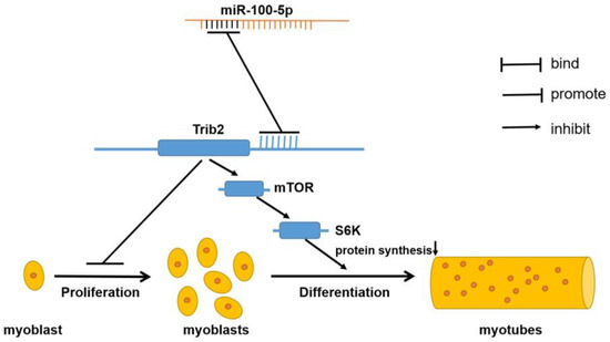
Figure 7.
Mechanism diagram of miR-100-5p regulating skeletal muscle myogenesis.
Author Contributions
Conceptualization, X.X. and K.W.; methodology, K.W. and X.L. (Xiaolin Liu); software, N.A. and X.L. (Xintong Li); validation, K.W., S.L. and B.C.; formal analysis, K.W.; data curation, K.W.; writing—original draft preparation, K.W. and S.L.; writing—review and editing, H.M. and Y.Z.; visualization, K.W. and Z.Y.; funding acquisition, H.M. and Y.Y. All authors have read and agreed to the published version of the manuscript.
Funding
This research was funded by the Laboratory of Lingnan Modern Agriculture Project (NT2021005), Hunan Provincial Natural Science Joint Foundation (2022JJ50018), and Major science and technology projects in Yunnan Province (202202AE090032).
Institutional Review Board Statement
The Institutional Animal Care and Use Committee of Hunan Agricultural University evaluated and approved the experimental protocols under approval number 2013-06.
Informed Consent Statement
Not applicable.
Data Availability Statement
Data presented in this study are available on request from the corresponding author.
Conflicts of Interest
The authors declare no conflict of interest.
References
- Frontera, W.R.; Ochala, J. Skeletal muscle: A brief review of structure and function. Calcif. Tissue Int. 2015, 96, 183–195. [Google Scholar] [CrossRef] [PubMed]
- Braun, T.; Gautel, M. Transcriptional mechanisms regulating skeletal muscle differentiation, growth and homeostasis. Nat. Rev. Mol. Cell Biol. 2011, 12, 349–361. [Google Scholar] [CrossRef] [PubMed]
- Singh, K.; Dilworth, F.J. Differential modulation of cell cycle progression distinguishes members of the myogenic regulatory factor family of transcription factors. FEBS J. 2013, 280, 3991–4003. [Google Scholar] [CrossRef] [PubMed]
- Günther, S.; Kim, J.; Kostin, S.; Lepper, C.; Fan, C.M.; Braun, T. Myf5-positive satellite cells contribute to Pax7-dependent long-term maintenance of adult muscle stem cells. Cell Stem Cell 2013, 13, 590–601. [Google Scholar] [CrossRef] [PubMed]
- Tapscott, S.J. The circuitry of a master switch: Myod and the regulation of skeletal muscle gene transcription. Development 2005, 132, 2685–2695. [Google Scholar] [CrossRef] [PubMed]
- Seale, P.; Sabourin, L.A.; Girgis-Gabardo, A.; Mansouri, A.; Gruss, P.; Rudnicki, M.A. Pax7 is required for the specification of myogenic satellite cells. Cell 2000, 102, 777–786. [Google Scholar] [CrossRef]
- Archacka, K.; Ciemerych, M.A.; Florkowska, A.; Romanczuk, K. Non-Coding RNAs as Regulators of Myogenesis and Postexercise Muscle Regeneration. Int. J. Mol. Sci. 2021, 22, 11568. [Google Scholar] [CrossRef]
- Zhao, H.; Li, P.; Wang, J. The role of muscle-specific MicroRNAs in patients with chronic obstructive pulmonary disease and skeletal muscle dysfunction. Front. Physiol. 2022, 13, 954364. [Google Scholar] [CrossRef]
- Yan, S.; Pei, Y.; Li, J.; Tang, Z.; Yang, Y. Recent Progress on Circular RNAs in the Development of Skeletal Muscle and Adipose Tissues of Farm Animals. Biomolecules 2023, 13, 314. [Google Scholar] [CrossRef]
- Lv, W.; Jin, J.; Xu, Z.; Luo, H.; Guo, Y.; Wang, X.; Wang, S.; Zhang, J.; Zuo, H.; Bai, W.; et al. lncMGPF is a novel positive regulator of muscle growth and regeneration. J. Cachexia Sarcopenia Muscle 2020, 11, 1723–1746. [Google Scholar] [CrossRef]
- Shukla, G.C.; Singh, J.; Barik, S. MicroRNAs: Processing, Maturation, Target Recognition and Regulatory Functions. Mol. Cell. Pharmacol. 2011, 3, 83–92. [Google Scholar] [PubMed]
- Huntzinger, E.; Izaurralde, E. Gene silencing by microRNAs: Contributions of translational repression and mRNA decay. Nat. Rev. Genet. 2011, 12, 99–110. [Google Scholar] [CrossRef] [PubMed]
- Nguyen, M.T.; Lee, W. Role of MiR-325-3p in the Regulation of CFL2 and Myogenic Differentiation of C2C12 Myoblasts. Cells 2021, 10, 2725. [Google Scholar] [CrossRef] [PubMed]
- Elnour, I.E.; Wang, X.; Zhansaya, T.; Akhatayeva, Z.; Khan, R.; Cheng, J.; Hung, Y.; Lan, X.; Lei, C.; Chen, H. Circular RNA circMYL1 Inhibit Proliferation and Promote Differentiation of Myoblasts by Sponging miR-2400. Cells 2021, 10, 176. [Google Scholar] [CrossRef]
- Dey, P.; Soyer, M.A.; Dey, B.K. MicroRNA-24-3p promotes skeletal muscle differentiation and regeneration by regulating HMGA1. Cell. Mol. Life Sci. 2022, 79, 170. [Google Scholar] [CrossRef]
- Eniafe, J.; Jiang, S. MicroRNA-99 family in cancer and immunity. Wiley Interdiscip. Rev. RNA 2021, 12, e1635. [Google Scholar] [CrossRef]
- Mir, B.A.; Albrecht, E.; Ali, A.; Hansson, O.; Maak, S. MicroRNA-100 Reduced Fetal Bovine Muscle Satellite Cell Myogenesis and Augmented Intramuscular Lipid Deposition by Modulating IGF1R. Cells 2022, 11, 451. [Google Scholar] [CrossRef]
- Jin, Y.; Tymen, S.D.; Chen, D.; Fang, Z.J.; Zhao, Y.; Dragas, D.; Dai, Y.; Marucha, P.T.; Zhou, X. MicroRNA-99 family targets AKT/mTOR signaling pathway in dermal wound healing. PLoS ONE 2013, 8, e64434. [Google Scholar] [CrossRef]
- Jahangiri, B.; Khalaj-Kondori, M.; Asadollahi, E.; Purrafee Dizaj, L.; Sadeghizadeh, M. MSC-Derived exosomes suppress colorectal cancer cell proliferation and metastasis via miR-100/mTOR/miR-143 pathway. Int. J. Pharmaceut. 2022, 627, 122214. [Google Scholar] [CrossRef]
- Cao, X.; Tang, S.; Du, F.; Li, H.; Shen, X.; Li, D.; Wang, Y.; Zhang, Z.; Xia, L.; Zhu, Q.; et al. miR-99a-5p Regulates the Proliferation and Differentiation of Skeletal Muscle Satellite Cells by Targeting MTMR3 in Chicken. Genes 2020, 11, 369. [Google Scholar] [CrossRef]
- Mai, Z.; Mi, Y.; Jiang, M.; Wan, S.; Di, Q. Expression and Related Mechanisms of miR-100 and TRIB2 in COPD Patients. J. Healthc. Eng. 2022, 2022, 6556208. [Google Scholar] [CrossRef] [PubMed]
- Eyers, P.A.; Keeshan, K.; Kannan, N. Tribbles in the 21st Century: The Evolving Roles of Tribbles Pseudokinases in Biology and Disease. Trends Cell Biol. 2017, 27, 284–298. [Google Scholar] [CrossRef] [PubMed]
- Ferreira, B.I.; Santos, B.; Link, W.; De Sousa-Coelho, A.L. Tribbles Pseudokinases in Colorectal Cancer. Cancers 2021, 13, 2825. [Google Scholar] [CrossRef] [PubMed]
- Harris, J.A.; Fairweather, E.; Byrne, D.P.; Eyers, P.A. Analysis of human Tribbles 2 (TRIB2) pseudokinase. Method Enzymol. 2022, 667, 79–99. [Google Scholar] [CrossRef]
- Richmond, L.; Keeshan, K. Pseudokinases: A tribble-edged sword. FEBS J. 2020, 287, 4170–4182. [Google Scholar] [CrossRef]
- Warma, A.; Ndiaye, K. Functional effects of Tribbles homolog 2 in bovine ovarian granulosa cells. Biol. Reprod. 2020, 102, 1177–1190. [Google Scholar] [CrossRef]
- Salomé, M.; Magee, A.; Yalla, K.; Chaudhury, S.; Sarrou, E.; Carmody, R.J.; Keeshan, K. A Trib2-p38 axis controls myeloid leukaemia cell cycle and stress response signalling. Cell Death Dis. 2018, 9, 443. [Google Scholar] [CrossRef]
- Takaguri, A.; Ishizaka, R.; Maki, S.; Satoh, K. The role of tribbles homolog 2 in vascular smooth muscle cell proliferation. Cell Biol. Int. 2023, 47, 787–795. [Google Scholar] [CrossRef]
- Nakayama, K.; Ogawa, A.; Miyashita, H.; Tabara, Y.; Igase, M.; Kohara, K.; Miki, T.; Kagawa, Y.; Yanagisawa, Y.; Katashima, M.; et al. Positive natural selection of TRIB2, a novel gene that influences visceral fat accumulation, in East Asia. Hum. Genet. 2013, 132, 201–217. [Google Scholar] [CrossRef]
- Saxton, R.A.; Sabatini, D.M. mTOR Signaling in Growth, Metabolism, and Disease. Cell 2017, 168, 960–976. [Google Scholar] [CrossRef]
- Ma, X.M.; Blenis, J. Molecular mechanisms of mTOR-mediated translational control. Nat. Rev. Mol. Cell Biol. 2009, 10, 307–318. [Google Scholar] [CrossRef] [PubMed]
- Holz, M.K.; Ballif, B.A.; Gygi, S.P.; Blenis, J. mTOR and S6K1 mediate assembly of the translation preinitiation complex through dynamic protein interchange and ordered phosphorylation events. Cell 2021, 184, 2255. [Google Scholar] [CrossRef] [PubMed]
- Rion, N.; Castets, P.; Lin, S.; Enderle, L.; Reinhard, J.R.; Eickhorst, C.; Rüegg, M.A. mTOR controls embryonic and adult myogenesis via mTORC1. Development 2019, 146, dev172460. [Google Scholar] [CrossRef]
- Luo, W.; Lin, Z.; Chen, J.; Chen, G.; Zhang, S.; Liu, M.; Li, H.; He, D.; Liang, S.; Luo, Q.; et al. TMEM182 interacts with integrin beta 1 and regulates myoblast differentiation and muscle regeneration. J. Cachexia Sarcopenia Muscle 2021, 12, 1704–1723. [Google Scholar] [CrossRef] [PubMed]
- Miller, J.B.; Everitt, E.A.; Smith, T.H.; Block, N.E.; Dominov, J.A. Cellular and molecular diversity in skeletal muscle development: News from in vitro and in vivo. Bioessays 1993, 15, 191–196. [Google Scholar] [CrossRef] [PubMed]
- Rao, P.K.; Kumar, R.M.; Farkhondeh, M.; Baskerville, S.; Lodish, H.F. Myogenic factors that regulate expression of muscle-specific microRNAs. Proc. Natl. Acad. Sci. USA 2006, 103, 8721–8726. [Google Scholar] [CrossRef] [PubMed]
- Rodriguez, J.; Vernus, B.; Chelh, I.; Cassar-Malek, I.; Gabillard, J.C.; Hadj Sassi, A.; Seiliez, I.; Picard, B.; Bonnieu, A. Myostatin and the skeletal muscle atrophy and hypertrophy signaling pathways. Cell. Mol. Life Sci. CMLS 2014, 71, 4361–4371. [Google Scholar] [CrossRef]
- Silva, W.J.; Graça, F.A.; Cruz, A.; Silvestre, J.G.; Labeit, S.; Miyabara, E.H.; Yan, C.; Wang, D.Z.; Moriscot, A.S. miR-29c improves skeletal muscle mass and function throughout myocyte proliferation and differentiation and by repressing atrophy-related genes. Acta Physiol. 2019, 226, e13278. [Google Scholar] [CrossRef]
- Lim, S.; Lee, D.; Morena Da Silva, F.; Koopmans, P.; Vechetti, I.J.; von Walden, F.; Greene, N.P.; Murach, K.A. MicroRNA Control of the Myogenic Cell Transcriptome and Proteome: The Role of miR-16. Am. J. Physiol. Cell Physiol. 2023, 324, C1101–C1109. [Google Scholar] [CrossRef]
- Shintani-Ishida, K.; Tsurumi, R.; Ikegaya, H. Decrease in the expression of muscle-specific miRNAs, miR-133a and miR-1, in myoblasts with replicative senescence. PLoS ONE 2023, 18, e280527. [Google Scholar] [CrossRef]
- Lopez, M.A.; Si, Y.; Hu, X.; Williams, V.; Qushair, F.; Carlyle, J.; Alesce, L.; Conklin, M.; Gilbert, S.; Bamman, M.M.; et al. Smad8 Is Increased in Duchenne Muscular Dystrophy and Suppresses miR-1, miR-133a, and miR-133b. Int. J. Mol. Sci. 2022, 23, 7515. [Google Scholar] [CrossRef] [PubMed]
- Takebayashi, K.; Nasu, K.; Okamoto, M.; Aoyagi, Y.; Hirakawa, T.; Narahara, H. hsa-miR-100-5p, an overexpressed miRNA in human ovarian endometriotic stromal cells, promotes invasion through attenuation of SMARCD1 expression. Reprod. Biol. Endocrinol. RBE 2020, 18, 31. [Google Scholar] [CrossRef] [PubMed]
- Fuso, P.; Di Salvatore, M.; Santonocito, C.; Guarino, D.; Autilio, C.; Mulè, A.; Arciuolo, D.; Rinninella, A.; Mignone, F.; Ramundo, M.; et al. Let-7a-5p, miR-100-5p, miR-101-3p, and miR-199a-3p Hyperexpression as Potential Predictive Biomarkers in Early Breast Cancer Patients. J. Pers. Med. 2021, 11, 816. [Google Scholar] [CrossRef] [PubMed]
- Lai, Y.; Kacal, M.; Kanony, M.; Stukan, I.; Jatta, K.; Kis, L.; Norberg, E.; Vakifahmetoglu-Norberg, H.; Lewensohn, R.; Hydbring, P.; et al. miR-100-5p confers resistance to ALK tyrosine kinase inhibitors Crizotinib and Lorlatinib in EML4-ALK positive NSCLC. Biochem. Biophys. Res. Commun. 2019, 511, 260–265. [Google Scholar] [CrossRef]
- Huang, J.; Wang, S.; Feng, X.; Liu, X.; Zhao, J.; Zheng, Q.; Wei, X.; Ma, Y. miRNA transcriptome comparison between muscle and adipose tissues indicates potential miRNAs associated with intramuscular fat in Chinese swamp buffalo. Genome 2019, 62, 729–738. [Google Scholar] [CrossRef]
- Pek, S.L.; Sum, C.F.; Lin, M.X.; Cheng, A.K.; Wong, M.T.; Lim, S.C.; Tavintharan, S. Circulating and visceral adipose miR-100 is down-regulated in patients with obesity and Type 2 diabetes. Mol. Cell. Endocrinol. 2016, 427, 112–123. [Google Scholar] [CrossRef]
- Andriani, F.; Majorini, M.T.; Mano, M.; Landoni, E.; Miceli, R.; Facchinetti, F.; Mensah, M.; Fontanella, E.; Dugo, M.; Giacca, M.; et al. MiR-16 regulates the pro-tumorigenic potential of lung fibroblasts through the inhibition of HGF production in an FGFR-1- and MEK1-dependent manner. J. Hematol. Oncol. 2018, 11, 45. [Google Scholar] [CrossRef]
- Keeshan, K.; Shestova, O.; Ussin, L.; Pear, W.S. Tribbles homolog 2 (Trib2) and HoxA9 cooperate to accelerate acute myelogenous leukemia. Blood Cells Mol. Dis. 2008, 40, 119–121. [Google Scholar] [CrossRef]
- Bailey, F.P.; Byrne, D.P.; Oruganty, K.; Eyers, C.E.; Novotny, C.J.; Shokat, K.M.; Kannan, N.; Eyers, P.A. The Tribbles 2 (TRB2) pseudokinase binds to ATP and autophosphorylates in a metal-independent manner. Biochem. J. 2015, 467, 47–62. [Google Scholar] [CrossRef]
- Do, E.K.; Park, J.K.; Cheon, H.C.; Kwon, Y.W.; Heo, S.C.; Choi, E.J.; Seo, J.K.; Jang, I.H.; Lee, S.C.; Kim, J.H. Trib2 regulates the pluripotency of embryonic stem cells and enhances reprogramming efficiency. Exp. Mol. Med. 2017, 49, e401. [Google Scholar] [CrossRef]
- Dobens, L.L.; Nauman, C.; Fischer, Z.; Yao, X. Control of Cell Growth and Proliferation by the Tribbles Pseudokinase: Lessons from Drosophila. Cancers 2021, 13, 883. [Google Scholar] [CrossRef] [PubMed]
- Zhang, P.; Liang, X.; Shan, T.; Jiang, Q.; Deng, C.; Zheng, R.; Kuang, S. mTOR is necessary for proper satellite cell activity and skeletal muscle regeneration. Biochem. Biophys. Res. Commun. 2015, 463, 102–108. [Google Scholar] [CrossRef] [PubMed]
- Cong, X.X.; Gao, X.K.; Rao, X.S.; Wen, J.; Liu, X.C.; Shi, Y.P.; He, M.Y.; Shen, W.L.; Shen, Y.; Ouyang, H.; et al. Rab5a activates IRS1 to coordinate IGF-AKT-mTOR signaling and myoblast differentiation during muscle regeneration. Cell Death Differ. 2020, 27, 2344–2362. [Google Scholar] [CrossRef] [PubMed]
- Schiaffino, S.; Dyar, K.A.; Ciciliot, S.; Blaauw, B.; Sandri, M. Mechanisms regulating skeletal muscle growth and atrophy. FEBS J. 2013, 280, 4294–4314. [Google Scholar] [CrossRef] [PubMed]
- Yoshida, T.; Delafontaine, P. Mechanisms of IGF-1-Mediated Regulation of Skeletal Muscle Hypertrophy and Atrophy. Cells 2020, 9, 1970. [Google Scholar] [CrossRef] [PubMed]
- Roy, A.; Kumar, A. Supraphysiological activation of TAK1 promotes skeletal muscle growth and mitigates neurogenic atrophy. Nat. Commun. 2022, 13, 2201. [Google Scholar] [CrossRef]
Disclaimer/Publisher’s Note: The statements, opinions and data contained in all publications are solely those of the individual author(s) and contributor(s) and not of MDPI and/or the editor(s). MDPI and/or the editor(s) disclaim responsibility for any injury to people or property resulting from any ideas, methods, instructions or products referred to in the content. |
© 2023 by the authors. Licensee MDPI, Basel, Switzerland. This article is an open access article distributed under the terms and conditions of the Creative Commons Attribution (CC BY) license (https://creativecommons.org/licenses/by/4.0/).