Review

Disabled-2 (DAB2): A Key Regulator of Anti- and Pro-Tumorigenic Pathways

1
Discipline of Obstetrics and Gynaecology, Robinson Research Institute, Adelaide Medical School, University of Adelaide, Adelaide, SA 5005, Australia
2
Department of Obstetrics and Gynecology, Nagoya University Graduate School of Medicine, Nagoya 464-0813, Japan
3
Department of Gynaecological Oncology, Royal Adelaide Hospital, Adelaide, SA 5000, Australia
*
Author to whom correspondence should be addressed.
Int. J. Mol. Sci. 2023, 24(1), 696; https://doi.org/10.3390/ijms24010696
Submission received: 7 November 2022 / Revised: 22 December 2022 / Accepted: 23 December 2022 / Published: 31 December 2022
(This article belongs to the Special Issue The Role of the Tumor Microenvironment in Cancer)

Abstract

:
Disabled-2 (DAB2), a key adaptor protein in clathrin mediated endocytosis, is implicated in the regulation of key signalling pathways involved in homeostasis, cell positioning and epithelial to mesenchymal transition (EMT). It was initially identified as a tumour suppressor implicated in the initiation of ovarian cancer, but was subsequently linked to many other cancer types. DAB2 contains key functional domains which allow it to negatively regulate key signalling pathways including the mitogen activated protein kinase (MAPK), wingless/integrated (Wnt) and transforming growth factor beta (TGFβ) pathways. Loss of DAB2 is primarily associated with activation of these pathways and tumour progression, however this review also explores studies which demonstrate the complex nature of DAB2 function with pro-tumorigenic effects. A recent strong interest in microRNAs (miRNA) in cancer has identified DAB2 as a common target. This has reignited an interest in DAB2 research in cancer. Transcriptomics of tumour associated macrophages (TAMs) has also identified a pro-metastatic role of DAB2 in the tumour microenvironment. This review will cover the broad depth literature on the tumour suppressor role of DAB2, highlighting its complex relationships with different pathways. Furthermore, it will explore recent findings which suggest DAB2 has a more complex role in cancer than initially thought.

1. Introduction

Disabled-2 (DAB2) is a widely recognised tumour suppressor. It was initially discovered in 1994 when Mok et al. identified an 800bp cDNA fragment which was expressed in normal ovarian surface epithelial cell lines but not in ovarian cancer cell lines. They referred to it as differentially expressed in ovarian carcinoma 2 (DOC-2) [1]. The following year, Xu et al. identified a 96 kDa phosphoprotein in mouse macrophage cell line, BAC1.2F5 with an amino terminal end which shared homology to the Drosophila disabled gene [2,3]. The Drosophila disabled protein is important in embryogenesis and neural positioning [4]. DAB2 is one of two human orthologs of the Drosophila disabled gene, Disabled-1 is expressed almost exclusively in neural cells whereas DAB2 is expressed in a wide range of epithelial cells including those of the ovary, lung and breast [5,6,7]. Loss of DAB2 expression has been reported in a range of malignancies including ovarian, lung and breast cancer [5,6,7] Table 1. The loss of DAB2 is associated with activation of key signalling pathways including Wnt, MAPK and TGFβ which is associated with enhanced cell proliferation, chemotherapy resistance and tumour progression, supporting its role as a tumour suppressor. This review will discuss the role of DAB2 in regulating these key pathways and the resulting effects on cancer progression.

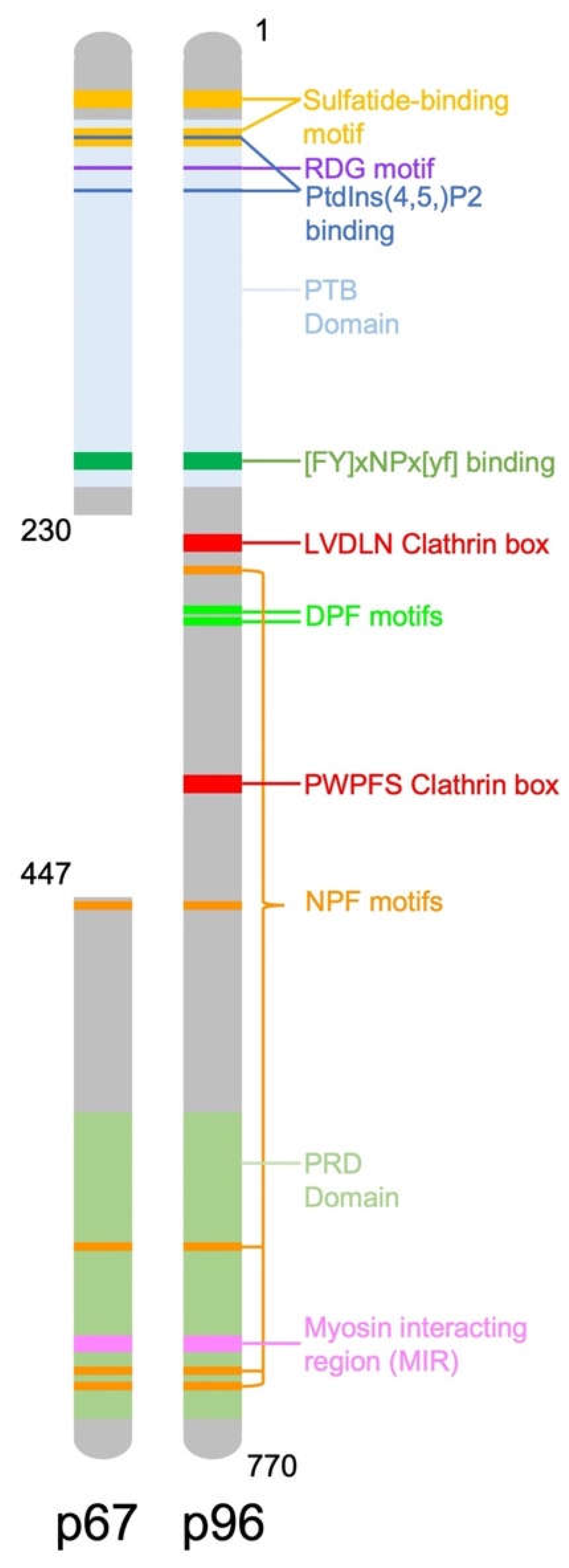
2. DAB2 Structure and Function

The human DAB2 gene, located on chromosome 5p13 consists of 15 exons, encoding a 770 amino acid protein [8]. The mouse DAB2 gene has 83% homology with the human gene, it also consists of 15 exons and encodes a 766 amino acid protein [9]. There are two isoforms of DAB2, including full length p96 (also known as p82) and spliced p67 (also known as p59) that is missing the central exon. DAB2 contains binding domains and motifs which allow it to recognise and recruit proteins to clathrin coated pits for endocytosis (Figure 1). Two key binding domains of DAB2 are a phosphotyrosine binding (PTB) domain at the N-terminus and a proline rich domain (PRD) at the carboxy terminal end of the protein which contains a myosin interacting region (MIR). The main function of DAB2 is as a clathrin associated sorting protein (CLASP) in clathrin mediated endocytosis. DAB2 interacts with clathrin via multiple binding sites including a type I LVDLN and type II PWPYP sequence [10]. DAB2 can interact with both pre-assembled clathrin cages and also soluble clathrin trimers, indicating a possible role in clathrin cage assembly [10]. The DAB2 PTB binds to phosphoinositide(4,5)P2 (PtdIns(4,5)P2) containing liposomes, further suggesting it is involved in clathrin cage assembly and vesicle budding [10].
The principle and first adaptor protein identified for clathrin mediated endocytosis (CME) was AP-2 tetramer, which recognises the YXXØ motif of target receptors. Two receptors which undergo CME, low density lipoprotein receptor (LDLR) and epidermal growth factor receptor (EGFR) cannot interact with AP-2 as they lack the YXXØ motif, however, both receptors contain FxNPxY motifs [11]. DAB2 is involved in the internalisation of both LDLR and EGFR through interactions between its PTB domain and their FxNPxY sequence [11,12]. The recruitment of LDLR via DAB2 occurs independent of AP-2 and ARH (LDLR adaptor protein) but via its interaction with clathrin and PtdIns(4,5)P2 [12]. Full length p96 DAB2 also contains DPF motifs within the central exon which interact with the ⍺-adaptin subunit of AP-2 [11]. DAB2 co-localises with AP-2 and LDLR in clathrin coated pits and early endosomes. In the cell DAB2 dissociates from LDLR before it reaches late endosomes or lysosomes [11].
The MIR domain of DAB2, spanning amino acids 675-713, contains two functional motifs 682SYF684 and 699DFD701 [13]. The SYF motif is required for binding to the myosin VI cargo binding domain (CBD) [13]. The DFD motif also interacts with the myosin VI CBD, however this interaction induces chemical changes within the myosin VI structure [13]. These interactions promote the homodimerisation of myosin VI which then transports clathrin coated vesicles throughout the cell along actin networks [14,15]. This interaction between DAB2 and myosin VI is dynamic allowing the transport of clathrin coated vesicles throughout the dense actin networks with minimal disruption to the actin fibres [14]. DAB2 has also been implicated as a negative regulator of myosin VI nuclear activity, such as the transcription of oestrogen receptor (ER) target genes in MCF-7 breast cancer cells [16].
DAB2 has also been shown to be involved in immune regulation. A review by Figliuolo da Paz et al. extensively explores the roles of DAB2 in immune regulation in both innate and adaptive immune responses [17].
DAB2 expression in antigen presenting cells (APCs) is downregulated during inflammation [17]. Under normal homeostasis, DAB2 expression is activated by binding of Ets- transcription factor and PU.1 to the DAB2 promoter [18]. During inflammation, interferon gamma (INF-γ), activates downstream transcription factor, interferon consensus sequence binding protein (ICSBP), which competes for binding to the promoter, inhibiting DAB2 expression [18]. DAB2 promotes cell spreading of RAW264.1 macrophage cell lines and enhances adhesion to ECM components collagen IV and laminin [18]. DAB2 regulates switching from a pro-inflammatory M1 macrophage phenotype to a M2 phenotype which promotes tissue repair and reduces inflammation [19]. DAB2 interacts with tumour necrosis factor receptor associated factor 6 (TRAF6) via 2 domains at aa226 and aa689, preventing activation of Nuclear factor kappa B (NF-κB) and subsequent expression of pro-inflammatory genes in M1 macrophages [19]. Loss of DAB2 was required for pro-inflammatory responses to Toll-like receptor (TLR) ligands lipoteichoic acid (LTA) and lipopolysaccharide (LPS) [19,20]. DAB2 is highly expressed on CD11b+CD103 dendritic cells (DC) which are involved in Th17 and Th1 responses in the gut [21]. Loss of DAB2 enhanced colitis in mouse models suggesting a role of DAB2 in immune tolerance [21]. TLR4 ligand LPS downregulates DAB2 expression and shifts the DCs to a mature, activated DC [21]. Furthermore, DAB2 silencing in bone marrow derived DC activates PI3K and NF-κB, enhancing the expression of pro-inflammatory cytokines IL-6 and IL-12 [22].
Overall, there is limited research on the role of DAB2 in T cells. DAB2 is expressed exclusively in FOX3P+CD4+CD8 T cells, with FOX3P promoting DAB2 expression by binding to its promoter [23]. DAB2 knock out (KO) in T cells has no effect on the overall number of Treg cells in vivo, however, adoptive transfer of the DAB2 KO Tregs had diminished efficacy against colitis in vivo [23]. This suggests DAB2 is not crucial for maintenance of Treg populations but is involved in their function.

3. Role of DAB2 as a Tumour Suppressor

3.1. Expression of DAB2 in Cancer

One of the initial studies which identified DAB2, found its expression was lost in 90% of ovarian cancer cell lines [1]. Loss of DAB2 expression is now considered an early step in the initiation of ovarian tumorigenesis [24]. DAB2 is mainly expressed in the ovary, brain, kidney and intestine and its downregulation has been observed in cancers including those of the ovary, breast, lung, bladder, prostate, cervix and stomach (summarised in Table 1). In ovarian cancer, the percentage of tumours with positive staining for DAB2 ranged from 0–26% [6,24,25,26]. Interestingly, 100% of mucinous tumours maintained DAB2 staining [25], suggesting DAB2 expression and function may vary between different ovarian cancer subtypes. This is also relevant in lung cancer where DAB2 expression has been suggested as a marker for epithelioid mesothelioma which has 80–98% DAB2 positivity compared to 3–23% for pulmonary adenocarcinoma cases [27,28]. Loss of the DAB2 p96 isoform was observed in breast cancer, but low expression of the p67 isoform was observed in both normal and cancerous breast tissue [5,29]. Approximately 25% of lung cancer patients had high DAB2 tumour expression compared to 56% in normal lung tissues [7,30]. DAB2 gene expression was also decreased in cervical, gastric and prostate cancer compared to normal controls [31,32,33]. Research into pancreatic cancer found no DAB2 expression in normal pancreatic tissues (n = 5), however in 7/8 pancreatic cancer tissues had low or high DAB2 expression [34].
DAB2 expression has also been associated with patient outcome in various cancers. In oesophageal cancer, low DAB2 expression was associated with reduced overall survival (OS), increased risk of recurrence, larger tumour size, advanced stage of disease and metastasis [35]. In lung cancer, low DAB2 protein expression was associated with reduced OS, reduced progression free survival (PFS), higher tumour stage and metastasis [30]. Low DAB2 gene expression was also associated with reduced OS and PFS in patients with non-small cell lung carcinoma (NSCLC) [36]. In urothelial carcinoma of the bladder (UCB), low DAB2 expression have been associated with high clinical stage and lymph node metastasis [37]. Another study was contradictory, finding high DAB2 was associated with high clinical grade and reduced OS and PSF [38].
Table 1. Expression of DAB2 in different cancers.
Cancer TypeDAB2 Expression Ref
BreastDAB2 p96 downregulated in cancer[5]
DAB2 p67 low expression in normal and cancer tissue[29]
OvarianDAB2 downregulated in serous ovarian cancer[25]
DAB2 maintained in mucinous ovarian cancer[25]
DAB2 downregulated in ovarian cancer[6,26]
DAB2 downregulated in serous, adenocarcinoma and mucinous cancer[24]
ChoriocarcinomaDAB2 increasingly downregulated from normal placental tissue, to partial mole, complete mole and choriocarcinoma[39]
Urothelial Carcinoma of the Bladder (UCB)DAB2 downregulated in UCB
Decreased DAB2 expression associated with poor patient prognosis
[37]
Urothelial Carcinoma of the Bladder (UCB)High DAB2 expression associated with poor patient prognosis[38]
LungLow DAB2 gene and protein expression associated with significantly reduced PFS and OS[40,41]
Low DAB2 associated with poor differentiation, higher tumour stage and lymph node metastasis[40]
DAB2 gene and protein expression downregulated in cancer[7,30,40,41]
Methylation of DAB2 promoter increased in cancer (93%) versus normal (35%)[40]
Oesophageal squamous cell carcinoma (ESCC)DAB2 downregulated in cancer[35,42]
Low DAB2 expression associated with poor patient prognosis[35]
CervicalDAB2 downregulated in cancer[31]
GastricDAB2 downregulated in cancer[32]
DAB2 downregulated in metastatic vs. primary tumours[43]
PancreaticDAB2 upregulated in cancer[34]
ProstateDAB2 downregulated in cancer[33]
Nasopharyngeal Carcinoma (NPC)DAB2 downregulated in cancer[44]

3.2. Mechanisms for DAB2 Deregulation in Cancer

3.2.1. Methylation of DAB2 Promoter

The DAB2 gene contains a CpG island at the 5′ end, suggesting promoter methylation may be one of the mechanisms regulating its expression [45]. A relationship between loss of DAB2 expression and promoter methylation status has been observed in cancers of the lung [30,40], nasopharynx [44], head and neck [45,46], vulva [45] and liver [47]. DAB2 promoter methylation was associated with poor cisplatin response and poor OS and PFS in squamous cell carcinomas (SCC) of the head and neck as well as the vulva [45]. Methylation of DAB2 promoter was significantly increased in hepatocellular carcinoma patients with OS survival less than 3 years [47]. The relationship between DAB2 expression and promoter methylation was not consistent for all cancer types. In NSCLC, DAB2 expression was lost in 95% of primary tumours compared to matched normal tissues, with 85% of tumours having a higher methylation status of the DAB2 promoter [48]. Another study in breast cancer found only 11% of patients had hypermethylation of the DAB2 promoter despite 74% of patients exhibiting loss of DAB2 expression [5]. In ESCC only 20% (n = 10) of patients with no DAB2 expression had hypermethylation of the DAB2 at the exon 1 promoter [42]. Another ESCC study found only 29% of patients with low DAB2 expression had promoter hypermethylation [35]. Together these findings indicate that methylation may be responsible for loss of DAB2 expression in some cancers. Re-expression of DAB2 through targeting DNA methylation presents a possible treatment mechanism in tumours where methylation downregulation occurs.

3.2.2. Translational Regulation of DAB2 Expression

Heterogenous nuclear ribonucleoprotein E1 (HnRNPE1) has been implicated in the regulation of DAB2 translation. hnRNPE1 binds to TGFβ activated translational (BAT) elements in the 3′UTR region of DAB2 mRNA, preventing its translation [49]. Activation of TGFβ signalling promotes phosphorylation of hnRNPE1 at Ser43, preventing binding to the BAT elements in DAB2 which promotes epithelial mesenchymal transition (EMT) in NMuMG and EpRas mammary epithelium [49]. Another mechanism for regulating DAB2 expression is through the GATA6 transcription factor, which has been shown to directly enhance DAB2 expression in transitional cell carcinomas (TCC) [50]. Loss of GATA6 was suggested as a precursor to pre-oncogenic transformation of serous, clear cell and endometrioid ovarian tumours but not mucinous ovarian tumours [51]. Furthermore, Mok et al. found that 100% of mucinous tumours were positive for DAB2 expression [25]. This suggests a potential relationship between loss of GATA6 and DAB2 expression in initiation of ovarian cancer. However, interestingly, this was not consistent in all ovarian cancer studies. Liu et al. found GATA6 to be significantly increased in high grade serous ovarian cancer (HGSOC) compared to non-serous subtypes (endometrioid, mucinous, clear cell, mixed, undifferentiated, malignant mixed mullerian tumour (MMMT)) [36]. Furthermore, in their study, HGSOC patients with GATA6 positive tumours had significantly reduced OS [36].

3.2.3. DAB2 Phosphorylation

Phosphorylation is another proposed mechanism for regulating DAB2 activity in the cells. DAB2 protein has 4 protein kinase C (PKC) phosphorylation sites at Ser24, Ser32, Ser241 and Ser249. PKC mediated phosphorylation of Ser24 inhibits the activity of AP-1 transcription factor [52]. The AP-1 transcription factor family includes c-Jun [53], c-FOS [44] and ATF which have previously been shown to be inhibited by DAB2 [54]. During mitosis DAB2 undergoes phosphorylation by cyclin-dependent kinase (cdc2) which causes it to dissociate from both the cell membrane and clathrin [55,56]. This inhibits DAB2 mediated endocytosis resulting in cell arrest [56].

3.2.4. MicroRNA Regulation of DAB2

Du et al. used TargetScan and miRmate programs to identify potential microRNAs that bind the 3′UTR of DAB2 and downregulate its translation. They identified 9 microRNAs, including miR-93, miR-145, miR-26a, miR-26b, miR-124, miR-187, miR-203 and miR-153 [41]. Expression analysis of microRNAs and DAB2 in lung cancer samples (n = 245) found a significant correlation between DAB2 and miR-93 expression [41]. Additionally, DAB2 overexpression significantly reduced cell proliferation via decreased Akt phosphorylation which was inhibited by overexpression of miR-93 [41]. This was consistent in acute myeloid leukaemia cells, where miR-93 downregulation enhanced DAB2 expression and subsequently enhanced cell apoptosis and reduced cell proliferation and in vivo tumorigenesis [57].
EMT has been indicated as a key process in the metastasis of tumours [58]. It is a conversion from an epithelial to a mesenchymal phenotype which is associated with loss of adherin molecules and a complex signature of transcription factors. TGFβ can promote EMT in cancer [59]. miR-106b has been shown to enhance TGFβ1 mediated migration in cervical cancer cell lines HeLa and SiHa [31,60]. miR-106b was also shown to enhance proliferation and migration of hepatocellular carcinoma (HCC) cells, which was inhibited by DAB2 expression [61]. miR-106b binds the 3′UTR of the DAB2 mRNA and there is a negative relationship between DAB2 and miR-106b expression [61]. miR-106b is part of novel microRNA cluster also consisting of miR-93 and miR-25 [62]. This microRNA cluster was shown to promote the switch from TGFβ growth suppression to TGFβ mediated EMT in MCF-7 breast cancer cells [63]. miRNA mediated DAB2 loss has also been reported in Epstein–Barr virus-associated gastric cancer by miR-BART1-3p [64]. miR-134-5p expression in stage I lung cancer was associated with early relapse [65]. miR-134-5p could silence DAB2 expression and was associated with reduced E-cadherin expression and enhanced migration, invasion, in vivo metastasis and resistance to cisplatin in lung cancer cell lines [65]. Oestrogen-induced cell proliferation was associated with increased miR-191 expression and the silencing of DAB2 expression in oestrogen receptor (ER) positive breast cancer cell lines [66]. Inhibiting miR-191 expression in ER+ breast cancer, enhanced DAB2 expression and reduced tumorigenesis in vivo [66]. 17β-estradiol enhanced expression of miR-378 in mouse ovarian surface and fallopian epithelium which was associated with a rapid and significant reduction in DAB2 expression and cell dysplasia [67]. DAB2 was reported to be a target of miR-145 [60,68]. Epigallocatechin gallate increased miR-145 in rat cardiomyocytes which in turn repressed DAB2 expression [69]. miR-149 also downregulates DAB2, activating Wnt signalling in mouse bone marrow derived mesenchymal stem cells [70].

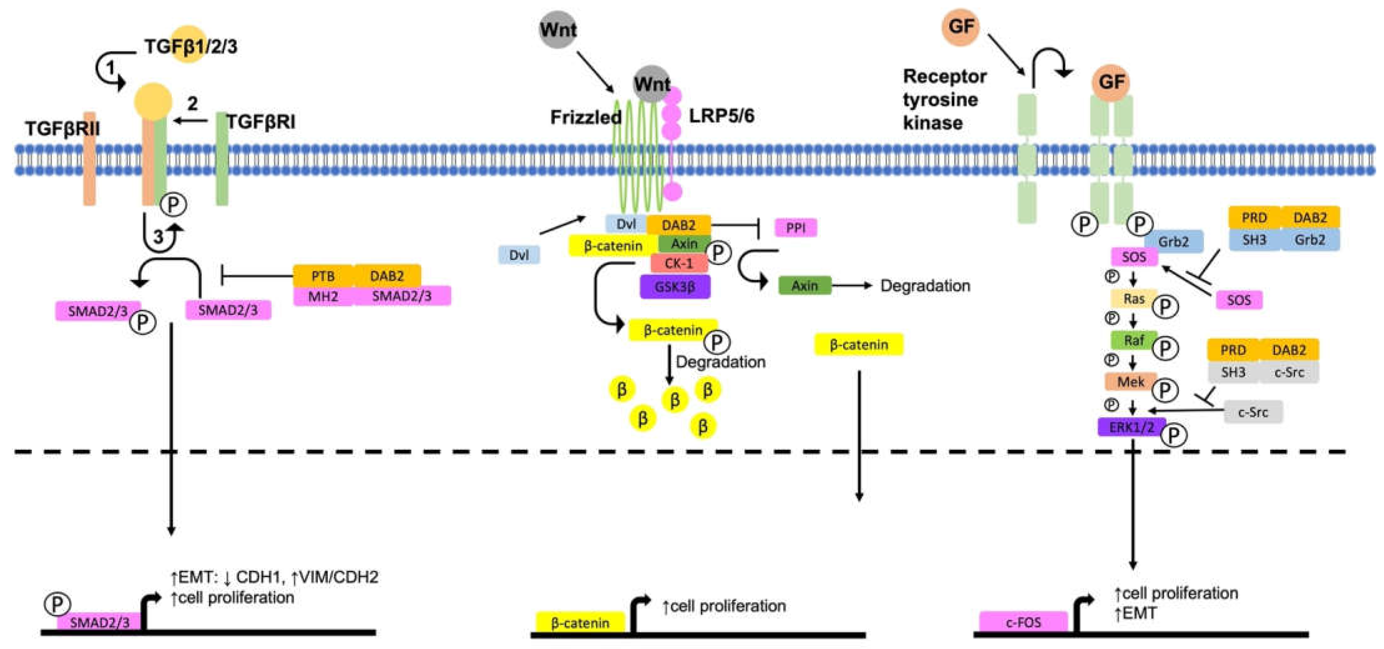
4. DAB2, a Negative Regulator of Pro-Tumorigenic Signalling Pathways

There is a complex network of extracellular and intracellular signalling pathways which are key to the progression of cancer. EMT is considered a key process involved in the progression and metastasis of cancer [71]. In the absence of DAB2, there is activation of key pro-tumorigenic and pro-EMT signalling pathways including the MAPK, Wnt/β-catenin and TGFβ pathways (Figure 2). The functional outcomes of DAB2 signalling in cancer is summarised in Table 2.

4.1. Activation of ERK/MAPK Signalling

Mitogen activated protein kinase (MAPK) signalling pathway involves binding of a range of stimuli including growth factors, cytokines and mitogens to a G-protein coupled receptor [76]. This activates a signalling cascade via mitogen-activated protein kinase kinase kinase (MAPKKK) which in turn activates MAPKK and then MAPK, promoting expression of target genes which leads to cell proliferation, differentiation, and migration [76]. In cancer, activation of MAPK signalling pathways is associated with chemotherapy resistance [77] and metastasis [78]. The extracellular signal-regulated kinase (ERK) family of MAPK are deregulated in approximately one third of human cancers [76,79]. Activation of ERK is initiated upon binding of a growth factor or mitogen to a receptor tyrosine kinase. Growth factor receptor-binding protein 2 (Grb2) is recruited and activated triggering a sigalling cascade of serial phosphorylation of different kinases from son of sevenless (SOS), to Ras, Raf, MEK/2 and finally ERK1/2 [76,79] (refer to Figure 2).
DAB2 inhibits the ERK/MAPK signalling pathway by disrupting the interaction between SOS and Grb2 [2]. DAB2 PRD interacts with the SH3 domains of Grb2, c-Src and Fg r [80]. DAB2 PRD can bind both the C-and N-terminal SH3 domains of Grb2 preventing the interaction of SOS with either SH3 domain [2,5,81]. Downregulation of DAB2 enhances free Grb2 for binding to SOS, activating the MAPK signalling pathway. Active ERK1/2 has been reported to promote EMT in two mammary epithelial cell lines (MCF10A1 and HME5-cdk4) [29]. EGF activated Erk2 via phosphorylation and activation of upstream c-Src at Tyr-416. The DAB2 PRD inhibited this activation by binding the SH3 domains within c-Src and subsequently inhibiting Erk2 [80]. Retinoic acid (RA) was shown to inhibit Erk1 activation in F9 embryonic stem cells which further inhibited activation of Elk-1 and c-Fos transcription [82]. RA treatment also enhanced DAB2 expression in F9 cells which was hypothesised as the mechanism for Erk1 inhibition [82]. Interestingly, DAB2 is a target of MAPK mediated phosphorylation [80]. DAB2 inhibition of ERK1/2 and c-Fos was confirmed in ovarian (OVCAR3, PA-1) and breast cancer cell lines (MCF-10, SK-Br-2 and MCF-7) [6]. DAB2 overexpression promotes cell death in ovarian cancer cells (OVCAR3) in normal tissue culture conditions, however when grown on a basement membrane, the inhibitory effect of DAB2 overexpression was reversed [6].

4.2. Activation of Wnt/β-Catenin Signalling

The Wnt signalling pathway is involved in cell proliferation, embryonic development, cell motility, differentiation, stem cell signalling and invasion [83,84]. Deregulation of Wnt signalling has been reported primarily in colorectal cancer but also in pancreatic and liver cancer [83,84]. Activation of the Wnt signalling pathway occurs by canonical and non-canonical pathways [83,84]. Canonical Wnt activation requires translocation of β-catenin to the nucleus where it activates its primary targets, cyclin D1 and c-myc [83,84]. Inactive Wnt signalling is maintained by a β-catenin destruction complex composed of Axin, glycogen synthase kinase 3 (GSK3), adenomatous polyposis coli (APC) and casein kinase I (CKI) which bind β-catenin and phosphorylate it, thereby targeting it for ubiquitinase mediated degradation [83,84]. Binding of Wnt ligand to the receptors frizzled and LRP5/6 promotes recruitment of Dishevled (Dvl) which disrupts the destruction complex, saving β-catenin from digestion [83,84].
DAB2 is a key regulator of Wnt signalling in differentiation of human embryonic stem cells into cardiomyocytes [85]. Expression of DAB2 and β-catenin are negatively correlated [30]. DAB2 overexpression and knockdown were associated with reduced and enhanced β-catenin expression, respectively, in lung and gastric cancer cells [7,30,43] and NIH-3T3 mouse fibroblasts [86]. A positive correlation between DAB2 and Axin expression was also observed in LK2 NSCLC cells [30]. Further analysis of the interactions between DAB2 and Wnt signalling components has demonstrated a direct interaction between DAB2 PTB and the Dvl-3 DEP domain and the Axin N-terminal region (aa 194-956) [86]. Upon Wnt3A signalling activation, Dvl-3 and Axin interact directly via their PDZ and N-terminal domains, respectively disrupting the β-catenin destruction complex. This allows nuclear translocation of β-catenin and transcription of targets c-Myc and cyclin D1 [86,87]. DAB2 binds both Dvl-3 and Axin, disrupting their interaction, maintaining the destruction complex and allowing glycogen synthase kinase 3β (GSK3β) phosphorylation and subsequent ubiquitin mediated digestion of β-catenin [86]. Axin is crucial for maintaining inactive Wnt signalling by stabilising the destruction complex and maintaining in-active Wnt signalling [88]. Phosphorylation of Axin is important for its own stability. Upon canonical Wnt activation, LRP5/6 is phosphorylated upon dimerisation with frizzled. This promotes recruitment of Axin which in turn is de-phosphorylated by protein phosphatase 1 (PP1) [88]. The destabilisation of Axin is further regulated by DAB2 [89]. DAB2 prevents the interaction between Axin with both PP1 and LRP5 [89]. PP1 and DAB2 bind to the C-terminal end of Axin indicating a competitive binding relationship [89]. The inhibition mechanism of Axin-LRP5 interactions by DAB2 is not known, but as the DAB2 PTB binds the FXNPXY sequence in other LDL family members, a direct interaction is hypothesised.
DAB2 has been shown to regulate Wnt signalling through direct interactions between DAB2 PTB domain and the intracellular domain of LRP6 [90]. In the absence of DAB2, Wnt3A signalling activates LRP6 via phosphorylation by GSK3β, LRP6 then undergoes calveolin mediated endocytosis and interacts with Axin, activating β-catenin signalling [91]. In the presence of DAB2, Wnt3A signalling promotes casein kinase 2 (CK2) phosphorylation of LRP6 at S1579A. This promotes binding of DAB2 to LRP6 and association with clathrin, thereby inhibiting the interaction of LRP6 with Axin and in turn inhibiting Wnt signalling resulting in reduced in vivo tumorigenesis as described in F9 teratocarcinoma cells [90]. In SGC gastric cancer cells, knockdown of DAB2 expression was associated with enhanced cell migration and enhanced expression of Wnt signalling components, including β-catenin, GSK3β and cyclinD1 [43].
Non-canonical Wnt signalling occurs independently of β-catenin and one of its primary targets is the planar cell polarity (PCP) signalling cascade which activates downstream Jun-N-terminal kinase (JNK) [92]. DAB2 has also been shown to regulate the non-canonical PCP-PE pathway [53,86]. DAB2 enhances Dvl1-3 activation of JNK via Wnt-5A signalling [86]. DAB2 has been shown in inhibit cholesterol-dependent activation of JNK and c-Jun by TGFβ1 through sequestering of TGFβRI [53].

4.3. Regulation of TGFβ Signalling Pathways

TGFβ is a key regulatory cytokine which is recognised by most human cells. TGFβ is responsible for maintaining normal homeostasis and in turn has tumour suppressive function [93]. Despite this, tumour cells are capable of evading TGFβ signalling and also utilising TGFβ signalling for their own benefit [94]. The TGFβ superfamily comprises over 30 members and has both canonical and non-canonical pathways, indicating a vast and complex network with diverse biological implications [93,94]. The principal mechanism of canonical TGFβ signalling involves binding of TGFβ ligand to a type II receptor, a serine threonine kinase which then recruits and phosphorylates a type I receptor [93]. Transcription factor SMAD is then phosphorylated and interacts with multiple other transcription factors to elicit and range of signals [93].
TGFβ signalling is complex and can act as both a tumour suppressor and a tumour promoter. Loss of DAB2 expression in tumour compared to normal tissues is well documented (Table 1). A particular study in head and neck and vulval human squamous cell carcinoma (HSCC) suggested loss of DAB2 expression acts as a switch for TGFβ signalling to change from a tumour suppressor to a tumour promoter role [45]. They found that with DAB2 expression, TGFβ activation of Smad2 and subsequent cell proliferation and motility were reduced. However, loss of DAB2 expression enhanced TGFβ Smad2 activation, reverting the effects on cell proliferation and enhanced cell motility [45]. A study in EpH4 mammary epithelium overexpressing Ras cells (EpRas), suggested cross talk between MAPK and TGFβ signalling in promoting EMT and tumour metastasis [95]. Another study in mammary epithelial cell lines (MCF10A1 and HME5-cdk4) demonstrated that DAB2 knockdown promoted activation of ERK which enhanced expression of TGFβ2 and promoted an EMT phenotype observed by reduced E-cadherin and enhanced N-cadherin and vimentin [29]. Hocevar et al. suggests that the tumour suppressive effects of DAB2 may occur through dual inhibition of more than one key signalling pathway [96]. In pancreatic cancer cell lines (COLO357 and PANC), DAB2 knock down enhanced TGFβ-mediated EMT through reduced E-cadherin and enhanced Snail, Slug and N-cadherin expression [96]. A functional mechanism for DAB2 in maintaining an epithelial phenotype is through co-localisation with E-cadherin at the plasma membrane, maintaining apical junctions [97]. Loss of DAB2 is associated with cytosolic localisation of E-cadherin and β-catenin [97].
TGFβ signalling can be activated through endocytosis, both clathrin and calveoli mediated, although Smad activation also occurs independently of endocytosis [98]. DAB2 was shown to regulate TGFβ clathrin mediated endocytosis of TGFβRI in ES-2 ovarian cancer cells [53]. DAB2 was not involved in the endocytosis of TGFβRII in NIH/3T3 mouse fibroblast, but it did mediate the intracellular trafficking of TGFβRII, particularly the transfer of TGFβRII from EEA1- positive early endosomes to Rab11-positive recycling endosomes [99]. DAB2 may also modulate TGFβ signalling through directly interacting with Smad effector proteins when TGFβ is impaired through loss of TGFβRI/II [100]. DAB2 PTB interacts with the MH2 domain of Smad2 and Smad3 but not Smad1 or 4 [100]. DAB2 expression is sufficient to recover TGFβ signalling and subsequently enhances phosphorylation of Smad2 and nuclear translocation of both Smad2 and Smad3 [100].

5. The Role of DAB2 as a Tumour Promoter

5.1. DAB2 and TGFβ Pro-Tumorigenic Signalling

There is strong evidence describing the tumour suppressive function of DAB2 particularly through inhibition of key signalling pathways involved in cell survival and cell fate determination. As shown in Table 1, the majority of cancer tissue have either a complete loss or significant reduction in DAB2 expression which has been associated with malignant transformation of cells. Despite most of the evidence supporting DAB2 is a tumour suppressor, some studies have found contradictory findings suggesting a tumour promoting role for DAB2 (Table 2). There is a strong relationship between DAB2 and TGFβ signalling [75]. It is well documented that TGFβ functions as both a tumour suppressor and tumour promoter [94]. In line with this, some studies have demonstrated that TGFβ together with DAB2 can have pro-tumorigenic effects [35,73,75].
TGFβ treatment in normal murine mammary gland epithelium (NMuMG) cells enhanced EMT and cell survival via DAB2 [75]. TGFβ via DAB2 enhanced activation of focal adhesion kinase (FAK) which in turn activated β1 integrin, preventing apoptosis [75]. Inhibition of DAB2 prevents TGFβ mediated EMT, through loss of N-cadherin and enhanced cell apoptosis [75]. Another study in ESCC found a negative correlation between DAB2 expression and cell migration associated with ERK activation [35]. Interestingly, when treated with TGFβ1 an EMT phenotype (enhanced CDH2, reduced CDH1) was observed in KYSE50 cells with high but not low DAB2 expression [35]. Activation of TGFβ signalling promoted phosphorylation of hnRNPE1 at Ser43, preventing binding to the BAT elements in DAB2 and ILEI which promoted DAB2 translation and EMT in NMuMG and EpRas mammary epithelium [49]. TGFβ enhanced expression of DAB2 and promoted localisation to the membrane and interacted with β1 integrin [75]. TGFβ via DAB2 promoted the activation of FAK kinase, activating β1 integrin, promoting cell survival [75]. DAB2 regulates internalisation of free and inactive integrin β1 in a clathrin independent mechanism and traffics integrin β1 to perinuclear recycling endosomes. Integrin β1 is then returned to the cell surface, where it interacts with vinculin (focal complex protein) enhancing migration of HeLa cancer cells [101].

5.2. DAB2 Promotes EMT and Metastasis

Another study demonstrated that DAB2 correlates with metastatic potential in the two prostate cancer cell lines, PC3 and LNCaP [73]. DAB2 overexpression was shown to promote migration and invasion in LNCaP cells and was associated with expression of migration associated genes, whereas knock down of DAB2 in the more metastatic PC3 cells had opposing effects on cell invasion and migration [73]. DAB2 expression was also associated with tumour progression in urothelial carcinoma [38]. High DAB2 expression associated with reduced PFS and OS, particularly in metastatic urothelial carcinoma which had invaded the muscle [38]. siRNA mediated knockdown of DAB2 expression in UM-UC-3 bladder cancer cells was associated with reduced tumour formation in vivo and subsequent enhanced expression of EMT marker KRT14 and reduced expression of the mesenchymal to epithelial transition (MET) marker occludin (OCLN) [38]. Secreted factors from DAB2 expressing stromal cells also promoted expression of EMT markers in Um-UC-3 cells [38]. DAB2 was also suggested to promote EMT in ovarian cancer [72]. Chao et al. found miR-187 to be associated with poor survival in ovarian cancer patients [72]. They found similar to other studies that miR-187-reduced DAB2 expression via the 3′UTR and was associated with reduced cell proliferation [72]. miR-187 overexpression in SKOV3 ovarian cancer cells promoted MET expression patterns including enhanced E-cadherin and reduced vimentin and phospho-FAK [72]. This was associated with reduced cell migration which could be reversed by overexpression of DAB2 [72]. In choriocarcinoma cell lines with similarly low endogenous DAB2 protein levels, DAB2 overexpression results in a 15%, 64% and 86% reduction in cell growth in BeWo, Jar and JEG choriocarcinoma cell lines, respectively, indicating that DAB2 expression can only inhibit cell proliferation in certain cell types [39]. Loss of DAB2 has been shown to inhibit cell migration of fibrosarcoma HT1080 cells as it is involved with AP-2 in the disassembly of the focal adhesion complex [102].
A small study in pancreatic cancer found DAB2 expression was enhanced in tumour versus normal pancreatic tissues. However, in metastatic tissues DAB2 expression was found to be reduced [34]. In grade 3, stage 1 (T1G3) bladder cancer there was a 2.85 fold increase in DAB2 expression in patients that progressed versus patients that did not [103]. DAB2 positivity for epithelioid mesothelioma was 80–98% compared with 3–23% inpulmonary adenocarcinoma [27,28]. DAB2 has therefore the potential to be used a novel marker for differentiating these two subtypes of lung malignancies.

5.3. Tumour Associated Macrophages (TAMs)

Recent research suggests DAB2 has tumour promoting effects in the tumour microenvironment (TME). DAB2 was highly expressed in TAMs and its knockdown significantly reduced lung metastasis in mouse fibrosarcoma and breast cancer models [104]. Immune cells elicit a range of pro-tumorigenic and tumour suppressive effects [105]. In particular, TAMs are well studied. M1 macrophages are thought to have a predominantly tumour suppressive function by actively promoting inflammation and directly targeting cancer cells [105]. M2 macrophages however are considered to have tumour promoting function as they enhance angiogenesis within the TME, and release a range of pro-metastatic secretory factors [105]. M2 macrophages have also been shown to enhance cell invasion and motility in various cancers such as those of the breast, stomach and lung [19,106]. A study by Adamson et al. demonstrated that DAB2 is involved in the polarisation of macrophages to a M2 phenotype, whereas DAB2-silencing promoted a pro-inflammatory M1 phenotype in both mouse and human bone marrow-derived macrophages (BMDM) [19]. High DAB2 macrophages in peritumoral and intratumoral areas was shown to associate poorly with disease free survival, lymph node metastases and tumour cell proliferation in breast cancer patients [104]. Colony stimulating factor-1 (CSF-1) also promotes an M2 phenotype [107]. CSF-1 enhanced DAB2 expression in TAMs isolated from MN-MCA1 fibrosarcoma mice models only in adherent conditions [104]. This was found to occur via the mechanotransductive YAP-TAZ transcription complex [104]. DAB2 KD in myeloid cells significantly reduced the invasiveness of E0771 breast cancer cells in vitro as well as number of lung metastases in vivo [104]. This was found to be a result of reduced internalisation and recycling of integrins and ECM components including collagen I, collagen IV, fibronectin and laminin [104]. Interestingly, PD-L inhibitors, which allow T-cell mediated death of tumour cells, further reduced the number of lung metastases in DAB2 KO mice but not WT mice [104]. This highlights a potential need to explore DAB2 inhibitors in combination with PD-L inhibitors in treating advanced staged cancers.

5.4. Regulation of Angiogenesis

Throughout tumour initiation, progression and metastasis, tumours cells release a range of factors which modulate their microenvironment [108]. The development of the TME includes the formation of new blood vessels which supply necessary oxygen and nutrients to the growing tumour [108,109]. TGFβ signalling is an important signalling pathway in angiogenesis, not only in the TME but also in embryogenesis [93]. DAB2 promotes TGFβ1 mediated expression of the angiogenic factors VEGF and FGF-2 and also activation of MAPK signalling, particularly phosphorylation of ERK [110]. Angiogenesis requires VEGFR signalling which is activated through internalisation of receptors VEGFR2 and VEGFR3 [111,112]. Ephrin-B2 is a key protein for VEGFR2 internalisation but it also requires PAR-3 and DAB2 which interacts directly with VEGFR2/3 via the PTB domain [111,112,113]. In mature vessels VEGFR2/3 internalisation is inhibited by atypical protein kinase C (aPKC) which phosphorylates the DAB2 PTB domain, preventing its interaction with VEGFR2/3 [114]. Syndecan-1 activates aPKC and subsequent phosphorylation of DAB2 inhibiting its activation of VEGF-VEGFRII signalling [114]. DAB2 promotes internalisation of VEGFRI and VEGFRII in liver sinusoidal endothelial cells which is required for their dedifferentiation, proliferation and migration during angiogenesis [115].

6. Targeting DAB2

Our review highlights how DAB2 in the majority of cases is associated with tumour suppressive phenotypes and hence is commonly downregulated. Re-expression of DAB2 in these cases should therefore be tumour suppressive and a potential treatment strategy. As discussed, there is growing evidence of the role of miRNAs targeting and downregulating DAB2. Targeting miRNAs in cancers and subsequent re-expression of DAB2 may be a suitable co-treatment with other current treatment strategies. Green tea has previously been shown to have anti-tumorigenic effects [50]. Yang et al. isolated crude polysaccharide from green tea which enhanced apoptosis in PC-3 prostate cancer cells [51]. They demonstrated the polysaccharide downregulated miR-93 which in turn enhanced DAB2 expression, activating both ERK and PI3K [25,51]. Re-expression of DAB2 through targeting DNA methylation presents another possible treatment mechanism in tumours where methylation downregulation occurs. In NSCLC cell line, LK2, x-ray irradiation promoted de-methylation of DAB2 CpG sites and enhanced DAB2 and Axin expression which inhibited Wnt signalling, cell proliferation and in vivo tumour formation [22]. DAB2 is important in regulating membrane integrity, immune regulation and key signalling pathways including MAPK, PI3K and Wnt and therefore will likely be difficult to directly target. A greater understanding of functional role of DAB2 in the TME may highlight potential treatment strategies to target DAB2 in cancer.

7. Conclusions

DAB2, initially identified as a tumour suppressor, is also an important adaptor molecule for clathrin-mediated endocytosis. Although the majority of literature has demonstrated either downregulation or loss of DAB2 expression in tumour tissues compared to normal tissues there are also several studies which have demonstrated an increase in DAB2 expression in cancer and an association with tumour progression. Additionally, some studies have shown that loss of DAB2 expression is not consistent amongst all subtypes of a particular cancer type. The majority of functional studies demonstrate that DAB2 is a negative regulator of the key signalling pathways Wnt, MAPK and TGFβ which elicit pro-tumorigenic effects. Some research contradicts these findings and describe a contrary effect, in particular through regulation of the TGFβ pathway which is known to be both pro-tumorigenic and tumour suppressive. DAB2 contains multiple binding domains and therefore has the ability to interact with multiple proteins simultaneously, explaining how different cell conditions may impact its function. More recent research has identified miRNA expression as a mechanism by which DAB2 is downregulated. Treatment strategies against pro-tumorigenic miRNAs are rapidly evolving offering a potential mechanism for re-activating DAB2 expression. A greater understanding of the functional role of DAB2 in the TME may lead to the development of novel strategies to block tumour progression.

Author Contributions

Conceptualisation Z.K.P., N.A.L., C.R., M.K.O., M.Y. and H.K.; investigation, Z.K.P., N.A.L. and C.R.; writing—original draft preparation, Z.K.P., N.A.L. and C.R.; writing—review and editing, Z.K.P., N.A.L., C.R. and M.K.O.; Supervision, C.R., M.Y. and H.K.; funding acquisition, M.K.O. All authors have read and agreed to the published version of the manuscript.

Funding

This research was funded by the Ovarian Cancer Research Foundation, Australia (OCRF.com.au).

Institutional Review Board Statement

Not applicable.

Informed Consent Statement

Not applicable.

Data Availability Statement

Not applicable.

Conflicts of Interest

The authors declare no conflict of interest.

References

  1. Mok, S.C.; Wong, K.K.; Chan, R.K.; Lau, C.C.; Tsao, S.W.; Knapp, R.C.; Berkowitz, R.S. Molecular cloning of differentially expressed genes in human epithelial ovarian cancer. Gynecol. Oncol. 1994, 52, 247–252. [Google Scholar] [CrossRef] [PubMed]
  2. Xu, X.X.; Yi, T.; Tang, B.; Lambeth, J.D. Disabled-2 (DAB2) is an SH3 domain-binding partner of Grb2. Oncogene 1998, 16, 1561–1569. [Google Scholar] [CrossRef] [Green Version]
  3. Xu, X.X.; Yang, W.; Jackowski, S.; Rock, C.O. Cloning of a novel phosphoprotein regulated by colony-stimulating factor 1 shares a domain with the Drosophila disabled gene product. J. Biol. Chem. 1995, 270, 14184–14191. [Google Scholar] [CrossRef] [PubMed] [Green Version]
  4. Gertler, F.B.; Bennett, R.L.; Clark, M.J.; Hoffmann, F.M. Drosophila abl tyrosine kinase in embryonic CNS axons: A role in axonogenesis is revealed through dosage-sensitive interactions with disabled. Cell 1989, 58, 103–113. [Google Scholar] [CrossRef] [PubMed]
  5. Bagadi, S.A.; Prasad, C.P.; Srivastava, A.; Prashad, R.; Gupta, S.D.; Ralhan, R. Frequent loss of DAB2 protein and infrequent promoter hypermethylation in breast cancer. Breast Cancer Res. Treat. 2007, 104, 277–286. [Google Scholar] [CrossRef]
  6. Sheng, Z.; Sun, W.; Smith, E.; Cohen, C.; Sheng, Z.; Xu, X.X. Restoration of positioning control following Disabled-2 expression in ovarian and breast tumor cells. Oncogene 2000, 19, 4847–4854. [Google Scholar] [CrossRef] [Green Version]
  7. Xu, H.T.; Yang, L.H.; Li, Q.C.; Liu, S.L.; Liu, D.; Xie, X.M.; Wang, E.H. Disabled-2 and Axin are concurrently colocalized and underexpressed in lung cancers. Hum. Pathol. 2011, 42, 1491–1498. [Google Scholar] [CrossRef]
  8. Sheng, Z.; He, J.; Tuppen, J.A.; Sun, W.; Fazili, Z.; Smith, E.R.; Dong, F.B.; Xu, X.X. Structure, sequence, and promoter analysis of human disabled-2 gene (DAB2). Genomics 2000, 70, 381–386. [Google Scholar] [CrossRef]
  9. Albertsen, H.M.; Smith, S.A.; Melis, R.; Williams, B.; Holik, P.; Stevens, J.; White, R. Sequence, genomic structure, and chromosomal assignment of human DOC-2. Genomics 1996, 33, 207–213. [Google Scholar] [CrossRef]
  10. Mishra, S.K.; Keyel, P.A.; Hawryluk, M.J.; Agostinelli, N.R.; Watkins, S.C.; Traub, L.M. Disabled-2 exhibits the properties of a cargo-selective endocytic clathrin adaptor. EMBO J. 2002, 21, 4915–4926. [Google Scholar] [CrossRef]
  11. Morris, S.M.; Cooper, J.A. Disabled-2 colocalizes with the LDLR in clathrin-coated pits and interacts with AP-2. Traffic 2001, 2, 111–123. [Google Scholar] [CrossRef]
  12. Maurer, M.E.; Cooper, J.A. The adaptor protein DAB2 sorts LDL receptors into coated pits independently of AP-2 and ARH. J Cell Sci. 2006, 119, 4235–4246. [Google Scholar] [CrossRef] [Green Version]
  13. Yu, C.; Feng, W.; Wei, Z.Y.; Miyanoiri, Y.; Wen, W.Y.; Zhao, Y.X.; Zhang, M.J. Myosin VI Undergoes Cargo-Mediated Dimerization. Cell 2009, 138, 537–548. [Google Scholar] [CrossRef] [PubMed] [Green Version]
  14. Rai, A.; Vang, D.; Ritt, M.; Sivaramakrishnan, S. Dynamic multimerization of DAB2-Myosin VI complexes regulates cargo processivity while minimizing cortical actin reorganization. J. Biol. Chem. 2021, 296, 100232:1–100232:13. [Google Scholar] [CrossRef] [PubMed]
  15. Morris, S.M.; Arden, S.D.; Roberts, R.C.; Kendrick-Jones, J.; Cooper, J.A.; Luzio, J.P.; Buss, F. Myosin VI binds to and localises with DAB2, potentially linking receptor-mediated endocytosis and the actin cytoskeleton. Traffic 2002, 3, 331–341. [Google Scholar] [CrossRef]
  16. Fili, N.; Hari-Gupta, Y.; Aston, B.; dos Santos, A.; Gough, R.E.; Alamad, B.; Wang, L.; Martin-Fernandez, M.L.; Toseland, C.P. Competition between two high-and low-affinity protein-binding sites in myosin VI controls its cellular function. J. Biol. Chem. 2020, 295, 337–347. [Google Scholar] [CrossRef] [PubMed]
  17. Da Paz, V.F.; Ghishan, F.K.; Kiela, P.R. Emerging Roles of Disabled Homolog 2 (DAB2) in Immune Regulation. Front. Immunol. 2020, 11, 580302:1–580302:11. [Google Scholar] [CrossRef]
  18. Rosenbauer, F.; Kallies, A.; Scheller, M.; Knobeloch, K.P.; Rock, C.O.; Schwieger, M.; Stocking, C.; Horak, I. Disabled-2 is transcriptionally regulated by ICSBP and augments macrophage spreading and adhesion. Embo J. 2002, 21, 211–220. [Google Scholar] [CrossRef] [Green Version]
  19. Adamson, S.E.; Griffiths, R.; Moravec, R.; Senthivinayagam, S.; Montgomery, G.; Chen, W.; Han, J.; Sharma, P.R.; Mullins, G.R.; Gorski, S.A.; et al. Disabled homolog 2 controls macrophage phenotypic polarization and adipose tissue inflammation. J. Clin. Investig. 2016, 126, 1311–1322. [Google Scholar] [CrossRef] [Green Version]
  20. Jokubaitis, V.G.; Gresle, M.M.; Kemper, D.A.; Doherty, W.; Perreau, V.M.; Cipriani, T.L.; Jonas, A.; Shaw, G.; Kuhlmann, T.; Kilpatrick, T.J.; et al. Endogenously regulated DAB2 worsens inflammatory injury in experimental autoimmune encephalomyelitis. Acta Neuropathol. Commun. 2013, 1, 32:1–32:14. [Google Scholar] [CrossRef]
  21. Figliuolo da Paz, V.; Jamwal, D.R.; Gurney, M.; Midura-Kiela, M.; Harrison, C.A.; Cox, C.; Wilson, J.M.; Ghishan, F.K.; Kiela, P.R. Rapid Downregulation of DAB2 by Toll-Like Receptor Activation Contributes to a Pro-Inflammatory Switch in Activated Dendritic Cells. Front. Immunol. 2019, 10, 304:1–304:18. [Google Scholar] [CrossRef] [PubMed] [Green Version]
  22. Lin, W.; Wang, W.; Wang, D.; Ling, W. Quercetin protects against atherosclerosis by inhibiting dendritic cell activation. Mol. Nutr. Food Res. 2017, 61, 1700031:1–1700031:12. [Google Scholar] [CrossRef] [PubMed]
  23. Jain, N.; Nguyen, H.; Friedline, R.H.; Malhotra, N.; Brehm, M.; Koyanagi, M.; Bix, M.; Cooper, J.A.; Chambers, C.A.; Kang, J. Cutting edge: DAB2 is a FOXP3 target gene required for regulatory T cell function. J. Immunol. 2009, 183, 4192–4196. [Google Scholar] [CrossRef] [PubMed] [Green Version]
  24. Fazili, Z.; Sun, W.; Mittelstaedt, S.; Cohen, C.; Xu, X.-X. Disabled-2 inactivation is an early step in ovarian tumorigenicity. Oncogene 1999, 18, 3104–3113. [Google Scholar] [CrossRef] [Green Version]
  25. Mok, S.C.; Chan, W.Y.; Wong, K.K.; Cheung, K.K.; Lau, C.C.; Ng, S.W.; Baldini, A.; Colitti, C.V.; Rock, C.O.; Berkowitz, R.S. DOC-2, a candidate tumor suppressor gene in human epithelial ovarian cancer. Oncogene 1998, 16, 2381–2387. [Google Scholar] [CrossRef] [Green Version]
  26. Yang, D.H.; Smith, E.R.; Cohen, C.; Wu, H.; Patriotis, C.; Godwin, A.K.; Hamilton, T.C.; Xu, X.X. Molecular events associated with dysplastic morphologic transformation and initiation of ovarian tumorigenicity. Cancer 2002, 94, 2380–2392. [Google Scholar] [CrossRef]
  27. Kuraoka, M.; Amatya, V.J.; Kushitani, K.; Mawas, A.S.; Miyata, Y.; Okada, M.; Kishimoto, T.; Inai, K.; Nishisaka, T.; Sueda, T.; et al. Identification of DAB2 and Intelectin-1 as Novel Positive Immunohistochemical Markers of Epithelioid Mesothelioma by Transcriptome Microarray Analysis for Its Differentiation From Pulmonary Adenocarcinoma. Am. J. Surg. Pathol. 2017, 41, 1045–1052. [Google Scholar] [CrossRef] [Green Version]
  28. Naso, J.R.; Cheung, S.; Ionescu, D.N.; Churg, A. Utility of SOX6 and DAB2 for the Diagnosis of Malignant Mesothelioma. Am. J. Surg. Pathol. 2021, 45, 1245–1251. [Google Scholar] [CrossRef]
  29. Martin, J.C.; Herbert, B.S.; Hocevar, B.A. Disabled-2 downregulation promotes epithelial-to-mesenchymal transition. Br. J. Cancer 2010, 103, 1716–1723. [Google Scholar] [CrossRef]
  30. Ma, S.; Zhang, W.L.; Leckey, B.D., Jr.; Xu, H.T.; Yang, L.H.; Wang, E. X-ray irradiation induced Disabled-2 gene promoter de-methylation enhances radiosensitivity of non-small-cell lung carcinoma cells. J. Exp. Clin. Cancer Res. 2018, 37, 315:1–315:12. [Google Scholar] [CrossRef]
  31. Cheng, Y.; Guo, Y.; Zhang, Y.; You, K.; Li, Z.; Geng, L. MicroRNA-106b is involved in transforming growth factor beta1-induced cell migration by targeting disabled homolog 2 in cervical carcinoma. J. Exp. Clin. Cancer Res. 2016, 35, 11:1–11:11. [Google Scholar] [CrossRef] [PubMed] [Green Version]
  32. Kleeff, J.; Huang Y Fau-Mok, S.C.; Mok Sc Fau-Zimmermann, A.; Zimmermann A Fau-Friess, H.; Friess H Fau-Büchler, M.W.; Büchler, M.W. Down-regulation of DOC-2 in colorectal cancer points to its role as a tumor suppressor in this malignancy. Dis. Colon. Rectum. 2002, 45, 1242–1248. [Google Scholar] [CrossRef]
  33. Yang, K.; Li, Y.-W.; Gao, Z.-Y.; Xiao, W.; Li, T.-Q.; Song, W.; Zheng, J.; Chen, H.; Chen, G.-H.; Zou, H.-Y. MiR-93 functions as a tumor promoter in prostate cancer by targeting disabled homolog 2 (DAB2) and an antitumor polysaccharide from green tea (Camellia sinensis) on their expression. Int. J. Biol. Macromol. 2019, 125, 557–565. [Google Scholar] [CrossRef] [PubMed]
  34. Huang, Y.; Friess, H.; Kleeff, J.; Esposito, I.; Zhu, Z.; Liu, S.; Mok, S.C.; Zimmermann, A.; Büchler, M.W. Doc-2/hDAB2 expression is up-regulated in primary pancreatic cancer but reduced in metastasis. Lab. Investig. 2001, 81, 863–873. [Google Scholar] [CrossRef] [Green Version]
  35. Wang, W.L.; Chang, W.L.; Yang, H.B.; Wang, Y.C.; Chang, I.W.; Lee, C.T.; Chang, C.Y.; Lin, J.T.; Sheu, B.S. Low disabled-2 expression promotes tumor progression and determines poor survival and high recurrence of esophageal squamous cell carcinoma. Oncotarget 2016, 7, 71169–71181. [Google Scholar] [CrossRef] [PubMed] [Green Version]
  36. Shen, W.; Niu, N.; Lawson, B.; Qi, L.; Zhang, J.; Li, T.; Zhang, H.; Liu, J. GATA6: A new predictor for prognosis in ovarian cancer. Hum. Pathol. 2019, 86, 163–169. [Google Scholar] [CrossRef] [PubMed]
  37. Karam, J.A.; Shariat, S.F.; Huang, H.Y.; Pong, R.C.; Ashfaq, R.; Shapiro, E.; Lotan, Y.; Sagalowsky, A.I.; Wu, X.R.; Hsieh, J.T. Decreased DOC-2/DAB2 expression in urothelial carcinoma of the bladder. Clin. Cancer Res. 2007, 13, 4400–4406. [Google Scholar] [CrossRef] [Green Version]
  38. Itami, Y.; Miyake, M.; Ohnishi, S.; Tatsumi, Y.; Gotoh, D.; Hori, S.; Morizawa, Y.; Iida, K.; Ohnishi, K.; Nakai, Y.; et al. Disabled Homolog 2 (DAB2) Protein in Tumor Microenvironment Correlates with Aggressive Phenotype in Human Urothelial Carcinoma of the Bladder. Diagnostics 2020, 10, 54. [Google Scholar] [CrossRef] [Green Version]
  39. Fulop, V.; Colitti, C.V.; Genest, D.; Berkowitz, R.S.; Yiu, G.K.; Ng, S.W.; Szepesi, J.; Mok, S.C. DOC-2/hDAB2, a candidate tumor suppressor gene involved in the development of gestational trophoblastic diseases. Oncogene 1998, 17, 419–424. [Google Scholar] [CrossRef] [Green Version]
  40. Xie, X.M.; Zhang, Z.Y.; Yang, L.H.; Yang, D.L.; Tang, N.; Zhao, H.Y.; Xu, H.T.; Li, Q.C.; Wang, E.H. Aberrant hypermethylation and reduced expression of disabled-2 promote the development of lung cancers. Int. J. Oncol. 2013, 43, 1636–1642. [Google Scholar] [CrossRef]
  41. Du, L.; Zhao, Z.; Ma, X.; Hsiao, T.H.; Chen, Y.; Young, E.; Suraokar, M.; Wistuba, I.; Minna, J.D.; Pertsemlidis, A. miR-93-directed downregulation of DAB2 defines a novel oncogenic pathway in lung cancer. Oncogene 2014, 33, 4307–4315. [Google Scholar] [CrossRef] [PubMed] [Green Version]
  42. Anupam, K.; Tusharkant, C.; Gupta, S.D.; Ranju, R. Loss of disabled-2 expression is an early event in esophageal squamous tumorigenesis. World J. Gastroenterol. 2006, 12, 6041–6045. [Google Scholar] [CrossRef] [PubMed]
  43. Wang, H.; Dong, S.; Liu, Y.; Ma, F.; Fang, J.; Zhang, W.; Shao, S.; Shen, H.; Jin, J. DAB2 suppresses gastric cancer migration by regulating the Wnt/beta-catenin and Hippo-YAP signaling pathways. Transl. Cancer Res. 2020, 9, 1174–1184. [Google Scholar] [CrossRef] [PubMed]
  44. Tong, J.H.; Ng, D.C.; Chau, S.L.; So, K.K.; Leung, P.P.; Lee, T.L.; Lung, R.W.; Chan, M.W.; Chan, A.W.; Lo, K.W.; et al. Putative tumour-suppressor gene DAB2 is frequently down regulated by promoter hypermethylation in nasopharyngeal carcinoma. BMC Cancer 2010, 10, 253. [Google Scholar] [CrossRef] [PubMed] [Green Version]
  45. Hannigan, A.; Smith, P.; Kalna, G.; Lo Nigro, C.; Orange, C.; O’Brien, D.I.; Shah, R.; Syed, N.; Spender, L.C.; Herrera, B.; et al. Epigenetic downregulation of human disabled homolog 2 switches TGF-beta from a tumor suppressor to a tumor promoter. J. Clin. Investig. 2010, 120, 2842–2857. [Google Scholar] [CrossRef] [Green Version]
  46. Paluszczak, J.; Kiwerska, K.; Mielcarek-Kuchta, D. Frequent methylation of DAB2, a Wnt pathway antagonist, in oral and oropharyngeal squamous cell carcinomas. Pathol. Res. Pract. 2018, 214, 314–317. [Google Scholar] [CrossRef]
  47. Calvisi, D.F.; Ladu, S.; Gorden, A.; Farina, M.; Lee, J.S.; Conner, E.A.; Schroeder, I.; Factor, V.M.; Thorgeirsson, S.S. Mechanistic and prognostic significance of aberrant methylation in the molecular pathogenesis of human hepatocellular carcinoma. J. Clin. Investig. 2007, 117, 2713–2722. [Google Scholar] [CrossRef] [Green Version]
  48. Li, C.; Chen J Fau-Chen, T.; Chen T Fau-Xu, Z.; Xu Z Fau-Xu, C.; Xu C Fau-Ding, C.; Ding C Fau-Wang, Y.; Wang Y Fau-Lei, Z.; Lei Z Fau-Zhang, H.-T.; Zhang Ht Fau-Zhao, J.; Zhao, J. Aberrant Hypermethylation at Sites -86 to 226 of DAB2 Gene in Non-Small Cell Lung Cancer. Am. J. Med. Sci. 2015, 349, 425–431. [Google Scholar] [CrossRef]
  49. Chaudhury, A.; Hussey Gs Fau-Ray, P.S.; Ray Ps Fau-Jin, G.; Jin G Fau-Fox, P.L.; Fox Pl Fau-Howe, P.H.; Howe, P.H. TGF-beta-mediated phosphorylation of hnRNP E1 induces EMT via transcript-selective translational induction of DAB2 and ILEI. Nat. Cell Biol. 2010, 13, 286–293. [Google Scholar] [CrossRef] [Green Version]
  50. Zhou, J.; Hernandez, G.; Tu, S.W.; Scholes, J.; Chen, H.; Tseng, C.P.; Hsieh, J.T. Synergistic induction of DOC-2/DAB2 gene expression in transitional cell carcinoma in the presence of GATA6 and histone deacetylase inhibitor. Cancer Res. 2005, 65, 6089–6096. [Google Scholar] [CrossRef]
  51. Cai, K.Q.; Caslini, C.; Capo-chichi, C.D.; Slater, C.; Smith, E.R.; Wu, H.; Klein-Szanto, A.J.; Godwin, A.K.; Xu, X.X. Loss of GATA4 and GATA6 expression specifies ovarian cancer histological subtypes and precedes neoplastic transformation of ovarian surface epithelia. PLoS ONE 2009, 4, e6454. [Google Scholar] [CrossRef] [PubMed] [Green Version]
  52. Tseng, C.P.; Ely, B.D.; Pong, R.C.; Wang, Z.; Zhou, J.; Hsieh, J.T. The role of DOC-2/DAB2 protein phosphorylation in the inhibition of AP-1 activity. An underlying mechanism of its tumor-suppressive function in prostate cancer. J. Biol. Chem. 1999, 274, 31981–31986. [Google Scholar] [CrossRef] [Green Version]
  53. Shapira, K.E.; Hirschhorn, T.; Barzilay, L.; Smorodinsky, N.I.; Henis, Y.I.; Ehrlich, M. DAB2 inhibits the cholesterol-dependent activation of JNK by TGF-beta. Mol. Biol. Cell 2014, 25, 1620–1628. [Google Scholar] [CrossRef] [PubMed]
  54. Garces de Los Fayos Alonso, I.; Liang, H.C.; Turner, S.D.; Lagger, S.; Merkel, O.; Kenner, L. The Role of Activator Protein-1 (AP-1) Family Members in CD30-Positive Lymphomas. Cancers 2018, 10, 93. [Google Scholar] [CrossRef] [Green Version]
  55. He, J.; Xu, J.; Xu, X.X.; Hall, R.A. Cell cycle-dependent phosphorylation of Disabled-2 by cdc2. Oncogene 2003, 22, 4524–4530. [Google Scholar] [CrossRef] [PubMed] [Green Version]
  56. Chetrit, D.; Barzilay, L.; Horn, G.; Bielik, T.; Smorodinsky, N.I.; Ehrlich, M. Negative regulation of the endocytic adaptor disabled-2 (DAB2) in mitosis. J. Biol. Chem. 2011, 286, 5392–5403. [Google Scholar] [CrossRef] [PubMed] [Green Version]
  57. Huang, J.; Xiao, R.; Wang, X.; Khadka, B.; Fang, Z.; Yu, M.; Zhang, L.; Wu, J.; Liu, J. MicroRNA93 knockdown inhibits acute myeloid leukemia cell growth via inactivating the PI3K/AKT pathway by upregulating DAB2. Int. J. Oncol. 2021, 59. [Google Scholar] [CrossRef]
  58. Huang, Y.; Hong, W.; Wei, X. The molecular mechanisms and therapeutic strategies of EMT in tumor progression and metastasis. J. Hematol. Oncol. 2022, 15, 129:1–129:27. [Google Scholar] [CrossRef]
  59. Kim, B.N.; Ahn, D.H.; Kang, N.; Yeo, C.D.; Kim, Y.K.; Lee, K.Y.; Kim, T.J.; Lee, S.H.; Park, M.S.; Yim, H.W.; et al. TGF-beta induced EMT and stemness characteristics are associated with epigenetic regulation in lung cancer. Sci. Rep. 2020, 10, 10597:1–10597:11. [Google Scholar] [CrossRef]
  60. Piao, J.L.; You, K.; Guo, Y.L.; Zhang, Y.Y.; Li, Z.J.; Geng, L. Substrate stiffness affects epithelial-mesenchymal transition of cervical cancer cells through miR-106b and its target protein DAB2. Int. J. Oncol. 2017, 50, 2033–2042. [Google Scholar] [CrossRef]
  61. Sun, C.; Yao, X.; Jiang, Q.; Sun, X. miR-106b targets DAB2 to promote hepatocellular carcinoma cell proliferation and metastasis. Oncol. Lett. 2018, 16, 3063–3069. [Google Scholar] [CrossRef] [PubMed] [Green Version]
  62. Cioffi, M.; Trabulo, S.M.; Vallespinos, M.; Raj, D.; Kheir, T.B.; Lin, M.L.; Begum, J.; Baker, A.M.; Amgheib, A.; Saif, J.; et al. The miR-25-93-106b cluster regulates tumor metastasis and immune evasion via modulation of CXCL12 and PD-L1. Oncotarget 2017, 8, 21609–21625. [Google Scholar] [CrossRef] [PubMed] [Green Version]
  63. Smith, A.L.; Iwanaga, R.; Drasin, D.J.; Micalizzi, D.S.; Vartuli, R.L.; Tan, A.C.; Ford, H.L. The miR-106b-25 cluster targets Smad7, activates TGF-beta signaling, and induces EMT and tumor initiating cell characteristics downstream of Six1 in human breast cancer. Oncogene 2012, 31, 5162–5171. [Google Scholar] [CrossRef] [PubMed] [Green Version]
  64. Min, K.; Kim, J.Y.; Lee, S.K. Epstein-Barr virus miR-BART1-3p suppresses apoptosis and promotes migration of gastric carcinoma cells by targeting DAB2. Int. J. Biol. Sci. 2020, 16, 694–707. [Google Scholar] [CrossRef] [PubMed] [Green Version]
  65. Zhang, L.; Huang, P.; Li, Q.; Wang, D.; Xu, C.X. miR-134-5p Promotes Stage I Lung Adenocarcinoma Metastasis and Chemoresistance by Targeting DAB2. Mol. Nucleic Acids 2019, 18, 627–637. [Google Scholar] [CrossRef] [Green Version]
  66. Tian, X.; Zhang, Z. miR-191/DAB2 axis regulates the tumorigenicity of estrogen receptor-positive breast cancer. IUBMB Life 2018, 70, 71–80. [Google Scholar] [CrossRef] [PubMed] [Green Version]
  67. Vuong, N.H.; Salah Salah, O.; Vanderhyden, B.C. 17beta-Estradiol sensitizes ovarian surface epithelium to transformation by suppressing Disabled-2 expression. Sci. Rep. 2017, 7, 16702. [Google Scholar] [CrossRef] [Green Version]
  68. Wang, B.W.; Fang, W.J.; Shyu, K.G. MicroRNA-145 regulates disabled-2 and Wnt3a expression in cardiomyocytes under hyperglycaemia. Eur. J. Clin. Investig 2018, 48, e12867. [Google Scholar] [CrossRef]
  69. Lin, C.M.; Fang, W.J.; Wang, B.W.; Pan, C.M.; Chua, S.K.; Hou, S.W.; Shyu, K.G. (-)-Epigallocatechin Gallate Promotes MicroRNA 145 Expression against Myocardial Hypoxic Injury through DAB2/Wnt3a/β-catenin. Am. J. Chin. Med. 2020, 48, 341–356. [Google Scholar] [CrossRef]
  70. Lu, M.; Xu, L.; Wang, M.; Guo, T.; Luo, F.; Su, N.; Yi, S.; Chen, T. miR149 promotes the myocardial differentiation of mouse bone marrow stem cells by targeting DAB2. Mol. Med. Rep. 2018, 17, 8502–8509. [Google Scholar] [CrossRef]
  71. Dongre, A.; Weinberg, R.A. New insights into the mechanisms of epithelial–mesenchymal transition and implications for cancer. Nat. Rev. Mol. Cell Biol. 2019, 20, 69–84. [Google Scholar] [CrossRef] [PubMed]
  72. Chao, A.; Lin, C.Y.; Lee, Y.S.; Tsai, C.L.; Wei, P.C.; Hsueh, S.; Wu, T.I.; Tsai, C.N.; Wang, C.J.; Chao, A.S.; et al. Regulation of ovarian cancer progression by microRNA-187 through targeting Disabled homolog-2. Oncogene 2012, 31, 764–775. [Google Scholar] [CrossRef] [PubMed] [Green Version]
  73. Xie, Y.Y.; Zhang, Y.L.; Jiang, L.; Zhang, M.M.; Chen, Z.W.; Liu, D.; Huang, Q.H. Disabled homolog 2 is required for migration and invasion of prostate cancer cells. Front. Med. 2015, 9, 312–321. [Google Scholar] [CrossRef]
  74. Wang, S.C.; Makino, K.; Xia, W.Y.; Kim, J.S.; Im, S.A.; Peng, H.; Mok, S.C.; Singletary, S.E.; Hung, M.C. DOC-2/hDab-2 inhibits ILK activity and induces anoikis in breast cancer cells through an Akt-independent pathway. Oncogene 2001, 20, 6960–6964. [Google Scholar] [CrossRef] [Green Version]
  75. Prunier, C.; Howe, P.H. Disabled-2 (DAB2) is required for transforming growth factor beta-induced epithelial to mesenchymal transition (EMT). J. Biol. Chem. 2005, 280, 17540–17548. [Google Scholar] [CrossRef] [PubMed] [Green Version]
  76. Braicu, C.; Buse, M.; Busuioc, C.; Drula, R.; Gulei, D.; Raduly, L.; Rusu, A.; Irimie, A.; Atanasov, A.G.; Slaby, O.; et al. A Comprehensive Review on MAPK: A Promising Therapeutic Target in Cancer. Cancers 2019, 11, 1618. [Google Scholar] [CrossRef] [PubMed] [Green Version]
  77. McGivern, N.; El-Helali, A.; Mullan, P.; McNeish, I.A.; Paul Harkin, D.; Kennedy, R.D.; McCabe, N. Activation of MAPK signalling results in resistance to saracatinib (AZD0530) in ovarian cancer. Oncotarget 2018, 9, 4722–4736. [Google Scholar] [CrossRef] [Green Version]
  78. Zhao, J.; Chen, G.; Li, J.; Liu, S.; Jin, Q.; Zhang, Z.; Qi, F.; Zhang, J.; Xu, J. Loss of PR55alpha promotes proliferation and metastasis by activating MAPK/AKT signaling in hepatocellular carcinoma. Cancer Cell Int. 2021, 21, 107. [Google Scholar] [CrossRef]
  79. Yuan, J.; Dong, X.; Yap, J.; Hu, J. The MAPK and AMPK signalings: Interplay and implication in targeted cancer therapy. J. Hematol. Oncol. 2020, 13, 113. [Google Scholar] [CrossRef]
  80. Zhou, J.; Scholes, J.; Hsieh, J.T. Characterization of a novel negative regulator (DOC-2/DAB2) of c-Src in normal prostatic epithelium and cancer. J. Biol. Chem. 2003, 278, 6936–6941. [Google Scholar] [CrossRef]
  81. Zhou, J.; Hsieh, J.T. The inhibitory role of DOC-2/DAB2 in growth factor receptor-mediated signal cascade. DOC-2/DAB2-mediated inhibition of ERK phosphorylation via binding to Grb2. J. Biol. Chem. 2001, 276, 27793–27798. [Google Scholar] [CrossRef] [PubMed] [Green Version]
  82. Smith, E.R.; Smedberg, J.L.; Rula, M.E.; Hamilton, T.C.; Xu, X.X. Disassociation of MAPK activation and c-Fos expression in F9 embryonic carcinoma cells following retinoic acid-induced endoderm differentiation. J. Biol. Chem. 2001, 276, 32094–32100. [Google Scholar] [CrossRef] [PubMed] [Green Version]
  83. Wang, Z.; Zhao, T.T.; Zhang, S.H.; Wang, J.K.; Chen, Y.Y.; Zhao, H.Z.; Yang, Y.X.; Shi, S.L.; Chen, Q.; Liu, K.C. The Wnt signaling pathway in tumorigenesis, pharmacological targets, and drug development for cancer therapy. Biomark. Res. 2021, 9, 1–16. [Google Scholar] [CrossRef] [PubMed]
  84. Zhang, Y.; Wang, X. Targeting the Wnt/beta-catenin signaling pathway in cancer. J. Hematol. Oncol. 2020, 13, 165. [Google Scholar] [CrossRef] [PubMed]
  85. Hofsteen, P.; Robitaille Aaron, M.; Chapman Daniel, P.; Moon Randall, T.; Murry Charles, E. Quantitative proteomics identify DAB2 as a cardiac developmental regulator that inhibits WNT/β-catenin signaling. Proc. Natl. Acad. Sci. USA 2016, 113, 1002–1007. [Google Scholar] [CrossRef] [Green Version]
  86. Hocevar, B.A.; Mou, F.; Rennolds, J.L.; Morris, S.M.; Cooper, J.A.; Howe, P.H. Regulation of the Wnt signaling pathway by disabled-2 (DAB2). EMBO J. 2003, 22, 3084–3094. [Google Scholar] [CrossRef] [Green Version]
  87. Li, L.; Yuan, H.; Weaver, C.D.; Mao, J.; Farr, G.H., 3rd; Sussman, D.J.; Jonkers, J.; Kimelman, D.; Wu, D. Axin and Frat1 interact with dvl and GSK, bridging Dvl to GSK in Wnt-mediated regulation of LEF-1. EMBO J. 1999, 18, 4233–4240. [Google Scholar] [CrossRef] [Green Version]
  88. Kim, S.E.; Huang, H.; Zhao, M.; Zhang, X.; Zhang, A.; Semonov, M.V.; MacDonald, B.T.; Zhang, X.; Garcia Abreu, J.; Peng, L.; et al. Wnt stabilization of beta-catenin reveals principles for morphogen receptor-scaffold assemblies. Science 2013, 340, 867–870. [Google Scholar] [CrossRef] [Green Version]
  89. Jiang, Y.; Luo, W.; Howe, P.H. DAB2 stabilizes Axin and attenuates Wnt/beta-catenin signaling by preventing protein phosphatase 1 (PP1)-Axin interactions. Oncogene 2009, 28, 2999–3007. [Google Scholar] [CrossRef] [Green Version]
  90. Jiang, Y.; He X Fau-Howe, P.H.; Howe, P.H. Disabled-2 (DAB2) inhibits Wnt/β-catenin signalling by binding LRP6 and promoting its internalization through clathrin. EMBO J. 2012, 31, 2336–2349. [Google Scholar] [CrossRef]
  91. Yamamoto, H.; Komekado, H.; Kikuchi, A. Caveolin Is Necessary for Wnt-3a-Dependent Internalization of LRP6 and Accumulation of β-Catenin. Dev. Cell 2006, 11, 213–223. [Google Scholar] [CrossRef] [PubMed] [Green Version]
  92. Patel, S.; Alam, A.; Pant, R.; Chattopadhyay, S. Wnt Signaling and Its Significance Within the Tumor Microenvironment: Novel Therapeutic Insights. Front. Immunol. 2019, 10, 2872. [Google Scholar] [CrossRef] [PubMed] [Green Version]
  93. French, R.; Feng, Y.; Pauklin, S. Targeting TGFbeta Signalling in Cancer: Toward Context-Specific Strategies. Trends Cancer 2020, 6, 538–540. [Google Scholar] [CrossRef] [PubMed]
  94. Baba, A.B.; Rah, B.; Bhat, G.R.; Mushtaq, I.; Parveen, S.; Hassan, R.; Hameed Zargar, M.; Afroze, D. Transforming Growth Factor-Beta (TGF-β) Signaling in Cancer-A Betrayal Within. Front. Pharmacol. 2022, 13, 791272. [Google Scholar] [CrossRef]
  95. Janda, E.; Lehmann, K.; Killisch, I.; Jechlinger, M.; Herzig, M.; Downward, J.; Beug, H.; Grünert, S. Ras and TGF[beta] cooperatively regulate epithelial cell plasticity and metastasis: Dissection of Ras signaling pathways. J. Cell Biol. 2002, 156, 299–313. [Google Scholar] [CrossRef] [Green Version]
  96. Hocevar, B.A. Loss of Disabled-2 Expression in Pancreatic Cancer Progression. Sci. Rep. 2019, 9, 7532:1–7532:11. [Google Scholar] [CrossRef] [Green Version]
  97. Vazquez-Carretero, M.D.; Garcia-Miranda, P.; Balda, M.S.; Matter, K.; Ilundain, A.A.; Peral, M.J. Proper E-cadherin membrane location in colon requires DAB2 and it modifies by inflammation and cancer. J. Cell Physiol. 2021, 236, 1083–1093. [Google Scholar] [CrossRef]
  98. Yakymovych, I.; Yakymovych, M.; Heldin, C.H. Intracellular trafficking of transforming growth factor beta receptors. Acta Biochim. Biophys. Sin. 2018, 50, 3–11. [Google Scholar] [CrossRef] [Green Version]
  99. Penheiter, S.G.; Singh Rd Fau-Repellin, C.E.; Repellin Ce Fau-Wilkes, M.C.; Wilkes Mc Fau-Edens, M.; Edens M Fau-Howe, P.H.; Howe Ph Fau-Pagano, R.E.; Pagano Re Fau-Leof, E.B.; Leof, E.B. Type II transforming growth factor-beta receptor recycling is dependent upon the clathrin adaptor protein DAB2. Mol. Biol. Cell 2010, 21, 4009–4019. [Google Scholar] [CrossRef] [Green Version]
  100. Hocevar, B.A.; Smine, A.; Xu, X.X.; Howe, P.H. The adaptor molecule Disabled-2 links the transforming growth factor beta receptors to the Smad pathway. EMBO J. 2001, 20, 2789–2801. [Google Scholar] [CrossRef]
  101. Teckchandani, A.; Toida, N.; Goodchild, J.; Henderson, C.; Watts, J.; Wollscheid, B.; Cooper, J.A. Quantitative proteomics identifies a DAB2/integrin module regulating cell migration. J. Cell Biol. 2009, 186, 99–111. [Google Scholar] [CrossRef] [PubMed] [Green Version]
  102. Chao, W.T.; Kunz, J. Focal adhesion disassembly requires clathrin-dependent endocytosis of integrins. FEBS Lett. 2009, 583, 1337–1343. [Google Scholar] [CrossRef] [PubMed] [Green Version]
  103. van der Heijden, A.G.; Mengual, L.; Lozano, J.J.; Ingelmo-Torres, M.; Ribal, M.J.; Fernandez, P.L.; Oosterwijk, E.; Schalken, J.A.; Alcaraz, A.; Witjes, J.A. A five-gene expression signature to predict progression in T1G3 bladder cancer. Eur. J. Cancer 2016, 64, 127–136. [Google Scholar] [CrossRef]
  104. Marigo, I.; Trovato, R.; Hofer, F.; Ingangi, V.; Desantis, G.; Leone, K.; De Sanctis, F.; Ugel, S.; Cane, S.; Simonelli, A.; et al. Disabled Homolog 2 Controls Prometastatic Activity of Tumor-Associated Macrophages. Cancer Discov. 2020, 10, 1758–1773. [Google Scholar] [CrossRef] [PubMed]
  105. Mantovani, A.; Allavena, P.; Marchesi, F.; Garlanda, C. Macrophages as tools and targets in cancer therapy. Nat. Rev. Drug Discov. 2022, 21, 799–820. [Google Scholar] [CrossRef] [PubMed]
  106. Guo, Z.; Song, J.; Hao, J.; Zhao, H.; Du, X.; Li, E.; Kuang, Y.; Yang, F.; Wang, W.; Deng, J.; et al. M2 macrophages promote NSCLC metastasis by upregulating CRYAB. Cell Death Dis. 2019, 10, 377:1–377:11. [Google Scholar] [CrossRef] [Green Version]
  107. Cai, H.; Zhang, Y.C.; Wang, J.; Gu, J.Y. Defects in Macrophage Reprogramming in Cancer Therapy: The Negative Impact of PD-L1/PD-1. Front. Immunol. 2021, 12. [Google Scholar] [CrossRef]
  108. Jin, M.Z.; Jin, W.L. The updated landscape of tumor microenvironment and drug repurposing. Signal Transduct.Target 2020, 5, 166:1–166:16. [Google Scholar] [CrossRef]
  109. Kim, H.J.; Ji, Y.R.; Lee, Y.M. Crosstalk between angiogenesis and immune regulation in the tumor microenvironment. Arch. Pharmacal. Res. 2022, 45, 401–416. [Google Scholar] [CrossRef]
  110. Cheong, S.M.; Choi H Fau-Hong, B.S.; Hong Bs Fau-Gho, Y.S.; Gho Ys Fau-Han, J.-K.; Han, J.K. DAB2 is pivotal for endothelial cell migration by mediating VEGF expression in cancer cells. Exp. Cell Res. 2012, 318, 550–557. [Google Scholar] [CrossRef]
  111. Sawamiphak, S.; Seidel, S.; Essmann, C.L.; Wilkinson, G.A.; Pitulescu, M.E.; Acker, T.; Acker-Palmer, A. Ephrin-B2 regulates VEGFR2 function in developmental and tumour angiogenesis. Nature 2010, 465, 487–491. [Google Scholar] [CrossRef] [PubMed]
  112. Wang, Y.; Nakayama, M.; Pitulescu, M.E.; Schmidt, T.S.; Bochenek, M.L.; Sakakibara, A.; Adams, S.; Davy, A.; Deutsch, U.; Luthi, U.; et al. Ephrin-B2 controls VEGF-induced angiogenesis and lymphangiogenesis. Nature 2010, 465, 483–486. [Google Scholar] [CrossRef] [PubMed]
  113. Nakayama, M.; Nakayama, A.; van Lessen, M.; Yamamoto, H.; Hoffmann, S.; Drexler, H.C.; Itoh, N.; Hirose, T.; Breier, G.; Vestweber, D.; et al. Spatial regulation of VEGF receptor endocytosis in angiogenesis. Nat. Cell Biol. 2013, 15, 249–260. [Google Scholar] [CrossRef] [PubMed] [Green Version]
  114. Jing, Z.; Jia-jun, W.; Wei-jie, Y. Phosphorylation of DAB2 is involved in inhibited VEGF-VEGFR-2 signaling induced by downregulation of syndecan-1 in glomerular endothelial cell. Cell Biol. Int. 2020, 44, 894–904. [Google Scholar] [CrossRef]
  115. Lao, Y.; Li, Y.; Hou, Y.; Chen, H.; Qiu, B.; Lin, W.; Sun, A.; Wei, H.; Jiang, Y.; He, F. Proteomic Analysis Reveals DAB2 Mediated Receptor Endocytosis Promotes Liver Sinusoidal Endothelial Cell Dedifferentiation. Sci. Rep. 2017, 7, 13456. [Google Scholar] [CrossRef] [PubMed]
Figure 1. Diagram representing the structure of the DAB2 protein and location of important functional domains.
Ijms 24 00696 g001
Figure 2. Regulatory role of DAB2 in MAPK, Wnt/β-catenin and canonical TGFβ pathways. In the absence of DAB2, TGFβ1/2/3 activate canonical signalling pathways through dimerisation of receptors TGFβRI and TGFβRII. This activates SMAD2 and enhances cell proliferation and EMT through expression of target genes. DAB2 PTB domain directly interacts with the MH2 domain of SMAD2/3, preventing its activation. DAB2 inhibits the canonical Wnt signalling pathway by preventing the interaction between Dvl and Axin, preventing destruction of the β-catenin destruction complex. In the absence of DAB2, Dvl and Axin interact separating the complex. β-catenin is then free to translocate to the nucleus where it promotes cell proliferation through expression of target c-myc and cyclin D1. DAB2 PRD interacts with the SH3 domains of SOS and c-Src in the MAPK pathway, inhibiting the signal transduction and in turn inhibiting cell proliferation and EMT.
Ijms 24 00696 g002
Table 2. The functional roles of DAB2 in cancer.
Cancer TypeObservationRoleRef
Cell proliferation
Acute Myeloid LeukaemiaKnockdown of miR-93 enhances DAB2 expression and inhibits cell proliferation in THP-1 cells in vitro and in vivo TS *[57]
Hepatocellular carcinoma (HCC)miR-106b knockdown of DAB2 enhances Hep3B cell proliferation in vitroTP **[61]
Lung adenocarcinomaKnockdown of DAB2 inhibits A549 and H1299 cell growth and overexpression of DAB2 enhance A549 and H1299 cell growthTP[65]
Breast cancerOestrogen enhances miR-191 and silences DAB2 expression and promotes cell proliferation in ER positive breast cancerTS[66]
Head and Neck and Vulval Squamous cell carcinoma (SCC)TGFβ inhibits cell proliferation in cell lines (HN30, H376, H413, Procotor, UMSCV1A, UMSCV1B and UMSCV7) that have high levels of DAB2TS[45]
Urothelial Carcinoma of the Bladder (UCB)Downregulation of DAB2 decreases the proliferation of UM-UC3, J82 and T24 cellsTP[38]
Ovarian cancermiR-187 in SKOV-3 cells suppressed DAB2 expression and enhanced cell proliferation TS[72]
Migration
Hepatocellular carcinoma (HCC)miR-106b knockdown of DAB2 enhances Hep3B cell migration TS[61]
Head and Neck and Vulval SCCTGFβ inhibits cell motility in cell lines (HN30, H413, UMSCV1A, UMSCV1B and UMSCV7) that express high levels of DAB2TS[45]
Lung adenocarcinomaSilencing DAB2 enhances cell migration in A549 and H1299 cells in vitro and overexpression of DAB2 reduced cell migration in A549 and H1299 cellsTS[65]
Prostate cancerDAB2 overexpression enhanced LNCaP cell migration and DAB2 knockdown by shRNA inhibited PC3 cell migrationTP[73]
Urothelial
UCB
Downregulation of DAB2 decrease the migration of UM-UC3 and T24 cellsTP[38]
Ovarian cancermiR-187 suppressed DAB2 expression and inhibited cell migration in SKOV-3 cellsTP[72]
Gastric cancerDownregulation of DAB2 promote SGC cell migration via Wnt/β-catenin and Hippo-YAP signalling pathwaysTS[43]
Invasion
Prostate cancerDAB2 expression enhanced LNCaP cell invasion and DAB2 knockdown inhibited PC3 cell invasionTP[73]
Urothelial
UCB
Downregulation of DAB2 decreased cell invasion of J82 and T24 cellsTP[38]
Apoptosis
Breast Cancer DAB2 promotes anoikis in SK-BR-3 and MDA-MB-453 cellsTS[74]
Normal murine mammary gland (NMuMG)Down regulation of DAB2 enhance TGFβ induced apoptosisTP[75]
Breast cancerDAB2 sensitises SK-BR-3 and MDA-MB-453 cells to apoptosis by inhibiting the activity of integrin-linked kinas (ILK)TS[74]
In vivo tumour growth and metastasis
Lung adenocarcinomaDAB2 is a target for miR-134-5p. Overexpression of miR-134-5p increased A549 cells tumour growth in mouse model TS[65]
Prostate cancerDAB2 knockdown inhibits PC3 cells tumour growth and metastasis in the mouse modelTP[73]
Urothelial
UCB
Reduced tumour growth and invasion in xenograft tumours of UM-UC-3 cells treated with DAB2 targeting siRNATP[38]
OvarianDAB2 overexpression reduces SKOV3 tumour formation in nude miceTS[25]
* TS = tumour suppressor; ** TP = tumour promoter.
Disclaimer/Publisher’s Note: The statements, opinions and data contained in all publications are solely those of the individual author(s) and contributor(s) and not of MDPI and/or the editor(s). MDPI and/or the editor(s) disclaim responsibility for any injury to people or property resulting from any ideas, methods, instructions or products referred to in the content.

