
Inclusion of 11-Oxygenated Androgens in a Clinical Routine LC-MS/MS Setup for Steroid Hormone Profiling

 ,
,  and
and
Abstract
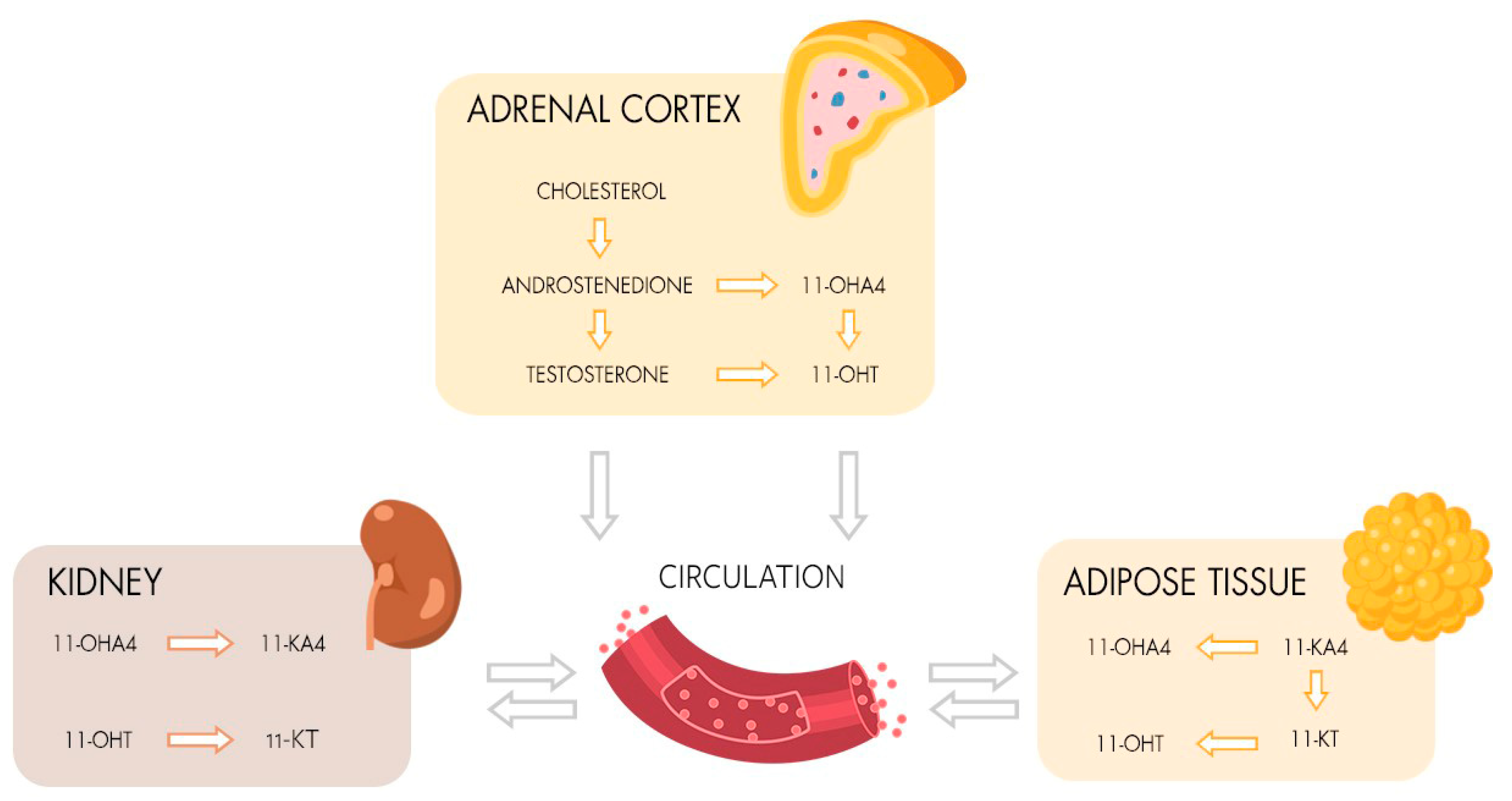
1. Introduction
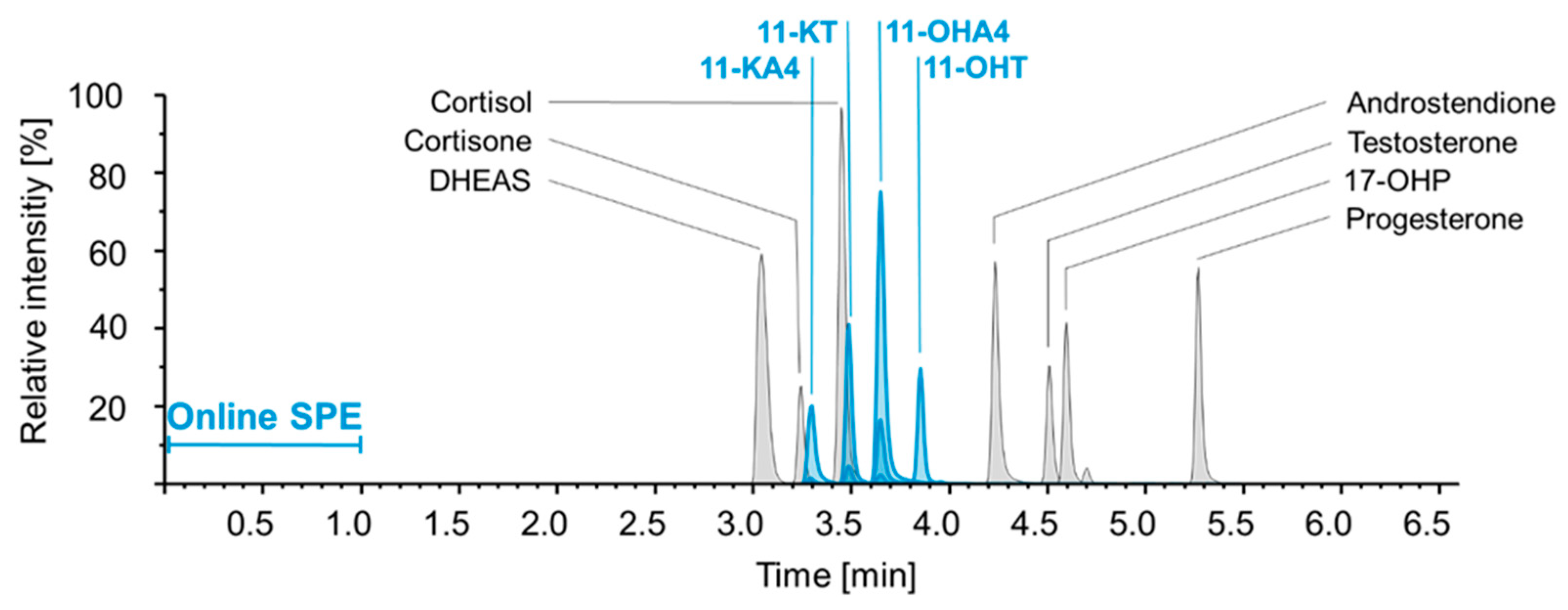
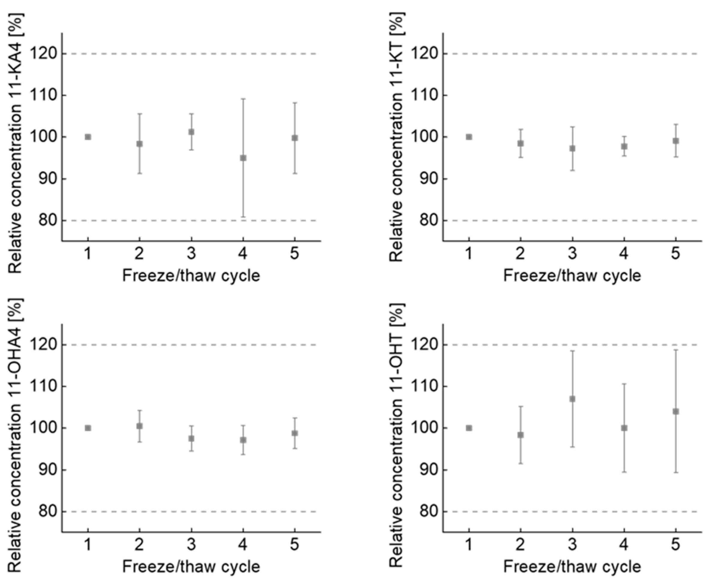
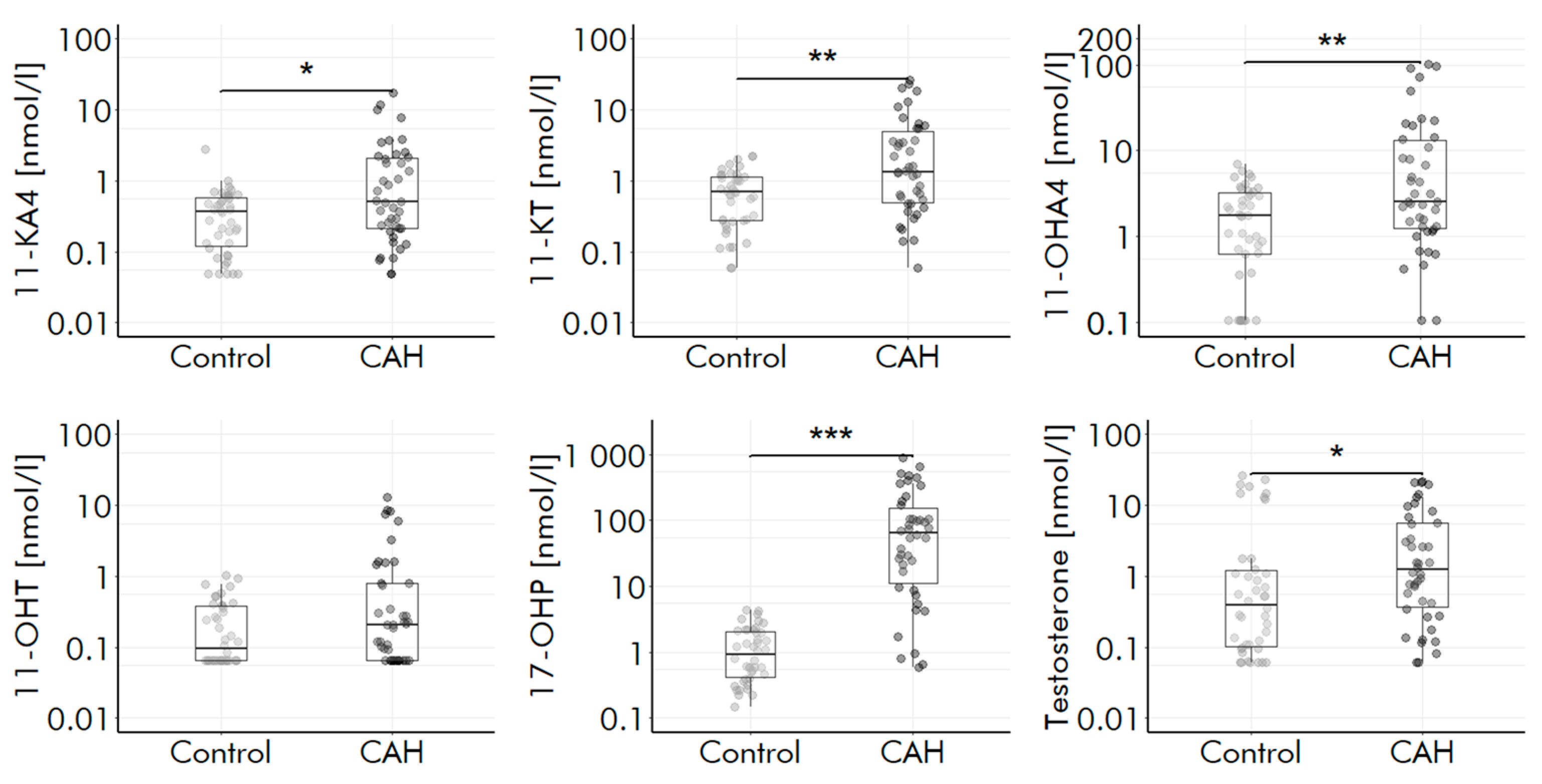
2. Results
3. Discussion
4. Materials and Methods
4.1. Chemicals and Reagents
4.2. Human Samples
4.3. Sample Preparation
4.4. LC-MS/MS
4.5. Validation
4.6. Clinical Verification
Supplementary Materials
Author Contributions
Funding
Institutional Review Board Statement
Informed Consent Statement
Data Availability Statement
Acknowledgments
Conflicts of Interest
References
- Mornet, E.; Dupont, J.; Vitek, A.; White, P.C. Characterization of Two Genes Encoding Human Steroid 11β-Hydroxylase (P-450(11β)). J. Biol. Chem. 1989, 264, 20961–20967. [Google Scholar] [CrossRef] [PubMed]
- Schiffer, L.; Arlt, W.; Storbeck, K.H. Intracrine Androgen Biosynthesis, Metabolism and Action Revisited. Mol. Cell. Endocrinol. 2018, 465, 4–26. [Google Scholar] [CrossRef] [PubMed]
- Turcu, A.F.; Rege, J.; Auchus, R.J.; Rainey, W.E. 11-Oxygenated Androgens in Health and Disease. Nat. Rev. Endocrinol. 2020, 16, 284–296. [Google Scholar] [CrossRef] [PubMed]
- Turcu, A.F.; Nanba, A.T.; Auchus, R.J. The Rise, Fall, and Resurrection of 11-Oxygenated Androgens in Human Physiology and Disease. Horm. Res. Paediatr. 2018, 89, 284–291. [Google Scholar] [CrossRef]
- Rege, J.; Nakamura, Y.; Wang, T.; Merchen, T.D.; Sasano, H.; Rainey, W.E. Transcriptome profiling reveals differentially expressed transcripts between the human adrenal zona fasciculata and zona reticularis. J. Clin. Endocrinol. Metab. 2014, 99, E518–E527. [Google Scholar] [CrossRef]
- Kelly, D.M.; Jones, T.H. Testosterone and Obesity. Obes. Rev. 2015, 16, 581–606. [Google Scholar] [CrossRef]
- Davio, A.; Woolcock, H.; Nanba, A.T.; Rege, J.; O’day, P.; Ren, J.; Zhao, L.; Ebina, H.; Auchus, R.; Rainey, W.E.; et al. Sex Differences in 11-Oxygenated Androgen Patterns across Adulthood. J. Clin. Endocrinol. Metab. 2020, 105, e2921–e2929. [Google Scholar] [CrossRef]
- Conway, G.; Dewailly, D.; Diamanti-Kandarakis, E.; Escobar-Morreale, H.F.; Franks, S.; Gambineri, A.; Kelestimur, F.; Macut, D.; Micic, D.; Pasquali, R.; et al. The polycystic ovary syndrome: A position statement from the European society of endocrinology. Eur. J. Endocrinol. 2014, 171, P1–P29. [Google Scholar] [CrossRef]
- Turcu, A.F.; Nanba, A.T.; Chomic, R.; Upadhyay, S.K.; Giordano, T.J.; Shields, J.J.; Merke, D.P.; Rainey, W.E.; Auchus, R.J. Adrenal-derived 11-oxygenated 19-carbon steroids are the dominant androgens in classic 21-hydroxylase deficiency. Eur. J. Endocrinol. 2016, 174, 601–609. [Google Scholar] [CrossRef]
- Turcu, A.F.; Rege, J.; Chomic, R.; Liu, J.; Nishimoto, H.K.; Else, T.; Moraitis, A.G.; Palapattu, G.S.; Rainey, W.E.; Auchus, R.J. Profiles of 21-carbon steroids in 21-hydroxylase deficiency. J. Clin. Endocrinol. Metab. 2015, 100, 2283–2290. [Google Scholar] [CrossRef]
- Rege, J.; Garber, S.; Conley, A.J.; Elsey, R.M.; Turcu, A.F.; Auchus, R.J.; Rainey, W.E. Circulating 11-oxygenated androgens across species. J. Steroid Biochem. Mol. Biol. 2019, 190, 242–249. [Google Scholar] [CrossRef] [PubMed]
- Carmina, E.; Stanczyk, F.Z.; Chang, L.; Miles, R.A.; Lobo, R.A. The ratio of androstenedione: 11β-Hydroxyandrostenedione Is an important marker of adrenal androgen excess in women. Fertility and Sterility 1992, 58, 148–152. [Google Scholar] [CrossRef] [PubMed]
- Nanba, A.T.; Rege, J.; Ren, J.; Auchus, R.J.; Rainey, W.E.; Turcu, A.F. 11-Oxygenated C19 steroids do not decline with age in women. J. Clin. Endocrinol. Metab. 2019, 104, 2615–2622. [Google Scholar] [CrossRef] [PubMed]
- du Toit, T.; Bloem, L.M.; Quanson, J.L.; Ehlers, R.; Serafin, A.M.; Swart, A.C. Profiling adrenal 11β-Hydroxyandrostenedione metabolites in prostate cancer cells, tissue and plasma: UPC2-MS/MS quantification of 11β-Hydroxytestosterone, 11keto-testosterone and 11keto-dihydrotestosterone. J. Steroid Biochem. Mol. Biol. 2017, 166, 54–67. [Google Scholar] [CrossRef] [PubMed]
- Han, B.; Zhu, H.; Yao, H.; Ren, J.; O’Day, P.; Wang, H.; Zhu, W.; Cheng, T.; Auchus, R.J.; Qiao, J. Differences of adrenal-derived androgens in 5α-reductase deficiency versus androgen insensitivity syndrome. Clin. Transl. Sci. 2021, 15, 658–666. [Google Scholar] [CrossRef] [PubMed]
- Blasco, M.; Carriquiriborde, P.; Marino, D.; Ronco, A.E.; Somoza, G.M. A Quantitative HPLC-MS Method for the Simultaneous Determination of Testosterone, 11-Ketotestosterone and 11-β Hydroxyandrostenedione in Fish Serum. J. Chromatogr. B: Anal. Technol. Biomed. Life Sci. 2009, 877, 1509–1515. [Google Scholar] [CrossRef] [PubMed]
- Flores-Valverde, A.M.; Hill, E.M. Methodology for profiling the steroid metabolome in animal tissues using ultraperformance liquid chromatography-electrospray-time-of-flight mass spectrometry. Anal. Chem. 2008, 80, 8771–8779. [Google Scholar] [CrossRef]
- Schloms, L.; Storbeck, K.H.; Swart, P.; Gelderblom, W.C.A.; Swart, A.C. The influence of aspalathus linearis (rooibos) and dihydrochalcones on adrenal steroidogenesis: Quantification of steroid intermediates and end products in H295R Cells. J. Steroid Biochem. Mol. Biol. 2012, 128, 128–138. [Google Scholar] [CrossRef]
- Xing, Y.; Edwards, M.A.; Ahlem, C.; Kennedy, M.; Cohen, A.; Gomez-Sanchez, C.E.; Rainey, W.E. The effects of ACTH on steroid metabolomic profiles in human adrenal cells. J. Endocrinol. 2011, 209, 327–335. [Google Scholar] [CrossRef]
- Zheng, J.; Islam, R.M.; Skiba, M.A.; Zheng, J.; Islam, R.M.; Skiba, M.A.; Bell, R.J.; Davis, S.R. Associations between androgens and sexual function in premenopausal women: A cross-sectional study. Lancet Diabetes Endocrinol. 2020, 8, 693–702. [Google Scholar] [CrossRef]
- Caron, P.; Turcotte, V.; Guillemette, C. A quantitative analysis of total and free 11-oxygenated androgens and its application to human serum and plasma specimens using liquid-chromatography tandem mass spectrometry. J. Chromatogr. A 2021, 1650, 462228. [Google Scholar] [CrossRef] [PubMed]
- Zhang, X.; Zhou, C.; Xu, H.; Feng, Y.; Yang, P.; Zhai, S.; Song, J.; Yang, L. A Sensitive HPLC-DMS/MS/MS method for multiplex analysis of androgens in human serum without derivatization and its application to PCOS patients. J. Pharm. Biomed. Anal. 2021, 192, 113680. [Google Scholar] [CrossRef] [PubMed]
- Hawley, J.M.; Adaway, J.E.; Owen, L.J.; Keevil, B.G. Development of a Total Serum Testosterone, Androstenedione, 17-Hydroxyprogesterone, 11β-Hydroxyandrostenedione and 11-Ketotestosterone LC-MS/MS Assay and Its Application to Evaluate Pre-Analytical Sample Stability. Clin. Chem. Lab. Med. 2020, 58, 741–752. [Google Scholar] [CrossRef]
- Wright, C.; O’Day, P.; Alyamani, M.; Sharifi, N.; Auchus, R.J. Abiraterone acetate treatment lowers 11-Oxygenated androgens. Eur. J. Endocrinol. 2020, 182, 413–421. [Google Scholar] [CrossRef] [PubMed]
- Häkkinen, M.R.; Murtola, T.; Voutilainen, R.; Poutanen, M.; Linnanen, T.; Koskivuori, J.; Lakka, T.; Jääskeläinen, J.; Auriola, S. Simultaneous analysis by LC–MS/MS of 22 ketosteroids with hydroxylamine derivatization and underivatized estradiol from human plasma, serum and prostate tissue. J. Pharm. Biomed. Anal. 2019, 164, 642–652. [Google Scholar] [CrossRef]
- Houghton, L.C.; Howland, R.E.; Wei, Y.; Ma, X.; Kehm, R.D.; Chung, W.K.; Genkinger, J.M.; Santella, R.M.; Hartmann, M.F.; Wudy, S.A.; et al. The steroid metabolome and breast cancer risk in women with a family history of breast cancer: The novel role of adrenal androgens and glucocorticoids. Cancer Epidemiol. Biomark. Prevention 2021, 30, 89–96. [Google Scholar] [CrossRef] [PubMed]
- Quanson, J.L.; Stander, M.A.; Pretorius, E.; Jenkinson, C.; Taylor, A.E.; Storbeck, K.H. High-throughput analysis of 19 endogenous androgenic steroids by ultra-performance convergence chromatography tandem mass spectrometry. J. Chromatogr. B Anal. Technol. Biomed. Life Sci. 2016, 1031, 131–138. [Google Scholar] [CrossRef]
- O’Reilly, M.W.; Kempegowda, P.; Jenkinson, C.; Taylor, A.E.; Quanson, J.L.; Storbeck, K.H.; Arlt, W. 11-oxygenated C19 steroids are the predominant androgens in polycystic ovary syndrome. J. Clin. Endocrinol. Metab. 2017, 102, 840–848. [Google Scholar] [CrossRef]
- du Toit, T.; Stander, M.A.; Swart, A.C. A High-Throughput UPC2-MS/MS method for the separation and quantification of C19 and C21 steroids and Their C11-Oxy steroid metabolites in the classical, alternative, backdoor and 11OHA4 steroid pathways. J. Chromatogr. B Anal. Technol. Biomed. Life Sci. 2018, 1080, 71–81. [Google Scholar] [CrossRef]
- Yoshida, T.; Matsuzaki, T.; Miyado, M.; Saito, K.; Iwasa, T.; Matsubara, Y.; Ogata, T.; Irahara, M.; Fukami, M. 11-Oxygenated C19 steroids as circulating androgens in women with polycystic ovary syndrome. Endocr. J. 2018, 65, 979–990. [Google Scholar] [CrossRef]
- Skiba, M.A.; Bell, R.J.; Islam, R.M.; Handelsman, D.J.; Desai, R.; Davis, S.R. Androgens during the reproductive years: What is normal for women? J. Clin. Endocrinol. Metab. 2019, 104, 5382–5392. [Google Scholar] [CrossRef] [PubMed]
- Rege, J.; Turcu, A.F.; Kasa-Vubu, J.Z.; Lerario, A.M.; Auchus, G.C.; Auchus, R.J.; Smith, J.M.; White, P.C.; Rainey, W.E. 11-ketotestosterone is the dominant circulating bioactive androgen during normal and premature adrenarche. J. Clin. Endocrinol. Metab. 2018, 103, 4589–4598. [Google Scholar] [CrossRef] [PubMed]
- Gaudl, A.; Kratzsch, J.; Bae, Y.J.; Kiess, W.; Thiery, J.; Ceglarek, U. Liquid chromatography quadrupole linear ion trap mass spectrometry for quantitative steroid hormone analysis in Plasma, Urine, Saliva and Hair. J. Chromatogr. A 2016, 1464, 64–71. [Google Scholar] [CrossRef] [PubMed]
- Gaudl, A.; Kratzsch, J.; Ceglarek, U. Advancement in Steroid Hormone Analysis by LC–MS/MS in clinical routine diagnostics—A three year recap from serum cortisol to dried blood 17α-hydroxyprogesterone. J. Steroid Biochem. Mol. Biol. 2019, 192, 105389. [Google Scholar] [CrossRef] [PubMed]
- CLSI Liquid Chromatography-Mass Spectrometry Methods; Approved Guideline; CLSI document C62-A 2014; Clinical and Laboratory Standards Institute: Wayne, PA, USA, 2014.
- Auer, M.K.; Paizoni, L.; Neuner, M.; Lottspeich, C.; Schmidt, H.; Hawley, J.; Keevil, B.; Reisch, N. Elevated 11-oxygenated androgens are not a major contributor to HPG-Axis disturbances in adults with congenital adrenal hyperplasia due to 21-Hydroxylase deficiency. medRxiv 2021. [Google Scholar] [CrossRef]


| 11-KA4 | 11-KT | 11-OHA4 | 11-OHT | |||||||||
|---|---|---|---|---|---|---|---|---|---|---|---|---|
| Mean [nmol/L] | CV | Recovery | Mean [nmol/L] | CV | Recovery | Mean [nmol/L] | CV | Recovery | Mean [nmol/L] | CV | Recovery | |
| Serum Level 1 | 0.34 | 13% | 102% | 1.5 | 7% | 91% | 6.0 | 5% | 113% | 0.44 | 10% | 108% |
| Serum Level 2 | 0.9 | 10% | 107% | 1.9 | 4% | 86% | 12 | 6% | 114% | 0.62 | 9% | 113% |
| Serum Level 3 | 19 | 7% | 115% | 16 | 3% | 88% | 188 | 4% | 111% | 1.2 | 6% | 116% |
| QK Level 1 | 0.18 | 7% | 109% | 0.14 | 10% | 85% | 1.8 | 5% | 107% | 0.18 | 13% | 109% |
| QK Level 2 | 0.7 | 10% | 108% | 0.6 | 4% | 85% | 7.1 | 3% | 107% | 0.69 | 5% | 105% |
| QK Level 3 | 19 | 8% | 113% | 15 | 5% | 90% | 165 | 6% | 100% | 16 | 3% | 99% |
Disclaimer/Publisher’s Note: The statements, opinions and data contained in all publications are solely those of the individual author(s) and contributor(s) and not of MDPI and/or the editor(s). MDPI and/or the editor(s) disclaim responsibility for any injury to people or property resulting from any ideas, methods, instructions or products referred to in the content. |
© 2022 by the authors. Licensee MDPI, Basel, Switzerland. This article is an open access article distributed under the terms and conditions of the Creative Commons Attribution (CC BY) license (https://creativecommons.org/licenses/by/4.0/).
Share and Cite
Zeidler, R.; Biemann, R.; Ceglarek, U.; Kratzsch, J.; Isermann, B.; Gaudl, A. Inclusion of 11-Oxygenated Androgens in a Clinical Routine LC-MS/MS Setup for Steroid Hormone Profiling. Int. J. Mol. Sci. 2023, 24, 539. https://doi.org/10.3390/ijms24010539
Zeidler R, Biemann R, Ceglarek U, Kratzsch J, Isermann B, Gaudl A. Inclusion of 11-Oxygenated Androgens in a Clinical Routine LC-MS/MS Setup for Steroid Hormone Profiling. International Journal of Molecular Sciences. 2023; 24(1):539. https://doi.org/10.3390/ijms24010539
Chicago/Turabian StyleZeidler, Robert, Ronald Biemann, Uta Ceglarek, Jürgen Kratzsch, Berend Isermann, and Alexander Gaudl. 2023. "Inclusion of 11-Oxygenated Androgens in a Clinical Routine LC-MS/MS Setup for Steroid Hormone Profiling" International Journal of Molecular Sciences 24, no. 1: 539. https://doi.org/10.3390/ijms24010539
APA StyleZeidler, R., Biemann, R., Ceglarek, U., Kratzsch, J., Isermann, B., & Gaudl, A. (2023). Inclusion of 11-Oxygenated Androgens in a Clinical Routine LC-MS/MS Setup for Steroid Hormone Profiling. International Journal of Molecular Sciences, 24(1), 539. https://doi.org/10.3390/ijms24010539