Abstract
Despite the recent successes and durable responses with immune checkpoint inhibitors (ICI), many cancer patients, including those with melanoma, do not derive long-term benefits from ICI therapies. The lack of predictive biomarkers to stratify patients to targeted treatments has been the driver of primary treatment failure and represents an unmet medical need in melanoma and other cancers. Understanding genomic correlations with response and resistance to ICI will enhance cancer patients’ benefits. Building on insights into interplay with the complex tumor microenvironment (TME), the ultimate goal should be assessing how the tumor ’instructs’ the local immune system to create its privileged niche with a focus on genomic reprogramming within the TME. It is hypothesized that this genomic reprogramming determines the response to ICI. Furthermore, emerging genomic signatures of ICI response, including those related to neoantigens, antigen presentation, DNA repair, and oncogenic pathways, are gaining momentum. In addition, emerging data suggest a role for checkpoint regulators, T cell functionality, chromatin modifiers, and copy-number alterations in mediating the selective response to ICI. As such, efforts to contextualize genomic correlations with response into a more insightful understanding of tumor immune biology will help the development of novel biomarkers and therapeutic strategies to overcome ICI resistance.
1. Introduction
Immune checkpoint inhibitors (ICIs) are revolutionizing the treatment of melanoma and other types of cancers, with 7 agents now approved in the US and a rich pipeline of new agents and mechanisms in development [1,2,3,4]. Notwithstanding the excitement around these developments, there is a significant unmet medical need in the form of patient stratification and therapy resistance. Melanoma affects more than 1 million Americans, and there is an increasing incidence of melanoma worldwide. Approx. 300,000 new cases are diagnosed in the US each year [5], with the average annual cost for treatment estimated at $3.3 billion [6].
Numerous successes have been achieved with anti-PD1, anti-CTLA4, or combination therapies [5,7] in treating melanoma and other types of cancers. The groundbreaking finding by Leach et al. [8] showed that antibodies blocking the T cell co-inhibitory receptor CTLA-4 can augment immune responses against tumor cells in mice. This finding gave rise to ipilimumab, the first ICI to increase the survival of patients with metastatic melanoma [9,10] which was granted FDA approval in 2011 for the treatment of metastatic melanoma.
In 2014, additional T cell immune checkpoint blocking antibodies, anti-PD-1 (pembrolizumab) and anti-PD-L1 (nivolumab) [11], received FDA approval. The combination of anti-PD-1 (pembrolizumab) and anti-PD-L1 (nivolumab) resulted in the augmented survival of patients with metastatic melanoma compared to patients treated with ipilimumab alone or chemotherapy alone [12,13,14].
In 2022, relatlimab, which targets lymphocyte-activation gene 3 (LAG-3), was approved by the FDA for adult and pediatric use with metastatic melanoma [15].
Following those developments, high levels of PD-L1 expression in cancer cells and tumor mutational burden (TMB) have been shown in melanoma to correlate with clinical responses to ICI [13].
Despite these data, a substantial number of melanoma patients fail to respond to ICI, leading to premature death [16,17,18]. Considerable effort has been made to identify biomarkers that predict clinical response/resistance to ICI [19]. Despite the successes of ICI, even with the combination of ipilimumab and nivolumab, the five-year survival for the intention-to-treat (ITT) population was only 53%. This means that 47% of patients do not reach long-lasting benefits and succumb to the disease [20], suggesting the need for predictive biomarkers of response to overcome ICI resistance.
Despite the approval of all of these new ICIs and combination therapy, physicians are nowhere near having predictive biomarkers of response to ICI. Presently, physicians have no idea which patients will or will not respond to ICI in the absence of predictive biomarkers of response.
Recently, Carlino et al. [21] demonstrated that combining anti-PD-1 and anti-CTLA-4 ICI in stage IV melanoma resulted in the highest 5-year overall survival rate of all the other therapies. Increased PD-L1 expression and TMB have been shown to correlate well with melanoma response to ICI [13]. Unfortunately, both PD-1 and CTLA4 are unable to predict the outcome [22]. As a result, we run the risk of undertreating some patients who might benefit from ICI.
To date, there are no reliable predictive biomarkers able to stratify responders and non-responders to ICI. Clinical factors, such as volume and disease sites, serum lactate dehydrogenase levels, and BRAF mutation status, used to select the initial therapy for patients with advanced melanoma have been rather unreliable [21].
The clinical trial data of melanoma response to ICI have identified three almost equal populations of patients: (1) those who respond (responders), (2) those who fail to ever respond (innate resistance), and (3) those who initially respond or have a prolonged period of disease stabilization, but eventually develop disease progression (acquired resistance) [7,16,23,24,25].
The current stratification strategies, including genetic mutations and variations in mutational load, have shown some correlation with response to ICI. However, they are unable in predicting patient response to ICI [26]. It has been shown that tumor cells possess genetic and epigenetic traits that facilitate immune evasion [27]. Tumors can mutate to evade the innate and adaptive immune response [19], rendering ICI therapy ineffective [23,28,29]. ICI resistance can start from different cells and their interactions in the tumor ecosystem [29,30]. Recent studies have revealed insights into the mechanism of ICI resistance [7,29,30]. As a result, ICI therapies elicit significantly limited efficacy in many of the cancer types due to drug resistance and toxicity.
The lack of predictive biomarkers to stratify patients to targeted treatments has been the driver of primary treatment failure and represents an unmet medical need in melanoma and other cancers. Therefore, understanding genomic correlations with response and resistance to ICI will be beneficial to cancer patients.
Here, we review recent studies involving ICI therapies in melanoma and other cancers that have revealed important insights about the tumor immune microenvironment in melanoma and other cancers, the mechanisms supporting these findings, and genomic correlations with response and resistance to ICI.
We further discuss how the insights gained from these studies are guiding more precise analyses of the mechanisms of action of ICI therapy and novel immunotherapy approaches, including novel combination therapies to overcome resistance to ICI therapy and turning “cold tumors” into “hot tumors”.
Consequently, these efforts will help us better understand the mechanisms involved in response and resistance to ICI, thus achieving more effective and durable use of immunotherapy.
2. Immune Checkpoint Inhibitors
As illustrated in Figure 1, immunosuppressive mechanisms involving the expression of checkpoint proteins (e.g., PD-1, CTLA-4, LAG-3, TIM-3, and NKG2A/B), activation of cell death programs, and accumulation of various immunosuppressive cells are instrumental in maintaining immune homeostasis and self-tolerance [31,32,33,34]. Cancer cells exploit those immune homeostasis mechanisms to evade the immune system [35]. Several ICI treatments are successful against a wide variety of cancers, given the durability of response and improved side effect profile compared to chemotherapeutic agents. Nonetheless, only a few ICIs have been approved by the FDA [34].
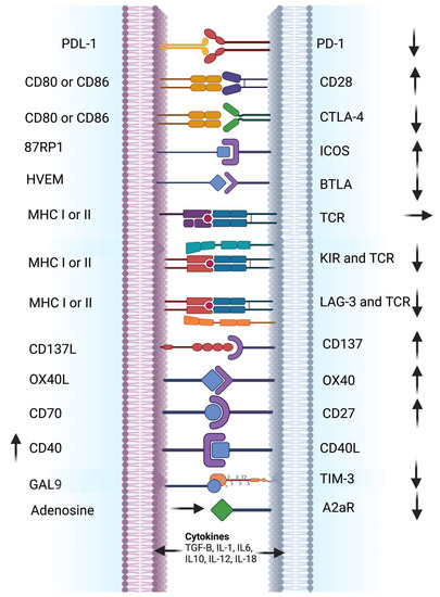
Figure 1.
Immune cell and cancer cell or antigen-presenting cell receptor-ligand interactions involved in immune checkpoint modulation. The figure illustrates an overview of implicated receptor-ligand interactions and their general effects on the immune response. Several immune cells, such as CD8 + T cells, CD4 + T cells, NK cells, and Tregs, express specific receptors, which are recognized and bound by their specific ligands present on the surface of various cancer cells or antigen-presenting cells. The illustration also depicts examples of different receptors and ligands involved in ICI modulation, along with generalized stimulatory (↑) or inhibitory (↓) effects. ICIs block various inhibitory receptor-ligand interactions leading to the activation of immune cells, which leads to tumor regression. ICI therapies, including anti-PD-1/PD-L1 and anti-CTLA-4, have shown clinical efficacy for many cancers, which provided opportunities for developing alternative ICIs (e.g., anti-LAG-3, anti-TIM-3, and anti-NKG2). Adapted from ref. [3,34]. Created with BioRender.com (accessed on 18 November 2022). Definitions of abbreviations are found in the abbreviations list at the end of the text.
3. Melanoma Biomarkers of Response to ICI
Physicians would greatly benefit in therapeutic decision-making by having predictive biomarker(s) of response to stratify melanoma and other types of cancer patients into responders and non-responders. A general representation of predictive biomarkers of response to ICIs in several cancers is shown in Table 1. However, the significant proportion of non-responders and treatment-associated toxicities and drug resistance remain one of the major obstacles to therapeutic success of ICI in melanoma and other types of cancers.

Table 1.
A general representation of predictive biomarkers of response to immune checkpoint inhibitors.
3.1. Established Clinical Biomarkers
Different types of biomarkers and their clinical utility in metastatic melanoma, including those approved by the FDA and others still at the experimental stage, are shown in Table 2 along with the clinical setting in which they are used.

Table 2.
Types of biomarkers and their clinical utility in metastatic melanoma.
As summarized in Table 2, the earliest biomarkers that helped to diagnose the presence of melanoma include human melanoma black-45 (HMB-45), melan-A, tyrosinase, microphthalmia transcription factor (MITF), S100, SM5-1, chondroitin sulfate proteoglycan 4 (CSPG4), loss of p16 protein expression, biomarker panels, and gene arrays. Those biomarkers have been used to screen healthy patients before the diagnosis of melanoma in order to help stratify patients into benign vs. malignant stages of the disease.
The earliest approved clinical biomarkers that helped to inform the prognosis of metastatic melanoma relied on baseline clinical characteristics, such as serum levels of lactate dehydrogenase (LDH), a marker of tumor burden [41]. Elevated serum LDH levels have been demonstrated to be a negative prognostic marker, irrespective of the given treatment [42,43,44,45]. Generally, high LDH levels are associated with poor overall survival (OS) compared with normal LDH levels. Elevated levels of LDH have been used as a biomarker to assess patient staging [46]. For example, it has been shown that among ca. 30% of patients with 4–5-year OS following treatment with BRAF and MEK inhibitors, only a few of those patients had high LDH levels before the initiation of therapy [47].
Likewise, elevated serum levels of S100B have demonstrated to have a prognostic value in both metastatic [48,49] and high-risk resected melanoma settings [50].
Gene expression profiling has shown to have a prognostic value that complements existing biomarkers in patients with melanoma [47,51,52].
In metastatic melanoma, a well-characterized predictive biomarker of response guiding the therapeutic decision process is the BRAF V600 mutation, which is somehow predictive of response to BRAF ± MEK inhibition with low rates of primary resistance [53,54,55]. The response rate to BRAF and MEK inhibitors in metastatic melanoma patients with the BRAF V600 mutation is ca. 70% in selected patients, with less than 10% of patients having the highest response to progressive disease [55,56,57,58].
Several oncogenic driver mutations have been identified as predictive biomarkers of response from targeted agents, including NRAS, NF-1, and c-KIT, that provide insight into the probability of therapeutic response to a specific treatment [59,60,61,62,63]. Presently, the presence of a BRAF V600 mutation is the only validated predictive biomarker for melanoma patients.
3.2. Emerging Predictive Biomarkers
As shown in Table 2, many emerging biomarkers are currently being evaluated in clinical studies. Du et al. [64] reported that several genomic and transcriptomic-based biomarkers have been explored as potential predictors of ICI response.
Predictive biomarkers of response include TMB, neoantigen load [7,65,66,67,68,69], HLA-I genotype [70,71], cytolytic activity [72], aneuploidy [73], and T cell repertoire [46], which exhibit high predictability to ICI response. Other predictive biomarkers of response are PDL-1 expression, LAG-3 expression, CD8+ T cells at tumor invasive margin, IL-17 expression, immune-related gene expression signatures, and T-cell receptor (TCR) signature.
Additional biomarkers that are used to assess ICI response include ctDNA profiles, absolute lymphocyte count, proliferating CD8+ T cells, increase of T-cell subsets and checkpoint molecules (PDL-1, LAG-3), granzyme B expression, and T-cell receptor (TCR) signature [37,38,39,40].
Unfortunately, many of those potential biomarkers of ICI response have not yet been validated [74,75,76,77,78].
A recent study by Carter et al. [74] questioned the validity of the immuno-predictive score (IMPRES), a predictor of ICI response in melanoma consisting of 15 pairwise transcriptomic signatures that analyze the relationship between immune checkpoint genes reported by Auslander et al. [76]. The IMPRES is context-dependent and could not reproducibly predict ICI response in the context of metastatic melanoma [76].
Moreover, Xiao et al. [77] questioned the reproducibility of the Immune Cells.Sig [79] signature in melanoma, demonstrating inconsistencies in the prediction capability of ImmuneCells.Sig across different RNA-seq datasets [77]. The performance of the ImmuneCells.Sig signature in predicting ICI outcomes in four melanoma patient datasets, using the same implementation scheme as Xiong et al. [79], showed that there were inconsistencies across different datasets [77].
3.2.1. Gene Expression Signatures
Gene expression signatures (GES) have also been identified as predictive biomarkers of response to ICI and have been validated in several independent datasets (e.g., immune-predictive score, IMPRES consisting of 15 immune genes). IFN-γ-responsive genes were also used to predict ICI response in metastatic melanoma [30,46,72,73,76,80,81,82,83,84,85,86].
A panel of pan-tumor T cell-inflamed GES consisting of 18 IFN-γ-responsive genes was validated and confirmed to predict the response to ICI in pre-treatment tumor specimens from nine types of cancers, including melanoma [83].
MHC-I/II gene signatures have also been explored as predictive biomarkers of ICI response in melanoma [85,87,88].
Carter et al. [74] reported that immuno-predictive score (IMPRES), a predictor of ICI response in melanoma encompassing 15 pairwise transcriptomics relations between immune checkpoint gene [76], did not reproducibly predict the response to ICI in metastatic melanoma.
It was argued that many factors may contribute to the limited successes of those biomarkers, such as: (1) the predictive biomarkers have been derived from pre-clinical studies; (2) evaluation of the biomarkers in clinical specimens only included baseline biopsies and peripheral blood samples; (3) batch effect, lack of reproducibility might have contributed to the failure of the biomarkers for ICI response [74,75,77,78].
To address these issues, numerous researchers have developed predictive biomarkers to reduce batch noise and other technical issues. Expanded predictive biomarker panels have resulted in higher reproducibility as opposed to predictive signatures based on individual biomarkers [89,90,91,92].
Tian et al. [91] reported that the combined BRAF, KRAS, and PI3KCA mutation signature resulted in a favorable predictive response to cetuximab for patients with colorectal cancer [91].
3.2.2. Gene Expression Signatures at Baseline and On-Treatment Tumor Specimens
Genome-wide analysis of transcriptomic and genomic profiles of baseline and on-therapy tumor specimens from patients treated with ICI provides a comprehensive view into the mechanisms underlying tumor response and resistance to ICI [93].
Grasso et al. [93] reported that the mechanism of action of ICI is based on the interaction between immune effector cells and cancer cell targets. Tumor studies conducting comprehensive analyses of transcriptomic and genomic profiling have focused not only on the genetic alterations and gene expression profiles of cancer cells [25,30,85,88], but also on the composition of immune infiltrates and expression of immune-activating gene programs [36,46,67,75,80,82,83,85,86,88,94,95,96,97,98].
Du et al. [64] reported that pathway-based signatures derived from on-treatment tumor specimens were predictive of the response to anti-PD1 blockade in patients with metastatic melanoma.
Other studies of breast cancer suggested that post-treatment tumor samples were more informative than pre-treatment samples [99,100,101].
Conversely, Wallin et al. developed adaptive immune signatures based on tumor samples obtained during the early course of treatment, showing that the signatures were highly predictive of the response to ICI in patients with metastatic melanoma [102].
Auslander et al. built an immune-predictive score (IMPRES), which encompasses 15 pairwise transcriptomic relations between immune checkpoint genes, to predict the response of metastatic melanoma to ICB therapy [76].
The IMPRES signature produced better predictive scores with post-treatment samples than with pre-treatment samples in two independent datasets [76].
In support of these findings, a recent proteome profiling study of samples from patients with metastatic melanoma undergoing either tumor infiltrating lymphocyte based or anti-PD1 immunotherapy demonstrated that the fatty acid oxidation pathway was significantly enriched in responder patients. These results underlined the critical role of mitochondrial metabolism, including fatty acid metabolism, in conferring response to immunotherapy [103].
It is now widely accepted that post-treatment tumor specimens are generally much more informative than pre-treatment specimens and may provide more valuable insight into dynamic changes at the transcriptional level that correlate with clinical response, resulting in a higher predictive score.
In conclusion, although pathway signatures derived from post-treatment samples are highly predictive of therapeutic response to anti-PD1 in patients with metastatic melanoma, further studies are warranted to confirm the predictive value of those signatures in larger cohorts of patients with metastatic melanoma.
3.2.3. Pathway Signatures
Du et al. [64] developed pathway-specific signatures in pre-treatment (PASS-PRE) and on-treatment (PASS-ON) tumor specimens based on transcriptomic data and clinical information from a large dataset of metastatic melanoma patients treated with anti-PD1. Both PASS-PRE and PASS-ON signatures were validated in three independent datasets of metastatic melanoma. Compared to existing molecular signatures, it was concluded that the on-treatment (PASS-ON) tumor specimen signature exhibited a robust and better predictive value for metastatic melanoma patients who responded to anti-PD1 across all four datasets.
The pre-treatment pathway signatures included six pathways for predicting the response to anti-PD1 treatment, including: (1) complement cascade; (2) regulation of insulin-like growth factor IGF transport and uptake by insulin-like growth factor binding proteins IGFBPS; (3) binding and uptake of ligands by scavenger receptors; (4) plasma lipoprotein remodeling; (5) IL2 family signaling; and (6) retinoic acid (RA) biosynthesis pathways. Complement cascade, binding, and uptake of ligands by scavenger receptors and IL2 family signaling pathways are related to immune and inflammation, whereas plasma lipoprotein remodeling and the RA biosynthesis pathways are related to metabolism.
In contrast to the pathway-based signature analysis of on-treatment samples, Du et al. [64] identified four pathways, including: (1) peroxisomal lipid metabolism; (2) generation of second messenger molecules; (3) fatty acid metabolism; and (4) PD1 signaling. Of note, peroxisomal lipid metabolism and fatty acid metabolism are related to fatty acid and lipid metabolism [103]. Generation of second messenger molecules is a central signaling pathway in T-cell receptor (TCR) stimulation. Likewise, PD1 signaling plays an important role in immunoregulation as an immunoregulatory signaling pathway.
To further validate the predictive performance of a pathway-based super signature for on-treatment samples (PASS-ON), Du et al. [64] tested three independent datasets with RNA-seq data available for on-treatment samples, including those of Gide et al. and Lee et al. [96,104], and the MGH cohort demonstrated the effectiveness of PASS-ON in predicting patient response to anti-PD1. Patients with high PASS-ON signature scores were associated with significantly improved PFS compared to those with low signature scores in all tested patients.
Furthermore, Du et al. [64] demonstrated that the time-response interaction pathway-based super signature for pre- and on-treatment samples had reasonable predictive power. The study suggested that pathway-based biomarker signatures derived from on-treatment tumor specimens compared to pretreatment tumor specimens were better predictors of response to anti-PD1 therapies in metastatic melanoma patients.
3.2.4. Tumor Antigens
Huang et al. [22] investigated several melanoma-relevant tumor-specific antigens, cancer germline genes, melanocyte differentiation antigens, overexpressed antigens, neoantigens, neuropeptides, and other sources of immunogenic antigens, such as immunogenic epitopes, have also been explored as novel predictive biomarkers of ICI response to melanoma.
Neoantigens are derived from tumor-specific somatic mutations and are exclusively expressed in cancer cells and absent in normal human tissue. The majority (95%) of somatic mutations are single-nucleotide variants (SNVs), which lead to aberrant protein and peptide expression with single amino acid substitutions [105].
Neopeptides also arise from nucleotide insertions or deletions (indels), leading to the expression of aberrant proteins and peptides with frameshift or non-frameshift sequences depending on the number of nucleotides added and deleted. While the minority of mutations are indels (<5% for melanoma) [106,107], frameshift mutations can generate a number of immunogenic neoepitopes that are highly distinct from the self.
Other sources of immunogenic antigens, including immunogenic epitopes, can also derive from mutations associated with gene fusion, aberrant messenger-RNA splicing with retained introns, or aberrant translation resulting in cryptic antigens, and genomically integrated endogenous retroviral sequences as a result of previous retroviral infections, although they are epigenetically silenced, can be reactivated in tumors [106], as in the case of cancer germline antigens.
Furthermore, tumors often present aberrant patterns of DNA methylation, resulting in the demethylation, ectopic expression, and presentation of cancer germline genes to T cells relevant in immune recognition [106,108].
For example, cancer germline genes such as MAGEA1 and NY-ESO-1 are silenced epigenetically through methylation in human tissue, with the exception of male germ cells and trophoblastic cells, which lack MHC-I molecules.
PRAME (preferentially expressed antigen in melanoma), a member of the cancer-testis antigen family, has been reported to be frequently overexpressed in many cancers, including melanoma, which indicates advanced cancer stages and poor clinical prognosis [109,110]. As such, overexpressed PRAME is a potential immunotherapy target. PRAME-specific immunotherapies are currently in development for many cancers, including melanoma. For example, a recent study demonstrated that uterine carcinosarcoma, synovial sarcoma, and leiomyosarcoma patients would potentially benefit from PRAME-specific immunotherapies [109].
3.2.5. Genomic Alterations
Considerable effort has been made to identify genomic alterations and transcriptome profiles as predictive biomarkers of ICI response. Numerous studies have identified distinct stages of CD8+ T cells linked to positive response or failure to ICI treatment [98].
Moreover, tumors from patients responding to ICI showed a higher number of cancer-associated somatic mutations (i.e., mutated antigens or neoantigens) targeted by T cells [65]. The IFN-γ signature has also been shown to predict the response to ICI (e.g., anti-PD-1) in melanoma [111] and in other types of cancers [6,93]. Gene expression signatures obtained from bulk melanoma tumor or single-cell profiling and the TME have been shown to be correlated with sensitivity and resistance to several ICIs [86,88,98,112,113,114].
Collectively, the gene expression signatures associated with response to ICI in metastatic melanoma represent distinct characteristics and play an important function in different signaling pathways, including the inflammatory response, type I interferon signaling pathway, cytokines, and others.
4. Who Is Responding to ICI?
Recent data have demonstrated that immunotherapies against immune checkpoints (e.g., CTLA-4 or PD-1) downregulate two main negative regulators of the anti-tumor immune response [93,115,116,117], resulting in durable anti-tumor responses in a subset of cancer patients, including those with melanoma [2,118].
Another key factor contributing to anti-tumor immune response following ICI treatment [93] is the pre-existing level of T cell infiltration of the tumor [119,120,121], representing the immunogenicity of the cancer cells.
Analysis of tumor biopsies from ICI-treated patients showed that clinical responses associated with ICI were mediated by tumor-infiltrating T cells reactivated following ICI treatment [121,122].
A combination of immunohistochemical (IHC) analysis with RNA-seq performed on cancer biopsies from patients treated with anti-CTLA-4 antibody (ipilimumab) before or after treatment with anti-PD-1 antibody (nivolumab) demonstrated that a major response to anti-CTLA-4 requires cancer cells with high levels of MHC-I expression at baseline, whereas the response to anti-PD-1 was more strongly associated with a pre-existing interferon-γ gene expression signature [88].
Liu et al. demonstrated that the response to anti-PD-1 therapy (with or without prior anti-CTLA-4 treatment) was associated with increased MHC-I and MHC-II expression [85]. This study demonstrated that patients not responding to therapy have occasional genetic alterations in antigen presentation genes [85].
Biopsies from patients with metastatic melanoma treated with anti-PD-1 monotherapy (nivolumab) in part 1 of the CheckMate 038 study showed an increase in immune cell subtypes with elevated immune activation gene signatures seen in responders to therapy [25].
The transcriptome analysis of tumor biopsies from patients treated with anti-PD-1 monotherapy (nivolumab) or in combination anti-PD1 plus anti-CTLA-4 therapy (ipilimumab) correlated well with the in vitro analysis of gene expression signatures of melanoma cell lines following exposure to interferon-γ [93].
It appears that cancer cells become enablers of the immune response via the expression of IFN-γ-response genes, triggering the upregulation of antigen presentation, amplification of the interferon response, and induction of chemokines (i.e., CXCL9 and CXCL10) to entice immune cells to the TME. Thus, T cell-induced IFN-γ correlates with ICI therapy response [93]. Collectively, the degree of the anti-tumor T cell response and downstream IFN-γ signaling are the main drivers of response or resistance to ICI therapy [93].
However, little is known about how tumor-intrinsic loss of IFN-γ signaling impacts TILs. The question remains whether tumor-intrinsic IFN-γ signaling actively regulates the infiltration or function of TILs?
Shen et al. [123] demonstrated that IFN-γR1 knockout melanomas and IFN-γR1KO melanomas in B6 mice had reduced infiltration and function of tumor-infiltrating lymphocytes (TILs). Furthermore, long-distance effects of IFN-γ on tumor cells also play a crucial role in anti-tumor immunity [123]. These recent findings revealed an important role of tumor-intrinsic IFN-γ signaling and IFN-γ-response genes in shaping TILs.
5. Tumor-Immunity Cycle and Resistance Mechanisms Involving Tumor Immunophenotypes
As discussed in the literature [124] and illustrated in Figure 2 and Figure 3, the anti-tumor-immunity cycle is a gradual process mediated to a large extent by CD8+ T lymphocytes and involves a multi-step process.
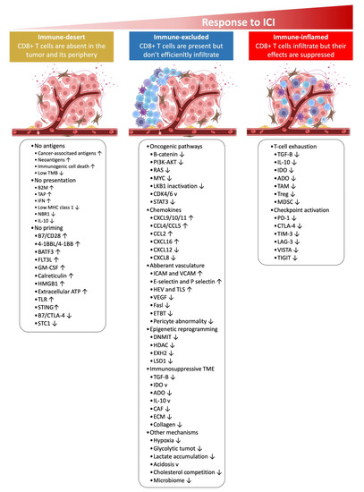
Figure 2.
Mechanisms of distinct tumor phenotypes, including immune-inflamed, immune-desert, and immune-excluded tumors, are associated with specific inhibitory and stimulatory biological mechanisms. Immune-inflamed tumors are permissive to immune cell infiltration; however, immune cells in the TME can be suppressed due to checkpoint activation. On the other hand, immune-desert tumors may be devoid of T cell priming due to the lack of tumor antigens, defective antigen processing and presentation processes, or impaired interactions between dendritic cells and T cells. The immune-excluded tumors, on the other hand, may display aberrant chemokines, activated oncogenic pathways, hypoxia, aberrant vasculature, or an immunosuppressive TME (e.g., stromal barriers). Adapted from ref. [124]. ↓ (downward arrow), inhibitors; ↑ (upward arrow), stimulatory factors. Created with BioRender.com (URL accessed on 18 November 2022). Definitions of abbreviations are found in the abbreviations list at the end of the text.
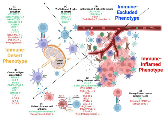
Figure 3.
Stimulatory and inhibitory factors in the cancer-immunity cycle. Each step of the cancer-immunity cycle requires the coordination of several stimulatory and inhibitory factors. Stimulatory factors shown in green with the upward arrow (↑) indicating promotion of immunity. Inhibitors shown in red with the downward arrow (↓) help keep the process in check and reduce immune activity and/or prevent autoimmunity. Examples of such factors and the primary steps at which they can act are shown. Antitumor immunity is mediated predominantly by CD8+ T cells and tumor immunity involves: (1) tumor antigen release, (2) tumor antigen processing and presentation by APCs (e.g., dendritic cells), (3) priming and activation of T cells, (4) trafficking of T cells via the bloodstream to tumors, (5) infiltration of activated T cells into the tumor parenchyma from the vasculature or tumor periphery, (6) recognition of tumor cells through antigenic peptide-MHC complexes on the surface of tumor cells by T cells, and (7) killing of tumor cells by cytotoxic T cells through granule exocytosis and release of perforin and granzyme or through the Fas/FasL pathway by inducing ferroptosis and pyroptosis. The release of additional antigens from dead tumor cells allows the continuation of the tumor-immunity cycle. Tumors with the immune-desert phenotype cannot pass steps 1–3 due to the absence of T cells in both the tumor and its margins. On the other hand, tumors with the immune-excluded phenotype cannot pass steps 4–5 due to the absence of T cells in the tumor bed. On the other hand, tumors with the immune-inflamed phenotype cannot pass steps 6–7 because of T cell exhaustion and checkpoint upregulation. Immune checkpoint proteins, such as PD1 and CTLA-4, can suppress the development of an active immune response by acting primarily at the level of T cell development and proliferation (step 3). Adapted from ref. [124,125]. Created with BioRender.com (URL (accessed on 18 November 2022). Definitions of abbreviations are found in the abbreviations list at the end of the text.
In the immune-desert phenotype, immune cells are absent from the tumor and its periphery. In the immune-excluded phenotype, immune cells accumulate but do not efficiently infiltrate. In the immune-inflamed phenotype, immune cells infiltrate but their effects are inhibited. Notably, the three different phenotypes have different response rates to immune checkpoint inhibitors.
Based on the spatial distribution of cytotoxic immune cells in the TME, a tumor is classified as an immune-inflamed, immune-excluded, or immune-desert phenotype (Figure 2 and Figure 3) [126]. Immune-inflamed tumors (i.e., “hot tumors”) are characterized by increased T cell infiltration, high interferon-γ (IFN-γ) signaling, elevated expression of PD-L1, and increased TMB [127]. Inflamed tumors are more responsive to ICIs than non-inflamed tumors [119,128]. Immune-deserted tumors (i.e., “cold tumors”), on the other hand, exhibit characteristics where CD8+ T lymphocytes localize only at the tumor margin and do not infiltrate the tumor [129]. In immune-desert tumors, CD8+ T lymphocytes are absent from the actual tumor and its periphery [129]. In addition to poor T cell infiltration, “cold tumors” display low mutational load, decreased MHC class I expression, and reduced PD-L1 expression [127].
Cold tumors also harbor immunosuppressive cells, including T-regulatory cells (Tregs) and myeloid-derived suppressor cells (MDSCs), and tumor-associated macrophages (TAMs) are key sources of many of these inhibitory factors [127]. As a result, cold tumors lack innate immunity or the innate antitumor immune features present in cold tumors’ may be ineffective due to the lack of immune cells [126]. The three tumor immune phenotypes have different response rates to ICIs and cold tumors respond poorly to ICI monotherapy [119].
5.1. Tumor Cell-Intrinsic and Tumor Cell-Extrinsic Resistance Mechanisms
Many factors play a role in T cells driving tumor resistance, ultimately leading to a noninflamed T cell phenotype and failed antitumor immunity (Figure 3 and Figure 4).
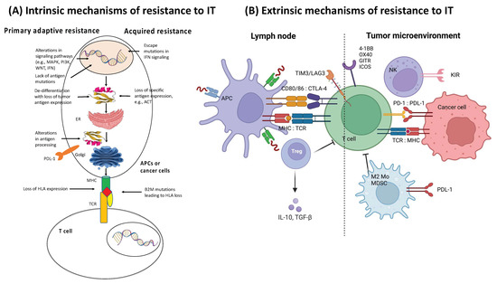
Figure 4.
Mechanisms of resistance to immunotherapy. (A) Intrinsic mechanisms of resistance to immunotherapy. Examples of intrinsic mechanisms of adaptive resistance involve altered signaling pathways, limited mutational burden, de-differentiation of tumor resulting in loss of neoantigen expression, defective antigen processing, constitutive PD-L1 expression, and loss of HLA expression. Examples of intrinsic mechanisms of acquired resistance include loss of antigenic target, loss of HLA expression, and escape mutations in IFN signaling. (B) Extrinsic mechanisms of resistance to ICI therapy (right panel). Examples of extrinsic mechanisms of resistance involve upregulated or constitutive immune checkpoint expression, immunosuppressive cytokine release (e.g., CSF-1, TGFβ, adenosine) within the TME, T cell exhaustion and phenotypic switching, as well as elevation of immunosuppressive cell populations (e.g., Treg, MDSC, and M2 macrophages). Adapted from ref. [34]. Created with BioRender.com (URL (accessed on 18 November 2022). Definitions of abbreviations are found in the abbreviations list at the end of the text.
Several other resistance mechanisms involving both tumor cell-intrinsic and tumor cell-extrinsic sources have been described (Figure 3 and Figure 4 and Table 3) [34]. In the case of tumor cell-intrinsic mechanisms, a lack of neoantigen development, impaired antigen presentation, and other primary factors contribute to the resistance to immunotherapy [130,131,132,133,134,135,136]. Tumor cell-extrinsic mechanisms encompass increased recruitment and activity of inhibitory immune cells within the TME and upregulation of LAG-3 and TIM-3 [16,47,137,138,139,140].

Table 3.
Genomic correlations with response and resistance based on primary location.
5.2. Immune Resistance Mechanisms in Melanoma
As illustrated in Figure 2, Figure 3, Figure 4 and Figure 5 and Table 3, resistance to ICI therapy is one of the most significant challenges to achieving a durable tumor response in many types of cancers [16,24,34,182,183].
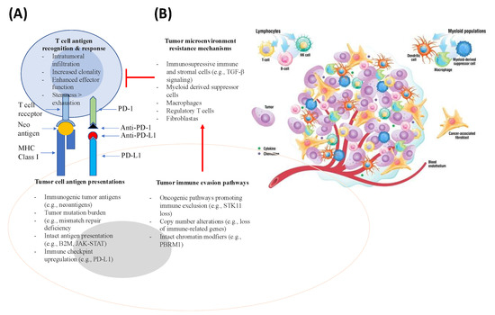
Figure 5.
Genomic correlations with response and resistance to ICI therapy within the immune TME. The panel (A) represents correlations with response, focusing on antigen presentation and recognition. The panel (B) represents resistance pathways that promote tumor immune evasion mechanisms that induce immunosuppressive cells, leading to the inhibition of T cell-mediated anti-tumor response (right side). Adapted from [113]. The panel (B) also illustrates the TME, which consists of cellular and non-cellular components. The cellular component consists of cancer cells, endothelial cells, pericytes, carcinoma-associated fibroblasts, and immune cells. The immune compartment comprises many immune cell populations (e.g., T cells, B cells, natural killer cells, tumor-associated macrophages, myeloid-derived suppressor cells, and dendritic cells). The non-cellular component of the TME, on the other hand, is represented by the extracellular matrix and functions as a scaffold. Components of the TME interact via the extracellular matrix, cell-cell contacts, and through the release of cytokines, chemokines, extracellular vesicles, and others. Adapted from ref. [113,184,185]. Definitions of abbreviations are found in the abbreviations list at the end of the text.
Several mechanisms, including primary (lack of tumor response to initial immunotherapy) and secondary/acquired resistance to cancer immunotherapy (initial response to tumor followed by a lack of response) have been described as the major resistance mechanisms to ICI therapy [16] (Figure 2, Figure 3, Figure 4 and Figure 5 and Table 3).
As for melanoma, Huang et al. [22] demonstrated that several key mechanisms are involved in the immune resistance to ICI, such as immune tolerance, T cell exhaustion, immune cell-mediated immunosuppression, expression of immune checkpoint ligands, and tumor escape.
Immune tolerance describes the lack of response by the immune system to substances or antigens that have the potential to induce an immune response. Immune tolerance to an individual’s own antigens occurs through both central and peripheral tolerance mechanisms. While central tolerance occurs via thymic deletion of high-affinity auto-reactive T cells, peripheral tolerance is maintained by other mechanisms (e.g., suppression by Treg cells and anergy) and induced by many mechanisms, including sub-optimal T cell co-stimulation, deletion via apoptosis, or conversion into Treg cells. The dose of antigen and TCR affinity are considered to be the major drivers of these mechanisms [22]. Notably, both the lymph node and tumor environments blunt T-cell effector functions and offer a rationale for the failure of tumor-specific responses to effectively counter tumor progression [186].
T cell exhaustion is a specific T cell differentiation process mediated by chronic antigen stimulation, which leads to increased expression of co-inhibitory immune receptors that are presumed to decrease chronic TCR signaling and regulate activation-induced cell death.
In this state of simultaneous TCR stimulation and co-inhibitory pathway stimulation, exhausted T cells (TEX cells) exhibit reduced effector functions (i.e., cytokine production and proliferative potential), but can survive in the hostile TME. Notably, T cell exhaustion appears to be a dynamic and progressive process that includes intermediate reversible states more permissive to stimulation by ICI.
Cell-mediated immunosuppression involves immunosuppressive cells, including MDSCs, Treg cells, and tolerogenic DCs, instructing effector T and B cells to not respond to positive immune stimuli.
Upregulation of immune checkpoint ligands PD-L1 and PD-L2 is often seen in melanoma and other cancers in response to robust inflammatory signals as a homeostatic mechanism adopted by cancer cells to shield themselves from immune attack. Interaction of PD-L1 and PD-L2 ligands through binding to PD-1 receptors expressed on tumor-specific T cells initiate a negative signaling process downstream of PD-1, which reduces T cell activation and impairs tumor-killing function [11]. As a consequence, expression of PD-L1 or PD-L2 in melanoma cells can neutralize the positive T cell signals mediated by the MHC-I and MHC-II antigen presentation process.
Tumor escape is a mechanism that cancer cells utilize to escape from anti-tumor immunity and immune surveillance. Tumor escape can be mediated by tumor-extrinsic mechanisms in the TME and by the tumor itself, which can evolve to evade immune recognition. Under strong immune selective pressure, heterogeneous tumor cells can result in clonal evolution, selection, and enrichment of specific tumor cells that can evade immune recognition, leading to immunotherapy resistance. The evasion of immune recognition by these treatment-resistant cells can take place via inactivation of antigen-presentation processes (e.g., B2M, HLA, TAP, etc.) and/or IFN-γ-response genes (e.g., JAK1, JAK2).
6. Therapeutic Strategies to Turn “Cold Tumors” into “Hot Tumors”
Several strategies have been investigated to elucidate the mechanisms underlying how T cells are driven into “hot tumors” in order to improve the efficacy of ICI therapy (Figure 6 and Table 4) [124]. Several clinical trials have tested these novel therapeutic modalities as interventions in combination with ICI to overcome ICI monotherapy resistance and attempt to turn “cold tumors’ into “hot tumors” (Table 5).
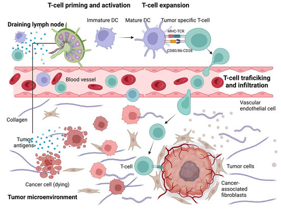
Figure 6.
Approaches to turn a “cold tumor” into a “hot tumor”. Some representative therapeutic strategies to increase T cell infiltration and improve the efficacy of ICI. T cell priming and activation: Oncolytic viruses, local thermal ablation therapy (e.g., radiofrequency ablation), chemotherapy, and radiotherapy are all capable of inducing immunogenic cell death to promote T cell priming and activation. Local administration of immune adjuvants, such as TLR agonists, promotes the activation of DCs. Epigenetic modification inhibitors can promote T cell priming by increasing the expression of tumor antigens and by restoring antigen processing and presentation mechanisms. T cell expansion: Cancer vaccines and adoptive cellular therapies, such as CAR-T cells, can promote the expansion of tumor-specific T cells. T cell trafficking and infiltration: Intrinsic oncogenic pathway inhibitors, antiangiogenic therapies, TGFβ inhibitors, CXCR4 inhibitors, and epigenetic modification inhibitors can promote T cell trafficking and enable T cells to infiltrate the tumor more effectively. Adapted from ref. [124]. Created with Briorender.com (URL (accessed on 18 November 2022). Definitions of abbreviations are found in the abbreviations list at the end of the text.

Table 4.
Therapeutic strategies to turn “cold tumors” into “hot tumors”.

Table 5.
Representative examples of ICI therapy in combination with other drugs to overcome resistance.
Grasso et al. [93] showed that a robust anti-tumor immune response relies on the interplay of key factors that can be modulated with innovative interventions.
Recent preclinical and clinical findings have provided insight into the immunological implications of canonical cancer signaling pathways (e.g., WNT-beta-catenin signaling, cell cycle regulatory signaling, mitogen-activated protein kinase signaling, and pathways activated by loss of the tumor suppressor phosphoinositide phosphatase PTEN), thus providing new opportunities for the development of new treatments for those patients who do not respond to ICI monotherapies [218].
Combined therapeutic strategies from preclinical [115,117,219] and clinical studies [14] have shown that anti-PD-1 plus anti-CTLA-4 (nivolumab and ipilimumab) treatments elicit stronger immune stimulation in stage IV melanoma than monotherapy alone, resulting in a favorable anti-tumor immune response. Response to ICI, either following anti-CTLA-4 monotherapy or in combination with anti-PD-1 therapy, triggers a robust T cell response that generated an appreciable antitumor response [218].
Another approach to turn “cold tumors” into “hot tumors” is through the intra-tumoral delivery of oncolytic viruses or Toll-like receptor agonists capable of inducing intra-tumoral interferon production, which triggers the pattern recognition pathways with consequent boosting of the anti-PD1 immune response rate [187,190,220].
Recently, different approaches have been used to boost the response to ICI. Activation of the STING pathway [221], inhibition of immune suppressive factors (e.g., WNT signaling or the adenosine pathway) [111,209,222,223], as well as the release of other immune checkpoints (e.g., LAG-3, TIM-3, or TIGIT, etc.) in T cells [111] have been explored, but no favorable clinical outcomes have yet been reported in patients.
Altogether, these new therapeutic strategies provide new opportunities for cancer immunotherapy for patients who do not respond to ICI [111].
7. Lessons Learned: ICI Therapies in Melanoma
Huang et al. [22] reported that several factors, including the immune TME, tumor-associated immune cells, and different host factors, contribute to the ICI resistance. Melanoma resistance has armed us with a handful of information that can be applied to other types of cancers, such as: (1) the ability to present cancer antigens through MHC-I and elevated TMB; (2) tumor-antigen-specific T cells play a crucial role in the response to ICI; (3) reactivation of terminally exhausted T cells could be considered a biomarker for PD-1 blockade, which is detectable as early as one week after ICI dosing; (4) melanoma immunosuppressive mechanisms are complicated and need additional research to remodel their interaction, cooperation, and dynamics during tumor progression and in immunotherapy resistance; and (5) Treg cells are emerging as a key mechanism of resistance to PD-1 blockade, but not necessarily CTLA-4 blockade; (6) emerging neoadjuvant immunotherapy trials are anticipated to provide new insight into pharmacodynamic immune responses and advance the development of rational immunotherapy and neoadjuvant combination regimens while avoiding toxicity and significantly improving patient management; (7) longitudinal assessment of pre- and on-treatment patient specimens is required to determine prognostic vs. predictive use of immune and other parameters, including genomic parameters correlating with patient outcomes, and deduce their biologic role in response to ICI therapy based on their modulation during treatment; and last but not least (8) melanoma-specific oncogenic programs supporting metabolic plasticity and fitness, together with clinical and preclinical evidence of differential activity of ICI therapy depending on the tumor metabolic state, should provide new research opportunities to evaluate these relationships as potential biomarkers for patient stratification and treatment allocation, and formulate novel precision-medicine combinations depending on metabolic and immune therapies.
8. Conclusions and Future Perspectives
Even with a high level of immunogenicity, metastatic melanoma grows and spreads rapidly via escape mutations and immunosuppression. Although combination ICI therapies targeting CTLA-4 and PD-1 can efficiently target some ICI-resistant mechanisms by improving T cell priming, re-activating PD-1 high CD8 + exhausted T cells, and reversing Treg suppression, many patients still do not derive durable clinical benefit.
Therefore, with the availability of novel immunotherapeutic agents, the mechanism of single-agent therapies needs to be better characterized in order to guide effective rational ICI combinations. In addition, since the immunologic effects of ICI therapy occur early, we need to focus on these early events to identify specific biomarkers, mechanisms of resistance, and neoadjuvant therapies.
Furthermore, toxicity from current and new ICI combinations remains a critical step to be addressed [224]. Because of concerns regarding adverse events from current and new ICI combinations, insight gained from molecular mediators of immune toxicity (e.g., antibody- vs. T cell-mediated) resulting from combination ICI therapies would help in avoiding these side effects and the development of novel combination ICI therapies to other cancers.
As a result, new efficacy and toxicity data deriving from different ICI therapies across various tumor types will help to characterize key parameters for predicting ICI response, thus limiting toxicity and enhancing therapeutic decision processes to overcome ICI resistance. In fact, new efficacy and toxicity data from many ICI therapies across various tumor types are helping in the characterization of key parameters for predicting response, limiting toxicity, and informing therapeutic decisions to overcome resistance.
Furthermore, the identification of novel genomic correlations with resistance to ICI requires well-annotated data from diverse patient cohorts and tumor histology to detect rare response-associated variants [159,225]. With a greater focus on detailed genomics as well as epigenomics, proteomics, and metabolomics, microbiome and biomarker studies will better characterize the relationships among host immunity, tumor biology, and mechanisms of resistance and response to ICI. Ultimately, molecular and clinical data from these studies will have to be adequately integrated and blended into preclinical studies.
For this reason, systems biology, computational biology, biostatistics, as well as machine learning and artificial intelligence approaches must be coordinated to integrate and help interpret this large dataset, thus translating the findings into actionable novel biomarkers that capture the complexity of multiple alterations affecting response and therapeutic success.
In addition, several other key challenges face ICI therapy that must be addressed to move the field ahead [226], including: (1) development of preclinical models that translate to human immunity; (2) identification and validation of the dominant drivers involved in cancer immunity; (3) deeper insights into organ-specific immune TME; (4) exploration of differences in molecular and cellular drivers between primary and secondary immune escape; (5) characterization of the benefit of endogenous vs. synthetic immunity; (6) investigation of ICI therapy combinations with other drugs (Figure 6, Table 4 and Table 5) in early-phase clinical studies; (7) investigation of steroids and immune suppression on immunotherapy and autoimmune adverse events and toxicities; (8) boosting personalized medicine approaches by using composite biomarkers; (9) developing and implementing refined regulatory endpoints for immunotherapy; and (10) optimizing durable survival with a combination of multi-agent immunotherapy regimens [226].
Consequently, all of these efforts will result in a better understanding of the mechanisms involved in response and resistance to ICI, hence facilitating the development of biomarkers and novel therapies.
In conclusion, these efforts should result in a deeper quantitative and conceptual understanding of the mechanisms involved in ICI response and resistance, thus facilitating the development of rational biomarkers and therapies.
Author Contributions
Conceptualization, A.A.S. and C.C.; methodology, A.A.S. and C.C.; software, A.A.S.; validation, A.A.S. and C.C.; formal analysis, A.A.S. and C.C.; investigation, A.A.S. and C.C.; resources, A.A.S. and C.C.; data curation, A.A.S.; writing—original draft preparation, A.A.S.; writing—review and editing, A.A.S. and C.C.; visualization, A.A.S.; supervision, A.A.S. and C.C.; project administration, A.A.S. and C.C. All authors have read and agreed to the published version of the manuscript.
Funding
No research funding received from specific grant from any funding agency in the public, commercial, or not-for-profit sectors was used in the preparation of this review article.
Institutional Review Board Statement
Not applicable.
Data Availability Statement
The authors grant the publisher the sole and exclusive license of the full copyright in the Contribution, which license the publisher hereby accepts.
Acknowledgments
We thank the Legorreta Cancer Center at Brown University for providing the environment for the preparation of this review article. The contents of this manuscript are solely the responsibility of the authors.
Conflicts of Interest
The authors declare no conflict of interest.
Abbreviations
| A2aR | adenosine A2a receptor |
| ADO | adenosine |
| AE | adverse event |
| APC | antigen-presenting cell |
| ATP | adenosine triphosphate |
| B2M | beta-2-microglobulin |
| B7RP1 | B7-related protein 1 |
| BATF3 | basic leucine zipper ATF-like transcription factor 3 |
| BTLA | B and T lymphocyte attenuator |
| CAF | cancer-associated fibroblast |
| CCR4 | C-C chemokine receptor type 4 |
| CD40L | CD40 ligand |
| CDN | cyclic dinucleotide |
| CRT | Calreticulin |
| CSF-1 | colony stimulating factor 1 |
| CTCs | circulating tumor cells |
| ctDNA | circulating tumor DNA |
| CTLA-4 | cytotoxic T-lymphocyte-associated protein 4 |
| CXCL/CCL | chemokine motif ligands; |
| dMMR | defective mismatch repair |
| DNMT | DNA methyltransferase |
| ECM | extracellular matrix |
| EGFR | epidermal growth factor receptor |
| ER | endoplasmic reticulum |
| ETBR | endothelin B receptor |
| EZH2 | enhancer of zeste homolog 2 |
| FDA | Food and Drug Administration |
| FLT3L | Fms-like tyrosine kinase 3 ligand |
| GAL9 | galectin 9 |
| GITR | glucocorticoid-induced tumor necrosis factor receptor |
| GM-CSF | granulocyte-macrophage colony-stimulating factor |
| HDAC | histone deacetylase |
| HER2 | human epidermal growth factor receptor 2 |
| HEV | high endothelial venule |
| HLA | human leukocyte antigen |
| HMGB1 | high mobility family protein B1 |
| HR | hormone receptor positive |
| HVEM | herpesvirus entry mediator |
| ICAM | intercellular adhesion molecule |
| ICOS | inducible T cell co-stimulator |
| IDO | indoleamine 2,3-dioxygenase |
| IFN | interferon |
| IL | interleukin |
| ITT | intention-to-treat |
| KIR | killer cell immunoglobulin-like receptors |
| LAG-3 | lymphocyte activation gene 3 |
| LFA1 | lymphocyte function-associated antigen-1 |
| mAb | monoclonal antibody |
| M-CSF | macrophage colony-stimulating factor |
| MDSC | myeloid-derived suppressor cell |
| MHC | major histocompatibility complex |
| MMR | mismatch repair |
| MSI | microsatellite instability |
| MSI-H | microsatellite instability-high |
| MɸII | type II macrophage |
| NK | natural killer |
| OS | overall survival |
| OX40L | OX40 ligand |
| PD-1 | programmed cell death protein-1 |
| PD-L1 | programmed death-ligand 1 |
| PD-L2 | programmed death-ligand 2 |
| PFS | progression-free survival |
| PI3K | phosphatidyl-inositol 3-kinase |
| PTEN | phosphatase and tensin homolog |
| STC1 | stanniocalcin 1 |
| TAM | tumor-associated macrophage |
| TAP | transporter associated with antigen processing |
| TCR | T-cell receptor |
| TGF | transforming growth factor |
| TGFβ | transforming growth factor-beta |
| TILs | tumor-infiltrating lymphocytes |
| TIM-3 | T cell immunoglobulin and mucin-domain containing-3 |
| TLR | Toll-like receptor |
| TLS | tertiary lymphoid structure |
| TMB | tumor mutation burden |
| TME | tumor microenvironment |
| TNF | tumor necrosis factor |
| TNFRSF14 | tumor necrosis factor receptor superfamily member 14 |
| Treg | T-regulatory cell |
| VCAM | vascular cell adhesion molecule |
| VEGF | vascular endothelial growth factor |
| VISTA | V-domain Ig suppressor of T cell activation |
References
- Sharma, P.; Wagner, K.; Wolchok, J.D.; Allison, J.P. Novel cancer immunotherapy agents with survival benefit: Recent successes and next steps. Nat. Rev. Cancer 2011, 11, 805–812. [Google Scholar] [CrossRef] [PubMed]
- Sharma, P.; Allison, J.P. Immune checkpoint targeting in cancer therapy: Toward combination strategies with curative potential. Cell 2015, 161, 205–214. [Google Scholar] [CrossRef] [PubMed]
- Pardoll, D.M. The blockade of immune checkpoints in cancer immunotherapy. Nat. Rev. Cancer 2012, 12, 252–264. [Google Scholar] [CrossRef] [PubMed]
- Darvin, P.; Toor, S.M.; Sasidharan Nair, V.; Elkord, E. Immune checkpoint inhibitors: Recent progress and potential biomarkers. Exp. Mol. Med. 2018, 50, 1–11. [Google Scholar] [CrossRef]
- Siegel, R.L.; Miller, K.D.; Jemal, A. Cancer statistics, 2018. CA Cancer J. Clin. 2018, 68, 7–30. [Google Scholar] [CrossRef] [PubMed]
- Ayers, M.; Lunceford, J.; Nebozhyn, M.; Murphy, E.; Loboda, A.; Albright, A.; Cheng, J.; Kang, S.P.; Ebbinghaus, S.; Yearley, J.; et al. Relationship between immune gene signatures and clinical response to PD-1 blockade with pembrolizumab (MK-3475) in patients with advanced solid tumors. J. ImmunoTherapy Cancer 2015, 3, P80. [Google Scholar] [CrossRef]
- Van Allen, E.M.; Miao, D.; Schilling, B.; Shukla, S.A.; Blank, C.; Zimmer, L.; Sucker, A.; Hillen, U.; Foppen, M.H.G.; Goldinger, S.M.; et al. Genomic correlates of response to CTLA-4 blockade in metastatic melanoma. Science 2015, 350, 207–211. [Google Scholar] [CrossRef]
- Leach, D.R.; Krummel, M.F.; Allison, J.P. Enhancement of antitumor immunity by CTLA-4 blockade. Science 1996, 271, 1734–1736. [Google Scholar] [CrossRef]
- Hodi, F.S.; O’Day, S.J.; McDermott, D.F.; Weber, R.W.; Sosman, J.A.; Haanen, J.B.; Gonzalez, R.; Robert, C.; Schadendorf, D.; Hassel, J.C.; et al. Improved survival with ipilimumab in patients with metastatic melanoma. N. Engl. J. Med. 2010, 363, 711–723. [Google Scholar] [CrossRef]
- Robert, C.; Thomas, L.; Bondarenko, I.; O’Day, S.; Weber, J.; Garbe, C.; Lebbe, C.; Baurain, J.F.; Testori, A.; Grob, J.J.; et al. Ipilimumab plus dacarbazine for previously untreated metastatic melanoma. N. Engl. J. Med. 2011, 364, 2517–2526. [Google Scholar] [CrossRef]
- Pauken, K.E.; Torchia, J.A.; Chaudhri, A.; Sharpe, A.H.; Freeman, G.J. Emerging concepts in PD-1 checkpoint biology. Semin. Immunol. 2021, 52, 101480. [Google Scholar] [CrossRef]
- Schumacher, T.N.; Schreiber, R.D. Neoantigens in cancer immunotherapy. Science 2015, 348, 69–74. [Google Scholar] [CrossRef] [PubMed]
- Zappasodi, R.; Wolchok, J.D.; Merghoub, T. Correction to: Strategies for Predicting Response to Checkpoint Inhibitors. Curr. Hematol. Malig. Rep. 2019, 14, 62. [Google Scholar] [CrossRef] [PubMed]
- Larkin, J.; Chiarion-Sileni, V.; Gonzalez, R.; Grob, J.J.; Rutkowski, P.; Lao, C.D.; Cowey, C.L.; Schadendorf, D.; Wagstaff, J.; Dummer, R.; et al. Five-Year Survival with Combined Nivolumab and Ipilimumab in Advanced Melanoma. N. Engl. J. Med. 2019, 381, 1535–1546. [Google Scholar] [CrossRef]
- Paik, J. Nivolumab Plus Relatlimab: First Approval. Drugs 2022, 82, 925–931. [Google Scholar] [CrossRef] [PubMed]
- Sharma, P.; Hu-Lieskovan, S.; Wargo, J.A.; Ribas, A. Primary, Adaptive, and Acquired Resistance to Cancer Immunotherapy. Cell 2017, 168, 707–723. [Google Scholar] [CrossRef] [PubMed]
- Prieto, P.A.; Yang, J.C.; Sherry, R.M.; Hughes, M.S.; Kammula, U.S.; White, D.E.; Levy, C.L.; Rosenberg, S.A.; Phan, G.Q. CTLA-4 blockade with ipilimumab: Long-term follow-up of 177 patients with metastatic melanoma. Clin. Cancer Res. 2012, 18, 2039–2047. [Google Scholar] [CrossRef] [PubMed]
- Di Giacomo, A.M.; Calabro, L.; Danielli, R.; Fonsatti, E.; Bertocci, E.; Pesce, I.; Fazio, C.; Cutaia, O.; Giannarelli, D.; Miracco, C.; et al. Long-term survival and immunological parameters in metastatic melanoma patients who responded to ipilimumab 10 mg/kg within an expanded access programme. Cancer Immunol. Immunother. 2013, 62, 1021–1028. [Google Scholar] [CrossRef] [PubMed]
- Gibney, G.T.; Weiner, L.M.; Atkins, M.B. Predictive biomarkers for checkpoint inhibitor-based immunotherapy. Lancet Oncol. 2016, 17, e542–e551. [Google Scholar] [CrossRef]
- Wolchok, J.D.; Chiarion-Sileni, V.; Gonzalez, R.; Grob, J.J.; Rutkowski, P.; Lao, C.D.; Cowey, C.L.; Schadendorf, D.; Wagstaff, J.; Dummer, R.; et al. Long-Term Outcomes With Nivolumab Plus Ipilimumab or Nivolumab Alone Versus Ipilimumab in Patients With Advanced Melanoma. J. Clin. Oncol. 2022, 40, 127–137. [Google Scholar] [CrossRef]
- Carlino, M.S.; Larkin, J.; Long, G.V. Immune checkpoint inhibitors in melanoma. Lancet 2021, 398, 1002–1014. [Google Scholar] [CrossRef] [PubMed]
- Huang, A.C.; Zappasodi, R. A decade of checkpoint blockade immunotherapy in melanoma: Understanding the molecular basis for immune sensitivity and resistance. Nat. Immunol. 2022, 23, 660–670. [Google Scholar] [CrossRef] [PubMed]
- Gajewski, T.F.; Schreiber, H.; Fu, Y.X. Innate and adaptive immune cells in the tumor microenvironment. Nat. Immunol. 2013, 14, 1014–1022. [Google Scholar] [CrossRef] [PubMed]
- Pitt, J.M.; Vétizou, M.; Daillère, R.; Roberti, M.P.; Yamazaki, T.; Routy, B.; Lepage, P.; Boneca, I.G.; Chamaillard, M.; Kroemer, G.; et al. Resistance Mechanisms to Immune-Checkpoint Blockade in Cancer: Tumor-Intrinsic and -Extrinsic Factors. Immunity 2016, 44, 1255–1269. [Google Scholar] [CrossRef]
- Riaz, N.; Havel, J.J.; Makarov, V.; Desrichard, A.; Urba, W.J.; Sims, J.S.; Hodi, F.S.; Martin-Algarra, S.; Mandal, R.; Sharfman, W.H.; et al. Tumor and Microenvironment Evolution during Immunotherapy with Nivolumab. Cell 2017, 171, 934–949.e16. [Google Scholar] [CrossRef]
- Zhang, N.; Song, J.; Liu, Y.; Liu, M.; Zhang, L.; Sheng, D.; Deng, L.; Yi, H.; Wu, M.; Zheng, Y.; et al. Photothermal therapy mediated by phase-transformation nanoparticles facilitates delivery of anti-PD1 antibody and synergizes with antitumor immunotherapy for melanoma. J. Control. Release 2019, 306, 15–28. [Google Scholar] [CrossRef]
- Khong, H.T.; Restifo, N.P. Natural selection of tumor variants in the generation of "tumor escape" phenotypes. Nat. Immunol. 2002, 3, 999–1005. [Google Scholar] [CrossRef]
- Pitt, J.M.; Marabelle, A.; Eggermont, A.; Soria, J.C.; Kroemer, G.; Zitvogel, L. Targeting the tumor microenvironment: Removing obstruction to anticancer immune responses and immunotherapy. Ann. Oncol. 2016, 27, 1482–1492. [Google Scholar] [CrossRef]
- Restifo, N.P.; Smyth, M.J.; Snyder, A. Acquired resistance to immunotherapy and future challenges. Nat. Rev. Cancer 2016, 16, 121–126. [Google Scholar] [CrossRef]
- Hugo, W.; Zaretsky, J.M.; Sun, L.; Song, C.; Moreno, B.H.; Hu-Lieskovan, S.; Berent-Maoz, B.; Pang, J.; Chmielowski, B.; Cherry, G.; et al. Genomic and Transcriptomic Features of Response to Anti-PD-1 Therapy in Metastatic Melanoma. Cell 2016, 165, 35–44. [Google Scholar] [CrossRef]
- Iliopoulos, D.; Kavousanaki, M.; Ioannou, M.; Boumpas, D.; Verginis, P. The negative costimulatory molecule PD-1 modulates the balance between immunity and tolerance via miR-21. Eur. J. Immunol. 2011, 41, 1754–1763. [Google Scholar] [CrossRef] [PubMed]
- Han, Y.; Liu, D.; Li, L. PD-1/PD-L1 pathway: Current researches in cancer. Am. J. Cancer Res. 2020, 10, 727–742. [Google Scholar] [PubMed]
- Carlsen, L.; Huntington, K.E.; El-Deiry, W.S. Immunotherapy for Colorectal Cancer: Mechanisms and Predictive Biomarkers. Cancers 2022, 14, 1028. [Google Scholar] [CrossRef] [PubMed]
- Gravbrot, N.; Gilbert-Gard, K.; Mehta, P.; Ghotmi, Y.; Banerjee, M.; Mazis, C.; Sundararajan, S. Therapeutic Monoclonal Antibodies Targeting Immune Checkpoints for the Treatment of Solid Tumors. Antibodies 2019, 8, 51. [Google Scholar] [CrossRef] [PubMed]
- Fan, J.; Shang, D.; Han, B.; Song, J.; Chen, H.; Yang, J.M. Adoptive Cell Transfer: Is it a Promising Immunotherapy for Colorectal Cancer? Theranostics 2018, 8, 5784–5800. [Google Scholar] [CrossRef] [PubMed]
- Helmink, B.A.; Reddy, S.M.; Gao, J.; Zhang, S.; Basar, R.; Thakur, R.; Yizhak, K.; Sade-Feldman, M.; Blando, J.; Han, G.; et al. B cells and tertiary lymphoid structures promote immunotherapy response. Nature 2020, 577, 549–555. [Google Scholar] [CrossRef]
- Tarhini, A.; Kudchadkar, R.R. Predictive and on-treatment monitoring biomarkers in advanced melanoma: Moving toward personalized medicine. Cancer Treat Rev. 2018, 71, 8–18. [Google Scholar] [CrossRef]
- Weinstein, D.; Leininger, J.; Hamby, C.; Safai, B. Diagnostic and prognostic biomarkers in melanoma. J. Clin. Aesthet. Dermatol. 2014, 7, 13–24. [Google Scholar]
- Louie, A.D.; Huntington, K.; Carlsen, L.; Zhou, L.; El-Deiry, W.S. Integrating Molecular Biomarker Inputs Into Development and Use of Clinical Cancer Therapeutics. Front. Pharmacol. 2021, 12, 747194. [Google Scholar] [CrossRef]
- Deacon, D.C.; Smith, E.A.; Judson-Torres, R.L. Molecular Biomarkers for Melanoma Screening, Diagnosis and Prognosis: Current State and Future Prospects. Front. Med. (Lausanne) 2021, 8, 642380. [Google Scholar] [CrossRef]
- Franzke, A.; Probst-Kepper, M.; Buer, J.; Duensing, S.; Hoffmann, R.; Wittke, F.; Volkenandt, M.; Ganser, A.; Atzpodien, J. Elevated pretreatment serum levels of soluble vascular cell adhesion molecule 1 and lactate dehydrogenase as predictors of survival in cutaneous metastatic malignant melanoma. Br. J. Cancer 1998, 78, 40–45. [Google Scholar] [CrossRef] [PubMed][Green Version]
- Long, G.V.; Eroglu, Z.; Infante, J.; Patel, S.; Daud, A.; Johnson, D.B.; Gonzalez, R.; Kefford, R.; Hamid, O.; Schuchter, L.; et al. Long-Term Outcomes in Patients With BRAF V600-Mutant Metastatic Melanoma Who Received Dabrafenib Combined With Trametinib. J. Clin. Oncol. 2018, 36, 667–673. [Google Scholar] [CrossRef] [PubMed]
- Long, G.V.; Grob, J.J.; Nathan, P.; Ribas, A.; Robert, C.; Schadendorf, D.; Lane, S.R.; Mak, C.; Legenne, P.; Flaherty, K.T.; et al. Factors predictive of response, disease progression, and overall survival after dabrafenib and trametinib combination treatment: A pooled analysis of individual patient data from randomised trials. Lancet Oncol. 2016, 17, 1743–1754. [Google Scholar] [CrossRef]
- Larkin, J.M.G.; McArthur, G.A.; Ribas, A.; Ascierto, P.A.; Gallo, J.D.; Rooney, I.A.; Chang, I.; Dréno, B. Clinical predictors of response for coBRIM: A phase III study of cobimetinib (C) in combination with vemurafenib (V) in advanced BRAF-mutated melanoma (MM). J. Clin. Oncol. 2016, 34, 9528. [Google Scholar] [CrossRef]
- Long, G.V.; Blank, C.; Ribas, A.; Mortier, L.; Carlino, M.S.; Lotem, M.; Lorigan, P.; Neyns, B.; Petrella, T.M.; Puzanov, I.; et al. 1141 Impact of baseline serum lactate dehydrogenase concentration on the efficacy of pembrolizumab and ipilimumab in patients with advanced melanoma: Data from KEYNOTE-006. Eur. J. Cancer 2017, 72, s122–s123. [Google Scholar] [CrossRef]
- Roh, W.; Chen, P.L.; Reuben, A.; Spencer, C.N.; Prieto, P.A.; Miller, J.P.; Gopalakrishnan, V.; Wang, F.; Cooper, Z.A.; Reddy, S.M.; et al. Integrated molecular analysis of tumor biopsies on sequential CTLA-4 and PD-1 blockade reveals markers of response and resistance. Sci. Transl. Med. 2017, 9, eaah3560. [Google Scholar] [CrossRef]
- Zager, J.S.; Gastman, B.R.; Leachman, S.; Gonzalez, R.C.; Fleming, M.D.; Ferris, L.K.; Ho, J.; Miller, A.R.; Cook, R.W.; Covington, K.R.; et al. Performance of a prognostic 31-gene expression profile in an independent cohort of 523 cutaneous melanoma patients. BMC Cancer 2018, 18, 130. [Google Scholar] [CrossRef]
- Hauschild, A.; Michaelsen, J.; Brenner, W.; Glaser, R.; Henze, E.; Christophers, E. Prognostic significance of serum S100B detection compared with routine blood parameters in advanced metastatic melanoma patients. Melanoma Res. 1999, 9, 155–161. [Google Scholar] [CrossRef]
- Hauschild, A.; Engel, G.; Brenner, W.; Glaser, R.; Monig, H.; Henze, E.; Christophers, E. S100B protein detection in serum is a significant prognostic factor in metastatic melanoma. Oncology 1999, 56, 338–344. [Google Scholar] [CrossRef]
- Tarhini, A.A.; Stuckert, J.; Lee, S.; Sander, C.; Kirkwood, J.M. Prognostic significance of serum S100B protein in high-risk surgically resected melanoma patients participating in Intergroup Trial ECOG 1694. J. Clin. Oncol. 2009, 27, 38–44. [Google Scholar] [CrossRef]
- Gerami, P.; Cook, R.W.; Russell, M.C.; Wilkinson, J.; Amaria, R.N.; Gonzalez, R.; Lyle, S.; Jackson, G.L.; Greisinger, A.J.; Johnson, C.E.; et al. Gene expression profiling for molecular staging of cutaneous melanoma in patients undergoing sentinel lymph node biopsy. J. Am. Acad. Dermatol. 2015, 72, 780–785.e3. [Google Scholar] [CrossRef] [PubMed]
- Gerami, P.; Cook, R.W.; Wilkinson, J.; Russell, M.C.; Dhillon, N.; Amaria, R.N.; Gonzalez, R.; Lyle, S.; Johnson, C.E.; Oelschlager, K.M.; et al. Development of a prognostic genetic signature to predict the metastatic risk associated with cutaneous melanoma. Clin. Cancer Res. 2015, 21, 175–183. [Google Scholar] [CrossRef] [PubMed]
- Chapman, P.B.; Hauschild, A.; Robert, C.; Haanen, J.B.; Ascierto, P.; Larkin, J.; Dummer, R.; Garbe, C.; Testori, A.; Maio, M.; et al. Improved survival with vemurafenib in melanoma with BRAF V600E mutation. N. Engl. J. Med. 2011, 364, 2507–2516. [Google Scholar] [CrossRef] [PubMed]
- Hauschild, A.; Grob, J.J.; Demidov, L.V.; Jouary, T.; Gutzmer, R.; Millward, M.; Rutkowski, P.; Blank, C.U.; Miller, W.H., Jr.; Kaempgen, E.; et al. Dabrafenib in BRAF-mutated metastatic melanoma: A multicentre, open-label, phase 3 randomised controlled trial. Lancet 2012, 380, 358–365. [Google Scholar] [CrossRef] [PubMed]
- Long, G.V.; Stroyakovskiy, D.; Gogas, H.; Levchenko, E.; de Braud, F.; Larkin, J.; Garbe, C.; Jouary, T.; Hauschild, A.; Grob, J.J.; et al. Dabrafenib and trametinib versus dabrafenib and placebo for Val600 BRAF-mutant melanoma: A multicentre, double-blind, phase 3 randomised controlled trial. Lancet 2015, 386, 444–451. [Google Scholar] [CrossRef]
- Ascierto, P.A.; McArthur, G.A.; Dréno, B.; Atkinson, V.; Liszkay, G.; Di Giacomo, A.M.; Mandalà, M.; Demidov, L.; Stroyakovskiy, D.; Thomas, L.; et al. Cobimetinib combined with vemurafenib in advanced BRAF(V600)-mutant melanoma (coBRIM): Updated efficacy results from a randomised, double-blind, phase 3 trial. Lancet Oncol. 2016, 17, 1248–1260. [Google Scholar] [CrossRef]
- Dummer, R.; Ascierto, P.A.; Gogas, H.J.; Arance, A.; Mandala, M.; Liszkay, G.; Garbe, C.; Schadendorf, D.; Krajsova, I.; Gutzmer, R.; et al. Encorafenib plus binimetinib versus vemurafenib or encorafenib in patients with BRAF-mutant melanoma (COLUMBUS): A multicentre, open-label, randomised phase 3 trial. Lancet Oncol. 2018, 19, 603–615. [Google Scholar] [CrossRef]
- Robert, C.; Karaszewska, B.; Schachter, J.; Rutkowski, P.; Mackiewicz, A.; Stroiakovski, D.; Lichinitser, M.; Dummer, R.; Grange, F.; Mortier, L.; et al. Improved overall survival in melanoma with combined dabrafenib and trametinib. N. Engl. J. Med. 2015, 372, 30–39. [Google Scholar] [CrossRef]
- Genomic Classification of Cutaneous Melanoma. Cell 2015, 161, 1681–1696. [CrossRef]
- Dummer, R.; Schadendorf, D.; Ascierto, P.A.; Arance, A.; Dutriaux, C.; Di Giacomo, A.M.; Rutkowski, P.; Del Vecchio, M.; Gutzmer, R.; Mandala, M.; et al. Binimetinib versus dacarbazine in patients with advanced NRAS-mutant melanoma (NEMO): A multicentre, open-label, randomised, phase 3 trial. Lancet Oncol. 2017, 18, 435–445. [Google Scholar] [CrossRef]
- Lebbé, C.; Dutriaux, C.; Lesimple, T.; Kruit, W.; Kerger, J.; Thomas, L.; Guillot, B.; Braud, F.; Garbe, C.; Grob, J.J.; et al. Pimasertib Versus Dacarbazine in Patients With Unresectable NRAS-Mutated Cutaneous Melanoma: Phase II, Randomized, Controlled Trial with Crossover. Cancers 2020, 12, 1727. [Google Scholar] [CrossRef] [PubMed]
- Maertens, O.; Johnson, B.; Hollstein, P.; Frederick, D.T.; Cooper, Z.A.; Messiaen, L.; Bronson, R.T.; McMahon, M.; Granter, S.; Flaherty, K.; et al. Elucidating distinct roles for NF1 in melanomagenesis. Cancer Discov. 2013, 3, 338–349. [Google Scholar] [CrossRef] [PubMed]
- Guo, J.; Si, L.; Kong, Y.; Flaherty, K.T.; Xu, X.; Zhu, Y.; Corless, C.L.; Li, L.; Li, H.; Sheng, X.; et al. Phase II, open-label, single-arm trial of imatinib mesylate in patients with metastatic melanoma harboring c-Kit mutation or amplification. J. Clin. Oncol. 2011, 29, 2904–2909. [Google Scholar] [CrossRef] [PubMed]
- Du, K.; Wei, S.; Wei, Z.; Frederick, D.T.; Miao, B.; Moll, T.; Tian, T.; Sugarman, E.; Gabrilovich, D.I.; Sullivan, R.J.; et al. Pathway signatures derived from on-treatment tumor specimens predict response to anti-PD1 blockade in metastatic melanoma. Nat. Commun. 2021, 12, 6023. [Google Scholar] [CrossRef]
- Snyder, A.; Makarov, V.; Merghoub, T.; Yuan, J.; Zaretsky, J.M.; Desrichard, A.; Walsh, L.A.; Postow, M.A.; Wong, P.; Ho, T.S.; et al. Genetic basis for clinical response to CTLA-4 blockade in melanoma. N. Engl. J. Med. 2014, 371, 2189–2199. [Google Scholar] [CrossRef]
- Rizvi, N.A.; Hellmann, M.D.; Snyder, A.; Kvistborg, P.; Makarov, V.; Havel, J.J.; Lee, W.; Yuan, J.; Wong, P.; Ho, T.S.; et al. Cancer immunology. Mutational landscape determines sensitivity to PD-1 blockade in non-small cell lung cancer. Science 2015, 348, 124–128. [Google Scholar] [CrossRef]
- Cristescu, R.; Mogg, R.; Ayers, M.; Albright, A.; Murphy, E.; Yearley, J.; Sher, X.; Liu, X.Q.; Lu, H.; Nebozhyn, M.; et al. Pan-tumor genomic biomarkers for PD-1 checkpoint blockade-based immunotherapy. Science 2018, 362, eaar3593. [Google Scholar] [CrossRef]
- Topalian, S.L.; Taube, J.M.; Anders, R.A.; Pardoll, D.M. Mechanism-driven biomarkers to guide immune checkpoint blockade in cancer therapy. Nat. Rev. Cancer 2016, 16, 275–287. [Google Scholar] [CrossRef]
- McGranahan, N.; Furness, A.J.; Rosenthal, R.; Ramskov, S.; Lyngaa, R.; Saini, S.K.; Jamal-Hanjani, M.; Wilson, G.A.; Birkbak, N.J.; Hiley, C.T.; et al. Clonal neoantigens elicit T cell immunoreactivity and sensitivity to immune checkpoint blockade. Science 2016, 351, 1463–1469. [Google Scholar] [CrossRef]
- Chowell, D.; Morris, L.G.T.; Grigg, C.M.; Weber, J.K.; Samstein, R.M.; Makarov, V.; Kuo, F.; Kendall, S.M.; Requena, D.; Riaz, N.; et al. Patient HLA class I genotype influences cancer response to checkpoint blockade immunotherapy. Science 2018, 359, 582–587. [Google Scholar] [CrossRef]
- Paulson, K.G.; Voillet, V.; McAfee, M.S.; Hunter, D.S.; Wagener, F.D.; Perdicchio, M.; Valente, W.J.; Koelle, S.J.; Church, C.D.; Vandeven, N.; et al. Acquired cancer resistance to combination immunotherapy from transcriptional loss of class I HLA. Nat. Commun. 2018, 9, 3868. [Google Scholar] [CrossRef] [PubMed]
- Rooney, M.S.; Shukla, S.A.; Wu, C.J.; Getz, G.; Hacohen, N. Molecular and genetic properties of tumors associated with local immune cytolytic activity. Cell 2015, 160, 48–61. [Google Scholar] [CrossRef] [PubMed]
- Davoli, T.; Uno, H.; Wooten, E.C.; Elledge, S.J. Tumor aneuploidy correlates with markers of immune evasion and with reduced response to immunotherapy. Science 2017, 355, eaaf8399. [Google Scholar] [CrossRef] [PubMed]
- Carter, J.A.; Gilbo, P.; Atwal, G.S. IMPRES does not reproducibly predict response to immune checkpoint blockade therapy in metastatic melanoma. Nat. Med. 2019, 25, 1833–1835. [Google Scholar] [CrossRef]
- Auslander, N.; Lee, J.S.; Ruppin, E. Reply to: ‘IMPRES does not reproducibly predict response to immune checkpoint blockade therapy in metastatic melanoma’. Nat. Med. 2019, 25, 1836–1838. [Google Scholar] [CrossRef]
- Auslander, N.; Zhang, G.; Lee, J.S.; Frederick, D.T.; Miao, B.; Moll, T.; Tian, T.; Wei, Z.; Madan, S.; Sullivan, R.J.; et al. Robust prediction of response to immune checkpoint blockade therapy in metastatic melanoma. Nat. Med. 2018, 24, 1545–1549. [Google Scholar] [CrossRef]
- Xiao, X.; Xu, C.; Yang, W.; Yu, R. Inconsistent prediction capability of ImmuneCells.Sig across different RNA-seq datasets. Nat. Commun. 2021, 12, 4167. [Google Scholar] [CrossRef]
- Xiong, D.; Wang, Y.; You, M. Reply to: "Inconsistent prediction capability of ImmuneCells.Sig across different RNA-seq datasets". Nat. Commun. 2021, 12, 4168. [Google Scholar] [CrossRef]
- Xiong, D.; Wang, Y.; You, M. A gene expression signature of TREM2(hi) macrophages and gammadelta T cells predicts immunotherapy response. Nat. Commun. 2020, 11, 5084. [Google Scholar] [CrossRef]
- Chen, P.L.; Roh, W.; Reuben, A.; Cooper, Z.A.; Spencer, C.N.; Prieto, P.A.; Miller, J.P.; Bassett, R.L.; Gopalakrishnan, V.; Wani, K.; et al. Analysis of Immune Signatures in Longitudinal Tumor Samples Yields Insight into Biomarkers of Response and Mechanisms of Resistance to Immune Checkpoint Blockade. Cancer Discov. 2016, 6, 827–837. [Google Scholar] [CrossRef]
- Huang, A.C.; Orlowski, R.J.; Xu, X.; Mick, R.; George, S.M.; Yan, P.K.; Manne, S.; Kraya, A.A.; Wubbenhorst, B.; Dorfman, L.; et al. A single dose of neoadjuvant PD-1 blockade predicts clinical outcomes in resectable melanoma. Nat. Med 2019, 25, 454–461. [Google Scholar] [CrossRef] [PubMed]
- Jiang, P.; Gu, S.; Pan, D.; Fu, J.; Sahu, A.; Hu, X.; Li, Z.; Traugh, N.; Bu, X.; Li, B.; et al. Signatures of T cell dysfunction and exclusion predict cancer immunotherapy response. Nat. Med. 2018, 24, 1550–1558. [Google Scholar] [CrossRef] [PubMed]
- Ayers, M.; Lunceford, J.; Nebozhyn, M.; Murphy, E.; Loboda, A.; Kaufman, D.R.; Albright, A.; Cheng, J.D.; Kang, S.P.; Shankaran, V.; et al. IFN-gamma-related mRNA profile predicts clinical response to PD-1 blockade. J. Clin. Invest. 2017, 127, 2930–2940. [Google Scholar] [CrossRef] [PubMed]
- Ock, C.Y.; Hwang, J.E.; Keam, B.; Kim, S.B.; Shim, J.J.; Jang, H.J.; Park, S.; Sohn, B.H.; Cha, M.; Ajani, J.A.; et al. Genomic landscape associated with potential response to anti-CTLA-4 treatment in cancers. Nat. Commun. 2017, 8, 1050. [Google Scholar] [CrossRef]
- Liu, D.; Schilling, B.; Liu, D.; Sucker, A.; Livingstone, E.; Jerby-Arnon, L.; Zimmer, L.; Gutzmer, R.; Satzger, I.; Loquai, C.; et al. Integrative molecular and clinical modeling of clinical outcomes to PD1 blockade in patients with metastatic melanoma. Nat. Med. 2019, 25, 1916–1927. [Google Scholar] [CrossRef]
- Jerby-Arnon, L.; Shah, P.; Cuoco, M.S.; Rodman, C.; Su, M.J.; Melms, J.C.; Leeson, R.; Kanodia, A.; Mei, S.; Lin, J.R.; et al. A Cancer Cell Program Promotes T Cell Exclusion and Resistance to Checkpoint Blockade. Cell 2018, 175, 984–997.e24. [Google Scholar] [CrossRef]
- Johnson, D.B.; Estrada, M.V.; Salgado, R.; Sanchez, V.; Doxie, D.B.; Opalenik, S.R.; Vilgelm, A.E.; Feld, E.; Johnson, A.S.; Greenplate, A.R.; et al. Melanoma-specific MHC-II expression represents a tumour-autonomous phenotype and predicts response to anti-PD-1/PD-L1 therapy. Nat. Commun. 2016, 7, 10582. [Google Scholar] [CrossRef]
- Rodig, S.J.; Gusenleitner, D.; Jackson, D.G.; Gjini, E.; Giobbie-Hurder, A.; Jin, C.; Chang, H.; Lovitch, S.B.; Horak, C.; Weber, J.S.; et al. MHC proteins confer differential sensitivity to CTLA-4 and PD-1 blockade in untreated metastatic melanoma. Sci. Transl. Med. 2018, 10, eaar3342. [Google Scholar] [CrossRef]
- Ben-Hamo, R.; Jacob Berger, A.; Gavert, N.; Miller, M.; Pines, G.; Oren, R.; Pikarsky, E.; Benes, C.H.; Neuman, T.; Zwang, Y.; et al. Predicting and affecting response to cancer therapy based on pathway-level biomarkers. Nat. Commun. 2020, 11, 3296. [Google Scholar] [CrossRef]
- Bild, A.H.; Yao, G.; Chang, J.T.; Wang, Q.; Potti, A.; Chasse, D.; Joshi, M.B.; Harpole, D.; Lancaster, J.M.; Berchuck, A.; et al. Oncogenic pathway signatures in human cancers as a guide to targeted therapies. Nature 2006, 439, 353–357. [Google Scholar] [CrossRef]
- Tian, S.; Simon, I.; Moreno, V.; Roepman, P.; Tabernero, J.; Snel, M.; van’t Veer, L.; Salazar, R.; Bernards, R.; Capella, G. A combined oncogenic pathway signature of BRAF, KRAS and PI3KCA mutation improves colorectal cancer classification and cetuximab treatment prediction. Gut 2013, 62, 540–549. [Google Scholar] [CrossRef] [PubMed]
- Haider, S.; Yao, C.Q.; Sabine, V.S.; Grzadkowski, M.; Stimper, V.; Starmans, M.H.W.; Wang, J.; Nguyen, F.; Moon, N.C.; Lin, X.; et al. Pathway-based subnetworks enable cross-disease biomarker discovery. Nat. Commun. 2018, 9, 4746. [Google Scholar] [CrossRef] [PubMed]
- Grasso, C.S.; Tsoi, J.; Onyshchenko, M.; Abril-Rodriguez, G.; Ross-Macdonald, P.; Wind-Rotolo, M.; Champhekar, A.; Medina, E.; Torrejon, D.Y.; Shin, D.S.; et al. Conserved Interferon-gamma Signaling Drives Clinical Response to Immune Checkpoint Blockade Therapy in Melanoma. Cancer Cell 2020, 38, 500–515.e3. [Google Scholar] [CrossRef] [PubMed]
- Cabrita, R.; Lauss, M.; Sanna, A.; Donia, M.; Skaarup Larsen, M.; Mitra, S.; Johansson, I.; Phung, B.; Harbst, K.; Vallon-Christersson, J.; et al. Tertiary lymphoid structures improve immunotherapy and survival in melanoma. Nature 2020, 577, 561–565. [Google Scholar] [CrossRef]
- Fehrenbacher, L.; Spira, A.; Ballinger, M.; Kowanetz, M.; Vansteenkiste, J.; Mazieres, J.; Park, K.; Smith, D.; Artal-Cortes, A.; Lewanski, C.; et al. Atezolizumab versus docetaxel for patients with previously treated non-small-cell lung cancer (POPLAR): A multicentre, open-label, phase 2 randomised controlled trial. Lancet 2016, 387, 1837–1846. [Google Scholar] [CrossRef]
- Gide, T.N.; Quek, C.; Menzies, A.M.; Tasker, A.T.; Shang, P.; Holst, J.; Madore, J.; Lim, S.Y.; Velickovic, R.; Wongchenko, M.; et al. Distinct Immune Cell Populations Define Response to Anti-PD-1 Monotherapy and Anti-PD-1/Anti-CTLA-4 Combined Therapy. Cancer Cell 2019, 35, 238–255.e6. [Google Scholar] [CrossRef]
- Petitprez, F.; de Reyniès, A.; Keung, E.Z.; Chen, T.W.; Sun, C.M.; Calderaro, J.; Jeng, Y.M.; Hsiao, L.P.; Lacroix, L.; Bougoüin, A.; et al. B cells are associated with survival and immunotherapy response in sarcoma. Nature 2020, 577, 556–560. [Google Scholar] [CrossRef]
- Sade-Feldman, M.; Yizhak, K.; Bjorgaard, S.L.; Ray, J.P.; de Boer, C.G.; Jenkins, R.W.; Lieb, D.J.; Chen, J.H.; Frederick, D.T.; Barzily-Rokni, M.; et al. Defining T Cell States Associated with Response to Checkpoint Immunotherapy in Melanoma. Cell 2018, 175, 998–1013.e20. [Google Scholar] [CrossRef]
- Bownes, R.J.; Turnbull, A.K.; Martinez-Perez, C.; Cameron, D.A.; Sims, A.H.; Oikonomidou, O. On-treatment biomarkers can improve prediction of response to neoadjuvant chemotherapy in breast cancer. Breast Cancer Res. 2019, 21, 73. [Google Scholar] [CrossRef]
- Turnbull, A.K.; Arthur, L.M.; Renshaw, L.; Larionov, A.A.; Kay, C.; Dunbier, A.K.; Thomas, J.S.; Dowsett, M.; Sims, A.H.; Dixon, J.M. Accurate Prediction and Validation of Response to Endocrine Therapy in Breast Cancer. J. Clin. Oncol. 2015, 33, 2270–2278. [Google Scholar] [CrossRef]
- Ellis, M.J.; Suman, V.J.; Hoog, J.; Goncalves, R.; Sanati, S.; Creighton, C.J.; DeSchryver, K.; Crouch, E.; Brink, A.; Watson, M.; et al. Ki67 Proliferation Index as a Tool for Chemotherapy Decisions During and After Neoadjuvant Aromatase Inhibitor Treatment of Breast Cancer: Results From the American College of Surgeons Oncology Group Z1031 Trial (Alliance). J. Clin. Oncol. 2017, 35, 1061–1069. [Google Scholar] [CrossRef] [PubMed]
- Wallin, J.J.; Bendell, J.C.; Funke, R.; Sznol, M.; Korski, K.; Jones, S.; Hernandez, G.; Mier, J.; He, X.; Hodi, F.S.; et al. Atezolizumab in combination with bevacizumab enhances antigen-specific T-cell migration in metastatic renal cell carcinoma. Nat. Commun. 2016, 7, 12624. [Google Scholar] [CrossRef]
- Harel, M.; Ortenberg, R.; Varanasi, S.K.; Mangalhara, K.C.; Mardamshina, M.; Markovits, E.; Baruch, E.N.; Tripple, V.; Arama-Chayoth, M.; Greenberg, E.; et al. Proteomics of Melanoma Response to Immunotherapy Reveals Mitochondrial Dependence. Cell 2019, 179, 236–250.e18. [Google Scholar] [CrossRef]
- Lee, J.H.; Shklovskaya, E.; Lim, S.Y.; Carlino, M.S.; Menzies, A.M.; Stewart, A.; Pedersen, B.; Irvine, M.; Alavi, S.; Yang, J.Y.H.; et al. Transcriptional downregulation of MHC class I and melanoma de-differentiation in resistance to PD-1 inhibition. Nat. Commun. 2020, 11, 1897. [Google Scholar] [CrossRef] [PubMed]
- Vogelstein, B.; Papadopoulos, N.; Velculescu, V.E.; Zhou, S.; Diaz, L.A., Jr.; Kinzler, K.W. Cancer genome landscapes. Science 2013, 339, 1546–1558. [Google Scholar] [CrossRef]
- Leko, V.; Rosenberg, S.A. Identifying and Targeting Human Tumor Antigens for T Cell-Based Immunotherapy of Solid Tumors. Cancer Cell 2020, 38, 454–472. [Google Scholar] [CrossRef] [PubMed]
- Turajlic, S.; Litchfield, K.; Xu, H.; Rosenthal, R.; McGranahan, N.; Reading, J.L.; Wong, Y.N.S.; Rowan, A.; Kanu, N.; Al Bakir, M.; et al. Insertion-and-deletion-derived tumour-specific neoantigens and the immunogenic phenotype: A pan-cancer analysis. Lancet Oncol. 2017, 18, 1009–1021. [Google Scholar] [CrossRef] [PubMed]
- Coulie, P.G.; Van den Eynde, B.J.; van der Bruggen, P.; Boon, T. Tumour antigens recognized by T lymphocytes: At the core of cancer immunotherapy. Nat. Rev. Cancer 2014, 14, 135–146. [Google Scholar] [CrossRef] [PubMed]
- Roszik, J.; Wang, W.L.; Livingston, J.A.; Roland, C.L.; Ravi, V.; Yee, C.; Hwu, P.; Futreal, A.; Lazar, A.J.; Patel, S.R.; et al. Overexpressed PRAME is a potential immunotherapy target in sarcoma subtypes. Clin. Sarcoma Res. 2017, 7, 11. [Google Scholar] [CrossRef]
- Zhang, W.; Li, L.; Cai, L.; Liang, Y.; Xu, J.; Liu, Y.; Zhou, L.; Ding, C.; Zhang, Y.; Zhao, H.; et al. Tumor-associated antigen Prame targets tumor suppressor p14/ARF for degradation as the receptor protein of CRL2(Prame) complex. Cell Death Differ. 2021, 28, 1926–1940. [Google Scholar] [CrossRef]
- Smyth, M.J.; Ngiow, S.F.; Ribas, A.; Teng, M.W. Combination cancer immunotherapies tailored to the tumour microenvironment. Nat. Rev. Clin. Oncol. 2016, 13, 143–158. [Google Scholar] [CrossRef] [PubMed]
- Hogan, S.A.; Levesque, M.P.; Cheng, P.F. Melanoma Immunotherapy: Next-Generation Biomarkers. Front. Oncol. 2018, 8, 178. [Google Scholar] [CrossRef]
- Keenan, T.E.; Burke, K.P.; Van Allen, E.M. Genomic correlates of response to immune checkpoint blockade. Nat. Med. 2019, 25, 389–402. [Google Scholar] [CrossRef] [PubMed]
- Trujillo, J.A.; Luke, J.J.; Zha, Y.; Segal, J.P.; Ritterhouse, L.L.; Spranger, S.; Matijevich, K.; Gajewski, T.F. Secondary resistance to immunotherapy associated with beta-catenin pathway activation or PTEN loss in metastatic melanoma. J. Immunother. Cancer 2019, 7, 295. [Google Scholar] [CrossRef] [PubMed]
- Curran, M.A.; Montalvo, W.; Yagita, H.; Allison, J.P. PD-1 and CTLA-4 combination blockade expands infiltrating T cells and reduces regulatory T and myeloid cells within B16 melanoma tumors. Proc. Natl. Acad. Sci. USA 2010, 107, 4275–4280. [Google Scholar] [CrossRef] [PubMed]
- Sharma, P.; Allison, J.P. The future of immune checkpoint therapy. Science 2015, 348, 56–61. [Google Scholar] [CrossRef] [PubMed]
- Wei, S.C.; Levine, J.H.; Cogdill, A.P.; Zhao, Y.; Anang, N.A.S.; Andrews, M.C.; Sharma, P.; Wang, J.; Wargo, J.A.; Pe’er, D.; et al. Distinct Cellular Mechanisms Underlie Anti-CTLA-4 and Anti-PD-1 Checkpoint Blockade. Cell 2017, 170, 1120–1133.e17. [Google Scholar] [CrossRef]
- Ribas, A.; Wolchok, J.D. Cancer immunotherapy using checkpoint blockade. Science 2018, 359, 1350–1355. [Google Scholar] [CrossRef]
- Herbst, R.S.; Soria, J.C.; Kowanetz, M.; Fine, G.D.; Hamid, O.; Gordon, M.S.; Sosman, J.A.; McDermott, D.F.; Powderly, J.D.; Gettinger, S.N.; et al. Predictive correlates of response to the anti-PD-L1 antibody MPDL3280A in cancer patients. Nature 2014, 515, 563–567. [Google Scholar] [CrossRef]
- Taube, J.M.; Klein, A.; Brahmer, J.R.; Xu, H.; Pan, X.; Kim, J.H.; Chen, L.; Pardoll, D.M.; Topalian, S.L.; Anders, R.A. Association of PD-1, PD-1 ligands, and other features of the tumor immune microenvironment with response to anti-PD-1 therapy. Clin. Cancer Res. 2014, 20, 5064–5074. [Google Scholar] [CrossRef]
- Tumeh, P.C.; Harview, C.L.; Yearley, J.H.; Shintaku, I.P.; Taylor, E.J.; Robert, L.; Chmielowski, B.; Spasic, M.; Henry, G.; Ciobanu, V.; et al. PD-1 blockade induces responses by inhibiting adaptive immune resistance. Nature 2014, 515, 568–571. [Google Scholar] [CrossRef] [PubMed]
- Sharma, A.; Subudhi, S.K.; Blando, J.; Vence, L.; Wargo, J.; Allison, J.P.; Ribas, A.; Sharma, P. Anti-CTLA-4 Immunotherapy Does Not Deplete FOXP3(+) Regulatory T Cells (Tregs) in Human Cancers-Response. Clin. Cancer Res. 2019, 25, 3469–3470. [Google Scholar] [CrossRef] [PubMed]
- Shen, H.; Huang, F.; Zhang, X.; Ojo, O.A.; Li, Y.; Trummell, H.Q.; Anderson, J.C.; Fiveash, J.; Bredel, M.; Yang, E.S.; et al. Selective suppression of melanoma lacking IFN-gamma pathway by JAK inhibition depends on T cells and host TNF signaling. Nat. Commun. 2022, 13, 5013. [Google Scholar] [CrossRef] [PubMed]
- Liu, Y.T.; Sun, Z.J. Turning cold tumors into hot tumors by improving T-cell infiltration. Theranostics 2021, 11, 5365–5386. [Google Scholar] [CrossRef] [PubMed]
- Chen, D.S.; Mellman, I. Oncology meets immunology: The cancer-immunity cycle. Immunity 2013, 39, 1–10. [Google Scholar] [CrossRef] [PubMed]
- Chen, D.S.; Mellman, I. Elements of cancer immunity and the cancer-immune set point. Nature 2017, 541, 321–330. [Google Scholar] [CrossRef] [PubMed]
- Hegde, P.S.; Karanikas, V.; Evers, S. The Where, the When, and the How of Immune Monitoring for Cancer Immunotherapies in the Era of Checkpoint Inhibition. Clin. Cancer Res. 2016, 22, 1865–1874. [Google Scholar] [CrossRef]
- Galon, J.; Bruni, D. Approaches to treat immune hot, altered and cold tumours with combination immunotherapies. Nat. Rev. Drug Discov. 2019, 18, 197–218. [Google Scholar] [CrossRef]
- Hegde, P.S.; Chen, D.S. Top 10 Challenges in Cancer Immunotherapy. Immunity 2020, 52, 17–35. [Google Scholar] [CrossRef]
- Ansell, S.M.; Lesokhin, A.M.; Borrello, I.; Halwani, A.; Scott, E.C.; Gutierrez, M.; Schuster, S.J.; Millenson, M.M.; Cattry, D.; Freeman, G.J.; et al. PD-1 blockade with nivolumab in relapsed or refractory Hodgkin’s lymphoma. N. Engl. J. Med. 2015, 372, 311–319. [Google Scholar] [CrossRef]
- Gubin, M.M.; Zhang, X.; Schuster, H.; Caron, E.; Ward, J.P.; Noguchi, T.; Ivanova, Y.; Hundal, J.; Arthur, C.D.; Krebber, W.J.; et al. Checkpoint blockade cancer immunotherapy targets tumour-specific mutant antigens. Nature 2014, 515, 577–581. [Google Scholar] [CrossRef] [PubMed]
- Marincola, F.M.; Jaffee, E.M.; Hicklin, D.J.; Ferrone, S. Escape of human solid tumors from T-cell recognition: Molecular mechanisms and functional significance. Adv. Immunol. 2000, 74, 181–273. [Google Scholar] [PubMed]
- Sucker, A.; Zhao, F.; Real, B.; Heeke, C.; Bielefeld, N.; Maβen, S.; Horn, S.; Moll, I.; Maltaner, R.; Horn, P.A.; et al. Genetic evolution of T-cell resistance in the course of melanoma progression. Clin. Cancer Res. 2014, 20, 6593–6604. [Google Scholar] [CrossRef] [PubMed]
- Liu, C.; Peng, W.; Xu, C.; Lou, Y.; Zhang, M.; Wargo, J.A.; Chen, J.Q.; Li, H.S.; Watowich, S.S.; Yang, Y.; et al. BRAF inhibition increases tumor infiltration by T cells and enhances the antitumor activity of adoptive immunotherapy in mice. Clin. Cancer Res. 2013, 19, 393–403. [Google Scholar] [CrossRef] [PubMed]
- Spranger, S.; Bao, R.; Gajewski, T.F. Melanoma-intrinsic beta-catenin signalling prevents anti-tumour immunity. Nature 2015, 523, 231–235. [Google Scholar] [CrossRef]
- Shin, D.S.; Zaretsky, J.M.; Escuin-Ordinas, H.; Garcia-Diaz, A.; Hu-Lieskovan, S.; Kalbasi, A.; Grasso, C.S.; Hugo, W.; Sandoval, S.; Torrejon, D.Y.; et al. Primary Resistance to PD-1 Blockade Mediated by JAK1/2 Mutations. Cancer Discov. 2017, 7, 188–201. [Google Scholar] [CrossRef]
- He, Y.; Cao, J.; Zhao, C.; Li, X.; Zhou, C.; Hirsch, F.R. TIM-3, a promising target for cancer immunotherapy. Onco. Targets Ther. 2018, 11, 7005–7009. [Google Scholar] [CrossRef]
- Kryczek, I.; Zou, L.; Rodriguez, P.; Zhu, G.; Wei, S.; Mottram, P.; Brumlik, M.; Cheng, P.; Curiel, T.; Myers, L.; et al. B7-H4 expression identifies a novel suppressive macrophage population in human ovarian carcinoma. J. Exp. Med. 2006, 203, 871–881. [Google Scholar] [CrossRef]
- Kuang, D.M.; Zhao, Q.; Peng, C.; Xu, J.; Zhang, J.P.; Wu, C.; Zheng, L. Activated monocytes in peritumoral stroma of hepatocellular carcinoma foster immune privilege and disease progression through PD-L1. J. Exp. Med. 2009, 206, 1327–1337. [Google Scholar] [CrossRef]
- Meyer, C.; Cagnon, L.; Costa-Nunes, C.M.; Baumgaertner, P.; Montandon, N.; Leyvraz, L.; Michielin, O.; Romano, E.; Speiser, D.E. Frequencies of circulating MDSC correlate with clinical outcome of melanoma patients treated with ipilimumab. Cancer Immunol. Immunother. 2014, 63, 247–257. [Google Scholar] [CrossRef]
- Şenbabaoğlu, Y.; Gejman, R.S.; Winer, A.G.; Liu, M.; Van Allen, E.M.; de Velasco, G.; Miao, D.; Ostrovnaya, I.; Drill, E.; Luna, A.; et al. Tumor immune microenvironment characterization in clear cell renal cell carcinoma identifies prognostic and immunotherapeutically relevant messenger RNA signatures. Genome Biol. 2016, 17, 231. [Google Scholar] [CrossRef] [PubMed]
- Charoentong, P.; Finotello, F.; Angelova, M.; Mayer, C.; Efremova, M.; Rieder, D.; Hackl, H.; Trajanoski, Z. Pan-cancer Immunogenomic Analyses Reveal Genotype-Immunophenotype Relationships and Predictors of Response to Checkpoint Blockade. Cell Rep. 2017, 18, 248–262. [Google Scholar] [CrossRef] [PubMed]
- Prat, A.; Navarro, A.; Paré, L.; Reguart, N.; Galván, P.; Pascual, T.; Martínez, A.; Nuciforo, P.; Comerma, L.; Alos, L.; et al. Immune-Related Gene Expression Profiling After PD-1 Blockade in Non-Small Cell Lung Carcinoma, Head and Neck Squamous Cell Carcinoma, and Melanoma. Cancer Res. 2017, 77, 3540–3550. [Google Scholar] [CrossRef] [PubMed]
- Danaher, P.; Warren, S.; Lu, R.; Samayoa, J.; Sullivan, A.; Pekker, I.; Wallden, B.; Marincola, F.M.; Cesano, A. Pan-cancer adaptive immune resistance as defined by the Tumor Inflammation Signature (TIS): Results from The Cancer Genome Atlas (TCGA). J. Immunother. Cancer 2018, 6, 63. [Google Scholar] [CrossRef]
- McDermott, D.F.; Huseni, M.A.; Atkins, M.B.; Motzer, R.J.; Rini, B.I.; Escudier, B.; Fong, L.; Joseph, R.W.; Pal, S.K.; Reeves, J.A.; et al. Clinical activity and molecular correlates of response to atezolizumab alone or in combination with bevacizumab versus sunitinib in renal cell carcinoma. Nat. Med. 2018, 24, 749–757. [Google Scholar] [CrossRef]
- Anagnostou, V.; Smith, K.N.; Forde, P.M.; Niknafs, N.; Bhattacharya, R.; White, J.; Zhang, T.; Adleff, V.; Phallen, J.; Wali, N.; et al. Evolution of Neoantigen Landscape during Immune Checkpoint Blockade in Non-Small Cell Lung Cancer. Cancer Discov. 2017, 7, 264–276. [Google Scholar] [CrossRef]
- Im, S.J.; Hashimoto, M.; Gerner, M.Y.; Lee, J.; Kissick, H.T.; Burger, M.C.; Shan, Q.; Hale, J.S.; Lee, J.; Nasti, T.H.; et al. Defining CD8+ T cells that provide the proliferative burst after PD-1 therapy. Nature 2016, 537, 417–421. [Google Scholar] [CrossRef]
- Seiwert, T.Y.; Burtness, B.; Mehra, R.; Weiss, J.; Berger, R.; Eder, J.P.; Heath, K.; McClanahan, T.; Lunceford, J.; Gause, C.; et al. Safety and clinical activity of pembrolizumab for treatment of recurrent or metastatic squamous cell carcinoma of the head and neck (KEYNOTE-012): An open-label, multicentre, phase 1b trial. Lancet Oncol. 2016, 17, 956–965. [Google Scholar] [CrossRef]
- Fehlings, M.; Simoni, Y.; Penny, H.L.; Becht, E.; Loh, C.Y.; Gubin, M.M.; Ward, J.P.; Wong, S.C.; Schreiber, R.D.; Newell, E.W. Checkpoint blockade immunotherapy reshapes the high-dimensional phenotypic heterogeneity of murine intratumoural neoantigen-specific CD8(+) T cells. Nat. Commun. 2017, 8, 562. [Google Scholar] [CrossRef]
- van Rooij, N.; van Buuren, M.M.; Philips, D.; Velds, A.; Toebes, M.; Heemskerk, B.; van Dijk, L.J.; Behjati, S.; Hilkmann, H.; El Atmioui, D.; et al. Tumor exome analysis reveals neoantigen-specific T-cell reactivity in an ipilimumab-responsive melanoma. J. Clin. Oncol. 2013, 31, e439–e442. [Google Scholar] [CrossRef]
- Le, D.T.; Durham, J.N.; Smith, K.N.; Wang, H.; Bartlett, B.R.; Aulakh, L.K.; Lu, S.; Kemberling, H.; Wilt, C.; Luber, B.S.; et al. Mismatch repair deficiency predicts response of solid tumors to PD-1 blockade. Science 2017, 357, 409–413. [Google Scholar] [CrossRef] [PubMed]
- Le, D.T.; Uram, J.N.; Wang, H.; Bartlett, B.R.; Kemberling, H.; Eyring, A.D.; Skora, A.D.; Luber, B.S.; Azad, N.S.; Laheru, D.; et al. PD-1 Blockade in Tumors with Mismatch-Repair Deficiency. N. Engl. J. Med. 2015, 372, 2509–2520. [Google Scholar] [CrossRef] [PubMed]
- Nghiem, P.T.; Bhatia, S.; Lipson, E.J.; Kudchadkar, R.R.; Miller, N.J.; Annamalai, L.; Berry, S.; Chartash, E.K.; Daud, A.; Fling, S.P.; et al. PD-1 Blockade with Pembrolizumab in Advanced Merkel-Cell Carcinoma. N. Engl. J. Med. 2016, 374, 2542–2552. [Google Scholar] [CrossRef] [PubMed]
- Łuksza, M.; Riaz, N.; Makarov, V.; Balachandran, V.P.; Hellmann, M.D.; Solovyov, A.; Rizvi, N.A.; Merghoub, T.; Levine, A.J.; Chan, T.A.; et al. A neoantigen fitness model predicts tumour response to checkpoint blockade immunotherapy. Nature 2017, 551, 517–520. [Google Scholar] [CrossRef]
- Panda, A.; de Cubas, A.A.; Stein, M.; Riedlinger, G.; Kra, J.; Mayer, T.; Smith, C.C.; Vincent, B.G.; Serody, J.S.; Beckermann, K.E.; et al. Endogenous retrovirus expression is associated with response to immune checkpoint blockade in clear cell renal cell carcinoma. JCI Insight 2018, 3, e121522. [Google Scholar] [CrossRef]
- Rosenberg, J.E.; Hoffman-Censits, J.; Powles, T.; van der Heijden, M.S.; Balar, A.V.; Necchi, A.; Dawson, N.; O’Donnell, P.H.; Balmanoukian, A.; Loriot, Y.; et al. Atezolizumab in patients with locally advanced and metastatic urothelial carcinoma who have progressed following treatment with platinum-based chemotherapy: A single-arm, multicentre, phase 2 trial. Lancet 2016, 387, 1909–1920. [Google Scholar] [CrossRef]
- Hellmann, M.D.; Callahan, M.K.; Awad, M.M.; Calvo, E.; Ascierto, P.A.; Atmaca, A.; Rizvi, N.A.; Hirsch, F.R.; Selvaggi, G.; Szustakowski, J.D.; et al. Tumor Mutational Burden and Efficacy of Nivolumab Monotherapy and in Combination with Ipilimumab in Small-Cell Lung Cancer. Cancer Cell 2018, 33, 853–861.e4. [Google Scholar] [CrossRef]
- Riaz, N.; Havel, J.J.; Kendall, S.M.; Makarov, V.; Walsh, L.A.; Desrichard, A.; Weinhold, N.; Chan, T.A. Recurrent SERPINB3 and SERPINB4 mutations in patients who respond to anti-CTLA4 immunotherapy. Nat. Genet. 2016, 48, 1327–1329. [Google Scholar] [CrossRef]
- Miao, D.; Margolis, C.A.; Vokes, N.I.; Liu, D.; Taylor-Weiner, A.; Wankowicz, S.M.; Adeegbe, D.; Keliher, D.; Schilling, B.; Tracy, A.; et al. Genomic correlates of response to immune checkpoint blockade in microsatellite-stable solid tumors. Nat. Genet. 2018, 50, 1271–1281. [Google Scholar] [CrossRef]
- Wang, S.; Jia, M.; He, Z.; Liu, X.S. APOBEC3B and APOBEC mutational signature as potential predictive markers for immunotherapy response in non-small cell lung cancer. Oncogene 2018, 37, 3924–3936. [Google Scholar] [CrossRef]
- Chen, R.; Zinzani, P.L.; Fanale, M.A.; Armand, P.; Johnson, N.A.; Brice, P.; Radford, J.; Ribrag, V.; Molin, D.; Vassilakopoulos, T.P.; et al. Phase II Study of the Efficacy and Safety of Pembrolizumab for Relapsed/Refractory Classic Hodgkin Lymphoma. J. Clin. Oncol. 2017, 35, 2125–2132. [Google Scholar] [CrossRef] [PubMed]
- Green, M.R.; Monti, S.; Rodig, S.J.; Juszczynski, P.; Currie, T.; O’Donnell, E.; Chapuy, B.; Takeyama, K.; Neuberg, D.; Golub, T.R.; et al. Integrative analysis reveals selective 9p24.1 amplification, increased PD-1 ligand expression, and further induction via JAK2 in nodular sclerosing Hodgkin lymphoma and primary mediastinal large B-cell lymphoma. Blood 2010, 116, 3268–3277. [Google Scholar] [CrossRef] [PubMed]
- Nayak, L.; Iwamoto, F.M.; LaCasce, A.; Mukundan, S.; Roemer, M.G.M.; Chapuy, B.; Armand, P.; Rodig, S.J.; Shipp, M.A. PD-1 blockade with nivolumab in relapsed/refractory primary central nervous system and testicular lymphoma. Blood 2017, 129, 3071–3073. [Google Scholar] [CrossRef] [PubMed]
- Goodman, A.M.; Piccioni, D.; Kato, S.; Boichard, A.; Wang, H.Y.; Frampton, G.; Lippman, S.M.; Connelly, C.; Fabrizio, D.; Miller, V.; et al. Prevalence of PDL1 Amplification and Preliminary Response to Immune Checkpoint Blockade in Solid Tumors. JAMA Oncol. 2018, 4, 1237–1244. [Google Scholar] [CrossRef]
- Zhang, J.; Bu, X.; Wang, H.; Zhu, Y.; Geng, Y.; Nihira, N.T.; Tan, Y.; Ci, Y.; Wu, F.; Dai, X.; et al. Cyclin D-CDK4 kinase destabilizes PD-L1 via cullin 3-SPOP to control cancer immune surveillance. Nature 2018, 553, 91–95. [Google Scholar] [CrossRef]
- Mezzadra, R.; Sun, C.; Jae, L.T.; Gomez-Eerland, R.; de Vries, E.; Wu, W.; Logtenberg, M.E.W.; Slagter, M.; Rozeman, E.A.; Hofland, I.; et al. Identification of CMTM6 and CMTM4 as PD-L1 protein regulators. Nature 2017, 549, 106–110. [Google Scholar] [CrossRef]
- Burr, M.L.; Sparbier, C.E.; Chan, Y.C.; Williamson, J.C.; Woods, K.; Beavis, P.A.; Lam, E.Y.N.; Henderson, M.A.; Bell, C.C.; Stolzenburg, S.; et al. CMTM6 maintains the expression of PD-L1 and regulates anti-tumour immunity. Nature 2017, 549, 101–105. [Google Scholar] [CrossRef]
- Pan, D.; Kobayashi, A.; Jiang, P.; Ferrari de Andrade, L.; Tay, R.E.; Luoma, A.M.; Tsoucas, D.; Qiu, X.; Lim, K.; Rao, P.; et al. A major chromatin regulator determines resistance of tumor cells to T cell-mediated killing. Science 2018, 359, 770–775. [Google Scholar] [CrossRef]
- Shen, J.; Ju, Z.; Zhao, W.; Wang, L.; Peng, Y.; Ge, Z.; Nagel, Z.D.; Zou, J.; Wang, C.; Kapoor, P.; et al. ARID1A deficiency promotes mutability and potentiates therapeutic antitumor immunity unleashed by immune checkpoint blockade. Nat. Med. 2018, 24, 556–562. [Google Scholar] [CrossRef]
- Jelinic, P.; Ricca, J.; Van Oudenhove, E.; Olvera, N.; Merghoub, T.; Levine, D.A.; Zamarin, D. Immune-Active Microenvironment in Small Cell Carcinoma of the Ovary, Hypercalcemic Type: Rationale for Immune Checkpoint Blockade. J. Natl. Cancer Inst. 2018, 110, 787–790. [Google Scholar] [CrossRef]
- Shukla, S.A.; Bachireddy, P.; Schilling, B.; Galonska, C.; Zhan, Q.; Bango, C.; Langer, R.; Lee, P.C.; Gusenleitner, D.; Keskin, D.B.; et al. Cancer-Germline Antigen Expression Discriminates Clinical Outcome to CTLA-4 Blockade. Cell 2018, 173, 624–633.e8. [Google Scholar] [CrossRef] [PubMed]
- Rizvi, H.; Sanchez-Vega, F.; La, K.; Chatila, W.; Jonsson, P.; Halpenny, D.; Plodkowski, A.; Long, N.; Sauter, J.L.; Rekhtman, N.; et al. Molecular Determinants of Response to Anti-Programmed Cell Death (PD)-1 and Anti-Programmed Death-Ligand 1 (PD-L1) Blockade in Patients With Non-Small-Cell Lung Cancer Profiled With Targeted Next-Generation Sequencing. J. Clin. Oncol. 2018, 36, 633–641. [Google Scholar] [CrossRef] [PubMed]
- Koyama, S.; Akbay, E.A.; Li, Y.Y.; Aref, A.R.; Skoulidis, F.; Herter-Sprie, G.S.; Buczkowski, K.A.; Liu, Y.; Awad, M.M.; Denning, W.L.; et al. STK11/LKB1 Deficiency Promotes Neutrophil Recruitment and Proinflammatory Cytokine Production to Suppress T-cell Activity in the Lung Tumor Microenvironment. Cancer Res. 2016, 76, 999–1008. [Google Scholar] [CrossRef] [PubMed]
- Peng, W.; Chen, J.Q.; Liu, C.; Malu, S.; Creasy, C.; Tetzlaff, M.T.; Xu, C.; McKenzie, J.A.; Zhang, C.; Liang, X.; et al. Loss of PTEN Promotes Resistance to T Cell-Mediated Immunotherapy. Cancer Discov. 2016, 6, 202–216. [Google Scholar] [CrossRef]
- George, S.; Miao, D.; Demetri, G.D.; Adeegbe, D.; Rodig, S.J.; Shukla, S.; Lipschitz, M.; Amin-Mansour, A.; Raut, C.P.; Carter, S.L.; et al. Loss of PTEN Is Associated with Resistance to Anti-PD-1 Checkpoint Blockade Therapy in Metastatic Uterine Leiomyosarcoma. Immunity 2017, 46, 197–204. [Google Scholar] [CrossRef]
- Spranger, S.; Dai, D.; Horton, B.; Gajewski, T.F. Tumor-Residing Batf3 Dendritic Cells Are Required for Effector T Cell Trafficking and Adoptive T Cell Therapy. Cancer Cell 2017, 31, 711–723.e4. [Google Scholar] [CrossRef]
- Gainor, J.F.; Shaw, A.T.; Sequist, L.V.; Fu, X.; Azzoli, C.G.; Piotrowska, Z.; Huynh, T.G.; Zhao, L.; Fulton, L.; Schultz, K.R.; et al. EGFR Mutations and ALK Rearrangements Are Associated with Low Response Rates to PD-1 Pathway Blockade in Non-Small Cell Lung Cancer: A Retrospective Analysis. Clin. Cancer Res. 2016, 22, 4585–4593. [Google Scholar] [CrossRef]
- Lee, C.K.; Man, J.; Lord, S.; Cooper, W.; Links, M.; Gebski, V.; Herbst, R.S.; Gralla, R.J.; Mok, T.; Yang, J.C. Clinical and Molecular Characteristics Associated With Survival Among Patients Treated With Checkpoint Inhibitors for Advanced Non-Small Cell Lung Carcinoma: A Systematic Review and Meta-analysis. JAMA Oncol. 2018, 4, 210–216. [Google Scholar] [CrossRef]
- Dong, Z.Y.; Zhong, W.Z.; Zhang, X.C.; Su, J.; Xie, Z.; Liu, S.Y.; Tu, H.Y.; Chen, H.J.; Sun, Y.L.; Zhou, Q.; et al. Potential Predictive Value of TP53 and KRAS Mutation Status for Response to PD-1 Blockade Immunotherapy in Lung Adenocarcinoma. Clin. Cancer Res. 2017, 23, 3012–3024. [Google Scholar] [CrossRef]
- Mariathasan, S.; Turley, S.J.; Nickles, D.; Castiglioni, A.; Yuen, K.; Wang, Y.; Kadel, E.E., III; Koeppen, H.; Astarita, J.L.; Cubas, R.; et al. TGFβ attenuates tumour response to PD-L1 blockade by contributing to exclusion of T cells. Nature 2018, 554, 544–548. [Google Scholar] [CrossRef]
- Chakravarthy, A.; Khan, L.; Bensler, N.P.; Bose, P.; De Carvalho, D.D. TGF-β-associated extracellular matrix genes link cancer-associated fibroblasts to immune evasion and immunotherapy failure. Nat. Commun. 2018, 9, 4692. [Google Scholar] [CrossRef] [PubMed]
- O’Donnell, J.S.; Long, G.V.; Scolyer, R.A.; Teng, M.W.; Smyth, M.J. Resistance to PD1/PDL1 checkpoint inhibition. Cancer Treat. Rev. 2017, 52, 71–81. [Google Scholar] [CrossRef] [PubMed]
- Jenkins, R.W.; Barbie, D.A.; Flaherty, K.T. Mechanisms of resistance to immune checkpoint inhibitors. Br. J. Cancer 2018, 118, 9–16. [Google Scholar] [CrossRef] [PubMed]
- Cui, Y.; Guo, G. Immunomodulatory Function of the Tumor Suppressor p53 in Host Immune Response and the Tumor Microenvironment. Int. J. Mol. Sci. 2016, 17, 1942. [Google Scholar] [CrossRef]
- Zhang, J.; Veeramachaneni, N. Targeting interleukin-1beta and inflammation in lung cancer. Biomark Res. 2022, 10, 5. [Google Scholar] [CrossRef]
- Zippelius, A.; Batard, P.; Rubio-Godoy, V.; Bioley, G.; Lienard, D.; Lejeune, F.; Rimoldi, D.; Guillaume, P.; Meidenbauer, N.; Mackensen, A.; et al. Effector function of human tumor-specific CD8 T cells in melanoma lesions: A state of local functional tolerance. Cancer Res. 2004, 64, 2865–2873. [Google Scholar] [CrossRef]
- Ribas, A.; Medina, T.; Kummar, S.; Amin, A.; Kalbasi, A.; Drabick, J.J.; Barve, M.; Daniels, G.A.; Wong, D.J.; Schmidt, E.V.; et al. SD-101 in Combination with Pembrolizumab in Advanced Melanoma: Results of a Phase Ib, Multicenter Study. Cancer Discov. 2018, 8, 1250–1257. [Google Scholar] [CrossRef]
- Chin, E.N.; Yu, C.; Vartabedian, V.F.; Jia, Y.; Kumar, M.; Gamo, A.M.; Vernier, W.; Ali, S.H.; Kissai, M.; Lazar, D.C.; et al. Antitumor activity of a systemic STING-activating non-nucleotide cGAMP mimetic. Science 2020, 369, 993–999. [Google Scholar] [CrossRef]
- Twumasi-Boateng, K.; Pettigrew, J.L.; Kwok, Y.Y.E.; Bell, J.C.; Nelson, B.H. Oncolytic viruses as engineering platforms for combination immunotherapy. Nat. Rev. Cancer 2018, 18, 419–432. [Google Scholar] [CrossRef]
- Ribas, A.; Dummer, R.; Puzanov, I.; VanderWalde, A.; Andtbacka, R.H.I.; Michielin, O.; Olszanski, A.J.; Malvehy, J.; Cebon, J.; Fernandez, E.; et al. Oncolytic Virotherapy Promotes Intratumoral T Cell Infiltration and Improves Anti-PD-1 Immunotherapy. Cell 2018, 174, 1031–1032. [Google Scholar] [CrossRef]
- Barker, H.E.; Paget, J.T.; Khan, A.A.; Harrington, K.J. The tumour microenvironment after radiotherapy: Mechanisms of resistance and recurrence. Nat. Rev. Cancer 2015, 15, 409–425. [Google Scholar] [CrossRef] [PubMed]
- Yamazaki, T.; Buqué, A.; Ames, T.D.; Galluzzi, L. PT-112 induces immunogenic cell death and synergizes with immune checkpoint blockers in mouse tumor models. Oncoimmunology 2020, 9, 1721810. [Google Scholar] [CrossRef] [PubMed]
- Maatouk, D.M.; Kellam, L.D.; Mann, M.R.; Lei, H.; Li, E.; Bartolomei, M.S.; Resnick, J.L. DNA methylation is a primary mechanism for silencing postmigratory primordial germ cell genes in both germ cell and somatic cell lineages. Development 2006, 133, 3411–3418. [Google Scholar] [CrossRef] [PubMed]
- Ritter, C.; Fan, K.; Paschen, A.; Reker Hardrup, S.; Ferrone, S.; Nghiem, P.; Ugurel, S.; Schrama, D.; Becker, J.C. Epigenetic priming restores the HLA class-I antigen processing machinery expression in Merkel cell carcinoma. Sci. Rep. 2017, 7, 2290. [Google Scholar] [CrossRef]
- Luo, N.; Nixon, M.J.; Gonzalez-Ericsson, P.I.; Sanchez, V.; Opalenik, S.R.; Li, H.; Zahnow, C.A.; Nickels, M.L.; Liu, F.; Tantawy, M.N.; et al. DNA methyltransferase inhibition upregulates MHC-I to potentiate cytotoxic T lymphocyte responses in breast cancer. Nat. Commun. 2018, 9, 248. [Google Scholar] [CrossRef]
- Seth, P.; Csizmadia, E.; Hedblom, A.; Vuerich, M.; Xie, H.; Li, M.; Longhi, M.S.; Wegiel, B. Deletion of Lactate Dehydrogenase-A in Myeloid Cells Triggers Antitumor Immunity. Cancer Res. 2017, 77, 3632–3643. [Google Scholar] [CrossRef]
- Pilon-Thomas, S.; Kodumudi, K.N.; El-Kenawi, A.E.; Russell, S.; Weber, A.M.; Luddy, K.; Damaghi, M.; Wojtkowiak, J.W.; Mulé, J.J.; Ibrahim-Hashim, A.; et al. Neutralization of Tumor Acidity Improves Antitumor Responses to Immunotherapy. Cancer Res. 2016, 76, 1381–1390. [Google Scholar] [CrossRef]
- Qi, X.; Yang, M.; Ma, L.; Sauer, M.; Avella, D.; Kaifi, J.T.; Bryan, J.; Cheng, K.; Staveley-O’Carroll, K.F.; Kimchi, E.T.; et al. Synergizing sunitinib and radiofrequency ablation to treat hepatocellular cancer by triggering the antitumor immune response. J. Immunother. Cancer 2020, 8, e001038. [Google Scholar] [CrossRef]
- Chen, Q.; He, Y.; Wang, Y.; Li, C.; Zhang, Y.; Guo, Q.; Zhang, Y.; Chu, Y.; Liu, P.; Chen, H.; et al. Penetrable Nanoplatform for "Cold" Tumor Immune Microenvironment Reeducation. Adv. Sci. (Weinh) 2020, 7, 2000411. [Google Scholar] [CrossRef]
- Sun, Y.; Zhang, Y.; Gao, Y.; Wang, P.; He, G.; Blum, N.T.; Lin, J.; Liu, Q.; Wang, X.; Huang, P. Six Birds with One Stone: Versatile Nanoporphyrin for Single-Laser-Triggered Synergistic Phototheranostics and Robust Immune Activation. Adv. Mater. 2020, 32, e2004481. [Google Scholar] [CrossRef]
- Zanganeh, S.; Hutter, G.; Spitler, R.; Lenkov, O.; Mahmoudi, M.; Shaw, A.; Pajarinen, J.S.; Nejadnik, H.; Goodman, S.; Moseley, M.; et al. Iron oxide nanoparticles inhibit tumour growth by inducing pro-inflammatory macrophage polarization in tumour tissues. Nat. Nanotechnol. 2016, 11, 986–994. [Google Scholar] [CrossRef] [PubMed]
- Liu, X.; Zheng, J.; Sun, W.; Zhao, X.; Li, Y.; Gong, N.; Wang, Y.; Ma, X.; Zhang, T.; Zhao, L.Y.; et al. Ferrimagnetic Vortex Nanoring-Mediated Mild Magnetic Hyperthermia Imparts Potent Immunological Effect for Treating Cancer Metastasis. ACS Nano 2019, 13, 8811–8825. [Google Scholar] [CrossRef] [PubMed]
- Chavez, M.; Silvestrini, M.T.; Ingham, E.S.; Fite, B.Z.; Mahakian, L.M.; Tam, S.M.; Ilovitsh, A.; Monjazeb, A.M.; Murphy, W.J.; Hubbard, N.E.; et al. Distinct immune signatures in directly treated and distant tumors result from TLR adjuvants and focal ablation. Theranostics 2018, 8, 3611–3628. [Google Scholar] [CrossRef] [PubMed]
- Ilovitsh, T.; Feng, Y.; Foiret, J.; Kheirolomoom, A.; Zhang, H.; Ingham, E.S.; Ilovitsh, A.; Tumbale, S.K.; Fite, B.Z.; Wu, B.; et al. Low-frequency ultrasound-mediated cytokine transfection enhances T cell recruitment at local and distant tumor sites. Proc. Natl. Acad. Sci. USA 2020, 117, 12674–12685. [Google Scholar] [CrossRef]
- Lai, J.; Mardiana, S.; House, I.G.; Sek, K.; Henderson, M.A.; Giuffrida, L.; Chen, A.X.Y.; Todd, K.L.; Petley, E.V.; Chan, J.D.; et al. Adoptive cellular therapy with T cells expressing the dendritic cell growth factor Flt3L drives epitope spreading and antitumor immunity. Nat. Immunol. 2020, 21, 914–926. [Google Scholar] [CrossRef]
- Rosenberg, S.A.; Yang, J.C.; Sherry, R.M.; Kammula, U.S.; Hughes, M.S.; Phan, G.Q.; Citrin, D.E.; Restifo, N.P.; Robbins, P.F.; Wunderlich, J.R.; et al. Durable complete responses in heavily pretreated patients with metastatic melanoma using T-cell transfer immunotherapy. Clin. Cancer Res. 2011, 17, 4550–4557. [Google Scholar] [CrossRef]
- Adachi, K.; Kano, Y.; Nagai, T.; Okuyama, N.; Sakoda, Y.; Tamada, K. IL-7 and CCL19 expression in CAR-T cells improves immune cell infiltration and CAR-T cell survival in the tumor. Nat. Biotechnol. 2018, 36, 346–351. [Google Scholar] [CrossRef]
- Ott, P.A.; Hu-Lieskovan, S.; Chmielowski, B.; Govindan, R.; Naing, A.; Bhardwaj, N.; Margolin, K.; Awad, M.M.; Hellmann, M.D.; Lin, J.J.; et al. A Phase Ib Trial of Personalized Neoantigen Therapy Plus Anti-PD-1 in Patients with Advanced Melanoma, Non-small Cell Lung Cancer, or Bladder Cancer. Cell 2020, 183, 347–362.e24. [Google Scholar] [CrossRef]
- Abril-Rodriguez, G.; Torrejon, D.Y.; Liu, W.; Zaretsky, J.M.; Nowicki, T.S.; Tsoi, J.; Puig-Saus, C.; Baselga-Carretero, I.; Medina, E.; Quist, M.J.; et al. PAK4 inhibition improves PD-1 blockade immunotherapy. Nat. Cancer 2020, 1, 46–58. [Google Scholar] [CrossRef]
- Sumimoto, H.; Imabayashi, F.; Iwata, T.; Kawakami, Y. The BRAF-MAPK signaling pathway is essential for cancer-immune evasion in human melanoma cells. J. Exp. Med. 2006, 203, 1651–1656. [Google Scholar] [CrossRef]
- Peng, D.; Kryczek, I.; Nagarsheth, N.; Zhao, L.; Wei, S.; Wang, W.; Sun, Y.; Zhao, E.; Vatan, L.; Szeliga, W.; et al. Epigenetic silencing of TH1-type chemokines shapes tumour immunity and immunotherapy. Nature 2015, 527, 249–253. [Google Scholar] [CrossRef] [PubMed]
- Nagarsheth, N.; Peng, D.; Kryczek, I.; Wu, K.; Li, W.; Zhao, E.; Zhao, L.; Wei, S.; Frankel, T.; Vatan, L.; et al. PRC2 Epigenetically Silences Th1-Type Chemokines to Suppress Effector T-Cell Trafficking in Colon Cancer. Cancer Res. 2016, 76, 275–282. [Google Scholar] [CrossRef] [PubMed]
- Topper, M.J.; Vaz, M.; Chiappinelli, K.B.; DeStefano Shields, C.E.; Niknafs, N.; Yen, R.C.; Wenzel, A.; Hicks, J.; Ballew, M.; Stone, M.; et al. Epigenetic Therapy Ties MYC Depletion to Reversing Immune Evasion and Treating Lung Cancer. Cell 2017, 171, 1284–1300.e21. [Google Scholar] [CrossRef] [PubMed]
- Tauriello, D.V.F.; Palomo-Ponce, S.; Stork, D.; Berenguer-Llergo, A.; Badia-Ramentol, J.; Iglesias, M.; Sevillano, M.; Ibiza, S.; Cañellas, A.; Hernando-Momblona, X.; et al. TGFβ drives immune evasion in genetically reconstituted colon cancer metastasis. Nature 2018, 554, 538–543. [Google Scholar] [CrossRef]
- Vanpouille-Box, C.; Diamond, J.M.; Pilones, K.A.; Zavadil, J.; Babb, J.S.; Formenti, S.C.; Barcellos-Hoff, M.H.; Demaria, S. TGFβ Is a Master Regulator of Radiation Therapy-Induced Antitumor Immunity. Cancer Res. 2015, 75, 2232–2242. [Google Scholar] [CrossRef]
- Feig, C.; Jones, J.O.; Kraman, M.; Wells, R.J.; Deonarine, A.; Chan, D.S.; Connell, C.M.; Roberts, E.W.; Zhao, Q.; Caballero, O.L.; et al. Targeting CXCL12 from FAP-expressing carcinoma-associated fibroblasts synergizes with anti-PD-L1 immunotherapy in pancreatic cancer. Proc. Natl. Acad. Sci. USA 2013, 110, 20212–20217. [Google Scholar] [CrossRef] [PubMed]
- Bockorny, B.; Semenisty, V.; Macarulla, T.; Borazanci, E.; Wolpin, B.M.; Stemmer, S.M.; Golan, T.; Geva, R.; Borad, M.J.; Pedersen, K.S.; et al. BL-8040, a CXCR4 antagonist, in combination with pembrolizumab and chemotherapy for pancreatic cancer: The COMBAT trial. Nat. Med. 2020, 26, 878–885. [Google Scholar] [CrossRef]
- Kalbasi, A.; Ribas, A. Tumour-intrinsic resistance to immune checkpoint blockade. Nat. Rev. Immunol. 2020, 20, 25–39. [Google Scholar] [CrossRef]
- Wei, S.C.; Anang, N.A.S.; Sharma, R.; Andrews, M.C.; Reuben, A.; Levine, J.H.; Cogdill, A.P.; Mancuso, J.J.; Wargo, J.A.; Pe’er, D.; et al. Combination anti-CTLA-4 plus anti-PD-1 checkpoint blockade utilizes cellular mechanisms partially distinct from monotherapies. Proc. Natl. Acad. Sci. USA 2019, 116, 22699–22709. [Google Scholar] [CrossRef]
- Vanpouille-Box, C.; Hoffmann, J.A.; Galluzzi, L. Pharmacological modulation of nucleic acid sensors - therapeutic potential and persisting obstacles. Nat. Rev. Drug Discov. 2019, 18, 845–867. [Google Scholar] [CrossRef]
- Li, T.; Chen, Z.J. The cGAS-cGAMP-STING pathway connects DNA damage to inflammation, senescence, and cancer. J. Exp. Med. 2018, 215, 1287–1299. [Google Scholar] [CrossRef] [PubMed]
- Grasso, C.S.; Giannakis, M.; Wells, D.K.; Hamada, T.; Mu, X.J.; Quist, M.; Nowak, J.A.; Nishihara, R.; Qian, Z.R.; Inamura, K.; et al. Genetic Mechanisms of Immune Evasion in Colorectal Cancer. Cancer Discov. 2018, 8, 730–749. [Google Scholar] [CrossRef]
- Spranger, S.; Gajewski, T.F. Impact of oncogenic pathways on evasion of antitumour immune responses. Nat. Rev. Cancer 2018, 18, 139–147. [Google Scholar] [CrossRef] [PubMed]
- Wang, H.; Franco, F.; Tsui, Y.C.; Xie, X.; Trefny, M.P.; Zappasodi, R.; Mohmood, S.R.; Fernández-García, J.; Tsai, C.H.; Schulze, I.; et al. CD36-mediated metabolic adaptation supports regulatory T cell survival and function in tumors. Nat. Immunol. 2020, 21, 298–308. [Google Scholar] [CrossRef]
- Landry, L.G.; Ali, N.; Williams, D.R.; Rehm, H.L.; Bonham, V.L. Lack Of Diversity In Genomic Databases Is A Barrier To Translating Precision Medicine Research Into Practice. Health Aff. (Millwood) 2018, 37, 780–785. [Google Scholar] [CrossRef] [PubMed]
- Berger, T.R.; Wen, P.Y.; Lang-Orsini, M.; Chukwueke, U.N. World Health Organization 2021 Classification of Central Nervous System Tumors and Implications for Therapy for Adult-Type Gliomas: A Review. JAMA Oncol. 2022, 8, 1493–1501. [Google Scholar] [CrossRef] [PubMed]
Disclaimer/Publisher’s Note: The statements, opinions and data contained in all publications are solely those of the individual author(s) and contributor(s) and not of MDPI and/or the editor(s). MDPI and/or the editor(s) disclaim responsibility for any injury to people or property resulting from any ideas, methods, instructions or products referred to in the content. |
© 2022 by the authors. Licensee MDPI, Basel, Switzerland. This article is an open access article distributed under the terms and conditions of the Creative Commons Attribution (CC BY) license (https://creativecommons.org/licenses/by/4.0/).