Activity of Silver Nanoparticles against Staphylococcus spp.


Abstract
1. Introduction
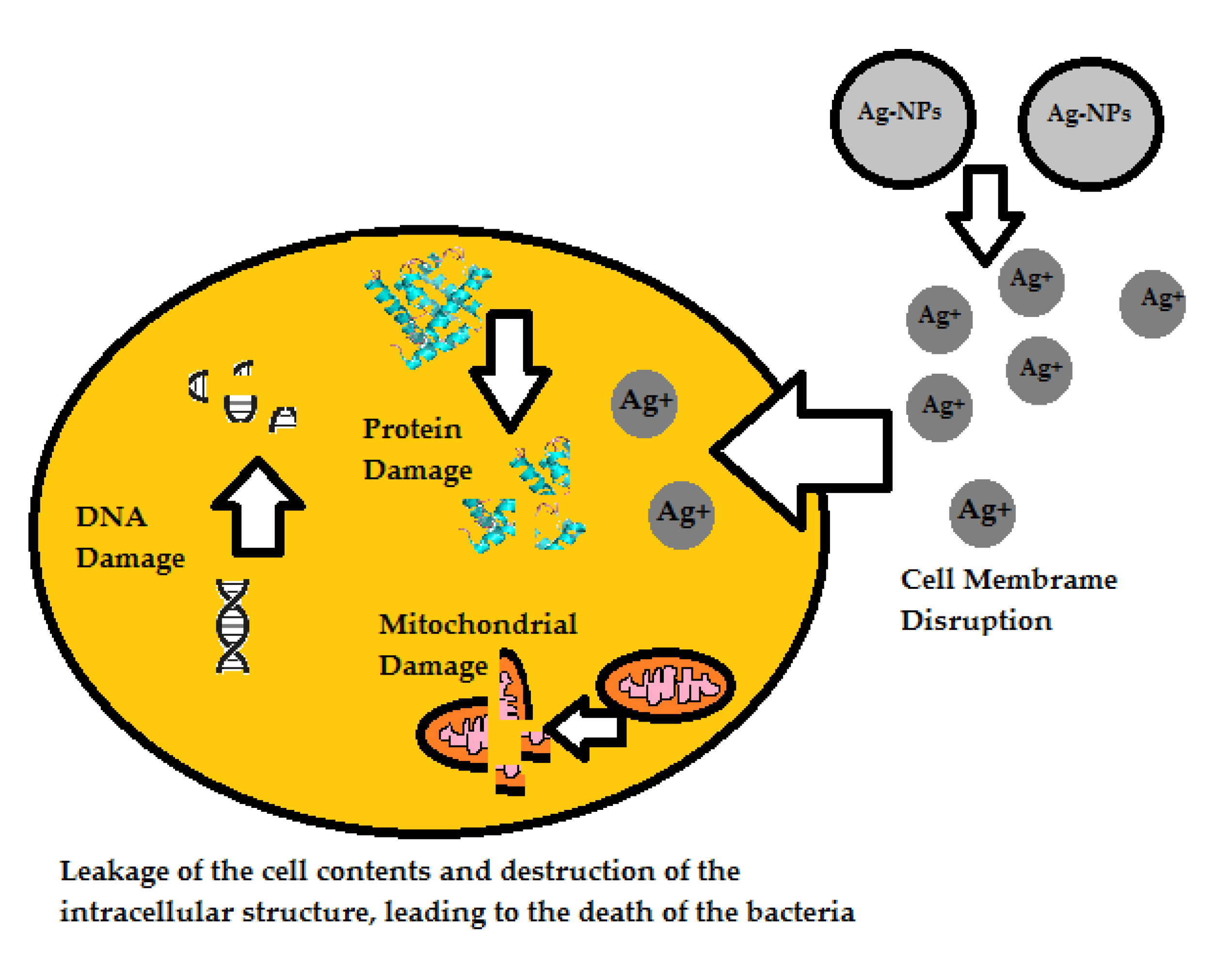
2. Mechanism of Action
3. Properties of Nanoparticles
4. Synthesis of Nanoparticles
5. Application of Nanoparticles
6. Discussion
7. Conclusions
Author Contributions
Funding
Conflicts of Interest
References
- Gonzalez-Martina, M.; Corberaa, J.A.; Suarez-Bonnetb, A.; Tejedor-Junco, M.T. Virulence factors in coagulase-positive staphylococci of veterinary interest other than Staphylococcus aureus. Vet. Q. 2020, 40, 118–131. [Google Scholar] [CrossRef] [Scilit] [PubMed]
- Fernandes, L.F.; Souza, G.; Almeida, A.C.; Cardoso, L.; Xavier, M.; Pinheiro, T.; Cruz, G.; Dourado, H.; Silva, W.S.; Xavier, A. Identification and characterization of methicillin-resistant Staphylococcus spp. isolated from surfaces near patients in an intensive care unit of a hospital in southeastern Brazil. Rev. Soc. Bras. Med. 2020, 53, e20200244. [Google Scholar] [CrossRef] [Scilit] [PubMed]
- Oliveira, W.F.; Silva, P.M.S.; Silva, R.C.S.; Silva, G.M.M.; Machado, G.; Coelho, L.C.B.B.; Correia, M.T.S. Staphylococcus aureus and Staphylococcus epidermidis infections on implants. J. Hosp. Infect. 2018, 98, 111–117. [Google Scholar] [CrossRef] [Scilit] [PubMed]
- Słowik, R.; Kołpa, M.; Wałaszek, M.; Różańska, A.; Jagiencarz-Starzec, B.; Zieńczuk, W.; Kawik, Ł.; Wolak, Z.; Wójkowska-Mach, J. Epidemiology of Surgical Site Infections Considering the NHSN Standardized Infection Ratio in Hip and Knee Arthroplasties. Int. J. Environ. Res. 2020, 17, 3167. [Google Scholar] [CrossRef] [Scilit]
- Cordeiro, L.; Figueiredo, P.; Souza, H.; Sousa, A.; Andrade-Júnior, F.; Medeiros, D.; Nóbrega, J.; Silva, D.; Martins, E.; Barbosa-Filho, J. Terpinen-4-ol as an Antibacterial and Antibiofilm Agent against Staphylococcus aureus. Int. J. Mol. Sci. 2020, 21, 4531. [Google Scholar] [CrossRef] [Scilit]
- Guo, Y.; Song, G.; Sun, M.; Wang, J.; Wang, Y. Prevalence and Therapies of Antibiotic-Resistance in Staphylococcus aureus. Front. Cell. Infect. 2020, 10, 107. [Google Scholar] [CrossRef] [Scilit]
- Caballero, A.R.; Tang, A.; Bierdeman, M.; O’Callaghan, R.; Marquart, M. Correlation of Staphylococcus epidermidis Phenotype and Its Corneal Virulence. Curr. Eye Res. 2021, 46, 638–647. [Google Scholar] [CrossRef] [Scilit]
- Fournière, M.; Latire, T.; Souak, D.; Feuilloley, M.G.J.; Bedoux, G. Staphylococcus epidermidis and Cutibacterium acnes: Two Major Sentinels of Skin Microbiota and the Influence of Cosmetics. Microorganisms 2020, 8, 1752. [Google Scholar] [CrossRef] [Scilit]
- Wang, P.J.; Xie, C.B.; Sun, F.H.; Guo, L.J.; Dai, M.; Cheng, X.; Ma, Y.X. Molecular Characteristics of Methicillin-Resistant Staphylococcus epidermidis on the Abdominal Skin of Females before Laparotomy. Int. J. Mol. Sci. 2016, 17, 992. [Google Scholar] [CrossRef] [Scilit]
- Koch, J.A.; Pust, T.M.; Cappellini, A.J.; Mandell, J.B.; Ma, D.; Shah, N.B.; Brothers, K.M.; Urish, K.L. Staphylococcus epidermidis Biofilms Have a High Tolerance to Antibiotics in Periprosthetic Joint Infection. Life 2020, 10, 253. [Google Scholar] [CrossRef] [Scilit]
- Schilcher, K.; Horswill, A.R. Staphylococcal Biofilm Development: Structure, Regulation, and Treatment Strategies. Microbiol. Mol. Biol. Rev. 2020, 84, e00026-19. [Google Scholar] [CrossRef] [Scilit]
- Ranieri, M.R.; Whitchurch, C.B.; Burrows, L.L. Mechanisms of biofilm stimulation by subinhibitory concentrations of antimicrobials. Curr. Opin. Microbiol. 2018, 45, 164–169. [Google Scholar] [CrossRef] [Scilit] [PubMed]
- Swolana, D.; Kępa, M.; Kabała-Dzik, A.; Dzik, R.; Wojtyczka, R.D. Sensitivity of Staphylococcal Biofilm to Selected Compounds of Plant Origin. Antibiotics 2021, 10, 607. [Google Scholar] [CrossRef] [Scilit] [PubMed]
- Ramasamy, K.; Dhavamani, S.; Natesan, G.; Sengodan, K.; Sengottayan, S.-N.; Tiwari, M.; Sahi, S.; Perumal, V. A potential role of green engineered TiO2 nanocatalyst towards enhanced photocatalytic and biomedical applications. Environ. Sci. Pollut. Res. 2021, 28, 41207–41223. [Google Scholar] [CrossRef] [Scilit] [PubMed]
- Khan, F.; Lee, J.W.; Pham, D.N.T.; Khan, M.M.; Park, S.K.; Shin, I.S.; Kim, Y.M. Antibiofilm Action of ZnO, SnO2 and CeO2 Nanoparticles towards Grampositive Biofilm Forming Pathogenic Bacteria. Recent Pat. Nanotechnol. 2020, 14, 239–249. [Google Scholar] [CrossRef] [Scilit]
- Otari, S.V.; Patel, S.K.S.; Jeong, J.-H.; Lee, J.H.; Lee, J.-K. A green chemistry approach for synthesizing thermostable antimicrobial peptide-coated gold nanoparticles immobilized in an alginate biohydrogel. RSC Adv. 2016, 6, 86808–86816. [Google Scholar] [CrossRef] [Scilit]
- Ji, H.; Zhou, S.; Fu, Y.; Wang, Y.; Mi, J.; Lu, T.; Wang, X.; Lü, C. Size-controllable preparation and antibacterial mechanism of thermo-responsive copolymer-stabilized silver nanoparticles with high antimicrobial activity. Mater. Sci. Eng. C Mater. Biol. Appl. 2020, 110, 110735. [Google Scholar] [CrossRef] [Scilit]
- Franci, G.; Falanga, A.; Galdiero, S.; Palomba, L.; Rai, M.; Morelli, G.; Galdiero, M. Silver Nanoparticles as Potential Antibacterial Agents. Molecules 2015, 20, 8856–8874. [Google Scholar] [CrossRef] [Scilit]
- Leng, D.; Li, Y.; Zhu, J.; Liang, R.; Zhang, C.; Zhou, Y.; Li, M.; Wang, Y.; Rong, D.; Wu, D.; et al. The Antibiofilm Activity and Mechanism of Nanosilver- and Nanozinc-Incorporated Mesoporous Calcium-Silicate Nanoparticles. Int. J. Nanomed. 2020, 15, 3921–3936. [Google Scholar] [CrossRef] [Scilit]
- Sheng, Z.; Liu, Y. Potential impacts of silver nanoparticles on bacteria in the aquatic environment. J. Environ. Manag. 2017, 191, 290–296. [Google Scholar] [CrossRef] [Scilit]
- Gunawan, C.; Faiz, M.B.; Mann, R.; Ting, S.R.S.; Sotiriou, G.A.; Marquis, C.P.; Amal, R. Nanosilver Targets the Bacterial Cell Envelope: The Link with Generation of Reactive Oxygen Radicals. ACS Appl. Mater. Interfaces 2020, 12, 5557–5568. [Google Scholar] [CrossRef] [Scilit] [PubMed]
- Tanvir, F.; Yaqub, A.; Tanvir, S.; Anderson, W.A. Poly-L-arginine Coated Silver Nanoprisms and Their Anti-Bacterial Properties. Nanomaterials 2017, 7, 296. [Google Scholar] [CrossRef] [Scilit] [PubMed]
- Pal, S.; Tak, Y.K.; Song, J.M. Does the antibacterial activity of silver nanoparticles depend on the shape of the nanoparticle? A study of the Gram-negative bacterium Escherichia coli. Appl. Environ. Microbiol. 2007, 73, 1712–1720. [Google Scholar] [CrossRef] [Scilit] [PubMed]
- Alshareef, A.; Laird, K.; Cross, R.B.M. Shape-dependent antibacterial activity of silver nanoparticles on Escherichia coli and Enterococcus faecium bacterium. Appl. Surf. Sci. 2017, 424, 310–315. [Google Scholar] [CrossRef] [Scilit]
- Jeevanandam, J.; Krishnan, S.; Hii, Y.S.; Pan, S.; Chan, Y.S.; Acquah, C.; Danquah, M.K.; Rodrigues, J. Synthesis approach-dependent antiviral properties of silver nanoparticles and nanocomposites. J. Nanostruct. Chem. 2022, 1–23. [Google Scholar] [CrossRef] [Scilit]
- El-Zahry, M.R.; Mahmoud, A.; Refaat, I.H.; Mohamed, H.A.; Bohlmann, H.; Lendl, B. Antibacterial effect of various shapes of silver nanoparticles monitored by SERS. Talanta 2015, 138, 183–189. [Google Scholar] [CrossRef] [Scilit]
- Platania, V.; Kaldeli-Kerou, A.; Karamanidou, T.; Kouki, M.; Tsouknidas, A.; Chatzinikolaidou, M. Antibacterial Effect of Colloidal Suspensions Varying in Silver Nanoparticles and Ions Concentrations. Nanomaterials 2021, 12, 31. [Google Scholar] [CrossRef] [Scilit]
- Courrol, D.D.S.; Lopes, C.R.B.; Pereira, C.B.P.; Franzolin, M.R.; Silva, F.R.O.; Courrol, L.C. Tryptophan Silver Nanoparticles Synthesized by Photoreduction Method: Characterization and Determination of Bactericidal and Anti-Biofilm Activities on Resistant and Susceptible Bacteria. Int. J. Tryptophan Res. 2019, 12, 1178646919831677. [Google Scholar] [CrossRef] [Scilit]
- Habash, M.B.; Goodyear, M.C.; Park, A.J.; Surette, M.D.; Vis, E.C.; Harris, R.J.; Khursigara, C.M. Potentiation of tobramycin by silver nanoparticles against Pseudomonas aeruginosa biofilms. Antimicrob. Agents Chemother. 2017, 61, e00415-17. [Google Scholar] [CrossRef] [Scilit]
- Haiyan Yu, H.; Haoyu Sun, H.; Chunsheng Yin, C.; Zhifen Lin, Z. Combination of sulfonamides, silver antimicrobial agents and quorum sensing inhibitors as a preferred approach for improving antimicrobial efficacy against Bacillus subtilis. Ecotoxicol. Environ. Saf. 2019, 181, 43–48. [Google Scholar]
- Nadhe, S.B.; Singh, R.; Wadhwani, S.A.; Chopade, B.A. Acinetobacter sp. mediated synthesis of AgNPs, its optimization, characterization and synergistic antifungal activity against C. albicans. J. Appl. Microbiol. 2019, 127, 445–458. [Google Scholar] [CrossRef] [Scilit] [PubMed]
- Khodadadi, S.; Mahdinezhad, N.; Fazeli-Nasab, B.; Heidari, M.J.; Fakheri, B.; Miri, A. Investigating the Possibility of Green Synthesis of Silver Nanoparticles Using Vaccinium arctostaphlyos Extract and Evaluating Its Antibacterial Properties. BioMed Res. Int. 2021, 2021, 5572252. [Google Scholar] [CrossRef] [Scilit]
- Jia, X.; Yao, Y.; Yu, G.; Qu, L.; Li, T.; Li, Z.; Xu, C. Synthesis of gold-silver nanoalloys under microwave-assisted irradiation by deposition of silver on gold nanoclusters/triple helix glucan and antifungal activity. Carbohydr. Polym. 2020, 238, 116169. [Google Scholar] [CrossRef] [Scilit] [PubMed]
- Laghrib, F.; Aghris, S.; Ajermoun, N.; Hrioua, A.; Bakasse, M.; Lahrich, S.; El Mhammedi, M.A. Recent progress in controlling the synthesis and assembly of nanostructures: Application for electrochemical determination of p-nitroaniline in water. Talanta 2020, 219, 121234. [Google Scholar] [CrossRef] [Scilit] [PubMed]
- Slavin, Y.N.; Ivanova, K.; Hoyo, J.; Perelshtein, I.; Owen, G.; Haegert, A.; Lin, Y.-Y.; LeBihan, S.; Gedanken, A.; Häfeli, U.O.; et al. Novel Lignin-Capped Silver Nanoparticles against Multidrug-Resistant Bacteria. ACS Appl. Mater. Interfaces 2021, 13, 22098–22109. [Google Scholar] [CrossRef] [Scilit]
- Haidari, H.; Kopecki, Z.; Bright, R.; Cowin, A.J.; Garg, S.; Goswami, N.; Vasilev, K. Ultrasmall AgNP-Impregnated Biocompatible Hydrogel with Highly Effective Biofilm Elimination Properties. ACS Appl. Mater. Interfaces 2020, 12, 41011–41025. [Google Scholar] [CrossRef] [Scilit]
- Osonga, F.J.; Akgul, A.; Yazgan, I.; Akgul, A.; Eshun, G.B.; Sakhaee, L.; Sadik, O.A. Size and Shape-Dependent Antimicrobial Activities of Silver and Gold Nanoparticles: A Model Study as Potential Fungicides. Molecules 2020, 25, 2682. [Google Scholar] [CrossRef] [Scilit]
- El-Faham, A.; Al-Rasheed, H.H.; Sholkamy, E.N.; Osman, S.M.; ALOthman, Z.A. Simple Approaches for the Synthesis of AgNPs in Solution and Solid Phase Using Modified Methoxypolyethylene Glycol and Evaluation of Their Antimicrobial Activity. Int. J. Nanomed. 2020, 15, 2353–2362. [Google Scholar] [CrossRef] [Scilit]
- Bartel, M.; Markowska, K.; Strawski, M.; Wolska, K.; Mazur, M. Silver-decorated gel-shell nanobeads: Physicochemical characterization and evaluation of antibacterial properties. Beilstein J. Nanotechnol. 2020, 11, 620–630. [Google Scholar] [CrossRef] [Scilit]
- Raho, R.; Nguyen, N.Y.; Zhang, N.; Jiang, W.; Sannino, A.; Liu, H.; Pollini, M.; Paladini, F. Photo-assisted green synthesis of silver doped silk fibroin/carboxymethyl cellulose nanocomposite hydrogels for biomedical applications. Mater. Sci. Eng. C Mater. Biol. Appl. 2020, 107, 110219. [Google Scholar] [CrossRef] [Scilit]
- Swolana, D.; Kępa, M.; Idzik, D.; Dziedzic, A.; Kabała-Dzik, A.; Wąsik, T.J.; Wojtyczka, R.D. The Antibacterial Effect of Silver Nanoparticles on Staphylococcus epidermidis Strains with Different Biofilm-Forming Ability. Nanomaterials 2020, 10, 1010. [Google Scholar] [CrossRef] [Scilit] [PubMed]
- Bertoglio, F.; De Vita, L.; D’Agostino, A.; Diaz Fernandez, Y.; Falqui, A.; Casu, A.; Merli, D.; Milanese, C.; Rossi, S.; Taglietti, A.; et al. Increased Antibacterial and Antibiofilm Properties of Silver Nanoparticles Using Silver Fluoride as Precursor. Molecules 2020, 25, 3494. [Google Scholar] [CrossRef] [Scilit] [PubMed]
- Tavares, T.D.; Antunes, J.C.; Padrão, J.; Ribeiro, A.I.; Zille, A.; Amorim, M.T.P.; Ferreira, F.; Felgueiras, H.P. Activity of Specialized Biomolecules against Gram-Positive and Gram-Negative Bacteria. Antibiotics 2020, 9, 314. [Google Scholar] [CrossRef] [Scilit] [PubMed]
- Nocchetti, M.; Donnadio, A.; Vischini, E.; Posati, T.; Ravaioli, S.; Arciola, C.R.; Campoccia, D.; Vivani, R. Zirconium Carboxyaminophosphonate Nanosheets as Support for Ag Nanoparticles. Materials 2019, 12, 3185. [Google Scholar] [CrossRef] [Scilit]
- Sprynskyy, M.; Sokol, H.; Rafińska, K.; Brzozowska, W.; Railean-Plugaru, V.; Pomastowski, P.; Buszewski, B. Preparation of AgNPs/saponite nanocomposites without reduction agents and study of its antibacterial activity. Colloids Surf. B 2019, 180, 457–465. [Google Scholar] [CrossRef] [Scilit]
- Elashnikov, R.; Radocha, M.; Panov, I.; Rimpelova, S.; Ulbrich, P.; Michalcova, A.; Svorcik, V.; Lyutakov, O. Porphyrin-silver nanoparticles hybrids: Synthesis, characterization and antibacterial activity. Mater. Sci. Eng. C Mater. Biol. Appl. 2019, 102, 192–199. [Google Scholar] [CrossRef] [Scilit]
- Babushkina, I.V.; Mamonova, I.A.; Ulyanov, V.Y. Local Treatment of Local Staphylococcal Infection with Complex Preparations Based on Metal Nanoparticles in the Experiment. Bull. Exp. Biol. Med. 2019, 167, 784–786. [Google Scholar] [CrossRef] [Scilit]
- Jose, M.; Sienkiewicz, P.; Szymańska, K.; Darowna, D.; Moszyński, D.; Lendzion-Bieluń, Z.; Szymański, K.; Mozia, S. Influence of Preparation Procedure on Physicochemical and Antibacterial Properties of Titanate Nanotubes Modified with Silver. Nanomaterials 2019, 9, 795. [Google Scholar] [CrossRef] [Scilit]
- Jaworski, S.; Wierzbicki, M.; Sawosz, E.; Jung, A.; Gielerak, G.; Biernat, J.; Jaremek, H.; Łojkowski, W.; Woźniak, B.; Wojnarowicz, J.; et al. Graphene Oxide-Based Nanocomposites Decorated with Silver Nanoparticles as an Antibacterial Agent. Nanoscale Res. Lett. 2018, 13, 116. [Google Scholar] [CrossRef] [Scilit]
- Chen, Y.C.; Liao, C.H.; Shen, W.T.; Su, C.; Wu, Y.C.; Tsai, M.H.; Hsiao, S.S.; Yu, K.P.; Tseng, C.H. Effective disinfection of airborne microbial contamination in hospital wards using a zero-valent nano-silver/TiO2 -chitosan composite. Indoor Air 2019, 29, 439–449. [Google Scholar] [CrossRef] [Scilit]
- Porrelli, D.; Travan, A.; Turco, G.; Crosera, M.; Borgogna, M.; Donati, I.; Paoletti, S.; Adami, G.; Marsich, E. Antibacterial-nanocomposite bone filler based on silver nanoparticles and polysaccharides. J. Tissue Eng. Regen. Med. 2018, 12, e747–e759. [Google Scholar] [CrossRef] [Scilit]
- Dai, T.; Wang, C.; Wang, Y.; Xu, W.; Hu, J.; Cheng, Y. A Nanocomposite Hydrogel with Potent and Broad-Spectrum Antibacterial Activity. ACS Appl. Mater. Interfaces 2018, 10, 15163–15173. [Google Scholar] [CrossRef] [Scilit] [PubMed]
- Takahashi, C.; Matsubara, N.; Akachi, Y.; Ogawa, N.; Kalita, G.; Asaka, T.; Tanemura, M.; Kawashima, Y.; Yamamoto, H. Visualization of silver-decorated poly (DL-lactide-co-glycolide) nanoparticles and their efficacy against Staphylococcus epidermidis. Mater. Sci. Eng. C Mater. Biol. Appl. 2017, 72, 143–149. [Google Scholar] [CrossRef] [Scilit] [PubMed]
- Pallavicini, P.; Arciola, C.R.; Bertoglio, F.; Curtosi, S.; Dacarro, G.; D’Agostino, A.; Ferrari, F.; Merli, D.; Milanese, C.; Rossi, S.; et al. Silver nanoparticles synthesized and coated with pectin: An ideal compromise for anti-bacterial and anti-biofilm action combined with wound-healing properties. J. Colloid Interface Sci. 2017, 498, 271–281. [Google Scholar] [CrossRef] [Scilit] [PubMed]
- Jyoti, K.; Singh, A. Evaluation of antibacterial activity from phytosynthesized silver nanoparticles against medical devices infected with Staphylococcus spp. J. Taibah Univ. Med. Sci. 2016, 12, 47–54. [Google Scholar] [CrossRef] [Scilit] [PubMed]
- Chen, Y.C.; Yu, K.P. Enhanced antimicrobial efficacy of thermal-reduced silver nanoparticles supported by titanium dioxide. Colloids Surf. B Biointerfaces 2017, 154, 195–202. [Google Scholar] [CrossRef] [Scilit] [PubMed]
- Prema, P.; Thangapandiyan, S.; Immanuel, G. CMC stabilized nano silver synthesis, characterization and its antibacterial and synergistic effect with broad spectrum antibiotics. Carbohydr. Polym. 2017, 158, 141–148. [Google Scholar] [CrossRef] [Scilit]
- Ambi, A.; Parikh, N.; Vera, C.; Burns, K.; Montano, N.; Sciorra, L.; Epstein, J.; Zeng, D.; Traba, C. Anti-infection silver nanoparticle immobilized biomaterials facilitated by argon plasma grafting technology. Biofouling 2018, 34, 273–286. [Google Scholar] [CrossRef] [Scilit]
- Yuan, Y.G.; Peng, Q.L.; Gurunathan, S. Effects of Silver Nanoparticles on Multiple Drug-Resistant Strains of Staphylococcus aureus and Pseudomonas aeruginosa from Mastitis-Infected Goats: An Alternative Approach for Antimicrobial Therapy. Int. J. Mol. Sci. 2017, 18, 569. [Google Scholar] [CrossRef] [Scilit]
- Gurunathan, S.; Han, J.W.; Kwon, D.N.; Kim, J.H. Enhanced antibacterial and anti-biofilm activities of silver nanoparticles against Gram-negative and Gram-positive bacteria. Nanoscale Res. Lett. 2014, 9, 373. [Google Scholar] [CrossRef] [Scilit]
- Choi, J.S.; Jung, H.C.; Baek, Y.J.; Kim, B.Y.; Lee, M.W.; Kim, H.D.; Kim, S.W. Antibacterial Activity of Green-Synthesized Silver Nanoparticles Using Areca catechu Extract against Antibiotic-Resistant Bacteria. Nanomaterials 2021, 11, 205. [Google Scholar] [CrossRef] [Scilit] [PubMed]
- Otari, S.V.; Patel, S.; Kalia, V.C.; Kim, I.W.; Lee, J.K. Antimicrobial Activity of Biosynthesized Silver Nanoparticles Decorated Silica Nanoparticles. Indian J. Microbiol. 2019, 59, 379–382. [Google Scholar] [CrossRef] [Scilit] [PubMed]
- Barros, C.H.N.; Fulaz, S.; Stanisic, D.; Tasic, L. Biogenic Nanosilver against Multidrug-Resistant Bacteria (MDRB). Antibiotics 2018, 7, 69. [Google Scholar] [CrossRef] [Scilit] [PubMed]
- Bahrulolum, H.; Nooraei, S.; Javanshir, N.; Tarrahimofrad, H.; Sadat Mirbagheri, V.; Easton, A.J.; Ahmadian, G. Green synthesis of metal nanoparticles using microorganisms and their application in the agrifood sector. J. Nanobiotechnol. 2021, 19, 1–26. [Google Scholar] [CrossRef] [Scilit] [PubMed]
- Ittibenjapong, C.; Kanjanahitanon, P.; Chaichamni, P.; Panich, S.; Thamsumet, N. Green synthesis of silver nanoparticles from Catunaregam tomentosa extract. Phys. Sci. Rev. 2021. [Google Scholar] [CrossRef] [Scilit]
- Tyavambiza, C.; Elbagory, A.M.; Madiehe, A.M.; Meyer, M.; Meyer, S. The Antimicrobial and Anti-Inflammatory Effects of Silver Nanoparticles Synthesised from Cotyledon orbiculata Aqueous Extract. Nanomaterials 2021, 11, 1343. [Google Scholar] [CrossRef] [Scilit]
- Arshad, H.; Sami, M.A.; Sadaf, S.; Hassan, U. Salvadora persica mediated synthesis of silver nanoparticles and their antimicrobial efficacy. Sci. Rep. 2021, 11, 5996. [Google Scholar] [CrossRef] [Scilit]
- Hafeez, M.; Zeb, M.; Khan, A.; Akram, B.; Abdin, Z.U.; Haq, S.; Zaheer, M.; Ali, S. Populus ciliata mediated synthesis of silver nanoparticles and their antibacterial activity. Microsc. Res. Tech. 2021, 84, 480–488. [Google Scholar] [CrossRef] [Scilit]
- Devanesan, S.; AlSalhi, M.S. Green Synthesis of Silver Nanoparticles Using the Flower Extract of Abelmoschus esculentus for Cytotoxicity and Antimicrobial Studies. Int. J. Nanomed. 2021, 14, 3343–3356. [Google Scholar] [CrossRef] [Scilit]
- de Carvalho Bernardo, W.L.; Boriollo, M.F.G.; Tonon, C.C.; da Silva, J.J.; Cruz, F.M.; Martins, A.L.; Höfling, J.F.; Spolidorio, D.M.P. Antimicrobial effects of silver nanoparticles and extracts of Syzygium cumini flowers and seeds: Periodontal, cariogenic and opportunistic pathogens. Arch. Oral Biol. 2021, 125, 105101. [Google Scholar] [CrossRef] [Scilit]
- Ansar, S.; Tabassum, H.; Aladwan, N.S.M.; Naiman Ali, M.; Almaarik, B.; AlMahrouqi, S.; Abudawood, M.; Banu, N.; Alsubki, R. Eco friendly silver nanoparticles synthesis by Brassica oleracea and its antibacterial, anticancer and antioxidant properties. Sci. Rep. 2020, 10, 18564. [Google Scholar] [CrossRef] [Scilit] [PubMed]
- Chahardoli, A.; Qalekhani, F.; Shokoohinia, Y.; Fattahi, A. Biological and Catalytic Activities of Green Synthesized Silver Nanoparticles from the Leaf Infusion of Dracocephalum kotschyi Boiss. Glob. Chall. 2021, 5, 2000018. [Google Scholar] [CrossRef] [Scilit] [PubMed]
- Saravanan, M.; Arokiyaraj, S.; Lakshmi, T.; Pugazhendhi, A. Synthesis of silver nanoparticles from Phenerochaete chrysosporium (MTCC-787) and their antibacterial activity against human pathogenic bacteria. Microb. Pathog. 2018, 117, 68–72. [Google Scholar] [CrossRef] [Scilit]
- Ovais, M.; Khalil, A.T.; Raza, A.; Islam, N.U.; Ayaz, M.; Saravanan, M.; Ali, M.; Ahmad, I.; Shahid, M.; Shinwari, Z.K. Multifunctional theranostic applications of biocompatible green-synthesized colloidal nanoparticles. Appl. Microbiol. Biotechnol. 2018, 102, 4393–4408. [Google Scholar] [CrossRef] [Scilit] [PubMed]
- Ahmed, F.; Qayyum, S.; Husain, Q. Benign nano-assemblages of silver induced by β-galactosidase with augmented antimicrobial and industrial dye degeneration potential. Mater. Sci. Eng. C Mater. Biol. Appl. 2018, 1, 570–578. [Google Scholar] [CrossRef] [Scilit]
- Devanesan, S.; AlSalhi, M.S.; Balaji, R.V.; Ranjitsingh, A.J.A.; Ahamed, A.; Alfuraydi, A.A.; AlQahtani, F.Y.; Aleanizy, F.S.; Othman, A.H. Antimicrobial and Cytotoxicity Effects of Synthesized Silver Nanoparticles from Punica granatum Peel Extract. Nanoscale Res. Lett. 2018, 13, 315. [Google Scholar] [CrossRef] [Scilit]
- Skandalis, N.; Dimopoulou, A.; Georgopoulou, A.; Gallios, N.; Papadopoulos, D.; Tsipas, D.; Theologidis, I.; Michailidis, N.; Chatzinikolaidou, M. The Effect of Silver Nanoparticles Size, Produced Using Plant Extract from Arbutus unedo, on Their Antibacterial Efficacy. Nanomaterials 2017, 7, 178. [Google Scholar] [CrossRef] [Scilit]
- Karatoprak, G.S.; Aydin, G.; Altinsoy, B.; Altinkaynak, C.; Koşar, M.; Ocsoy, I. The Effect of Pelargonium endlicherianum Fenzl. root extracts on formation of nanoparticles and their antimicrobial activities. Enzym. Microb. Technol. 2017, 97, 21–26. [Google Scholar] [CrossRef] [Scilit]
- Alavi, M.; Karimi, N. Characterization, antibacterial, total antioxidant, scavenging, reducing power and ion chelating activities of green synthesized silver, copper and titanium dioxide nanoparticles using Artemisia haussknechtii leaf extract. Artif. Cell. Nanomed. Biotechnol. 2018, 46, 2066–2081. [Google Scholar] [CrossRef] [Scilit]
- McShan, D.; Ray, P.C.; Yu, H. Molecular toxicity mechanism of nanosilver. J. Food Drug Anal. 2014, 22, 116–127. [Google Scholar] [CrossRef] [Scilit]
- Aguilera-Correa, J.J.; Esteban, J.; Vallet-Regí, M. Inorganic and Polymeric Nanoparticles for Human Viral and Bacterial Infections Prevention and Treatment. Nanomaterials 2021, 11, 137. [Google Scholar] [CrossRef] [Scilit] [PubMed]
- Valentin, E.; Bottomley, A.L.; Chilambi, G.S.; Harry, E.J.; Amal, R.; Sotiriou, G.A.; Rice, S.A.; Gunawan, C. Heritable nanosilver resistance in priority pathogen: A unique genetic adaptation and comparison with ionic silver and antibiotics. Nanoscale 2020, 12, 2384–2392. [Google Scholar] [CrossRef] [Scilit] [PubMed]
- Mosselhy, D.A.; Assad, M.; Sironen, T.; Elbahri, M. Nanotheranostics: A Possible Solution for Drug-Resistant Staphylococcus aureus and their Biofilms? Nanomaterials 2021, 11, 82. [Google Scholar] [CrossRef] [Scilit] [PubMed]
- Naseer, Q.A.; Xue, X.; Wang, X.; Dang, S.; Din, S.U.; Kalsoom, W.; Jamil, J. Synthesis of silver nanoparticles using Lactobacillus bulgaricus and assessment of their antibacterial potential. Braz. J. Biol. 2021, 82, e232434. [Google Scholar] [CrossRef] [Scilit] [PubMed]
- Alsamhary, K.I. Eco-friendly synthesis of silver nanoparticles by Bacillus subtilis and their antibacterial activity. Saudi J. Biol. Sci. 2020, 27, 2185–2191. [Google Scholar] [CrossRef] [Scilit]
- Al-Dhabi, N.A.; Mohammed Ghilan, A.-K.; Arasu, M.V. Characterization of Silver Nanomaterials Derived from Marine Streptomyces sp. Al-Dhabi-87 and Its In Vitro Application against Multidrug Resistant and Extended-Spectrum Beta-Lactamase Clinical Pathogens. Nanomaterials 2018, 8, 279. [Google Scholar] [CrossRef] [Scilit]
- Rogowska, A.; Rafińska, K.; Pomastowski, P.; Walczak, J.; Railean-Plugaru, V.; Buszewska-Forajta, M.; Buszewski, B. Silver nanoparticles functionalized with ampicillin. Electrophoresis 2017, 38, 2757–2764. [Google Scholar] [CrossRef] [Scilit]
- Viorica, R.-P.; Pomastowski, P.; Meller, K.; Złoch, M.; Rafinska, K.; Buszewski, B. Lactococcus lactis as a safe and inexpensive source of bioactive silver composites. Appl. Microbiol. Biotechnol. 2017, 101, 7141–7153. [Google Scholar] [CrossRef] [Scilit]
- Sheikholeslami, S.; Mousavi, S.E.; Ahmadi Ashtiani, H.R.; Hosseini Doust, S.R.; Mahdi Rezayat, S. Antibacterial Activity of Silver Nanoparticles and Their Combination with Zataria multiflora Essential Oil and Methanol Extract. Jundishapur J. Microbiol. 2016, 9, e36070. [Google Scholar] [CrossRef] [Scilit]


| AgNPs | PEG AgNPs | Citrate AgNPs | Borohydride AgNPs | Deionized H2O (Negative Control) |
|---|---|---|---|---|
| Shape | hexagonal | spherical | triangular | - |
| Inhibition zone size (cm) ±SD | 2.45 ± 0.24 | 2.02 ± 0.55 | 0.08 ± 0.14 | 0.00 ± 0.00 |
| Synthesis or Source | Size [nm] | Bacterial Strain | Concentrations Used and Results | Ref. |
|---|---|---|---|---|
| Lignin-Silver nanoparticles | 20 | S. epidermidis MDR | MIC = 10 µg/mL; MBC = 10 µg/mL | [35] |
| S. aureus MDR | MIC = 10 µg/mL; MBC = 10 µg/mL | |||
| S. aureus ATCC 700788 | MIC = 5 µg/mL; MBC = 10 µg/mL | |||
| Mercaptosuccinic acid (MSA)-coated silver nanoparticles | 2.98 | S. aureus | ZOI = 8 mm; viability of bacterial cells reduced to 95%; significant biofilm disruption and elimination with 50 μg/g AgNPs loaded gel | [36] |
| S. epidermidis | ZOI = 13 mm; viability of bacterial cells reduced to >96%; significant biofilm disruption and elimination with 50 μg/g AgNPs loaded gel | |||
| Luteolin tetraphosphate derived silver nanoparticles | 9; 21 | S. epidermidis ATCC | viability below 70% for concentrations from 1 to 15 µM | [37] |
| The modified methoxypolyethylene glycol bishydrazino-s-triazine silver nanoparticles (mPEGTH2-AgNPs 2:1) | 7–10 | S. epidermidis ATCC 12228 | MIC = 0.094 mg/mL; MBC = 0.188 mg/mL | [38] |
| Sulfonated polystyrene beads with embedded silver nanoparticles (PSSAg) | 5 | S. aureus ATCC 29213 | MIC = 1.14 µg/mL; MBIC = 3.04 µg/mL | [39] |
| S. epidermidis ATCC 12228 | MIC = 0.76 µg/mL; MBIC = 0.76 µg/mL | |||
| Composite hydrogel (CoHy) of RSF stabilized with Carboxymethyl Cellulose-Na (CMC-Na) and loaded with silver nanoparticles | 92 | S. epidermidis ATCC 35984 | ZOI = ~4.5 mm | [40] |
| S. aureus ATCC 29737 | ZOI = ~6.0 mm | |||
| MRSA ATCC 33591 | ZOI = ~4.0 mm | |||
| Silver nanoparticles | 10 | S. epidermidis ATCC 12228 | MIC = 3 µg/mL | [41] |
| S. epidermidis ATCC 35983 | MIC = 4 µg/mL | |||
| S. epidermidis ATCC 35984 | MIC = 5 µg/mL | |||
| Silver nanoparticles produced with AgF (pAgNP-F) | 7.0–7.2 | S. epidermidis RP62A | MIC = 250 µM | [42] |
| Nanopowder of silver nanoparticles (AgNPs) coated with polyvinyl pyrrolidone (PVP) | 20–30 | S. aureus ATCC 6538 | ZOI = 11.5 mm; MIC = 4000 µg/mL | [43] |
| S. epidermidis ATCC 35984 | ZOI = 10.6 mm; MIC = 4000 µg/mL | |||
| Silver nanoparticles synthesized with the zirconium phosphate glycine-N,N-bismethylphosphonate | 4.5 | S. epidermidis ATCC 35984 | ZOI = 6.2 mm | [44] |
| Tryptophan Silver nanoparticles (2:1) | ~13.4 | S. epidermidis | 100% inhibition of growth after 24 h incubation; inhibition of biofilm formation of 20.0% to 40.2% | [28] |
| S. aureus | 100% inhibition of growth after 24 h incubation; inhibition of biofilm formation of 67.2% to 71.8 % | |||
| Silver nanoparticles/saponite nanocomposites obtained from initial silver ion concentration of 500 mg/L | 2–4 | S. aureus ATCC 11632 | ZOI = 23.50 ± 0.87 mm | [45] |
| S. epidermidis | ZOI = 30.67 ± 0.58 mm | |||
| Porphyrin-silver nanoparticles with LED illumination | 5 to 30 range, the majority of silver nanoparticles had a size of about 10 | S. epidermidis DBM 3179 | the proposed AgNPs-porphyrin conjugate show pronounced antibacterial activity, which exceeds the activity of each separated antibacterial agents; LED illumination enhances the activity against the S. epidermidis strain | [46] |
| Powder preparation containing 0.03–0.05 wt.% silver nanoparticles | 40–50 | 10 MRSE strains | microbial contamination of wounds decreased by 100 or more times in comparison the bacterial load before treatment | [47] |
| Titanate Nanotubes Modified with Silver Nanoparticles | 1–10 | S. epidermidis ATCC 49461 | the samples containing higher Ag loading were more active than samples with low silver amount | [48] |
| Graphene Oxide-Silver Nanoparticles mixture-coated polyurethane foil | 251 ± 10 | S. aureus ATCC 25923 | 79.6% growth inhibition | [49] |
| S. epidermidis ATCC 14990 | 76.5% growth inhibition | |||
| Zero-valent nano-silver/TiO2-chitosan composite | 6.69–8.84 | S. epidermidis | ZOI = 8.56 mm for 2 wt%; critical concentrations: 7.57 µg Ag/mm3 for 0.5 wt%; 2.96 µg Ag/mm3 for 1 wt%; 1.51 µg Ag/mm3 for 2 wt% | [50] |
| Microbeads with silver nanoparticles, obtained by reducing silver ions with ascorbic acid in Chitlac solution | 270 ± 40 | S. epidermidis ATCC 12228 | Growth inhibition of more than four orders of magnitude | [51] |
| S. aureus ATCC 25923 | Growth inhibition of nearly four orders of magnitude | |||
| The dendrimer-encapsulated Ag nanoparticles (G5-AgNPs) at available Ag–G5 feeding molar ratio of 30:1 | 3.33 | S. aureus USA300 | MIC = 128 µg/mL | [52] |
| The dendrimer-encapsulated Ag nanoparticles (G5-AgNPs) at available Ag–G5 feeding molar ratio 30:1 reacted with oxidized dextran (Dex-CHO)-Dex-G5–Ag (30) hydrogel | 3.33 | S. epidermidis 1457 | 99.8% inhibition of growth at the concentration 16 µg/mL | [53] |
| S. aureus USA300 | 99.9% inhibition of growth at the concentration 16 µg/mL | |||
| Silver-decorated submicron-sized biodegradable poly (DL-lactide-coglycolide) nanoparticles (Ag PLGA nanoparticles) | 299.6 | S. epidermidis ATCC 14990T | 38.5% viability of bacteria after treatment with Ag PLGA nanoparticles; viable colony counts for the biofilm decreased from 6.4 × 10⁷ CFU/mL to 1.4 × 10⁷ CFU/mL (more than four orders of magnitude); viable colony count for the bacterial cells after various treatment for 2 h decreased from 6.4 × 10⁶ CFU/mL to 3.7 CFU/mL (1.7 million times less) | [54] |
| Silver nanoparticles synthesized and coated with pectin | 8.0 | S. epidermidis RP62A | MIC = 500 µM | [55] |
| Phytosynthesized silver nanoparticles | 15–35; approximately 27 | S. epidermidis | BE = 3.36 (after 18 h of contact, calculated by growing bacterial suspensions and count viable cells using a digital colony counter) | [56] |
| S. aureus | BE = 3.60 (after 18 h of contact, calculated by growing bacterial suspensions and count viable cells using a digital colony counter) | |||
| Silver nanoparticles synthesized and supported by TiO2 powder (P25) using heat-treatment temperature 200 °C | 3.02–5.74 | S. epidermidis BCRC 11030 | ZOI = 4.6 mm; 7.4 mm; 10.0 mm; 16.5 mm; 13.7 mm in Ag weight percentage: 0.5; 1; 2; 5; 10 wt%, respectively | [57] |
| Carboxy methyl cellulose stabilized silver nanoparticles | 5–15 | S. aureus MTCC 3160 | ZOI = 28.23 mm; MIC = 60 µg/mL; MBC = 60 µg/mL; MBC/MIC ratio = 1.00 | [58] |
| S. epidermidis MTCC 435 | ZOI = 25.03 mm; MIC = 70 µg/mL; MBC = 70 µg/mL; MBC/MIC ratio = 1.00 | |||
| Silver nanoparticles immobilized onto biomaterial | 25 | S. aureus MRSA ATCC 43300 | 100% growth reduction after 45 min of bacteria exposure to the action of silver nanoparticles at a concentration of 5 mM | [17] |
| S. epidermidis ATCC 700567 | 100% growth reduction after 45 min of bacteria exposure to the action of silver nanoparticles at a concentration of 5 mM |
| Synthesis or Source | Size [nm] | Bacterial Strain | Concentrations used and Results | Ref. |
|---|---|---|---|---|
| Silver nanoparticles synthesized from the aqueous extract of Cotyledon orbiculata | 106–137 ± 2 | S. aureus | MIC = 20 µg/mL; MBC = 40 µg/mL | [66] |
| S. epidermidis | MIC = 20 µg/mL; MBC = 20 µg/mL | |||
| MRSA | MIC = 40 µg/mL; MBC = 80 µg/mL | |||
| Silver nanoparticles synthesized from aqueous ‘root extract’ of Salvadora persica | 10–70 | S. epidermidis ATCC 12228 | MIC = 0.19 µg/mL; MBC = 0.39 µg/mL | [67] |
| Silver nanoparticles synthesized from Populus ciliata leaf extract | 4 | S. epidermidis | ZOI = 17.8 mm | [68] |
| Silver nanoparticles synthesized using A. esculentus flower extract | 5.52–24.65; average 18.24 | S. aureus ATCC 29213 | MIC = 85 µg/mL; MBC = 90 µg/mL | [69] |
| S. epidermidis MTCC 3615 | MIC = 150 µg/mL; MBC = 165 µg/mL | |||
| Silver nanoparticles synthesized using lyophilized hydroalcoholic extract of S. cumini seeds | 36.25–77.01 | S. aureus ATCC 25923 | MIC = 125 µg/mL | [70] |
| S. epidermidis ATCC 12228 | MIC = 31.2 µg/mL | |||
| Silver nanoparticles synthesized using lyophilized hydroalcoholic extract of S. cumini flowers | 37.21–46.48 | S. aureus ATCC 25923 | MIC = 250 µg/mL | [70] |
| S. epidermidis ATCC 12228 | MIC = 250 µg/mL | |||
| Silver nanoparticles synthesized with Brassica oleracea | 20 | S. aureus ATCC 6538 | ZOI = 10.1 mm; MIC = 25 µg/mL | [71] |
| S. epidermidis ATCC 12228 | ZOI = 14.33 mm; MIC = 6.25 µg/mL | |||
| Silver nanoparticles synthesized with the leaf infusion of Dracocephalum kotschyi Boiss | 5–63; average 19.41 | S. epidermidis ATCC 12228 | ZOI = 13 mm; MIC = 15.63 µg/mL | [72] |
| S. aureus ATCC 43300 | ZOI = 18 mm; MIC = 15.63 µg/mL | |||
| Silver nanoparticles synthesized by P. chrysosphorium | 34–90 | S. epidermidis | ZOI = 11 mm | [73] |
| S. aureus | ZOI = 13 mm | |||
| Biocompatible green-synthesized colloidal nanoparticles synthesized from Olax nana Wall. ex Benth. (family: Olacaceae) aqueous extract | 26 | S. epidermidis | MIC = 7.14 µg/mL | [74] |
| Silver nanoparticles synthesized with enzyme β-galactosidase from Aspergillus oryzae | 12.89 | S. aureus | MIC = 32 µg/mL; MBC = 64 µg/mL; over 80% inhibition of biofilm formation at the concentration 16 µg/mL | [75] |
| S. epidermidis | MIC = 64 µg/mL; MBC = 128 µg/mL; the bacterial cells completely lose their count at a concentration of 32 µg/mL within 12 h; over 60% inhibition of biofilm formation at the concentration 16 µg/mL | |||
| Silver nanoparticles synthesized from pomegranate fruit peel (Punica granatum) | 20–40; average 26.95 | S. aureus ATCC 29213 | ZOI = 10 mm, 14 mm, 19 mm, 21 mm for the AgNPs concentration 25 µL, 50 µL, 75 µL, 100 µL, respectively | [76] |
| S. epidermidis MTCC 3615 | ZOI = 6 mm, 7 mm, 11 mm, 12 mm for the AgNPs concentration 25 µL, 50 µL, 75 µL, 100 µL, respectively | |||
| Silver nanoparticles produced using the leaf extract of the plant Arbutus unedo | 50–60 | S. epidermidis isolate C5M6 | MIC = 15 µg/mL MIC50 = 6.6 µg/mL MBC = 15 µg/mL | [77] |
| Silver nanoparticles produced using the leaf extract of the plant Arbutus unedo | 40 | S. epidermidis isolate C5M6 | MIC = 15 µg/mL MIC50 = 6.3 µg/mL MBC = 15 µg/mL | [77] |
| Silver nanoparticles produced using gallic acid and apocynin from Pelargonium endlicherianum Fenzl. root extract | 60 | S. epidermidis ATCC 11228 | MIC = 7.81 µg/mL; ∼81% of cell growth inhibition at concentration 62.5 µg/mL | [78] |
| Silver nanoparticles produced using gallic acid, apocynin and quercetin from Pelargonium endlicherianum Fenzl. root extract | 25 and 80 | S. epidermidis ATCC 11228 | MIC = 6.25 µg/mL; ∼88% reduction of bacterial cell density at concentration 62.5 µg/mL | [78] |
| Silver nanoparticles synthesized using Artemisia haussknechtii leaf aqueous extract | 10.69 | S. aureus ATCC 43300 | ZOI = 12 mm; 10 mm; 8 mm in concentrations: 0.1 M; 0.01 M; 0.001 M, respectively; MIC = 10 µg/mL; MBC = 60 µg/mL | [79] |
| S. epidermidis ATCC 12258 | ZOI = LG in concentrations: 0.1 M; 0.01 M; 0.001 MIC = 4 µg/mL; MBC = 20 µg/mL |
| Synthesis or Source | Size [nm] | Bacterial Strain | Concentrations Used and Results | Ref. |
|---|---|---|---|---|
| Silver nanoparticles obtained from Lactobacillus bulgaricus | 30–100 | S. aureus | ZOI = 15 mm | [84] |
| S. epidermidis | ZOI = 17 mm | |||
| Silver nanoparticles synthesized from Bacillus subtilis | 3–20 | S. aureus (MRSA) | ZOI = 30.12 mm; MIC = 230 µg/mL; MLC = 380 µg/mL | [85] |
| S. epidermidis | ZOI = 39.0 mm; MIC = 180 µg/mL; MLC = 220 µg/mL | |||
| Silver nanoparticles derived from marine Streptomyces sp. Al-Dhabi-87 | 20–50 | S. aureus ATCC 29213 | MIC = 0.039 mg/mL; 100% inhibition of bacterial growth | [86] |
| S. epidermidis ATCC 12228 | MIC = 1.25 mg/mL; 100% inhibition of bacterial growth | |||
| Multidrug Resistant S. aureus WC 25 V 880854 | MIC = 0.039 mg/mL | |||
| Multidrug Resistant S. aureus V 552 | MIC = 0.039 mg/mL | |||
| S. aureus ATCC 43300 | MIC = 0.039 mg/mL | |||
| S. aureus TC 7692 | MIC = 0.039 mg/mL | |||
| Biologically obtained nanoparticles synthesized by Actinomycetes strain called CGG 11n | 76.63 | S. aureus ATCC 6338 | ZOI = 7 mm; MIC = 25 µg/mL | [87] |
| S. epidermidis | ZOI = 12 mm; MIC = 6.25 µg/mL | |||
| Biologically obtained nanoparticles synthesized by Actinomycetes strain called CGG 11n functionalized with Ampicillin | 95.04 | S. aureus ATCC 6338 | ZOI = 5 mm; MIC = 50 µg/mL | [87] |
| S. epidermidis | ZOI = 12 mm; MIC = 6.25 µg/mL | |||
| Silver nanoparticles synthesized by Lactococcus lactis 56 KY484989 | 5–50; average 19 | S. epidermidis ATCC 49461 | ZOI = 16 mm (in concentration 15 μg/mL); MIC90 = 3.13 µg/mL | [88] |
| S. aureus MSSA ATCC 29213 | ZOI = 12 mm (in concentration 15 μg/mL); MIC90 = 12.5 µg/mL | |||
| S. aureus ATCC 6338 | ZOI = 14 mm (in concentration 15 μg/mL); MIC90 = 3.13 µg/mL |
Publisher’s Note: MDPI stays neutral with regard to jurisdictional claims in published maps and institutional affiliations. |
© 2022 by the authors. Licensee MDPI, Basel, Switzerland. This article is an open access article distributed under the terms and conditions of the Creative Commons Attribution (CC BY) license (https://creativecommons.org/licenses/by/4.0/).
Share and Cite
Swolana, D.; Wojtyczka, R.D. Activity of Silver Nanoparticles against Staphylococcus spp. Int. J. Mol. Sci. 2022, 23, 4298. https://doi.org/10.3390/ijms23084298
Swolana D, Wojtyczka RD. Activity of Silver Nanoparticles against Staphylococcus spp. International Journal of Molecular Sciences. 2022; 23(8):4298. https://doi.org/10.3390/ijms23084298
Chicago/Turabian StyleSwolana, Denis, and Robert D. Wojtyczka. 2022. "Activity of Silver Nanoparticles against Staphylococcus spp." International Journal of Molecular Sciences 23, no. 8: 4298. https://doi.org/10.3390/ijms23084298
APA StyleSwolana, D., & Wojtyczka, R. D. (2022). Activity of Silver Nanoparticles against Staphylococcus spp. International Journal of Molecular Sciences, 23(8), 4298. https://doi.org/10.3390/ijms23084298