Review

The Endocannabinoid System and Physical Activity—A Robust Duo in the Novel Therapeutic Approach against Metabolic Disorders

by
Tomasz Charytoniuk
1,2,
Hubert Zywno
1,*,
Klaudia Berk
1,
Wiktor Bzdega
1,
Adrian Kolakowski
1,
Adrian Chabowski
1 and
Karolina Konstantynowicz-Nowicka
1
1
Department of Physiology, Medical University of Bialystok, 15-089 Bialystok, Poland
2
Department of Ophthalmology, Antoni Jurasz University Hospital No. 1, 85-094 Bydgoszcz, Poland
*
Author to whom correspondence should be addressed.
Int. J. Mol. Sci. 2022, 23(6), 3083; https://doi.org/10.3390/ijms23063083
Submission received: 16 February 2022 / Revised: 9 March 2022 / Accepted: 9 March 2022 / Published: 12 March 2022
(This article belongs to the Special Issue Molecular Relationship between Endocannabinoid System and Disease)

Abstract

:
Rapidly increasing worldwide prevalence of obesity and related pathologies encompassing coronary heart disease, hypertension, metabolic syndrome, or type 2 diabetes constitute serious threats to global health and are associated with a significantly elevated risk of premature death. Considering the enormous burden of these pathologies, novel therapeutic and preventive patterns are indispensable. Dysregulation of one of the most complex biological systems in the human body namely, the endocannabinoid system (ECS) may result in metabolic imbalance and development of insulin resistance, type 2 diabetes, or non-alcoholic fatty liver disease. Furthermore, many studies showed that physical exercises, depending on their type, intensity, and frequency, exert various alterations within the ECS. Emerging evidence suggests that targeting the ECS via physical activity may produce robust beneficial effects on the course of metabolic pathologies. However, the data showing a direct correlation between the ECS and physical activity in the aspect of metabolic health are very scarce. Therefore, the aim of this review was to provide the most up-to-date state of knowledge about the interplay between the ECS activity and physical exercises in the novel therapeutic and preventive approach toward metabolic pathologies. We believe that this paper, at least in part, will fulfill the existing gap in knowledge and encourage researchers to further explore this very complex yet interesting link between the ECS, its action in physical activity, and subsequent positive outcomes for metabolic health.

1. Introduction

Nowadays, metabolic disorders, e.g., obesity, metabolic syndrome, or type 2 diabetes mellitus (T2DM) are some of the major medical concerns that occur among different populations, especially those with a predominance of the Western dietary pattern [1,2]. It is estimated that about 2 billion people across the world are overweight or obese. Consequently, obesity and its associated pathologies constitute serious threats to global health and are widely regarded as a worldwide epidemic, thereby posing as one of the leading causes of death in well-developed countries [3]. Various and complex molecular mechanisms are involved in the pathogenesis of these disorders, including insulin resistance, chronic inflammation, defective metabolism within adipose tissue, excessive oxidative stress, and alterations in the endocannabinoid system (ECS) [4,5]. The ECS constitutes a versatile and complex regulator of human energy homeostasis, which is involved in metabolic health functions, for instance, appetite, food intake, energy expenditure, metabolism of carbohydrates and lipids, likewise hedonic rewards such as palatability [6,7]. Recent studies indicate that dysregulation in the components of ECS is linked with the metabolic imbalance and, subsequently, the development of obesity and related disorders. Interestingly, the ECS is widely known for its interplay with physical activity (PA) [7,8]. Although the correlation between the ECS and PA was described in the literature, underlying molecular alterations remain unknown and deserve to be reviewed more extensively. A body of evidence suggests that targeting the ECS signaling via exercise may result in significant beneficial outcomes either in the supportive treatment or prophylaxis of metabolic disorders [9,10]. Therefore, considering the scarcity of studies showing this relationship, this review aimed to provide the most up to date state of knowledge about the interplay between the ECS and PA in the novel therapeutic and preventive approach towards metabolic pathologies, such as obesity, insulin resistance, type 2 diabetes mellitus, and non-alcoholic fatty liver disease (NAFLD).

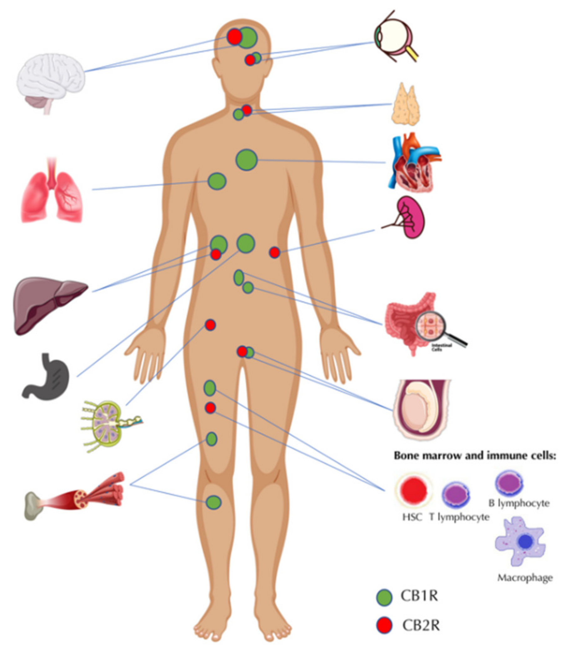
2. The Endocannabinoid System—A Brief Overview of the Key Functions and Elements

The ECS constitutes one of the most complex biological systems in the human body, creating a milieu responsible for the plethora of essential functions, including maintaining metabolic and cardiovascular homeostasis, mediating immune responses, and modulating the signaling within reward systems, as well as playing a relevant role in the brain physiology components, such as mood, cognition, and neurogenesis [11,12,13]. Furthermore, the ECS is involved in fertility, reproduction, and pregnancy [14]. In general, the ECS comprises (1) cannabinoid receptors (CBRs), (2) endocannabinoids (eCBs)—lipid ligands of cannabinoid receptors originating from omega-3 and omega-6 polyunsaturated fatty acids, among which anandamide (N-arachidonoyl-ethanolamine, AEA) and 2-arachidonoylglycerol (2-AG) received the most attention and were best described in the literature, (3) enzymes involved in bioformation and degradation of eCBs [15,16]. Cannabinoid receptors type 1 and type 2 (CB1R and CB2R) belong to the family of 7-transmembrane G-protein-coupled receptors (GPCRs) (Gi/o); thus, their activation, by suitable ligand, leads to the inhibition of adenyl cyclase and decreases in the concentration of cyclic adenosine monophosphate (cAMP). As a result, cAMP intensifies the activity of p42/p44 mitogen-activated protein kinase (MAPK) and Jun N-terminal kinase (JNK) which activate divergent nuclear transcription factors and alter the cellular metabolism. Additionally, cannabinoid receptors activate potassium and inhibit calcium channels in the presynaptic end of neurons that results in the overall inhibition of neurotransmitter release [17,18,19]. The distribution of CB1R is mainly clustered within brain structures, especially the neocortex, hippocampus, basal ganglia, and brain stem; likewise, its presence was confirmed in the skeletal muscles, lungs, testes, gastrointestinal tract, liver, pancreas, and adipose tissue. In turn, CB2R is mainly expressed on the immune cells (macrophages, T lymphocytes, B lymphocytes), hematopoietic stem, and progenitor cells present in bone marrow, as well as in the thymus and lymph nodes; thereby its activation triggers wide-range of immunomodulatory effects (Figure 1) [19,20,21,22]. Endocannabinoids are synthesized on demand in response to the elevated intracellular Ca2+ concentration [18]. 2-AG, which is synthesized from diacylglycerol (DAG), acts as a full agonist with a moderate affinity of cannabinoid receptors, whereas is hydrolyzed by monoacylglycerol lipase (MAGL) [23,24]. In turn, AEA constitutes a partial agonist of cannabinoid receptors synthesized from N-arachidonoyl phosphatidylethanolamine (NAPE) coordinated by N-acyl-phosphatidylethanolamine phospholipase D-like esterase (NAPE-PLD) and is degraded to arachidonic acid (AA) by the activity of fatty acid amid hydrolase (FAAH) [15,18]. Next to the well-known eCBs, analogous “endocannabinoid-like” lipid ligands were described namely oleoylethanolamine (OEA) and palmitoylethanolamide (PEA). Although OEA and PEA do not bind typical cannabinoid receptors, they possess a high affinity toward peroxisome proliferator-activated receptor alpha (PPARα) and orphan GPCRs. These compounds play an important role in hypoxia-induced intestinal permeability and suppression of the inflammation within the gastrointestinal tract, which highlights their potential importance in the aspect of cancer research [25,26]. Recently some researchers provided the term “endocannabinoidome” (eCBome), which refers to the broad and expanded signaling system that, apart from the aforementioned elements, contains a large number of different membrane and nuclear receptors, i.e., PPARs, transient receptor potential vanniloid 1 ion channel (TRPV1), various GPCRs, their endogenous ligands, and dozens of enzymes playing important roles in endocannabinoid signaling. Endocannabinoidome may constitute a promising target in the therapy of various pathologies in which the components of ECS seem to be involved; therefore, further research on the emerging new metabolic implications of eCBome remains in high demand [16,17,27,28].

3. The Endocannabinoid System and Metabolic Pathologies

Many researchers indicated that ECS might be widely involved in the regulation of energy balance and feeding behavior through both central and peripheral mechanisms [27]. Central mechanisms include hedonic energy regulation in the nucleus accumbens (NAc) and the ventral tegmental area that results in a higher motivation via the reward system. Moreover, another essential mechanism that leads to an increased appetite and higher food intake is homeostatic regulation in the hypothalamus, a major site of ECS action. Conversely, in the peripheral levels of action, ECS is widely associated with reduced expression of adiponectin, a protein hormone involved in suppressing glucose production in the liver and enhancing fatty acid oxidation in skeletal muscles. Another mechanism of ECS action is decreased AMPK activity (5’AMP-activated protein kinase), which is involved in insulin sensitivity, and its inhibition regulates lipogenesis and fatty acids or cholesterol synthesis in both the liver and adipose tissue [28]. Interestingly, the concentration of anandamide might be decreased via leptin action (an appetite-reducing adipose tissue-origin hormone that plays an essential role in food intake, body weight control, and metabolism), which enhances FAAH expression and leads to a lower concentration of anandamide in adipose tissue [29,30]. It is commonly known that low circulating levels of the leptin, as well as leptin resistance, are widely correlated with a higher risk of obesity and its aftermath [31,32]. Interestingly, a recent study conducted by Tam et al. indicated that a peripheral inhibition of CB1R in mice with diet-induced obesity (DIO) leads to an increase in the leptin sensitivity, which results in hypophagia via the reactivation of melanocortin signaling in the arcuate nucleus (ARC) in the hypothalamus [33]. Some studies reported that anandamide may lead to increased liver lipogenesis via the CB1R [34,35]. Another study by Karaliota et al. demonstrated that AEA may act as a PPAR-γ agonist and subsequently leads to the development of ECS-related adipogenesis in primary rat preadipocytes isolated from epididymal adipose tissue of male Wistar rats [36]. Numerous studies have indicated that endocannabinoids widely affect proliferation, as well as differentiation of adipocytes [33,37,38]. Amidst various components of endocannabinoidome, researchers point out the relevance of the link between the ECS and ion channel TRPV1 in ensuring undisturbed energy homeostasis. It is highly expressed within the central nervous system as well as peripheral tissues, including stomach and adipose tissue. TRPV1 is a non-selective cation channel activated by capsaicin, high temperature, and acidosis that belong to the transient receptor potential protein superfamily [39]. Moreover, among various stimuli which interact with TRPV1, N-acyldopamines, leukotriene B4 and N-acylethanoloamines (e.g., AEA) may be distinguished as TRPV1 agonists. The effect of endocannabinoids on TRPV1 may be exerted through direct activation on this channel or indirectly through activation of the CB1R that activates the phospholipase C-PKC pathway. However, endocannabinoids may also activate CB1R, which leads to the inhibition of the adenylate cyclase-PKA pathway, resulting in diminished TRPV1 activity. Those correlations are still unclear and, at least in part, depend on the concentration of AEA [37,40]. The metabolic functions of TRPV1 include its influence on glucose metabolism and lipid oxidation, regulation of the appetite (via interaction with appetite-regulating hormones e.g., ghrelin and leptin) as well as activation of thermogenesis [40]. Numerous studies highlighted that impaired signaling within TRPV1 may contribute to the development of various metabolic disorders, including obesity and T2DM [38,41]. Therefore, the complex interactions between the ECS and TRPV1 are still a subject of discussion and should be clarified in future studies.

3.1. Obesity and Non-Alcoholic Fatty Liver Disease (NAFLD)

A substantial correlation between metabolic disturbances and endocannabinoid system overactivation was unequivocally demonstrated in both in vivo animal studies and clinical trials [42,43]. So far, a vast number of studies have linked obesity with higher circulating levels of endocannabinoids [44,45]. It is known that obesity and higher food intake might lead to an overactivation of ECS by increased cannabinoid receptor activity. Moreover, this might become a vicious circle—obesity-related ECS activation might contribute to an intensified lipogenesis, higher food intake, and consequently lead to further fat storage. Increased fat accumulation in adipose tissue might be associated with activation of CB1R since it was indicated that those receptors might increase the activity of lipoprotein lipase, a key enzyme regulating triglyceride hydrolysis to free fatty acids, which are subsequently transported and deposited in the liver [46]. Thus, inhibition of CB receptors seems to be relevant.
Interestingly, rimonabant was the first CB1R antagonist approved for the treatment of obesity since clinical trials revealed that it might promote weight loss in obese patients. However, due to a number of serious psychiatric adverse effects, e.g., anxiety or depression, the clinical use of rimonabant was discontinued, and the drug was withdrawn from the worldwide market [47]. A human-based study by Bennetzen et al. demonstrated that levels of 2-arachidonoylglycerol were significantly reduced in the abdominal adipose tissue, whereas weight loss led to a normalization of this endocannabinoid. Furthermore, the same study indicated that the low expression of CB1 in abdominal adipose tissue was normalized after weight loss, whereas in gluteal adipose tissue the CB1 expression was decreased after weight loss. Thus, this research, together with other related studies, presented substantial results that ECS is widely dysregulated among individuals suffering from obesity [48,49] Moreover, it indicates that not only CB receptor expression is affected by obesity but also levels of ECs. Indeed, some researchers reported that in obese patients, both AEA and 2-AG levels are increased, and elevated plasma concentrations of endocannabinoids (ECs) might be correlated with obesity aftermath, e.g., cardiovascular diseases, as well as non-alcoholic fatty liver disease (NAFLD) [50,51,52]. Moreover, increased concentrations of endocannabinoids observed in small intestinal mucosa and plasma of high-fat diet-fed obese mice inhibited may change TRPV1 activity, which is involved in energy homeostasis [53]. However, because of various interactions of this channel with other systems, the precise role of TRPV1 in regulating of food intake and energy expenditure needs further investigation. All these outcomes highlight those alterations in dysregulated ECS signaling are very complex and should be investigated in depth.
Non-alcoholic fatty liver disease is an obesity-related metabolic pathology induced by enhanced triacylglycerols accumulation leading to hepatic steatosis, excluding alcohol abuse as a pathogenic factor [54]. Numerous studies have indicated that NAFLD occurrence is widely correlated with the dysfunction of the endocannabinoid system because lipid accumulation in the liver might be mediated via alterations in ECS [55,56]. It is known that the basal hepatic expression of cannabinoid receptors is indistinct, with low levels of both CB1R and CB2R. However, an increased expression of the cannabinoid receptors was proven in NAFLD. Moreover, CB1R was shown as a key mediator of insulin resistance development, increased liver lipogenesis, and steatosis in both animal and human studies, whereas CB2R might be considered as a promoter of inflammatory processes underlying the pathogenesis of liver steatosis [57]. It is known that blockage of cannabinoid receptor type 1 may result in attenuation of hepatic oxidative stress, as well as the impairment of inflammatory response by inhibiting the production of pro-inflammatory cytokines [58,59] Furthermore, Irungbam et al. recently demonstrated in both in vitro (AML12 liver cell line) and in vivo studies on mice with a global CB1R receptor knockout that lower CB1R signaling reduced liver steatosis via downregulation of perilipin 2—the protein that is involved in the suppression of lipolysis and lipid accumulation [60]. Shi et al. conducted in vitro studies in which application of CB1R antagonist in HepG2 cells resulted in a significant decrease in factors widely involved in lipogenesis, i.e., sterol regulatory element-binding protein (SREBP1c), carbohydrate responsive element-binding protein (ChREBP), liver X receptors (LXRs), acetyl-CoA carboxylase (ACC1) and fatty acid synthase (FAS) [61]. Moreover, Deveaux et al. conducted a study on mice which indicated that the inactivation of cannabinoid receptor type 2 reduced steatosis, liver triacylglycerol concentration, and obesity-associated inflammation caused by a high-fat diet [62]. Interestingly, CB1R was defined as a pro-fibrogenic activator, while cannabinoid receptor type 2 was proposed to have anti-fibrogenic properties in the liver [35,63]. Thus, ECs and their receptors might be considered as a possible and promising target for the treatment of liver steatosis and its metabolic aftermath.

3.2. Insulin Resistance and Type 2 Diabetes

Insulin resistance (IR), defined as a condition with impaired insulin action and response in tissues, is widely considered to be one of the most fundamental factors associated with the development of many metabolic disorders, e.g., type 2 diabetes mellitus or metabolic syndrome [64,65]. A vast number of studies has provided evidence indicating that activation of peripheral CB1R located in insulin-dependent tissues, i.e., liver, adipose tissue, or skeletal muscles, is widely associated with the development of dyslipidemia, insulin resistance, and type 2 diabetes [66]. Additionally, plenty of studies reported that CB1R blockage leads to increased insulin sensitivity and, subsequently, improves both glucose and lipid metabolism [67,68]. Considering the pathophysiological basis of IR development, Liu et al. demonstrated that it is correlated with activation of CB1R that inhibits insulin signaling via endoplasmic reticulum stress-dependent suppression of enzymes involved in the insulin signaling pathway, e.g., PKB/Akt [69]. Interestingly, further Liu et al. studies provided notable results that blockage of CB1R in obese mice improved glycemic control through the hepatic Sirt1/mTORC2/Akt pathway, as well as increased fatty acid β-oxidation via LKB1/AMPK signaling route [70]. A study conducted by Jourdan et al. demonstrated that intravenous administration of β-D-glucan-encapsulated siRNA to knock down CB1R gene expression, improved insulin sensitivity in Kupfer cells of mice with obesity. Thus, it might be said that the CB1R played a significant role in the development of liver insulin resistance via Kupffer cell-related inflammatory mechanisms [71]. Considering the CB2R, principally expressed in immune tissues, several in vitro and in vivo studies demonstrated CB2R-mediated anti-inflammatory effects of endocannabinoids, i.e., AEA and 2-AG, as well as synthetic CB2R agonists in models of various metabolic diseases, e.g., obesity [72,73,74]. Furthermore, Verty et al. reported that activation of CB2R reduced the inflammatory response and promoted anti-obesity effects by reducing body weight, while not changing mood-related behaviors [75]. Although plenty of studies support the claim that endocannabinoids are anti-inflammatory mediators, a limited amount of evidence demonstrated the pro-inflammatory action of endocannabinoids via CB2R activation [76,77]. Moreover, Turcotte et al. showed that most of the pro-inflammatory effects involve 2-AG, but not AEA [78].
Considering the correlation between the endocannabinoid system and oxidative stress, a study conducted by Mendizabal-Zubiaga et al. showed that the mitochondrial activation of CB1R in muscle cells might be correlated with the mitochondrial regulation of oxidative activity. Thus, this might be highly associated with the development of insulin resistance, since mitochondrial dysfunction is correlated with a loss of muscular oxidative capacity in response to a high intake of fatty acids, and therefore may be involved in the development of metabolic disorders [79,80]. Notably, Dipanjan et al. demonstrated that activation of the CB1R is involved in gluconeogenesis via direct activation of an endoplasmic reticulum (ER) membrane-localized stress-dependent liver-specific transcription factor called CREBH—cyclic AMP-responsive element-binding protein H, which is considered to be one of the key mediators in both glucose and lipid metabolism [81,82]. Thus, it may be said that there is a molecular association between the activation of the endocannabinoid system and levels of circulating glucose.
An endocannabinoid-related study conducted by Jourdan et al. on the Zucker diabetic rats indicated that the loss of pancreatic beta cell functions and its amount is associated with the release of pro-inflammatory cytokines, i.e., IL-1β and IL-18. Those molecules, acting as paracrine substances that induce beta-cell apoptosis, are released from infiltrating M1 macrophages as a result of macrophages-origin CB1R signaling that participates in selective activation of the Nlrp3-ASC inflammasome [83]. Moreover, a recent study by Kim et al. conducted on pancreatic beta-cell lines MIN6 and βTC6 incubated with synthetic CB1R agonist WIN55,212-2 showed a decreased expression of anti-apoptotic protein Bcl-2 and cell cycle regulator cyclin D2, together with caspase-3-dependent apoptosis afterward [84]. Thus, these two studies unequivocally determine the effect of CB1R activation in beta-cell apoptosis and therefore insulin signaling deficiency and the development of type 2 diabetes. It is commonly known that insulin resistance, as well as T2DM, are widely associated with an increased tissue concentration of sphingolipids—a class of lipids whose fractions are also involved in cellular growth, differentiation, and regulation of apoptosis in addition to playing an essential role as structural molecules in cellular membranes [85,86]. Cinar et al. linked the action of the endocannabinoid system and insulin resistance development together with excessive sphingolipids deposition. It was indicated in a study conducted on C57Bl6/J mice with high-fat diet-induced obesity (DIO) that the factor, which is widely involved in the development of hepatic IR, is increased de novo ceramides synthesis, which is mediated by endoplasmic reticulum (ER) stress-dependent activation of CB1R [87]. Thus, it may be assumed that changes in CB receptors and other components of the ECS may be associated with a favorable prognosis not only of obesity and NAFLD but also IR and type 2 diabetes.

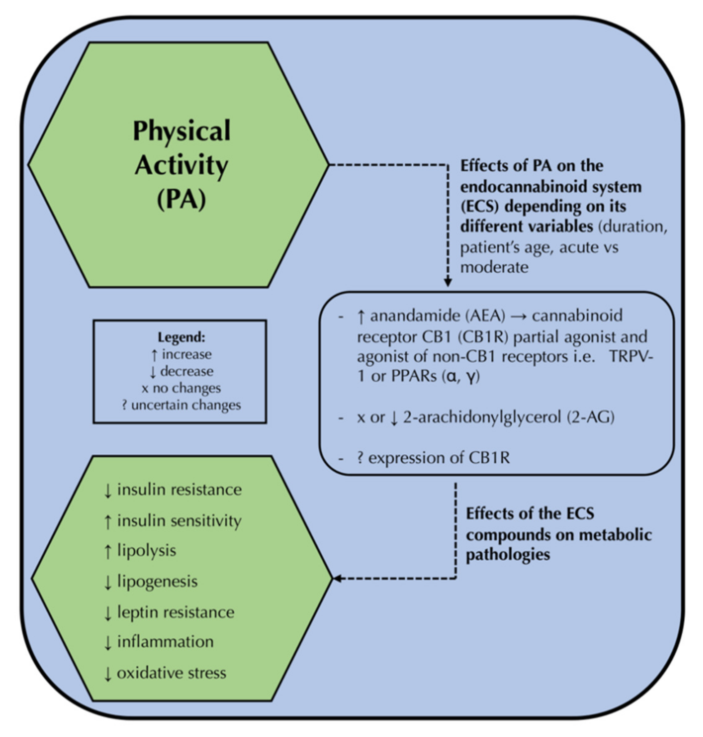
4. The Endocannabinoid System and Physical Activity

Physical activity is broadly portrayed as one of the most popular, potent, cost-friendly, and pleasant ways to maintain a prolonged and healthy life. It constitutes a major pillar in the treatment of metabolic disorders next to the proper diet, pharmacotherapy, chemoprevention, and in the most severe cases, bariatric interventions [88]. What is more, it affects human mental health, leading to mood, perception, concentration, and creativity improvement as well as producing a very interesting phenomenon called “runner’s high” [89]. Despite widely known positive effects in the therapy of various diseases, the exact molecular mechanisms induced by PA are still a subject of discussion. Interestingly, many studies outlined the influence of different types of physical activity on endocannabinoid signaling. These results, however, seem to be partially unclear and confusing; therefore, they deserve to be gathered and described comprehensively. The study conducted by Sparling et al. was arguably the first showing that moderate exercises led to the increased level of AEA but not 2-AG in the sera of healthy subjects after 50 min of treadmill running or cycling on a stationary bike at the level of 70–80% of maximum heart rate [90]. Other studies also indicated that PA, especially at medium intensity, led to the increased blood level of AEA, whereas 2-AG remained unchanged [91,92,93,94]. Heyman et al. revealed the link between significantly increased plasma AEA, OEA, and PEA levels during intense exercise followed by 15 min of recovery and elevated serum brain-derived neurotrophic factor (BDNF) levels, thereby indicating an important role of the ECS in the production of neuroplastic and antidepressant effects triggered by PA. A simultaneous increase in OEA and PEA along with AEA probably comes from the fact that these compounds share similar synthesis pathways, whereas 2-AG formation seems to be regulated by different metabolic routes [95]. Conversely, the study conducted by Cedernaes et al. showed elevated blood 2-AG and OEA levels after acute ergometer exercises, while AEA remained stable [96]. The possible reason for this discrepancy may lie in the fasted status of the participants before activity [97]. Brellenthin et al. observed that both AEA and 2-AG are increased after aerobic exercises in the blood of healthy individuals, but the increase in AEA was more substantial in the prescribed by the investigator activity in comparison with self-selected ones, based on personal preferences [98]. Thompson et al. in their study on mice showed that males after exercise tended to have more increased 2-AG serum levels, whereas AEA content was higher in female subjects. These results raised an interesting concern that alterations in the circulating eCBs induced by PA may be at least partially, sex-dependent; however, more data is needed to clarify this issue [99]. It may be suspected that various changes in the levels of eCBs may be a result of different type and time of exercise, but they all exerted changes in the nervous system. Furthermore, Crombie et al. showed that acute isometric activity led to the elevated plasma levels of both AEA and 2-AG as well as OEA and PEA, which resulted in overall analgesic effects [100]. On the other hand, most animal studies pointed out that aerobic activity, i.e., swimming or treadmill running, was associated with increased expression and density of both CB1R and CB2R within the central nervous system, namely the hippocampus, striatum, and spinal cord, that were associated with positive neurological outcomes, including improved cognition, analgesia, and reduced neuroinflammation [101,102,103]. Nevertheless, there is evidence, that PA may lead to the downregulated expression of cannabinoid receptors in the aforementioned structures which suggests that further investigation is required to elucidate these discrepancies [101,104]. Physical activity, depending on such factors as type, frequency, intensity, and duration leads to the (I) activation of the ECS signaling, (II) significantly affects circulating blood levels of the endocannabinoids, mainly AEA and 2-AG, as well as (III) change in expression of the cannabinoid receptors (CB1R and CB2R). Despite the fact, that molecular interaction between the ECS and PA has been well described in the literature, it is still difficult to point out clear, unequivocal changes within endocannabinoid signaling induced by various types of exercise; thus, this topic needs further investigation in future studies.

5. The Triad—Physical Activity, the Endocannabinoid System, and the Novel Therapeutic Approach to Metabolic Disorders—How All These Components May Be Linked?

The summary of the correlation between physical activity, its effect on the ECS, and subsequent metabolic pathologies is presented in the Figure 2.
There is no doubt that the involvement of the ECS in the pathogenesis of metabolic pathologies is crucial [105]. Therefore, targeting the elements of ECS, including receptors, endocannabinoids, and enzymes, by physical exercises and a healthy diet may exert robust beneficial outcomes in either prevention or supportive treatment of metabolic disorders [9]. To date, only a few studies conducted on humans (Table 1) and animal models, namely Wistar rats and C57B1/6J mice (Table 2), showed the positive influence of chronic and acute PA (e.g., treadmill running, swimming, and aerobic exercises) or combined with the appropriate caloric restriction on the endocannabinoid signaling and subsequently, auspicious course of the metabolic syndrome, for instance, body weight loss, decreased waist circumference and visceral adipose tissue percentage, improved insulin signaling, or enhanced lipid profile. Additionally, it is noteworthy that physical exercises and accompanying increased concentration of eCBs significantly improved mood, sense of well-being, and increased motivation through reward systems that may constitute important factors for maintaining patient cooperation with a physician, thereby providing efficient patient compliance, which is crucial during therapy targeting metabolic diseases. These effects probably arise from the fact that CB1R shares similar localization in the midbrain with dopamine receptors and may enhance the activity of dopaminergic neurons in ventral tegmentum and substantia nigra [106]. Interestingly, observed decreased blood eCBs levels after PA among obese individuals are opposite to those described in the studies involving healthy patients, which, as was mentioned above, indicated increased concentrations of AEA and/or 2-AG [91,92,93,94,98,99]. A similar concern deals with the altered expression of cannabinoid receptors. Indeed, a lot of experts suggested that PA in healthy animals was correlated with upregulated CB1R expression within brain structures which, on the contrary, was lowered in the brain and subcutaneous/visceral adipose tissue of obese mice. Neither animal nor human studies revealed consistent outcomes concerning the expression of genes encoding both cannabinoid receptors and enzymes involved in eCBs metabolism like FAAH, DAGL, MAGL, and NAPE-PLD (Table 1 and Table 2). However, Schönke et al., using the online tool MetaMEx, found downregulated skeletal muscle expression of DAGLβ as well as catabolic enzymes, namely MAGL and FAAH, in healthy individuals and patients with metabolic syndrome who underwent chronic aerobic or resistant exercises [107]. It is noteworthy that in vivo studies conducted by Gamelin et al. revealed that obese Wistar rats after a 12-week running period showed an increased expression of TRPV1 and CB1R both in the hippocampus or subcutaneous adipose tissue, which suggests potentially enhanced signaling within this ion channel and may result in the favorable course of obesity and other metabolic conditions [10,108]. In conclusion, all discrepancies and concerns related to ECS signaling suggest unclear and paradoxical involvement of the ECS in both physiological and impaired energy balance and might indicate possible compensation mechanisms in response to the significantly diminished metabolic homeostasis. It strengthens the evidence that the ECS itself constitutes a complex molecular apparatus that may be involved in producing beneficial therapeutic effects and may act as a culprit due to confirmed dysregulation in metabolic diseases [107]. A mutual correlation between the ECS and PA should be discussed more in-depth in the future considering the significant deficit of the studies in this area.

6. Conclusions

To the best of our knowledge, this is the first review directly and comprehensively discussing the uncharted link between physical activity and its influence on the endocannabinoid signaling in the aspect of beneficial effects in the management of metabolic disorders. Considering the very alarming worldwide prevalence of these diseases as well as the unexplored potential of the topic, we believe that this paper, at least in part, will encourage researchers toward investigating this interesting, yet very complicated interplay. ECS and physical activity constitute robust and valuable therapeutic and preventive approaches that may significantly contribute to the decreased socioeconomic burden and the reduced annual number of patients suffering from obesity and other metabolic disorders. The future investigation should primarily encompass further discovery of the link between physical activity, alterations within endocannabinoid signaling and subsequently improved metabolic status of overweight, obese, and diabetic individuals.

Author Contributions

Conceptualization, T.C., H.Z. and K.K.-N.; resources, K.B., A.K. and W.B.; writing—original draft preparation, T.C. and H.Z.; writing—review and editing, K.K.-N. and A.C.; visualization, T.C. and H.Z.; supervision, A.C.; project administration, T.C. All authors have read and agreed to the published version of the manuscript.

Funding

This work was supported by the Medical University of Bialystok (grant number: SUB/1/DN/20/002/1118).

Institutional Review Board Statement

Not applicable.

Informed Consent Statement

Not applicable.

Data Availability Statement

Not applicable.

Conflicts of Interest

The authors declare no conflict of interest.

References

  1. Caballero, B. Humans against Obesity: Who Will Win? Adv. Nutr. 2019, 10, S4–S9. [Google Scholar] [CrossRef] [PubMed]
  2. Mayoral, L.P.-C.; Andrade, G.B.; Mayoral, E.P.-C.; Teresa, H.H.; Socorro, P.C.; Francisco, J.R.C.; Héctor, A.C.-F.; Cruz, M.M.; Santiago, A.D.P.; Alpuche, J.J.; et al. Obesity subtypes, related biomarkers & heterogeneity. Indian J. Med. Res. 2020, 151, 11–21. [Google Scholar] [CrossRef] [PubMed]
  3. Smith, K.B.; Smith, M.S. Obesity Statistics. Prim. Care Clin. Off. Pract. 2016, 43, 121–135. [Google Scholar] [CrossRef] [PubMed]
  4. Yaribeygi, H.; Butler, A.E. Insulin resistance: Review of the underlying molecular mechanisms. J. Cell. Physiol. 2019, 234, 8152–8161. [Google Scholar] [CrossRef]
  5. Antonopoulos, A.S.; Tousoulis, D. Themolecularmechanisms of obesity paradox. Cardiovasc. Res. 2017, 113, 1074–1086. [Google Scholar] [CrossRef]
  6. Mazier, W.; Saucisse, N.; Gatta-Cherifi, B.; Cota, D. The Endocannabinoid System: Pivotal Orchestrator of Obesity and Metabolic Disease. Trends Endocrinol. Metab. 2015, 26, 524–537. [Google Scholar] [CrossRef]
  7. Tantimonaco, M.; Ceci, R.; Sabatini, S.; Catani, M.V.; Rossi, A.; Gasperi, V.; Maccarrone, M. Physical activity and the endocannabinoid system: An overview. Cell. Mol. Life Sci. 2014, 71, 2681–2698. [Google Scholar] [CrossRef]
  8. Watkins, B.A. Diet, endocannabinoids, and health. Nutr. Res. 2019, 70, 32–39. [Google Scholar] [CrossRef]
  9. Heyman, E.; Gamelin, F.X.; Aucouturier, J.; Di Marzo, V. The role of the endocannabinoid system in skeletal muscle and metabolic adaptations to exercise: Potential implications for the treatment of obesity. Obes. Rev. 2012, 13, 1110–1124. [Google Scholar] [CrossRef]
  10. Gamelin, F.X.; Aucouturier, J.; Iannotti, F.A.; Piscitelli, F.; Mazzarella, E.; Aveta, T.; Leriche, M.; Dupont, E.; Cieniewski-Bernard, C.; Montel, V.; et al. Effects of chronic exercise on the endocannabinoid system in Wistar rats with high-fat diet-induced obesity. J. Physiol. Biochem. 2016, 72, 183–199. [Google Scholar] [CrossRef]
  11. Meccariello, R. Endocannabinoid system in health and disease: Current situation and future perspectives. Int. J. Mol. Sci. 2020, 21, 3549. [Google Scholar] [CrossRef] [PubMed]
  12. Nesto, R.W.; Mackie, K. Endocannabinoid system and its implications for obesity and cardiometabolic risk. Eur. Hearth J. Suppl. 2008, 10, 34–41. [Google Scholar] [CrossRef] [Green Version]
  13. Mechoulam, R.; Parker, L.A. The Endocannabinoid System and the Brain. Annu. Rev. Psychol. 2013, 64, 21–47. [Google Scholar] [CrossRef] [Green Version]
  14. Bovolin, P.; Cottone, E.; Pomatto, V.; Fasano, S.; Pierantoni, R.; Cobellis, G.; Meccariello, R. Endocannabinoids are involved in male vertebrate reproduction: Regulatory mechanisms at central and gonadal level. Front. Endocrinol. 2014, 5, 1–9. [Google Scholar] [CrossRef] [PubMed]
  15. Iannotti, F.A.; Di Marzo, V. The gut microbiome, endocannabinoids and metabolic disorders. J. Endocrinol. 2021, 248, R83–R97. [Google Scholar] [CrossRef] [PubMed]
  16. Freitas, H.R.; Isaac, A.R.; Malcher-Lopes, R.; Diaz, B.L.; Trevenzoli, I.H.; De Melo Reis, R.A. Polyunsaturated fatty acids and endocannabinoids in health and disease. Nutr. Neurosci. 2018, 21, 695–714. [Google Scholar] [CrossRef] [PubMed]
  17. Cristino, L.; Bisogno, T.; Di Marzo, V. Cannabinoids and the expanded endocannabinoid system in neurological disorders. Nat. Rev. Neurol. 2020, 16, 9–29. [Google Scholar] [CrossRef]
  18. Berk, K.; Bzdega, W.; Konstantynowicz-nowicka, K.; Charytoniuk, T.; Zywno, H.; Chabowski, A. Phytocannabinoids—A green approach toward non-alcoholic fatty liver disease treatment. J. Clin. Med. 2021, 10, 393. [Google Scholar] [CrossRef]
  19. Bozkurt, T.E. Endocannabinoid system in the airways. Molecules 2019, 24, 4626. [Google Scholar] [CrossRef] [Green Version]
  20. Joshi, N.; Onaivi, E.S. Endocannabinoid System Components: Overview and Tissue Distribution. Adv. Exp. Med. Biol. 2019, 1162, 1–12. [Google Scholar] [CrossRef]
  21. Mackie, K. Cannabinoid receptors: Where they are and what they do. J. Neuroendocrinol. 2008, 20, 10–14. [Google Scholar] [CrossRef] [PubMed]
  22. Jiang, S.; Zagozdzon, R.; Jorda, M.A.; Parmar, K.; Fu, Y.; Williams, J.S.; Wood, J.A.T.; Makriyannis, A.; Banu, N.; Avraham, S.; et al. Endocannabinoids are expressed in bone marrow stromal niches and play a role in interactions of hematopoietic stem and progenitor cells with the bone marrow microenvironment. J. Biol. Chem. 2010, 285, 35471–35478. [Google Scholar] [CrossRef] [PubMed] [Green Version]
  23. Zou, S.; Kumar, U. Cannabinoid receptors and the endocannabinoid system: Signaling and function in the central nervous system. Int. J. Mol. Sci. 2018, 19, 833. [Google Scholar] [CrossRef] [PubMed] [Green Version]
  24. Di Marzo, V. New approaches and challenges to targeting the endocannabinoid system. Nat. Rev. Drug Discov. 2018, 17, 623–639. [Google Scholar] [CrossRef] [PubMed]
  25. Karwad, M.A.; Macpherson, T.; Wang, B.; Theophilidou, E.; Sarmad, S.; Barrett, D.A.; Larvin, M.; Wright, K.L.; Lund, J.N.; O’Sullivan, S.E. Oleoylethanolamine and palmitoylethanolamine modulate intestinal permeability in vitro via TRPV1 and PPARα. FASEB J. 2017, 31, 469–481. [Google Scholar] [CrossRef] [Green Version]
  26. Karwad, M.A.; Couch, D.G.; Wright, K.L.; Tufarelli, C.; Larvin, M.; Lund, J.; O’Sullivan, S.E. Endocannabinoids and endocannabinoid-like compounds modulate hypoxia-induced permeability in CaCo-2 cells via CB1, TRPV1, and PPARα. Biochem. Pharmacol. 2019, 168, 465–472. [Google Scholar] [CrossRef]
  27. Gatta-Cherifi, B.; Cota, D. New insights on the role of the endocannabinoid system in the regulation of energy balance. Int. J. Obes. 2016, 40, 210–219. [Google Scholar] [CrossRef]
  28. Bermudez-Silva, F.J.; Cardinal, P.; Cota, D. The role of the endocannabinoid system in the neuroendocrine regulation of energy balance. J. Psychopharmacol. 2012, 26, 114–124. [Google Scholar] [CrossRef]
  29. Balsevich, G.; Sticht, M.; Bowles, N.P.; Singh, A.; Lee, T.T.Y.; Li, Z.; Chelikani, P.K.; Lee, F.S.; Borgland, S.L.; Hillard, C.J.; et al. Role for fatty acid amide hydrolase (faah) in the leptin-mediated effects on feeding and energy balance. Proc. Natl. Acad. Sci. USA 2018, 115, 7605–7610. [Google Scholar] [CrossRef] [Green Version]
  30. Buettner, C.; Muse, E.D.; Cheng, A.; Chen, L.; Scherer, T.; Pocai, A.; Su, K.; Cheng, B.; Li, X.; Harvey-White, J.; et al. Leptin controls adipose tissue lipogenesis via central, STAT3-independent mechanisms. Nat. Med. 2008, 14, 667–675. [Google Scholar] [CrossRef]
  31. Rossi, F.; Punzo, F.; Umano, G.R.; Argenziano, M.; Miraglia Del Giudice, E. Role of cannabinoids in obesity. Int. J. Mol. Sci. 2018, 19, 2690. [Google Scholar] [CrossRef] [PubMed] [Green Version]
  32. Gruzdeva, O.; Borodkina, D.; Uchasova, E.; Dyleva, Y.; Barbarash, O. Leptin resistance: Underlying mechanisms and diagnosis. Diabetes, Metab. Syndr. Obes. Targets Ther. 2019, 12, 191–198. [Google Scholar] [CrossRef] [PubMed] [Green Version]
  33. Tam, J.; Szanda, G.; Drori, A.; Liu, Z.; Cinar, R.; Kashiwaya, Y.; Reitman, M.L.; Kunos, G. Peripheral cannabinoid-1 receptor blockade restores hypothalamic leptin signaling. Mol. Metab. 2017, 6, 1113–1125. [Google Scholar] [CrossRef]
  34. DiPatrizio, N.V.; Piomelli, D. The thrifty lipids: Endocannabinoids and the neural control of energy conservation. Trends Neurosci. 2012, 35, 403–411. [Google Scholar] [CrossRef] [Green Version]
  35. Bazwinsky-Wutschke, I.; Zipprich, A.; Dehghani, F. Endocannabinoid system in hepatic glucose metabolism, fatty liver disease, and cirrhosis. Int. J. Mol. Sci. 2019, 20, 2516. [Google Scholar] [CrossRef] [Green Version]
  36. Karaliota, S.; Siafaka-Kapadai, A.; Gontinou, C.; Psarra, K.; Mavri-Vavayanni, M. Anandamide increases the differentiation of rat adipocytes and causes PPARλ and cB1 receptor upregulation. Obesity 2009, 17, 1830–1838. [Google Scholar] [CrossRef] [PubMed]
  37. Muller, C.; Lynch, D.L.; Hurst, D.P.; Reggio, P.H. A Closer Look at Anandamide Interaction With TRPV1. Front. Mol. Biosci. 2020, 7, 1–8. [Google Scholar] [CrossRef]
  38. Lee, E.; Jung, D.Y.; Kim, J.H.; Patel, P.R.; Hu, X.; Lee, Y.; Azuma, Y.; Wang, H.F.; Tsitsilianos, N.; Shafiq, U.; et al. Transient receptor potential vanilloid type-1 channel regulates diet-induced obesity, insulin resistance, and leptin resistance. FASEB J. 2015, 29, 3182–3192. [Google Scholar] [CrossRef] [Green Version]
  39. Lotteau, S.; Ducreux, S.; Romestaing, C.; Legrand, C.; Van Coppenolle, F. Characterization of Functional TRPV1 Channels in the Sarcoplasmic Reticulum of Mouse Skeletal Muscle. PLoS ONE 2013, 8, e58673. [Google Scholar] [CrossRef] [Green Version]
  40. Christie, S.; Wittert, G.A.; Li, H.; Page, A.J. Involvement of TRPV1 Channels in Energy Homeostasis. Front. Endocrinol. 2018, 9, 1–14. [Google Scholar] [CrossRef] [Green Version]
  41. Park, S.; Zhang, X.; Lee, N.R.; Jin, H.S. TRPV1 Gene Polymorphisms Are Associated with Type 2 Diabetes by Their Interaction with Fat Consumption in the Korean Genome Epidemiology Study. J. Nutrigenet. Nutrigenomics 2016, 9, 47–61. [Google Scholar] [CrossRef] [PubMed]
  42. Muller, T.; Demizieux, L.; Troy-Fioramonti, S.; Gresti, J.; de Barros, J.P.P.; Berger, H.; Vergès, B.; Degrace, P. Overactivation of the endocannabinoid system alters the antilipolytic action of insulin in mouse adipose tissue. Am. J. Physiol. Endocrinol. Metab. 2017, 313, E26–E36. [Google Scholar] [CrossRef] [Green Version]
  43. Blüher, M.; Engeli, S.; Klöting, N.; Berndt, J.; Fasshauer, M.; Bátkai, S.; Pacher, P.; Schön, M.R.; Jordan, J.; Stumvoll, M. Dysregulation of the peripheral and adipose tissue endocannabinoid system in human abdominal obesity. Diabetes 2006, 55, 3053–3060. [Google Scholar] [CrossRef] [PubMed] [Green Version]
  44. Côté, M.; Matias, I.; Lemieux, I.; Petrosino, S.; Alméras, N.; Després, J.P.; Di Marzo, V. Circulating endocannabinoid levels, abdominal adiposity and related cardiometabolic risk factors in obese men. Int. J. Obes. 2007, 31, 692–699. [Google Scholar] [CrossRef] [PubMed] [Green Version]
  45. Vähätalo, L.H.; Ruohonen, S.T.; Mäkelä, S.; Ailanen, L.; Penttinen, A.M.; Stormi, T.; Kauko, T.; Piscitelli, F.; Silvestri, C.; Savontaus, E.; et al. Role of the endocannabinoid system in obesity induced by neuropeptide y overexpression in noradrenergic neurons. Nutr. Diabetes 2015, 5, 1–8. [Google Scholar] [CrossRef] [PubMed] [Green Version]
  46. Cota, D.; Marsicano, G.; Tschöp, M.; Grübler, Y.; Flachskamm, C.; Schubert, M.; Auer, D.; Yassouridis, A.; Thöne-Reineke, C.; Ortmann, S.; et al. The endogenous cennabinoid system affects energy balance via central orexigenic drive and peripheral lipogenesis. J. Clin. Invest. 2003, 112, 423–431. [Google Scholar] [CrossRef]
  47. Müller, T.D.; Clemmensen, C.; Finan, B.; Dimarchi, R.D.; Tschöp, M.H. Anti-obesity therapy: From rainbow pills to polyagonists. Pharmacol. Rev. 2018, 70, 712–746. [Google Scholar] [CrossRef] [Green Version]
  48. Bennetzen, M.F.; Wellner, N.; Ahmed, S.S.; Ahmed, S.M.; Diep, T.A.; Hansen, H.S.; Richelsen, B.; Pedersen, S.B. Investigations of the human endocannabinoid system in two subcutaneous adipose tissue depots in lean subjects and in obese subjects before and after weight loss. Int. J. Obes. 2011, 35, 1377–1384. [Google Scholar] [CrossRef] [Green Version]
  49. Izzo, A.A.; Piscitelli, F.; Capasso, R.; Aviello, G.; Romano, B.; Borrelli, F.; Petrosino, S.; Di Marzo, V. Peripheral endocannabinoid dysregulation in obesity: Relation to intestinal motility and energy processing induced by food deprivation and re-feeding. Br. J. Pharmacol. 2009, 158, 451–461. [Google Scholar] [CrossRef] [Green Version]
  50. Little, T.J.; Cvijanovic, N.; Dipatrizio, N.V.; Argueta, D.A.; Rayner, C.K.; Feinle-Bisset, C.; Young, R.L. Endocannabinoids and cannabinoid receptors as regulators of endocrine functions and tissue metabolism: Plasma endocannabinoid levels in lean, overweight, and obese humans: Relationships to intestinal permeability markers, inflammation, and incretin secret. Am. J. Physiol. Endocrinol. Metab. 2018, 315, E489–E495. [Google Scholar] [CrossRef]
  51. Sipe, J.C.; Scott, T.M.; Murray, S.; Harismendy, O.; Simon, G.M.; Cravatt, B.F.; Waalen, J. Biomarkers of endocannabinoid system activation in severe obesity. PLoS ONE 2010, 5, e8792. [Google Scholar] [CrossRef] [Green Version]
  52. Quercioli, A.; Pataky, Z.; Vincenti, G.; Makoundou, V.; Di Marzo, V.; Montecucco, F.; Carballo, S.; Thomas, A.; Staub, C.; Steffens, S.; et al. Elevated endocannabinoid plasma levels are associated with coronary circulatory dysfunction in obesity. Eur. Heart J. 2011, 32, 1369–1378. [Google Scholar] [CrossRef] [Green Version]
  53. Argueta, D.A.; DiPatrizio, N.V. Peripheral endocannabinoid signaling controls hyperphagia in western diet-induced obesity. Physiol. Behav. 2017, 171, 32–39. [Google Scholar] [CrossRef] [Green Version]
  54. Charytoniuk, T.; Drygalski, K.; Konstantynowicz-Nowicka, K.; Berk, K.; Chabowski, A. Alternative treatment methods attenuate the development of NAFLD: A review of resveratrol molecular mechanisms and clinical trials. Nutrition 2017, 34, 108–117. [Google Scholar] [CrossRef]
  55. Auguet, T.; Berlanga, A.; Guiu-Jurado, E.; Terra, X.; Martinez, S.; Aguilar, C.; Filiu, E.; Alibalic, A.; Sabench, F.; Hernández, M.; et al. Endocannabinoid receptors gene expression in morbidly obese women with nonalcoholic fatty liver disease. BioMed Res. Int. 2014, 2014, 502542. [Google Scholar] [CrossRef]
  56. Purohit, V.; Rapaka, R.; Shurtleff, D. Role of cannabinoids in the development of fatty liver (steatosis). AAPS J. 2010, 12, 233–237. [Google Scholar] [CrossRef] [Green Version]
  57. Alswat, K.A. The role of endocannabinoids system in fatty liver disease and therapeutic potentials. Saudi J. Gastroenterol. 2013, 19, 144–151. [Google Scholar] [CrossRef]
  58. Jorgačević, B.; Vučević, D.; Samardžić, J.; Mladenović, D.; Vesković, M.; Vukićević, D.; Ješić, R.; Radosavljević, T. The Effect of CB1 Antagonism on Hepatic Oxidative/Nitrosative Stress and Inflammation in Nonalcoholic Fatty Liver Disease. Curr. Med. Chem. 2020, 28, 169–180. [Google Scholar] [CrossRef]
  59. Chang, E.; Kim, D.H.; Yang, H.; Lee, D.H.; Bae, S.H.; Park, C.Y. CB1 receptor blockade ameliorates hepatic fat infiltration and inflammation and increases Nrf2-AMPK pathway in a rat model of severely uncontrolled diabetes. PLoS ONE 2018, 13, e0206152. [Google Scholar] [CrossRef]
  60. Irungbam, K.; Churin, Y.; Matono, T.; Weglage, J.; Ocker, M.; Glebe, D.; Hardt, M.; Koeppel, A.; Roderfeld, M.; Roeb, E. Cannabinoid receptor 1 knockout alleviates hepatic steatosis by downregulating perilipin 2. Lab. Investig. 2020, 100, 454–465. [Google Scholar] [CrossRef]
  61. Shi, D.; Zhan, X.; Yu, X.; Jia, M.; Zhang, Y.; Yao, J.; Hu, X.; Bao, Z. Inhibiting CB1 receptors improves lipogenesis in an in vitro non-alcoholic fatty liver disease model. Lipids Health Dis. 2014, 13, 173. [Google Scholar] [CrossRef] [Green Version]
  62. Deveaux, V.; Cadoudal, T.; Ichigotani, Y.; Teixeira-Clerc, F.; Louvet, A.; Manin, S.; Van Nhieu, J.T.; Belot, M.P.; Zimmer, A.; Even, P.; et al. Cannabinoid CB2 receptor potentiates obesity-associated inflammation, insulin resistance and hepatic steatosis. PLoS ONE 2009, 4, e5844. [Google Scholar] [CrossRef]
  63. Julien, B.; Grenard, P.; Teixeira-Clerc, F.; Van Nhieu, J.T.; Li, L.; Karsak, M.; Zimmer, A.; Mallat, A.; Lotersztajn, S. Antifibrogenic role of the cannabinoid receptor CB2 in the liver. Gastroenterology 2005, 128, 742–755. [Google Scholar] [CrossRef]
  64. Groop, L.C. Insulin resistance: The fundamental trigger of type 2 diabetes. Diabetes Obes. Metab. 1999, 1, 1–7. [Google Scholar] [CrossRef]
  65. Ndisang, J.F.; Vannacci, A.; Rastogi, S. Insulin Resistance, Type 1 and Type 2 Diabetes, and Related Complications 2017. J. Diabetes Res. 2017, 2017, 1478294. [Google Scholar] [CrossRef]
  66. Nagappan, A.; Shin, J.; Jung, M.H. Role of cannabinoid receptor type 1 in insulin resistance and its biological implications. Int. J. Mol. Sci. 2019, 20, 2109. [Google Scholar] [CrossRef] [Green Version]
  67. Jourdan, T.; Demizieux, L.; Gresti, J.; Djaouti, L.; Gaba, L.; Vergès, B.; Degrace, P. Antagonism of peripheral hepatic cannabinoid receptor-1 improves liver lipid metabolism in mice: Evidence from cultured explants. Hepatology 2012, 55, 790–799. [Google Scholar] [CrossRef]
  68. Lipina, C.; Vaanholt, L.M.; Davidova, A.; Mitchell, S.E.; Storey-Gordon, E.; Hambly, C.; Irving, A.J.; Speakman, J.R.; Hundal, H.S. CB1 receptor blockade counters age-induced insulin resistance and metabolic dysfunction. Aging Cell 2016, 15, 325–335. [Google Scholar] [CrossRef] [Green Version]
  69. Liu, J.; Zhou, L.; Xiong, K.; Godlewski, G.; Mukhopadhyay, B.; Tam, J.; Yin, S.; Gao, P.; Shan, X.; Pickel, J.; et al. Hepatic cannabinoid receptor-1 mediates diet-induced insulin resistance via inhibition of insulin signaling and clearance in mice. Gastroenterology 2012, 142, 1218–1228. [Google Scholar] [CrossRef] [Green Version]
  70. Liu, J.; Godlewski, G.; Jourdan, T.; Liu, Z.; Cinar, R.; Xiong, K.; Kunos, G. Cannabinoid-1 Receptor Antagonism Improves Glycemic Control and Increases Energy Expenditure Through Sirtuin-1/Mechanistic Target of Rapamycin Complex 2 and 5′Adenosine Monophosphate–Activated Protein Kinase Signaling. Hepatology 2019, 69, 1535–1548. [Google Scholar] [CrossRef]
  71. Jourdan, T.; Nicoloro, S.M.; Zhou, Z.; Shen, Y.; Liu, J.; Coffey, N.J.; Cinar, R.; Godlewski, G.; Gao, B.; Aouadi, M.; et al. Decreasing CB1 receptor signaling in Kupffer cells improves insulin sensitivity in obese mice. Mol. Metab. 2017, 6, 1517–1528. [Google Scholar] [CrossRef]
  72. Lopez-Rodriguez, A.B.; Mela, V.; Acaz-Fonseca, E.; Garcia-Segura, L.M.; Viveros, M.P. CB2 cannabinoid receptor is involved in the anti-inflammatory effects of leptin in a model of traumatic brain injury. Exp. Neurol. 2016, 279, 274–282. [Google Scholar] [CrossRef]
  73. Ashton, J.; Glass, M. The Cannabinoid CB2 Receptor as a Target for Inflammation-Dependent Neurodegeneration. Curr. Neuropharmacol. 2007, 5, 73–80. [Google Scholar] [CrossRef] [Green Version]
  74. Javed, H.; Azimullah, S.; Haque, M.E.; Ojha, S.K. Cannabinoid type 2 (CB2) receptors activation protects against oxidative stress and neuroinflammation associated dopaminergic neurodegeneration in rotenone model of parkinson’s disease. Front. Neurosci. 2016, 10. [Google Scholar] [CrossRef] [Green Version]
  75. Verty, A.N.A.; Stefanidis, A.; McAinch, A.J.; Hryciw, D.H.; Oldfield, B. Anti-obesity effect of the CB2 receptor agonist JWH-015 in diet-induced obese mice. PLoS ONE 2015, 10. [Google Scholar] [CrossRef] [Green Version]
  76. Oka, S.; Wakui, J.; Ikeda, S.; Yanagimoto, S.; Kishimoto, S.; Gokoh, M.; Nasui, M.; Sugiura, T. Involvement of the Cannabinoid CB2 Receptor and Its Endogenous Ligand 2-Arachidonoylglycerol in Oxazolone-Induced Contact Dermatitis in Mice. J. Immunol. 2006, 177, 8796–8805. [Google Scholar] [CrossRef]
  77. Maestroni, G.J.M. The endogenous cannabinoid 2-arachidonoyl glycerol as in vivo chemoattractant for dendritic cells and adjuvant for Th1 response to a soluble protein. FASEB J. 2004, 18, 1914–1916. [Google Scholar] [CrossRef] [Green Version]
  78. Turcotte, C.; Chouinard, F.; Lefebvre, J.S.; Flamand, N. Regulation of inflammation by cannabinoids, the endocannabinoids 2-arachidonoyl-glycerol and arachidonoyl-ethanolamide, and their metabolites. J. Leukoc. Biol. 2015, 97, 1049–1070. [Google Scholar] [CrossRef]
  79. Mendizabal-Zubiaga, J.; Melser, S.; Bénard, G.; Ramos, A.; Reguero, L.; Arrabal, S.; Elezgarai, I.; Gerrikagoitia, I.; Suarez, J.; De Fonseca, F.R.; et al. Cannabinoid CB1 receptors are localized in striated muscle mitochondria and regulate mitochondrial respiration. Front. Physiol. 2016, 7. [Google Scholar] [CrossRef] [Green Version]
  80. Cavuoto, P.; McAinch, A.J.; Hatzinikolas, G.; Cameron-Smith, D.; Wittert, G.A. Effects of cannabinoid receptors on skeletal muscle oxidative pathways. Mol. Cell. Endocrinol. 2007, 267, 63–69. [Google Scholar] [CrossRef]
  81. Chanda, D.; Kim, D.K.; Li, T.; Kim, Y.H.; Koo, S.H.; Lee, C.H.; Chiang, J.Y.L.; Choi, H.S. Cannabinoid Receptor Type 1 (CB1R) signaling regulates hepatic gluconeogenesis via induction of endoplasmic reticulum-bound transcription factor cAMP-responsive element-binding protein H (CREBH) in primary hepatocytes. J. Biol. Chem. 2011, 286, 27971–27979. [Google Scholar] [CrossRef] [Green Version]
  82. Nakagawa, Y.; Shimano, H. CREBH regulates systemic glucose and lipid metabolism. Int. J. Mol. Sci. 2018, 19, 1396. [Google Scholar] [CrossRef] [Green Version]
  83. Jourdan, T.; Godlewski, G.; Cinar, R.; Bertola, A.; Szanda, G.; Liu, J.; Tam, J.; Han, T.; Mukhopadhyay, B.; Skarulis, M.C.; et al. Activation of the Nlrp3 inflammasome in infiltrating macrophages by endocannabinoids mediates beta cell loss in type 2 diabetes. Nat. Med. 2013, 19, 1132–1140. [Google Scholar] [CrossRef] [Green Version]
  84. Kim, J.; Lee, K.J.; Kim, J.S.; Rho, J.G.; Shin, J.J.; Song, W.K.; Lee, E.K.; Egan, J.M.; Kim, W. Cannabinoids regulate Bcl-2 and cyclin D2 expression in pancreatic β cells. PLoS ONE 2016, 11, e0150981. [Google Scholar] [CrossRef]
  85. Zywno, H.; Bzdega, W.; Kolakowski, A.; Kurzyna, P.; Harasim-Symbor, E.; Sztolsztener, K.; Chabowski, A.; Konstantynowicz-Nowicka, K. The influence of coumestrol on sphingolipid signaling pathway and insulin resistance development in primary rat hepatocytes. Biomolecules 2021, 11, 268. [Google Scholar] [CrossRef]
  86. Brozinick, J.T.; Hawkins, E.; Hoang Bui, H.; Kuo, M.S.; Tan, B.; Kievit, P.; Grove, K. Plasma sphingolipids are biomarkers of metabolic syndrome in non-human primates maintained on a Western-style diet. Int. J. Obes. 2013, 37, 1064–1070. [Google Scholar] [CrossRef] [Green Version]
  87. Cinar, R.; Godlewski, G.; Liu, J.; Tam, J.; Jourdan, T.; Mukhopadhyay, B.; Harvey-White, J.; Kunos, G. Hepatic cannabinoid-1 receptors mediate diet-induced insulin resistance by increasing de novo synthesis of long-chain ceramides. Hepatology 2014, 59, 143–153. [Google Scholar] [CrossRef] [Green Version]
  88. Jakicic, J.M.; Davis, K.K. Obesity and physical activity. Psychiatr. Clin. N. Am. 2011, 34, 829–840. [Google Scholar] [CrossRef]
  89. Charytoniuk, T.; Zywno, H.; Konstantynowicz-Nowicka, K.; Berk, K.; Bzdega, W.; Chabowski, A. Can physical activity support the endocannabinoid system in the preventive and therapeutic approach to neurological disorders? Int. J. Mol. Sci. 2020, 21, 4221. [Google Scholar] [CrossRef]
  90. Sparling, P.B.; Giu, A.; Piomelli, D.; Rosskopf, L.; Ca, A.D. Exercise activates the endocannabinoid system. Neuroreport 2003, 14, 1–3. [Google Scholar] [CrossRef]
  91. Raichlen, D.A.; Foster, A.D.; Seillier, A.; Giuffrida, A.; Gerdeman, G.L. Exercise-induced endocannabinoid signaling is modulated by intensity. Eur. J. Appl. Physiol. 2013, 113, 869–875. [Google Scholar] [CrossRef] [PubMed]
  92. Feuerecker, M.; Hauer, D.; Toth, R.; Demetz, F.; Hölzl, J.; Thiel, M.; Kaufmann, I.; Schelling, G.; Choukèr, A. Effects of exercise stress on the endocannabinoid system in humans under field conditions. Eur. J. Appl. Physiol. 2012, 112, 2777–2781. [Google Scholar] [CrossRef] [PubMed]
  93. Raichlen, D.A.; Foster, A.D.; Gerdeman, G.L.; Seillier, A.; Giuffrida, A. Wired to run: Exercise-induced endocannabinoid signaling in humans and cursorial mammals with implications for the “runner’s high”. J. Exp. Biol. 2012, 215, 1331–1336. [Google Scholar] [CrossRef] [Green Version]
  94. Hill, M.N.; Titterness, A.K.; Morrish, A.C.; Carrier, E.J.; Lee, T.T.Y.; Gil-Mohapel, J.; Gorzalka, B.B.; Hillard, C.J.; Christie, B.R. Endogenous cannabinoid signaling is required for voluntary exercise-induced enhancement of progenitor cell proliferation in the hippocampus. Hippocampus 2010, 20, 513–523. [Google Scholar] [CrossRef] [PubMed] [Green Version]
  95. Heyman, E.; Gamelin, F.; Goekint, M.; Piscitelli, F. Intense exercise increases circulating endocannabinoid and BDNF levels in humans. Possible implications for reward and depression. Psychoneuroendocrinology 2012, 37, 844–851. [Google Scholar] [CrossRef] [PubMed]
  96. Cedernaes, J.; Fanelli, F.; Fazzini, A.; Pagotto, U.; Broman, J.E.; Vogel, H.; Dickson, S.L.; Schiöth, H.B.; Benedict, C. Sleep restriction alters plasma endocannabinoids concentrations before but not after exercise in humans. Psychoneuroendocrinology 2016, 74, 258–268. [Google Scholar] [CrossRef] [PubMed]
  97. Di Marzo, V.; Silvestri, C. Lifestyle and metabolic syndrome: Contribution of the endocannabinoidome. Nutrients 2019, 11, 1956. [Google Scholar] [CrossRef] [PubMed] [Green Version]
  98. Brellenthin, A.G.; Crombie, K.M.; Hillard, C.J.; Koltyn, K.F. Endocannabinoid and mood responses to exercise in adults with varying activity levels. Med. Sci. Sports Exerc. 2017, 49, 1688–1696. [Google Scholar] [CrossRef]
  99. Thompson, Z.; Argueta, D.; Garland, T.; DiPatrizio, N. Circulating levels of endocannabinoids respond acutely to voluntary exercise, are altered in mice selectively bred for high voluntary wheel running, and differ between the sexes. Physiol. Behav. 2017, 170, 141–150. [Google Scholar] [CrossRef] [Green Version]
  100. Crombie, K.M.; Brellenthin, A.G.; Hillard, C.J.; Koltyn, K.F. Endocannabinoid and opioid system interactions in exercise-induced hypoalgesia. Pain Med. 2018, 19, 118–123. [Google Scholar] [CrossRef]
  101. Moosavi Sohroforouzani, A.; Shakerian, S.; Ghanbarzadeh, M.; Alaei, H. Treadmill exercise improves LPS-induced memory impairments via endocannabinoid receptors and cyclooxygenase enzymes. Behav. Brain Res. 2020, 380, 112440. [Google Scholar] [CrossRef]
  102. Ferreira-Vieira, T.H.; Bastos, C.P.; Pereira, G.S.; Moreira, F.A.; Massensini, A.R. A role for the endocannabinoid system in exercise-induced spatial memory enhancement in mice. Hippocampus 2014, 24, 79–88. [Google Scholar] [CrossRef] [PubMed]
  103. Dos Santos, R.S.; Sorgi, C.A.; Peti, A.P.F.; Veras, F.P.; Faccioli, L.H.; Galdino, G. Involvement of Spinal Cannabinoid CB2 Receptors in Exercise-Induced Antinociception. Neuroscience 2019, 418, 177–188. [Google Scholar] [CrossRef] [PubMed]
  104. Gomes da Silva, S.; Araujo, B.H.S.; Cossa, A.C.; Scorza, F.A.; Cavalheiro, E.A.; Naffah-Mazzacoratti, M.d.G.; Arida, R.M. Physical exercise in adolescence changes CB1 cannabinoid receptor expression in the rat brain. Neurochem. Int. 2010, 57, 492–496. [Google Scholar] [CrossRef]
  105. Richey, J.M.; Woolcott, O. Re-visiting the Endocannabinoid System and Its Therapeutic Potential in Obesity and Associated Diseases. Curr. Diab. Rep. 2017, 17. [Google Scholar] [CrossRef]
  106. Dietrich, A.; McDaniel, W.F. Endocannabinoids and exercise. Br. J. Sports Med. 2004, 38, 536–541. [Google Scholar] [CrossRef]
  107. Schönke, M.; Martinez-Tellez, B.; Rensen, P.C. Role of the endocannabinoid system in the regulation of the skeletal muscle response to exercise. Curr. Opin. Pharmacol. 2020, 52, 52–60. [Google Scholar] [CrossRef]
  108. Gamelin, F.X.; Aucouturier, J.; Iannotti, F.A.; Piscitelli, F.; Mazzarella, E.; Aveta, T.; Leriche, M.; Dupont, E.; Cieniewski-Bernard, C.; Leclair, E.; et al. Exercise training and high-fat diet elicit endocannabinoid system modifications in the rat hypothalamus and hippocampus. J. Physiol. Biochem. 2016, 73, 335–347. [Google Scholar] [CrossRef]
  109. Di Marzo, V.; Côté, M.; Matias, I.; Lemieux, I.; Arsenault, B.J.; Cartier, A.; Piscitelli, F.; Petrosino, S.; Alméras, N.; Després, J.P. Changes in plasma endocannabinoid levels in viscerally obese men following a 1 year lifestyle modification programme and waist circumference reduction: Associations with changes in metabolic risk factors. Diabetologia 2009, 52, 213–217. [Google Scholar] [CrossRef] [Green Version]
  110. You, T.; Disanzo, B.L.; Wang, X.; Yang, R.; Gong, D. Adipose tissue endocannabinoid system gene expression: Depot differences and effects of diet and exercise. Lipids Health Dis. 2011, 10, 1–8. [Google Scholar] [CrossRef] [Green Version]
  111. Fernández-Aranda, F.; Sauchelli, S.; Pastor, A.; Gonzalez, M.L.; De La Torre, R.; Granero, R.; Jiménez-Murcia, S.; Baños, R.; Botella, C.; Fernández-Real, J.M.; et al. Moderate-vigorous physical activity across body mass index in females: Moderating effect of endocannabinoids and temperament. PLoS ONE 2014, 9, e104534. [Google Scholar] [CrossRef] [PubMed] [Green Version]
  112. Yan, Z.C.; Liu, D.Y.; Zhang, L.L.; Shen, C.Y.; Ma, Q.L.; Cao, T.B.; Wang, L.J.; Nie, H.; Zidek, W.; Tepel, M.; et al. Exercise reduces adipose tissue via cannabinoid receptor type 1 which is regulated by peroxisome proliferator-activated receptor-δ. Biochem. Biophys. Res. Commun. 2007, 354, 427–433. [Google Scholar] [CrossRef] [PubMed]
  113. Ju, F.; Zhu, X.; Zhao, G.; Yang, Q. Effect of diet and physical exercise on endocannabinoid system and energy homeostasis in obese mice. Int. J. Clin. Exp. Med. 2021, 14, 1445–1454. [Google Scholar]
Figure 1. The distribution of cannabinoid receptors within the human body. CB1R—cannabinoid receptor type 1, CB2R—cannabinoid receptor type 2, HSC—hematopoietic stem cell (some graphic was acquired from vecteezy.com, accessed date: 1 November 2021).
Ijms 23 03083 g001
Figure 2. The effect of physical activity on the endocannabinoid system components and, subsequently, its possible impact on the attenuation of metabolic pathologies. PA: physical activity, ECS: endocannabinoid system, AEA: anandamide, CB1R: cannabinoid receptor type 1, ECS: the endocannabinoid system, TRPV-1: transient receptor potential vanilloid 1 ion channel, PPAR: peroxisome proliferator-activated receptors α, 2-AG: 2-arachidonoylglycerol.
Ijms 23 03083 g002
Table 1. A summary of the clinical studies showing the correlation between physical activity, alterations in the endocannabinoid system, and subsequent beneficial therapeutic effects in metabolic pathologies.
SubjectsPerformed ActivityMain OutcomesReference
Viscerally obese men (n = 49)1-year lifestyle modification program including regular physical activity and healthy dietECS:
↓ plasma AEA and 2-AG
Metabolic effects:
↓ body weight and waist circumference
↓ visceral adipose tissue
↑ HDL3−C
[109]
Overweight or obese women
(n = 30)
20 weeks of moderate (HRmax = 45–50%) or intense aerobic exercises (HRmax = 70–75%) combined with caloric restrictionECS:
cb1r gluteal adipose tissue gene expression
faah abdominal adipose tissue gene expression
Metabolic effects:
↓ body weight and waist circumference
↓ glucose
↓ insulin
[110]
Obese women
(n = 77)
6 days of normal, daytime physical activity and 6 days of moderate–vigorous physical activityECS:
↓ plasma 2-AG
↑ plasma AEA and OEA (only for moderate–vigorous physical activity)
Metabolic effects:
↓ BMI and waist circumference (only for moderate–vigorous physical activity)
[111]
2-AG—2-arachidonylglycerol, AEA—anandamide, BMI—body mass index, cb1r—cannabinoid type 1 receptor gene, ECS—endocannabinoid system, faah—fatty acid amide hydrolase gene, HDL3-C—high density lipoprotein—cholesterol, HRmax—maximum heart rate, OEA—oleoylethanolamide, ↑—increase, ↓—decrease.
Table 2. A summary of the animal studies showing the correlation between physical activity, alterations in the endocannabinoid system, and subsequent beneficial therapeutic effects in metabolic pathologies.
SubjectsPerformed ActivityMain OutcomesReference
Male Wistar rats fed with HFD1 h of swimming, 3 times a week for 6 monthsECS:
↓ expression of CB1R in VAT and SAT
↑ expression of PPARδ in VAT
Metabolic effects:
↓ body weight
↓ visceral adipose tissue percentage
↓ blood pressure
[112]
C57Bl/6J male mice fed with HFD1 h of treadmill running, 6 times a week for 6 weeksECS:
↓ plasma AEA and 2-AG
↓ CB1R and CB2R expression in the brain
↓ CB2R expression in the epididymal fat
↓ MAGL, DAGL- α and β, FAAH, and NAPE-PLD expression in the brain and epididymal fat
Metabolic effects:
↓ body weight
↓ body fat percentage
↓ LDL-C
↓ TG
↑ HDL-C
[113]
Male Wistar rats fed with HFD1 h of treadmill running, 5 times a week for 12 weeks (70–80% MAV)ECS:
no effect on AEA and 2-AG in SAT and VAT
↑ DAGL-α and FAAH expression in SAT
cb1r and trpv1 gene expression in SAT
Metabolic effects:
↓ body weight
↓ fasting plasma glucose
[10]
Male Wistar rats fed with HFD1 h of treadmill running 5 times a week for 12 weeks (70–80% MAV)ECS:
cb1r and trpv1 gene expression in hippocampus
↑ DAGL-α expression in hippocampus
faah gene expression in hippocampus
napepld gene expression in hippocampus
Metabolic effects:↓ body weight
↓ fasting plasma glucose
[108]
2-AG—2-arachidonylglycerol, AEA—anandamide, cb1r—cannabinoid receptor type 1 gene, CB1R—cannabinoid receptor type 1, CB2R—cannabinoid receptor type 2, DAGL α and β—diacylglycerol lipase α and β, ECS—the endocannabinoid system, faah—fatty acid amide hydrolase gene, FAAH—fatty acid amide hydrolase, HDL-C—high-density lipoprotein cholesterol, HFD—high-fat diet, LDL-C—low-density lipoprotein cholesterol, MAGL—monoacyl glycerol lipase, MAV—maximal aerobic velocity, NAPE-PLD—N-Acyl phosphatidylethanolamine phospholipase D, napepld—N-acyl phosphatidylethanolamine phospholipase D gene, PPARδ—peroxisome proliferator activation receptor δ, SAT—subcutaneous adipose tissue, TG—triglyceride, trpv1—transient receptor potential vanniloid 1 ion channel gene, VAT—visceral adipose tissue, ↑—increase, ↓—decrease.
Publisher’s Note: MDPI stays neutral with regard to jurisdictional claims in published maps and institutional affiliations.

